Geography of the Forehead
|
|
|
- Evelyn Gilbert
- 5 years ago
- Views:
Transcription
1 5. Brain Areas
2 Geography of the Forehead Everyone thinks the brain is so complicated, but let s look at the facts. The frontal lobe, for example, is located in the front! And the temporal lobe is where the clock is. What could be simpler? The hippocampal fissure is where big, dumb thoughts camp, while at the Fissure of Rolando dark-skinned men with one gold earring lie around the fire and play guitars. The superior frontal convolution is where a lot of really nice houses are set back off a twisty road, while the inferior frontal convolution is a kind of trailer park, regularly leveled by brainstorms. The area of Broca is pretty much off limits. And if you know Broca, you know why. Geography of the Forehead by Ron Koertge from Geography of the Forehead. The University of Arkansas Press, /8/18 COSC 494/594 CCN 2
3 Outline A. Functional Anatomy of the Brain B. Perception and Attention C. Motor Control D. Learning and Memory E. Language F. Executive Function 2/8/18 COSC 494/594 CCN 3
4 A. Functional Anatomy of the Brain Comparing and Contrasting Major Brain Areas 2/8/18 COSC 494/594 CCN 4

5 2/8/18 COSC 494/594 CCN 5 (fig. from internet)
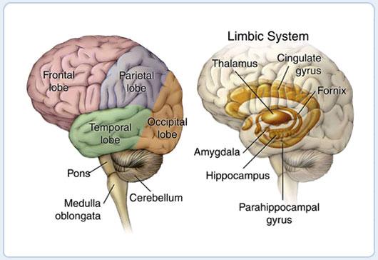
6 Other (Subcortical) Areas l l l l l l Hippocampus: Thalamus: Amygdala: Basal Ganglia: Cerebellum: Reward prediction system: rapid learning sensory input, attention emotion, fear/desire motor control, gating of PFC coordinating movements dopamine release 2/8/18 COSC 494/594 CCN 6 (slide < O Reilly)


7 anterior inferior 2/8/18 Subcortical Areas COSC 494/594 CCN 7
8 Interactive Images Atlas of Brain Injury and Anatomy Brain Anatomy (Koshland Science Museum) 2/8/18 COSC 494/594 CCN 8

9 Left Hemisphere 2/8/18 COSC 494/594 CCN 9
 Uncinate fasciculus, (6) Sagittal stratum, (7) Inferior longitudinal fasciculus 2/8/18 COSC 494/594")
10 Intercortical Connections (1) Short arcuate bundles, (2) Superior longitudinal fasciculus, (3) External capsule, (4) Inferior occipitofrontal fasciculus, (5) Uncinate fasciculus, (6) Sagittal stratum, (7) Inferior longitudinal fasciculus 2/8/18 COSC 494/594 CCN 10
")

11 Intercortical Connections (diffusion spectrum imaging) 2/8/18 COSC 494/594 CCN 11 Published by AAAS G. Miller Science 330, 164 (2010) (2010)

12 Brodmann s Areas 2/8/18 COSC 494/594 CCN 12

 2/8/18 COSC 494/594 CCN")
13 General Functions of Cortical Lobes (how is this determined?) 2/8/18 COSC 494/594 CCN 13 (slide < O Reilly)

14 Motor Movements Motor Plan Plan Object Meaning Objects Shapes lines+colors Visual input Hierarchical Sequences of Transformations Motor 1 Frontal Supp. Motor Frontal PFC Frontal Ant. Pole Temporal Inferior Temporal V4 Occipital V1/V2 Occipital 2/8/18 COSC 494/594 CCN 14 (slide < O Reilly) Retina

15 Tripartite Cognitive Architecture 2/8/18 COSC 494/594 CCN 15 (slide < O Reilly)
16 Large Scale Distributed Organizations Knowledge is distributed across multiple brain areas Multiple areas participate in representing a given thing (e.g., apple) Each area represents multiple things Same idea as distributed representation among units for individual items, but in this case across multiple areas/modalities, etc. 2/8/18 COSC 494/594 CCN 16 (slide based on Frank)
17 Learning Across the Brain Learning Signal Dynamics Area Reward Error Self Org Separator Integrator Attractor Basal Ganglia Cerebellum Hippocampus Neocortex /8/18 COSC 494/594 CCN 17
18 B. Perception and Attention What versus Where 2/8/18 COSC 494/594 CCN 18

19 Hierarchy of Visual Detectors 2/8/18 COSC 494/594 CCN 19

20 Macaque Visual System 2/8/18 COSC 494/594 CCN 20 (fig. from Clark, Being There, 1997)


21 Hierarchy of Macaque Visual Areas 2/8/18 COSC 494/594 CCN 21 (fig. from Van Essen & al. 1992)
22 What vs. Where Pathways What ignores differences in location, illumination, size, rotation Where emphasizes location, size, and ignores object identity 2/8/18 COSC 494/594 CCN 22
23 Multiple Interacting Pathways Maintaining Abstract Information (plans, sensory info, etc) Movement Spatial Processing Touch Visual Object Recognition Audition Abstract Semantics 2/8/18 COSC 494/594 CCN 23 (slide < O Reilly) Early Vision
24 Principal Regions in What Pathway Occipital Lobe V1: Primary visual cortex encodes image in terms of oriented edges V2: Secondary visual cortex encodes in terms of intersections & junctions V4: Third cortical area in ventral stream Temporal Lobe PIT: Posterior inferotemporal (IT) cortex location & size invariant object recognition includes FFA (fusiform face area) AIT: Anterior IT cortex abstract/semantic visual information more complex features over wider range of locations modulation by attention 2/8/18 COSC 494/594 CCN 24
25 C. Motor Control Parietal and Motor Cortex Interacting with Basal Ganglia and Cerebellum 2/8/18 COSC 494/594 CCN 25
26 Motor Control Motor cortex (frontal to parietal) high-level metrical processing of sensory information, integrating multiple modalities and translating between different reference frames arrives at a range of possible responses to the current sensory environment Basal ganglia action selection: receives sensory inputs and potential responses being considered in frontal cortex; triggers disinhibitory signal enabling best action action selection is shaped by reinforcement learning driven by dopamine signal amygdala plays key role in driving these dopamine signals in response to sensory cues associated with reward and punishment Cerebellum uses error-driven learning to acquire high-resolution metrical maps between sensory inputs and motor outputs 2/8/18 COSC 494/594 CCN 26

27 Somatosensory & Motor Homunculi 2/8/18 COSC 494/594 CCN 27
 several months later 2/8/18 COSC 494/594 CCN 28 (fig. < McClelland & al, Par. Distr.")
28 Reorganization of Cortex Median nerve sectioned to show fluidity of cortical organization (C) before (D) immediately after (E) several months later 2/8/18 COSC 494/594 CCN 28 (fig. < McClelland & al, Par. Distr. Proc. II)
29 D. Learning and Memory Temporal Cortex and the Hippocampus 2/8/18 COSC 494/594 CCN 29
30 Computational Trade-offs in Learning & Memory Computational objectives that are mutually incompatible and thus cannot be achieved by a single brain system Learning must be slow to capture statistical structure (averaging) But you have to be able to learn rapidly too Tradeoff solved by two systems: cortex learns slowly hippocampus learns rapidly Third system: active memory (prefrontal cortex) fastest (immediately accessible) 2/8/18 COSC 494/594 CCN 30 (slide < Frank)
31 Tripartite Functional Organization HC PC FC PC = posterior perceptual cortex: slow integrative learning HC = hippocampus and related structures: rapid memorization FC = prefrontal cortex: active maintenance ( working memory ) Defined by set of functional trade-offs. 2/8/18 COSC 494/594 CCN 31 (slide < Frank)

32 Hippocampal Memory Formation extremely sparse representations pattern separation 2/8/18 COSC 494/594 CCN 32
33 E. Language 2/8/18 COSC 494/594 CCN 33
34 Language Involves many of the foregoing functions perception, memory, executive function, motor control Models (ch. 9) will address: small scale model of reading, incorporating orthographic, phonological, and semantic aspects regular behavior without rules self-organization of semantic representations interaction of syntax and semantics 2/8/18 COSC 494/594 CCN 34

35 Distributed Representation of Words 2/8/18 COSC 494/594 CCN 35
36 F. Executive Function Prefrontal Cortex and Basal Ganglia 2/8/18 COSC 494/594 CCN 36
37 Executive Function Builds on motor control functions of frontal cortex (FC) and basal ganglia (BG) Areas of FC oriented to what vs. how processing hot emotional vs. cold cognitive processing Prefrontal cortex (PFC) control over posterior cortex PFC and BG interact to implement dynamically gated working memory system 2/8/18 COSC 494/594 CCN 37
38 Ventral vs. Dorsal Organization of PFC 2/8/18 COSC 494/594 CCN 38
2 Summary of Part I: Basic Mechanisms. 4 Dedicated & Content-Specific
 1 Large Scale Brain Area Functional Organization 2 Summary of Part I: Basic Mechanisms 2. Distributed Representations 5. Error driven Learning 1. Biological realism w = δ j ai+ aia j w = δ j ai+ aia j
Computational Explorations in Cognitive Neuroscience Chapter 7: Large-Scale Brain Area Functional Organization
 Computational Explorations in Cognitive Neuroscience Chapter 7: Large-Scale Brain Area Functional Organization 1 7.1 Overview This chapter aims to provide a framework for modeling cognitive phenomena based
Neocortex. Hemispheres 9/22/2010. Psychology 472 Pharmacology of Psychoactive Drugs. Structures are divided into several section or lobes.
 Neocortex Psychology 472 Pharmacology of Psychoactive Drugs 1 Is the most developed in Humans Has many folds and fissures The folds of tissue are called gyri or a gyrus (single) The fissures or valleys
CISC 3250 Systems Neuroscience
 CISC 3250 Systems Neuroscience Levels of organization Central Nervous System 1m 10 11 neurons Neural systems and neuroanatomy Systems 10cm Networks 1mm Neurons 100μm 10 8 neurons Professor Daniel Leeds
The Frontal Lobes. Anatomy of the Frontal Lobes. Anatomy of the Frontal Lobes 3/2/2011. Portrait: Losing Frontal-Lobe Functions. Readings: KW Ch.
 The Frontal Lobes Readings: KW Ch. 16 Portrait: Losing Frontal-Lobe Functions E.L. Highly organized college professor Became disorganized, showed little emotion, and began to miss deadlines Scores on intelligence
Cerebral Cortex 1. Sarah Heilbronner
 Cerebral Cortex 1 Sarah Heilbronner heilb028@umn.edu Want to meet? Coffee hour 10-11am Tuesday 11/27 Surdyk s Overview and organization of the cerebral cortex What is the cerebral cortex? Where is each
P. Hitchcock, Ph.D. Department of Cell and Developmental Biology Kellogg Eye Center. Wednesday, 16 March 2009, 1:00p.m. 2:00p.m.
 Normal CNS, Special Senses, Head and Neck TOPIC: CEREBRAL HEMISPHERES FACULTY: LECTURE: READING: P. Hitchcock, Ph.D. Department of Cell and Developmental Biology Kellogg Eye Center Wednesday, 16 March
NEOCORTICAL CIRCUITS. specifications
 NEOCORTICAL CIRCUITS specifications where are we coming from? human-based computing using typically human faculties associating words with images -> labels for image search locating objects in images ->
Homework Week 2. PreLab 2 HW #2 Synapses (Page 1 in the HW Section)
") Homework Week 2 Due in Lab PreLab 2 HW #2 Synapses (Page 1 in the HW Section) Reminders No class next Monday Quiz 1 is @ 5:30pm on Tuesday, 1/22/13 Study guide posted under Study Aids section of website
Homework Week 2 Due in Lab PreLab 2 HW #2 Synapses (Page 1 in the HW Section) Reminders No class next Monday Quiz 1 is @ 5:30pm on Tuesday, 1/22/13 Study guide posted under Study Aids section of website
CEREBRUM Dr. Jamila Elmedany Dr. Essam Eldin Salama
 CEREBRUM Dr. Jamila Elmedany Dr. Essam Eldin Salama Objectives At the end of the lecture, the student should be able to: List the parts of the cerebral hemisphere (cortex, medulla, basal nuclei, lateral
THE CENTRAL NERVOUS SYSTEM. The Brain & Spinal Cord
 THE CENTRAL NERVOUS SYSTEM The Brain & Spinal Cord Review: Nervous System Parallel Distributed Processing Composition of the CNS Nuclei: Clusters of neurons in the CNS ( neighborhoods ) Fiber Tracts/Pathways:
Introduction to Physiological Psychology Review
 Introduction to Physiological Psychology Review ksweeney@cogsci.ucsd.edu www.cogsci.ucsd.edu/~ksweeney/psy260.html n Learning and Memory n Human Communication n Emotion 1 What is memory? n Working Memory:
CEREBRUM. Dr. Jamila EL Medany
 CEREBRUM Dr. Jamila EL Medany Objectives At the end of the lecture, the student should be able to: List the parts of the cerebral hemisphere (cortex, medulla, basal nuclei, lateral ventricle). Describe
The human brain. of cognition need to make sense gives the structure of the brain (duh). ! What is the basic physiology of this organ?
. ! What is the basic physiology of this organ?") The human brain The human brain! What is the basic physiology of this organ?! Understanding the parts of this organ provides a hypothesis space for its function perhaps different parts perform different
The human brain The human brain! What is the basic physiology of this organ?! Understanding the parts of this organ provides a hypothesis space for its function perhaps different parts perform different
25/09/2012. Capgras Syndrome. Chapter 2. Capgras Syndrome - 2. The Neural Basis of Cognition
 Chapter 2 The Neural Basis of Cognition Capgras Syndrome Alzheimer s patients & others delusion that significant others are robots or impersonators - paranoia Two brain systems for facial recognition -
The Nervous System. Divisions of the Nervous System. Branches of the Autonomic Nervous System. Central versus Peripheral
 The Nervous System Divisions of the Nervous System Central versus Peripheral Central Brain and spinal cord Peripheral Everything else Somatic versus Autonomic Somatic Nerves serving conscious sensations
Chapter 14, Part 2! Chapter 14 Part 2 Brain/Cranial Nerves! The Cerebrum and Cranial Nerves! pp !
 Chapter 14, Part 2! The Cerebrum and Cranial pp. 482 505! SECTION 14-9! The cerebrum, the largest region of the brain, contains motor, sensory, and association areas! 2! White Matter of the Cerebrum! 1.
Myers Psychology for AP*
 Myers Psychology for AP* David G. Myers PowerPoint Presentation Slides by Kent Korek Germantown High School Worth Publishers, 2010 *AP is a trademark registered and/or owned by the College Board, which
Chapter 14, Part 2! The Cerebrum and Cranial Nerves! pp !
 Chapter 14, Part 2! The Cerebrum and Cranial pp. 482 505! SECTION 14-9! The cerebrum, the largest region of the brain, contains motor, sensory, and association areas! 2! 1! ! Chapter 14 Part 2 Brain/Cranial
Brain-Behavior Network. Central Nervous System. Cerebral Cortex Gyrus and Sulcus. Nervous System
 Brain-Behavior Network Nervous System Sensory information comes into and decisions come out of the central nervous system (CNS) Central Nervous System The nerves outside the CNS are called the peripheral
Neuroscience Tutorial
 Neuroscience Tutorial Brain Organization : cortex, basal ganglia, limbic lobe : thalamus, hypothal., pituitary gland : medulla oblongata, midbrain, pons, cerebellum Cortical Organization Cortical Organization
Overview of Brain Structures
 First Overview of Brain Structures Psychology 470 Introduction to Chemical Additions Steven E. Meier, Ph.D. All parts are interrelated. You need all parts to function normally. Neurons = Nerve cells Listen
Memory, Attention, and Decision-Making
 Memory, Attention, and Decision-Making A Unifying Computational Neuroscience Approach Edmund T. Rolls University of Oxford Department of Experimental Psychology Oxford England OXFORD UNIVERSITY PRESS Contents
Sensorimotor Functioning. Sensory and Motor Systems. Functional Anatomy of Brain- Behavioral Relationships
 Sensorimotor Functioning Sensory and Motor Systems Understanding brain-behavior relationships requires knowledge of sensory and motor systems. Sensory System = Input Neural Processing Motor System = Output
Fundamentals of Cognitive Psychology, 3e by Ronald T. Kellogg Chapter 2. Multiple Choice
 Multiple Choice 1. Which structure is not part of the visual pathway in the brain? a. occipital lobe b. optic chiasm c. lateral geniculate nucleus *d. frontal lobe Answer location: Visual Pathways 2. Which
EDGE DETECTION. Edge Detectors. ICS 280: Visual Perception
 EDGE DETECTION Edge Detectors Slide 2 Convolution & Feature Detection Slide 3 Finds the slope First derivative Direction dependent Need many edge detectors for all orientation Second order derivatives
FRONTAL LOBE. Central Sulcus. Ascending ramus of the Cingulate Sulcus. Cingulate Sulcus. Lateral Sulcus
 FRONTAL LOBE Central Ascending ramus of the Cingulate Cingulate Lateral Lateral View Medial View Motor execution and higher cognitive functions (e.g., language production, impulse inhibition, reasoning
Essentials of Human Anatomy & Physiology. Seventh Edition. The Nervous System. Copyright 2003 Pearson Education, Inc. publishing as Benjamin Cummings
 Essentials of Human Anatomy & Physiology Seventh Edition The Nervous System Copyright 2003 Pearson Education, Inc. publishing as Benjamin Cummings Functions of the Nervous System 1. Sensory input gathering
Introduction to sensory pathways. Gatsby / SWC induction week 25 September 2017
 Introduction to sensory pathways Gatsby / SWC induction week 25 September 2017 Studying sensory systems: inputs and needs Stimulus Modality Robots Sensors Biological Sensors Outputs Light Vision Photodiodes
fmri (functional MRI)
") Lesion fmri (functional MRI) Electroencephalogram (EEG) Brainstem CT (computed tomography) Scan Medulla PET (positron emission tomography) Scan Reticular Formation MRI (magnetic resonance imaging) Thalamus
Lesion fmri (functional MRI) Electroencephalogram (EEG) Brainstem CT (computed tomography) Scan Medulla PET (positron emission tomography) Scan Reticular Formation MRI (magnetic resonance imaging) Thalamus
Cerebrum-Cerebral Hemispheres. Cuneyt Mirzanli Istanbul Gelisim University
 Cerebrum-Cerebral Hemispheres Cuneyt Mirzanli Istanbul Gelisim University The largest part of the brain. Ovoid shape. Two incompletely separated cerebral hemispheres. The outer surface of the cerebral
CEREBRUM & CEREBRAL CORTEX
 CEREBRUM & CEREBRAL CORTEX Seonghan Kim Dept. of Anatomy Inje University, College of Medicine THE BRAIN ANATOMICAL REGIONS A. Cerebrum B. Diencephalon Thalamus Hypothalamus C. Brain Stem Midbrain Pons
3/20/13. :: Slide 1 :: :: Slide 39 :: How Is the Nervous System Organized? Central Nervous System Peripheral Nervous System and Endocrine System
 :: Slide 1 :: :: Slide 39 :: How Is the Nervous System Organized? Central Nervous System Peripheral Nervous System and Endocrine System The nervous system is organized into several major branches, each
THE BRAIN HABIT BRIDGING THE CONSCIOUS AND UNCONSCIOUS MIND
 THE BRAIN HABIT BRIDGING THE CONSCIOUS AND UNCONSCIOUS MIND Mary ET Boyle, Ph. D. Department of Cognitive Science UCSD How did I get here? What did I do? Start driving home after work Aware when you left
PSY 302: CHAPTER 3 NOTES THE BRAIN (PART II) - 9/5/17. By: Joseline
 - 9/5/17. By: Joseline") PSY 302: CHAPTER 3 NOTES THE BRAIN (PART II) - 9/5/17 By: Joseline Left 3 MAJOR FISSURES : 2HEMISPHERES Right Lateral Ventricle Central Fissure Third Ventricle Sulcus Lateral Fissure Gyros Fissure- Fissures
PSY 302: CHAPTER 3 NOTES THE BRAIN (PART II) - 9/5/17 By: Joseline Left 3 MAJOR FISSURES : 2HEMISPHERES Right Lateral Ventricle Central Fissure Third Ventricle Sulcus Lateral Fissure Gyros Fissure- Fissures
Telencephalon (Cerebral Hemisphere)
") Telencephalon (Cerebral Hemisphere) OUTLINE The Cortex - Lobes, Sulci & Gyri - Functional Subdivisions - Limbic Lobe & Limbic System The Subcortex - Basal Ganglia - White Matter (Internal Capsule) - Relations
Telencephalon (Cerebral Hemisphere) OUTLINE The Cortex - Lobes, Sulci & Gyri - Functional Subdivisions - Limbic Lobe & Limbic System The Subcortex - Basal Ganglia - White Matter (Internal Capsule) - Relations
THE PREFRONTAL CORTEX. Connections. Dorsolateral FrontalCortex (DFPC) Inputs
 Inputs") THE PREFRONTAL CORTEX Connections Dorsolateral FrontalCortex (DFPC) Inputs The DPFC receives inputs predominantly from somatosensory, visual and auditory cortical association areas in the parietal, occipital
THE PREFRONTAL CORTEX Connections Dorsolateral FrontalCortex (DFPC) Inputs The DPFC receives inputs predominantly from somatosensory, visual and auditory cortical association areas in the parietal, occipital
CNS Tour (Lecture 12)
") A. Introduction CNS Tour (Lecture 12) There are to a chemical pathways in the nervous system. These pathways also form different neurological structures B. Spinal Cord Receives sensory neurons from skin
A. Introduction CNS Tour (Lecture 12) There are to a chemical pathways in the nervous system. These pathways also form different neurological structures B. Spinal Cord Receives sensory neurons from skin
Announcement. Danny to schedule a time if you are interested.
 Announcement If you need more experiments to participate in, contact Danny Sanchez (dsanchez@ucsd.edu) make sure to tell him that you are from LIGN171, so he will let me know about your credit (1 point).
Anatomy and Physiology (Bio 220) The Brain Chapter 14 and select portions of Chapter 16
 The Brain Chapter 14 and select portions of Chapter 16") Anatomy and Physiology (Bio 220) The Brain Chapter 14 and select portions of Chapter 16 I. Introduction A. Appearance 1. physical 2. weight 3. relative weight B. Major parts of the brain 1. cerebrum 2.
Anatomy and Physiology (Bio 220) The Brain Chapter 14 and select portions of Chapter 16 I. Introduction A. Appearance 1. physical 2. weight 3. relative weight B. Major parts of the brain 1. cerebrum 2.
The Brain. Its major systems, How we study them, How they make the mind
 The Brain Its major systems, How we study them, How they make the mind 9.00 Introduction to Psychology Joanne s Recitation Section Friday, February 11, 2011 Outline 1. Syllabus: Course Requirements, Exams,
skilled pathways: distal somatic muscles (fingers, hands) (brainstem, cortex) are giving excitatory signals to the descending pathway
 (brainstem, cortex) are giving excitatory signals to the descending pathway") L15 - Motor Cortex General - descending pathways: how we control our body - motor = somatic muscles and movement (it is a descending motor output pathway) - two types of movement: goal-driven/voluntary
L15 - Motor Cortex General - descending pathways: how we control our body - motor = somatic muscles and movement (it is a descending motor output pathway) - two types of movement: goal-driven/voluntary
Motor Systems I Cortex. Reading: BCP Chapter 14
 Motor Systems I Cortex Reading: BCP Chapter 14 Principles of Sensorimotor Function Hierarchical Organization association cortex at the highest level, muscles at the lowest signals flow between levels over
Forebrain Brain Structures Limbic System. Brain Stem Midbrain Basil Ganglia. Cerebellum Reticular Formation Medulla oblongata
 Brain structures (1) Cut out the following cards (2) Identify the three major divisions of the brain (as defined by your book). Initially, try this without any form of aid such as your textbook. (3) Organize
Introduction to the Nervous System. Code: HMP 100/ UPC 103/ VNP 100. Course: Medical Physiology. Level 1 MBChB/BDS/BPharm
 Introduction to the Nervous System. Code: HMP 100/ UPC 103/ VNP 100. Course: Medical Physiology Level 1 MBChB/BDS/BPharm Lecture 2. Functional Organisation of the Nervous System Lecture Outline 1.1 Introduction
Cognitive Modelling Themes in Neural Computation. Tom Hartley
 Cognitive Modelling Themes in Neural Computation Tom Hartley t.hartley@psychology.york.ac.uk Typical Model Neuron x i w ij x j =f(σw ij x j ) w jk x k McCulloch & Pitts (1943), Rosenblatt (1957) Net input:
Biological Bases of Behavior. 3: Structure of the Nervous System
 Biological Bases of Behavior 3: Structure of the Nervous System Neuroanatomy Terms The neuraxis is an imaginary line drawn through the spinal cord up to the front of the brain Anatomical directions are
Leah Militello, class of 2018
 Leah Militello, class of 2018 Objectives 1. Describe the general organization of cerebral hemispheres. 2. Describe the locations and features of the different functional areas of cortex. 3. Understand
Text to brain: predicting the spatial distribution of neuroimaging observations from text reports (submitted to MICCAI 2018)
") 1 / 22 Text to brain: predicting the spatial distribution of neuroimaging observations from text reports (submitted to MICCAI 2018) Jérôme Dockès, ussel Poldrack, Demian Wassermann, Fabian Suchanek, Bertrand
1 / 22 Text to brain: predicting the spatial distribution of neuroimaging observations from text reports (submitted to MICCAI 2018) Jérôme Dockès, ussel Poldrack, Demian Wassermann, Fabian Suchanek, Bertrand
Disorders of Object and Spatial perception. Dr John Maasch Brain Injury Rehabilitation Service Burwood Hospital.
 Disorders of Object and Spatial perception Dr John Maasch Brain Injury Rehabilitation Service Burwood Hospital. Take Home Message 1 Where there are lesions of the posterior cerebrum and posterior temporal
Lateral Geniculate Nucleus (LGN)
") Lateral Geniculate Nucleus (LGN) What happens beyond the retina? What happens in Lateral Geniculate Nucleus (LGN)- 90% flow Visual cortex Information Flow Superior colliculus 10% flow Slide 2 Information
Lateral Geniculate Nucleus (LGN) What happens beyond the retina? What happens in Lateral Geniculate Nucleus (LGN)- 90% flow Visual cortex Information Flow Superior colliculus 10% flow Slide 2 Information
The Integration of Features in Visual Awareness : The Binding Problem. By Andrew Laguna, S.J.
 The Integration of Features in Visual Awareness : The Binding Problem By Andrew Laguna, S.J. Outline I. Introduction II. The Visual System III. What is the Binding Problem? IV. Possible Theoretical Solutions
Topic 11 - Parietal Association Cortex. 1. Sensory-to-motor transformations. 2. Activity in parietal association cortex and the effects of damage
 Topic 11 - Parietal Association Cortex 1. Sensory-to-motor transformations 2. Activity in parietal association cortex and the effects of damage Sensory to Motor Transformation Sensory information (visual,
Neural plasticity in infants - relevance to baby swimming. Morten Overgaard
 Neural plasticity in infants - relevance to baby swimming Morten Overgaard Programme What is neuroscience? Totally superficial neuroanatomy Paradoxes of functional localization Mechanisms of neural plasticity
Vision II. Steven McLoon Department of Neuroscience University of Minnesota
 Vision II Steven McLoon Department of Neuroscience University of Minnesota 1 Ganglion Cells The axons of the retinal ganglion cells form the optic nerve and carry visual information into the brain. 2 Optic
Cerebral Cortex: Association Areas and Memory Tutis Vilis
 97 Cerebral Cortex: Association Areas and Memory Tutis Vilis a) Name the 5 main subdivisions of the cerebral cortex. Frontal, temporal, occipital, parietal, and limbic (on the medial side) b) Locate the
Contents. Boxes xii Preface xiii Acknowledgments. Background and Methods
 Contents Boxes xii Preface xiii Acknowledgments xv PARTI Background and Methods 1 A Brief History of Cognitive Neuroscience 2 A Historical Perspective 4 The Brain Story 5 The Psychological Story 10 The
Notes: Organization. Anatomy of the Nervous System. Cerebral cortex. Cortical layers. PSYC 2: Biological Foundations - Fall Professor Claffey
 PSYC 2: Biological Foundations - Fall 2012 - Professor Claffey Notes: Organization Version: 10/30/12 - original version Anatomy of the Nervous System Content covered in Hans's lecture: CNS & PNS Directions/Planes
The Central Nervous System
 The Central Nervous System Cellular Basis. Neural Communication. Major Structures. Principles & Methods. Principles of Neural Organization Big Question #1: Representation. How is the external world coded
Layered organization of cortex: Paleocortex 3 layers hippocampal formation / ventral & medial cortex closest to brainstem
 Layered organization of cortex: Paleocortex 3 layers hippocampal formation / ventral & medial cortex closest to brainstem Archicortex 3-4 layers hippocampal formation / amygdala Neocortex 6 layers more
a) Central sulcus- shallow groove that runs across brain sagitally
 Central sulcus- shallow groove that runs across brain sagitally") KEY BRAIN Brain Gross Anatomy Terms 1) Explain each of the following in terms of structure of the brain a) Central sulcus- shallow groove that runs across brain sagitally b) Lateral fissure- deep groove
KEY BRAIN Brain Gross Anatomy Terms 1) Explain each of the following in terms of structure of the brain a) Central sulcus- shallow groove that runs across brain sagitally b) Lateral fissure- deep groove
Cognitive Neuroscience Cortical Hemispheres Attention Language
 Cognitive Neuroscience Cortical Hemispheres Attention Language Based on: Chapter 18 and 19, Breedlove, Watson, Rosenzweig, 6e/7e. Cerebral Cortex Brain s most complex area with billions of neurons and
Supplementary Online Material Supplementary Table S1 to S5 Supplementary Figure S1 to S4
 Supplementary Online Material Supplementary Table S1 to S5 Supplementary Figure S1 to S4 Table S1: Brain regions involved in the adapted classification learning task Brain Regions x y z Z Anterior Cingulate
synapse neurotransmitters Extension of a neuron, ending in branching terminal fibers, through which messages pass to other neurons, muscles, or glands
 neuron synapse The junction between the axon tip of a sending neuron and the dendrite of a receiving neuron Building block of the nervous system; nerve cell Chemical messengers that cross the synaptic
Human Brain. Lateralization of Function. Cortex. Cerebral Hemispheres. An extension of the spinal cord. Dr. Coulson Cognitive Science Department UCSD
 Lateralization of Function Human Brain An extension of the spinal cord Dr. Coulson Cognitive Science Department UCSD Cerebral Hemispheres Two millimeters thick and has area of 1.5 square meters Corpus
Acetylcholine (ACh) Action potential. Agonists. Drugs that enhance the actions of neurotransmitters.
 Action potential. Agonists. Drugs that enhance the actions of neurotransmitters.") Acetylcholine (ACh) The neurotransmitter responsible for motor control at the junction between nerves and muscles; also involved in mental processes such as learning, memory, sleeping, and dreaming. (See
Acetylcholine (ACh) The neurotransmitter responsible for motor control at the junction between nerves and muscles; also involved in mental processes such as learning, memory, sleeping, and dreaming. (See
Making Things Happen: Simple Motor Control
 Making Things Happen: Simple Motor Control How Your Brain Works - Week 10 Prof. Jan Schnupp wschnupp@cityu.edu.hk HowYourBrainWorks.net The Story So Far In the first few lectures we introduced you to some
Human Paleoneurology and the Evolution of the Parietal Cortex
 PARIETAL LOBE The Parietal Lobes develop at about the age of 5 years. They function to give the individual perspective and to help them understand space, touch, and volume. The location of the parietal
Exam 1 PSYC Fall 1998
 Exam 1 PSYC 2022 Fall 1998 (2 points) Briefly describe the difference between a dualistic and a materialistic explanation of brain-mind relationships. (1 point) True or False. George Berkely was a monist.
Memory Development. Cognitive Development
 Memory Development Cognitive Development Memory as information storage Memory Why does our memory sometimes fail us? Memory Schachter s Seven Sins of Memory 1. Transience 2. Absent-Mindedness 3. Blocking
Inside Your Patient s Brain Michelle Peterson, APRN, CNP Centracare Stroke and Vascular Neurology
 Inside Your Patient s Brain Michelle Peterson, APRN, CNP Centracare Stroke and Vascular Neurology Activity Everyone stand up, raise your right hand, tell your neighbors your name 1 What part of the brain
Category Learning in the Brain
 Aline Richtermeier Category Learning in the Brain The key to human intelligence Based on the article by: Seger, C.A. & Miller, E.K. (2010). Annual Review of Neuroscience, 33, 203-219. Categorization Ability
To understand AD, it is important to
 To understand AD, it is important to know a bit about the brain. This part of Unraveling the Mystery gives an inside view of the normal brain, how it works, and what happens during aging. The brain is
 Peripheral facial paralysis (right side). The patient is asked to close her eyes and to retract their mouth (From Heimer) Hemiplegia of the left side. Note the characteristic position of the arm with
Regional and Lobe Parcellation Rhesus Monkey Brain Atlas. Manual Tracing for Parcellation Template
 Regional and Lobe Parcellation Rhesus Monkey Brain Atlas Manual Tracing for Parcellation Template Overview of Tracing Guidelines A) Traces are performed in a systematic order they, allowing the more easily
Nervous System: Part IV The Central Nervous System The Brain
 Nervous System: Part IV The Central Nervous System The Brain Can you survive when part of your brain is destroyed? 2 Essential Knowledge 3.D.2 2. Cells communicate with each other through direct contact
Fig.1: A, Sagittal 110x110 mm subimage close to the midline, passing through the cingulum. Note that the fibers of the corpus callosum run at a
 Fig.1 E Fig.1:, Sagittal 110x110 mm subimage close to the midline, passing through the cingulum. Note that the fibers of the corpus callosum run at a slight angle are through the plane (blue dots with
Memory. Psychology 3910 Guest Lecture by Steve Smith
 Memory Psychology 3910 Guest Lecture by Steve Smith Note: Due to copyright restrictions, I had to remove the images from the Weschler Memory Scales from the slides I posted online. Wechsler Memory Scales
LEC 1B ANATOMY OF THE NERVOUS SYSTEM. Cogs 17 * UCSD
 LEC 1B ANATOMY OF THE NERVOUS SYSTEM Cogs 17 * UCSD Cerebral Cortex A 6-layer sheet of cells, unfolded = < 1 m square X 3 mm thick Cortex 6 layers of cells Nissl Stain for Cell Bodies Info projected to
Systems Neuroscience CISC 3250
 Systems Neuroscience CISC 325 Memory Types of Memory Declarative Non-declarative Episodic Semantic Professor Daniel Leeds dleeds@fordham.edu JMH 328A Hippocampus (MTL) Cerebral cortex Basal ganglia Motor
Learning Objectives.
 Emilie O Neill, class of 2016 Learning Objectives 1. Describe the types of deficits that occur with lesions in association areas including: prosopagnosia, neglect, aphasias, agnosia, apraxia 2. Discuss
Cognitive Neuroscience Attention
 Cognitive Neuroscience Attention There are many aspects to attention. It can be controlled. It can be focused on a particular sensory modality or item. It can be divided. It can set a perceptual system.
Prof. Saeed Abuel Makarem & Dr.Sanaa Alshaarawy
 Prof. Saeed Abuel Makarem & Dr.Sanaa Alshaarawy 1 Objectives By the end of the lecture, you should be able to: Describe the anatomy and main functions of the thalamus. Name and identify different nuclei
correlates with social context behavioral adaptation.
 REVIEW OF FRONTAL LOBE STRUCTURES Main organization of frontal cortex: 1. Motor area (precentral gyrus). 2. Premotor & supplementary motor areas (immediately anterior to motor area). Includes premotor,
Perceptual Learning. Motor Learning. Stimulus-Response Learning. Relational Learning
 Introduction to Physiological Psychology Review ksweeney@cogsci.ucsd.edu www.cogsci.ucsd.edu/~ksweeney/psy260.html Learning and Memory Human Communication Emotion 1 Working Memory: What is memory? Limited
Resistance to forgetting associated with hippocampus-mediated. reactivation during new learning
 Resistance to Forgetting 1 Resistance to forgetting associated with hippocampus-mediated reactivation during new learning Brice A. Kuhl, Arpeet T. Shah, Sarah DuBrow, & Anthony D. Wagner Resistance to
By Lauren Stowe, PhD, CCC-SLP & Gina Rotondo, MS, CCC-SLP The Speech Therapy Group
 By Lauren Stowe, PhD, CCC-SLP & Gina Rotondo, MS, CCC-SLP The Speech Therapy Group http://www.acquiredbraininjury.com/interactive brain/interactivebrain.swf 1. Hormones make the science messy 2. Difference
Higher Cortical Function
 Emilie O Neill, class of 2016 Higher Cortical Function Objectives Describe the association cortical areas processing sensory, motor, executive, language, and emotion/memory information (know general location
1. Which part of the brain is responsible for planning and initiating movements?
 Section: Chapter 10: Multiple Choice 1. Which part of the brain is responsible for planning and initiating movements? p.358 frontal lobe hippocampus basal ganglia cerebellum 2. The prefrontal cortex is
Chapter 18: The Brain & Cranial Nerves. Origin of the Brain
 Chapter 18: The Brain & Cranial Nerves BIO 218 Fall 2015 Origin of the Brain The brain originates from a structure called the neural tube, which arises during a developmental stage called neurulation.
Outline of the next three lectures
 Outline of the next three lectures Lecture 35 Anatomy of the human cerebral cortex gross and microscopic cell types connections Vascular supply of the cerebral cortex Disorders involving the cerebral cortex
A Scientific Model of Consciousness that Explains Spirituality and Enlightened States
 A Scientific Model of Consciousness that Explains Spirituality and Enlightened States Frank Heile, Ph.D. Physics degrees from Stanford and MIT consciousness@frankheile.com April 2016 Outline Part 1: Agents
Overview of the Nervous System (some basic concepts) Steven McLoon Department of Neuroscience University of Minnesota
 Steven McLoon Department of Neuroscience University of Minnesota") Overview of the Nervous System (some basic concepts) Steven McLoon Department of Neuroscience University of Minnesota 1 Coffee Hour Tuesday (Sept 11) 10:00-11:00am Friday (Sept 14) 8:30-9:30am Surdyk s
Overview of the Nervous System (some basic concepts) Steven McLoon Department of Neuroscience University of Minnesota 1 Coffee Hour Tuesday (Sept 11) 10:00-11:00am Friday (Sept 14) 8:30-9:30am Surdyk s
9.14 Class 32 Review. Limbic system
 9.14 Class 32 Review Limbic system 1 Lateral view Medial view Brainstem, sagittal section Sensory- Perceptual Motor Behavior Major functional modules of the CNS Motivation Courtesy of MIT Press. Used with
Lighta part of the spectrum of Electromagnetic Energy. (the part that s visible to us!)
") Introduction to Physiological Psychology Vision ksweeney@cogsci.ucsd.edu cogsci.ucsd.edu/~ /~ksweeney/psy260.html Lighta part of the spectrum of Electromagnetic Energy (the part that s visible to us!)
Introduction to Physiological Psychology Vision ksweeney@cogsci.ucsd.edu cogsci.ucsd.edu/~ /~ksweeney/psy260.html Lighta part of the spectrum of Electromagnetic Energy (the part that s visible to us!)
Human Brain. Lateralization of Function. An extension of the spinal cord. Dr. Coulson Cognitive Science Department UCSD
 Lateralization of Function Human Brain An extension of the spinal cord Dr. Coulson Cognitive Science Department UCSD Cerebral Hemispheres Corpus Callosum Cerebral Lobes Neurons Brain composed of neurons
The Nervous System and the Endocrine System
 The Nervous System and the Endocrine System Neurons: The Building Blocks of the Nervous System Nervous System The electrochemical communication system of the body Sends messages from the brain to the
The Central Nervous System I. Chapter 12
 The Central Nervous System I Chapter 12 The Central Nervous System The Brain and Spinal Cord Contained within the Axial Skeleton Brain Regions and Organization Medical Scheme (4 regions) 1. Cerebral Hemispheres
Cerebral Cortex Structure, Function, Dysfunction Reading Ch 10 Waxman Dental Neuroanatomy Lecture. Suzanne Stensaas, Ph.D.
 Cerebral Cortex Structure, Function, Dysfunction Reading Ch 10 Waxman Dental Neuroanatomy Lecture Suzanne Stensaas, Ph.D. March 7, 2012 Anatomy Review Lobes and layers Brodmann s areas Vascular Supply
shows syntax in his language. has a large neocortex, which explains his language abilities. shows remarkable cognitive abilities. all of the above.
 Section: Chapter 14: Multiple Choice 1. Alex the parrot: pp.529-530 shows syntax in his language. has a large neocortex, which explains his language abilities. shows remarkable cognitive abilities. all