The Tools: Imaging the Living Brain
|
|
|
- Rosamund Martin
- 6 years ago
- Views:
Transcription
1 The Tools: Imaging the Living Brain I believe the study of neuroimaging has supported the localization of mental operations within the human brain. -Michael I. Posner, 2003
2 Neuroimaging methods Since Descarte mind vs body materialism what does the brain have to do with it? NEURO COGNITIVE GAP: Cells Fires > We Order Lunch
3 System level brain measures: Brain Imaging 1990s Brain Imaging technologies began to appear New Field emerged: Cognitive Neuroscience Promise: to look inside brain and see the underlying processes.
4 Neurons are Information Processing Structures
5 Integrate and Fire!
6 MOST NI Above Here
7 The time-space tradeoff: some techniques provide high temporal resolution of brain activity (such as EEG) while others provide higher spatial resolution (such as fmri).

8 Brain recording: more and less direct measurements How do brain recordings reflect human cognition? While they are indirect measures, each type of brain recording tells us part of the story of how the brain works. Here is an image of the brain using diffusion tensor imaging: this technique allows us to view white (myelinated) fiber tracts.
 during the delay period.")
9 A range of useful tools -measuring electric and magnetic signals Single-unit recording: Recording from individual neurons can tell us about spiking patterns in the brain. Here you see that the activity in this single unit is most active (shown in red) during the delay period. Such neurons are thought to be involved in the working memory system.

10 A range of useful tools -Homologies Animal and human studies cast light on each other Some macaque behaviors are similar to humans as well, such as close infant-mother bonding.

11 A range of useful tools -Homologies Animal and human studies cast light on each other While humans and monkeys are very different, some monkeys, such as the macaque, are extensively studied because of the similarity between their brains and human brains. Human Macaque
.")
12 A range of useful tools -measuring electric and magnetic signals Electroencephalography (EEG) recordings reveal brain rhythms such as Gamma (40Hz). Gamma activity is thought to signal exchange of information between cortical and subcortical regions.
 MEG")

13 A range of useful tools -measuring electric and magnetic signals Magnetoencephalography (MEG) MEG recordings reflect magnetic -- not electric -- cortical activity. MEG has higher source localization capabilities than EEG.

14 A range of useful tools -measuring electric and magnetic signals Zapping the brain -- Transcranial Magnetic Stimulation (TMS) TMS uses brief magnetic pulses over the scalp to inhibit or excite a small region of cortex. TMS is used to test causal hypotheses about the contribution of specific brain regions to complex cognitive processes.


15 3.0 fmri and PET: indirect signals for neural activity fmri provides a measure of hemodynamic (blood based) activity in the brain and is based on the premise that neuronal activation increases oxygen demand of neurons and related cells, leading to additional blood flow carrying oxygen molecules to the region. This can be measured using BOLD -- Blood Oxygen Level Dependent -activity. fmri is the dominant neuroimaging technique today in research.
 studies brain")
16 MRI studies brain anatomy. Functional MRI (fmri) studies brain function.
17
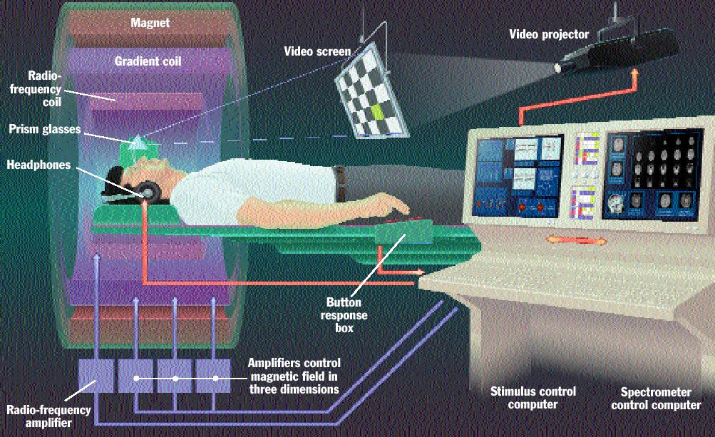
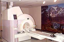
18 fmri Setup /home/jose/courses/scanner2.wav
19 Getting fmri Data Like a human Radio 1) Put subject in big magnetic field (leave him there) 2) Transmit radio waves into subject [about 3 ms at a specific frequency] 3) Turn off radio wave transmitter 4) Receive radio waves re transmitted by subject Manipulate re transmission with magnetic fields during this readout interval [ ms: MRI is not a snapshot] 5) Store measured radio wave data vs. time Now go back to 2) to get some more data 6) Process raw data to reconstruct images (Fourier
20 fmri depends on 3 lucky aspects of the human body 1. We're mostly made out of Water allowing for magnetic succesptibility in tissue (Protons) 3. Changes in neural activity produce changes in local blood flow 4. Local blood flow disrupts local tissue magnetic succeptibility allowing for localization


21 Protons Align in Magnetic Field Outside the Magnet Inside the Magnet M Brain is 70% water High abundance of H protons throughout tissue. When placed in a Magnetic Field (50k times> Earth's) Protons in the brain begin to align in particular HYDROGEN Protons A radio frequency Pulse at the frequency of the HYDROGEN PROTON (170.3 MHz) which perturbs the alignment and then slowly relaxes back to the aligned state: TIME CONSTANT T1
 = time to wait after excitation before sampling T1 Different Tissue have different T1 time constants.")
22 T1 and TR T1 = recovery of longitudinal (B0) magnetization used in anatomical images ~ msec (longer with bigger B0) TR (repetition time) = time to wait after excitation before sampling T1 Different Tissue have different T1 time constants.. CSF is slow 3seconds, white matter 500ms
23 Magnetic Susceptibility Impurities in the Magnetic Field Proton effect Homogenous Unless for some reason Brain is locally perturbed Neural Activity > produces Oxygen Consumption Blood Flow to local area to replenish Oxygen brings oxyhemoglobin (oxygen loaded hemoglobin) which is paramagnetic hence producing a local disturbance in the magnetic field (a NATURAL CONSTRAST AGENT) changes in Magnetic Susceptibility to surrounding tissue. Thus producing SLOWER Time Constants for the surrounding tissue and hence a STRONG MR SIGNAL (T2*) perturbance magnetic susceptibility
24 BOLD Genesis T2* is the decay due to local perturbance of magnetic field We have no idea how this happens
25 Ideal HDR The peak to optimize for Brain mapping sometimes called the task area. 6 8 seconds from neuronal event 0 0 Seconds % signal change = (point baseline)/baseline 20 time to rise signal begins to rise soon after stimulus begins usually 0.5 3% could be as high as 10% initial dip more focal and potentially a better measure time to peak signal peaks 4 6 sec after stimulus begins post stimulus undershoot
26 Properties of BOLD susceptibility of brain tissue The statistical interpretation of signals in BOLD* involve (1) the noise model for the signal: this effects detection, interpretation of the number of relevant areas, patterns of effects and the potential shape and size of the tissue sensitive to the measurement. (2) the impulse response temporal dynamics. Identifiabilty is critical in determining the temporal properties of the measurement


27 Despite lack of Identifiability: HRF Convolution is useful for maximizing signal Experimental Stimulus Function Hemodynamic Response Function Predicted Response Block Design Event Related

28 OVERVIEW OF BRAIN IMAGING ANALYSIS

29 Finger tapping
 low resolution (~3 mm but can be better) one image fmri many images Blood Oxygenation Level")
30 MRI vs. fmri high resolution MRI fmri (1 mm) low resolution (~3 mm but can be better) one image fmri many images Blood Oxygenation Level Dependent (BOLD) signal indirect measure of neural activity neural activity (e.g., every 2 sec for 5 mins) blood oxygen fmri signal

31 fmri Activation: AVERAGING Source: Posner & Raichle, Images of Mind
 e.g., 19.2 cm 3 mm 3 mm")
32 Slice Terminology VOXEL Slice Thickness (Volumetric Pix In plane resolution e.g., 6 mm e.g., 192 mm / 64 = 3 mm SAGITTAL SLICE IN PLANE SLICE Number of Slices e.g., 10 Matrix Size e.g., 64 x 64 Field of View (FOV) e.g., 19.2 cm 3 mm 3 mm
33 Activation Statistics Functional images ROI Time Course fmri ~2s Signal (% change) Time Co nd it ion Condition Statistical Map 1 superimposed on anatomical MRI image Time Co nd itio Region of interest (ROI) n 2... ~ 5 min


34 Statistical Maps & Time Courses Then extract the time course Use stat maps to pick region
35 What are the temporal limits? What is the briefest stimulus that fmri can detect? Blamire et al. (1992) 2 sec Bandettini (1993): 0.5 sec Savoy et al (1995): 34 msec With enough averaging, anything seems possible. The shape of the HRF is predictable. Event related potentials (ERPs) are based on averaging small responses over many trials.

36 Subject Safety Anyone going near the magnet subjects, staff and visitors must be thoroughly screened: Subjects must have no metal in their bodies: pacemaker aneurysm clips metal implants (e.g., cochlear implants) interuterine devices (IUDs) some dental work (fillings okay) Subjects must remove metal from their bodies jewellery, watch, piercings coins, etc. wallet This subject was wearing a hair band with a ~2 mm copper clamp. Left: with hair band. Right: without.

37 fmri and PET: indirect signals for neural activity Regions of interest: The brain is a dynamic, complex entity. How do you know which brain activity corresponds to your research experiment? One technique is to define regions of interest (ROIs) before scanning to identify which areas you expect to see changes in activation.
 at the anterior of the brain, the parietal lobe (orange) posterior to the frontal lobe at the superior aspect of the brain, the")
38 1.0 Introduction The geography of the brain The four major lobes of the brain are visible from a lateral view. A lateral view of the left hemisphere, with the frontal lobe (purple) at the anterior of the brain, the parietal lobe (orange) posterior to the frontal lobe at the superior aspect of the brain, the temporal lobe (blue) posterior to the frontal lobe and inferior to the parietal lobe, and the occipital lobe (yellow) which is posterior to both the parietal and temporal lobes.
 and a mid-sagittal perspective (right")
39 1.0 Introduction The geography of the brain Some important landmarks of the brain in the left hemisphere from a lateral perspective (left panel) and a mid-sagittal perspective (right panel).
40 The geography of the brain A schematic drawing of the six layers in the gray matter of the cortex. Some cortical neurons send their axons to the thalamus, while others receive input from thalamic neurons.

41 1.0 Introduction The geography of the brain The Brodmann classification (based on microscopic cell differences) of regions in the left hemisphere, shown in a lateral view. Areas 41 & 52 are indicated by lines. Some areas, like the insula and auditory region, are tucked away behind the temporal lobe.

42 1.0 Introduction The geography of the brain The Brodmann classification of regions in the left hemisphere in midsagittal view.
43 Growing a brain from the bottom up Building a brain from bottom to top The brain builds on the brainstem, with the thalami on top as a major input hub. The hippocampi and amygdalas are actually nestled inside each of the temporal lobes. The light blue ventricles have no neurons, but provide the brain s own circulatory system. The basal ganglia can be thought of as the output hub of the system. A great deal of traffic flows back to the cortex as well.

44 3.0 From where to what : the functional roles of brain regions Output and input: the front-back division The frontal lobe typically is involved in executive and motor (output) functions. The posterior half of the brain is involved in sensory processing. A view of functional areas in sensory regions of the cortex: the central sulcus separates the frontal lobe from the parietal. Immediately anterior to the central sulcus are motor areas, and just posterior to the central sulcus is the primary somatosensory area.
45 From where to what : the functional roles of brain regions Output and input: the front-back division A homonculus ( little man ) shows the body map for motor representation of different areas of the body. Note that some body areas, such as the fingers, have disproportionately larger representation than other body areas, such as the trunk.
46 From where to what : the functional roles of brain regions Output and input: the front-back division A homonculus ( little man ) shows the body map for somatosensory representation of different areas on the body. Note that some body areas, such as the face, have disproportionately larger representation than other body areas, such as the trunk.


47 The Little Man in the somatasensory cortex
48 From where to what : the functional roles of brain regions Output and input: the front-back division The close physical connection between motor cortex, located just anterior to the central sulcus, and the somatosensory cortex, located just posterior to it, allows for a tight coupling between the senses of touch, pressure, and pain and the action or motor system.

49 fmri and PET: indirect signals for neural activity The resting brain is not silent Many fmri and PET research protocols subtract an experimental condition from a baseline -- frequently a resting state where the subject does not have a task to perform. But, is the brain resting? While activity during a resting state may not be directly related to the tasks being performed in other states, we know that humans are constantly thinking, imagining, feeling, anticipating and remembering. Background activity in the left and right hemispheres during a resting state

50 4.0 Conscious and unconscious brain events A recent wave of brain studies are investigating conscious and unconscious phenomena in the brain. For example, a fmri study compared brain activation for conscious and unconscious events: unconscious viewing of words activated visual areas only, while conscious viewing activated expanded regions in the cortex.

51 Pessoa: Fear recognition..

52 Correlation and Causation Brain damage and causal inferences Brain injuries can provide evidence that areas are necessary for certain cognitive functions, however it is important to keep in mind that studies of brain-damaged individuals provide correlational, not causal, explanations about brain function.
53 Summary The advent of brain imaging has transformed the study of human cognition. New and refined methods are constantly being produced. There is a wide array of methods and techniques for brain recording: brain imaging techniques allow us measure single neurons as well as large cortical activations, brain structures as well as dynamic brain activity. A powerful use of brain imaging is to provide converging evidence, across techniques and research populations, to better understand human cognition.
The Central Nervous System
 The Central Nervous System Cellular Basis. Neural Communication. Major Structures. Principles & Methods. Principles of Neural Organization Big Question #1: Representation. How is the external world coded
The neurolinguistic toolbox Jonathan R. Brennan. Introduction to Neurolinguistics, LSA2017 1
 The neurolinguistic toolbox Jonathan R. Brennan Introduction to Neurolinguistics, LSA2017 1 Psycholinguistics / Neurolinguistics Happy Hour!!! Tuesdays 7/11, 7/18, 7/25 5:30-6:30 PM @ the Boone Center
P2 Visual - Perception
 P2 Visual - Perception 2014 SOSE Neuroimaging of high-level visual functions gyula.kovacs@uni-jena.de 11/09/06 Functional magnetic resonance imaging (fmri) The very basics What is fmri? What is MRI? The
Brain and Cognition. Cognitive Neuroscience. If the brain were simple enough to understand, we would be too stupid to understand it
 Brain and Cognition Cognitive Neuroscience If the brain were simple enough to understand, we would be too stupid to understand it 1 The Chemical Synapse 2 Chemical Neurotransmission At rest, the synapse
Table 1. Summary of PET and fmri Methods. What is imaged PET fmri BOLD (T2*) Regional brain activation. Blood flow ( 15 O) Arterial spin tagging (AST)
 Regional brain activation. Blood flow ( 15 O) Arterial spin tagging (AST)") Table 1 Summary of PET and fmri Methods What is imaged PET fmri Brain structure Regional brain activation Anatomical connectivity Receptor binding and regional chemical distribution Blood flow ( 15 O)
Table 1 Summary of PET and fmri Methods What is imaged PET fmri Brain structure Regional brain activation Anatomical connectivity Receptor binding and regional chemical distribution Blood flow ( 15 O)
Organization of the nervous system. The withdrawal reflex. The central nervous system. Structure of a neuron. Overview
 Overview The nervous system- central and peripheral The brain: The source of mind and self Neurons Neuron Communication Chemical messengers Inside the brain Parts of the brain Split Brain Patients Organization
Cerebral Cortex 1. Sarah Heilbronner
 Cerebral Cortex 1 Sarah Heilbronner heilb028@umn.edu Want to meet? Coffee hour 10-11am Tuesday 11/27 Surdyk s Overview and organization of the cerebral cortex What is the cerebral cortex? Where is each
CISC 3250 Systems Neuroscience
 CISC 3250 Systems Neuroscience Levels of organization Central Nervous System 1m 10 11 neurons Neural systems and neuroanatomy Systems 10cm Networks 1mm Neurons 100μm 10 8 neurons Professor Daniel Leeds
Myers Psychology for AP*
 Myers Psychology for AP* David G. Myers PowerPoint Presentation Slides by Kent Korek Germantown High School Worth Publishers, 2010 *AP is a trademark registered and/or owned by the College Board, which
fmri (functional MRI)
") Lesion fmri (functional MRI) Electroencephalogram (EEG) Brainstem CT (computed tomography) Scan Medulla PET (positron emission tomography) Scan Reticular Formation MRI (magnetic resonance imaging) Thalamus
Lesion fmri (functional MRI) Electroencephalogram (EEG) Brainstem CT (computed tomography) Scan Medulla PET (positron emission tomography) Scan Reticular Formation MRI (magnetic resonance imaging) Thalamus
Gross Organization I The Brain. Reading: BCP Chapter 7
 Gross Organization I The Brain Reading: BCP Chapter 7 Layout of the Nervous System Central Nervous System (CNS) Located inside of bone Includes the brain (in the skull) and the spinal cord (in the backbone)
Outline. Biological Psychology: Research Methods. Dr. Katherine Mickley Steinmetz
 Biological Psychology: Research Methods Dr. Katherine Mickley Steinmetz Outline Neuroscience Methods Histology Electrophysiological Recordings Lesion Neuroimaging Neuroanatomy Histology: Brain structure
STRUCTURAL ORGANIZATION OF THE NERVOUS SYSTEM
 STRUCTURAL ORGANIZATION OF THE NERVOUS SYSTEM STRUCTURAL ORGANIZATION OF THE BRAIN The central nervous system (CNS), consisting of the brain and spinal cord, receives input from sensory neurons and directs
1. Processes nutrients and provides energy for the neuron to function; contains the cell's nucleus; also called the soma.
 1. Base of brainstem; controls heartbeat and breathing 2. tissue destruction; a brain lesion is a naturally or experimentally caused destruction of brain tissue 3. A thick band of axons that connects the
ASSUMPTION OF COGNITIVE UNIFORMITY
 The Human Brain cerebral hemispheres: two most important divisions of the brain, separated by the longitudinal fissure corpus callosum: a large bundle of axons that constitutes the major connection between
Functional MRI Mapping Cognition
 Outline Functional MRI Mapping Cognition Michael A. Yassa, B.A. Division of Psychiatric Neuro-imaging Psychiatry and Behavioral Sciences Johns Hopkins School of Medicine Why fmri? fmri - How it works Research
Announcements. Final Exam will be a take-home exam. Format similar to the short assignment (no multiple choice, etc.)
") Announcements Final Exam will be a take-home exam Format similar to the short assignment (no multiple choice, etc.) Will be handed out at end of last class period (Thursday June 5 th ) Due by 6 pm June
Announcements Final Exam will be a take-home exam Format similar to the short assignment (no multiple choice, etc.) Will be handed out at end of last class period (Thursday June 5 th ) Due by 6 pm June
Define functional MRI. Briefly describe fmri image acquisition. Discuss relative functional neuroanatomy. Review clinical applications.
 Dr. Peter J. Fiester November 14, 2012 Define functional MRI. Briefly describe fmri image acquisition. Discuss relative functional neuroanatomy. Review clinical applications. Briefly discuss a few examples
Methods for assessing the brain basis of developmental disorders
 Announcements LIGN171: Child Language Acquisition http://ling.ucsd.edu/courses/lign171 Final Exam will be a take-home exam Format similar to the short assignment (no multiple choice, etc.) Will be handed
Introduction to Functional MRI
 Introduction to Functional MRI Douglas C. Noll Department of Biomedical Engineering Functional MRI Laboratory University of Michigan Outline Brief overview of physiology and physics of BOLD fmri Background
Introduction. Visual Perception Aditi Majumder, UCI. Perception is taken for granted!
 Introduction Visual Perception Perception is taken for granted! Slide 2 1 Perception is very complex Perceive Locate Identify/Recognize Different objects Their relationship with each other Qualitative
Announcement. Danny to schedule a time if you are interested.
 Announcement If you need more experiments to participate in, contact Danny Sanchez (dsanchez@ucsd.edu) make sure to tell him that you are from LIGN171, so he will let me know about your credit (1 point).
Acetylcholine (ACh) Action potential. Agonists. Drugs that enhance the actions of neurotransmitters.
 Action potential. Agonists. Drugs that enhance the actions of neurotransmitters.") Acetylcholine (ACh) The neurotransmitter responsible for motor control at the junction between nerves and muscles; also involved in mental processes such as learning, memory, sleeping, and dreaming. (See
Acetylcholine (ACh) The neurotransmitter responsible for motor control at the junction between nerves and muscles; also involved in mental processes such as learning, memory, sleeping, and dreaming. (See
Neural Basis of Motor Control
 Neural Basis of Motor Control Central Nervous System Skeletal muscles are controlled by the CNS which consists of the brain and spinal cord. Determines which muscles will contract When How fast To what
Brain and behaviour (Wk 6 + 7)
") Brain and behaviour (Wk 6 + 7) What is a neuron? What is the cell body? What is the axon? The basic building block of the nervous system, the individual nerve cell that receives, processes and transmits
Brain and behaviour (Wk 6 + 7) What is a neuron? What is the cell body? What is the axon? The basic building block of the nervous system, the individual nerve cell that receives, processes and transmits
The human brain. of cognition need to make sense gives the structure of the brain (duh). ! What is the basic physiology of this organ?
. ! What is the basic physiology of this organ?") The human brain The human brain! What is the basic physiology of this organ?! Understanding the parts of this organ provides a hypothesis space for its function perhaps different parts perform different
The human brain The human brain! What is the basic physiology of this organ?! Understanding the parts of this organ provides a hypothesis space for its function perhaps different parts perform different
Neural Correlates of Human Cognitive Function:
 Neural Correlates of Human Cognitive Function: A Comparison of Electrophysiological and Other Neuroimaging Approaches Leun J. Otten Institute of Cognitive Neuroscience & Department of Psychology University
Human Paleoneurology and the Evolution of the Parietal Cortex
 PARIETAL LOBE The Parietal Lobes develop at about the age of 5 years. They function to give the individual perspective and to help them understand space, touch, and volume. The location of the parietal
A U. Methods of Cognitive Neuroscience 2/8/2016. Neat Stuff! Cognitive Psychology
 Methods of Cognitive Neuroscience Neat Stuff! Optogenetics http://spie.org/newsroom/technical-articles-archive/videos/0411-boyden Stimulating the brain with light Cognitive Psychology Mental Representation
What is cognitive neuroscience?
 9.63 - Fall 2005 Lecture 7 Ben Balas Courtesy of Ben Balas. Used with permission. What is cognitive neuroscience? Experimental Psychology is more or less the study of subjects behavior subject to various
Methods to examine brain activity associated with emotional states and traits
 Methods to examine brain activity associated with emotional states and traits Brain electrical activity methods description and explanation of method state effects trait effects Positron emission tomography
Leah Militello, class of 2018
 Leah Militello, class of 2018 Objectives 1. Describe the general organization of cerebral hemispheres. 2. Describe the locations and features of the different functional areas of cortex. 3. Understand
Sincerely, Ms. Paoloni and Mrs. Whitney
 Dear Students, Welcome to AP Psychology! We will begin our course of study focusing on the nervous system with a particular emphasis on how the brain and neurotransmitters influence our behaviors. In preparation
Neuroimaging. BIE601 Advanced Biological Engineering Dr. Boonserm Kaewkamnerdpong Biological Engineering Program, KMUTT. Human Brain Mapping
 11/8/2013 Neuroimaging N i i BIE601 Advanced Biological Engineering Dr. Boonserm Kaewkamnerdpong Biological Engineering Program, KMUTT 2 Human Brain Mapping H Human m n brain br in m mapping ppin can nb
The Nervous System. Neuron 01/12/2011. The Synapse: The Processor
 The Nervous System Neuron Nucleus Cell body Dendrites they are part of the cell body of a neuron that collect chemical and electrical signals from other neurons at synapses and convert them into electrical
The Cognitive Neuroscientist s Toolkit
 The Cognitive Neuroscientist s Toolkit Jesse Rissman CS 182 Guest Lecture, 2/1/07 QuickTimeª and a TIFF (Uncompressed) decompressor are needed to see this picture. LESION PET fmri EEG/MEG TMS A little
The physiology of the BOLD signal What do we measure with fmri?
 The physiology of the BOLD signal What do we measure with fmri? Methods and Models in fmri, 10.11.2012 Jakob Heinzle Translational Neuromodeling Unit (TNU) Institute for Biomedical Engineering (IBT) University
To understand AD, it is important to
 To understand AD, it is important to know a bit about the brain. This part of Unraveling the Mystery gives an inside view of the normal brain, how it works, and what happens during aging. The brain is
P. Hitchcock, Ph.D. Department of Cell and Developmental Biology Kellogg Eye Center. Wednesday, 16 March 2009, 1:00p.m. 2:00p.m.
 Normal CNS, Special Senses, Head and Neck TOPIC: CEREBRAL HEMISPHERES FACULTY: LECTURE: READING: P. Hitchcock, Ph.D. Department of Cell and Developmental Biology Kellogg Eye Center Wednesday, 16 March
Neuroimaging vs. other methods
 BASIC LOGIC OF NEUROIMAGING fmri (functional magnetic resonance imaging) Bottom line on how it works: Adapts MRI to register the magnetic properties of oxygenated and deoxygenated hemoglobin, allowing
COGNITIVE SCIENCE 17. Peeking Inside The Head. Part 1. Jaime A. Pineda, Ph.D.
 COGNITIVE SCIENCE 17 Peeking Inside The Head Part 1 Jaime A. Pineda, Ph.D. Imaging The Living Brain! Computed Tomography (CT)! Magnetic Resonance Imaging (MRI)! Positron Emission Tomography (PET)! Functional
Cerebrum-Cerebral Hemispheres. Cuneyt Mirzanli Istanbul Gelisim University
 Cerebrum-Cerebral Hemispheres Cuneyt Mirzanli Istanbul Gelisim University The largest part of the brain. Ovoid shape. Two incompletely separated cerebral hemispheres. The outer surface of the cerebral
Chapter 3. Structure and Function of the Nervous System. Copyright (c) Allyn and Bacon 2004
 Allyn and Bacon 2004") Chapter 3 Structure and Function of the Nervous System 1 Basic Features of the Nervous System Neuraxis: An imaginary line drawn through the center of the length of the central nervous system, from the
Chapter 3 Structure and Function of the Nervous System 1 Basic Features of the Nervous System Neuraxis: An imaginary line drawn through the center of the length of the central nervous system, from the
Geography of the Forehead
 5. Brain Areas Geography of the Forehead Everyone thinks the brain is so complicated, but let s look at the facts. The frontal lobe, for example, is located in the front! And the temporal lobe is where
CEREBRUM. Dr. Jamila EL Medany
 CEREBRUM Dr. Jamila EL Medany Objectives At the end of the lecture, the student should be able to: List the parts of the cerebral hemisphere (cortex, medulla, basal nuclei, lateral ventricle). Describe
Mirror Neurons in Primates, Humans, and Implications for Neuropsychiatric Disorders
 Mirror Neurons in Primates, Humans, and Implications for Neuropsychiatric Disorders Fiza Singh, M.D. H.S. Assistant Clinical Professor of Psychiatry UCSD School of Medicine VA San Diego Healthcare System
Non-Invasive Techniques
 Non-Invasive Techniques Key: Does not hurt the organism Psychology 372 Physiological Psychology Steven E. Meier, Ph.D. Listen to the audio lecture while viewing these slides or view the video presentation
Non-Invasive Techniques
 Many Procedures Non-Invasive Techniques Key: Does not hurt the organism Psychology 372 Physiological Psychology Steven E. Meier, Ph.D. Listen to the audio lecture while viewing these slides or view the
Regional and Lobe Parcellation Rhesus Monkey Brain Atlas. Manual Tracing for Parcellation Template
 Regional and Lobe Parcellation Rhesus Monkey Brain Atlas Manual Tracing for Parcellation Template Overview of Tracing Guidelines A) Traces are performed in a systematic order they, allowing the more easily
Restoring Communication and Mobility
 Restoring Communication and Mobility What are they? Artificial devices connected to the body that substitute, restore or supplement a sensory, cognitive, or motive function of the nervous system that has
10/3/2016. T1 Anatomical structures are clearly identified, white matter (which has a high fat content) appears bright.
 appears bright.") H2O -2 atoms of Hydrogen, 1 of Oxygen Hydrogen just has one single proton and orbited by one single electron Proton has a magnetic moment similar to the earths magnetic pole Also similar to earth in that
H2O -2 atoms of Hydrogen, 1 of Oxygen Hydrogen just has one single proton and orbited by one single electron Proton has a magnetic moment similar to the earths magnetic pole Also similar to earth in that
The Brain. Its major systems, How we study them, How they make the mind
 The Brain Its major systems, How we study them, How they make the mind 9.00 Introduction to Psychology Joanne s Recitation Section Friday, February 11, 2011 Outline 1. Syllabus: Course Requirements, Exams,
Eavesdropping on the Mind. COGS 17 - Winter 2019 Andrew Shibata
 Eavesdropping on the Mind COGS 17 - Winter 2019 Andrew Shibata Announcements - Midterm I is next Tuesday! - Exam is worth 25% of your grade - Homework 1 is due at the exam (worth 2.5% of grade) - Review
Neuroimaging and Assessment Methods
 Psych 2200, Lecture 5 Experimental Design and Brain Imaging Methods Tues Sept 15, 2015 Revised TA office hours (Sam), today 4-5p, and wed 11:30-1:30. I will not have office hours this thurs but you should
Biocomputer Wired for Action MWABBYH CTBIR LOBES
 Biocomputer Wired for Action MWABBYH CTBIR LOBES 100 100 100 100 100 200 200 200 200 200 300 300 300 300 300 400 400 400 400 400 500 500 500 500 500 Biocomputer Wired for Action MWABBYH CTBIR LOBES 100
Laurent Itti: CS564 Brain Theory and Artificial Intelligence. Lecture 4: Experimental techniques in visual neuroscience. Reading Assignments: None!
 CS 564 Brain Theory and Artificial Intelligence Lecture 4: Experimental techniques in visual neuroscience Reading Assignments: None! 1 Today we will briefly review - electrophysiological recording and
TABLE OF CONTINENTS. PSYC1002 Notes. Neuroscience.2. Cognitive Processes Learning and Motivation. 37. Perception Mental Abilities..
 TABLE OF CONTINENTS Neuroscience.2 Cognitive Processes...21 Learning and Motivation. 37 Perception.....54 Mental Abilities.. 83 Abnormal Psychology....103 1 Topic 1: Neuroscience Outline 1. Gross anatomy
CS/NEUR125 Brains, Minds, and Machines. Due: Friday, April 14
 CS/NEUR125 Brains, Minds, and Machines Assignment 5: Neural mechanisms of object-based attention Due: Friday, April 14 This Assignment is a guided reading of the 2014 paper, Neural Mechanisms of Object-Based
Bio11: The Nervous System. Body control systems. The human brain. The human brain. The Cerebrum. What parts of your brain are you using right now?
 Bio11: The Nervous System Body control systems Nervous system Quick Sends message directly to target organ Endocrine system Sends a hormone as a messenger to the target organ Can target several organs
Est-ce que l'eeg a toujours sa place en 2019?
 Est-ce que l'eeg a toujours sa place en 2019? Thomas Bast Epilepsy Center Kork, Germany Does EEG still play a role in 2019? What a question 7T-MRI, fmri, DTI, MEG, SISCOM, Of ieeg course! /HFO, Genetics
PSYC& 100: Biological Psychology (Lilienfeld Chap 3) 1
 1") PSYC& 100: Biological Psychology (Lilienfeld Chap 3) 1 1 What is a neuron? 2 Name and describe the functions of the three main parts of the neuron. 3 What do glial cells do? 4 Describe the three basic
PSYC& 100: Biological Psychology (Lilienfeld Chap 3) 1 1 What is a neuron? 2 Name and describe the functions of the three main parts of the neuron. 3 What do glial cells do? 4 Describe the three basic
Homework Week 2. PreLab 2 HW #2 Synapses (Page 1 in the HW Section)
") Homework Week 2 Due in Lab PreLab 2 HW #2 Synapses (Page 1 in the HW Section) Reminders No class next Monday Quiz 1 is @ 5:30pm on Tuesday, 1/22/13 Study guide posted under Study Aids section of website
Homework Week 2 Due in Lab PreLab 2 HW #2 Synapses (Page 1 in the HW Section) Reminders No class next Monday Quiz 1 is @ 5:30pm on Tuesday, 1/22/13 Study guide posted under Study Aids section of website
biological psychology, p. 40 The study of the nervous system, especially the brain. neuroscience, p. 40
 biological psychology, p. 40 The specialized branch of psychology that studies the relationship between behavior and bodily processes and system; also called biopsychology or psychobiology. neuroscience,
NEUROSCIENCE. W1 Divisions of the nervous system PSYC1002 NOTES
 PSYC1002 NOTES NEUROSCIENCE W1 Divisions of the nervous system Nervous system: - CNS o Brain and spinal cord - Peripheral Nervous System o Sensory nerves o Motor nerves o Autonomic nervous system o Enteric
Biological Bases of Behavior. 3: Structure of the Nervous System
 Biological Bases of Behavior 3: Structure of the Nervous System Neuroanatomy Terms The neuraxis is an imaginary line drawn through the spinal cord up to the front of the brain Anatomical directions are
HST 583 fmri DATA ANALYSIS AND ACQUISITION
 HST 583 fmri DATA ANALYSIS AND ACQUISITION Neural Signal Processing for Functional Neuroimaging Neuroscience Statistics Research Laboratory Massachusetts General Hospital Harvard Medical School/MIT Division
PHYSICS OF MRI ACQUISITION. Alternatives to BOLD for fmri
 PHYSICS OF MRI ACQUISITION Quick Review for fmri HST-583, Fall 2002 HST.583: Functional Magnetic Resonance Imaging: Data Acquisition and Analysis Harvard-MIT Division of Health Sciences and Technology
Psychology in Your Life
 Sarah Grison Todd Heatherton Michael Gazzaniga Psychology in Your Life SECOND EDITION Chapter 2 The Role of Biology in Psychology 1 2016 W. W. Norton & Company, Inc. 2.1 How Do Our Nervous Systems Affect
Physiology Unit 2 CONSCIOUSNESS, THE BRAIN AND BEHAVIOR
 Physiology Unit 2 CONSCIOUSNESS, THE BRAIN AND BEHAVIOR In Physiology Today What the Brain Does The nervous system determines states of consciousness and produces complex behaviors Any given neuron may
The Sonification of Human EEG and other Biomedical Data. Part 3
 The Sonification of Human EEG and other Biomedical Data Part 3 The Human EEG A data source for the sonification of cerebral dynamics The Human EEG - Outline Electric brain signals Continuous recording
Chapter 5 The Research Methods of Biopsychology
 Chapter 5 The Research Methods of Biopsychology Understanding What Biopsychologists Do This multimedia product and its contents are protected under copyright law. The following are prohibited by law: any
Nervous system, integration: Overview, and peripheral nervous system:
 Nervous system, integration: Overview, and peripheral nervous system: Some review & misc. parts [Fig. 28.11B, p. 573]: - white matter --> looks white due to the myelinated sheaths, which are quite fatty.
Introduction to Brain Imaging
 Introduction to Brain Imaging Human Brain Imaging NEUR 570 & BIC lecture series September 9, 2013 Petra Schweinhardt, MD PhD Montreal Neurological Institute McGill University Montreal, Canada Various techniques
Stuttering Research. Vincent Gracco, PhD Haskins Laboratories
 Stuttering Research Vincent Gracco, PhD Haskins Laboratories Stuttering Developmental disorder occurs in 5% of children Spontaneous remission in approximately 70% of cases Approximately 1% of adults with
Computational Cognitive Neuroscience (CCN)
") introduction people!s background? motivation for taking this course? Computational Cognitive Neuroscience (CCN) Peggy Seriès, Institute for Adaptive and Neural Computation, University of Edinburgh, UK
introduction people!s background? motivation for taking this course? Computational Cognitive Neuroscience (CCN) Peggy Seriès, Institute for Adaptive and Neural Computation, University of Edinburgh, UK
3/1/18. Overview of the Talk. Important Aspects of Neuroimaging Technology
 3/1/18 Considerations for the Use of Neuroimaging for Predicting Recovery of Speech and Language in Aphasia Linda I. Shuster, Ph.D., CCC-SLP Overview of the Talk Important aspects of neuroimaging technology
Daniel Bulte. Centre for Functional Magnetic Resonance Imaging of the Brain. University of Oxford
 Daniel Bulte Centre for Functional Magnetic Resonance Imaging of the Brain University of Oxford Overview Signal Sources BOLD Contrast Mechanism of MR signal change FMRI Modelling Scan design details Factors
Anatomy & Physiology Central Nervous System Worksheet
 1. What are the two parts of the CNS? 2. What are the four functions of the CNS Anatomy & Physiology Central Nervous System Worksheet 3. What are the four functions of the meninges? (p430) 4. Starting
COGNITIVE NEUROSCIENCE
 HOW TO STUDY MORE EFFECTIVELY (P 187-189) Elaborate Think about the meaning of the information that you are learning Relate to what you already know Associate: link information together Generate and test
Chapter 6 Section 1. The Nervous System: The Basic Structure
 Chapter 6 Section 1 The Nervous System: The Basic Structure Essential Question: How does studying the biology of the brain give us an understanding of our behavior? Draw or type 2 things you already know
LESSON 1.3 WORKBOOK. How can we study the behaving brain?
 LESSON 1.3 WORKBOOK How can we study the behaving brain? We are in the middle of a technological revolution when it comes to how closely we can look at the behaving brain. Scientists and doctors now have
Department of Cognitive Science UCSD
 Department of Cognitive Science UCSD Verse 1: Neocortex, frontal lobe, Brain stem, brain stem, Hippocampus, neural node, Right hemisphere, Pons and cortex visual, Brain stem, brain stem, Sylvian fissure,
Event-Related fmri and the Hemodynamic Response
 Human Brain Mapping 6:373 377(1998) Event-Related fmri and the Hemodynamic Response Randy L. Buckner 1,2,3 * 1 Departments of Psychology, Anatomy and Neurobiology, and Radiology, Washington University,
Biological Process 9/7/10. (a) Anatomy: Neurons have three basic parts. 1. The Nervous System: The communication system of your body and brain
 Anatomy: Neurons have three basic parts. 1. The Nervous System: The communication system of your body and brain") Biological Process Overview 1. The Nervous System: s (a) Anatomy, (b) Communication, (c) Networks 2. CNS/PNS 3. The Brain (a) Anatomy, (b) Localization of function 4. Methods to study the brain (Dr. Heidenreich)
Biological Process Overview 1. The Nervous System: s (a) Anatomy, (b) Communication, (c) Networks 2. CNS/PNS 3. The Brain (a) Anatomy, (b) Localization of function 4. Methods to study the brain (Dr. Heidenreich)
MSc Neuroimaging for Clinical & Cognitive Neuroscience
 MSc Neuroimaging for Clinical & Cognitive Neuroscience School of Psychological Sciences Faculty of Medical & Human Sciences Module Information *Please note that this is a sample guide to modules. The exact
Announcements. Exam 1. VII. Imaging techniques of the brain. Anatomical/Structural Scans. Structural Scans: CT. Structural Scans: CT 2/17/2014
 Exam 1 None at the moment! Announcements Mean 78.0% Median 80% Mode 86% Min 26% Max 98% Std Dev 12.6% VII. Imaging techniques of the brain A. CT: anatomical B. MRI: anatomical C. fmri: functional D. SPECT
Bioscience in the 21st century
 Bioscience in the 21st century Lecture 2: Innovations and Challenges Dr. Michael Burger Outline: Review of last lecture Organization of the nervous system (in brief) The mapping concept Bionic implants
shows syntax in his language. has a large neocortex, which explains his language abilities. shows remarkable cognitive abilities. all of the above.
 Section: Chapter 14: Multiple Choice 1. Alex the parrot: pp.529-530 shows syntax in his language. has a large neocortex, which explains his language abilities. shows remarkable cognitive abilities. all
The neurvous system senses, interprets, and responds to changes in the environment. Two types of cells makes this possible:
 NERVOUS SYSTEM The neurvous system senses, interprets, and responds to changes in the environment. Two types of cells makes this possible: the neuron and the supporting cells ("glial cells"). Neuron Neurons
Exam 1. Mean 78.0% Median 80% Mode 86% Min 26% Max 98% Std Dev 12.6%
 Exam 1 Mean 78.0% Median 80% Mode 86% Min 26% Max 98% Std Dev 12.6% None at the moment! Announcements VII. Imaging techniques of the brain A. CT: anatomical B. MRI: anatomical C. fmri: functional D. SPECT
Amy Kruse, Ph.D. Strategic Analysis, Inc. LCDR Dylan Schmorrow USN Defense Advanced Research Projects Agency
 What can modern neuroscience technologies offer the forward-looking applied military psychologist? Exploring the current and future use of EEG and NIR in personnel selection and training. Amy Kruse, Ph.D.
Unit 3: The Biological Bases of Behaviour
 Unit 3: The Biological Bases of Behaviour Section 1: Communication in the Nervous System Section 2: Organization in the Nervous System Section 3: Researching the Brain Section 4: The Brain Section 5: Cerebral
Physiology Unit 2 CONSCIOUSNESS, THE BRAIN AND BEHAVIOR
 Physiology Unit 2 CONSCIOUSNESS, THE BRAIN AND BEHAVIOR What the Brain Does The nervous system determines states of consciousness and produces complex behaviors Any given neuron may have as many as 200,000
Neuroscience Tutorial
 Neuroscience Tutorial Brain Organization : cortex, basal ganglia, limbic lobe : thalamus, hypothal., pituitary gland : medulla oblongata, midbrain, pons, cerebellum Cortical Organization Cortical Organization
The Central Nervous System I. Chapter 12
 The Central Nervous System I Chapter 12 The Central Nervous System The Brain and Spinal Cord Contained within the Axial Skeleton Brain Regions and Organization Medical Scheme (4 regions) 1. Cerebral Hemispheres
Implantable Microelectronic Devices
 ECE 8803/4803 Implantable Microelectronic Devices Fall - 2015 Maysam Ghovanloo (mgh@gatech.edu) School of Electrical and Computer Engineering Georgia Institute of Technology 2015 Maysam Ghovanloo 1 Outline
Guided Reading Activities
 Name Period Chapter 28: Nervous Systems Guided Reading Activities Big idea: Nervous system structure and function Answer the following questions as you read modules 28.1 28.2: 1. Your taste receptors for
Lesson 14. The Nervous System. Introduction to Life Processes - SCI 102 1
 Lesson 14 The Nervous System Introduction to Life Processes - SCI 102 1 Structures and Functions of Nerve Cells The nervous system has two principal cell types: Neurons (nerve cells) Glia The functions
Prof. Greg Francis 5/23/08
 Brain parts The brain IIE 269: Cognitive Psychology Greg Francis Lecture 02 The source of cognition (consider transplant!) Weighs about 3 pounds Damage to some parts result in immediate death or disability
Submitted report on Sufi recordings at AAPB 2013 in Portland. Not for general distribution. Thomas F. Collura, Ph.D. July, 2013
 Submitted report on Sufi recordings at AAPB 2013 in Portland Not for general distribution. Thomas F. Collura, Ph.D. July, 2013 Summary of EEG findings The intent of the EEG monitoring was to see which