Homework Week 2. PreLab 2 HW #2 Synapses (Page 1 in the HW Section)
|
|
|
- Dora Vivien Lane
- 6 years ago
- Views:
Transcription
1 Homework Week 2 Due in Lab PreLab 2 HW #2 Synapses (Page 1 in the HW Section) Reminders No class next Monday Quiz 1 5:30pm on Tuesday, 1/22/13 Study guide posted under Study Aids section of website
2 Thought Questions 1. When you have one of your mandibular teeth worked on at the dentist and he gives you a shot to deaden half of your mouth, what division of the nervous system is being affected by the lidocaine? 2. What do you think it s mode of action is? Hint: Remember Physio-EX in lab? 3. Is it affecting a cranial or spinal nerve?
3 Thought Questions 1. What 3 structures maintain resting membrane potential in neurons? 2. What membrane structure is responsible for depolarization during an action potential? 3. What membrane structure is responsible for repolarization during an action potential?
4 The Nervous System T H E C E N T R A L N E R V O U S S Y S T E M T H E B R A I N
5 Introduction Integration Memory Learning Sensation and perception

6 Organization of Neural Tissue White matter versus gray matter Fiber bundles Nerves versus tracts Nerve cell bodies Nucleus versus ganglion
7 Organization of Neural Tissue Generally Central cavity surrounded by a gray matter core External white matter Composed of myelinated fiber tracts Brain has additional areas of gray matter Not present in spinal cord

8 Central cavity Migratory pattern of neurons Cerebrum Cerebellum Cortex of gray matter Inner gray matter Outer white matter Gray matter Region of cerebellum Central cavity Inner gray matter Outer white matter Brain stem Spinal cord Gray matter Central cavity Outer white matter Inner gray matter Copyright 2010 Pearson Education, Inc. Figure 12.4

9 Organization of Neural Tissue Similar pattern with additional areas of gray matter
10 The Brain Functions Conscious perception Internal regulation Average adult male 3.5 lbs Average adult female 3.2 lbs Same brain mass to body mass ratio!
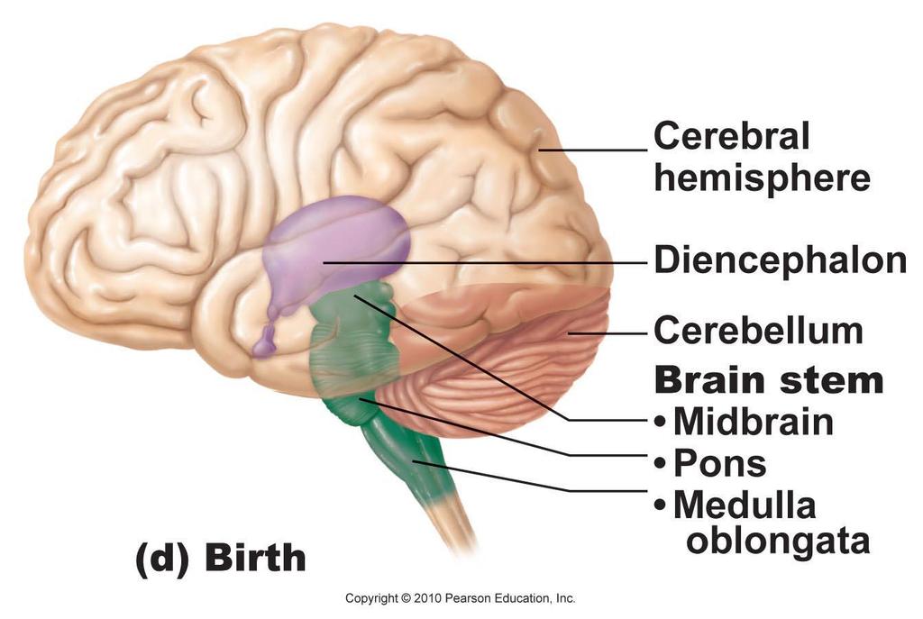
11 The Brain 4 Adult brain regions 1. Cerebral hemispheres (cerebrum) 2. Diencephalon 3. Cerebellum 4. Brain stem (midbrain, pons, and medulla)
12 The Brain

13 The Brain Four major regions are connected by ventricles and aqueducts

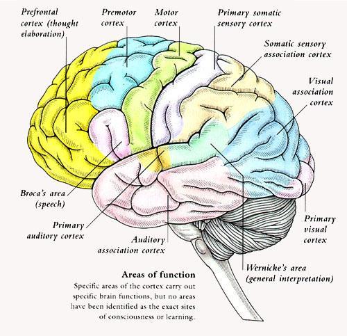
14 The Brain Ventricles Filled with cerebrospinal fluid Lined by ependymal cells Continuous with one another
15 The Cerebrum Cerebral hemispheres form superior part of brain About 80% of brain mass 3 tissue layers Superficial cortex = gray matter Internal white matter Basal nuclei = islands of gray matter
 Posterior Parietal lobe Right cerebral hemisphere Occipital lobe Figure")
16 Anterior Longitudinal fissure Frontal lobe Cerebral veins and arteries covered by arachnoid mater Left cerebral hemisphere (c) Posterior Parietal lobe Right cerebral hemisphere Occipital lobe Figure 12.6c
17 The Cerebrum Cerebral cortex Surface layer of cerebrum Executive Suite Convolutions Gyri elevated ridges Sulci shallow grooves Fissures deep grooves, separate larger regions of the brain

18 The Cerebrum Fissures divide cerebral hemispheres into 4 lobes
19 The Cerebrum 3 types of functional areas in the cerebral cortex 1. Motor areas Control voluntary movement 2. Sensory areas Conscious awareness of sensation 3. Association areas Integrate diverse information
20 The Cerebrum Functional areas of the cerebral cortex Contralateral orientation Hemispheres are functionally specialized Conscious behavior involves the entire cortex
21 Cerebral Motor Activity Motor areas Primary motor cortex Premotor cortex Frontal eye field Broca s area (outlined by dashes) Prefrontal cortex Working memory for spatial tasks Executive area for task management Working memory for object-recall tasks Solving complex, multitask problems (a) Lateral view, left cerebral hemisphere Central sulcus Sensory areas and related association areas Primary somatosensory cortex Somatosensory association cortex Gustatory cortex (in insula) Wernicke s area (outlined by dashes) Primary visual cortex Visual association area Auditory association area Primary auditory cortex Somatic sensation Taste Vision Hearing Primary motor cortex Motor association cortex Primary sensory cortex Sensory association cortex Multimodal association cortex Figure 12.8a
22 Cerebral Motor Activity Primary motor cortex Large pyramidal cells of the precentral gyrus Long axons pyramidal (corticospinal) tracts Allows conscious control Precise, skilled, voluntary movements

")
23 Motor Homunculus Somatotopy of precentral gyrus (primary motor cortex) Motor map in precentral gyrus Motor Posterior Anterior Jaw Toes Motor homunculi: upside-down caricatures representing the motor innervation of body regions Tongue Swallowing Primary motor cortex (precentral gyrus) Figure 12.9
24 Cerebral Motor Activity Premotor cortex Anterior to the precentral gyrus Controls learned, repetitious or patterned motor skills Coordinates simultaneous or sequential actions Involved in the planning of movements that depend on sensory feedback
 Motor speech area that directs muscles of the tongue Active as one prepares to")
25 Cerebral Motor Activity Broca s area Anterior to the inferior region of the premotor area Present in one hemisphere (usually the left) Motor speech area that directs muscles of the tongue Active as one prepares to speak
26 Cerebral Motor Activity Frontal eye field Anterior to the premotor cortex and superior to Broca s area Controls voluntary eye movements
27 Cerebral Motor Activity Motor areas Primary motor cortex Premotor cortex Frontal eye field Broca s area (outlined by dashes) Prefrontal cortex Working memory for spatial tasks Executive area for task management Working memory for object-recall tasks Solving complex, multitask problems (a) Lateral view, left cerebral hemisphere Central sulcus Sensory areas and related association areas Primary somatosensory cortex Somatosensory association cortex Gustatory cortex (in insula) Wernicke s area (outlined by dashes) Primary visual cortex Visual association area Auditory association area Primary auditory cortex Somatic sensation Taste Vision Hearing Primary motor cortex Motor association cortex Primary sensory cortex Sensory association cortex Multimodal association cortex Figure 12.8a
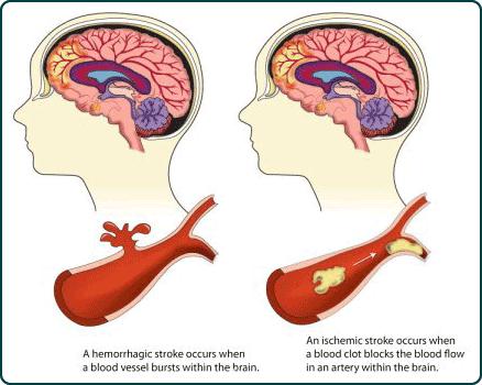
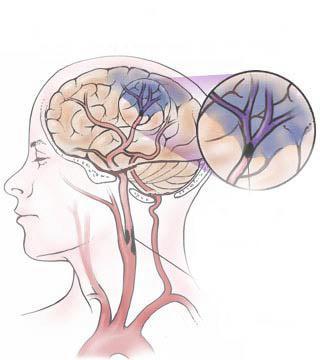
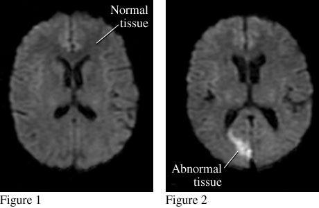
28 Cerebral Vascular Accident (Stroke) Types Ischemic stroke Hemorrhagic stroke Result Tissue death called an infarct Effects are determined by Where it occurs How large the area involved

29 Stroke
30 Stroke
31 Motor areas Primary motor cortex Premotor cortex Frontal eye field Broca s area (outlined by dashes) Prefrontal cortex Working memory for spatial tasks Executive area for task management Working memory for object-recall tasks Solving complex, multitask problems (a) Lateral view, left cerebral hemisphere Cerebral Sensory Activity Central sulcus Primary motor cortex Motor association cortex Primary sensory cortex Sensory association cortex Multimodal association cortex Sensory areas & related association areas Primary somatosensory cortex Somatosensory association cortex Gustatory cortex (in insula) Wernicke s area (outlined by dashes) Primary visual cortex Visual association area Auditory association area Primary auditory cortex Somatic sensation Taste Vision Hearing Figure 12.8a
32 Cerebral Sensory Activity Widely dispersed Parietal, temporal & occipital lobes Concerned with conscious awareness of sensation

33 Cerebral Sensory Activity Primary somatosensory cortex In the postcentral gyri, parietal lobe Stimuli from skin, skeletal muscles, and joints Capable of spatial discrimination Identification of body region being stimulated
 Intraabdominal Copyright 2010 Pearson Education, Inc. Figure 12.")
34 Posterior Anterior Sensory Sensory map in postcentral gyrus Genitals Primary somatosensory cortex (postcentral gyrus) Intraabdominal Copyright 2010 Pearson Education, Inc. Figure 12.9
35 Cerebral Sensory Activity Somatosensory association cortex Posterior to the primary somatosensory cortex Integrates sensory input from primary somatosensory cortex Integrates and analyzes inputs Temperature, size, texture Relationship of parts of objects being felt Example: keys in pocket
36
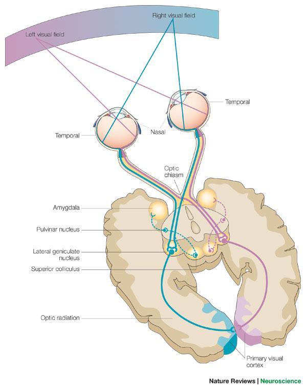
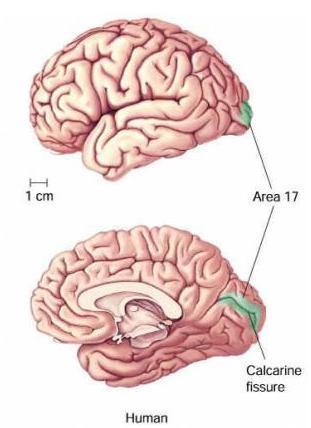
37 Cerebral Sensory Activity Visual areas Primary visual cortex Occipital lobe Receives visual information from the retinas Visual association area Surrounds the primary visual cortex Uses past visual experiences to interpret visual stimuli Example: color, form and movement Complex processing involves entire posterior half of the hemispheres
38
39 Cerebral Sensory Activity Auditory areas Primary auditory cortex Temporal lobes Interprets information from inner ear Pitch, loudness and location Auditory association area Posterior to the primary auditory cortex Stores memories of sounds and permits perception of sounds
40 Cerebral Sensory Activity Motor areas Primary motor cortex Premotor cortex Frontal eye field Broca s area (outlined by dashes) Prefrontal cortex Working memory for spatial tasks Executive area for task management Working memory for object-recall tasks Solving complex, multitask problems (a) Lateral view, left cerebral hemisphere Central sulcus Sensory areas and related association areas Primary somatosensory cortex Somatosensory association cortex Gustatory cortex (in insula) Wernicke s area (outlined by dashes) Primary visual cortex Visual association area Auditory association area Primary auditory cortex Somatic sensation Taste Vision Hearing Primary motor cortex Motor association cortex Primary sensory cortex Sensory association cortex Multimodal association cortex Figure 12.8a
41 Association Areas Receive inputs from multiple sensory areas Send outputs to multiple areas Including the premotor cortex Function Allows us to give meaning to information received, store it as memory, compare it to previous experience, and decide on action to take Multimodal association areas
42 Association Activity Three areas Prefrontal cortex Posterior association area (not discussed here) Limbic association area
43 Cerebral Association Activity Motor areas Primary motor cortex Premotor cortex Frontal eye field Broca s area (outlined by dashes) Prefrontal cortex Working memory for spatial tasks Executive area for task management Working memory for object-recall tasks Solving complex, multitask problems (a) Lateral view, left cerebral hemisphere Central sulcus Sensory areas and related association areas Primary somatosensory cortex Somatosensory association cortex Gustatory cortex (in insula) Wernicke s area (outlined by dashes) Primary visual cortex Visual association area Auditory association area Primary auditory cortex Somatic sensation Taste Vision Hearing Primary motor cortex Motor association cortex Primary sensory cortex Sensory association cortex Multimodal association cortex Figure 12.8a
44 Association Activity Prefrontal cortex Most complicated cortical region Involved with intellect, cognition, recall and personality Contains working memory needed for judgment, reasoning and conscience Development depends on feedback from social environment


45 Association Activity Limbic association area Part of the limbic system Provides emotional impact that helps establish memories Connections with prefrontal cortex regulate emotional expression
 Lateral view, left cerebral hemisphere Central sulcus Sensory areas and related association areas")
46 Cerebral Association Activity Motor areas Primary motor cortex Premotor cortex Frontal eye field Broca s area (outlined by dashes) Prefrontal cortex Working memory for spatial tasks Executive area for task management Working memory for object-recall tasks Solving complex, multitask problems (a) Lateral view, left cerebral hemisphere Central sulcus Sensory areas and related association areas Primary somatosensory cortex Somatosensory association cortex Gustatory cortex (in insula) Wernicke s area (outlined by dashes) Primary visual cortex Visual association area Auditory association area Primary auditory cortex Somatic sensation Taste Vision Hearing Primary motor cortex Motor association cortex Primary sensory cortex Sensory association cortex Multimodal association cortex Figure 12.8a


47 Cerebral Lateralization Left hemisphere Math Logic Language Controls right side of body Right hemisphere Visual-spatial skills Intuition Emotion Art and music Controls left side of body
48 Cerebral White Matter Projection tracts Connect cerebrum w/other body locations Association tracts Interconnect cerebral cortex (same side) Commissural tracts Connect two hemispheres

49 White Matter Tracts
50 Cerebral Gray Matter Basal Nuclei Association of gray matter deep in cerebral hemispheres Contribute to muscle coordination by excitatory innervation Ex. Parkinson s

51 Basal Nuclei
52 Complete CNS 23 #5, 1-6 CNS 21 Crossword puzzle Activity 52
CEREBRUM Dr. Jamila Elmedany Dr. Essam Eldin Salama
 CEREBRUM Dr. Jamila Elmedany Dr. Essam Eldin Salama Objectives At the end of the lecture, the student should be able to: List the parts of the cerebral hemisphere (cortex, medulla, basal nuclei, lateral
The Central Nervous System I. Chapter 12
 The Central Nervous System I Chapter 12 The Central Nervous System The Brain and Spinal Cord Contained within the Axial Skeleton Brain Regions and Organization Medical Scheme (4 regions) 1. Cerebral Hemispheres
Anatomy and Physiology (Bio 220) The Brain Chapter 14 and select portions of Chapter 16
 The Brain Chapter 14 and select portions of Chapter 16") Anatomy and Physiology (Bio 220) The Brain Chapter 14 and select portions of Chapter 16 I. Introduction A. Appearance 1. physical 2. weight 3. relative weight B. Major parts of the brain 1. cerebrum 2.
Anatomy and Physiology (Bio 220) The Brain Chapter 14 and select portions of Chapter 16 I. Introduction A. Appearance 1. physical 2. weight 3. relative weight B. Major parts of the brain 1. cerebrum 2.
The Nervous System PART B
 7 The Nervous System PART B PowerPoint Lecture Slide Presentation by Jerry L. Cook, Sam Houston University ESSENTIALS OF HUMAN ANATOMY & PHYSIOLOGY EIGHTH EDITION ELAINE N. MARIEB The Reflex Arc Reflex
BRAIN PART I (A & B): VENTRICLES & MENINGES
: VENTRICLES & MENINGES") BRAIN PART I (A & B): VENTRICLES & MENINGES Cranial Meninges Cranial meninges are continuous with spinal meninges Dura mater: inner layer (meningeal layer) outer layer (endosteal layer) fused to periosteum
BRAIN PART I (A & B): VENTRICLES & MENINGES Cranial Meninges Cranial meninges are continuous with spinal meninges Dura mater: inner layer (meningeal layer) outer layer (endosteal layer) fused to periosteum
CEREBRUM. Dr. Jamila EL Medany
 CEREBRUM Dr. Jamila EL Medany Objectives At the end of the lecture, the student should be able to: List the parts of the cerebral hemisphere (cortex, medulla, basal nuclei, lateral ventricle). Describe
Essentials of Human Anatomy & Physiology. Seventh Edition. The Nervous System. Copyright 2003 Pearson Education, Inc. publishing as Benjamin Cummings
 Essentials of Human Anatomy & Physiology Seventh Edition The Nervous System Copyright 2003 Pearson Education, Inc. publishing as Benjamin Cummings Functions of the Nervous System 1. Sensory input gathering
The Brain and Cranial Nerves Pg Three Main Regions of the Brain. Forebrain
 The Brain and Cranial Nerves Pg. 129 Three Main Regions of the Brain Forebrain Cerbral hemispheres Diencephalon Midbrain Brain stem Hindbrain Pons Cerebellum Medulla oblongata Interprets sensory inputs
The Brain and Cranial Nerves Pg. 129
 The Brain and Cranial Nerves Pg. 129 Three Main Regions of the Brain Forebrain Cerbral hemispheres Diencephalon Midbrain Brain stem Hindbrain Pons Cerebellum Medulla oblongata Forebrain Interprets sensory
meninges Outermost layer of the meninge dura mater arachnoid mater pia mater membranes located between bone and soft tissue of the nervous system
 membranes located between bone and soft tissue of the nervous system meninges Outermost layer of the meninge dura mater middle layer of the meninges, contains no blood vessels arachnoid mater Innermost
Chapter 14, Part 2! Chapter 14 Part 2 Brain/Cranial Nerves! The Cerebrum and Cranial Nerves! pp !
 Chapter 14, Part 2! The Cerebrum and Cranial pp. 482 505! SECTION 14-9! The cerebrum, the largest region of the brain, contains motor, sensory, and association areas! 2! White Matter of the Cerebrum! 1.
Chapter 14, Part 2! The Cerebrum and Cranial Nerves! pp !
 Chapter 14, Part 2! The Cerebrum and Cranial pp. 482 505! SECTION 14-9! The cerebrum, the largest region of the brain, contains motor, sensory, and association areas! 2! 1! ! Chapter 14 Part 2 Brain/Cranial
Organization of The Nervous System PROF. MOUSAED ALFAYEZ & DR. SANAA ALSHAARAWY
 Organization of The Nervous System PROF. MOUSAED ALFAYEZ & DR. SANAA ALSHAARAWY Objectives At the end of the lecture, the students should be able to: List the parts of the nervous system. List the function
The Nervous System 7PART B. PowerPoint Lecture Slide Presentation by Patty Bostwick-Taylor, Florence-Darlington Technical College
 PowerPoint Lecture Slide Presentation by Patty Bostwick-Taylor, Florence-Darlington Technical College The Nervous System 7PART B What is a reflex? What is a reflex? What is meant by the statement that
b. The groove between the two crests is called 2. The neural folds move toward each other & the fuse to create a
 Chapter 13: Brain and Cranial Nerves I. Development of the CNS A. The CNS begins as a flat plate called the B. The process proceeds as: 1. The lateral sides of the become elevated as waves called a. The
The Brain. Brain. Spinal Cord. Cauda Equina
 The Brain Brain Spinal Cord Cauda Equina The Brain Ventricles- cavities in the brain filled with cerebrospinal fluid connected to the subarachnoid space- fluid filled space surrounding the brain Brain
Good Morning! Take out your notes and vocab 1-10! Copyright 2003 Pearson Education, Inc. publishing as Benjamin Cummings
 Good Morning! Take out your notes and vocab 1-10! Functions of the Nervous System 1. Sensory input gathering information To monitor changes occurring inside and outside the body (changes = stimuli) 2.
Chapter 12 The Central Nervous System Chapter Outline
 Chapter 12 The Central Nervous System Chapter Outline Module 12.1 Overview of the Central Nervous System (Figures 12.1, 12.2, 12.3) A. The central nervous system (CNS) includes the and, and is involved
Parts of the Brain. Hindbrain. Controls autonomic functions Breathing, Heartbeat, Blood pressure, Swallowing, Vomiting, etc. Upper part of hindbrain
 Parts of the Brain The human brain is made up of three main parts: 1) Hindbrain (or brainstem) Which is made up of: Myelencephalon Metencephalon 2) Midbrain Which is made up of: Mesencephalon 3) Forebrain
Organization of The Nervous System PROF. SAEED ABUEL MAKAREM
 Organization of The Nervous System PROF. SAEED ABUEL MAKAREM Objectives By the end of the lecture, you should be able to: List the parts of the nervous system. List the function of the nervous system.
Lecture - Chapter 13: Central Nervous System
 Lecture - Chapter 13: Central Nervous System 1. Describe the following structures of the brain, what is the general function of each: a. Cerebrum b. Diencephalon c. Brain Stem d. Cerebellum 2. What structures
a) Central sulcus- shallow groove that runs across brain sagitally
 Central sulcus- shallow groove that runs across brain sagitally") KEY BRAIN Brain Gross Anatomy Terms 1) Explain each of the following in terms of structure of the brain a) Central sulcus- shallow groove that runs across brain sagitally b) Lateral fissure- deep groove
KEY BRAIN Brain Gross Anatomy Terms 1) Explain each of the following in terms of structure of the brain a) Central sulcus- shallow groove that runs across brain sagitally b) Lateral fissure- deep groove
49a A&P: Nervous System -! Synaptic Transmission and Central Nervous System
 49a A&P: Nervous System -! Synaptic Transmission and Central Nervous System 49a A&P: Nervous System -! Synaptic Transmission and Central Nervous System! Class Outline" 5 minutes" "Attendance, Breath of
P. Hitchcock, Ph.D. Department of Cell and Developmental Biology Kellogg Eye Center. Wednesday, 16 March 2009, 1:00p.m. 2:00p.m.
 Normal CNS, Special Senses, Head and Neck TOPIC: CEREBRAL HEMISPHERES FACULTY: LECTURE: READING: P. Hitchcock, Ph.D. Department of Cell and Developmental Biology Kellogg Eye Center Wednesday, 16 March
Cerebrum-Cerebral Hemispheres. Cuneyt Mirzanli Istanbul Gelisim University
 Cerebrum-Cerebral Hemispheres Cuneyt Mirzanli Istanbul Gelisim University The largest part of the brain. Ovoid shape. Two incompletely separated cerebral hemispheres. The outer surface of the cerebral
14 - Central Nervous System. The Brain Taft College Human Physiology
 14 - Central Nervous System The Brain Taft College Human Physiology Development of the Brain The brain begins as a simple tube, a neural tube. The tube or chamber (ventricle) is filled with cerebrospinal
Ch 13: Central Nervous System Part 1: The Brain p 374
 Ch 13: Central Nervous System Part 1: The Brain p 374 Discuss the organization of the brain, including the major structures and how they relate to one another! Review the meninges of the spinal cord and
The neurvous system senses, interprets, and responds to changes in the environment. Two types of cells makes this possible:
 NERVOUS SYSTEM The neurvous system senses, interprets, and responds to changes in the environment. Two types of cells makes this possible: the neuron and the supporting cells ("glial cells"). Neuron Neurons
Basic Brain Structure
 The Human Brain Basic Brain Structure Composed of 100 billion cells Makes up 2% of bodies weight Contains 15% of bodies blood supply Uses 20% of bodies oxygen and glucose Brain Protection Surrounded by
Nervous System: Part IV The Central Nervous System The Brain
 Nervous System: Part IV The Central Nervous System The Brain Can you survive when part of your brain is destroyed? 2 Essential Knowledge 3.D.2 2. Cells communicate with each other through direct contact
Biology 3201 Nervous System #2- Anatomy. Components of a Nervous System
 Biology 3201 Nervous System #2- Anatomy Components of a Nervous System In any nervous system, there are 4 main components: (1) sensors: gather information from the external environment (sense organs) (2)
Lab 12 Nervous System II
 Lab 12 Nervous System II Laboratory Objectives Identify the structural components of the central nervous system Label the functional areas of human cerebral cortex. Given a deficit affecting one or more
The Brain Worksheet Sections 5-7
 The Brain Worksheet Sections 5-7 1. neuroglia 2. autonomic nervous system 3. sensory neurons 4. oligodendrocytes 5. ascending tracts 6. descending tracts 7. saltatory propagation 8. continuous propagation
Human Anatomy. Brain and Cranial Nerves
 Human Anatomy Brain and Cranial Nerves 1 Brain and Cranial Nerves An adult brain weighs between 1.35 and 1.4 kilograms (kg) (around 3 pounds) and has a volume of about 1200 cubic centimeters (cc). Brain
The Nervous System. Divisions of the Nervous System. Branches of the Autonomic Nervous System. Central versus Peripheral
 The Nervous System Divisions of the Nervous System Central versus Peripheral Central Brain and spinal cord Peripheral Everything else Somatic versus Autonomic Somatic Nerves serving conscious sensations
Biological Bases of Behavior. 3: Structure of the Nervous System
 Biological Bases of Behavior 3: Structure of the Nervous System Neuroanatomy Terms The neuraxis is an imaginary line drawn through the spinal cord up to the front of the brain Anatomical directions are
Central Nervous System. January 7, 2016
 Central Nervous System January 7, 2016 Anatomy of a neuron Cell Body (soma) Receives information from the soma s extensions (dendrites) Passes on information away from the soma towards extensions (axons)
Central Nervous System
 Central Nervous System January 7, 2016 Anatomy of a neuron Cell Body (soma) Receives information from the soma s extensions (dendrites) Passes on information away from the soma towards extensions (axons)
Nervous system, integration: Overview, and peripheral nervous system:
 Nervous system, integration: Overview, and peripheral nervous system: Some review & misc. parts [Fig. 28.11B, p. 573]: - white matter --> looks white due to the myelinated sheaths, which are quite fatty.
Dissection of the Sheep Brain
 Dissection of the Sheep Brain Laboratory Objectives After completing this lab, you should be able to: 1. Identify the main structures in the sheep brain and to compare them with those of the human brain.
THE CENTRAL NERVOUS SYSTEM. The Brain & Spinal Cord
 THE CENTRAL NERVOUS SYSTEM The Brain & Spinal Cord Review: Nervous System Parallel Distributed Processing Composition of the CNS Nuclei: Clusters of neurons in the CNS ( neighborhoods ) Fiber Tracts/Pathways:
Central Nervous System
 Anatomy of a neuron Cell Body (soma) Receives information from the soma s extensions (dendrites) Central Nervous System January 7, 2016 Passes on information away from the soma towards extensions (axons)
C14 / CNS / MC3. What two stuctures make up the central nervous system?
 C14 / CNS / MC3 What two stuctures make up the central nervous system? The CNS starts embryonic development as a neural tube. What adult structures develop from the neural tube? What structures are part
Chapter 14: The Brain and Cranial Nerves. Copyright 2009, John Wiley & Sons, Inc.
 Chapter 14: The Brain and Cranial Nerves Development of the Brain Three to four-week embryo: prosencephalon, mesencephalon and rhombencephalon. Five-week embryo: telencephalon (cerebrum), diencephalon
-Zeina Assaf. -Omar Odeh. - Maha Beltagy
 -3 -Zeina Assaf -Omar Odeh - Maha Beltagy 1 P a g e The Inferior Surface Of The Brain The inferior surface of the brain is divide by the stem of the lateral fissure into 2 parts : The orbital surface and
Chapter 18: The Brain & Cranial Nerves. Origin of the Brain
 Chapter 18: The Brain & Cranial Nerves BIO 218 Fall 2015 Origin of the Brain The brain originates from a structure called the neural tube, which arises during a developmental stage called neurulation.
The Nervous System PART B
 7 The Nervous System PART B PowerPoint Lecture Slide Presentation by Jerry L. Cook, Sam Houston University ESSENTIALS OF HUMAN ANATOMY & PHYSIOLOGY EIGHTH EDITION ELAINE N. MARIEB Central Nervous System
Chapter 3. Structure and Function of the Nervous System. Copyright (c) Allyn and Bacon 2004
 Allyn and Bacon 2004") Chapter 3 Structure and Function of the Nervous System 1 Basic Features of the Nervous System Neuraxis: An imaginary line drawn through the center of the length of the central nervous system, from the
Chapter 3 Structure and Function of the Nervous System 1 Basic Features of the Nervous System Neuraxis: An imaginary line drawn through the center of the length of the central nervous system, from the
Principles of Anatomy and Physiology
 Principles of Anatomy and Physiology 14 th Edition CHAPTER 14 The Brain and Cranial Nerves Introduction The purpose of the chapter is to: 1. Understand how the brain is organized, protected, and supplied
MENTAL HOSPITAL PHONE MENU
 If you have low self-esteem, please hang up. Our operators are too busy to talk with you. MENTAL HOSPITAL PHONE MENU Hello and thank you for calling The State Mental Hospital. Please select from the following
Unit Three. The brain includes: cerebrum, diencephalon, brain stem, & cerebellum. The brain lies within the cranial cavity of the skull.
 Human Anatomy & Physiology 11 Divisions of the Nervous System Karen W. Smith, Instructor Unit Three BRAIN & SPINAL CORD Refer to the following URLs. Be sure to study these along with your book. http://www.sirinet.net/~jgjohnso/nervous.html
8.3 The Central Nervous System. SBI4U Ms. Ho-Lau
 8.3 The Central Nervous System SBI4U Ms. Ho-Lau The Central Nervous System the structural and functional centre for the entire nervous system the site of neural integration and processing The Central
Brain and Cranial Nerves (Ch. 15) Human Anatomy lecture. caudal = toward the spinal cord)
 Human Anatomy lecture. caudal = toward the spinal cord)") Insight: Some cranial nerve disorders Brain and Cranial Nerves (Ch. 15) Human Anatomy lecture I. Overview (Directional terms: rostral = toward the forehead caudal = toward the spinal cord) A. 3 Major parts
Insight: Some cranial nerve disorders Brain and Cranial Nerves (Ch. 15) Human Anatomy lecture I. Overview (Directional terms: rostral = toward the forehead caudal = toward the spinal cord) A. 3 Major parts
Gross Organization I The Brain. Reading: BCP Chapter 7
 Gross Organization I The Brain Reading: BCP Chapter 7 Layout of the Nervous System Central Nervous System (CNS) Located inside of bone Includes the brain (in the skull) and the spinal cord (in the backbone)
If I Only Had a Brain
 If I Only Had a Brain A Heart. (The Nerve!) Regions of the Brain Cerebral hemisphere Diencephalon Cerebellum (b) Adult brain Brain stem Regions of the Brain: Cerebrum Precentral gyrus Frontal lobe Central
THE ESSENTIAL BRAIN INJURY GUIDE
 THE ESSENTIAL BRAIN INJURY GUIDE Neuroanatomy & Neuroplasticity Section 2 Contributors Erin D. Bigler, PhD Michael R. Hoane, PhD Stephanie Kolakowsky-Hayner, PhD, CBIST, FACRM Dorothy A. Kozlowski, PhD
Overview of Brain Structures
 First Overview of Brain Structures Psychology 470 Introduction to Chemical Additions Steven E. Meier, Ph.D. All parts are interrelated. You need all parts to function normally. Neurons = Nerve cells Listen
Sheep Brain Dissection
 Sheep Brain Dissection Mammalian brains have many features in common. Human brains may not be available, so sheep brains often are dissected as an aid to understanding the mammalian brain since he general
Announcement. Danny to schedule a time if you are interested.
 Announcement If you need more experiments to participate in, contact Danny Sanchez (dsanchez@ucsd.edu) make sure to tell him that you are from LIGN171, so he will let me know about your credit (1 point).
CEREBRUM & CEREBRAL CORTEX
 CEREBRUM & CEREBRAL CORTEX Seonghan Kim Dept. of Anatomy Inje University, College of Medicine THE BRAIN ANATOMICAL REGIONS A. Cerebrum B. Diencephalon Thalamus Hypothalamus C. Brain Stem Midbrain Pons
STRUCTURAL ORGANIZATION OF THE NERVOUS SYSTEM
 STRUCTURAL ORGANIZATION OF THE NERVOUS SYSTEM STRUCTURAL ORGANIZATION OF THE BRAIN The central nervous system (CNS), consisting of the brain and spinal cord, receives input from sensory neurons and directs
Neocortex. Hemispheres 9/22/2010. Psychology 472 Pharmacology of Psychoactive Drugs. Structures are divided into several section or lobes.
 Neocortex Psychology 472 Pharmacology of Psychoactive Drugs 1 Is the most developed in Humans Has many folds and fissures The folds of tissue are called gyri or a gyrus (single) The fissures or valleys
Disorders affecting region: depression anxiety
 Amygdala Involved in learning, and the processing of emotional memories. Measures sensory input for potential threat level, then hypothalamus Regulates volatile emotions like fear and anger. Disorders
Primary Functions. Monitor changes. Integrate input. Initiate a response. External / internal. Process, interpret, make decisions, store information
 NERVOUS SYSTEM Monitor changes External / internal Integrate input Primary Functions Process, interpret, make decisions, store information Initiate a response E.g., movement, hormone release, stimulate/inhibit
Chapter 9. Nervous System
 Chapter 9 Nervous System Central Nervous System (CNS) vs. Peripheral Nervous System(PNS) CNS Brain Spinal cord PNS Peripheral nerves connecting CNS to the body Cranial nerves Spinal nerves Neurons transmit
Neurology study of the nervous system. nervous & endocrine systems work together to maintain homeostasis
 Nervous System Neurology study of the nervous system nervous & endocrine systems work together to maintain homeostasis Nervous System works very fast Uses electrical signals called nerve impulses Short-lived
Auditory and Vestibular Systems
 Auditory and Vestibular Systems Objective To learn the functional organization of the auditory and vestibular systems To understand how one can use changes in auditory function following injury to localize
Nervous System. Human Anatomy & Physiology P. Wilson
 Nervous System Human Anatomy & Physiology P. Wilson 1 2 Types of cells in the nervous system: Neurons & Neuroglial cells Neuroglial (aka glial) cells perform functions that are vital to neurons by filling
CNS consists of brain and spinal cord Cephalization Evolutionary development of rostral (anterior) portion of CNS Increased number of neurons in head
 portion of CNS Increased number of neurons in head") CNS consists of brain and spinal cord Cephalization Evolutionary development of rostral (anterior) portion of CNS Increased number of neurons in head Highest level reached in human brain 1 Mostly to orient
CNS consists of brain and spinal cord Cephalization Evolutionary development of rostral (anterior) portion of CNS Increased number of neurons in head Highest level reached in human brain 1 Mostly to orient
Department of Cognitive Science UCSD
 Department of Cognitive Science UCSD Verse 1: Neocortex, frontal lobe, Brain stem, brain stem, Hippocampus, neural node, Right hemisphere, Pons and cortex visual, Brain stem, brain stem, Sylvian fissure,
SHORT ANSWER. Write the word or phrase that best completes each statement or answers the question.
 Exam Name SHORT ANSWER. Write the word or phrase that best completes each statement or answers the question. Figure 12.3 Using Figure 12.3, match the following: 1) Site of efferent soma. 2) Site of axons
Composed of gray matter and arranged in raised ridges (gyri), grooves (sulci), depressions (fissures).
, grooves (sulci), depressions (fissures).") PSYC1020 Neuro and Pysc Notes Structure Description Major Functions Brainstem Stemlike portion of the brain, continuous with diencephalon above and spinal cord below. Composed of midbrain, pons, medulla
PSYC1020 Neuro and Pysc Notes Structure Description Major Functions Brainstem Stemlike portion of the brain, continuous with diencephalon above and spinal cord below. Composed of midbrain, pons, medulla
Nervous System and Brain Review. Bio 3201
 Nervous System and Brain Review Bio 3201 Dont worry about: glial cells Oligodendrocytes Satelite cells etc Nervous System - Vital to maintaining homeostasis in organisms - Comprised of : brain, spinal
Nervous System. 1. What N.S. division controls skeletal muscles? 3. What kind of neuroglia myelinates axons in the PNS?
 . What N.S. division controls skeletal muscles? Nervous System SRS Review %. Central nervous system %. Peripheral nervous system %. Afferent division %. Somatic division %. Autonomic division %. Sympathetic
WHAT ARE the COMPONENTS OF THE NERVOUS SYSTEM?
 The Nervous System WHAT ARE the COMPONENTS OF THE NERVOUS SYSTEM? The nervous system is made of: the brain & the spinal cord the nerves the senses There are lots of proteins and chemicals in your body
Bellringer: The central nervous system is comprised of: What is the name of the outermost layer of the brain? a. Brain. b.
 Bellringer: The central is comprised of: a. Brain b. Spinal cord c. Sensory receptors d. Both a and b What is the name of the outermost layer of the brain? a. Pia mater b. Dura mater c. Arachnoid d. Pons
Neuroanatomy lecture (1)
") Neuroanatomy lecture (1) Introduction: Neuroanatomy has two parts: the central and peripheral nervous system. The central nervous system is composed of brain and spinal cord. The brain has the following
Neuroanatomy lecture (1) Introduction: Neuroanatomy has two parts: the central and peripheral nervous system. The central nervous system is composed of brain and spinal cord. The brain has the following
Anatomy & Physiology Central Nervous System Worksheet
 1. What are the two parts of the CNS? 2. What are the four functions of the CNS Anatomy & Physiology Central Nervous System Worksheet 3. What are the four functions of the meninges? (p430) 4. Starting
M555 Medical Neuroscience Lab 1: Gross Anatomy of Brain, Crainal Nerves and Cerebral Blood Vessels
 M555 Medical Neuroscience Lab 1: Gross Anatomy of Brain, Crainal Nerves and Cerebral Blood Vessels Anatomical Directions Terms like dorsal, ventral, and posterior provide a means of locating structures
Anatomy of the Human Brain
 Anatomy of the Human Brain Overview Lobes of the brain (Forebrain) Midbrain/Hindbrain Protection and Blood supply Structure and Function of a neuron Synaptic Transmission Neurotransmitters The brain Most
The Nervous System: Sensory and Motor Tracts of the Spinal Cord
 15 The Nervous System: Sensory and Motor Tracts of the Spinal Cord PowerPoint Lecture Presentations prepared by Steven Bassett Southeast Community College Lincoln, Nebraska Introduction Millions of sensory
BIOL Dissection of the Sheep and Human Brain
 BIOL 2401 Dissection of the Sheep and Human Brain Laboratory Objectives After completing this lab, you should be able to: Identify the main structures in the sheep brain and to compare them with those
2401 : Anatomy/Physiology
 Dr. Chris Doumen Week 7 2401 : Anatomy/Physiology The Cerebrum Central Nervous System TextBook Readings Pages 434-456 and 460-461 Make use of the figures in your textbook ; a picture is worth a thousand
Functional Organization of the Central Nervous System
 Functional Organization of the Central Nervous System Hierarchical orgnization CNS consists of the brain and the spinal cord The brain analyzes and interprets the information Response messages are
LESSON 1.2 WORKBOOK How does brain structure impact its function?
 DEFINITIONS OF TERMS Central nervous system (CNS) contains the brain and spinal cord. Peripheral nervous system (PNS) includes all the nerves outside the brain and spinal cord. For a complete list of defined
The Human Brain: Anatomy, Functions, and Injury
 The Human Brain: Anatomy, Functions, and Injury Main Menu Brain Anatomy Brain Functions Injury Mechanisms Brain Anatomy Menu Skull Anatomy Interior Skull Surface Blood Vessels of the Brain Arteries of
Neural Basis of Motor Control
 Neural Basis of Motor Control Central Nervous System Skeletal muscles are controlled by the CNS which consists of the brain and spinal cord. Determines which muscles will contract When How fast To what
The Nervous System: Central Nervous System
 The Nervous System: Central Nervous System I. Anatomy of the nervous system A. The CNS & the body by: 1. monitoring of the body 2. & information between parts of the body 3. acting as a to gather, store,
Chapter 12b. Overview
 Chapter 12b Spinal Cord Overview Spinal cord gross anatomy Spinal meninges Sectional anatomy Sensory pathways Motor pathways Spinal cord pathologies 1 The Adult Spinal Cord About 18 inches (45 cm) long
Central N.S. Peripheral N.S. 2) List the functional subdivisions. 1) List the anatomical subdivisions.
 List the functional subdivisions. 1) List the anatomical subdivisions.") S T U D Y G U I D E 8 1. Divisions of the Nervous System 1) List the anatomical subdivisions. Central N.S. Peripheral N.S. 2) List the functional subdivisions. Somatic N.S. Autonomic N.S. 2. Nerve Tissue
S T U D Y G U I D E 8 1. Divisions of the Nervous System 1) List the anatomical subdivisions. Central N.S. Peripheral N.S. 2) List the functional subdivisions. Somatic N.S. Autonomic N.S. 2. Nerve Tissue
Student Lab #: Date. Lab: Gross Anatomy of Brain Sheep Brain Dissection Organ System: Nervous Subdivision: CNS (Central Nervous System)
") Lab: Gross Anatomy of Brain Sheep Brain Dissection Organ System: Nervous Subdivision: CNS (Central Nervous System) Student Lab #: Date 1 Objectives: 1. Learn the main components making up a motor neuron.
Lab: Gross Anatomy of Brain Sheep Brain Dissection Organ System: Nervous Subdivision: CNS (Central Nervous System) Student Lab #: Date 1 Objectives: 1. Learn the main components making up a motor neuron.
CISC 3250 Systems Neuroscience
 CISC 3250 Systems Neuroscience Levels of organization Central Nervous System 1m 10 11 neurons Neural systems and neuroanatomy Systems 10cm Networks 1mm Neurons 100μm 10 8 neurons Professor Daniel Leeds
Anatomy Lab (1) Theoretical Part. Page (2 A) Page (2B)
 Theoretical Part. Page (2 A) Page (2B)") Anatomy Lab (1) This sheet only includes the extra notes for the lab handout regarding the theoretical part, as for the practical part it includes everything the doctor mentioned. Theoretical Part Page
Anatomy Lab (1) This sheet only includes the extra notes for the lab handout regarding the theoretical part, as for the practical part it includes everything the doctor mentioned. Theoretical Part Page
Brain-Behavior Network. Central Nervous System. Cerebral Cortex Gyrus and Sulcus. Nervous System
 Brain-Behavior Network Nervous System Sensory information comes into and decisions come out of the central nervous system (CNS) Central Nervous System The nerves outside the CNS are called the peripheral
stored information, making decisions, and taking action. 1. It is also the center for intellect, emotions, behavior, and memory.
 Chapter 14 - Outline I. INTRODUCTION A. The brain is the center for registering sensations, correlating them with one another and with stored information, making decisions, and taking action. 1. It is
NOTES CHAPTER 9 (Brief) The Nervous System LECTURE NOTES
 The Nervous System LECTURE NOTES") NOTES CHAPTER 9 (Brief) The Nervous System LECTURE NOTES I. Divisions of the Nervous System two major divisions A. Central Nervous System (CNS) 1. brain 2. spinal cord B. Peripheral Nervous System (PNS)
NOTES CHAPTER 9 (Brief) The Nervous System LECTURE NOTES I. Divisions of the Nervous System two major divisions A. Central Nervous System (CNS) 1. brain 2. spinal cord B. Peripheral Nervous System (PNS)
Central Nervous System (CNS) -> brain and spinal cord. Major Divisions of the nervous system:
 -> brain and spinal cord. Major Divisions of the nervous system:") Central Nervous System (CNS) -> brain and spinal cord Major Divisions of the nervous system: Afferent (sensory input) -> cell bodies outside of the central nervous system (CNS), carry info into the CNS
Central Nervous System (CNS) -> brain and spinal cord Major Divisions of the nervous system: Afferent (sensory input) -> cell bodies outside of the central nervous system (CNS), carry info into the CNS
Gives few collaterals, it is mainly a single process surrounded by a myelin sheath
 Lecture 1 - Nerve fiber refers to both axons and dendrites, the dendrites are the afferent fibers (sensory); they receive impulses from neighbouring neurons, and the axon is the efferent fiber (motor);
2/22/2012. Cerebrum CNS
 Chapter 8 outline CNS: Consists of???? Structural organization of the brain Cerebrum Diencephalon Midbrain and hindbrain Spinal cord tracts Cranial and spinal nerves Receives input from???? neurons Directs
PSY 215 Lecture #5 (01/26/2011) (Anatomy of the Brain) Dr. Achtman PSY 215. Lecture 5 Anatomy of the Brain Chapter 4, pages 86-96
 (Anatomy of the Brain) Dr. Achtman PSY 215. Lecture 5 Anatomy of the Brain Chapter 4, pages 86-96") Corrections: none needed PSY 215 Lecture 5 Anatomy of the Brain Chapter 4, pages 86-96 Announcements: Reminder: The first midterm is in one week! Everyone is encouraged to start studying (recommend 30/night
Corrections: none needed PSY 215 Lecture 5 Anatomy of the Brain Chapter 4, pages 86-96 Announcements: Reminder: The first midterm is in one week! Everyone is encouraged to start studying (recommend 30/night