|
|
|
- Mavis Lamb
- 5 years ago
- Views:
Transcription
1
2
3
4
5
6
7
8
9

10 Peripheral facial paralysis (right side). The patient is asked to close her eyes and to retract their mouth (From Heimer)
11
12
13
14 Hemiplegia of the left side. Note the characteristic position of the arm with flexion in the elbow and wrist. The paretic leg is moved laterally in a semicircle during the swing phase to keep the foot of the ground (circumduction) (From Brodal) Inverted plantar reflex in central paresis (Babinski. A: normal. B: in a patient with damage of the pyramidal tract the big toe moves upward (dorsiflexion) (From Brodal)

15 In Brown s classic experiment, the spinal cord was severed so that the nerves to the hind legs were isolated from the brain. The cats were still able to produce stereotypic rhythmic movements with the hind legs when walking on a moving treadmill. Since all inputs from the brain were eliminated, the motor commands must have originated in the lower portion of the spinal cord.

16 Location and trajectory planning of motor commands. (a) in the hierarchical model, goals are specified initially as target locations. A translation process is then required to determine the trajectory and corresponding muscle activity required to move a limb to the goal. (b) An alternate hypothesis is that location and trajectory planning provide two different representations for determining muscle activity (From Gazzaniga).
17 Motor representations are not linked to particular effector system. These scripts were produced by moving a pen with (a) the right hand, (b) the right wrist, (c) the left hand, (d) the mouth, and (e) the right foot. (From Gazzaniga)
18 Motor cortex activity is correlated with movement direction. A The animal was trained to move a lever from the center location to one of 8 locations. The activity of motor cortex neuron is plotted next to each target location. Each row represent a single movement and the dots correspond to action potentials. The eight rasters show that this neuron s activity was related to movements in four of the 8 directions. The neuron discharged most intensely for movements down and to the right and was inhibited during movements up and to the left. B: For each of the 8 movements the discharge of each M1 neuron is shown as a line pointing in the neuron s preferred direction. Each line starts at the movement endpoint, and its length is proportional to the intensity of the discharge of that neuron during movement in that direction. Although the discharge of single neurons rarely identified ant single movement direction with accuracy the population vectors (arrows) summing the discharge of an ensemble of M1 neurons adequately specify each of the 8 movement directions. (Georgopoulos)


19 A C A: Divergence of M1outputs to multiple muscles. Tracing of a single corticospinal axon ramifying in the ventral horn of the spinal cord shows terminal fields in the motor neuron pools of four foerearm muscles (Shinoda et al). C: Output of single corticospinal neurons often diverges to influence multiple muscles (Cheney et al). B: Convergence of M1 outputs to single muscles. The cortical input to any muscle s motor neurons originates from a wide territory in M1 and that the cortical territory provifing input to other muscle overlaps extensively with the cortical territory providing input to other muscles in the same part of the body (Anderson).

20 Areas of metabolic activity associated with a variety of motor tasks. Blood flow increases were restricted to primary motor and somatic sensory cortical regions in the contralateral hemisphere during simple flexions and extension of the index finger of the right hand. When the subjects were asked to perform a complicated series of sequential finger movements with the right hand, blood flow increases also were observed bilaterally in the SMA and prefrontal areas. The SMA was also active, bilaterally, when the sequence was mentally rehearsed. During this imagery condition, no increases were present in M1 (Roland, 1993; Gazzaniga et al., 2002).


21 Activity of 3 neurons, - one in M1, one in PM and one in SMA- recorded as a monkey pressed 3 boutons in sequence. The sequence was first visually cued by lighting the buttons and then internally cued. The M1 neuron showed similar activity whether the monkey performed from visual or internal cues. The PM neuron, however, was much more active in response to visual than internal cues, while the opposite was true for SMA neuron (Mushiake et al., 1991). Movements may vary in terms of the contribution of internal and external sources of information. The external loop, including the cerebellum, parietal lobe, and lateral premotor cortex (PMC) dominates during visually guided movements. The internal loop, including the basal ganglia and SMA, dominate during self-guided, well learned movements (Gazzaniga et al., 2002).
 and during the course of learning of a movement sequence (New).")
22 Shifts in metabolic activity during motor learning. PET scans were obtained under two conditions: while subjects performed a well-learned movement sequence (Old) and during the course of learning of a movement sequence (New). Learning was associated with blood flow increases in lateral premotor and prefrontal areas; in contrast, performance of previously learned sequences was correlated with blood flow increases in SMA and hippocampus (Jenkins et al., 1994; Gazzaniga et al., 2002).
23 Lateral and medial views of the monkey cerebral cortex. The visuomotor stream for grasping is indicated by large arrows. F5 also receives somatosensory input from areas SII, and somatosensory and visual input from area 7b (circled areas). Cortical areas that control grasping are connected with basal ganglia and cerebellar circuits (not shown). AIP= anterior intarparietal area; VIP= ventral intarparietal area; MIP= medial intraparietal area; LIP= lateral intraparietal area; STs= superior temporal sulcus; Cs=central sulcus; AIs; ASs= inferior, superior arcuate sulcus (Jedannerod et al., 1995)

24 Visual and motor responses of a mirror neuron in area F5. a A piece of food is placed on a tray and presented to the monkey. The experimenter grasps the food, then moves the tray with the food towards the monkey. Strong activation is present in F5 during observation of the experimenter's grasping movements, and while the same action is performed by the monkey. Note that the neural discharge (lower panel) is absent when the food is presented and moved towards the monkey. b A similar experimental condition, except that the experimenter grasps the food with pliers. Note the absence of a neural response when the observed action is performed with a tool. Rasters and histograms show activity before and after the point at which the experimenter touched the food (vertical bar). Rizzolatti et al., 2001
25 Types of neurons in monkey intarparietal area (AIP) that are involved in hand manipulation. Activity of cells during hand manipulation in light and in dark, as well as during visual fixation objects, is shown with rasters and histograms. Key indicates the period of pressing the anchor key before moving to the object. Obj. indicates the period of holding the object to keep the switch on (Jeannerod et al., 1995)

: Patients with either anterior or posterior lesions who produced apraxic gestures on the motor task were selected.")
26 Model of the neural regions associated with the production of skilled actions. The premotor area of the contrlateral hemisphere are essential for skilled movements of the limbs. These areas receive input from the parietal lobe of the left hemisphere, an area, assumed to store the representations of the actions. Thus, a lesion in the posterior parietal area will lead to apraxic movements with both contra-and ipsilesional limbs (Gazzaniga et al., 2002). (a): Motor; (b) perception test. (c): Patients with either anterior or posterior lesions who produced apraxic gestures on the motor task were selected. Only the patients with posterior lesions showed impairment on the perception test. The apraxic patients with anterior lesions performed as well as nonapraxic (Heilman et al., 1982; Gazzaniga).
.")
27 Sensory receptive fields in M1and PMv A: Black regions show the tactile receptive fields of 8 M1 neurons recorded at loci where intaqrcortical microstimulation evoked flexion of the monkey s index and ring fingers. Other neurons at the same loci responded to passive extension of those fingers (Rosen and Asanuma). B: A single PMv neuron responded to visual stimuli moving near the mouth, to tactile stimulation of the lips and the skin between the thumb and index finger, and to flexion of the elbow. C: Another PMv neuron with a tactile receptive field covering the entire right arm had a visual receptive field for objects moving near the face. The visual receptive field shifted from right to left as the right arm was moved from right to left (Graziano et al).
28 Visuomotor pathways of the monkey brain. (a) Lateral view of macaque cerebraql cortex ahowing some of the cortical areas involved in the representation of visual space and visuomotor coordination. Major posterior sulci have been opened up to show the buried cortex (shaded areas). (b) Some of the neural pathways by which visual information entering the eye might guide movement of the eyes and limbs. Areas shown in black are in the posterior parietal lobe. FEF=frontal eye field; LGN= lateral geniculate body (Gross: in Current Opinion in Neurobiology )
29
Neurophysiology of systems
 Neurophysiology of systems Motor cortex (voluntary movements) Dana Cohen, Room 410, tel: 7138 danacoh@gmail.com Voluntary movements vs. reflexes Same stimulus yields a different movement depending on context
Cortical Control of Movement
 Strick Lecture 2 March 24, 2006 Page 1 Cortical Control of Movement Four parts of this lecture: I) Anatomical Framework, II) Physiological Framework, III) Primary Motor Cortex Function and IV) Premotor
Motor Systems I Cortex. Reading: BCP Chapter 14
 Motor Systems I Cortex Reading: BCP Chapter 14 Principles of Sensorimotor Function Hierarchical Organization association cortex at the highest level, muscles at the lowest signals flow between levels over
Brain Stem and cortical control of motor function. Dr Z Akbari
 Brain Stem and cortical control of motor function Dr Z Akbari Brain stem control of movement BS nuclear groups give rise to descending motor tracts that influence motor neurons and their associated interneurons
Motor Functions of Cerebral Cortex
 Motor Functions of Cerebral Cortex I: To list the functions of different cortical laminae II: To describe the four motor areas of the cerebral cortex. III: To discuss the functions and dysfunctions of
Human Paleoneurology and the Evolution of the Parietal Cortex
 PARIETAL LOBE The Parietal Lobes develop at about the age of 5 years. They function to give the individual perspective and to help them understand space, touch, and volume. The location of the parietal
At the highest levels of motor control, the brain represents actions as desired trajectories of end-effector
 At the highest levels of motor control, the brain represents actions as desired trajectories of end-effector Normal condition, using fingers and wrist Using elbow as folcrum Using shoulder as folcrum (outstretched
The Motor Systems. What s the motor system? Plan
 The Motor Systems What s the motor system? Parts of CNS and PNS specialized for control of limb, trunk, and eye movements Also holds us together From simple reflexes (knee jerk) to voluntary movements
Making Things Happen: Simple Motor Control
 Making Things Happen: Simple Motor Control How Your Brain Works - Week 10 Prof. Jan Schnupp wschnupp@cityu.edu.hk HowYourBrainWorks.net The Story So Far In the first few lectures we introduced you to some
Topic 11 - Parietal Association Cortex. 1. Sensory-to-motor transformations. 2. Activity in parietal association cortex and the effects of damage
 Topic 11 - Parietal Association Cortex 1. Sensory-to-motor transformations 2. Activity in parietal association cortex and the effects of damage Sensory to Motor Transformation Sensory information (visual,
The Physiology of the Senses Chapter 8 - Muscle Sense
 The Physiology of the Senses Chapter 8 - Muscle Sense www.tutis.ca/senses/ Contents Objectives... 1 Introduction... 2 Muscle Spindles and Golgi Tendon Organs... 3 Gamma Drive... 5 Three Spinal Reflexes...
CNS Control of Movement
 CNS Control of Movement Cognitive Neuroscience, Fall, 2011 Joel Kaplan, Ph.D. Dept of Clinical Neuroscience Karolinska Institute joel.kaplan@ki.se Charles Sherrington (1857-1952) Basic Concepts Localization
I: To describe the pyramidal and extrapyramidal tracts. II: To discuss the functions of the descending tracts.
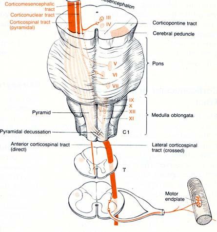
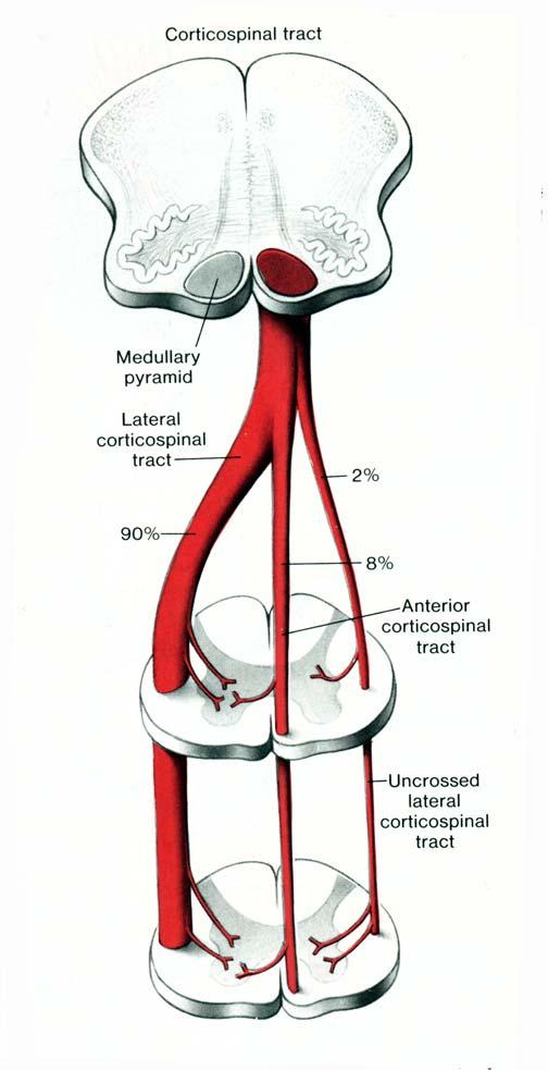
 Descending Tracts I: To describe the pyramidal and extrapyramidal tracts. II: To discuss the functions of the descending tracts. III: To define the upper and the lower motor neurons. 1. The corticonuclear
Voluntary Movements. Lu Chen, Ph.D. MCB, UC Berkeley. Outline. Organization of the motor cortex (somatotopic) Corticospinal projection
 Corticospinal projection") Voluntary Movements Lu Chen, Ph.D. MCB, UC Berkeley 1 Outline Organization of the motor cortex (somatotopic) Corticospinal projection Physiology of motor neurons Direction representation, population coding
Voluntary Movements Lu Chen, Ph.D. MCB, UC Berkeley 1 Outline Organization of the motor cortex (somatotopic) Corticospinal projection Physiology of motor neurons Direction representation, population coding
1. Which part of the brain is responsible for planning and initiating movements?
 Section: Chapter 10: Multiple Choice 1. Which part of the brain is responsible for planning and initiating movements? p.358 frontal lobe hippocampus basal ganglia cerebellum 2. The prefrontal cortex is
Sensory Pathways & Somatic Nervous System. Chapter 15
 Sensory Pathways & Somatic Nervous System Chapter 15 How Does Brain Differentiate Sensations? Pain impulses make brain aware of injuries and infections. Impulses from eye, ear, nose and tongue make brain
Clinical Learning Exercise #1
 Clinical Learning Exercise #1 Exercise: We are going to assume nothing is wrong with the peripheral nervous system and attempt to identify the central nervous system anatomical location for the following
skilled pathways: distal somatic muscles (fingers, hands) (brainstem, cortex) are giving excitatory signals to the descending pathway
 (brainstem, cortex) are giving excitatory signals to the descending pathway") L15 - Motor Cortex General - descending pathways: how we control our body - motor = somatic muscles and movement (it is a descending motor output pathway) - two types of movement: goal-driven/voluntary
L15 - Motor Cortex General - descending pathways: how we control our body - motor = somatic muscles and movement (it is a descending motor output pathway) - two types of movement: goal-driven/voluntary
Cerebrum-Cerebral Hemispheres. Cuneyt Mirzanli Istanbul Gelisim University
 Cerebrum-Cerebral Hemispheres Cuneyt Mirzanli Istanbul Gelisim University The largest part of the brain. Ovoid shape. Two incompletely separated cerebral hemispheres. The outer surface of the cerebral
Homework Week 2. PreLab 2 HW #2 Synapses (Page 1 in the HW Section)
") Homework Week 2 Due in Lab PreLab 2 HW #2 Synapses (Page 1 in the HW Section) Reminders No class next Monday Quiz 1 is @ 5:30pm on Tuesday, 1/22/13 Study guide posted under Study Aids section of website
Homework Week 2 Due in Lab PreLab 2 HW #2 Synapses (Page 1 in the HW Section) Reminders No class next Monday Quiz 1 is @ 5:30pm on Tuesday, 1/22/13 Study guide posted under Study Aids section of website
KINE 4500 Neural Control of Movement. Lecture #1:Introduction to the Neural Control of Movement. Neural control of movement
 KINE 4500 Neural Control of Movement Lecture #1:Introduction to the Neural Control of Movement Neural control of movement Kinesiology: study of movement Here we re looking at the control system, and what
The Nervous System: Sensory and Motor Tracts of the Spinal Cord
 15 The Nervous System: Sensory and Motor Tracts of the Spinal Cord PowerPoint Lecture Presentations prepared by Steven Bassett Southeast Community College Lincoln, Nebraska Introduction Millions of sensory
Lecturer. Prof. Dr. Ali K. Al-Shalchy MBChB/ FIBMS/ MRCS/ FRCS 2014
 Lecturer Prof. Dr. Ali K. Al-Shalchy MBChB/ FIBMS/ MRCS/ FRCS 2014 Dorsal root: The dorsal root carries both myelinated and unmyelinated afferent fibers to the spinal cord. Posterior gray column: Long
Role of brainstem in somatomotor (postural) functions
 functions") Role of brainstem in somatomotor (postural) functions (vestibular apparatus) The muscle tone and its regulation VESTIBULAR SYSTEM (Equilibrium) Receptors: Otolith organs Semicircular canals Sensation (information):
Role of brainstem in somatomotor (postural) functions (vestibular apparatus) The muscle tone and its regulation VESTIBULAR SYSTEM (Equilibrium) Receptors: Otolith organs Semicircular canals Sensation (information):
-Zeina Assaf. -Omar Odeh. - Maha Beltagy
 -3 -Zeina Assaf -Omar Odeh - Maha Beltagy 1 P a g e The Inferior Surface Of The Brain The inferior surface of the brain is divide by the stem of the lateral fissure into 2 parts : The orbital surface and
The Central Nervous System I. Chapter 12
 The Central Nervous System I Chapter 12 The Central Nervous System The Brain and Spinal Cord Contained within the Axial Skeleton Brain Regions and Organization Medical Scheme (4 regions) 1. Cerebral Hemispheres
Degree of freedom problem
 KINE 4500 Neural Control of Movement Lecture #1:Introduction to the Neural Control of Movement Neural control of movement Kinesiology: study of movement Here we re looking at the control system, and what
Motor systems III: Cerebellum April 16, 2007 Mu-ming Poo
 Motor systems III: Cerebellum April 16, 2007 Mu-ming Poo Population coding in the motor cortex Overview and structure of cerebellum Microcircuitry of cerebellum Function of cerebellum -- vestibulo-ocular
The motor system. To move things is all that mankind can do whether in whispering a syllable or in felling a forest C.
 The motor system To move things is all that mankind can do whether in whispering a syllable or in felling a forest C. Sherrington 1920 Principles Components: Muscles, Spinal cord and spinal tracts, Subcortical
Lecture X. Brain Pathways: Movement!
 Bio 3411 Readings (background only) Bio 3411 Monday Neuroscience 4 th ed Page(s) Feature 423-451Upper motor control of Brain Stem and Spinal Cord The Brain Atlas 3 rd ed Page(s) Feature 198-199 Vestibular
Lecture X. Brain Pathways: Movement!
 Bio 3411 Monday 1 Readings (background only) Neuroscience 5 th ed Page(s) Feature 353-398Upper motor control of Brain Stem and Spinal Cord Neuroscience 4 th ed Page(s) Feature 423-451Upper motor control
Cortical Organization. Functionally, cortex is classically divided into 3 general types: 1. Primary cortex:. - receptive field:.
 Cortical Organization Functionally, cortex is classically divided into 3 general types: 1. Primary cortex:. - receptive field:. 2. Secondary cortex: located immediately adjacent to primary cortical areas,
Thalamus and Sensory Functions of Cerebral Cortex
 Thalamus and Sensory Functions of Cerebral Cortex I: To describe the functional divisions of thalamus. II: To state the functions of thalamus and the thalamic syndrome. III: To define the somatic sensory
Voluntary Movement. Ch. 14: Supplemental Images
 Voluntary Movement Ch. 14: Supplemental Images Skeletal Motor Unit: The basics Upper motor neuron: Neurons that supply input to lower motor neurons. Lower motor neuron: neuron that innervates muscles,
Basal nuclei, cerebellum and movement
 Basal nuclei, cerebellum and movement MSTN121 - Neurophysiology Session 9 Department of Myotherapy Basal Nuclei (Ganglia) Basal Nuclei (Ganglia) Role: Predict the effects of various actions, then make
Chapter 3. Structure and Function of the Nervous System. Copyright (c) Allyn and Bacon 2004
 Allyn and Bacon 2004") Chapter 3 Structure and Function of the Nervous System 1 Basic Features of the Nervous System Neuraxis: An imaginary line drawn through the center of the length of the central nervous system, from the
Chapter 3 Structure and Function of the Nervous System 1 Basic Features of the Nervous System Neuraxis: An imaginary line drawn through the center of the length of the central nervous system, from the
Cognitive Modelling Themes in Neural Computation. Tom Hartley
 Cognitive Modelling Themes in Neural Computation Tom Hartley t.hartley@psychology.york.ac.uk Typical Model Neuron x i w ij x j =f(σw ij x j ) w jk x k McCulloch & Pitts (1943), Rosenblatt (1957) Net input:
Rules of apparent motion: The shortest-path constraint: objects will take the shortest path between flashed positions.
 Rules of apparent motion: The shortest-path constraint: objects will take the shortest path between flashed positions. The box interrupts the apparent motion. The box interrupts the apparent motion.
Exam 1 PSYC Fall 1998
 Exam 1 PSYC 2022 Fall 1998 (2 points) Briefly describe the difference between a dualistic and a materialistic explanation of brain-mind relationships. (1 point) True or False. George Berkely was a monist.
Parietofrontal Circuits for Action and Space Perception in the Macaque Monkey
 NeuroImage 14, S27 S32 (2001) doi:10.1006/nimg.2001.0835, available online at http://www.idealibrary.com on Parietofrontal Circuits for Action and Space Perception in the Macaque Monkey Massimo Matelli
Anatomy and Physiology (Bio 220) The Brain Chapter 14 and select portions of Chapter 16
 The Brain Chapter 14 and select portions of Chapter 16") Anatomy and Physiology (Bio 220) The Brain Chapter 14 and select portions of Chapter 16 I. Introduction A. Appearance 1. physical 2. weight 3. relative weight B. Major parts of the brain 1. cerebrum 2.
Anatomy and Physiology (Bio 220) The Brain Chapter 14 and select portions of Chapter 16 I. Introduction A. Appearance 1. physical 2. weight 3. relative weight B. Major parts of the brain 1. cerebrum 2.
Biological Bases of Behavior. 8: Control of Movement
 Biological Bases of Behavior 8: Control of Movement m d Skeletal Muscle Movements of our body are accomplished by contraction of the skeletal muscles Flexion: contraction of a flexor muscle draws in a
Notes: Organization. Anatomy of the Nervous System. Cerebral cortex. Cortical layers. PSYC 2: Biological Foundations - Fall Professor Claffey
 PSYC 2: Biological Foundations - Fall 2012 - Professor Claffey Notes: Organization Version: 10/30/12 - original version Anatomy of the Nervous System Content covered in Hans's lecture: CNS & PNS Directions/Planes
Chapter 8. Control of movement
 Chapter 8 Control of movement 1st Type: Skeletal Muscle Skeletal Muscle: Ones that moves us Muscles contract, limb flex Flexion: a movement of a limb that tends to bend its joints, contraction of a flexor
Motor systems.... the only thing mankind can do is to move things... whether whispering or felling a forest. C. Sherrington
 Motor systems... the only thing mankind can do is to move things... whether whispering or felling a forest. C. Sherrington 1 Descending pathways: CS corticospinal; TS tectospinal; RS reticulospinal; VS
CEREBRUM Dr. Jamila Elmedany Dr. Essam Eldin Salama
 CEREBRUM Dr. Jamila Elmedany Dr. Essam Eldin Salama Objectives At the end of the lecture, the student should be able to: List the parts of the cerebral hemisphere (cortex, medulla, basal nuclei, lateral
Chapter 14: Integration of Nervous System Functions I. Sensation.
 Chapter 14: Integration of Nervous System Functions I. Sensation A. General Organization 1. General senses have receptors a. The somatic senses provide information about & 1. Somatic senses include: a.
1) Drop off in the Bi 150 box outside Baxter 331 or to the head TA (jcolas).
 Drop off in the Bi 150 box outside Baxter 331 or to the head TA (jcolas).") Bi/CNS/NB 150 Problem Set 5 Due: Tuesday, Nov. 24, at 4:30 pm Instructions: 1) Drop off in the Bi 150 box outside Baxter 331 or e-mail to the head TA (jcolas). 2) Submit with this cover page. 3) Use a
Bi/CNS/NB 150 Problem Set 5 Due: Tuesday, Nov. 24, at 4:30 pm Instructions: 1) Drop off in the Bi 150 box outside Baxter 331 or e-mail to the head TA (jcolas). 2) Submit with this cover page. 3) Use a
A3.1.7 Motor Control. 10 November 2016 Institute of Psychiatry,Psychology and Neuroscience Marinela Vavla
 A3.1.7 Motor Control 10 November 2016 Institute of Psychiatry,Psychology and Neuroscience Marinela Vavla marinela.vavla@kcl.ac.uk Learning objectives Motor systems: components & organization Spinal cord
Announcement. Danny to schedule a time if you are interested.
 Announcement If you need more experiments to participate in, contact Danny Sanchez (dsanchez@ucsd.edu) make sure to tell him that you are from LIGN171, so he will let me know about your credit (1 point).
PHY3111 Mid-Semester Test Study. Lecture 2: The hierarchical organisation of vision
 PHY3111 Mid-Semester Test Study Lecture 2: The hierarchical organisation of vision 1. Explain what a hierarchically organised neural system is, in terms of physiological response properties of its neurones.
CEREBRUM. Dr. Jamila EL Medany
 CEREBRUM Dr. Jamila EL Medany Objectives At the end of the lecture, the student should be able to: List the parts of the cerebral hemisphere (cortex, medulla, basal nuclei, lateral ventricle). Describe
The Frontal Lobes. Anatomy of the Frontal Lobes. Anatomy of the Frontal Lobes 3/2/2011. Portrait: Losing Frontal-Lobe Functions. Readings: KW Ch.
 The Frontal Lobes Readings: KW Ch. 16 Portrait: Losing Frontal-Lobe Functions E.L. Highly organized college professor Became disorganized, showed little emotion, and began to miss deadlines Scores on intelligence
Pathways of proprioception
 The Autonomic Nervous Assess Prof. Fawzia Al-Rouq Department of Physiology College of Medicine King Saud University Pathways of proprioception System posterior column& Spinocerebellar Pathways https://www.youtube.com/watch?v=pmeropok6v8
Dr. Farah Nabil Abbas. MBChB, MSc, PhD
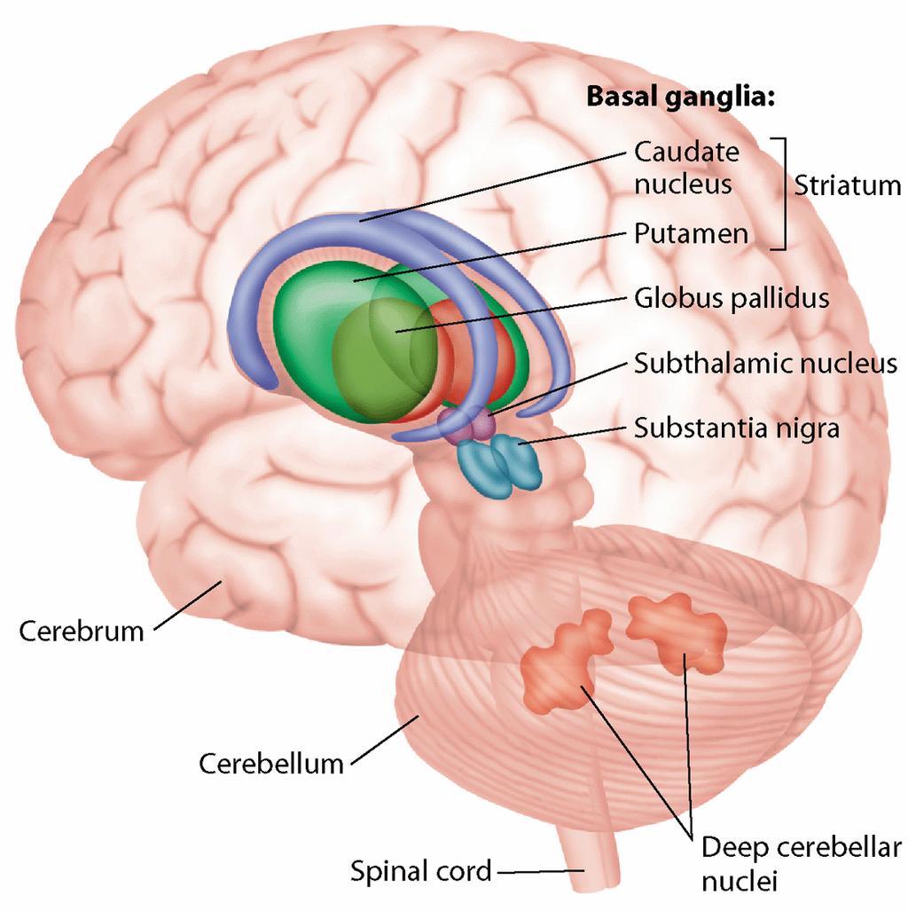
 Dr. Farah Nabil Abbas MBChB, MSc, PhD The Basal Ganglia *Functions in association with motor cortex and corticospinal pathways. *Regarded as accessory motor system besides cerebellum. *Receive most of
Neural Integration I: Sensory Pathways and the Somatic Nervous System
 15 Neural Integration I: Sensory Pathways and the Somatic Nervous System PowerPoint Lecture Presentations prepared by Jason LaPres Lone Star College North Harris An Introduction to Sensory Pathways and
CHAPTER 10 THE SOMATOSENSORY SYSTEM
 CHAPTER 10 THE SOMATOSENSORY SYSTEM 10.1. SOMATOSENSORY MODALITIES "Somatosensory" is really a catch-all term to designate senses other than vision, hearing, balance, taste and smell. Receptors that could
Unit VIII Problem 5 Physiology: Cerebellum
 Unit VIII Problem 5 Physiology: Cerebellum - The word cerebellum means: the small brain. Note that the cerebellum is not completely separated into 2 hemispheres (they are not clearly demarcated) the vermis
SAMPLE EXAMINATION QUESTIONS
 SAMPLE EXAMINATION QUESTIONS PLEASE NOTE, THE QUESTIONS BELOW SAMPLE THE ENTIRE LECTURE COURSE AND THEREORE INCLUDE QUESTIONS ABOUT TOPICS THAT WE HAVE NOT YET COVERED IN CLASS. 1. Which of the following
Brainstem. Steven McLoon Department of Neuroscience University of Minnesota
 Brainstem Steven McLoon Department of Neuroscience University of Minnesota 1 Course News Change in Lab Sequence Week of Oct 2 Lab 5 Week of Oct 9 Lab 4 2 Goal Today Know the regions of the brainstem. Know
Note: Waxman is very sketchy on today s pathways and nonexistent on the Trigeminal.
 Dental Neuroanatomy Thursday, February 3, 2011 Suzanne Stensaas, PhD Note: Waxman is very sketchy on today s pathways and nonexistent on the Trigeminal. Resources: Pathway Quiz for HyperBrain Ch. 5 and
Essentials of Human Anatomy & Physiology. Seventh Edition. The Nervous System. Copyright 2003 Pearson Education, Inc. publishing as Benjamin Cummings
 Essentials of Human Anatomy & Physiology Seventh Edition The Nervous System Copyright 2003 Pearson Education, Inc. publishing as Benjamin Cummings Functions of the Nervous System 1. Sensory input gathering
Cerebral Cortex 1. Sarah Heilbronner
 Cerebral Cortex 1 Sarah Heilbronner heilb028@umn.edu Want to meet? Coffee hour 10-11am Tuesday 11/27 Surdyk s Overview and organization of the cerebral cortex What is the cerebral cortex? Where is each
Circuits & Behavior. Daniel Huber
 Circuits & Behavior Daniel Huber How to study circuits? Anatomy (boundaries, tracers, viral tools) Inactivations (lesions, optogenetic, pharma, accidents) Activations (electrodes, magnets, optogenetic)
Somatosensation. Recording somatosensory responses. Receptive field response to pressure
 Somatosensation Mechanoreceptors that respond to touch/pressure on the surface of the body. Sensory nerve responds propotional to pressure 4 types of mechanoreceptors: Meissner corpuscles & Merkel discs
LISC-322 Neuroscience Cortical Organization
 LISC-322 Neuroscience Cortical Organization THE VISUAL SYSTEM Higher Visual Processing Martin Paré Assistant Professor Physiology & Psychology Most of the cortex that covers the cerebral hemispheres is
P. Hitchcock, Ph.D. Department of Cell and Developmental Biology Kellogg Eye Center. Wednesday, 16 March 2009, 1:00p.m. 2:00p.m.
 Normal CNS, Special Senses, Head and Neck TOPIC: CEREBRAL HEMISPHERES FACULTY: LECTURE: READING: P. Hitchcock, Ph.D. Department of Cell and Developmental Biology Kellogg Eye Center Wednesday, 16 March
LISC-322 Neuroscience. Visual Field Representation. Visual Field Representation. Visual Field Representation. Visual Field Representation
 LISC-3 Neuroscience THE VISUAL SYSTEM Central Visual Pathways Each eye sees a part of the visual space that defines its visual field. The s of both eyes overlap extensively to create a binocular. eye both
Key questions about attention
 Key questions about attention How does attention affect behavioral performance? Can attention affect the appearance of things? How does spatial and feature-based attention affect neuronal responses in
Anatomical Substrates of Somatic Sensation
 Anatomical Substrates of Somatic Sensation John H. Martin, Ph.D. Center for Neurobiology & Behavior Columbia University CPS The 2 principal somatic sensory systems: 1) Dorsal column-medial lemniscal system
HEAD AND NECK PART 2
 HEAD AND NECK PART 2 INTEGRATED CURRICULUM = Integrate Basic Science and Clinical Training 1- ENT PATIENT EXAM IN ICS COURSE - Today and next week - Review/Preview Anatomy underlying ENT exam 2- NEUROANATOMY/NEUROLOGY
Decoding a Perceptual Decision Process across Cortex
 Article Decoding a Perceptual Decision Process across Cortex Adrián Hernández, 1 Verónica Nácher, 1 Rogelio Luna, 1 Antonio Zainos, 1 Luis Lemus, 1 Manuel Alvarez, 1 Yuriria Vázquez, 1 Liliana Camarillo,
Supporting Information
 Supporting Information Lingnau et al. 10.1073/pnas.0902262106 Fig. S1. Material presented during motor act observation (A) and execution (B). Each row shows one of the 8 different motor acts. Columns in
Telencephalon (Cerebral Hemisphere)
") Telencephalon (Cerebral Hemisphere) OUTLINE The Cortex - Lobes, Sulci & Gyri - Functional Subdivisions - Limbic Lobe & Limbic System The Subcortex - Basal Ganglia - White Matter (Internal Capsule) - Relations
Telencephalon (Cerebral Hemisphere) OUTLINE The Cortex - Lobes, Sulci & Gyri - Functional Subdivisions - Limbic Lobe & Limbic System The Subcortex - Basal Ganglia - White Matter (Internal Capsule) - Relations
Auditory and Vestibular Systems
 Auditory and Vestibular Systems Objective To learn the functional organization of the auditory and vestibular systems To understand how one can use changes in auditory function following injury to localize
SENSORY (ASCENDING) SPINAL TRACTS
 SPINAL TRACTS") SENSORY (ASCENDING) SPINAL TRACTS Dr. Jamila El-Medany Dr. Essam Eldin Salama OBJECTIVES By the end of the lecture, the student will be able to: Define the meaning of a tract. Distinguish between the different
SENSORY (ASCENDING) SPINAL TRACTS Dr. Jamila El-Medany Dr. Essam Eldin Salama OBJECTIVES By the end of the lecture, the student will be able to: Define the meaning of a tract. Distinguish between the different
Fig.1: A, Sagittal 110x110 mm subimage close to the midline, passing through the cingulum. Note that the fibers of the corpus callosum run at a
 Fig.1 E Fig.1:, Sagittal 110x110 mm subimage close to the midline, passing through the cingulum. Note that the fibers of the corpus callosum run at a slight angle are through the plane (blue dots with
The neurvous system senses, interprets, and responds to changes in the environment. Two types of cells makes this possible:
 NERVOUS SYSTEM The neurvous system senses, interprets, and responds to changes in the environment. Two types of cells makes this possible: the neuron and the supporting cells ("glial cells"). Neuron Neurons
b. The groove between the two crests is called 2. The neural folds move toward each other & the fuse to create a
 Chapter 13: Brain and Cranial Nerves I. Development of the CNS A. The CNS begins as a flat plate called the B. The process proceeds as: 1. The lateral sides of the become elevated as waves called a. The
General Sensory Pathways of the Trunk and Limbs
 General Sensory Pathways of the Trunk and Limbs Lecture Objectives Describe gracile and cuneate tracts and pathways for conscious proprioception, touch, pressure and vibration from the limbs and trunk.
Objectives Neurophysiology of brain area related to movement and motor control
 Objectives Neurophysiology of brain area related to movement and motor control 1. Ascending pathways (sensory input) 2. Sensory input treatment, and thalamo-cortical & cortico-thalamic filter 3. Sensory
The origins of localization
 Association Cortex, Asymmetries, and Cortical Localization of Affective and Cognitive Functions Michael E. Goldberg, M.D. The origins of localization The concept that different parts of the brain did different
Neuroscience Tutorial
 Neuroscience Tutorial Brain Organization : cortex, basal ganglia, limbic lobe : thalamus, hypothal., pituitary gland : medulla oblongata, midbrain, pons, cerebellum Cortical Organization Cortical Organization
Attention: Neural Mechanisms and Attentional Control Networks Attention 2
 Attention: Neural Mechanisms and Attentional Control Networks Attention 2 Hillyard(1973) Dichotic Listening Task N1 component enhanced for attended stimuli Supports early selection Effects of Voluntary
Introduction to the Nervous System. Code: HMP 100/ UPC 103/ VNP 100. Course: Medical Physiology. Level 1 MBChB/BDS/BPharm
 Introduction to the Nervous System. Code: HMP 100/ UPC 103/ VNP 100. Course: Medical Physiology Level 1 MBChB/BDS/BPharm Lecture 2. Functional Organisation of the Nervous System Lecture Outline 1.1 Introduction
Association Cortex, Asymmetries, and Cortical Localization of Affective and Cognitive Functions. Michael E. Goldberg, M.D.
 Association Cortex, Asymmetries, and Cortical Localization of Affective and Cognitive Functions Michael E. Goldberg, M.D. The origins of localization The concept that different parts of the brain did different
Biology 218 Human Anatomy
 Chapter 21 Adapted form Tortora 10 th ed. LECTURE OUTLINE A. Overview of Sensations (p. 652) 1. Sensation is the conscious or subconscious awareness of external or internal stimuli. 2. For a sensation
SOMATIC SENSATION PART I: ALS ANTEROLATERAL SYSTEM (or SPINOTHALAMIC SYSTEM) FOR PAIN AND TEMPERATURE
 FOR PAIN AND TEMPERATURE") Dental Neuroanatomy Thursday, February 3, 2011 Suzanne S. Stensaas, PhD SOMATIC SENSATION PART I: ALS ANTEROLATERAL SYSTEM (or SPINOTHALAMIC SYSTEM) FOR PAIN AND TEMPERATURE Reading: Waxman 26 th ed, :
Dental Neuroanatomy Thursday, February 3, 2011 Suzanne S. Stensaas, PhD SOMATIC SENSATION PART I: ALS ANTEROLATERAL SYSTEM (or SPINOTHALAMIC SYSTEM) FOR PAIN AND TEMPERATURE Reading: Waxman 26 th ed, :
PSYC& 100: Biological Psychology (Lilienfeld Chap 3) 1
 1") PSYC& 100: Biological Psychology (Lilienfeld Chap 3) 1 1 What is a neuron? 2 Name and describe the functions of the three main parts of the neuron. 3 What do glial cells do? 4 Describe the three basic
PSYC& 100: Biological Psychology (Lilienfeld Chap 3) 1 1 What is a neuron? 2 Name and describe the functions of the three main parts of the neuron. 3 What do glial cells do? 4 Describe the three basic
Mechanosensation. Central Representation of Touch. Wilder Penfield. Somatotopic Organization
 Mechanosensation Central Representation of Touch Touch and tactile exploration Vibration and pressure sensations; important for clinical testing Limb position sense John H. Martin, Ph.D. Center for Neurobiology
Chapter 12b. Overview
 Chapter 12b Spinal Cord Overview Spinal cord gross anatomy Spinal meninges Sectional anatomy Sensory pathways Motor pathways Spinal cord pathologies 1 The Adult Spinal Cord About 18 inches (45 cm) long
Cerebral Cortex: Association Areas and Memory Tutis Vilis
 97 Cerebral Cortex: Association Areas and Memory Tutis Vilis a) Name the 5 main subdivisions of the cerebral cortex. Frontal, temporal, occipital, parietal, and limbic (on the medial side) b) Locate the
Nervous System C H A P T E R 2
 Nervous System C H A P T E R 2 Input Output Neuron 3 Nerve cell Allows information to travel throughout the body to various destinations Receptive Segment Cell Body Dendrites: receive message Myelin sheath
THE CENTRAL NERVOUS SYSTE M
 THE CENTRAL NERVOUS SYSTE M Structure and Functio n THIRD EDITIO N PER BRODAL A Brief Survey, x i Studying the Structures and Function of the Nervous System, xii i Animal Experiments Crucial for Progress,
Cognitive Neuroscience Attention
 Cognitive Neuroscience Attention There are many aspects to attention. It can be controlled. It can be focused on a particular sensory modality or item. It can be divided. It can set a perceptual system.
STRUCTURAL ORGANIZATION OF THE NERVOUS SYSTEM
 STRUCTURAL ORGANIZATION OF THE NERVOUS SYSTEM STRUCTURAL ORGANIZATION OF THE BRAIN The central nervous system (CNS), consisting of the brain and spinal cord, receives input from sensory neurons and directs
Cerebellum: little brain. Cerebellum. gross divisions
 Cerebellum The anatomy of the cerebellum and its gross divisions Its principal input and output pathways The organization of the cerebellar cortex Role of climbing vs. mossy fibre input The parallel-fibre/
Anatomy Lab (1) Theoretical Part. Page (2 A) Page (2B)
 Theoretical Part. Page (2 A) Page (2B)") Anatomy Lab (1) This sheet only includes the extra notes for the lab handout regarding the theoretical part, as for the practical part it includes everything the doctor mentioned. Theoretical Part Page
Anatomy Lab (1) This sheet only includes the extra notes for the lab handout regarding the theoretical part, as for the practical part it includes everything the doctor mentioned. Theoretical Part Page
Outline of the next three lectures
 Outline of the next three lectures Lecture 35 Anatomy of the human cerebral cortex gross and microscopic cell types connections Vascular supply of the cerebral cortex Disorders involving the cerebral cortex
CONTROL OF MOVEMENT BY THE BRAIN A. PRIMARY MOTOR CORTEX:
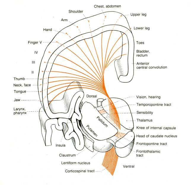
 CONTROL OF MOVEMENT BY THE BRAIN A. PRIMARY MOTOR CORTEX: - responsible for - like somatosensory cortex, primary motor cortex show (motor homunculus) - amount of cortex devoted to different parts of body