Adult attachment predicts maternal brain and oxytocin response to infant cues
|
|
|
- Audra Nichols
- 5 years ago
- Views:
Transcription
1 Adult attachment predicts maternal brain and oxytocin response to infant cues Lane Strathearn, MBBS, FRACP, 1,2,3 Peter Fonagy, PhD, 4,5 Janet Amico, MD, 6 P. Read Montague, PhD 2,5 1 The Meyer Center for Developmental Pediatrics and 2 Human Neuroimaging Laboratory, Department of Neuroscience, One Baylor Plaza, Baylor College of Medicine, Houston, Texas 77030, USA; 3 School of Medicine, The University of Queensland, Queensland 4072 Australia; 4 Research Department of Clinical, Educational and Health Psychology, University College London, Gower St, London WC1E 6BT, UK; 5 Menninger Department of Psychiatry and Behavioral Sciences, One Baylor Plaza, Baylor College of Medicine, Houston, Texas 77030, USA; 6 Department of Medicine, University of Pittsburgh School of Medicine, and Department of Pharmaceutical Sciences, University of Pittsburgh School of Pharmacy, 541A Salk Hall, University of Pittsburgh, 351 Terrace Street, Pittsburgh, Pennsylvania 15261, USA. Correspondence should be addressed to L.S. (lanes@bcm.edu). Running Head: Attachment and Neuroendocrine Response to Infant Cues Word Count: 4564; Abstract Word Count: 150 Figures: 5; Tables: 2 (including 1 Supplementary Table). 1
2 ABSTRACT Infant cues, such as smiling faces, are powerful motivators of human maternal behavior, activating dopamine-associated brain reward circuits. Oxytocin, a neurohormone of attachment, promotes maternal care in animals, although its role in human maternal behavior is unclear. We examined whether differences in attachment security of 30 firsttime mothers were related to brain reward and peripheral oxytocin response to infant cues. On viewing their own infant s smiling and crying faces during functional MRI scanning, mothers with secure attachment showed greater activation of brain reward regions, including the ventral striatum, and the oxytocin-associated hypothalamus/pituitary region. Peripheral oxytocin response to infant contact was also significantly higher in secure mothers, and was positively correlated with brain activation in both regions. Insecure/dismissing mothers showed insular activation in response to their own infant s sad faces. These results suggest that individual differences in maternal attachment may be linked with development of the dopaminergic and oxytocinergic neuroendocrine systems. KEY WORDS Attachment, mother-infant relations, dopamine, oxytocin, reward, functional MRI, maternal. 2
3 INTRODUCTION The attachment relationship between infants and their caregivers is critical for human development, ensuring infant survival and optimal social, emotional and cognitive development (Insel and Young 2001; Sroufe et al. 2005). The relationship between a mother and her infant is particularly salient, with evidence that the biological processes of pregnancy, parturition and lactation may all contribute to the establishment of the mother-infant bond (Strathearn et al. 2009; Kinsley et al. 2008). In both human and animal research, significant differences in early maternal caregiving have been observed ranging from sensitive and responsive infant care to maternally perpetrated abuse or neglect (Strathearn et al. 2009; Sroufe et al. 2005), with corresponding differences in infant health and developmental outcomes (Sroufe et al. 2005; Strathearn et al. 2001; Thompson 2008; Francis et al. 1999; Weaver et al. 2004). Understanding the neurobiological processes underlying these differences in maternal behavior may help us to provide more effective treatment and preventative services. The neurobiology of attachment behavior has been studied extensively in animal models (Insel and Young 2001; Swain et al. 2007), and more recently in humans using functional magnetic resonance imaging (fmri) (Lorberbaum et al. 2002; Bartels and Zeki 2004; Swain et al. 2007; Strathearn et al. 2008). Although there is likely to be a complex interaction of multiple neuroendocrine systems, two specific systems have been shown to consistently play a role in promoting and maintaining maternal behavior: 1) the dopaminergic reward processing system (Champagne et al. 2004; Strathearn et al. 2008; Ferris et al. 2005) and 2) the oxytocinergic system (Bartels and Zeki 2004; Champagne et al. 2001; Levine et al. 2007) Oxytocin, a neuromodulatory hormone produced in the hypothalamus, has well-described central actions associated with the onset of maternal behavior, as well as peripheral actions in stimulating uterine contraction during labor and milk ejection during lactation. It is rapidly released into the 3
4 peripheral circulation following stimuli such as infant suckling, the sight or sound of a nursing mother s infant (Lucas et al. 1980; McNeilly et al. 1983; Johnston and Amico 1986), or even tactile stimulation in rat dams (Uvnas-Moberg et al. 1993). In randomized, placebo-controlled trials, intranasal oxytocin produces a broad range of social effects, including enhanced social memory, eye gaze when viewing faces, increased recognition and memory of facial expressions and identity, and increased manifestations of trust (Domes et al. 2007; Savaskan et al. 2008; Baumgartner et al. 2008; Kosfeld et al. 2005; Guastella et al. 2008b; Guastella et al. 2008a). Oxytocin receptors are located in the ventral striatum, a key dopaminergic brain region, and receptor binding is linked functionally to maternal behavior in the rat (Olazabal and Young 2006a). Thus, oxytocin may link social cues, such as infant facial expressions, with dopamine-associated reinforcement pathways. The extent to which these biological systems explain differences in the quality of human attachment between mothers and infants, is yet to be explored (Strathearn 2006). In this study, we aimed to measure differences in maternal brain reward activation and peripheral oxytocin release in response to infant cues, based on the mother s adult attachment classification. We hypothesized that mothers with secure patterns of adult attachment would show an increased brain response to their own infant s face in mesocorticolimbic reward regions, including the midbrain ventral tegmental area, the ventral striatum and the medial prefrontal cortex, and that this would be true on viewing both happy and sad infant face cues. We also hypothesized that secure mothers would show an enhanced peripheral oxytocin response on interacting with their infants, which would correlate with maternal brain responses. 4
5 MATERIALS AND METHODS Study setting and participants. In this cohort study, we recruited first-time pregnant women during the third trimester of pregnancy from Houston, Texas, and monitored them for 14 months postnatally. Recruitment occurred between August 2004 and April 2006 and was through prenatal clinic visits and advertisements on billboards, in magazines and via the internet. We excluded potential subjects who were on psychotropic medications, using cigarettes during pregnancy, left-handed or had any contraindications to MRI scanning. Research was approved by the Institutional Review Board at Baylor College of Medicine, and all subjects provided written informed consent. Study design (Fig. 1). Visit 1: Pregnancy. During this visit, each enrolled woman participated in a modified version of the Adult Attachment Interview (AAI) (Crittenden 2004; George et al. 1996), a semi-structured 1½-2 hour-long interview involving specified questions and follow-up inquiries relating to childhood relationships with attachment figures, usually parents. The modified version was chosen because of its theoretical links with patterns of information processing in the brain (Strathearn 2006; Crittenden 2008). Each digitally recorded interview was transcribed (with personally identifying details altered to preserve anonymity), and coded blindly to classify each woman s adult attachment pattern, which was not revealed until study completion. During this visit we also collected sociodemographic data, and screening information for depression (Beck Depression Inventory, BDI) (Beck et al. 1996) and personality disorders (Personality Disorder Questionnaire 4+, PDQ) (see Supplementary Table 1 online). We repeated the BDI on each post-natal visit, and calculated a mean post-natal score. Visit 2: Videotaping and oxytocin sampling. Approximately 7 months postdelivery, each mother and infant attended a session at the Human Neuroimaging 5
6 Laboratory. We requested that mothers abstain from caffeine and tobacco for 2-3 hours prior to the visit. After separating from their infants, the mothers had an intravenous cannula inserted, and 20 minutes later had blood drawn for baseline measurements of serum oxytocin, free cortisol, epinephrine and norepinephrine. We also measured serum estradiol, progesterone and β human chorionic gonadotropin levels to exclude a current pregnancy and to assess menstrual status. During this separation period, we videotaped each infant to obtain still images for use in the subsequent fmri visit. Smiling, neutral and crying faces were elicited in a standardized setting, as described elsewhere (Strathearn et al. 2008). The mother and infant were then reunited for a 5-minute "freeplay" period in which they physically interacted on the floor, after which another blood sample was drawn. They then participated in a 6-minute modified still face procedure (Koos and Gergely 2001), during which mother and infant could hear and see each other (via a mirror) but not interact physically. We then obtained a third blood sample after the mother left the room, followed by a final blood draw after 20 minutes of separation. Before and after the interaction period, each mother rated their current feelings using the Positive and Negative Affect Schedule (PANAS) (Watson et al. 1988), a 5-point rating of 20 affect states, such as interested, excited, irritable and nervous. Each mother also completed a 120 item self-report questionnaire, the Parenting Stress Index (PSI) (Abidin 1995), designed to help identify potentially dysfunctional parent-child relations. We assessed adult and infant temperament using the self-report Adult Temperament Questionnaire Short form (ATQ) and the Infant Behavior Questionnaire Revised (IBQ) (Gartstein and Rothbart 2003). The mothers also reported their breastfeeding status, which was repeated at Visit 3. Visit 3: Scanning. At ~11 months post-delivery, a minimum of 3 months after the videotaping session, each mother underwent fmri scanning while viewing 60 unique infant face images, 30 of her own baby and 30 of the matched unknown infant face. 6
7 Each mother viewed randomly presented baby face images for 2 seconds each within a rapid event-related fmri design, with a random inter-stimulus interval of 2, 4 or 6 seconds (Fig. 2). Visual images were generated using a computer controlled LCD projector, and presented to the mother on an overhead mirror display. Visit 4: Child follow-up. Finally, at 14 months of age we performed a general assessment of child development using the Screening Test of the Bayley Scales of Infant and Toddler Development (Bayley 2006). Variables and statistical methods. Predictor variable Adult attachment. We determined each mother s adult attachment classification using the AAI (George et al. 1996; Fonagy et al. 1991) which categorizes the mother s capacity to form secure attachment relationships on the basis of a narrative of her own attachment experience. Over the past 25 years, over 200 studies have reported over 10,000 AAIs (van IJzendoorn and Bakermans-Kranenburg 2009). From both cross-sectional and prospective longitudinal studies, adult attachment has been shown to reliably predict maternal behavior patterns, the development of infant attachment (van IJzendoorn 1995), and infant social and emotional development (Sroufe et al. 2005). We chose to measure attachment during pregnancy using a longitudinal design to preclude the possibility that the infant s temperament or mother-infant interaction patterns might influence the way the mother discusses her own attachment experiences. The coding is based on the subject s coherence and consistency in describing attachment-related experiences and their effects on current functioning (Crittenden 2004). The 3 basic styles, which parallel Ainsworth s original classification of attachment in infancy (Ainsworth and Bell 1970) include Type A Insecure/Dismissing, Type B Secure and Type C Insecure/Preoccupied. Individuals with Type B attachment styles tend to provide balanced descriptions of childhood experiences, using both 7
8 temporal/causal order and affect to describe both positive and negative events and feeling states. Individuals with Type A attachment tend to describe events or feelings in more cognitive terms, avoiding or inhibiting displays of negative affect. In contrast, Type C individuals exaggerate affective responses, with omitted or distorted cognitive processing (Crittenden 2008). Fifty percent of the transcripts were double coded to ensure reliability, with an 87% agreement with regard to a 4-group classification (kappa=0.78). Discrepancies were resolved through conferencing between coders. Potential confounding variables. We measured a variety of socioeconomic and behavioral factors to compare the characteristics of women in the two attachment groups (see Supplementary Table 1 online). Continuous measures were evaluated using t- tests or the Mann-Whitney U-test for nonparametric data (as determined from histogram analysis). We compared categorical variables using the Chi-Square test, or Fisher Exact test when numbers were insufficient. We used the Kendall s tau-b test for ordinal or ranked nonparametric variables. We compared PANAS ratings of the mothers affect before and after contact with their infants between groups using a repeated measure analysis of variance (ANOVA). Serial measurements of cortisol, norepinephrine and epinephrine were also compared between attachment groups using linear mixed modeling. Analyses were performed using SPSS (version 15.0) and P < 0.05 (2-tailed) was considered statistically significant. Outcome Variables. 1) Oxytocin Response. We used linear mixed modeling to assess the effects of attachment group, interaction time point, breastfeeding status, and all 2-way interactions, on oxytocin response. Residual plots were used to confirm normality of distribution. Cases with missing data points were excluded (one Type B and two Type A subjects). The difference in mean oxytocin concentration between attachment groups, at each time point, was compared using a z-test (with Bonferroni 8
9 correction for multiple comparisons; alpha was considered statistically significant). The mean oxytocin concentration from the two mother-infant interaction time points (which were highly correlated: r S =0.77, P < 0.001) was recomputed as a percentage change from the first baseline measure, to provide a single index for correlation with fmri data. To determine the correlation between % change in oxytocin and fmri blood-oxygen-level-dependent (BOLD) activation measured 4 months later (ztransformed beta weights), we calculated a Spearman correlation coefficient. We used a Bonferroni correction to adjust the alpha level for multiple comparisons with differing beta weights for the 6 types of infant face (own-happy, unknown-neutral, etc). An alpha < was considered statistically significant. We measured oxytocin concentrations using a sensitive and specific liquid phase radioimmunoassay, in which oxytocin antiserum does not cross-react with arginine vasopressin or other oxytocin-like peptides (Amico et al. 1985). The lower limit for detectability of the assay is 0.5pg ml -1, inter- and intra-assay coefficients of variation are <10%. 2) Functional MRI Brain Response. We prepared thirty standardized face images from each infant (10 happy, 10 neutral and 10 sad) for use in the fmri scanning paradigm, along with 30 images from an unknown baby which were matched on age, race and independently-coded degree of affect (Fig. 2) (Strathearn et al. 2008). To ensure that the degree of infant facial affect did not vary between attachment groups, all faces were re-coded by three blinded raters using the 9-point Self-Assessment Manikin (Bradley and Lang 1994) (ICC = 0.90). Using a mixed model three-way ANOVA, we saw no main effects for attachment group (F 2,28 = 1.9, NS) or order of presentation (Wilk s Lamda =.502, F 9,20 = 2.0, NS). Similarly, none of the interactions with attachment security were significant. 9
10 Imaging was performed using a 3 Tesla Siemens Allegra head-only MRI scanner. High-resolution T1-weighted structural images (192 slices, in plane resolution 256 x 256; field of view [FOV] 245 mm; slice thickness 1 mm) were first acquired, followed by whole-brain functional runs of around 185 scans (gradient recalled echo planar imaging; 37 slices; repetition time 2000 msec; echo time 25 msec; flip angle, 90 ; 64 x 64 matrix [in plane resolution]; FOV 220 mm; slice thickness 3 mm; positioned at 30 degrees in the axial plane to the anterior commissure/posterior commissure line). Imaging data for each subject were preprocessed in BrainVoyager QX, version and analyzed in version , as previously described (Strathearn et al. 2008). A BrainVoyager protocol file was created for each functional run, representing the timing of each stimulus event. Each predictor was then convolved using a doublegamma hemodynamic response function. Using the General Linear Model (GLM), effects for the whole group (n = 30) and for each attachment subgroup (n = 15) were evaluated separately using a random effects analysis between subjects, and fixed effects for individual within factors. After specifying a particular contrast in stimulus types (e.g. own-happy vs. unknown-happy or own-sad vs. unknown-sad), a group t-map was generated onto a template 3-dimensional anatomical image. An activation map threshold was determined using a false discovery rate (FDR) of 5% to control for multiple comparisons, and a cluster threshold of 4 voxels. Smaller cluster thresholds were also examined in the striatum (3 voxels) and brainstem (1 voxel) to reveal activation of smaller nuclei. Anatomical regions were identified using the automated Talairach Daemon (Lancaster et al. 2000), and confirmed manually using a human brain atlas (Mai et al. 2004). Next, we compared activation patterns between attachment groups using an 2- factor random effects ANOVA model, with infant face category as a repeated measure within-factor variable and attachment group as a between-factor, assessing whole 10
11 brain differences in activation using a threshold of q(fdr) = Percentage BOLD signal change was calculated between the 2 attachment groups, in a priori regions of interest (midbrain, striatum, prefrontal cortex and hypothalamus/pituitary). 11
12 RESULTS Description of subjects Of 112 women recruited during pregnancy, 61 met eligibility criteria and were enrolled in the study, with 44 participating in fmri scanning. Ten women were unable to be scanned (9 due to a current pregnancy and one because of a past history of seizures) and 7 had withdrawn from the study or were lost to follow-up. Of the 44 scanned women, 15 were classified as having insecure/dismissing attachment (Type A). A further 16 women demonstrated secure patterns of attachment, without unresolved trauma or loss (Type B). A small group (n = 4) were classified as insecure/preoccupied (Type C), with the remaining 9 women having combined or atypical patterns. We specifically compared women from the two predominant attachment groups Type A and B. To ensure equal numbers in each group, one Type B mother was excluded. At Visit 1, the 30 women in this study were generally from middle to high socioeconomic backgrounds (based on the Four-Factor Index of Social Status [A. B. Hollingshead, PhD, working paper, 1985]: mean score 51.4 ± 9.4). Eighty percent had completed a college or graduate degree and 70% were married at the time of enrollment. The median WTAR-predicted IQ for the group was 112 (range ). Sixty percent identified themselves as non-hispanic White, one-quarter were Hispanic and one-tenth African American. Subjects within the two attachment groups did not differ in age, race, education, socioeconomic status, marital status or predicted IQ (see Methods and Supplementary Table 1 online). Both groups were also comparable in screening measures of personality disorder risk and parenting stress (at Visit 2) and depression (measured at each study visit). There were no significant differences seen in temperament subscales of either the mother or child, mothers ratings of emotions before and after mother-infant interaction (based on the PANAS during Visit 2) (Watson et al. 1988) or in scales of 12
13 infant development (measured during Visit 4). We also found no significant difference in breastfeeding status at Visits 2 or 3, although Type B mothers tended to breastfeed longer and Type A mothers were significantly more likely to be separated from their child for longer periods of time each week (P = 0.03). Oxytocin response to mother-infant interaction (Visit 2) During the 7 month postpartum visit, Type B mothers showed a significantly higher peripheral oxytocin response following periods of mother-infant interaction (Fig. 3a; time point by attachment group interaction effect adjusted for breastfeeding at this visit, F = 2.9, P = 0.04). Although there were no differences between attachment groups in the two baseline measurements, after the 5-minute free-play interaction Type B mothers had significantly higher oxytocin levels (P = 0.01). This difference persisted into an additional mirror-based interaction period, although it was no longer statistically significant (P = 0.07). There were no significant differences in serum free cortisol, epinephrine or norepinephrine, or in baseline serum estradiol or progesterone. Whole group analysis of maternal brain responses (Visit 3) On the whole brain analysis, when mothers viewed their own infant s happy faces, compared to unknown happy faces, key dopamine-associated reward processing regions were activated, overlapping previously reported regions (Strathearn et al. 2008) including the substantia nigra, dorsal putamen and thalamic nuclei. In addition, activation was seen in the ventral striatum, caudate nuclei, insular cortex, superior temporal gyrus and pre- and post-central gyri (P < 0. 05, FDR corrected). As in the prior study, no significant activation was seen on contrasting own vs. unknown sad or neutral infant faces. Combining all affect groups together, contrasting own vs. unknown faces, an activation pattern overlapping our previous study results (Strathearn et al. 2008) was 13
14 again seen, including both mesocorticolimbic (ventral tegmental area and ventral striatum) and nigrostriatal pathway (substantia nigra and dorsal striatum) activation, but without activation of the prefrontal or anterior cingulate cortex. Attachment group comparisons We next compared own vs. unknown infant face responses between the two attachment groups, to look specifically for hypothesized differences in activation of dopamineassociated brain reward regions (in the midbrain, striatum and forebrain) and the hypothalamus/pituitary region. When comparing happy and sad faces separately, between attachment groups, no significant differences in activation were seen on whole brain analysis (using an alpha of 0.05, FDR corrected). However, when all affect groups were combined, Type B mothers showed significantly more activation in the lateral prefrontal cortex bilaterally and the left medial prefrontal cortex (P < 0.05, FDR corrected). In addition, the hypothalamus/pituitary region was strongly activated in Type B mothers (Table 1; Fig. 3b), with the activation signal in response to neutral own-infant faces significantly correlated with the mother s peripheral oxytocin response on interaction with her infant (z-transformed beta weights and % change in oxytocin; r S =0.60, P = 0.001) (Fig. 3c). When attachment groups were compared in this correlation analysis, no differences in line slope (P = 0.80) or position (P = 0.12) were detected. No correlation was seen between oxytocin response and brain activation when viewing unknown infant faces, or any face type in the medial prefrontal cortex (mpfc). In post-hoc analyses, we then compared own-infant faces, in each affect state, directly between attachment groups (e.g. own-happy in Type A vs. Type B), without the inclusion of unknown infant face comparisons. From the hypothesized regions of interest, for the happy face contrast, significantly greater activation was seen in the ventral striatum for Type B mothers, as well as the orbitofrontal cortex (OFC) and mpfc 14
15 bilaterally (Table 2). An equal but opposite BOLD response was seen in Type A mothers in the ventral striatum (Fig. 4a). In the mpfc, Type B mothers had a much larger increase in percent signal change compared with Type A mothers. Type A mothers showed significantly more activation in the dorsolateral prefrontal cortex (dlpfc) bilaterally, compared with Type B mothers, in response to both happy and sad owninfant faces. In response to own infant sad faces, the right ventral striatum was also more active in Type B mothers (Table 2; Fig. 4b). Type A mothers again showed more activation of the dlpfc in response to own-sad faces, as well as a much stronger activation signal in the anterior insula bilaterally, compared with Type B mothers (Fig. 4b). Activation in the right ventral striatum to neutral own-infant faces was also highly correlated with peripheral oxytocin response (r S = 0.57, P = 0.002; Fig. 5). Unknowninfant faces produced no such correlation. None of these contrasts, for happy or sad faces, showed significant differences in activation of midbrain regions, across attachment groups. Overall, mothers with Type B attachment tended to show greater left hemisphere activation, whereas Type A had predominantly right hemisphere activation, especially for happy and sad infant faces (Table 1). 15
16 DISCUSSION This study demonstrates group differences in maternal brain and oxytocin responses to infant cues, based on adult attachment patterns measured prior to the birth of the mother s first child. As hypothesized, mothers with secure vs. insecure/dismissing attachment showed increased activation of mesocorticolimbic reward brain regions, on viewing their own infant s smiling face. Furthermore, they showed an increased peripheral oxytocin response while interacting with their infants, which was positively correlated with activation of oxytocinergic and dopamine-associated reward processing regions of the brain (hypothalamus/pituitary and ventral striatum). Finally, striking differences in brain activation were seen in response to their own infant s sad facial affect. Securely attached mothers continued to show greater activation in reward processing regions, while insecure/dismissing mothers showed increased activation of the anterior insula, a region associated with feelings of unfairness, pain and disgust (see review, Montague and Lohrenz 2007). The finding of reduced reward activation in mothers with insecure/dismissing attachment is consistent with a recent study of responses to smiling adult faces and positive task feedback (Vrticka et al. 2008), where activation of the ventral striatum was negatively correlated with dismissing attachment scores. In linking attachment security with ventral striatal activation, our findings suggest that for securely attached mothers, infant cues (whether positive or negative in affect) may act as an important signal of incentive salience (Berridge 2007), reinforcing and motivating responsive maternal care. In contrast, mothers with insecure/dismissing attachment styles showed greater activation of dlpfc and anterior insula in response to their own infant s sad face, suggesting cognitive control over a negative affective response (Greene et al. 2004; Sanfey et al. 2003). In line with our current understanding that activation of the anterior 16
17 insula may signal norm violations (Montague and Lohrenz 2007), insecure/dismissing mothers may cognitively appraise their infant s sad affect as a violation of an expected affect state. This may lead to avoidance or rejection of negative infant cues (Sanfey et al. 2003), rather than the approach responses seen in Type B secure mothers. While the ventral striatal activation seen in Type B mothers has been associated with anticipated gain, right anterior insula activation is seen in anticipation of loss (Knutson et al. 2007). These results are consistent with a previously published model of the cortical organization of the attachment system (Strathearn 2006; Crittenden 2008), which postulates that individuals with insecure/dismissing attachment are biased toward cognitive information processing, and tend to inhibit negative affective responses. Although anterior insula activation has also been linked with empathic responses to a loved one s feeling of physical pain (Singer et al. 2004), dismissing individuals score much lower on a scale of emotional empathy (Sonnby-Borgstrom and Jonsson 2004), making this interpretation less likely. Oxytocin has long been implicated as an important neuromodulatory hormone involved in maternal behavior (Insel 1992; Insel and Young 2001). Synthesized in the paraventricular nucleus of the hypothalamus, there are oxytocinergic projections to the posterior pituitary gland where it is released into the blood stream. In addition, oxytocin neurons project centrally to regions important in the manifestation of social and maternal behaviors (Numan 2006). There is some evidence to suggest that oxytocin neurons in the hypothalamus may directly project to the ventral striatum, facilitating dopamine release (Liu and Wang 2003) and thus linking social and maternally-related cues to reward processing and behavioral reinforcement (Insel 2003). Rodent studies have demonstrated that oxytocin receptor binding in the nucleus accumbens (a nucleus of the ventral striatum) facilitates the onset of maternal behavior (Olazabal and Young 2006a; Olazabal and Young 2006b). 17
18 While there has been some controversy surrounding the relationship between peripheral and central oxytocin production (McGregor et al. 2008), these results are consistent with the idea that differences in peripheral oxytocin response may reflect central oxytocin production and contribute to individual differences in maternal caregiving behavior. Other studies have shown reduced peripheral oxytocin responses in cocaine addicted mothers (Light et al. 2004) and in pregnant women with lower maternal-fetal attachment scores (Levine et al. 2007). Furthermore, reduced peripheral oxytocin levels have been seen in orphanage-adopted children with histories of early neglect, who display severe impairments in social reciprocity (Fries et al. 2005). The observation that oxytocin levels are higher in securely attached mothers following interaction with their infants suggests the importance of this neuropeptide in mediating attachment and social behaviors, as seen in human randomized placebo-controlled trials of intranasal oxytocin (Baumgartner et al. 2008; Guastella et al. 2008b), as well as in rodent studies (Insel and Young 2001; Champagne et al. 2001; Insel 1992; Liu and Wang 2003). In our study, the correlation of interaction-elicited peripheral oxytocin with the activation of reward regions in the brain suggests that oxytocin may be one mechanism by which socially-relevant cues activate dopaminergic pathways and thus reinforce behavior. Mothers with secure attachment patterns when interacting with their infants may produce more oxytocin which increases the experience of reward, and in turn may contribute to the mother s ability to provide consistent, nurturant care. One limitation of these findings is that oxytocin measurements during real-time interaction were collected 4 months prior to fmri scanning, providing no opportunity to examine simultaneous correlations. However, the correlation between oxytocin and hemodynamic response across time suggests that the oxytocin response may reflect an enduring trait difference associated with attachment security. 18
19 Numerous previous investigations have shown that mothers with insecure attachment patterns are less likely to establish secure relationships with their children, and that their children tend to have greater difficulties regulating affect, forming peer relationships and establishing secure attachment relationships themselves (Sroufe et al. 2005; van IJzendoorn 1995). While the transgenerational transmission of attachment has been frequently observed, its mechanism is still poorly understood (van IJzendoorn 1995). This study may help shed light on this question, with evidence that secure attachment is associated with more intense maternal reward activation to infant facial expressions, while insecure/dismissing mothers show greater insula response to negative infant cues. Additional research is needed to confirm these findings in larger cohorts of mothers, including mothers with insecure/preoccupied attachment. A randomized controlled trial of intranasal oxytocin may also help to clarify any causal relationship between oxytocin response and maternal brain activation. In conclusion, this study is the first to examine the neuroendocrine basis of human mother-infant attachment. As such, it may help us to better understand the transmission of attachment patterns across generations and how secure maternal attachment may confer developmental advantages on infant development (Sroufe et al. 2005; van IJzendoorn 1995). 19
20 DISCLOSURE / CONFLICT OF INTEREST The authors have no disclosures or conflicts of interest. ACKNOWLEDGEMENTS This research was supported by National Institute of Child Health and Human Development (K23 HD43097), General Clinical Research Center (MO1RR00188), Baylor Child Health Research Center: Pediatrics Mentored Research Program (K12 HD41648) (L. Strathearn); Kane Family Foundation, National Institute of Neurological Disorders and Stroke (NS ), National Institute of Drug Abuse (DA-11723) (P. R. Montague); and a Child and Family Center Program Grant from the Menninger Foundation (P. Fonagy). We would like to thank H. Cai for technical assistance in performing the radioimmunoassays of oxytocin, O. Smith for statistical advice, and technical staff at the Human Neuroimaging Laboratory for assistance with conducting the experiments. 20
21 References 1. Abidin RR (1995): Parenting stress index, professional manual. Psychological Assessment Resources: Lutz, FL. 2. Ainsworth MD, Bell SM (1970). Attachment, exploration, and separation: illustrated by the behavior of one-year-olds in a strange situation. Child Dev 41: Amico JA, Ervin MG, Leake RD, Fisher DA, Finn FM, Robinson AG (1985). A novel oxytocin-like and vasotocin-like peptide in human plasma after administration of estrogen. J Clin Endocrinol Metab 60: Bartels A, Zeki S (2004). The neural correlates of maternal and romantic love. Neuroimage 21: Baumgartner T, Heinrichs M, Vonlanthen A, Fischbacher U, Fehr E (2008). Oxytocin Shapes the Neural Circuitry of Trust and Trust Adaptation in Humans. Neuron 58: Bayley N (2006): Bayley Scales of Infant and Toddler Development. Harcourt Assessment: San Antonio, TX. 7. Beck AT, Steer RA, Brown GK (1996): Manual for the Beck Depression Inventory- II. Psychological Corporation: San Antonio, TX. 8. Berridge KC (2007). The debate over dopamine's role in reward: the case for incentive salience. Psychopharmacology (Berl) 191:
22 9. Bradley MM, Lang PJ (1994). Measuring emotion: The self-assessment manikin and the semantic differential. Journal of Behavior Therapy and Experimental Psychiatry 25: Champagne F, Diorio J, Sharma S, Meaney MJ (2001). Naturally occurring variations in maternal behavior in the rat are associated with differences in estrogen-inducible central oxytocin receptors. Proc Natl Acad Sci U S A 98: Champagne FA, Chretien P, Stevenson CW, Zhang TY, Gratton A, Meaney MJ (2004). Variations in nucleus accumbens dopamine associated with individual differences in maternal behavior in the rat. J Neurosci 24: Crittenden P (2008): Raising Parents. Attachment, parenting and child safety. Willan Publishing: Devon, U.K. 13. Crittenden PM (2004): Patterns of Attachment in Adulthood: A Dynamic- Maturational Approach to Analyzing The Adult Attachment Interview. In. 14. Domes G, Heinrichs M, Michel A, Berger C, Herpertz SC (2007). Oxytocin improves "mind-reading" in humans. Biol Psychiatry 61: Ferris CF, Kulkarni P, Sullivan JM, Jr., Harder JA, Messenger TL, Febo M (2005). Pup suckling is more rewarding than cocaine: evidence from functional magnetic resonance imaging and three-dimensional computational analysis. J Neurosci 25:
23 16. Fonagy P, Steele H, Steele M (1991). Maternal representations of attachment during pregnancy predict the organization of infant-mother attachment at one year of age. Child Dev 62: Francis D, Diorio J, Liu D, Meaney MJ (1999). Nongenomic transmission across generations of maternal behavior and stress responses in the rat. Science 286: Fries ABW, Ziegler TE, Kurian JR, Jacoris S, Pollak SD (2005). Early experience in humans is associated with changes in neuropeptides critical for regulating social behavior. PNAS 102: Gartstein MA, Rothbart MK (2003). Studying infant temperament via the Revised Infant Behavior Questionnaire. Infant Behavior & Development 26: George C, Kaplin N, Main M (1996): Adult Attachment Interview (third edition). In. 21. Greene JD, Nystrom LE, Engell AD, Darley JM, Cohen JD (2004). The neural bases of cognitive conflict and control in moral judgment. Neuron 44: Guastella AJ, Mitchell PB, Dadds MR (2008a). Oxytocin Increases Gaze to the Eye Region of Human Faces. Biological Psychiatry 63: Guastella AJ, Mitchell PB, Mathews F (2008b). Oxytocin Enhances the Encoding of Positive Social Memories in Humans. Biological Psychiatry 64: Insel TR (1992). Oxytocin--a neuropeptide for affiliation: evidence from behavioral, receptor autoradiographic, and comparative studies. Psychoneuroendocrinology 17:
24 25. Insel TR, Young LJ (2001). The neurobiology of attachment. Nat Rev Neurosci 2: Insel TR (2003). Is social attachment an addictive disorder? Physiology & Behavior 79: Johnston JM, Amico JA (1986). A prospective longitudinal study of the release of oxytocin and prolactin in response to infant suckling in long term lactation. J Clin Endocrinol Metab 62: Kinsley CH, Bardi M, Karelina K, Rima B, Christon L, Friedenberg J, et al. (2008). Motherhood induces and maintains behavioral and neural plasticity across the lifespan in the rat. Arch Sex Behav 37: Knutson B, Rick S, Wimmer GE, Prelec D, Loewenstein G (2007). Neural Predictors of Purchases. Neuron 53: Koos O, Gergely G (2001). A contingency-based approach to the etiology of 'disorganized' attachment: the 'flickering switch' hypothesis. Bull Menninger Clin 65: Kosfeld M, Heinrichs M, Zak PJ, Fischbacher U, Fehr E (2005). Oxytocin increases trust in humans. Nature 435: Lancaster JL, Woldorff MG, Parsons LM, Liotti M, Freitas CS, Rainey L, et al. (2000). Automated Talairach atlas labels for functional brain mapping. Hum Brain Mapp 10:
25 33. Levine A, Zagoory-Sharon O, Feldman R, Weller A (2007). Oxytocin during pregnancy and early postpartum: Individual patterns and maternal-fetal attachment. Peptides 28: Light KC, Grewen KM, Amico JA, Boccia M, Brownley KA, Johns JM (2004). Deficits in plasma oxytocin responses and increased negative affect, stress, and blood pressure in mothers with cocaine exposure during pregnancy. Addict Behav 29: Liu Y, Wang ZX (2003). Nucleus accumbens oxytocin and dopamine interact to regulate pair bond formation in female prairie voles. Neuroscience 121: Lorberbaum JP, Newman JD, Horwitz AR, Dubno JR, Lydiard RB, Hamner MB, et al. (2002). A potential role for thalamocingulate circuitry in human maternal behavior. Biol Psychiatry 51: Lucas A, Drewett RB, Mitchell MD (1980). Breast-feeding and plasma oxytocin concentrations. Br Med J 281: Mai JK, Assheuer J, Paxinos G (2004): Atlas of the Human Brain. Elsevier: San Diego, CA. 39. McGregor IS, Callaghan PD, Hunt GE (2008). From ultrasocial to antisocial: a role for oxytocin in the acute reinforcing effects and long-term adverse consequences of drug use? Br J Pharmacol 154: McNeilly AS, Robinson IC, Houston MJ, Howie PW (1983). Release of oxytocin and prolactin in response to suckling. Br Med J (Clin Res Ed) 286:
26 41. Montague PR, Lohrenz T (2007). To detect and correct: norm violations and their enforcement. Neuron 56: Numan M (2006). Hypothalamic neural circuits regulating maternal responsiveness toward infants. Behav Cogn Neurosci Rev 5: Olazabal DE, Young LJ (2006a). Oxytocin receptors in the nucleus accumbens facilitate "spontaneous" maternal behavior in adult female prairie voles. Neuroscience 141: Olazabal DE, Young LJ (2006b). Species and individual differences in juvenile female alloparental care are associated with oxytocin receptor density in the striatum and the lateral septum. Hormones and Behavior 49: Sanfey AG, Rilling JK, Aronson JA, Nystrom LE, Cohen JD (2003). The Neural Basis of Economic Decision-Making in the Ultimatum Game. Science 300: Savaskan E, Ehrhardt R, Schulz A, Walter M, SchΣchinger H (2008). Post-learning intranasal oxytocin modulates human memory for facial identity. Psychoneuroendocrinology 33: Singer T, Seymour B, O'Doherty J, Kaube H, Dolan RJ, Frith CD (2004). Empathy for pain involves the affective but not sensory components of pain. Science 303: Sonnby-Borgstrom M, Jonsson P (2004). Dismissing-avoidant pattern of attachment and mimicry reactions at different levels of information processing. Scand J Psychol 45:
27 49. Sroufe LA, Egeland B, Carlson E, Collin WA (2005): The development of the person: The Minnesota study of risk and adaptation from birth to adulthood. Guilford: New York. 50. Strathearn L (2006): Exploring the Neurobiology of Attachment. In: Mayes LC, Fonagy P, Target M (eds). Developmental Science and Psychoanalysis: Integration and Innovation. Karnac Press. 51. Strathearn L, Abdullah M, Najman JM, O'Callaghan M (2009). Does breastfeeding protect against substantiated child abuse and neglect? A 15-year cohort study. Pediatrics 123: Strathearn L, Gray PH, O'Callaghan M, Wood DO (2001). Childhood neglect and cognitive development in extremely low birth weight infants: a prospective study. Pediatrics 108: Strathearn L, Li J, Fonagy P, Montague PR (2008). What's in a smile? Maternal brain responses to infant facial cues. Pediatrics 122: Swain JE, Lorberbaum JP, Kose S, Strathearn L (2007). Brain basis of early parent-infant interactions: psychology, physiology, and in vivo functional neuroimaging studies. J Child Psychol & Psychiat 48: Thompson RA (2008): Early Attachment and Later Development: Familiar Questions, New Answers. In: Cassidy J, Shaver PR (eds). Handbook of Attachment. Guilford Press: New York. 27
28 56. Uvnas-Moberg K, Bruzelius G, Alster P, Lundeberg T (1993). The antinociceptive effect of non-noxious sensory stimulation is mediated partly through oxytocinergic mechanisms. Acta Physiol Scand 149: van IJzendoorn MH (1995). Adult attachment representations, parental responsiveness, and infant attachment: a meta-analysis on the predictive validity of the Adult Attachment Interview. Psychol Bull 117: van IJzendoorn MH, Bakermans-Kranenburg MJ (2009): The first 10,000 Adult Attachment Interviews: Distributions of adult attachment representations in clinical and non-clinical groups. In. 59. Vrticka P, Andersson F, Grandjean D, Sander D, Vuilleumier P (2008). Individual attachment style modulates human amygdala and striatum activation during social appraisal. PLoS ONE 3: e Watson D, Clark LA, Tellegen A (1988). Development and validation of brief measures of positive and negative affect: the PANAS scales. J Pers Soc Psychol 54: Weaver ICG, Cervoni N, Champagne FA, D'Alessio AC, Sharma S, Seckl JR, et al. (2004). Epigenetic programming by maternal behavior. Nat Neurosci 7:
29 Table 1 Areas of significant activation within the prefrontal cortex, striatum and midbrain, when comparing Type A and Type B attachment groups. All regions-of-interest P ; voxel threshold=4, except as noted. Talairach coordinates (x, y, z) represent centre-of-gravity mean values for each region-of-interest. Region-of-Interest / Cluster (Brodmann Area, BA) Right Hemisphere Mean x, y, z t-score Left Hemisphere Mean x, y, z t-score A. Own > Unknown (all affect groups combined): Secure > Insecure/Dismissing Prefrontal cortex Middle frontal gyrus (BA 10) 44, 46, Medial frontal gyrus (BA 10) , 58, Superior frontal gyrus (BA 10) , 51, Insula / Frontal operculum (BA 13) , 17, Hypothalamus / pituitary region , Insecure/Dismissing > Secure Dorsolateral prefrontal cortex Precentral gyrus (BA 6) 44, -16, , -16, Precentral gyrus (BA 9) , 5, Superior frontal gyrus (BA 9) 39, 34, Inferior frontal gyrus (BA 9) 36, Middle frontal gyrus (BA 8/6) 24, 21, , 6, Middle frontal gyrus (BA 46) , 29, Medial prefrontal cortex Superior frontal gyrus (BA 8) 13, 36,
30 Anterior Insula (BA 13) , -3, B. Own-Happy Faces: Secure > Insecure/Dismissing Medial prefrontal cortex Medial frontal gyrus (BA 10) 7, 64, * -6, 60, Orbitofrontal cortex Inferior frontal gyrus (BA 46/45) 48, 40, , 17, * Superior frontal gyrus (BA 10) , 55, Medial frontal gyrus (BA 9) , 36, Striatum Ventral striatum / nucleus , 10, * accumbens Insecure/Dismissing > Secure Dorsolateral prefrontal cortex Middle frontal gyrus (BA 46) 44, 30, , 37, Middle frontal gyrus (BA 9) 28, 24, , 21, * Middle frontal gyrus (BA 9) 36, 32, Middle frontal gyrus (BA 6) 21, 18, Subcallosal gyrus 1, 13, C. Own-Sad Faces: Secure > Insecure/Dismissing Lateral prefrontal cortex Inferior frontal gyrus , 41, Middle frontal gyrus (BA 9) , 40, Striatum 30
31 Ventral striatum / Nucleus 12, 10, accumbens Insecure/Dismissing > Secure Dorsolateral prefrontal cortex Inferior frontal gyrus (BA 44) , 4, Middle frontal gyrus (BA 9) 35, 31, Middle frontal gyrus (BA 8) 23, 20, Medial frontal gyrus (BA 6) 18, 6, Medial frontal gyrus (BA 9) 10, 47, Precentral gyrus (BA 44) 47, 12, Anterior insula Anterior Insula (BA 13) 37, 19, , 27, Anterior Insula (BA 13) 38, 17, Anterior Insula (BA 13) 27, 18, Medial frontal lobe Medial frontal gyrus posterior 14, -13, (BA 6) Uncus (BA 28) 15, -9, Anterior cingulate cortex (BA 32) 14, 30, Medial frontal gyrus / Gyrus rectus 3, 10, (BA 25) * Only seen at a threshold of 3 voxels. 31
32 Supplementary Table 1 Comparison of Cohort Groups: Type A and Type B mothers. Variable Insecure/ Dismissing Attachment (Type A) (N=15) Secure Attachment (Type B) (N=15) Age of mother, Visit 1, y ± SD 29.6± ±4.3 Timing of visits, mo ± SD Visit 2 (months after birth) 6.2± ±2.2 Visit 3 (months after Visit 2) 4.0± ±1.5 Visit 4 (months after Visit 3) 3.5± ±2.8 Maternal race, N (%) White Non-white Maternal education, N (%) Graduate professional training Some college experience 8 (53%) 7 (47%) 7 (47%) 8 (53%) 10 (67%) 5 (33%) 4 (27%) 11 (73%) Married, Visit 1, N (%) 10 (67%) 11 (73%) Hollingshead SES (joint with 51.3± ±13.1 partner), Visit 1, score ± SD Maternal IQ (WTAR-predicted 108.3± ±7.7 WAIS-III) ± SD Personality disorder screener (PDQ- 4+), Visit 1, N No positive screens
33 1 or 2 positive screens or more positive screens 6 6 Depression (BDI), N Prenatal depression, Visit 1 Minimal Mild 1 2 Moderate 2 0 Postnatal depression (mean score, Visits 2-4) Minimal Mild 1 1 Parenting Stress Index (PSI), Visit 2, raw score ± SD (N=13) (N=14) Child Domain 95.9± ±15.8 Parent Domain 116.3± ±25.6 Total Stress 212.2± ±35.9 Temperament of Mothers (ATQ), raw score ± SD Negative Affect 4.0± ±0.6 Extraversion/Surgency 4.7± ±0.6 Effortful Control 4.7± ±0.6 Orienting Sensitivity 4.9± ±0.7 Infant Temperament (IBQ), raw score ± SD Approach 5.5± ±0.6 33
34 Vocal reactivity 5.4± ±0.7 High intensity pleasure 6.1± ±0.5 Activity level 5.0± ±0.7 Perceptual sensitivity 4.4± ±0.8 Distress to limitations 4.0± ±0.7 Fear 2.8± ±0.7 Low intensity pleasure 5.1± ±1.5 Cuddliness/affiliation 5.7± ±0.7 Duration of orienting 4.0± ±1.1 Mother-infant separation, >20 hrs per week, Visit 3, N (%) a 10 (67%) 4 (27%) Still breastfeeding, N (%) Visit 2 8 (53%) 11 (73%) Visit 3 8 (53%) 9 (60%) Child development, Visit 4, Bayley mean raw score ± SD (N=13) (N=15) Cognitive 17.2± ±1.6 Receptive communication 13.8± ±2.4 Expressive communication 13.3± ±1.8 Fine motor 14.8± ±1.7 Gross motor 17.8± ±1.3 a P=0.03. No significant differences seen between groups in all other comparisons. 34
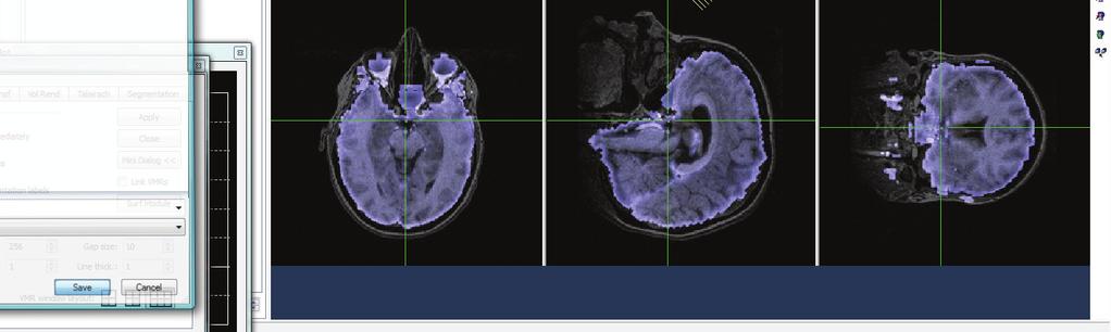
35 FIGURE LEGENDS Figure 1 Study timeline and data collected at each of 4 study visits. Abbreviations: AAI, Adult Attachment Interview; PDQ, Personality Disorder Questionnaire 4+; BDI, Beck Depression Inventory; PANAS, Positive and Negative Affect Schedule; ATQ, Adult Temperament Questionnaire Short form; IBQ, Infant Behavior Questionnaire Revised; PSI, Parenting Stress Index; WTAR, Wechsler Test of Adult Reading. Figure 2 Baby face presentation paradigm in functional MRI experiment. Infant face images were presented for 2 seconds, followed by a variable 2-6 second period of a plain black screen. Six stimulus types were presented in random order: own-happy (OH), own-neutral, own-sad, unknown-happy, unknown-neutral, unknown-sad. Reproduced with permission from Pediatrics, Vol. 122, Pages 40-51, Copyright 2008 by the AAP. Figure 3 Peripheral oxytocin and related brain activation in response to infant cues. (a) Mothers with Type B (secure) attachment patterns show a greater peripheral oxytocin response during an episode of physical interaction with their infant (mean ± sem; Bonferroni corrected comparison at free play time point, P = 0.01). The first baseline sample was collected 20 minutes after mother-infant separation; the second immediately after a 5-minute free-play involving direct physical contact between the mother and infant. The third sample was after a modified still-face procedure, in which the mother was in direct visual and auditory contact with her infant (via a mirror) but was physically separated by a screen divider. The final sample was collected after a further 20-minute period of complete mother-infant separation. (b) Type B mothers show greater activation of the hypothalamus/pituitary region in response to own vs. unknown infant face images (all affect groups combined) (mean ± sem, t=4.2, P < 0.001). Structural brain image 35
36 created from average of all subjects. Inset of magnified hypothalamic/pituitary region (single subject image to improve anatomical clarity). (c) Peripheral oxytocin response correlates with activation of hypothalamus/pituitary region in response to neutral own infant face cues (r S = 0.60, P = 0.001). A single outlying value was omitted from the graph, but not the statistical calculations. Figure 4 Brain responses to happy and sad own-infant faces, contrasting mothers with Type A (insecure/dismissing) and B (secure) attachment classifications (% signal change ± sem) (a) Type B mothers show greater activation of the ventral striatum (VS; t=3.1, P<0.005) and medial prefrontal cortex (mpfc; t=3.0, P<0.01) in response to happy own-infant faces. (b) Type B mothers show greater activation of the right ventral striatum (t=3.0, P<0.01) in response to sad own-infant faces. Type A mothers show greater activation of the right anterior insula (t=-3.9, P<0.0005). Figure 5 Peripheral oxytocin response after episodes of mother-infant interaction correlates with activation in the right ventral striatum (area shown in Figure 4b) in response to neutral own infant face cues (r S =0.57, P=0.002). Percent oxytocin change calculated from the first baseline measurement and a mean of the second and third samples, which were taken during episodes of mother-infant interaction. 36
37 Visit 1: Pregnancy Visit 2: Videotaping Visit 3: Scanning Visit 4: Follow-Up 3 rd trimester 7 mo 11 mo 14 mo Study Timeline BIRTH 20 min 5 min 6 min 20 min Mother-infant separation 1 Free play interaction Mirror-based interaction Mother-infant separation 2 Data Collected AAI Demographics PDQ BDI PANAS (1) Demographics ATQ BDI Infant face images Blood draws Oxytocin Cortisol Adrenaline Noradrenaline PANAS (2) IBQ PSI Breastfeeding duration WTAR BDI Breastfeeding duration Hours separated per week Bayley Scales of Infant Development BDI
")
 IDENTITY Unknown Infant Happy OH UH")
 2 sec Unknown: Neutral")
38 2 sec 2 sec 2 sec 2 6 sec random inter-stimulus interval Own: Happy (OH) STIMULUS TYPES A F F E C T Own Infant Unknown: Happy (UH) IDENTITY Unknown Infant Happy OH UH Neutral ON UN Sad OS US Unknown: Sad (US) 2 sec Own: Neutral (ON) 2 sec Unknown: Neutral (UN) 2 sec Own: Sad (OS)
 A 2.5 2 * B OWN > UNKNOWN 1.")
 Type A")

39 % signal change Serum oxytocin (pg/ml) A * B OWN > UNKNOWN Type B (Secure) Type A (Insecure) Optic chiasm Hypothalamus Thalamus Pons C Pituitary region (-3,2,-16) Secure Insecure


 Type A")

40 % signal change % signal change A y=11 VS mpfc x= R Ventral Striatum R mpfc Type B (Secure) Type A (Insecure) 1.2 Anterior Insula B VS y=6 y= R Ventral Striatum R Insula
41
42 Response to Reviewers We appreciate the reviewer s thoughtful comments and their positive overall assessment of a well written article with extremely interesting findings, rare and admirable [in] attempt[ing] to relate brain activity to real life maternal behavior and physiology and an elegant and demanding study design to address an issue at the very core of biological psychiatry. We have addressed each of the reviewer s suggestions as outlined below: Reviewer #1: 1. Figure 3b shows a stronger hypothalamic response in type B compared with type A mothers for the contrast own unknown. Given this, it would seem that the data points for type B mothers in figure 3c should lie mostly above y=0, but this is not the case. How can this be? Also, the figure legend indicates that the contrast between own and unknown is being plotted in figure 3c, but the y axis is labeled beta weight. Is this the beta weight for own or for the contrast value for own unknown? This needs to be clarified. The only way I can reconcile these two findings is if 3c is plotting the beta value for own and 3b is plotting the contrast value. But in this case, the difference between maternal types would be driven by a differential response to unknown faces and it is hard to see why this would be correlated with the OT response to interacting with one's own infant (Figure 3c). We appreciate the insightful feedback from this reviewer. Figure 3c was incorrectly labeled, giving rise to these inconsistencies. As correctly suggested by the reviewer, Figure 3c plots the beta values of own (neutral) faces, whereas Figure 3b is the contrast between own and unknown infant faces (happy/neutral/sad affect combined). Thus, the hypothalamic activation in response to own vs. unknown infant faces is greater in mothers with secure attachment, and the activation in response to neutral own infant faces is correlated significantly with peripheral oxytocin response when the mothers interact with their child. This is now clarified in the Figure 3 legend and text (page 14). 2. The ventral striatum is activated for own vs. unknown happy faces for the combined sample of type A and type B mothers (page 13), but figure 4a shows it deactivating for own happy faces in the type A mothers? How can these two findings be reconciled? A number of factors may contribute to this apparent inconsistency. Firstly, the two analyses differ in the contrast used. The first is a contrast between face types (own vs. unknown happy faces), while the second is a contrast between attachment groups (Type B vs. Type A). Each contrast uses different analysis methods and has different numbers of contrasted groups (i.e. a random effects analysis of 30 individual subjects in the first analysis, vs. a fixed effects analysis of 2 attachment groups in the second). There were also differences in the specific location of activation within the ventral striatum. The most likely explanation is that mothers in general show greater activation of the ventral striatum when viewing their own vs. an unknown infant s smiling face, but the subgroup of insecure/dismissing mothers show more deactivation in this region, compared with secure mothers. These two results link the findings of our previous paper (Strathearn, 2008) with the present study.
43 3. In discussing anterior insula activation when dismissing mothers viewed their own infant's sad faces, (page 17), the authors state, "Thus, in contrast with reward/motivation responses seen in mothers with secure attachment, sad infant affect may inhibit normal caregiving responses in dismissing mothers, and predispose to the development of insecure attachment in infancy." However, the anterior insula is also implicated in responding empathically to a loved one's distress (Singer et al 2004), and this is of course a crucial component of successful mothering. The insula has also been implicated in aversive conditioning and could be part of the mechanism by which mothers are taught how to meet their infants' needs more effectively. Therefore, the assumption that the insula activation in the dismissive mothers is dysfunctional seems premature. The valuable point that anterior insula activation has also been linked with empathic responses to a loved one s feeling of physical pain has now been included in the discussion, along with the caveat that dismissing individuals score much lower on a scale of emotional empathy, making this interpretation less likely (page 17). The premature conclusion quoted by the reviewer has now been deleted. 4. Please explain why a fixed effects rather than a random effects model was used when comparing brain activation across attachment groups. - Thank you for identifying this issue which is in need of clarification. The analysis was, in fact, a random effects analysis, in which the subjects were treated as a random factor in all ANOVA models. However, the experimental factors of the multi factorial design were fixed, as they were not a sample from a larger population of potential factor levels, and we were not interested in drawing inferences about the population of factor levels. This has now been clarified in the manuscript (page 10). 5. On page 16, the authors state,..., our findings suggest that for securely attached mothers, infant cues (whether positive or negative in affect) may act as an important learning signal (Schultz 1998) to reinforce and motivate responsive maternal care. This is an interesting idea, but if this were true then viewing a picture of one's own infant crying should deactivate the ventral striatum to teach the mother to avoid whatever behavior precipitated the infant's negative reaction. - Although prior studies have suggested that activation of the ventral striatum is related primarily to positive reward activation, more recent work has shown that negative but highly salient cues also activate this region (e.g. Faure, 2008; Berridge, 2007), suggesting a role of the striatum in incentive salience. This point has been clarified in the discussion (page 16). 6. Please describe the kinetics of OT release into peripheral blood. How long after an OT releasing event does it show up in blood? Are the levels following the maternal infant interaction reflecting that interaction or something that happened earlier? - Oxytocin is rapidly released into the peripheral circulation following a stimulus such as infant suckling. Studies that have used frequent sampling of blood (every 1 to 3 min) during a bout of nursing have identified a pulsatile pattern of oxytocin release in breastfeeding women (Lucas, 1980; Johnston, 1986). Oxytocin pulses also have been reported to occur in nursing women before the onset of infant suckling, usually in response to the sight or sound of the baby (McNeilly, 1983). Thus
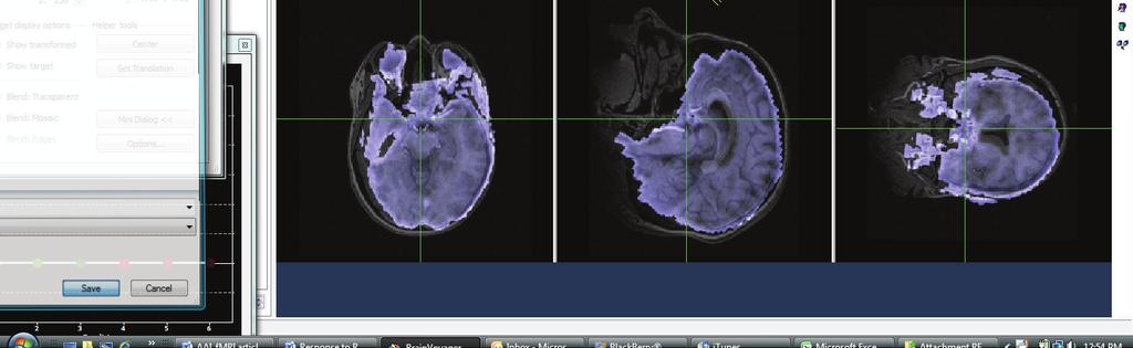
44 in the present study, it seems plausible that the release of oxytocin likely relates to emotional cues triggered by mother infant interaction. This has been explained in detail on pages Type A mothers activated DLPFC more than type B mothers when viewing their own infant's face. What is the authors' interpretation of this? In the model proposed in our original paper (Figure 6, Strathearn, 2008), we suggested that DLPFC activation in relation to attachment stimuli were indicative of more cognitive, and less affect focused, responses to infant face cues. In line with this model, we now propose in the paper that DLPFC activation in insecure/dismissing mothers is related to a more cognitive, impersonal response triggered by seeing their own baby s face (page 16 17). 8. Please indicate what was happening to the infants when they were separated from their mother? During the separation of mother and infant, smiling, neutral and crying faces were elicited while the infant was videotaped while in a car seat. Smiling faces were elicited using age appropriate toys and blowing bubbles. If the infant did not cry during this period, he/she was videotaped while being briefly left alone in the room, but observed from behind a one way mirror. The infant was not left to cry for more than 30 seconds before being picked up and pacified. The infant was constantly in the supervision and care of the study investigator or a trained research assistant. These details are referenced on page 6 to the methods section of a prior paper (Strathearn, 2008). 9. The Talairach coordinate listed for the hypothalamic activation in figure 3b is off the brain in my atlas, or perhaps on the optic chiasm. Nevertheless, considering smoothing and registration errors, it is plausible that the activation reflects hypothalamic activity. However, please show the hypothalamic/pituitary activation on an EPI functional image to verify that it is on the brain in a region with good signal. Also, a close up view of the activation with relevant structures labeled would be helpful. We recognize that this region of activation does not precisely correspond with the hypothalamus and pituitary in some brain atlases. However, when localized onto an averaged structural brain image created from all of the study subjects, the activation corresponds well with this brain region (see Figure 3b). On the recommendation of this reviewer, we have confirmed that the hypothalamic/pituitary activation does, in fact, lie within the functional maps of individual subjects, as illustrated below. A close up, labeled view of these regions has now been incorporated into Figure 3b, as suggested by Reviewer 2.

45 Subject AA

46 Subject BB Subject J
HHS Public Access Author manuscript Neuropsychopharmacology. Author manuscript; available in PMC 2011 February 18.
 Adult attachment predicts maternal brain and oxytocin response to infant cues Lane Strathearn, MBBS, FRACP 1,2,3, Peter Fonagy, PhD 4,5, Janet Amico, MD 6, and P. Read Montague, PhD 2,5 1 The Meyer Center
Drug addicted mothers show reduced brain reward response to their infants: Can oxytocin reverse the trend?
 Drug addicted mothers show reduced brain reward response to their infants: Can oxytocin reverse the trend? Dr. Lane Strathearn, MBBS FRACP PhD Stead Family Professor, Department of, University of Iowa
Supplementary Information
 Supplementary Information The neural correlates of subjective value during intertemporal choice Joseph W. Kable and Paul W. Glimcher a 10 0 b 10 0 10 1 10 1 Discount rate k 10 2 Discount rate k 10 2 10
Resistance to forgetting associated with hippocampus-mediated. reactivation during new learning
 Resistance to Forgetting 1 Resistance to forgetting associated with hippocampus-mediated reactivation during new learning Brice A. Kuhl, Arpeet T. Shah, Sarah DuBrow, & Anthony D. Wagner Resistance to
2/28/2017. Attachment in Substance Use. Objectives. Social Attachments
 Attachment in Substance Use Kalpana Miriyala, MD Zero to Three Fellow 2016-2018 Asst. Professor, Child and Adolescent Psychiatry Marshall University School of Medicine Objectives To review the theory of
9.01 Introduction to Neuroscience Fall 2007
 MIT OpenCourseWare http://ocw.mit.edu 9.01 Introduction to Neuroscience Fall 2007 For information about citing these materials or our Terms of Use, visit: http://ocw.mit.edu/terms. Emotion Sebastian Seung
This article appeared in a journal published by Elsevier. The attached copy is furnished to the author for internal non-commercial research and
 This article appeared in a journal published by Elsevier. The attached copy is furnished to the author for internal non-commercial research and education use, including for instruction at the authors institution
Methods to examine brain activity associated with emotional states and traits
 Methods to examine brain activity associated with emotional states and traits Brain electrical activity methods description and explanation of method state effects trait effects Positron emission tomography
The Neural Correlates of Moral Decision-Making in Psychopathy
 University of Pennsylvania ScholarlyCommons Neuroethics Publications Center for Neuroscience & Society 1-1-2009 The Neural Correlates of Moral Decision-Making in Psychopathy Andrea L. Glenn University
NIH Public Access Author Manuscript Pediatrics. Author manuscript; available in PMC 2009 July 1.
 NIH Public Access Author Manuscript Published in final edited form as: Pediatrics. 2008 July ; 122(1): 40 51. doi:10.1542/peds.2007-1566. What s in a Smile? Maternal Brain Responses to Infant Facial Cues
Searching for the Cause of Autism:
 Searching for the Cause of Autism: How genetics and social experience may intersect Dr Lane Strathearn, MBBS FRACP PhD Professor, Department of Pediatrics, University of Iowa Physician Director, Center
Title of file for HTML: Supplementary Information Description: Supplementary Figures, Supplementary Tables and Supplementary References
 Title of file for HTML: Supplementary Information Description: Supplementary Figures, Supplementary Tables and Supplementary References Supplementary Information Supplementary Figure 1. The mean parameter
Hallucinations and conscious access to visual inputs in Parkinson s disease
 Supplemental informations Hallucinations and conscious access to visual inputs in Parkinson s disease Stéphanie Lefebvre, PhD^1,2, Guillaume Baille, MD^4, Renaud Jardri MD, PhD 1,2 Lucie Plomhause, PhD
Supplementary Information Methods Subjects The study was comprised of 84 chronic pain patients with either chronic back pain (CBP) or osteoarthritis
 or osteoarthritis") Supplementary Information Methods Subjects The study was comprised of 84 chronic pain patients with either chronic back pain (CBP) or osteoarthritis (OA). All subjects provided informed consent to procedures
Supplementary Information Methods Subjects The study was comprised of 84 chronic pain patients with either chronic back pain (CBP) or osteoarthritis (OA). All subjects provided informed consent to procedures
Oxytocin - Is It Truly The Love Hormone? Wilson Tran, Oslín Licea Chávez, Jose Sandoval, Jon Zhang, Nari Kim, Denise Barroga, Chau Dang
 Oxytocin - Is It Truly The Love Hormone? Wilson Tran, Oslín Licea Chávez, Jose Sandoval, Jon Zhang, Nari Kim, Denise Barroga, Chau Dang What is Love? Love of Love The Chemistry Love is an emotion that
The Neural Basis of Economic Decision- Making in The Ultimatum Game
 The Neural Basis of Economic Decision- Making in The Ultimatum Game Sanfey, Rilling, Aronson, Nystrom, & Cohen (2003), The neural basis of economic decisionmaking in the Ultimatum game, Science 300, 1755-1758
Altruistic Behavior: Lessons from Neuroeconomics. Kei Yoshida Postdoctoral Research Fellow University of Tokyo Center for Philosophy (UTCP)
") Altruistic Behavior: Lessons from Neuroeconomics Kei Yoshida Postdoctoral Research Fellow University of Tokyo Center for Philosophy (UTCP) Table of Contents 1. The Emergence of Neuroeconomics, or the Decline
Altruistic Behavior: Lessons from Neuroeconomics Kei Yoshida Postdoctoral Research Fellow University of Tokyo Center for Philosophy (UTCP) Table of Contents 1. The Emergence of Neuroeconomics, or the Decline
Study No: Title : Rationale: Phase: Study Period: Study Design: Centres: Indication: Treatment: Objectives: Statistical Methods: Sample Size
 The study listed may include approved and non-approved uses, formulations or treatment regimens. The results reported in any single study may not reflect the overall results obtained on studies of a product.
Supplementary information Detailed Materials and Methods
 Supplementary information Detailed Materials and Methods Subjects The experiment included twelve subjects: ten sighted subjects and two blind. Five of the ten sighted subjects were expert users of a visual-to-auditory
The Role of Oxytocin in Mother-Infant Relations: A Systematic Review of Human Studies
 REVIEW The Role of Oxytocin in Mother-Infant Relations: A Systematic Review of Human Studies Megan Galbally, MBBS, MPM, FRANZCP, Andrew James Lewis, PhD, Marinus van IJzendoorn, PhD, and Michael Permezel,
Procedia - Social and Behavioral Sciences 159 ( 2014 ) WCPCG 2014
 WCPCG 2014") Available online at www.sciencedirect.com ScienceDirect Procedia - Social and Behavioral Sciences 159 ( 2014 ) 743 748 WCPCG 2014 Differences in Visuospatial Cognition Performance and Regional Brain Activation
Available online at www.sciencedirect.com ScienceDirect Procedia - Social and Behavioral Sciences 159 ( 2014 ) 743 748 WCPCG 2014 Differences in Visuospatial Cognition Performance and Regional Brain Activation
9/13/2018. Neurobiological Aspects of Attention Deficit Hyperactivity Disorder (ADHD) DSM-5 Diagnostic Criteria
 DSM-5 Diagnostic Criteria") DSM-5 Diagnostic Criteria Neurobiological Aspects of Attention Deficit Hyperactivity Disorder (ADHD) Neil P. Jones 7th Annual Conference on ADHD and Executive Function September 14, 218 Diagnosis Child
DSM-5 Diagnostic Criteria Neurobiological Aspects of Attention Deficit Hyperactivity Disorder (ADHD) Neil P. Jones 7th Annual Conference on ADHD and Executive Function September 14, 218 Diagnosis Child
SUPPLEMENTARY METHODS. Subjects and Confederates. We investigated a total of 32 healthy adult volunteers, 16
 SUPPLEMENTARY METHODS Subjects and Confederates. We investigated a total of 32 healthy adult volunteers, 16 women and 16 men. One female had to be excluded from brain data analyses because of strong movement
What Happens to a Woman's Brain When She Becomes a Mother
 What Happens to a Woman's Brain When She Becomes a Mother From joy and attachment to anxiety and protectiveness, mothering behavior begins with biochemical reactions. Bridget Coila/Flickr Adrienne LaFrance
Supplementary Online Content
 Supplementary Online Content Redlich R, Opel N, Grotegerd D, et al. Prediction of individual response to electroconvulsive therapy via machine learning on structural magnetic resonance imaging data. JAMA
Neural activity to positive expressions predicts daily experience of schizophrenia-spectrum symptoms in adults with high social anhedonia
 1 Neural activity to positive expressions predicts daily experience of schizophrenia-spectrum symptoms in adults with high social anhedonia Christine I. Hooker, Taylor L. Benson, Anett Gyurak, Hong Yin,
Brain Imaging studies in substance abuse. Jody Tanabe, MD University of Colorado Denver
 Brain Imaging studies in substance abuse Jody Tanabe, MD University of Colorado Denver NRSC January 28, 2010 Costs: Health, Crime, Productivity Costs in billions of dollars (2002) $400 $350 $400B legal
NIH Public Access Author Manuscript Nat Neurosci. Author manuscript; available in PMC 2006 September 5.
 NIH Public Access Author Manuscript Published in final edited form as: Nat Neurosci. 2006 August ; 9(8): 1004 1006. Maternal presence serves as a switch between learning fear and attraction in infancy
Twelve right-handed subjects between the ages of 22 and 30 were recruited from the
 Supplementary Methods Materials & Methods Subjects Twelve right-handed subjects between the ages of 22 and 30 were recruited from the Dartmouth community. All subjects were native speakers of English,
Neuroimaging vs. other methods
 BASIC LOGIC OF NEUROIMAGING fmri (functional magnetic resonance imaging) Bottom line on how it works: Adapts MRI to register the magnetic properties of oxygenated and deoxygenated hemoglobin, allowing
QUANTIFYING CEREBRAL CONTRIBUTIONS TO PAIN 1
 QUANTIFYING CEREBRAL CONTRIBUTIONS TO PAIN 1 Supplementary Figure 1. Overview of the SIIPS1 development. The development of the SIIPS1 consisted of individual- and group-level analysis steps. 1) Individual-person
Theory of mind skills are related to gray matter volume in the ventromedial prefrontal cortex in schizophrenia
 Theory of mind skills are related to gray matter volume in the ventromedial prefrontal cortex in schizophrenia Supplemental Information Table of Contents 2 Behavioral Data 2 Table S1. Participant demographics
smokers) aged 37.3 ± 7.4 yrs (mean ± sd) and a group of twelve, age matched, healthy
 aged 37.3 ± 7.4 yrs (mean ± sd) and a group of twelve, age matched, healthy") Methods Participants We examined a group of twelve male pathological gamblers (ten strictly right handed, all smokers) aged 37.3 ± 7.4 yrs (mean ± sd) and a group of twelve, age matched, healthy males,
Methods Participants We examined a group of twelve male pathological gamblers (ten strictly right handed, all smokers) aged 37.3 ± 7.4 yrs (mean ± sd) and a group of twelve, age matched, healthy males,
Epigenetic and Neurodevelopmental Perspectives on Variation in Parenting Behavior
 Parenting Science and Practice ISSN: 1529-5192 (Print) 1532-7922 (Online) Journal homepage: http://www.tandfonline.com/loi/hpar20 Epigenetic and Neurodevelopmental Perspectives on Variation in Parenting
PSYC 470. THE NEUROBIOLOGY OF AFFILIATIVE BEHAVIOUR
 PSYC 470. THE NEUROBIOLOGY OF AFFILIATIVE BEHAVIOUR 2016 WINTER SYLLABUS Instructor: Email: Office hours: Prof. Viara Mileva-Seitz milevav@queensu.ca by appointment Lectures: Humphrey Hall 223 Tuesday
Supplemental Information. Triangulating the Neural, Psychological, and Economic Bases of Guilt Aversion
 Neuron, Volume 70 Supplemental Information Triangulating the Neural, Psychological, and Economic Bases of Guilt Aversion Luke J. Chang, Alec Smith, Martin Dufwenberg, and Alan G. Sanfey Supplemental Information
The Neurobiology of Attachment. Cambridge, IASA Kim Barthel, OTR. Magic of Connection. Genes
 The Neurobiology of Attachment Cambridge, IASA Kim Barthel, OTR Magic of Connection Genes 1 Genome Malleable and flexible protein structure that bends and flows in response to the environment. The attachment
nucleus accumbens septi hier-259 Nucleus+Accumbens birnlex_727
 Nucleus accumbens From Wikipedia, the free encyclopedia Brain: Nucleus accumbens Nucleus accumbens visible in red. Latin NeuroNames MeSH NeuroLex ID nucleus accumbens septi hier-259 Nucleus+Accumbens birnlex_727
Ghosts in the NICU. Types of Stress 9/12/2018. Mom s History of ACEs and It s Impact on Attachment
 Mom s History of ACEs and It s Impact on Attachment Alan D. Mease MD, FAAP Medical Director, Child/Adolescent Heath Arkansas Department of Health Ghosts in the NICU alan.mease@arkansas.gov Learning Objectives
Supplementary Online Content
 Supplementary Online Content Green SA, Hernandez L, Tottenham N, Krasileva K, Bookheimer SY, Dapretto M. The neurobiology of sensory overresponsivity in youth with autism spectrum disorders. Published
Supplemental Data. Inclusion/exclusion criteria for major depressive disorder group and healthy control group
 1 Supplemental Data Inclusion/exclusion criteria for major depressive disorder group and healthy control group Additional inclusion criteria for the major depressive disorder group were: age of onset of
Veronika Grimm, Friederike Mengel. Let me sleep on it: Delay reduces rejection rates in Ultimatum Games RM/10/017
 Veronika Grimm, Friederike Mengel Let me sleep on it: Delay reduces rejection rates in Ultimatum Games RM/10/017 Let me sleep on it: Delay reduces rejection rates in Ultimatum Games Veronika Grimm Friederike
A possible mechanism for impaired joint attention in autism
 A possible mechanism for impaired joint attention in autism Justin H G Williams Morven McWhirr Gordon D Waiter Cambridge Sept 10 th 2010 Joint attention in autism Declarative and receptive aspects initiating
The Neuroscience of Addiction: A mini-review
 The Neuroscience of Addiction: A mini-review Jim Morrill, MD, PhD MGH Charlestown HealthCare Center Massachusetts General Hospital Disclosures Neither I nor my spouse/partner has a relevant financial relationship
Reward Systems: Human
 Reward Systems: Human 345 Reward Systems: Human M R Delgado, Rutgers University, Newark, NJ, USA ã 2009 Elsevier Ltd. All rights reserved. Introduction Rewards can be broadly defined as stimuli of positive
Supporting Information
 Supporting Information Braver et al. 10.1073/pnas.0808187106 SI Methods Participants. Participants were neurologically normal, righthanded younger or older adults. The groups did not differ in gender breakdown
How does adversity in childhood get under the skin
 How does adversity in childhood get under the skin What can we learn from neuroscience and epigenetics? Eamon McCrory Professor of Developmental Neuroscience & Psychopathology, UCL Consultant Clinical
Supplementary Material for The neural basis of rationalization: Cognitive dissonance reduction during decision-making. Johanna M.
 Supplementary Material for The neural basis of rationalization: Cognitive dissonance reduction during decision-making Johanna M. Jarcho 1,2 Elliot T. Berkman 3 Matthew D. Lieberman 3 1 Department of Psychiatry
Schizophrenic twin. Normal twin
 Brain anatomy and activity are often abnormal in schizophrenics - many studies have found the ventricles in schizophrenic patients enlarged (see below). - at the structural level, several brain areas have
Differences in brain structure and function between the sexes has been a topic of
 Introduction Differences in brain structure and function between the sexes has been a topic of scientific inquiry for over 100 years. In particular, this topic has had significant interest in the past
SUPPLEMENTARY MATERIAL. Table. Neuroimaging studies on the premonitory urge and sensory function in patients with Tourette syndrome.
 SUPPLEMENTARY MATERIAL Table. Neuroimaging studies on the premonitory urge and sensory function in patients with Tourette syndrome. Authors Year Patients Male gender (%) Mean age (range) Adults/ Children
How rational are your decisions? Neuroeconomics
 How rational are your decisions? Neuroeconomics Hecke CNS Seminar WS 2006/07 Motivation Motivation Ferdinand Porsche "Wir wollen Autos bauen, die keiner braucht aber jeder haben will." Outline 1 Introduction
Role of the ventral striatum in developing anorexia nervosa
 Role of the ventral striatum in developing anorexia nervosa Anne-Katharina Fladung 1 PhD, Ulrike M. E.Schulze 2 MD, Friederike Schöll 1, Kathrin Bauer 1, Georg Grön 1 PhD 1 University of Ulm, Department
SUPPLEMENT: DYNAMIC FUNCTIONAL CONNECTIVITY IN DEPRESSION. Supplemental Information. Dynamic Resting-State Functional Connectivity in Major Depression
 Supplemental Information Dynamic Resting-State Functional Connectivity in Major Depression Roselinde H. Kaiser, Ph.D., Susan Whitfield-Gabrieli, Ph.D., Daniel G. Dillon, Ph.D., Franziska Goer, B.S., Miranda
Distinct valuation subsystems in the human brain for effort and delay
 Supplemental material for Distinct valuation subsystems in the human brain for effort and delay Charlotte Prévost, Mathias Pessiglione, Elise Météreau, Marie-Laure Cléry-Melin and Jean-Claude Dreher This
Epigenetic Pathways Linking Parental Effects to Offspring Development. Dr. Frances A. Champagne Department of Psychology, Columbia University
 Epigenetic Pathways Linking Parental Effects to Offspring Development Dr. Frances A. Champagne Department of Psychology, Columbia University Prenatal & Postnatal Experiences Individual differences in brain
Psych3BN3 Topic 4 Emotion. Bilateral amygdala pathology: Case of S.M. (fig 9.1) S.M. s ratings of emotional intensity of faces (fig 9.
 S.M. s ratings of emotional intensity of faces (fig 9.") Psych3BN3 Topic 4 Emotion Readings: Gazzaniga Chapter 9 Bilateral amygdala pathology: Case of S.M. (fig 9.1) SM began experiencing seizures at age 20 CT, MRI revealed amygdala atrophy, result of genetic
Psych3BN3 Topic 4 Emotion Readings: Gazzaniga Chapter 9 Bilateral amygdala pathology: Case of S.M. (fig 9.1) SM began experiencing seizures at age 20 CT, MRI revealed amygdala atrophy, result of genetic
The Parental Brain Regulation of Mother and Father Behavior that Influences Infant Development
 Contexts for Development & Child Flourishing University of Notre Dame - September 28, 2014 The Parental Brain Regulation of Mother and Father Behavior that Influences Infant Development James E. Swain
Supporting Online Material for
 www.sciencemag.org/cgi/content/full/324/5927/646/dc1 Supporting Online Material for Self-Control in Decision-Making Involves Modulation of the vmpfc Valuation System Todd A. Hare,* Colin F. Camerer, Antonio
WHAT DOES THE BRAIN TELL US ABOUT TRUST AND DISTRUST? EVIDENCE FROM A FUNCTIONAL NEUROIMAGING STUDY 1
 SPECIAL ISSUE WHAT DOES THE BRAIN TE US ABOUT AND DIS? EVIDENCE FROM A FUNCTIONAL NEUROIMAGING STUDY 1 By: Angelika Dimoka Fox School of Business Temple University 1801 Liacouras Walk Philadelphia, PA
0-3 DEVELOPMENT. By Drina Madden. Pediatric Neuropsychology 1
 0-3 DEVELOPMENT By Drina Madden DrinaMadden@hotmail.com www.ndcbrain.com 1 PHYSICAL Body Growth Changes in height and weight are rapid in the first two years of life. Development moves from head to tail
Emotional Development
 Emotional Development How Children Develop Chapter 10 Emotional Intelligence A set of abilities that contribute to competent social functioning: Being able to motivate oneself and persist in the face of
Neurobiological Foundations of Reward and Risk
 Neurobiological Foundations of Reward and Risk... and corresponding risk prediction errors Peter Bossaerts 1 Contents 1. Reward Encoding And The Dopaminergic System 2. Reward Prediction Errors And TD (Temporal
The Adolescent Developmental Stage
 The Adolescent Developmental Stage o Physical maturation o Drive for independence o Increased salience of social and peer interactions o Brain development o Inflection in risky behaviors including experimentation
Supporting Information
 Supporting Information Carhart-Harris et al. 10.1073/pnas.1119598109 Fig. S1. Slices for arterial spin labeling (ASL) result. Lightbox display of slices showing regions where there were significant decreases
Supplemental information online for
 Supplemental information online for Sleep contributes to the strengthening of some memories over others, depending on hippocampal activity at learning. Géraldine Rauchs (1,2), Dorothée Feyers (1), Brigitte
Investigating directed influences between activated brain areas in a motor-response task using fmri
 Magnetic Resonance Imaging 24 (2006) 181 185 Investigating directed influences between activated brain areas in a motor-response task using fmri Birgit Abler a, 4, Alard Roebroeck b, Rainer Goebel b, Anett
CEREBRUM & CEREBRAL CORTEX
 CEREBRUM & CEREBRAL CORTEX Seonghan Kim Dept. of Anatomy Inje University, College of Medicine THE BRAIN ANATOMICAL REGIONS A. Cerebrum B. Diencephalon Thalamus Hypothalamus C. Brain Stem Midbrain Pons
The Frontal Lobes. Anatomy of the Frontal Lobes. Anatomy of the Frontal Lobes 3/2/2011. Portrait: Losing Frontal-Lobe Functions. Readings: KW Ch.
 The Frontal Lobes Readings: KW Ch. 16 Portrait: Losing Frontal-Lobe Functions E.L. Highly organized college professor Became disorganized, showed little emotion, and began to miss deadlines Scores on intelligence
Talk 2. Neurocognitive differences in children with or without CU- traits 05/12/2013. Psychological. Behavioural
 Neurocognitive differences in children with or without CU- traits Prof Essi Viding Developmental Risk and Resilience Unit, Psychology and Language Sciences, UCL e.viding@ucl.ac.uk Talk 2. Environment Psychological
Supplementary Online Material Supplementary Table S1 to S5 Supplementary Figure S1 to S4
 Supplementary Online Material Supplementary Table S1 to S5 Supplementary Figure S1 to S4 Table S1: Brain regions involved in the adapted classification learning task Brain Regions x y z Z Anterior Cingulate
Hierarchically Organized Mirroring Processes in Social Cognition: The Functional Neuroanatomy of Empathy
 Hierarchically Organized Mirroring Processes in Social Cognition: The Functional Neuroanatomy of Empathy Jaime A. Pineda, A. Roxanne Moore, Hanie Elfenbeinand, and Roy Cox Motivation Review the complex
HST.583 Functional Magnetic Resonance Imaging: Data Acquisition and Analysis Fall 2008
 MIT OpenCourseWare http://ocw.mit.edu HST.583 Functional Magnetic Resonance Imaging: Data Acquisition and Analysis Fall 2008 For information about citing these materials or our Terms of Use, visit: http://ocw.mit.edu/terms.
Session Goals. Principles of Brain Plasticity
 Presenter: Bryan Kolb Canadian Centre for Behavioural Neuroscience University of Lethbridge Date: January 12, 2011 The FASD Learning Series is part of the Alberta government s commitment to programs and
Intelligence moderates reinforcement learning: a mini-review of the neural evidence
 Articles in PresS. J Neurophysiol (September 3, 2014). doi:10.1152/jn.00600.2014 1 2 3 4 5 6 7 8 9 10 11 12 13 14 15 16 17 18 19 20 21 22 23 24 25 26 27 28 29 30 31 32 33 34 35 36 37 38 39 40 41 42 43
Biomarkers Workshop In Clinical Trials Imaging for Schizophrenia Trials
 Biomarkers Workshop In Clinical Trials Imaging for Schizophrenia Trials Research focused on the following areas Brain pathology in schizophrenia and its modification Effect of drug treatment on brain structure
Define functional MRI. Briefly describe fmri image acquisition. Discuss relative functional neuroanatomy. Review clinical applications.
 Dr. Peter J. Fiester November 14, 2012 Define functional MRI. Briefly describe fmri image acquisition. Discuss relative functional neuroanatomy. Review clinical applications. Briefly discuss a few examples
The Role of Working Memory in Visual Selective Attention
 Goldsmiths Research Online. The Authors. Originally published: Science vol.291 2 March 2001 1803-1806. http://www.sciencemag.org. 11 October 2000; accepted 17 January 2001 The Role of Working Memory in
Oxytocin and Early Experience. Sue Carter The Brain Body Center Department of Psychiatry University of Illinois at Chicago
 Oxytocin and Early Experience Sue Carter The Brain Body Center Department of Psychiatry University of Illinois at Chicago At the center of the neuroendocrine mechanisms for parental behavior (both sexes)
HST.583 Functional Magnetic Resonance Imaging: Data Acquisition and Analysis Fall 2006
 MIT OpenCourseWare http://ocw.mit.edu HST.583 Functional Magnetic Resonance Imaging: Data Acquisition and Analysis Fall 2006 For information about citing these materials or our Terms of Use, visit: http://ocw.mit.edu/terms.
Introduction to Systems Neuroscience. Nov. 28, The limbic system. Daniel C. Kiper
 Introduction to Systems Neuroscience Nov. 28, 2017 The limbic system Daniel C. Kiper kiper@ini.phys.ethz.ch http: www.ini.unizh.ch/~kiper/system_neurosci.html LIMBIC SYSTEM The term limbic system mean
Academic year Lecture 16 Emotions LECTURE 16 EMOTIONS
 Course Behavioral Economics Academic year 2013-2014 Lecture 16 Emotions Alessandro Innocenti LECTURE 16 EMOTIONS Aim: To explore the role of emotions in economic decisions. Outline: How emotions affect
The effects of intranasal oxytocin on reward circuitry responses in children with autism spectrum disorder
 Greene et al. Journal of Neurodevelopmental Disorders (2018) 10:12 https://doi.org/10.1186/s11689-018-9228-y RESEARCH Open Access The effects of intranasal oxytocin on reward circuitry responses in children
Jeffry A. Simpson, W. Andrew Collins, and Jessica E. Salvatore University of Minnesota. Introduction. An Organizational Perspective on Development
 The Impact of Early Interpersonal Experience on Adult Romantic Relationship Functioning: Recent Findings From the Minnesota Longitudinal Study of Risk and Adaptation Current Directions in Psychological
Supporting Families to Build Secure Attachment Relationships : Comments on Benoit, Dozier, and Egeland
 ATTACHMENT Supporting Families to Build Secure Attachment Relationships : Comments on Benoit, Dozier, and Egeland Femmie Juffer, PhD, Marian J. Bakermans-Kranenburg, PhD, & Marinus H. van IJzendoorn, PhD
Title of file for HTML: Peer Review File Description:
 Title of file for HTML: Supplementary Information Description: Supplementary Figure, Supplementary Tables, Supplementary Methods and Supplementary References Title of file for HTML: Peer Review File Description:
Endocrine Glands: Hormone-secreting organs are called endocrine glands
 University of Jordan Department of Physiology and Biochemistry Nursing students, Academic year 2017/2018. ******************************************************************* Ref: Principles of Anatomy
When is an embryo a person?
 AKC Autumn 2008 Lecture 2 When is an embryo a person? roger.morris@kcl.ac.uk Head, School of Biomedical & Health Sciences In essence Human mental and social development occurs postnatally in critical periods
correlates with social context behavioral adaptation.
 REVIEW OF FRONTAL LOBE STRUCTURES Main organization of frontal cortex: 1. Motor area (precentral gyrus). 2. Premotor & supplementary motor areas (immediately anterior to motor area). Includes premotor,
Neural Basis of Decision Making. Mary ET Boyle, Ph.D. Department of Cognitive Science UCSD
 Neural Basis of Decision Making Mary ET Boyle, Ph.D. Department of Cognitive Science UCSD Phineas Gage: Sept. 13, 1848 Working on the rail road Rod impaled his head. 3.5 x 1.25 13 pounds What happened
Supplementary Materials for
 Supplementary Materials for Folk Explanations of Behavior: A Specialized Use of a Domain-General Mechanism Robert P. Spunt & Ralph Adolphs California Institute of Technology Correspondence may be addressed
Brain Mechanisms of Emotion 1 of 6
 Brain Mechanisms of Emotion 1 of 6 I. WHAT IS AN EMOTION? A. Three components (Oately & Jenkins, 1996) 1. caused by conscious or unconscious evaluation of an event as relevant to a goal that is important
Supporting Information. Demonstration of effort-discounting in dlpfc
 Supporting Information Demonstration of effort-discounting in dlpfc In the fmri study on effort discounting by Botvinick, Huffstettler, and McGuire [1], described in detail in the original publication,
Neocortex. Hemispheres 9/22/2010. Psychology 472 Pharmacology of Psychoactive Drugs. Structures are divided into several section or lobes.
 Neocortex Psychology 472 Pharmacology of Psychoactive Drugs 1 Is the most developed in Humans Has many folds and fissures The folds of tissue are called gyri or a gyrus (single) The fissures or valleys
Cover Page. The handle holds various files of this Leiden University dissertation
 Cover Page The handle http://hdl.handle.net/1887/32078 holds various files of this Leiden University dissertation Author: Pannekoek, Nienke Title: Using novel imaging approaches in affective disorders
Social and Pragmatic Language in Autistic Children
 Parkland College A with Honors Projects Honors Program 2015 Social and Pragmatic Language in Autistic Children Hannah Li Parkland College Recommended Citation Li, Hannah, "Social and Pragmatic Language
Mindfulness-Based Relapse Prevention: A Dialogue Between Clinical Science and Neuroscience in the Treatment of Addictive Behavior.
 Mindfulness-Based Relapse Prevention: A Dialogue Between Clinical Science and Neuroscience in the Treatment of Addictive Behavior. Presented at the Western Psychological Association Meeting, April 2010,
The effect of intragastric fructose infusion on homeostatic and hedonic brain regions interacts with the effect of emotional state
 The effect of intragastric fructose infusion on homeostatic and hedonic brain regions interacts with the effect of emotional state J. Iven, J.R. Biesiekierski, D. Zhao, J. Tack, L. Van Oudenhove Translational
biological psychology, p. 40 The study of the nervous system, especially the brain. neuroscience, p. 40
 biological psychology, p. 40 The specialized branch of psychology that studies the relationship between behavior and bodily processes and system; also called biopsychology or psychobiology. neuroscience,
Connect with amygdala (emotional center) Compares expected with actual Compare expected reward/punishment with actual reward/punishment Intuitive
 Compares expected with actual Compare expected reward/punishment with actual reward/punishment Intuitive") Orbitofronal Notes Frontal lobe prefrontal cortex 1. Dorsolateral Last to myelinate Sleep deprivation 2. Orbitofrontal Like dorsolateral, involved in: Executive functions Working memory Cognitive flexibility
Orbitofronal Notes Frontal lobe prefrontal cortex 1. Dorsolateral Last to myelinate Sleep deprivation 2. Orbitofrontal Like dorsolateral, involved in: Executive functions Working memory Cognitive flexibility