Mechanisms of anti-sm B cell activation in autoimmune Fas-deficient mice
|
|
|
- Sabina Bradford
- 5 years ago
- Views:
Transcription
1 Mechanisms of anti-sm B cell activation in autoimmune Fas-deficient mice Kara Lynne Conway A dissertation submitted to the faculty of the University of North Carolina at Chapel Hill in partial fulfillment of the requirements for the degree of Doctor of Philosophy in the Department of Microbiology and Immunology Chapel Hill 2007 Approved by: Larry W. Arnold, Ph.D. Stephen H. Clarke, Ph.D. Jeffrey A. Frelinger, Ph.D. Glenn K. Matsushima, Ph.D. Roland M. Tisch, Ph.D
2 2007 Kara Lynne Conway ALL RIGHTS RESERVED ii
3 ABSTRACT Kara Lynne Conway: Mechanisms of anti-sm B cell activation in autoimmune Fas-deficient mice (Under the direction of Dr. Stephen H. Clarke) Systemic lupus erythematosus (SLE) is an autoimmune disease characterized by the production of anti-nuclear autoantibodies to Smith (Sm), a component of a ribonucleoprotein complex. Anti-Sm transgenic mice (2-12H) develop large numbers of anti-sm B cells but do not develop elevated anti-sm titers, indicating that anti-sm B cells are regulated. However, 2-12H mice with the lpr mutation of the pro-apoptosis gene Fas (Fas lpr ) develop high anti- Sm titers and anti-sm antibody secreting cells (ASCs) in multiple tissues. This loss of tolerance is coincident with an autoantigen-specific depletion of marginal zone (MZ) and B-1 B cells and a bypass in the early pre-plasma cell (PC) tolerance checkpoint. To demonstrate the involvement of each mature B cell subset in the anti-sm response in Fas lpr mice, I adoptively transferred sorted 2-12H splenic B cells to Fas lpr or Fas wt recipients. I demonstrate that both anti-sm follicular (FO) and MZ B cells are precursors to early pre-pcs. In addition, MZ B cells and early pre-pcs give rise to immediate, short-lived ASC responses, while anti-sm FO B cells give rise to a delayed, long-lived ASC response after transfer to Fas lpr recipients. These findings indicate that all anti-sm B cell subsets are activated in autoimmunity but follow different activation programs, paralleling their responses to foreign antigens. I also demonstrate that immature bone marrow-derived DCs (BMDCs) and ex vivo DCs that have phagocytized Sm-bearing apoptotic cells (ACs) induce anti-sm MZ B cell iii
4 differentiation to ASCs in vivo and in vitro. This MZ B cell activation is antigen-specific, T cell-dependent, cell-contact dependent, and dependent upon IL-1β, CD40L, and BAFF production. Interestingly, BMDCs and ex vivo DCs from Fas lpr mice activate anti-sm MZ B cells regardless of AC phagocytosis or maturation status. The restricted ability of immature, non-autoimmune DCs to activate anti-sm MZ B cells after AC phagocytosis is likely to prevent concomitant T cell activation and to induce only a short-lived B cell response. In contrast, the loss of these restrictions by Fas lpr DCs is likely to allow concomitant T cell activation, resulting in long-lived, T cell-dependent responses. These data provide a possible mechanism for long-lived, anti-self responses in SLE. iv
5 To my loving family and future husband in gratitude for their unending support, love, and patience. v
6 TABLE OF CONTENTS LIST OF TABLES...x LIST OF FIGURES...xi LIST OF ABBREVIATIONS AND SYMBOLS xiii CHAPTER 1: INTRODUCTION...1 A. B CELL DEVELOPMENT Central B Cell Development Peripheral B Cell Development.4 3. Mature B Cell Subsets....5 a. Follicular (FO) B Cells.5 b. Marginal Zone B Cells..6 c. B-1 B Cells 7 B. B CELL FUNCTIONS Immunoglobulin (Ig) Production Complement Cascade Initiation Antigen Presenting Cell (APC) Capability Cytokine Production. 10 C. B CELL ACTIVATION Toll-like Receptor (TLR)-Mediated Activation BCR-Mediated Activation Germinal Center (GC) and Extrafollicular Responses..13 vi
7 4. Memory B Cells Plasma Cells (PCs) 15 a. PC Transcription Program.15 b. PC Homing..16 c. PC Intermediates. 16 D. B CELL TOLERANCE Receptor Editing and Central Deletion Peripheral Deletion Anergy. 19 E. DENDRITIC CELLS DC Function DC Maturation DC Subsets Apoptotic Cells (ACs) and DCs DCs and B Cells.23 a. DCs and B Cell Activation..23 b. BAFF F. B CELLS AND AUTOIMMUNITY Systemic Lupus Erythematosus (SLE) Murine Models of SLE Anti-Sm Response in SLE a. Anti-Sm B Cell Regulation..29 b. Anti-Sm B Cell Activation vii
8 G. REFERENCES..31 CHAPTER 2: AUTOREACTIVE MZ AND B-1 B CELL ACTIVATION BY FAS lpr IS COINCIDENT WITH AN INCREASED FREQUENCY OF APOPTOTIC LYMPHOCYTES AND A DEFECT IN MACROPHAGE CLEARANCE 51 A. ABSTRACT.. 52 B. INTRODUCTION. 53 C. MATERIALS AND METHODS..56 D. RESULTS...59 E. DISCUSSION F. REFERENCES...85 CHAPTER 3: ANTI-Sm FOLLICULAR, MARGINAL ZONE, AND EARLY PRE-PC B CELL ACTIVATION IN FAS lpr MICE...90 A. ABSTRACT B. INTRODUCTION..92 C. MATERIALS AND METHODS D. RESULTS E. DISCUSSION 107 F. REFERENCES CHAPTER 4: DENDRITIC CELL ACTIVATION OF ANTI-Sm MARGINAL ZONE B CELLS A. ABSTRACT B. INTRODUCTION C. MATERIALS AND METHODS D. RESULTS viii
9 E. DISCUSSION..147 F. REFERENCES CHAPTER 5: CONCLUSIONS AND FUTURE DIRECTIONS APPENDIX 1: EARLY PRE-PLASMA CELLS DEFINE A TOLERANCE CHECKPOINT FOR AUTOREACTIVE B CELLS. 181 A. ABSTRACT..182 B. INTRODUCTION C. MATERIALS AND METHODS 185 D. RESULTS E. DISCUSSION F. REFERENCES. 221 APPENDIX 2: EBV LATENT MEMBRANE PROTEIN 2A INDUCES AUTOREACTIVE B CELL ACTIVATION AND TLR HYPERSENSITIVITY 227 A. ABSTRACT.228 B. INTRODUCTION C. MATERIALS AND METHODS 232 D. RESULTS. 236 E. DISCUSSION F. REFERENCES. 260 ix
10 LIST OF TABLES Table 3.1. Donor cell detection and ASC secretion in the spleen after 2-12H early pre-pc transfers Table 3.2. Donor cell detection and ASC secretion in the spleen after 2-12H MZ and FO transfers x
11 LIST OF FIGURES Figure 2.1. Anti-Sm antibody ASC production in autoimmune mice Figure 2.2. Anti-Sm B cell development in spleen of 2-12H Tg, 2-12H MRL, 2-12H/Fas lpr, and 2-12H MRL/Fas lpr mice Figure 2.3. ELISpot analysis of anti-sm ASCs in 2-12H, 2-12H/Fas lpr, and 2-12HMRL/Fas lpr mice Figure 2.4. Peritoneal anti-sm B-1 cells in 2-12H Tg, 2-12H MRL, 2-12H/Fas lpr, and 2-12H MRL/Fas lpr mice Figure 2.5. Anti-Sm B-1 cell activation in Fas lpr mice after peritoneal cell transfer Figure 2.6. The effect of Fas lpr on PtC-specific B cells Figure 2.7. Apoptotic cells in wt and Fas lpr mice Figure 3.1. Anti-Sm pre-pcs are functional and regulated by extrinsic mechanisms Figure 3.2. Anti-Sm follicular (FO) and marginal zone (MZ) B cells can both acquire an early pre-pc phenotype Figure 3.3. Anti-Sm MZ are activated in autoimmune mice to induce a rapid, short-lived ASC response Figure 3.4. FO B cells are activated in autoimmune mice to induce a delayed, long-lived ASC response Figure 4.1. Apoptotic cell (AC)-pulsed BMDCs induce anti-sm antibody secreting cell (ASC) formation in vivo Figure 4.2. AC-pulsed BMDCs affect B cell activation at two stages Figure 4.3. Fas-deficient BMDC activation of anti-sm B cells is not limited by ingestion of ACs or to the immature stage Figure 4.4. In vitro activation of anti-sm B cells by BMDCs recapitulates in vivo activation Figure 4.5. Cell sorting of splenic B cell and splenic DC populations demonstrates the primary responding subsets involved in anti-sm B cell activation by BMDCs in vitro xi
12 Figure 4.6. Both cell contact and secreted cytokines are required for optimal anti-sm B cell activation by AC-pulsed BMDCs Figure 4.7. Exogenous BAFF and IL-1β can replace AC-pulsing in anti-sm ASC induction by BMDCs FIGURE A1.1. CD138 int cells are present at a high frequency in the spleens and BM of non-tg and 2-12H mice FIGURE A1.2. Immunofluorescence analysis of nonautoimmune and autoimmune mice FIGURE A1.3. A subset of CD138 int B cells are IC IgM high and secrete Ab, but anti-sm CD138 int B cells do not secrete Ab FIGURE A1.4. Anti-Sm CD138 int B cells are ASCs in autoimmune mice FIGURE A1.5. Anti-Sm CD138 int B cells have a high turnover rate and a high frequency are undergoing apoptosis FIGURE A1.6. Blimp-1 mrna is up-regulated in CD138 int B cells that secrete Ab FIGURE A1.7. Differential expression of BAFF-R, TACI, and BCMA on anti-sm and non-tg CD138 int B cells FIGURE A2.1. Activation of anti-sm B cells in 2-12H/LMP2A mice FIGURE A2.2. LMP2A significantly affects B cell phenotype analysis FIGURE A2.3. Analysis of CD138-expressing B cells indicates that LMP2A allows differentiation beyond the pre-pc checkpoint FIGURE A2.4. Real-time PCR comparison of Blimp-1, XBP-1, and PAX-5 expression in sorted B cell subpopulations indicates differentiation beyond the pre-pc tolerance checkpoint FIGURE A H/LMP2A B cells do not proliferate in response to BCR signals but are nevertheless BCR signaling competent FIGURE A2.6. LMP2A induces B cell hyperresponsiveness to CpG ODN, LPS, and imiquimod FIGURE A2.7. LMP2A induces higher TLR expression levels. One parameter histograms are shown for TLR4, RP105, and TLR9 expression xii
13 LIST OF ABBREVIATIONS AND SYMBOLS Ab Antibody Abs Antibodies AC(s) Apoptotic cell(s) Ag Antigen APC Antigen presenting cell Apops Apoptotic cells ASC Antibody secreting cell B6 C57BL/6 murine strain BAFF B cell activating factor BCR B cell receptor Blimp-1 B lymphocyte induced maturation protein 1 BLyS B lymphocyte stimulator BM Bone marrow BMDC(s) Bone marrow-derived dendritic cell(s) BSAP B cell specific activating protein Ca 2+ CpG CSR DC dsdna Fc FcR FDC FO FSC GC HEL IC IC IgM Calcium divalent cation Cytosine-phosphate-guanine motif Class switch recombination Dendritic cell Double-stranded DNA Fragment crystallizable (constant) region Fc receptor Follicular dendritic cell Follicular Forward scatter Germinal center Hen egg lysozyme Immune complex Intracellular IgM xiii
14 IFN Interferon Ig Immunoglobulin IL Interleukin IRF-4 Interferon regulatory factor 4 ITAM Immunoreceptor tyrosine-based activation motif i.v. Intravenous L.P. Lamina propria LPS Lipopolysaccharide MAC Membrane attack complex MFI Median fluorescence intensity MHC Major histocompatibility complex mhel Mmbrane-bound hen egg lysozyme MLN Mesenteric lymph node MZ Marginal zone NF-κB Nuclear factor κb PALS Periarterial lymphoid sheath PAMP Pattern associated recognition motif PC Plasma cell RAG Recombination activating genes RNP Ribonucleoprotein SHM Somatic hypermutation sig Surface immunoglobulin SLE Systemic lupus erythematosis Sm Smith antigen shel soluble hen egg lysozyme snrnp Small nuclear ribonuclear protein ssrna Single stranded RNA SSC Side scatter T regs Regulatory T cells T1 Transitional 1 T2 Transitional 2 xiv
15 T3 Transitional 3 TCR T cell receptor TD T-dependent Th T helper TI T-independent TLR Toll-like receptor TNF Tumor necrosis factor XBP-1 X-box protein 1 α alpha/antiβ beta γ gamma κ kappa xv
16 CHAPTER 1: INTRODUCTION
17 B lymphocytes, the effectors of the humoral immune response, have evolved to recognize a variety of foreign antigens from viral, bacterial, and fungal pathogens. These antibody-producing cells are thus able to assist in mounting efficient, specific immune responses against invading organisms. In creating this diverse repertoire of B cells, autoreactive specificities are inevitably generated. Many mechanisms of regulation have evolved to prevent anti-self B cell activation. Understanding how autoreactive B cells are regulated and how these mechanisms are compromised in autoimmunity is important for treatment of autoimmune diseases such as systemic lupus erythematosus (SLE). A. B CELL DEVELOPMENT B cell development is a series of intricate processes aimed at creating a diverse repertoire of B cells capable of responding to a seemingly infinite number of foreign antigens. This goal must be achieved while maintaining tolerance to self, which is controlled by both positive and negative selection processes. Stromal cells in the bone marrow (BM) provide signals to guide the generation of B cells from lymphoid progenitors. B cell lymphopoiesis begins in the BM and fetal liver and, unlike T cells, most of their development is completed in these lymphoid compartments before migrating to the periphery. 1. Central B Cell Development 2
18 Common lymphoid progenitors begin B cell differentiation after receiving both membrane-bound and secreted signals from BM stromal cells, the most important of which is interleukin-7 (IL-7). Upon receiving stromal survival signals, common lymphoid progenitors enter the pro-b cell stage under the direction of EBF, E2A, and Pax-5, transcription factors controlling B cell development(1-5). Pax-5, which is involved in suppressing lineage-inappropriate genes, has been shown to be indispensable for pro-b cell development(3, 6, 7). B cell immunoglobulin (Ig) diversification begins in this pro-b cell stage, as recombination activating genes (RAG) 1 and 2 aid in V H D H J H heavy chain recombination(8-12). If productive heavy chain rearrangement occurs, the B cell progresses into the pre-b cell stage in which the µ heavy chain is expressed. The µ heavy chain can pair with the surrogate light chain, forming the pre-b cell receptor (pre-bcr)(13-15). The pre-bcr associates with the Igα and Igβ signaling components, and signals through this pre-bcr will lead to light chain V L J L rearrangement(16-19). Pre-BCR signals are imperative for B cell progression and survival, as B cells from µmt mice, which do not express H chain on the surface, and Igα/β-deficient mice, which cannot transduce signals through the pre-bcr, do not survive past the pre-b cell stage(20-22). Once a complete BCR with heavy and light chains has been rearranged, assembled, and expressed on the surface, the cell is now considered an immature B cell. Immature B cells are involved in both positive and negative selection in the BM(22-29). As I mentioned above, at the pre-b cell stage, immature B cells are also eliminated if incapable of signaling through the BCR, allowing only productive rearrangements to progress. This process is referred to as positive selection and is defined by the BCR 3
19 interacting with self antigen weakly (25, 29, 30). On the other hand, immature B cells that strongly bind self antigen can undergo a secondary light chain rearrangement, called receptor editing, in an attempt to avoid autoreactivity by its BCR(31). If editing does not eliminate self-antigen binding, the immature B cell will undergo apoptosis (25, 29). This process is referred to as central deletion, a mechanism of negative selection. Immature B cells that possess a functional and non-autoreactive BCR are then able to migrate to the peripheral lymphoid organs where they are referred to as transitional B cells(32). 2. Peripheral B Cell Development Newly generated immature B cells migrate to the spleen to continue B cell development and differentiate into mature B cells. The spleen is composed of the red pulp, which is the region responsible for red blood cell clearance and recycling, and the white pulp, which is the region where lymphocytes reside(33). In the white pulp, T cells are located around a central arteriole in an area called the periarterial lymphoid sheath (PALS), while the B cells are organized outside of this region in follicles(33). The marginal zone (MZ) area, straddling both the red and white pulp of the spleen, is composed of B cells, dendritic cells (DCs), and macrophages(33). Immature B cells, now termed transitional B cells, home to the follicles via chemokine receptor expression(33-36). There are several sub-stages of transitional B cells that are somewhat controversial and definitively unclear. T1, T2, and T3 subsets have been described and re-classified recently, however new evidence suggests the professed T3 subset may not even be a transitional population but a non-functional 4
20 (anergic) group of autoreactive B cells (37). The T1 and T2 transitional subsets can each undergo further selection processes, representing late stage regulation of autoreactive B cells(38-40). Autoreactive T1 B cells undergo peripheral deletion, as BCR ligation leads to apoptosis, while T2 B cells undergo positive selection (30, 38-42). This evidence supports the idea that tonic BCR signals are required throughout all stages of B cell development. Transitional B cells express high levels of surface IgM and begin to increase expression of CD23 and CD21 at later stages before becoming mature(38). 3. Mature B Cell Subsets While the phenotype and functions of the mature B cell subsets are fairly wellestablished, much controversy remains in determining how these mature subsets arise. Through the use of transgenic models overexpressing BCR positive regulators or lacking BCR negative regulators, several groups have provided evidence suggesting MZ and B-1 B cells arise from the transitional subset after receiving strong BCR signals, while follicular B cells arise after receiving low BCR signals(43, 44). Other groups argue just the opposite, highlighting the intricacy of the process used to generate mature B cells(43, 44). The properties of these mature subsets are outlined below, yet clearly much remains to be learned about these populations. a. Follicular (FO) B Cells FO B cells (sigm low, CD21 int, CD23 + ), as their name suggests, primarily reside in the follicles in the spleen, however they also re-circulate in the blood and lymph(20, 41, 45). FO B cells respond to T-dependent (TD) antigens to produce high-affinity, isotypeswitched antibodies. Unlike transitional B cells, BCR engagement of FO B cells leads to 5
21 activation, proliferation, and upregulation of the co-stimulatory molecules CD80, CD86, and CD40(40, 46-50). The primary cells responsible for germinal center (GC) formation, FO B cells migrate to the T-B border upon activation where they proliferate before returning to the follicle to initiate the GC reaction(51, 52). FO B cells in GC responses undergo somatic hypermutation (SHM) in response to antigen, resulting in affinity maturation, the generation of higher affinity immunoglobulins(51-54). The products of a GC are long-lived plasma cells (PCs) and memory B cells(51). This process will be further described below. b. Marginal Zone B Cells B cells (sigm hi, CD21 hi, CD23 lo/int ) are non-circulating, antigen-experienced B cells that respond primarily to T-independent type 2 (TI-2) antigens(55-58). This population resides in the MZ and thus are some of the first cells to come into contact with circulating antigens and immune complexes (ICs)(57, 59). The MZ repertoire is highly autoreactive and polyreactive, suggesting that self-antigens may be involved in their positive selection. These anti-self cells may have a normal physiological function such as to assist in apoptotic cell (AC) clearance and/or to mount a response against potential cross-reactive epitopes on pathogens(55). These cells are pre-activated and thus able to rapidly differentiate upon antigen exposure, aiding in rapid, early TI responses to blood-borne pathogens(59, 60). MZ B cells also migrate to the T-B border when activated where they proliferate, yet instead of initiating a GC reaction, these cells quickly differentiate to short-lived PCs(59, 60). While generally thought of as TI antigen responders, MZ B cells have also been shown to contribute to TD responses and 6
22 are able to undergo somatic hypermutation (SHM) (59, 61, 62). Interestingly, as described below, many groups have highlighted the involvement of MZ B cells in the development of autoimmunity. c. B-1 B cells B-1 B cells (IgM hi, Mac-1 +, CD5 +/- ) primarily reside in the peritoneal cavity and have also been implicated in autoimmunity, as many of these B cells are also autoreactive(63, 64). There are varying schools of thought on B-1 B cell development: some propose a distinct progenitor cell subtype differentiates into the B-1 cell compartment, while others suggest these cells are derived along normal B cell development pathways but delineate after being positively selected(65, 66). Interestingly, these cells have the ability to self-renew and are long-lived B cells that are the source of most of the circulating IgM and natural antibodies(63, 64). The B-1 B cell compartment is composed of two subsets based on CD5 expression: B-1a (CD5 + ) and B- 1b (CD5 - )(63, 64). B-1a B cells are similar in function to the splenic MZ compartment, as they are usually self-reactive and are thought to be involved in protection from TI antigens, such as enteric bacteria(63, 67-71). B. B CELL FUNCTIONS B cells have many different functions apart from their most important role as antibody producers. While the predominant effector cell type in the humoral immune 7
23 response, B cells also assist in innate immune defense as well as in adaptive immune responses. Highlighted below are the major B cell functions in our immune system. 1. Immunoglobulin (Ig) Production Igs, or antibodies, are produced by B cells and are simply secreted forms of the B cell s BCR. Igs possess two identical heavy chains and two identical light chains, and both the heavy and light chains have constant and variable regions. The variable regions determine antigen specificity while the constant region of the heavy chain (Fc) determines the class of Ig(72). As described above, there are several processes during B cell development that aid in antigen diversity, creating a large repertoire of antigenic specificities, including VDJ/VJ rearrangement, the pairing of the heavy and light chains, and SHM in the periphery. There are five Ig subtypes: IgM, IgD, IgE, IgA, and IgG(72). Only one type of Ig is expressed on and/or in the B cell at a time, with the observed exception of IgM and IgD co-expression. IgM, which exists primarily as a pentamer when secreted, is the first class of Ig made by every B cell. Upon antigen encounter and cognate T cell help, the B cell can undergo class switch recombination (CSR) to acquire a more appropriate or potent antibody needed for the current immune response(73-75). Functionally, IgM is known to provide assistance in primary, innate responses and is responsible for the production of natural antibodies. The function of IgD is unknown yet exists only as a surface receptor and is not secreted. IgE is involved in allergy responses where it is captured by mast cells and/or eosinophils via Fc receptors to assist in removal of parasites or worms, often through antibody-depedent cell-mediated cytotoxicity, or 8
24 ADCC. IgA, which exists most often as a dimer, is involved in mucosal immunity. IgG is produced in response to TD antigens and provides long-term protective immunity. The effector functions of the various Ig subtypes further increases the specificity of a particular immune response, adding more complexity to an already intricate process(72). 2. Complement Cascade Initiation B cells are also involved in initiating one branch of the complement cascade pathway. The complement system is comprised of many small, plasma proteins that are involved in initiation of inflammation and assistance in opsonization(76-79). Complement is activated in response to intricate cleavage events which eventually lead to destruction of a pathogen. There are three complement pathways: the classical pathway, the alternative pathway, and the mannose-binding lectin pathway. The classical pathway is the one branch that is initiated by B cells, where complement forms immune complexes (ICs) with IgM or IgG antibodies to initiate the complement signaling cascade. This antibody-mediated event ends in the production of the membrane attack complex (MAC), effectively lysing some pathogens(76-80). The alternative and mannose-binding lectin pathways do not involve B cells, however, and are solely part of the innate immune system. 3. Antigen Presenting Cell (APC) Capability B cells are also able to be APCs, as they are capable of ingesting, processing, and presenting antigen on MHC class II molecules(81-83). While B cells themselves do not recognize processed peptides, they are able as APCs to present antigen to cognate T cells 9
25 as well as provide the second signal needed for T cell activation through CD80 and CD86 signals. Moreover, several studies have demonstrated that B cells are the necessary APC required for the break in T cell tolerance in several autoimmune diseases(81-83). 4. Cytokine Production While much focus is put on the cytokine signals B cells require for development, activation, and differentiation, B cells themselves produce a variety of Th1 and Th2 cytokines. For example, B cells produce IL-10 and IL-6, both typically thought of as Th2 cytokines, which can act on B cells in an autocrine fashion as well as affect other lymphocyte populations(84-87). B cells can also produce a very potent Th1-family cytokine, TNF-α, which has been demonstrated to have profound effects on macrophages and CD8 + T cells(86, 87). Clearly B cells do not merely respond to the polarizing Th1 or Th2 cytokines, but actually contribute to the cytokine microenvironment and perhaps skew the response(88-90). C. B CELL ACTIVATION B cells can be activated by several different types of signals contributing to both the innate and adaptive immune response. T-independent (TI) responses occur without T cell help and are characterized as an innate, rapid response. For example, B cells can detect pattern-associated molecular patterns (PAMPs) on pathogens and immediately proliferate and secrete antibody(91-93). T-dependent (TD) responses, on the other hand, 10
26 require T cell help and are characterized by a longer, typically germinal center (GC) response. The nature of some of these B cell activation responses are outlined below. 1. Toll-like Receptor (TLR)-Mediated Activation TLRs are a set of highly conserved pattern recognition receptors (PRRs) that detects pathogen-associated molecular patterns (PAMPs) including ligands on bacteria, viruses, parasites, and fungi(76, 94). There are 10 (TLR1-10) of these innate detectors expressed in a variety of epithelial and immune cells in mice and man, three of which are expressed intracellularly (TLR3, 7, and 9). TLRs provide an immediate, innate defense to invasive pathogens and are thus indispensable in many ways, yet these receptors also play a crucial role in adaptive immunity(95-100). MyD88 is the key adaptor protein involved in the signaling in most TLR pathways and thus is a very important immune system regulator(94, 101). Activation of NF-κB is the primary consequence of TLR signaling, resulting in the production of cytokines and other crucial mediators(102, 103). TLRs have recently been implicated in autoimmunity as several of them can recognize self ligands, initiating an immune response against host antigens. B cells express and typically respond robustly to several TLR ligands, including LPS, ssrna, and CpG DNA. LPS, whose receptor is TLR4, has long been known as a polyclonal B cell activator, yet recent attention has focused on TLR7 and TLR9 ligands and B cell activation(92, 93, ). Interestingly, B cell tolerance was lost after simultaneous stimulation of BCR and TLR7 or TLR9 which results in B cell activation and antibody secreting cell (ASC) formation(92, ). As mentioned above, this establishes an important link between TLRs and the development of autoimmunity. 11
27 2. BCR-Mediated Activation Unlike T cells, which require antigen processing and peptide presentation, B cells recognize soluble antigens. Interesting new data is emerging suggesting that membranebound, unprocessed antigen on dendritic cells (DCs) is even more efficient at eliciting B cell activation than free, soluble antigen, proposing a direct B cell/dc relationship(108). Upon specific-antigen exposure, BCR crosslinking occurs, initiating B cell activation. The BCR possesses a co-receptor complex, which includes CD19 and CD21, that enhances BCR signals( ). Additionally, the membrane Ig is associated noncovalently with immunoreceptor tyrosine-based activation motif (ITAM)-containing heterodimers, Igα/β, which are responsible for the initiation of BCR signals(112). Once the BCR is crosslinked, the BCR signals induce upregulation of co-stimulatory molecules, including CD80, CD86, CD40, and MHC class II. These cell surface receptors poise the partially activated B cell to receive the needed signals from T cells to fully initiate responses. The second signal required for B cell activation after BCR cross-linking is engagement of CD80/86 with CD28 expressed on Th cells. Antigenexperienced B cells upregulate CD40 to allow for efficient ligation of CD40L on T cells. This is an essential interaction for proficient B cell activation and downstream GC reactions( ). While much is known about this initial B cell activation process, it has become increasingly clear that many factors dictate how the B cell will respond, including the nature of the antigen (affinity/avidity), the nature of the response (TI/TD), the cellular and cytokine environment in which the response is occurring, and the actual subset of the responding B cell. 12
28 3. Germinal Center (GC) and Extrafollicular Responses An activated B cell can then partake in one of two different responses: an extrafollicular response or a GC response. In an extrafollicular response, an activated B cell forms a foci at the B-T border and bridging channels, rapidly differentiates, and becomes an ASC. This is achieved by the induction of a plasma cell (PC) transcription program. These responses are usually short-lived and seem to be predominantly generated from MZ B cells in a TI-manner(53, 116). The availability of T cell-help leads most antigen-activated B cells to form a germinal center (GC). Two of the most important transcription factors involved in the GC reaction are Bcl-6 and Pax-5(117, 118). Bcl-6 promotes B cell proliferation and inhibits B lymphocyte induced maturation protein 1 (Blimp-1), the terminal plasma cell (PC) differentiation factor( ). Moreover, mice lacking Bcl-6 do not develop GCs, suggesting this transcription factor is crucial to this process(118, 120, 122). Pax-5 is also important in that it represses X-box binding protein 1 (XBP-1), another factor essential for PC differentiation through its involvement in the unfolded protein response(117, 121, 123, 124). After approximately 8-10 days, this focus of cells polarizes with proliferating B cells (centroblasts) on the T cell side and with the resting B cells (centrocytes) on the other. The centroblasts are in an area now called the dark zone, while the centrocytes are in an area referred to as the light zone(51, 52). Centroblasts will undergo SHM and cycle into the light zone for selection via antigen on the surface of follicular dendritic cells (FDCs)(52-54). Those B cells with a high affinity BCR survive, while those with a 13
29 weak or moderate affinity either die immediately or cycle back through the dark zone and the light zone(52, 116, 125). T cells provide signals to the high affinity B cells to undergo CSR to increase the specificity of the response(52-54). Once selected to survive, the B cells from the GC reaction either become memory B cells or long-lived PCs(51, 126, 127). What signals dictate whether a particular B cell will become a memory B cell or a PC are unknown. While PCs are immediate antibody secretors, usually long-lived PCs when exiting from a GC reaction and short-lived when deriving from an extrafollicular response, memory B cells differentiate to PCs after a secondary antigen challenge. Both memory B cell and PC humoral cell functions are essential in mounting and maintaining an efficient immune response. 4. Memory B Cells Memory B cells generated in a GC response are long-lived, Ig-expressing cells that are poised to rapidly respond without T cell assistance upon subsequent antigen exposure(128). These cells maintain Pax-5 expression, perhaps in response to CD40 signals, until reactivation at which time the responding cells can either differentiate into PCs, and likewise acquire a PC transcriptional program (described below), or replenish the memory B cell population(121, 129). Amidst much debate, it is now fairly wellaccepted that antigen is not required for memory B cell persistence in humans(130). Importantly, memory B cells secrete high-affinity, mutated, and often class-switched antibodies, again aiding in the rapid and robust secondary response(128). While there is evidence of several different subsets of murine memory B cells that exhibit differential 14
30 responses upon secondary challenge, these are not well characterized or understood(54, 129). 5. Plasma Cells (PCs) The B cell effector population, PCs, is defined by decreased surface IgM, B220, CD19, and MHC class II and increased expression of intracellular IgM and CD138, the archetypal PC marker(116). PCs generated from extrafollicular responses are generally short-lived while those generated from a GC reaction are long-lived, often homing back to the BM where they can persist for months. These long-lived PCs have increased survival due to the upregulation of receptors for survival factors, such as BAFF and other stromal cell signals(53, 116). a. PC Transcription Program Unlike GC and memory B cells, PCs have low levels of Pax-5 expression, as other transcription factors repress this transcriptional repressor(117, 123). Likewise, Bcl-6, the GC transcription factor that promotes cellular proliferation, is also downregulated(118, 120). Bcl-6 and Pax-5 downregulation relieves repression of Blimp-1 expression, and Blimp-1 acts to further repress Pax-5 and Bcl-6. Blimp-1 is required for PC differentiation of B cells, since Blimp-1 deficient mice fail to develop PCs in response to a TD antigen(117, 131). While it has long been thought that Blimp-1 is the master regulator of PC differentiation, recent studies have revealed that this factor, perhaps required for terminal differentiation, may not be necessary for PC commitment. Other factors are also required for this PC differentiation process, 15
31 including XBP-1 and IRF-4(124). Recent studies have highlighted the importance of IRF-4, proving it is required for terminal differentiation through its ability to upregulate Blimp-1 expression. Mice lacking IRF-4 expression consequently lack serum Ig(132, 133). Continuing to identify how this complex network of transcription factors is promoting PC differentiation is imperative to truly understand the end stages of B cell activation. b. PC Homing One interesting and very important change that occurs upon PC differentiation is the alteration of chemokine receptor expression. These alterations are essential as they allow PCs to home back to the BM where they persist as long-lived, antibody-secreting PCs. PCs downregulate the chemokine receptor CXCR5 to allow these effector cells to migrate from the follicle, as they will no longer be sensitive to CXCL13 signals provided by FDCs and stromal cells(134, 135). Concurrently, PCs upregulate the chemokine receptor CXCR4 which promotes PC migration to the bone marrow where they can interact with the CXCR4 ligand, stromal derived factor 1 (SDF-1), and most likely reside until they turnover(133, 136). c. PC Intermediates While much effort has focused on the generation and function of mature B cells, much remains to be learned about the steps incurred by a mature B cell in becoming a PC. Over the last 5-7 years several groups have attempted to describe PC intermediates, many of which possess different phenotypical and functional attributes, 16
32 suggesting mature B cell differentiation and activation is more complex than previously appreciated. PC intermediates are generated in response to both TD and TI antigens. One example of PC intermediates generated in response to TD antigens includes a population identified in the BM. These cells express the PC marker CD138 and low levels of IgM but are not antibody secreting cells (ASCs)(137). Interestingly, another study shows immunization with mouse mammary tumor virus (MMTV) results in the induction of an extrafollicular response in which CD138 int cells are generated, yet these cells migrate to the BM before acquiring a full PC phenotype(138). Many groups have also demonstrated the generation of PC intermediates in extrafollicular responses to TI antigens(117, 139, 140). Our group has recently described an early pre-pc intermediate population in mice defined by CD19 and B220 expression accompanied by CD138 intermediate expression(141). These PC precursors are antigen-experienced and highly autoreactive but are not ASCs (141). These cells form a relatively stable population (half-life=21 days) and appear to be a population of antigen-experienced cells that are capable of rapid PC differentiation. Clearly many PC intermediate populations exist and these and other recent studies have elucidated the importance of better defining these stages of late B cell differentiation into PCs. D. B CELL TOLERANCE The V(D)J rearrangement process to produce a diverse B cell repertoire inevitably generates autoreactive B cells. In healthy individuals and mice, several tolerance mechanisms exist to prevent anti-self B cell activation and subsequent 17
33 secretion of pathogenic autoantibodies. However, in some individuals these tolerance mechanisms fail and cause autoimmune diseases. Here I will briefly discuss some of these central and peripheral tolerance mechanisms. Much of our knowledge of these mechanisms has come from studies in transgenic models that bind self- or neoselfantigens. 1. Receptor Editing and Central Deletion As described above, B cells at the immature B cell stage in the BM will undergo receptor editing if they possess an anti-self BCR(31, 142, 143). The B cells undergoing this process typically rearrange their light chain, although there are reports of heavy chain rearrangement, in an attempt to become non-self(31, 142, 144). If the B cells are unable to successfully acquire a non-autoreactive BCR specificity, they undergo apoptosis, a process referred to as central deletion. Central deletion is clearly illustrated in the neo-self antigen hen egg lysozyme (HEL) system, as anti-hel B cells are deleted only in the presence of membrane-bound HEL (mhel) antigen(145, 146, 147). B cells that successfully rearrange their BCR to no longer be self-reactive will survive and continue differentiation. 2. Peripheral Deletion Peripheral deletion is discussed in depth above and is a mechanism of peripheral self-tolerance employed at several B cell development stages. Importantly, anti-self B cells appear to be sensitive to negative selection via BCR ligation at any stage, suggesting autoreactive B cells that escape from the BM are still able to be deleted at the 18
34 transitional B cell stage and perhaps beyond. BCR signals at the transitional B cell stage induce cell death rather than proliferation. Approximately 60% of transitional B cells are deleted at the T2 to mature B cell differentiative step, indicating a major tolerance checkpoint for autoreactive B cells. 3. Anergy Anergy, defined as functional unresponsiveness, is an important regulation mechanism for autoreactive B cells in the periphery(145, ). There appear to be multiple mechanisms of anergy and which is employed by a given B cell is likely dependent upon the nature of the antigen and the antigen concentration in the environment. The HEL system again provides insight into this regulation mechanism as anti-hel B cells become mature, FO B cells but are anergic in the presence of soluble HEL (shel) antigen. These anti-hel B cells were classified as anergic due to their lack of BCR responsiveness, low sigm expression and negligible response to LPS(145, 151, 152). Another form of anergy was described in the Ars/A1 (anti-ssdna crossreactive antigen) model in which the B cells, despite expressing normal levels of IgM, possess attenuated BCR signals and a reduced half-life. In addition, anti-sm B cells in the 2-12H/Vκ8 are able to mature, maintain sigm expression, and signal in response to BCR stimulation yet do not proliferate in response to BCR signals. Moreover, they are instrinsically defective in responses to LPS. The mechanism(s) by which anergy is induced in autoreactive B cells is not entirely clear, but it appears the presence of antigen is required for these processes(145, , 153). For example, constant antigen engagement has been shown to induce BCR uncoupling and subsequent desensitization, 19
35 as the Igα/β signaling components of the BCR are sequestered from the external µ receptor( ). Recently DC-secreted factors, including IL-6, CD40L, and TNF-α, have been shown to repress anti-sm B cells, providing evidence for the involvement of extrinsic factors in the maintenance of B cell anergy(157, 158). E. DENDRITIC CELLS Dendritic cells (DCs) are professional antigen presenting cells (APCs) that provide a crucial link between innate and acquired immunity as they are able to both initiate and dictate immune responses(164). There are many different DC subsets with distinct functions during an immune response. Here I will offer further insight into DC immunobiology with particular emphasis on their involvement in B cell regulation. 1. DC Function DCs, like B cells, are generated in the BM and migrate into peripheral lymphoid and non-lymphoid tissues upon differentiation(165). These professional APCs are largely responsible for ingestion and antigen presentation of foreign antigens and possess specialized detectors, including TLRs (discussed in depth above), that continually sample their local environment(76). DCs phagocytose foreign pathogens through the use of phagocytic receptors, including Fc receptors (FcRs), CD14, and others( ). The goal of DC microbe ingestion is to process these pathogenic antigens and present microbial peptides on their surface MHC molecules to elicit specific T cell help. As I mentioned above, DCs harnass membrane-bound, intact antigen to aid in B cell 20
36 stimulation, however T cell activation requires DC-processing and presentation of a particular peptide, as opposed to whole, soluble antigen. 2. DC Maturation DC function is highly influenced by their activation or maturation status. Immature DCs, which have not been exposed to foreign antigen, have not upregulated co-stimulatory molecules such as CD80, CD86, and CD40, and thus are unable to engage T cells and subsequently elicit T cell help(173). However, importantly, immature DCs are very efficient at phagocytosis and thus are the important initiators of an immune response(173), since they will be the first antigen presenting cell (APC) to acquire antigen. Signals through several receptors can induce DC maturation, including CD40, TLRs, and Fas(95, ). Upregulation of these receptors leads to an increase in coreceptor expression, making mature DCs very efficient T cell activators, unlike immature DCs. After foreign pathogen signals and/or ingestion, DCs acquire a mature phenotype, defined by co-stimulatory molecule upregulation and pro-inflammatory cytokine secretion, including TNF-α, IL-12 and IL-1β(164). Activated, mature DCs are highly migratory and migrate to lymphoid tissues where they encounter T cells. DCs that already reside in lymphoid organs, such as the spleen, tend to be non-migratory and continually sample the local lymphoid environment to aid in immune response initiation( ). 3. DC Subsets 21
37 In addition to maturity status, there are many different DC subsets that are also phenotypically and functionally distinct. As my dissertation focuses primarily on murine splenic DCs, I will briefly introduce the splenic subsets: lymphoid, myeloid, and plasmacytoid DCs. Lymphoid DCs (CD11c + CD11b - CD8α + CD4 - ) are located primarily in the T cell-rich areas of the spleen, including the PALS, and are involved in activating CD8 + CTLs, initiating Th1 differentiation. Myeloid DCs (CD11c + CD11b + CD8α - CD4 +/- ) are located in the MZ area and thus are able to sample foreign antigens in the blood as it empties into the spleen. Myeloid DCs are able to present to both CD4 + and CD8 + T cells yet predominantly induce Th2 differentiation. Lastly, plasmacytoid DCs (CD11c + CD11b - B220 + Gr1 +/- ) are responsible for extensive type 1 interferon production during anti-viral responses(184, 185). Outside of the spleen, specialized DC subsets also exist that are involved in ingesting pathogens from peripheral tissues, including the blood and skin. It is clear distinct anatomical locations, receptor expression profiles, and presentation capabilities demonstrate the highly specialized and vast amount of DC functions in the murine spleen. 4. Apoptotic Cells (ACs) and DCs DCs not only ingest and process foreign antigens to mount immune responses but are also efficient phagocytes of ACs. Efficient phagocytosis of ACs by DCs, through receptors such as the receptor tyrosine kinanse MerTK, complement receptor C1qR, ανβ 5 integrin, and the phosphatidlyserine receptor, is ideal as ACs expose nuclear proteins on their outer leaflets, providing a pool of self-antigen( ). Immature, but not mature, DCs are able to efficiently ingest ACs, and this process itself does not 22
38 mature the DCs(173). DCs that have phagocytized ACs possess an impaired ability to stimulate T lymphocytes and exhibit a reduced capacity to produce pro-inflammatory cytokines after stimulation(189, 190). This is important in that the failure of DCs to mature after AC ingestion prevents cognate autoreactive T cells from responding and initiating a pathogenic response. Moreover, DCs that phagocytized ACs for hours are refractory to LPS stimulation, however in the data presented in this dissertation, we show DCs pulsed with ACs for 24 hours are able to mature in response to TNF-α(173). 5. DCs and B Cells While DC-mediated T cell activation has garnered much focus over the years, recent studies have revealed direct DC interactions with B cells. a. DCs and B Cell Activation DCs have been shown to play a role in Ig class-switch, B cell activation, and B cell antibody production( ). Germain and colleagues demonstrated through 2- photon intravital imaging that antigen-bearing DCs that come in direct contact with B cells results in B cell Ca 2+ signaling, antigen acquisition, and extrafollicular accumulation(194). Kearney and colleagues have demonstrated DC involvement in responses to a TI-2 antigen by showing blood DCs transporting bacteria to the MZ where they activate MZ B cells(195). They also showed that this activation is dependent upon BAFF secretion by DCs(195). Moreover, Vilen and colleagues showed a DCmediated repression of anergic, autoreactive B cells that requires direct cell contact and secretion of the cytokines IL-6 and TNF-α(157, 158). Thus, a picture is emerging of 23
39 DCs having a direct role in B cell activation. Chapter 4 of my dissertation will discuss our recent finding contributing to this growing body of literature. b. BAFF B cell activating factor (BAFF), also known as BLyS, TALL-1, and THANK, is a member of the TNF family and is a strong promoter of B cell survival( ). BAFF is produced by many cell types including monocytes, macrophages, DCs, and radiation-resistant stromal cells and exists in both secreted and membrane-bound forms. BAFF s pro-survival functions are illustrated by groups that have shown transgenic mice with excess BAFF have increased mature B cell numbers( ). Moreover, the administration of recombinant BAFF to mice induces a similar B cell expansion(197, 202). A proliferation-inducing ligand (APRIL) is a member of this TNF family and competes for two of BAFF s three receptors(199). BAFF and APRIL share two receptors, transmembrane activator and CAML interactor (TACI) and B cell maturation antigen (BCMA), and BAFF s additional receptor for which it is the only ligand is BAFF-R(199, ). TACI, expressed on both FO and MZ mature B cell subsets, behaves as a negative B cell regulator, as mice deficient in this receptor develop an increased number of peripheral B cells and autoimmunity(207). BCMA, on the other hand, is expressed highly on pre-pcs and PCs, suggesting this receptor plays a role in promoting PC survival(207). BAFF-R, the receptor for which BAFF is the sole ligand, is a promoter of B cell survival, as B cell development is halted at the T1 stage in BAFF- R deficient mice(207). While several different signaling pathways seem to be involved 24
40 in BAFF-induced survial, the inhibition of Bim, a pro-apoptotic protein, seems to be the common effector. Beyond providing survival signals during B cell development, BAFF is also important in B cell activation and PC survival. BAFF signals allow the B cell to respond more vigorously upon activation by promoting survival as well as enhancing cell metabolism and cell-cycling. BAFF has also been shown to promote other B cell activation processes including CSR. Important to my dissertation, DCs that have ingested ACs have increased expression of surface BAFF, thus creating a scenario in which autoantigen and increased BAFF could stimulate autoreactive B cells(208). Combined with the association between increased BAFF levels and autoimmunity in mice and man, DCs providing autoantigen and BAFF may lead to autoreactive B cell tolerance loss(209). F. B CELLS AND AUTOIMMUNITY Autoimmune diseases develop as a result of immune responses against selfantigens. The development of autoreactive BCRs and TCRs during lymphocyte development is inevitable as a result of the randomness involved in receptor gene rearrangements. Multiple forms of B cell regulation have evolved to prevent anti-self B cell maturation and activation. However, loss of tolerance occurs in some individuals, resulting in autoimmune disease. Similarly, mechanisms of T cell regulation have been described as well which are important in preventing T cell-mediated autoimmune diseases, including Type 1 Diabetes( ). Both genetic and environmental factors 25
41 have been shown to contribute to autoimmune diseases. While many human autoimmune diseases are classified as B cell- or T cell-mediated, it has become increasingly clear that development of autoimmunity is a complex process frequently involving both B and T cells. 1. Systemic Lupus Erythematosus (SLE) SLE is a human autoimmune disease characterized by the development of antinuclear autoantibodies including anti-dna, anti-chromatin, and anti-smith (Sm). Sm is a component of a ribonucleoprotein complex involved in RNA splicing and is unique, and thus diagnostic of, SLE. SLE is a systemic disease, thus affecting several target organs throughout the body. Immune complex deposition in vital organs, particularly in the kidney glomeruli, ultimately leads to inflammation, tissue destruction, and possibly death. Recent studies in humans, as well as in mice, have focused on the role of hormones in development of disease, as SLE primarily affects women (10:1)(213, 214). Both estrogen and prolactin, female hormones, have been shown to play a role in anti- DNA B cell maturation and activation(215, 216). Interestingly, SLE is also skewed in terms of race distribution, as individuals of African American descent are three times more likely to develop the disease than Caucasians(215). While many factors contribute to the development of SLE, recent studies have elucidated that the production of antinuclear antibodies may be a result of exposure of these self-antigens during cell apoptosis( ). Indeed, increases in AC burdens correlate with the development of SLE and other autoimmune diseases. 26
42 2. Murine models of SLE Studying SLE in humans is very difficult, as acquiring tissues in which pertinent affected cell populations are present is not practical or common. Hence most human studies are limited to analysis of only the circulating B cell populations isolated from peripheral blood samples. Therefore, much investigation of SLE-specific autoreactive B cell regulation has been and continues to be done in murine SLE models. Many of these strains, similar to human SLE, are characterized by hyperactivated B cells, pathogenic autoantibody production, and subsequent immune complex deposition in vital organs(220). The two most intensely studied murine autoimmune strains that develop spontaneous SLE-like diseases are the following: (a) (NZBxNZW) F1 and NZM2410 and (b) MRL/lpr (220, 221). Studies in the NZBxNZW F1 strain mimics human SLE in that there is a female bias for the development of disease, autoantibodies against nuclear components develop, and immune complex deposition in the kidneys leads to severe glomerulonephritis(220). MRL/Fas lpr mice develop a disease that closely resembles human SLE including the production of anti-sm antibody, a marker SLE antibody(220, 221). Unlike the NZBxNZW F1 strain, disease incidence between males and females in MRL/Fas lpr mice is equal. MRL background genes are required for anti-sm production, but the genes predisposing to autoimmunity have not yet been identified. One gene that can contribute to both murine and human autoimmunity is the pro-apoptosis gene Fas. The lpr mutation of Fas (Fas lpr ) is sufficient to induce SLE, but the severity is dependent upon the background genes. It accelerates and exacerbates the disease induced by MRL 27
43 background genes, but induces only a mild disease on the otherwise non-autoimmune C57BL/6 (B6) background (220, 221). In addition to these spontaneous SLE-like murine models, many groups have developed transgenic and knockout mice to further study B cells specific for SLEassociated self-antigens. The focus for my dissertation has been on the regulation and activation of anti-sm B cells, and I have done this through the use of a heavy chain transgenic mouse, 2-12H(222). A summary of our findings in this mouse are detailed in the following section. 3. Anti-Sm Response in SLE Sm is part of the small nuclear ribonuclear protein (snrnp) complex involved in RNA splicing and is composed of seven proteins with three of them, SmB, SmB, and SmD, targeted in human SLE(223). While only ~30% of SLE patients develop an anti- Sm titer, it is specific for, and thus diagnostic of, SLE. Those individuals who develop anti-sm antibodies, however, typically have exacerbated disease symptoms and an overall poorer prognosis of disease( ). Because of this correlation and the exclusivity of anti-sm antibodies in SLE, our lab became interested in studying the regulation and activation of this particular autoreactive B cell specificity fixed heavy chain transgenic mice were generated from the 2-12 hybridoma of MRL/Fas lpr origin using a rearranged, unmutated V H J558(222). Over 30% of peripheral B cells in the 2-12H mice bind Sm strongly but these mice do not have elevated anti-sm antibodies in circulation. Thus, 2-12H mice are a useful system for studying anti-sm B cell regulation and activation(222). 28
44 a. Anti-Sm B Cell Regulation While 2-12H mice develop large numbers of anti-sm B cells, these mice do not develop elevated anti-sm titers, indicating that the autoreactive B cells are regulated. At least some anti-sm B cells in these mice are functional, however, as (a) they respond to LPS stimulation in vitro and (b) AC immunization induces a transient anti-sm response in vivo(222, 228). We have demonstrated that unlike many other autoreactive B cell specificities, anti-sm B cells are both positively and negatively selected. We have demonstrated that 2-12H mice employ both central and peripheral deletion of anti-sm B cells possessing either high- or moderate-affinity BCRs(70, 228). Anti-Sm B cells with low-affinity BCRs migrate to the spleen but are rendered anergic. Anti-Sm MZ and B-1 B cells, on the other hand, are positively selected and functionally responsive to TLR stimulation and in vivo AC immunization, yet are not actively secreting in vivo(70, 229, 230). We have recently identified early pre-pc anti-sm B cells (CD19 + CD138 int ) which are also positively selected and activated yet fail to terminally differentiate into PCs in vivo(231). These cells have a short half-life and are VAD-FMK positive suggesting the induction of apoptosis. In contrast, early pre-pcs in non-transgenic mice, which have a longer half-life and low frequency of apoptosis, have upregulated Blimp-1 and are able to secrete antibodies. Therefore, we believe this early pre-pc population represents a late-stage of autoreactive B cell regulation(231). In Chapters 2 and 4 I will demonstrate that this population is functional as well as demonstrate how it contributes to the development of autoimmunity. 29
45 b. Anti-Sm B Cell Activation Tolerance to Sm was lost in 2-12H mice with autoimmune backgrounds or susceptibility genes(230). Fas lpr mice, which do not typically develop an anti-sm response, acquired elevated anti-sm titers and developed anti-sm antibody secreting cells (ASCs) in the BM, spleen, mesenteric lymph node (MLN) and lamina propria (LP). We have demonstrated that B-1 B cells are activated to produce ASCs in the MLN and LP and that the early pre-pc tolerance checkpoint is bypassed. Moreover, marginal zone (MZ) B cell numbers, like B-1 B cell numbers, decrease, concurrent with an increase in serum anti-sm, suggesting that anti-sm MZ B cells are also activated(230). We demonstrated the spleen and lymph nodes of Fas lpr mice have an increased AC burden which we hypothesize is a contributing factor to the development of disease(230, 232). These studies detailing anti-sm B cell activation in autoimmune strains are described in Chapter 2. 30
46 G. REFERENCES 1. Bain, G., E. C. Maandag, D. J. Izon, D. Amsen, A. M. Kruisbeek, B. C. Weintraub, I. Krop, M. S. Schlissel, A. J. Feeney, M. van Roon, and et al E2A proteins are required for proper B cell development and initiation of immunoglobulin gene rearrangements. Cell 79: Lin, H., and R. Grosschedl Failure of B-cell differentiation in mice lacking the transcription factor EBF. Nature 376: Nutt, S. L., B. Heavey, A. G. Rolink, and M. Busslinger Commitment to the B-lymphoid lineage depends on the transcription factor Pax5. Nature 401: Urbanek, P., Z. Q. Wang, I. Fetka, E. F. Wagner, and M. Busslinger Complete block of early B cell differentiation and altered patterning of the posterior midbrain in mice lacking Pax5/BSAP. Cell 79: O'Riordan, M., and R. Grosschedl Coordinate regulation of B cell differentiation by the transcription factors EBF and E2A. Immunity 11: Mikkola, I., B. Heavey, M. Horcher, and M. Busslinger Reversion of B cell commitment upon loss of Pax5 expression. Science 297: Nutt, S. L., S. Vambrie, P. Steinlein, Z. Kozmik, A. Rolink, A. Weith, and M. Busslinger Independent regulation of the two Pax5 alleles during B-cell development. Nat Genet 21: Brack, C., M. Hirama, R. Lenhard-Schuller, and S. Tonegawa A complete immunoglobulin gene is created by somatic recombination. Cell 15:1. 9. Lu, L., G. Smithson, P. W. Kincade, and D. G. Osmond Two models of murine B lymphopoiesis: a correlation. Eur J Immunol 28: Lu, L. S., and R. Auerbach Characterization and differentiation of an early murine yolk sac-derived IL-7-independent pre-pro-b cell line. J Immunol 161: Oettinger, M. A., D. G. Schatz, C. Gorka, and D. Baltimore RAG-1 and RAG-2, adjacent genes that synergistically activate V(D)J recombination. Science 248: Schatz, D. G., M. A. Oettinger, and D. Baltimore The V(D)J recombination activating gene, RAG-1. Cell 59:
47 13. Kudo, A., and F. Melchers A second gene, VpreB in the lambda 5 locus of the mouse, which appears to be selectively expressed in pre-b lymphocytes. Embo J 6: Kudo, A., N. Sakaguchi, and F. Melchers Organization of the murine Igrelated lambda 5 gene transcribed selectively in pre-b lymphocytes. Embo J 6: Sakaguchi, N., and F. Melchers Lambda 5, a new light-chain-related locus selectively expressed in pre-b lymphocytes. Nature 324: Gong, S., M. Sanchez, and M. C. Nussenzweig Counterselection against D mu is mediated through immunoglobulin (Ig)alpha-Igbeta. J Exp Med 184: Gong, S., and M. C. Nussenzweig Regulation of an early developmental checkpoint in the B cell pathway by Ig beta. Science 272: Nagata, K., T. Nakamura, F. Kitamura, S. Kuramochi, S. Taki, K. S. Campbell, and H. Karasuyama The Ig alpha/igbeta heterodimer on mu-negative prob cells is competent for transducing signals to induce early B cell differentiation. Immunity 7: Kline, G. H., L. Hartwell, G. B. Beck-Engeser, U. Keyna, S. Zaharevitz, N. R. Klinman, and H. M. Jack Pre-B cell receptor-mediated selection of pre-b cells synthesizing functional mu heavy chains. J Immunol 161: Pillai, S., A. Cariappa, and S. T. Moran Positive selection and lineage commitment during peripheral B-lymphocyte development. Immunol Rev 197: Pillai, S The chosen few? Positive selection and the generation of naive B lymphocytes. Immunity 10: Rolink, A. G., E. ten Boekel, T. Yamagami, R. Ceredig, J. Andersson, and F. Melchers B cell development in the mouse from early progenitors to mature B cells. Immunol Lett 68: Melchers, F., A. Rolink, U. Grawunder, T. H. Winkler, H. Karasuyama, P. Ghia, and J. Andersson Positive and negative selection events during B lymphopoiesis. Curr Opin Immunol 7: Harnett, M. M., E. Katz, and C. A. Ford Differential signalling during B- cell maturation. Immunol Lett 98:33. 32
48 25. Niiro, H., and E. A. Clark Regulation of B-cell fate by antigen-receptor signals. Nat Rev Immunol 2: Matthias, P., and A. G. Rolink Transcriptional networks in developing and mature B cells. Nat Rev Immunol 5: Rolink, A. G., F. Melchers, and J. Andersson The transition from immature to mature B cells. Curr Top Microbiol Immunol 246: Rolink, A. G., T. Brocker, H. Bluethmann, M. H. Kosco-Vilbois, J. Andersson, and F. Melchers Mutations affecting either generation or survival of cells influence the pool size of mature B cells. Immunity 10: Rolink, A. G., C. Schaniel, J. Andersson, and F. Melchers Selection events operating at various stages in B cell development. Curr Opin Immunol 13: Srivastava, B., R. C. Lindsley, N. Nikbakht, and D. Allman Models for peripheral B cell development and homeostasis. Semin Immunol 17: Nemazee, D Receptor editing in B cells. Adv Immunol 74: Levine, M. H., A. M. Haberman, D. B. Sant'Angelo, L. G. Hannum, M. P. Cancro, C. A. Janeway, Jr., and M. J. Shlomchik A B-cell receptorspecific selection step governs immature to mature B cell differentiation. Proc Natl Acad Sci U S A 97: Mebius, R. E., and G. Kraal Structure and function of the spleen. Nat Rev Immunol 5: Cyster, J. G Chemokines and cell migration in secondary lymphoid organs. Science 286: von Andrian, U. H., and T. R. Mempel Homing and cellular traffic in lymph nodes. Nat Rev Immunol 3: Okada, T., and J. G. Cyster B cell migration and interactions in the early phase of antibody responses. Curr Opin Immunol 18: Merrell, K. T., R. J. Benschop, S. B. Gauld, K. Aviszus, D. Decote-Ricardo, L. J. Wysocki, and J. C. Cambier Identification of anergic B cells within a wild-type repertoire. Immunity 25: Carsetti, R., G. Kohler, and M. C. Lamers Transitional B cells are the target of negative selection in the B cell compartment. J Exp Med 181:
49 39. King, L. B., A. Norvell, and J. G. Monroe Antigen receptor-induced signal transduction imbalances associated with the negative selection of immature B cells. J Immunol 162: Sater, R. A., P. C. Sandel, and J. G. Monroe B cell receptor-induced apoptosis in primary transitional murine B cells: signaling requirements and modulation by T cell help. Int Immunol 10: Allman, D., R. C. Lindsley, W. DeMuth, K. Rudd, S. A. Shinton, and R. R. Hardy Resolution of three nonproliferative immature splenic B cell subsets reveals multiple selection points during peripheral B cell maturation. J Immunol 167: Su, T. T., and D. J. Rawlings Transitional B lymphocyte subsets operate as distinct checkpoints in murine splenic B cell development. J Immunol 168: Kanayama, N., M. Cascalho, and H. Ohmori Analysis of marginal zone B cell development in the mouse with limited B cell diversity: role of the antigen receptor signals in the recruitment of B cells to the marginal zone. J Immunol 174: Wen, L., J. Brill-Dashoff, S. A. Shinton, M. Asano, R. R. Hardy, and K. Hayakawa Evidence of marginal-zone B cell-positive selection in spleen. Immunity 23: Loder, F., B. Mutschler, R. J. Ray, C. J. Paige, P. Sideras, R. Torres, M. C. Lamers, and R. Carsetti B cell development in the spleen takes place in discrete steps and is determined by the quality of B cell receptor-derived signals. J Exp Med 190: Allman, D. M., S. E. Ferguson, V. M. Lentz, and M. P. Cancro Peripheral B cell maturation. II. Heat-stable antigen(hi) splenic B cells are an immature developmental intermediate in the production of long-lived marrow-derived B cells. J Immunol 151: Allman, D. M., S. E. Ferguson, and M. P. Cancro Peripheral B cell maturation. I. Immature peripheral B cells in adults are heat-stable antigenhi and exhibit unique signaling characteristics. J Immunol 149: Kurosaki, T Functional dissection of BCR signaling pathways. Curr Opin Immunol 12: Nemazee, D., and K. Buerki Clonal deletion of autoreactive B lymphocytes in bone marrow chimeras. Proc Natl Acad Sci U S A 86:
50 50. Hartley, S. B., M. P. Cooke, D. A. Fulcher, A. W. Harris, S. Cory, A. Basten, and C. C. Goodnow Elimination of self-reactive B lymphocytes proceeds in two stages: arrested development and cell death. Cell 72: McHeyzer-Williams, M. G B cells as effectors. Curr Opin Immunol 15: McHeyzer-Williams, L. J., D. J. Driver, and M. G. McHeyzer-Williams Germinal center reaction. Curr Opin Hematol 8: McHeyzer-Williams, M. G., and R. Ahmed B cell memory and the longlived plasma cell. Curr Opin Immunol 11: McHeyzer-Williams, M. G., M. J. McLean, P. A. Lalor, and G. J. Nossal Antigen-driven B cell differentiation in vivo. J Exp Med 178: Martin, F., and J. F. Kearney B-cell subsets and the mature preimmune repertoire. Marginal zone and B1 B cells as part of a "natural immune memory". Immunol Rev 175: Lopes-Carvalho, T., J. Foote, and J. F. Kearney Marginal zone B cells in lymphocyte activation and regulation. Curr Opin Immunol 17: Cyster, J. G B cells on the front line. Nat Immunol 1: Martin, F., A. M. Oliver, and J. F. Kearney Marginal zone and B1 B cells unite in the early response against T-independent blood-borne particulate antigens. Immunity 14: Martin, F., and J. F. Kearney Marginal-zone B cells. Nat Rev Immunol 2: Gunn, K. E., and J. W. Brewer Evidence that marginal zone B cells possess an enhanced secretory apparatus and exhibit superior secretory activity. J Immunol 177: Song, H., and J. Cerny Functional heterogeneity of marginal zone B cells revealed by their ability to generate both early antibody-forming cells and germinal centers with hypermutation and memory in response to a T-dependent antigen. J Exp Med 198: Phan, T. G., S. Gardam, A. Basten, and R. Brink Altered migration, recruitment, and somatic hypermutation in the early response of marginal zone B cells to T cell-dependent antigen. J Immunol 174:
51 63. Hayakawa, K., and R. R. Hardy Development and function of B-1 cells. Curr Opin Immunol 12: Martin, F., and J. F. Kearney B1 cells: similarities and differences with other B cell subsets. Curr Opin Immunol 13: Montecino-Rodriguez, E., and K. Dorshkind New perspectives in B-1 B cell development and function. Trends Immunol 27: Tung, J. W., and L. A. Herzenberg Unraveling B-1 progenitors. Curr Opin Immunol 19: Casali, P., S. E. Burastero, M. Nakamura, G. Inghirami, and A. L. Notkins Human lymphocytes making rheumatoid factor and antibody to ssdna belong to Leu-1+ B-cell subset. Science 236: Mercolino, T. J., L. W. Arnold, L. A. Hawkins, and G. Haughton Normal mouse peritoneum contains a large population of Ly-1+ (CD5) B cells that recognize phosphatidyl choline. Relationship to cells that secrete hemolytic antibody specific for autologous erythrocytes. J Exp Med 168: Haughton, G., L. W. Arnold, A. C. Whitmore, and S. H. Clarke B-1 cells are made, not born. Immunol Today 14: Qian, Y., C. Santiago, M. Borrero, T. F. Tedder, and S. H. Clarke Lupusspecific antiribonucleoprotein B cell tolerance in nonautoimmune mice is maintained by differentiation to B-1 and governed by B cell receptor signaling thresholds. J Immunol 166: Duan, B., and L. Morel Role of B-1a cells in autoimmunity. Autoimmun Rev 5: Mix, E., R. Goertsches, and U. K. Zett Immunoglobulins-Basic considerations. J Neurol 253 Suppl 5:v Honjo, T., K. Kinoshita, and M. Muramatsu Molecular mechanism of class switch recombination: linkage with somatic hypermutation. Annu Rev Immunol 20: Okazaki, I. M., K. Kinoshita, M. Muramatsu, K. Yoshikawa, and T. Honjo The AID enzyme induces class switch recombination in fibroblasts. Nature 416: Martin, A., and M. D. Scharff Somatic hypermutation of the AID transgene in B and non-b cells. Proc Natl Acad Sci U S A 99:
52 76. Janeway, C. A., Travers, P., Walport, M., and Shlomchik, M Immunobiology: The Immune System in Health and Disease. Garland Science Publishing. 77. Dempsey, P. W., M. E. Allison, S. Akkaraju, C. C. Goodnow, and D. T. Fearon C3d of complement as a molecular adjuvant: bridging innate and acquired immunity. Science 271: Carroll, M. C The role of complement and complement receptors in induction and regulation of immunity. Annu Rev Immunol 16: Barrington, R., M. Zhang, M. Fischer, and M. C. Carroll The role of complement in inflammation and adaptive immunity. Immunol Rev 180: Carroll, M. C A protective role for innate immunity in systemic lupus erythematosus. Nat Rev Immunol 4: Taneja, V., C. J. Krco, M. D. Behrens, H. S. Luthra, M. M. Griffiths, and C. S. David B cells are important as antigen presenting cells for induction of MHC-restricted arthritis in transgenic mice. Mol Immunol 44: Serreze, D. V., S. A. Fleming, H. D. Chapman, S. D. Richard, E. H. Leiter, and R. M. Tisch B lymphocytes are critical antigen-presenting cells for the initiation of T cell-mediated autoimmune diabetes in nonobese diabetic mice. J Immunol 161: Chan, O. T., M. P. Madaio, and M. J. Shlomchik The central and multiple roles of B cells in lupus pathogenesis. Immunol Rev 169: O'Garra, A., G. Stapleton, V. Dhar, M. Pearce, J. Schumacher, H. Rugo, D. Barbis, A. Stall, J. Cupp, K. Moore, and et al Production of cytokines by mouse B cells: B lymphomas and normal B cells produce interleukin 10. Int Immunol 2: Van Snick, J Interleukin-6: an overview. Annu Rev Immunol 8: Ware, C. F., T. L. VanArsdale, P. D. Crowe, and J. L. Browning The ligands and receptors of the lymphotoxin system. Curr Top Microbiol Immunol 198: Pistoia, V Production of cytokines by human B cells in health and disease. Immunol Today 18: Harris, D. P., S. Goodrich, K. Mohrs, M. Mohrs, and F. E. Lund Cutting edge: the development of IL-4-producing B cells (B effector 2 cells) is controlled by IL-4, IL-4 receptor alpha, and Th2 cells. J Immunol 175:
53 89. Harris, D. P., L. Haynes, P. C. Sayles, D. K. Duso, S. M. Eaton, N. M. Lepak, L. L. Johnson, S. L. Swain, and F. E. Lund Reciprocal regulation of polarized cytokine production by effector B and T cells. Nat Immunol 1: Lee, B. O., J. Rangel-Moreno, J. E. Moyron-Quiroz, L. Hartson, M. Makris, F. Sprague, F. E. Lund, and T. D. Randall CD4 T cell-independent antibody response promotes resolution of primary influenza infection and helps to prevent reinfection. J Immunol 175: Akira, S., and K. Takeda Toll-like receptor signalling. Nat Rev Immunol 4: Marshak-Rothstein, A Toll-like receptors in systemic autoimmune disease. Nat Rev Immunol 6: Hayashi, E. A., S. Akira, and A. Nobrega Role of TLR in B cell development: signaling through TLR4 promotes B cell maturation and is inhibited by TLR2. J Immunol 174: Ozinsky, A., D. M. Underhill, J. D. Fontenot, A. M. Hajjar, K. D. Smith, C. B. Wilson, L. Schroeder, and A. Aderem The repertoire for pattern recognition of pathogens by the innate immune system is defined by cooperation between toll-like receptors. Proc Natl Acad Sci U S A 97: Boule, M. W., C. Broughton, F. Mackay, S. Akira, A. Marshak-Rothstein, and I. R. Rifkin Toll-like receptor 9-dependent and -independent dendritic cell activation by chromatin-immunoglobulin G complexes. J Exp Med 199: Lau, C. M., C. Broughton, A. S. Tabor, S. Akira, R. A. Flavell, M. J. Mamula, S. R. Christensen, M. J. Shlomchik, G. A. Viglianti, I. R. Rifkin, and A. Marshak- Rothstein RNA-associated autoantigens activate B cells by combined B cell antigen receptor/toll-like receptor 7 engagement. J Exp Med 202: Pasare, C., and R. Medzhitov Control of B-cell responses by Toll-like receptors. Nature 438: Viglianti, G. A., C. M. Lau, T. M. Hanley, B. A. Miko, M. J. Shlomchik, and A. Marshak-Rothstein Activation of autoreactive B cells by CpG dsdna. Immunity 19: Vollmer, J., S. Tluk, C. Schmitz, S. Hamm, M. Jurk, A. Forsbach, S. Akira, K. M. Kelly, W. H. Reeves, S. Bauer, and A. M. Krieg Immune stimulation mediated by autoantigen binding sites within small nuclear RNAs involves Tolllike receptors 7 and 8. J Exp Med 202:
54 100. Lang, K. S., M. Recher, T. Junt, A. A. Navarini, N. L. Harris, S. Freigang, B. Odermatt, C. Conrad, L. M. Ittner, S. Bauer, S. A. Luther, S. Uematsu, S. Akira, H. Hengartner, and R. M. Zinkernagel Toll-like receptor engagement converts T-cell autoreactivity into overt autoimmune disease. Nat Med 11: Trinchieri, G., and A. Sher Cooperation of Toll-like receptor signals in innate immune defence. Nat Rev Immunol 7: Iwasaki, A., and R. Medzhitov Toll-like receptor control of the adaptive immune responses. Nat Immunol 5: Hayden, M. S., and S. Ghosh Signaling to NF-kappaB. Genes Dev 18: Fields, M. L., M. H. Metzgar, B. D. Hondowicz, S. A. Kang, S. T. Alexander, K. D. Hazard, A. C. Hsu, Y. Z. Du, E. L. Prak, M. Monestier, and J. Erikson Exogenous and endogenous TLR ligands activate anti-chromatin and polyreactive B cells. J Immunol 176: Ehlers, M., H. Fukuyama, T. L. McGaha, A. Aderem, and J. V. Ravetch TLR9/MyD88 signaling is required for class switching to pathogenic IgG2a and 2b autoantibodies in SLE. J Exp Med 203: Leadbetter, E. A., I. R. Rifkin, A. M. Hohlbaum, B. C. Beaudette, M. J. Shlomchik, and A. Marshak-Rothstein Chromatin-IgG complexes activate B cells by dual engagement of IgM and Toll-like receptors. Nature 416: Viau, M., and M. Zouali B-lymphocytes, innate immunity, and autoimmunity. Clin Immunol 114: Batista, F. D., D. Iber, and M. S. Neuberger B cells acquire antigen from target cells after synapse formation. Nature 411: Kurosaki, T Regulation of B-cell signal transduction by adaptor proteins. Nat Rev Immunol 2: Tedder, T. F., M. Inaoki, and S. Sato The CD19-CD21 complex regulates signal transduction thresholds governing humoral immunity and autoimmunity. Immunity 6: Tedder, T. F., J. C. Poe, M. Fujimoto, K. M. Haas, and S. Sato The CD19- CD21 signal transduction complex of B lymphocytes regulates the balance between health and autoimmune disease: systemic sclerosis as a model system. Curr Dir Autoimmun 8:55. 39
55 112. Monroe, J. G ITAM-mediated tonic signalling through pre-bcr and BCR complexes. Nat Rev Immunol 6: Foy, T. M., M. McIlraith, S. R. Masters, J. J. Dunn, A. A. Rossini, L. D. Shultz, R. A. Hesselton, E. J. Wagar, P. E. Lipsky, R. J. Noelle, and D. L. Greiner Blockade of CD40-CD154 interferes with human T cell engraftment in scid mice. Cell Transplant 7: Foy, T. M., J. D. Laman, J. A. Ledbetter, A. Aruffo, E. Claassen, and R. J. Noelle gp39-cd40 interactions are essential for germinal center formation and the development of B cell memory. J Exp Med 180: Quezada, S. A., L. Z. Jarvinen, E. F. Lind, and R. J. Noelle CD40/CD154 interactions at the interface of tolerance and immunity. Annu Rev Immunol 22: O'Connor, B. P., M. W. Gleeson, R. J. Noelle, and L. D. Erickson The rise and fall of long-lived humoral immunity: terminal differentiation of plasma cells in health and disease. Immunol Rev 194: Shapiro-Shelef, M., K. I. Lin, D. Savitsky, J. Liao, and K. Calame Blimp- 1 is required for maintenance of long-lived plasma cells in the bone marrow. J Exp Med 202: Dent, A. L., A. L. Shaffer, X. Yu, D. Allman, and L. M. Staudt Control of inflammation, cytokine expression, and germinal center formation by BCL-6. Science 276: Shaffer, A. L., X. Yu, Y. He, J. Boldrick, E. P. Chan, and L. M. Staudt BCL-6 represses genes that function in lymphocyte differentiation, inflammation, and cell cycle control. Immunity 13: Cattoretti, G., C. C. Chang, K. Cechova, J. Zhang, B. H. Ye, B. Falini, D. C. Louie, K. Offit, R. S. Chaganti, and R. Dalla-Favera BCL-6 protein is expressed in germinal-center B cells. Blood 86: Wakatsuki, Y., M. F. Neurath, E. E. Max, and W. Strober The B cellspecific transcription factor BSAP regulates B cell proliferation. J Exp Med 179: Ye, B. H., G. Cattoretti, Q. Shen, J. Zhang, N. Hawe, R. de Waard, C. Leung, M. Nouri-Shirazi, A. Orazi, R. S. Chaganti, P. Rothman, A. M. Stall, P. P. Pandolfi, and R. Dalla-Favera The BCL-6 proto-oncogene controls germinal-centre formation and Th2-type inflammation. Nat Genet 16:
56 123. Calame, K. L Plasma cells: finding new light at the end of B cell development. Nat Immunol 2: Iwakoshi, N. N., A. H. Lee, and L. H. Glimcher The X-box binding protein-1 transcription factor is required for plasma cell differentiation and the unfolded protein response. Immunol Rev 194: Manser, T Textbook germinal centers? J Immunol 172: McHeyzer-Williams, M. G Immune response decisions at the single cell level. Semin Immunol 9: Tarlinton, D Germinal centers: form and function. Curr Opin Immunol 10: Liu, Y. J., and J. Banchereau Regulation of B-cell commitment to plasma cells or to memory B cells. Semin Immunol 9: Tarlinton, D B-cell memory: are subsets necessary? Nat Rev Immunol 6: Crotty, S., and R. Ahmed Immunological memory in humans. Semin Immunol 16: Turner, C. A., Jr., D. H. Mack, and M. M. Davis Blimp-1, a novel zinc finger-containing protein that can drive the maturation of B lymphocytes into immunoglobulin-secreting cells. Cell 77: Fanzo, J. C., W. Yang, S. Y. Jang, S. Gupta, Q. Chen, A. Siddiq, S. Greenberg, and A. B. Pernis Loss of IRF-4-binding protein leads to the spontaneous development of systemic autoimmunity. J Clin Invest 116: Sciammas, R., A. L. Shaffer, J. H. Schatz, H. Zhao, L. M. Staudt, and H. Singh Graded expression of interferon regulatory factor-4 coordinates isotype switching with plasma cell differentiation. Immunity 25: Cyster, J. G., K. M. Ansel, K. Reif, E. H. Ekland, P. L. Hyman, H. L. Tang, S. A. Luther, and V. N. Ngo Follicular stromal cells and lymphocyte homing to follicles. Immunol Rev 176: Ansel, K. M., V. N. Ngo, P. L. Hyman, S. A. Luther, R. Forster, J. D. Sedgwick, J. L. Browning, M. Lipp, and J. G. Cyster A chemokine-driven positive feedback loop organizes lymphoid follicles. Nature 406:
57 136. Hargreaves, D. C., P. L. Hyman, T. T. Lu, V. N. Ngo, A. Bidgol, G. Suzuki, Y. R. Zou, D. R. Littman, and J. G. Cyster A coordinated change in chemokine responsiveness guides plasma cell movements. J Exp Med 194: O'Connor, B. P., M. Cascalho, and R. J. Noelle Short-lived and long-lived bone marrow plasma cells are derived from a novel precursor population. J Exp Med 195: Wehrli, N., D. F. Legler, D. Finke, K. M. Toellner, P. Loetscher, M. Baggiolini, I. C. MacLennan, and H. Acha-Orbea Changing responsiveness to chemokines allows medullary plasmablasts to leave lymph nodes. Eur J Immunol 31: Angelin-Duclos, C., G. Cattoretti, K. I. Lin, and K. Calame Commitment of B lymphocytes to a plasma cell fate is associated with Blimp-1 expression in vivo. J Immunol 165: Soro, P. G., A. P. Morales, M. J. Martinez, A. S. Morales, S. G. Copin, M. A. Marcos, and M. L. Gaspar Differential involvement of the transcription factor Blimp-1 in T cell-independent and -dependent B cell differentiation to plasma cells. J Immunol 163: Culton, D. A., M. W. Nicholas, D. O. Bunch, Q. L. Zhen, T. B. Kepler, M. A. Dooley, C. Mohan, P. H. Nachman, and S. H. Clarke Similar CD19 Dysregulation in Two Autoantibody-Associated Autoimmune Diseases Suggests a Shared Mechanism of B-Cell Tolerance Loss. J Clin Immunol 27: Gay, D., T. Saunders, S. Camper, and M. Weigert Receptor editing: an approach by autoreactive B cells to escape tolerance. J Exp Med 177: Casellas, R., T. A. Shih, M. Kleinewietfeld, J. Rakonjac, D. Nemazee, K. Rajewsky, and M. C. Nussenzweig Contribution of receptor editing to the antibody repertoire. Science 291: Chen, C., E. L. Prak, and M. Weigert Editing disease-associated autoantibodies. Immunity 6: Goodnow, C. C., J. Crosbie, S. Adelstein, T. B. Lavoie, S. J. Smith-Gill, R. A. Brink, H. Pritchard-Briscoe, J. S. Wotherspoon, R. H. Loblay, K. Raphael, and et al Altered immunoglobulin expression and functional silencing of selfreactive B lymphocytes in transgenic mice. Nature 334: Melamed, D., J. A. Kench, K. Grabstein, A. Rolink, and D. Nemazee A functional B cell receptor transgene allows efficient IL-7-independent maturation of B cell precursors. J Immunol 159:
58 147. Hartley, S. B., J. Crosbie, R. Brink, A. B. Kantor, A. Basten, and C. C. Goodnow Elimination from peripheral lymphoid tissues of self-reactive B lymphocytes recognizing membrane-bound antigens. Nature 353: Gauld, S. B., K. T. Merrell, and J. C. Cambier Silencing of autoreactive B cells by anergy: a fresh perspective. Curr Opin Immunol 18: Cyster, J. G., and C. C. Goodnow Antigen-induced exclusion from follicles and anergy are separate and complementary processes that influence peripheral B cell fate. Immunity 3: Gauld, S. B., R. J. Benschop, K. T. Merrell, and J. C. Cambier Maintenance of B cell anergy requires constant antigen receptor occupancy and signaling. Nat Immunol 6: Adams, E., A. Basten, and C. C. Goodnow Intrinsic B-cell hyporesponsiveness accounts for self-tolerance in lysozyme/anti-lysozyme double-transgenic mice. Proc Natl Acad Sci U S A 87: Fulcher, D. A., and A. Basten Reduced life span of anergic self-reactive B cells in a double-transgenic model. J Exp Med 179: Cambier, J. C., S. B. Gauld, K. T. Merrell, and B. J. Vilen B-cell anergy: from transgenic models to naturally occurring anergic B cells? Nat Rev Immunol 7: Vilen, B. J., K. M. Burke, M. Sleater, and J. C. Cambier Transmodulation of BCR signaling by transduction-incompetent antigen receptors: implications for impaired signaling in anergic B cells. J Immunol 168: Vilen, B. J., S. J. Famiglietti, A. M. Carbone, B. K. Kay, and J. C. Cambier B cell antigen receptor desensitization: disruption of receptor coupling to tyrosine kinase activation. J Immunol 159: Vilen, B. J., T. Nakamura, and J. C. Cambier Antigen-stimulated dissociation of BCR mig from Ig-alpha/Ig-beta: implications for receptor desensitization. Immunity 10: Kilmon, M. A., N. J. Wagner, A. L. Garland, L. Lin, K. Aviszus, L. J. Wysocki, and B. J. Vilen Macrophages prevent the differentiation of autoreactive B cells by secreting CD40 ligand and IL-6. Blood Kilmon, M. A., J. A. Rutan, S. H. Clarke, and B. J. Vilen Low-affinity, Smith antigen-specific B cells are tolerized by dendritic cells and macrophages. J Immunol 175:37. 43
59 159. Goodnow, C. C., J. Crosbie, H. Jorgensen, R. A. Brink, and A. Basten Induction of self-tolerance in mature peripheral B lymphocytes. Nature 342: Goodnow, C. C Transgenic mice and analysis of B-cell tolerance. Annu Rev Immunol 10: Akkaraju, S., K. Canaan, and C. C. Goodnow Self-reactive B cells are not eliminated or inactivated by autoantigen expressed on thyroid epithelial cells. J Exp Med 186: Cyster, J. G., S. B. Hartley, and C. C. Goodnow Competition for follicular niches excludes self-reactive cells from the recirculating B-cell repertoire. Nature 371: Ekland, E. H., R. Forster, M. Lipp, and J. G. Cyster Requirements for follicular exclusion and competitive elimination of autoantigen-binding B cells. J Immunol 172: Banchereau, J., and R. M. Steinman Dendritic cells and the control of immunity. Nature 392: Fogg, D. K., C. Sibon, C. Miled, S. Jung, P. Aucouturier, D. R. Littman, A. Cumano, and F. Geissmann A clonogenic bone marrow progenitor specific for macrophages and dendritic cells. Science 311: Tobar, J. A., P. A. Gonzalez, and A. M. Kalergis Salmonella escape from antigen presentation can be overcome by targeting bacteria to Fc gamma receptors on dendritic cells. J Immunol 173: Schuurhuis, D. H., A. Ioan-Facsinay, B. Nagelkerken, J. J. van Schip, C. Sedlik, C. J. Melief, J. S. Verbeek, and F. Ossendorp Antigen-antibody immune complexes empower dendritic cells to efficiently prime specific CD8+ CTL responses in vivo. J Immunol 168: Regnault, A., D. Lankar, V. Lacabanne, A. Rodriguez, C. Thery, M. Rescigno, T. Saito, S. Verbeek, C. Bonnerot, P. Ricciardi-Castagnoli, and S. Amigorena Fcgamma receptor-mediated induction of dendritic cell maturation and major histocompatibility complex class I-restricted antigen presentation after immune complex internalization. J Exp Med 189: Bretscher, P. A A two-step, two-signal model for the primary activation of precursor helper T cells. Proc Natl Acad Sci U S A 96: Sharpe, A. H., and G. J. Freeman The B7-CD28 superfamily. Nat Rev Immunol 2:
60 171. Albert, M. L., B. Sauter, and N. Bhardwaj Dendritic cells acquire antigen from apoptotic cells and induce class I-restricted CTLs. Nature 392: Albert, M. L., S. F. Pearce, L. M. Francisco, B. Sauter, P. Roy, R. L. Silverstein, and N. Bhardwaj Immature dendritic cells phagocytose apoptotic cells via alphavbeta5 and CD36, and cross-present antigens to cytotoxic T lymphocytes. J Exp Med 188: Steinman, R. M., S. Turley, I. Mellman, and K. Inaba The induction of tolerance by dendritic cells that have captured apoptotic cells. J Exp Med 191: Steinman, R. M., D. Hawiger, and M. C. Nussenzweig Tolerogenic dendritic cells. Annu Rev Immunol 21: Sparwasser, T., R. M. Vabulas, B. Villmow, G. B. Lipford, and H. Wagner Bacterial CpG-DNA activates dendritic cells in vivo: T helper cellindependent cytotoxic T cell responses to soluble proteins. Eur J Immunol 30: Kalergis, A. M., and J. V. Ravetch Inducing tumor immunity through the selective engagement of activating Fcgamma receptors on dendritic cells. J Exp Med 195: Guo, Z., M. Zhang, H. An, W. Chen, S. Liu, J. Guo, Y. Yu, and X. Cao Fas ligation induces IL-1beta-dependent maturation and IL-1beta-independent survival of dendritic cells: different roles of ERK and NF-kappaB signaling pathways. Blood 102: Geissmann, F The origin of dendritic cells. Nat Immunol 8: Ardavin, C Origin, precursors and differentiation of mouse dendritic cells. Nat Rev Immunol 3: Liu, K., C. Waskow, X. Liu, K. Yao, J. Hoh, and M. Nussenzweig Origin of dendritic cells in peripheral lymphoid organs of mice. Nat Immunol 8: Naik, S. H., D. Metcalf, A. van Nieuwenhuijze, I. Wicks, L. Wu, M. O'Keeffe, and K. Shortman Intrasplenic steady-state dendritic cell precursors that are distinct from monocytes. Nat Immunol 7: Ashany, D., A. Savir, N. Bhardwaj, and K. B. Elkon Dendritic cells are resistant to apoptosis through the Fas (CD95/APO-1) pathway. J Immunol 163:
61 183. Ma, Y., H. Liu, H. Tu-Rapp, H. J. Thiesen, S. M. Ibrahim, S. M. Cole, and R. M. Pope Fas ligation on macrophages enhances IL-1R1-Toll-like receptor 4 signaling and promotes chronic inflammation. Nat Immunol 5: Shortman, K., and Y. J. Liu Mouse and human dendritic cell subtypes. Nat Rev Immunol 2: Blander, J. M., and R. Medzhitov Regulation of phagosome maturation by signals from toll-like receptors. Science 304: Li, M. O., M. R. Sarkisian, W. Z. Mehal, P. Rakic, and R. A. Flavell Phosphatidylserine receptor is required for clearance of apoptotic cells. Science 302: Cohen, P. L., R. Caricchio, V. Abraham, T. D. Camenisch, J. C. Jennette, R. A. Roubey, H. S. Earp, G. Matsushima, and E. A. Reap Delayed apoptotic cell clearance and lupus-like autoimmunity in mice lacking the c-mer membrane tyrosine kinase. J Exp Med 196: Scott, R. S., E. J. McMahon, S. M. Pop, E. A. Reap, R. Caricchio, P. L. Cohen, H. S. Earp, and G. K. Matsushima Phagocytosis and clearance of apoptotic cells is mediated by MER. Nature 411: Stuart, L. M., M. Lucas, C. Simpson, J. Lamb, J. Savill, and A. Lacy-Hulbert Inhibitory effects of apoptotic cell ingestion upon endotoxin-driven myeloid dendritic cell maturation. J Immunol 168: Kim, S., K. B. Elkon, and X. Ma Transcriptional suppression of interleukin-12 gene expression following phagocytosis of apoptotic cells. Immunity 21: Gerloni, M., D. Lo, and M. Zanetti DNA immunization in relb-deficient mice discloses a role for dendritic cells in IgM-->IgG1 switch in vivo. Eur J Immunol 28: MacPherson, G., N. Kushnir, and M. Wykes Dendritic cells, B cells and the regulation of antibody synthesis. Immunol Rev 172: Wykes, M., and G. MacPherson Dendritic cell-b-cell interaction: dendritic cells provide B cells with CD40-independent proliferation signals and CD40- dependent survival signals. Immunology 100: Qi, H., J. G. Egen, A. Y. Huang, and R. N. Germain Extrafollicular activation of lymph node B cells by antigen-bearing dendritic cells. Science 312:
62 195. Balazs, M., F. Martin, T. Zhou, and J. Kearney Blood dendritic cells interact with splenic marginal zone B cells to initiate T-independent immune responses. Immunity 17: Schneider, P., F. MacKay, V. Steiner, K. Hofmann, J. L. Bodmer, N. Holler, C. Ambrose, P. Lawton, S. Bixler, H. Acha-Orbea, D. Valmori, P. Romero, C. Werner-Favre, R. H. Zubler, J. L. Browning, and J. Tschopp BAFF, a novel ligand of the tumor necrosis factor family, stimulates B cell growth. J Exp Med 189: Moore, P. A., O. Belvedere, A. Orr, K. Pieri, D. W. LaFleur, P. Feng, D. Soppet, M. Charters, R. Gentz, D. Parmelee, Y. Li, O. Galperina, J. Giri, V. Roschke, B. Nardelli, J. Carrell, S. Sosnovtseva, W. Greenfield, S. M. Ruben, H. S. Olsen, J. Fikes, and D. M. Hilbert BLyS: member of the tumor necrosis factor family and B lymphocyte stimulator. Science 285: Mukhopadhyay, A., J. Ni, Y. Zhai, G. L. Yu, and B. B. Aggarwal Identification and characterization of a novel cytokine, THANK, a TNF homologue that activates apoptosis, nuclear factor-kappab, and c-jun NH2- terminal kinase. J Biol Chem 274: Gross, J. A., J. Johnston, S. Mudri, R. Enselman, S. R. Dillon, K. Madden, W. Xu, J. Parrish-Novak, D. Foster, C. Lofton-Day, M. Moore, A. Littau, A. Grossman, H. Haugen, K. Foley, H. Blumberg, K. Harrison, W. Kindsvogel, and C. H. Clegg TACI and BCMA are receptors for a TNF homologue implicated in B-cell autoimmune disease. Nature 404: Khare, S. D., I. Sarosi, X. Z. Xia, S. McCabe, K. Miner, I. Solovyev, N. Hawkins, M. Kelley, D. Chang, G. Van, L. Ross, J. Delaney, L. Wang, D. Lacey, W. J. Boyle, and H. Hsu Severe B cell hyperplasia and autoimmune disease in TALL-1 transgenic mice. Proc Natl Acad Sci U S A 97: Mackay, F., S. A. Woodcock, P. Lawton, C. Ambrose, M. Baetscher, P. Schneider, J. Tschopp, and J. L. Browning Mice transgenic for BAFF develop lymphocytic disorders along with autoimmune manifestations. J Exp Med 190: Parry, T. J., T. A. Riccobene, S. J. Strawn, R. Williams, R. Daoud, J. Carrell, S. Sosnovtseva, R. C. Miceli, C. M. Poortman, L. Sekut, Y. Li, J. Fikes, and C. Sung Pharmacokinetics and immunological effects of exogenously administered recombinant human B lymphocyte stimulator (BLyS) in mice. J Pharmacol Exp Ther 296: Marsters, S. A., M. Yan, R. M. Pitti, P. E. Haas, V. M. Dixit, and A. Ashkenazi Interaction of the TNF homologues BLyS and APRIL with the TNF receptor homologues BCMA and TACI. Curr Biol 10:
63 204. Wu, Y., D. Bressette, J. A. Carrell, T. Kaufman, P. Feng, K. Taylor, Y. Gan, Y. H. Cho, A. D. Garcia, E. Gollatz, D. Dimke, D. LaFleur, T. S. Migone, B. Nardelli, P. Wei, S. M. Ruben, S. J. Ullrich, H. S. Olsen, P. Kanakaraj, P. A. Moore, and K. P. Baker Tumor necrosis factor (TNF) receptor superfamily member TACI is a high affinity receptor for TNF family members APRIL and BLyS. J Biol Chem 275: Thompson, J. S., S. A. Bixler, F. Qian, K. Vora, M. L. Scott, T. G. Cachero, C. Hession, P. Schneider, I. D. Sizing, C. Mullen, K. Strauch, M. Zafari, C. D. Benjamin, J. Tschopp, J. L. Browning, and C. Ambrose BAFF-R, a newly identified TNF receptor that specifically interacts with BAFF. Science 293: Yan, M., J. R. Brady, B. Chan, W. P. Lee, B. Hsu, S. Harless, M. Cancro, I. S. Grewal, and V. M. Dixit Identification of a novel receptor for B lymphocyte stimulator that is mutated in a mouse strain with severe B cell deficiency. Curr Biol 11: Bossen, C., and P. Schneider BAFF, APRIL and their receptors: structure, function and signaling. Semin Immunol 18: Diaz-de-Durana, Y., G. T. Mantchev, R. J. Bram, and A. Franco TACI- BLyS signaling via B-cell-dendritic cell cooperation is required for naive CD8+ T-cell priming in vivo. Blood 107: Cancro, M. P Peripheral B-cell maturation: the intersection of selection and homeostasis. Immunol Rev 197: Yan, J., B. P. Harvey, R. J. Gee, M. J. Shlomchik, and M. J. Mamula B cells drive early T cell autoimmunity in vivo prior to dendritic cell-mediated autoantigen presentation. J Immunol 177: Silveira, P. A., E. Johnson, H. D. Chapman, T. Bui, R. M. Tisch, and D. V. Serreze The preferential ability of B lymphocytes to act as diabetogenic APC in NOD mice depends on expression of self-antigen-specific immunoglobulin receptors. Eur J Immunol 32: Silveira, P. A., J. Dombrowsky, E. Johnson, H. D. Chapman, D. Nemazee, and D. V. Serreze B cell selection defects underlie the development of diabetogenic APCs in nonobese diabetic mice. J Immunol 172: Ward, M. M., E. Pyun, and S. Studenski Long-term survival in systemic lupus erythematosus. Patient characteristics associated with poorer outcomes. Arthritis Rheum 38:
64 214. Whitacre, C. C Sex differences in autoimmune disease. Nat Immunol 2: Yu, C. Y., and C. C. Whitacre Sex, MHC and complement C4 in autoimmune diseases. Trends Immunol 25: Cohen-Solal, J. F., V. Jeganathan, C. M. Grimaldi, E. Peeva, and B. Diamond Sex hormones and SLE: influencing the fate of autoreactive B cells. Curr Top Microbiol Immunol 305: Miranda, M. E., C. E. Tseng, W. Rashbaum, R. L. Ochs, C. A. Casiano, F. Di Donato, E. K. Chan, and J. P. Buyon Accessibility of SSA/Ro and SSB/La antigens to maternal autoantibodies in apoptotic human fetal cardiac myocytes. J Immunol 161: Pellicciari, C., M. G. Bottone, and M. Biggiogera Restructuring and extrusion of nuclear ribonucleoproteins (RNPs) during apoptosis. Gen Physiol Biophys 18 Suppl 1: Casciola-Rosen, L. A., G. Anhalt, and A. Rosen Autoantigens targeted in systemic lupus erythematosus are clustered in two populations of surface structures on apoptotic keratinocytes. J Exp Med 179: Nguyen, C., N. Limaye, and E. K. Wakeland Susceptibility genes in the pathogenesis of murine lupus. Arthritis Res 4 Suppl 3:S Cohen, P. L., and R. A. Eisenberg Lpr and gld: single gene models of systemic autoimmunity and lymphoproliferative disease. Annu Rev Immunol 9: Santulli-Marotto, S., M. W. Retter, R. Gee, M. J. Mamula, and S. H. Clarke Autoreactive B cell regulation: peripheral induction of developmental arrest by lupus-associated autoantigens. Immunity 8: Stark, H., P. Dube, R. Luhrmann, and B. Kastner Arrangement of RNA and proteins in the spliceosomal U1 small nuclear ribonucleoprotein particle. Nature 409: Homma, M., T. Mimori, Y. Takeda, H. Akama, T. Yoshida, T. Ogasawara, and M. Akizuki Autoantibodies to the Sm antigen: immunological approach to clinical aspects of systemic lupus erythematosus. J Rheumatol 14 Suppl 13: Yasuma, M., Y. Takasaki, K. Matsumoto, A. Kodama, H. Hashimoto, and S. Hirose Clinical significance of IgG anti-sm antibodies in patients with systemic lupus erythematosus. J Rheumatol 17:
65 226. Boey, M. L., C. L. Peebles, G. Tsay, P. H. Feng, and E. M. Tan Clinical and autoantibody correlations in Orientals with systemic lupus erythematosus. Ann Rheum Dis 47: Winn, D. M., J. F. Wolfe, D. A. Lindberg, F. H. Fristoe, L. Kingsland, and G. C. Sharp Identification of a clinical subset of systemic lupus erythematosus by antibodies to the SM antigen. Arthritis Rheum 22: Qian, Y., H. Wang, and S. H. Clarke Impaired clearance of apoptotic cells induces the activation of autoreactive anti-sm marginal zone and B-1 B cells. J Immunol 172: Qian, Y., H. Wang, and S. H. Clarke Impaired clearance of apoptotic cells induces the activation of autoreactive anti-sm marginal zone and B-1 B cells. In J Immunol Qian, Y., K. L. Conway, X. Lu, H. M. Seitz, G. K. Matsushima, and S. H. Clarke Autoreactive MZ and B-1 B-cell activation by Faslpr is coincident with an increased frequency of apoptotic lymphocytes and a defect in macrophage clearance. Blood 108: Culton, D. A., B. P. O'Conner, K. L. Conway, R. Diz, J. Rutan, B. J. Vilen, and S. H. Clarke Early preplasma cells define a tolerance checkpoint for autoreactive B cells. J Immunol 176: Santulli-Marotto, S., Y. Qian, S. Ferguson, and S. H. Clarke Anti-Sm B cell differentiation in Ig transgenic MRL/Mp-lpr/lpr mice: altered differentiation and an accelerated response. J Immunol 166:
66 CHAPTER 2: AUTOREACTIVE MZ AND B-1 B CELL ACTIVATION BY Fas lpr IS COINCIDENT WITH AN INCREASED FREQUENCY OF APOPTOTIC LYMPHOCYTES AND A DEFECT IN MACROPHAGE CLEARANCE This research was originally published in Blood. Qian Y, Conway KL, Lu X, Seitz HM, Matsushima GK, and Clarke SH. Autoreactive MZ and B-1 B cell activation by Fas lpr is coincident with an increased frequency of apoptotic lymphocytes and a defect in macrophage clearance. Blood. 2006; 108(3): The American Society of Hematology
67 A. ABSTRACT Murine autoreactive anti-smith (Sm) B cells are negatively regulated by anergy and developmental arrest, but are also positively selected into the marginal zone (MZ) and B-1 B cell populations. Despite positive selection, anti-sm production only occurs in autoimmune-prone mice. To investigate autoreactive B cell activation, an anti-sm transgene was combined with the lpr mutation, a mutation of the pro-apoptotic gene Fas (Fas lpr ), on both autoimmune (MRL) and non-autoimmune backgrounds. Fas lpr induces a progressive and autoantigen-specific loss of anti-sm MZ and B-1 B cells in young adult Fas lpr and MRL/Fas lpr mice that does not require that Fas lpr be B cell intrinsic. This loss is accompanied by a bypass of the early pre-plasma cell tolerance checkpoint. Although the MRL background does not lead to a progressive loss of anti-sm MZ or B-1 B cells, it induces a robust bypass of the early pre-pc tolerance checkpoint. Fas lpr mice have a high frequency of apoptotic lymphocytes in secondary lymphoid tissues and a macrophage defect in apoptotic cell phagocytosis. Since Sm is exposed on the surface of apoptotic cells, we hypothesize that anti-sm MZ and B-1 B cell activation is the result of a Fas lpr -induced defect in apoptotic cell clearance. 52
68 B. INTRODUCTION A goal of B cell tolerance studies is to understand how tolerance is lost in autoimmune diseases such as SLE. Mice of SLE-susceptible strains carry unique sets of susceptibility genes 1. MRL/Fas lpr mice develop a disease that closely resembles human SLE including the production of anti-sm, a marker SLE antibody 2. The MRL background is required for anti-sm production 3, but the genes predisposing to autoimmunity have not yet been identified. One gene that can contribute to both murine and human autoimmunity is the pro-apoptosis gene Fas 4 5,6. The lpr mutation of Fas (Fas lpr ) causes a loss of function 4 and is sufficient to induce SLE, but the severity is dependent on the background genes. It accelerates and exacerbates the disease induced by the MRL background 7, but induces only a mild disease in C57BL/6 (B6) mice 7. Despite producing a wide spectrum of autoantibodies in B6 mice, Fas lpr does not induce anti-sm production in these mice 8. To examine how autoreactive B cells are regulated, we developed 2-12H transgenic (Tg) mice expressing an anti-sm H chain transgene 9. Both positively and negatively selected anti-sm B cells co-exist in 2-12H mice, yet they are not activated to secrete antibody. Anti-Sm B cells constitute ~30% of the follicular (FO) B cell subset, a majority of MZ B cells, and ~30% of peritoneal B-1 cells in 2-12H mice. Regulation of FO anti-sm B cells occurs by anergy and developmental arrest 9,10, but how the anti-sm MZ and B-1 B cells are regulated is unknown. We have recently determined that anti- Sm B cells are activated and begin plasma cell (PC) differentiation, but arrest at a pre- PC stage before becoming antibody secreting cells (ASCs) 11. However, in 2-12H 53
69 MRL/Fas lpr mice, anti-sm pre-pcs overcome this block and become ASCs, producing elevated anti-sm levels with complete penetrance by 2 months of age 12. MZ and B-1 cells are likely sources of autoantibodies in MRL/Fas lpr mice, as the normal B-1 and MZ B cell repertoires contain a high frequency of anti-self B cells including anti-dna 13,14. MZ B cell involvement in autoantibody production has been documented in an Ig Tg model where these cells produce anti-ssdna upon estrogen treatment 15. Also, excess BAFF expression rescues self-reactive B cells from peripheral deletion and leads to autoimmunity 16,17, and interestingly induces an expansion of the MZ B cell subset 18 implying a role for this population. Anti-erythrocyte B-1 cells are responsible for the production of the hemolytic autoantibodies in Fas lpr mice 19, implicating a role for B-1 cells in autoantibody production. Also, the sle2 locus of NZW origin contributes to autoimmunity and is responsible for, among other effects, an expansion of the B-1a cell population 20,21. We have recently demonstrated anti-sm MZ and B-1 B cells are activated in 2-12H Tg mice carrying a mutation of the receptor tyrosine kinase Mer (mertk kd ) 22. mertk kd macrophages are impaired in their ability to phagocytize apoptotic cells, and mertk kd mice develop a mild lupus-like disease with the production of autoantibodies 22,23. The anti-sm MZ population is expanded in 2-12H/mertk kd mice, and there is a progressive loss of peritoneal anti-sm B-1 cells in young adult mice, which is caused at least in part by B-1 cell activation and differentiation to ASCs of the mesenteric lymph nodes (MLN) and lamina propria (LP). Apoptotic cells are a likely source of Sm antigen in vivo, as Sm is exposed on the surface of apoptotic cell blebs 22, similar to other nuclear antigens 24, and immunization of non-autoimmune mice results in a transient 54
70 anti-sm response 22. Thus, the increased availability of apoptotic cells caused by mertk kd mice could contribute to the induction of an anti-sm response in 2-12H/ mertk kd mice, or mertk kd could alter the function of dendritic cells or macrophages which indirectly lead to defects in T cell and B cell regulation. Since Fas has an opposing function in apoptosis to Mer, we sought to understand how Fas lpr differs from mertk kd in the dysregulation of anti-sm B cells. In this report we show that similar to mertk kd, Fas lpr induces the activation of anti-sm MZ and B-1 B cells by an antigen-specific mechanism. This unexpected similarity can be explained by the observations that Fas lpr also induces an increase in apoptotic cell frequency in lymphoid tissues and a defect in macrophage phagocytosis of apoptotic cells. 55
71 C. MATERIALS AND METHODS Mice. 2-12H, 2-12H MRL/Fas lpr, 2-12H/ mertk kd, and 6-1 mice have been described 9,12,22,25, H/MRL mice were generated by backcrossing 2-12H MRL/Fas lpr mice with Fas intact MRL/Mp-+/+ (MRL) mice (Jackson Laboratory, Bar Harbor, ME) through two generations to eliminate the Fas lpr mutation. 2-12H and 6-1 mice were crossed with C57BL/6 Fas lpr mice (Jackson Laboratory, Bar Harbor, ME) to generate 2-12H/Fas lpr and 6-1/Fas lpr mice. Ig H chain transgenes of offspring were identified by PCR as previously described 9,25. Fas and Fas lpr were identified by analysis of tail genomic DNA using the forward primer, CAA GCC GTG CCC TAG GAA ACA CAG, and reverse primers, GCA GAG ATG CTA AGC AGC AGC CGG, and GTG GAG CTC CAA TGC AGC GTT CCT. All animal experiments were carried out with institutional IACUC approval. Flow Cytometry. The antibodies specific for IgM a (DS-1), IgM b (AF6-78), B220 (RA3-6B2), CD11b (M1/70), CD21(7G6), CD23 (B3B4), CD43 (S7), and CD5 (53-7.3) were obtained from PharMingen (San Diego, CA), and were labeled with FITC, PE, APC, or biotin. Biotinylated Sm (Immunovision, Scottsdale AR) was used to stain for Sm specific B cells. Biotinylated reagents were revealed with streptavidin-percp. Cells were stained as described before 22,26. Liposomes encapsulating FITC and composed of membranes containing PtC were used as described to identify PtC-specific B cells 25,27. CaspACE FITC-VAD-FMK was purchased from Promega (Madison, WI), and cells 56
72 were stained according to manufacturer s instructions. Data were analyzed using WinMDI (Scripps Institute, La Jolla, CA). All data represent cells that fall within the lymphocyte gate determined by forward and 90 light scatter cells per sample were analyzed. Cell sorting. For cell sorting of FO, MZ and CD138 + B cells for ELISpot analysis, spleen cells were stained for CD19, CD21, CD23, and CD138. The CD19 +, CD21 hi, CD23 -/lo cells were sorted as MZ B cells, the CD19 +, CD21 low, CD23 +, CD138 - cells were sorted as FO B cells, and the CD19 +, CD138 + cells sorted as pre-pcs. For cell sorting of peritoneal B-1 and B-2 cells for transfer, peritoneal were stained for CD19 and CD11b and the CD19 + CD11b + (B-1) and CD19 + CD11b - (B-2) cells sorted. The cells were sorted using a MoFlo high speed sorter (DakoCytomation, Fort Collins, CO). Sorted populations were >90% pure as determined by reanalysis. Mouse peritoneal cells transfer. 1x10 5 sorted peritoneal B-1 and B-2 cells were injected intraperitoneally into non-tg recipients. In other experiments, approximately 5x10 6 peritoneal cells isolated from 2-12H Tg or 2-12H/Fas lpr mice were transferred intraperitoneally into non-tg wild type or non-tg Fas lpr mice. MLN and LP ELISpot assays were conducted at 2 weeks post transfer. ELISAs and ELISPOTs. Quantification of anti-sm antibodies and total IgM in mouse serum was performed by ELISA as previously described 9,22,26. A capture ELISA was used to measure levels of serum IL-10 levels as described previously 28. Anti-Sm and 57
73 IgM ELISpot assays were conducted as described previously using Sm and anti-igm coated plates 22. Small ELISpots were between 1 x10⁻⁴ to 3.2 x 10⁻³ mm 2 and large ELISpots were >3.2 x 10⁻³ mm 2. TUNEL assay. Spleen and MLN tissues were fixed in 10% formalin, embedded in paraffin and sectioned. TUNEL assays were performed using ApopTag PLUS peroxidase In Situ Apoptosis Detection Kit (Chemicon, Temecula, CA) according to manufacturer s instructions. Methyl Green was used for counterstaining. Phagocytosis assay. In vitro phagocytosis experiments were done as described by Scott et al. 23. Statistical analysis. Statistical analysis was performed using one-tailed Student s t test. A value of p<0.05 was considered significant. 58
74 D. RESULTS Anti-Sm antibody production in autoimmune 2-12H mice. We previously demonstrated that serum anti-sm levels were elevated in 2-12H MRL/Fas lpr mice 12. To investigate the contribution of Fas lpr and the MRL background to anti-sm production we examined mice of a mixed C57BL/6-BALB/c background (2-12H and 2-12H/Fas lpr ) and an autoimmune MRL background (2-12H MRL and 2-12H MRL/Fas lpr ). Compared to 2-12H mice, serum anti-sm levels were significantly elevated in all 2-12H/Fas lpr, 2-12H MRL, and 2-12H MRL/Fas lpr mice examined indicating that both the MRL background and Fas lpr induced anti-sm production (Fig. 2.1A). The levels were not significantly different between 2-12H MRL and 2-12H MRL/Fas lpr mice suggesting that the MRL background and Fas lpr were not additive. Fas lpr did not induce anti-sm production in non-tg mice, as previously demonstrated 8. Anti-Sm ASCs of 2-12H/Fas lpr and 2-12H MRL/Fas lpr mice were demonstrated in the bone marrow (BM), spleen, MLN, and LP (Fig. 2.1B). Fas lpr preferentially affects anti-sm MZ B cells in the spleen. To understand the effects of the MRL background and Fas lpr on anti-sm B cells, we examined their effects on B cell differentiation. We observed a higher frequency and number of anti-sm B cells in 2-12H MRL than 2-12H mice (41%±3 vs. 30%± 3, respectively; p=0.013) (Figs. 2.2A and 2.2B). This increase was against the backdrop of an overall 2-fold increase in the total number of splenic B cells in 2-12H MRL and non-tg MRL mice compared to their non-autoimmune counterparts (Fig. 2.2B). Thus, the MRL background 59
75 preferentially expanded the anti-sm B cell subset. This expansion was opposed by Fas lpr, as both the frequency (16%±3 in 2-12H MRL/ Fas lpr vs. 41%±3 in 2-12H MRL mice; p=0.002) and number of anti-sm B cells were reduced by half in 2-12H MRL/Fas lpr mice (Figs. 2.2A and B). This Fas lpr -induced reduction of anti-sm B cells in MRL mice did not occur in non-autoimmune 2-12H mice (30%±3 in 2-12H vs. 36%±4 in 2-12H/Fas lpr mice; p=0.114; Figs 2A and 2B). Thus, Fas lpr prevented the expansion of both the splenic B cell and anti-sm B cell populations induced by the MRL background, but had no effect on the sizes of these populations in non-autoimmune mice. The MRL background and Fas lpr preferentially affected the anti-sm MZ B cell subset. MRL mice had an ~4-fold expansion of the MZ B cell subset compared to nonautoimmune mice (Fig. 2.2C and 2.2D, bottom), as has been previously reported 29,30, but had an ~8-fold expansion of the anti-sm MZ B cell subset (Fig. 2.2D, top). These increases exceeded the ~2-fold expansion of total splenic B cell numbers (Fig. 2.2B), indicating a preferential effect on MZ B cells and in particular anti-sm MZ B cells. Again, Fas lpr opposed the expansion of the anti-sm MZ B cell subset, as 2-12H MRL/ Fas lpr mice had one-seventh the number of anti-sm MZ B cells as 2-12H MRL mice (Fig. 2.2D). Fas lpr only affected the MRL-induced expansion of MZ B cells specific for Sm, since non-tg/fas lpr and non-tg mice did not differ in the number of MZ B cells (Fig. 2.2D). Thus, the MRL background induced a preferential expansion the anti-sm MZ B cell subset that was opposed by Fas lpr. While Fas lpr opposed the expansion of the anti-sm MZ B cell subset in autoimmune 2-12H MRL mice, this same mutation induced a loss of anti-sm MZ B cells 60
76 in 2-12H/Fas lpr mice. 2-12H and 2-12H/Fas lpr mice have equal numbers of anti-sm MZ B cells up to 2 months of age, but while 2-12H anti-sm MZ numbers remained the same thereafter, 2-12H/ Fas lpr numbers declined to background levels by 3 months of age (Fig. 2.2C and 2.2D). Because there was no decline in MZ B cell numbers in non-tg Fas lpr mice (Fig. 2.2D, bottom) and the majority of MZ B cells in 2-12H, but not non-tg mice, were anti-sm (Fig. 2.2E), we conclude that Fas lpr induces an autoantigen-specific loss of MZ B cells. Fas lpr and the MRL background promoted bypass of the pre-pc tolerance checkpoint. We previously determined that anti-sm B cells in 2-12H mice differentiate to an early pre-pc stage. Similar to PCs, these cells express intermediate levels of the PC marker CD138 (CD138 int ), have upregulated CXCR4, and have become larger and more granular; yet similar to B cells, they still express near normal levels of CD19, B220, and IgM 11. However, PC differentiation is arrested prior to becoming ASCs. In contrast, the anti-sm B cells of autoimmune 2-12H MRL/Fas lpr mice can differentiate beyond this checkpoint, become CD138 hi, increase in size and granularity, and express low levels of CD19, B220, and IgM 11. Most significantly, some differentiate to ASCs 11. To assess the role of the MRL background and Fas lpr on pre-pc differentiation we examined anti-sm pre-pcs in these autoimmune mice. We found that the frequency of CD138 int anti-sm B cells was similar in 2-12H and 2-12H/Fas lpr mice (p=0.48) (Fig. 2.3A) and that there were no differences in size or expression of IgM, CXCR4, CD19, CD80 (Fig. 2.3B and data not shown). Moreover, Fas lpr did not induce an increase in CD138 hi pre-pcs (p=0.38). In contrast, the MRL background induced a significant 61
77 decrease in the frequency of CD138 int anti-sm B cells in 2-12H MRL mice (p=0.008 versus 2-12H) and a significant increase in CD138 hi anti-sm B cells (p=0.049 versus 2-12H). Compared to CD138 int pre-pcs, the CD138 hi pre-pcs were larger, more granular, had higher levels of CXCR4 and lower levels of CD19, B220, and IgM (Fig. 2.3B and data not shown). This phenotype was similar to that of the anti-sm CD138 hi pre-pcs of 2-12H MRL/Fas lpr mice (Fig. 2.3A and B and ref. 11). Thus, only the MRL background appears to induce significant differentiation beyond the early pre-pc tolerance checkpoint. Since 2-12H/Fas lpr mice generate anti-sm ASCs (Fig. 2.1B), we sought to determine whether bypass of the early pre-pc checkpoint occurs inefficiently, so as not to alter the frequency of CD138 int and CD138 hi pre-pcs, yet still generate ASCs. Thus, we determined the frequency of anti-sm ASCs among CD138 + pre-pcs from both nonautoimmune and autoimmune 2-12H mice by ELISpot analysis. Since anti-sm MZ B cells in non-autoimmune mice secrete small amounts of antibody 22, we also examined the frequency of ASCs among FO and MZ B cells. The sorting criteria were as illustrated in Fig. 2.3C. PCs were excluded from the sorted CD138 int B cell population since the sorted population excluded all CD19 - cells (Fig. 2.3C). The FO and MZ B cells of 2-12H and 2-12H/Fas lpr formed only small anti-sm ELISpots with the highest frequency among MZ B cells (Fig. 2.3D). Interestingly, 2-12H/Fas lpr mice had a significantly lower frequency of anti-sm ELISpots among MZ B cells than 2-12H mice, and anti-sm ELISpots were undetectable among MZ and FO B cells of 2-12H MRL/Fas lpr mice. In contrast, large anti-sm ELISpots were only detectable only among the CD138 + pre-pcs from 2-12H/Fas lpr and 2-12H MRL/Fas lpr mice, with the highest 62
78 frequency among the latter (Fig. 2.3D). Thus, although Fas lpr does not induce an increase in CD138 hi pre-pcs, it induces some pre-pcs to become ASCs indicating bypass of the early pre-pc tolerance checkpoint. Moreover, the inverse relationship between the numbers of small anti-sm ELISpots of FO and MZ B cells and large anti- Sm ELISpots of the CD138 + pre-pcs suggests that MZ and FO B cells are induced to differentiate to CD138 int pre-pcs in autoimmune mice. Fas lpr opposed the MRL-induced expansion of the B-1 cell population and induced anti-sm B-1 cell activation. Fas lpr and the MRL background also affected the number of anti-sm B-1 cells. B-1 cells were identified as CD5 +, CD43 +, CD23 -, and CD11b + (Fig. 2.4A and data not shown). As with the MZ B cell population, the MRL background genes induced a fold expansion of both the anti-sm B-1 cell population and the total B-1 cell population (Fig. 2.4B), and Fas lpr opposed this increase (Fig. 2.4B). However, it had no effect on the number of B-1 cells in non-autoimmune 2-12H or non-tg mice (Fig. 2.4B). Also similar to anti-sm MZ B cells, the anti-sm B-1 cell population underwent limited expansion in perinatal 2-12H/Fas lpr and 2-12H MRL/Fas lpr mice and declined to background numbers by 3 months of age, whereas the anti-sm B-1 cell numbers were constant in 2-12H and 2-12H MRL mice. Thus, Fas lpr induces the loss of anti-sm B-1 cells, but not the total number of B-1 cells, suggesting that like the loss of anti-sm MZ B cells, the loss of anti-sm B-1 cells is autoantigen specific. We previously showed that peritoneal anti-sm B-1 cell numbers declined in 2-12H/ mertk kd mice after ~2 months of age and that this was due at least in part to 63
79 differentiation to ASCs of the MLNs and LP 22. To determine whether a similar explanation accounted for the loss of peritoneal anti-sm B-1 cells in 2-12H/Fas lpr mice, sorted B-1 and B-2 cells from 2-12H/Fas lpr mice were transferred i.p. to Fas lpr recipient mice. As shown in Figure 2.5A, only B-1 cell transfers generated anti-sm ASCs in the MLN and LP. Thus, Fas lpr induced anti-sm B-1 cells, but not B-2 cells, to differentiate to MLN and LP ASCs. Since these data indicate that only B-1 cells are activated after transfer, all subsequent experiments used unsorted peritoneal cells to maximize the number of B-1 cells transferred. To determine whether the Fas lpr environment is required for activation, 2-12H/Fas lpr peritoneal cells were transferred to wt and Fas lpr recipient mice. Two weeks after transfer, Fas lpr recipients, but not wt recipients, had an elevated level of serum anti- Sm (Fig. 2.5B, inset) and an elevated number of anti-sm ASCs of donor origin in the MLN and LP of Fas lpr recipients (Fig. 2.5B). Anti-Sm ASCs were not detected in the spleen or BM of either Fas lpr or wt recipients. B-1 cell activation did not require that they be Fas-deficient, since transfer of 2-12H peritoneal cells to Fas lpr, but not wt recipient mice, generated serum anti-sm and anti-sm ASCs of donor origin (Fig. 2.5C). Thus, anti-sm B-1 cells are only activated in Fas lpr recipients and they need not be Fasdeficient. B-1 cell activation by Fas lpr was autoantigen specific. That Fas lpr had no effect on total B-1 cell numbers in non-autoimmune mice (Fig. 2.4B, lower panel) suggested that the loss of anti-sm B-1 cells caused by Fas lpr is antigen specific. To further test this possibility we examined B-1 cells specific for phosphatidyl choline (PtC), a common 64
80 membrane phospholipid to which as many as 7% of B-1 cells are specific 27. For these experiments we used mice with a H chain transgene that encodes anti-ptc antibodies (6-1). 6-1 Tg mice have a high frequency of splenic and peritoneal anti-ptc B-1 cells. These cells were identified as CD43 +, CD5 + B cells that also stained with PtC-containing liposomes. Fas lpr had no effect on the percentage of peritoneal anti-ptc B-1 cells (Fig. 2.6A) or on the expansion of the anti-ptc B-1 cell population in neonatal and perinatal mice (Fig. 2.6B), and it did not induce a loss of PtC-specific B-1 cells (Fig. 2.6B). To determine whether Fas lpr increased the activation of anti-ptc B-1 cells to ASCs, we compared the activation of anti-ptc B-1 cells in 6-1 and 6-1/Fas lpr mice by cell transfer. 6-1 peritoneal cells were transferred to wt and Fas lpr recipient mice. Anti- PtC B-1 cells are normally activated in non-autoimmune mice 27, and thus, two weeks after transfer ASCs of donor origin were present in wt recipients (Fig. 2.6C). However, the numbers of ASCs of donor origin in Fas lpr mice were not significantly different from those in wt recipients (Fig. 2.6C). Thus, the activation and depletion of B-1 cells in Fas lpr mice is limited to cells of certain specificities, consistent with antigen-dependence. Fas lpr mice have a high frequency of apoptotic cells in the spleen and MLNs. The activation and loss of anti-sm B-1 cells of 2-12H/Fas lpr mice were remarkably similar to our findings with anti-sm B-1 cells of 2-12H/mertk kd mice 22. We attributed this loss in mertk kd mice to an increase in availability of apoptotic cell antigens such as Sm and a defect in macrophage phagocytosis of apoptotic cells. Since Fas has non-apoptotic functions 31,32 we sought to determine whether Fas lpr mice also increased the availability of apoptotic cell antigens. The frequency of apoptotic cells in wt, Fas lpr, and 65
81 MRL/Fas lpr mice was measured by flow cytometry using the fluorescence-labeled caspase inhibitor VAD-FMK. Both 2 and 3 month old mice were analyzed to span the time frame for anti-sm MZ and B-1 B cell loss (Figs. 2.2 and 2.4). No difference in the frequency of splenic of MLN apoptotic cells were evident at 2 months, whereas at a higher frequency of apoptotic cells was noted at 3 months Fas lpr spleens and MLNs (Fig. 2.7A). This was corroborated by the detection of cells with fragmented DNA by TUNEL assay of frozen tissue sections of 3 month old Fas lpr mice (Fig. 2.7B). In the spleen TUNEL + cells were located predominantly in the extra-follicular space, whereas in the MLN they were located predominantly in B cell-rich follicles and in germinal centers. Defective phagocytosis of apoptotic cells by Fas lpr macrophages. The increase in apoptotic cell frequency seen in Fas lpr mice could be due to a defect in their clearance by phagocytic cells. Since macrophages play a key role in apoptotic cell clearance 23 we compared the ability of macrophages from 2 and 3 month old Fas lpr and wt mice to phagocytize apoptotic cells in an in vitro assay. As shown in Fig. 2.7C, both 2 and 3 month old Fas lpr macrophages were significantly less able to phagocytize apoptotic cells than their wt counterparts, although their defect at both ages was less severe than that of mertk kd macrophages. Nevertheless, these data implicate Fas lpr macrophages in the high frequency of apoptotic lymphocytes in Fas lpr mice and in the activation of autoreactive B cells. 66
82 E. DISCUSSION We show here that MRL, MRL/Fas lpr, and Fas lpr mice with the 2-12H transgene develop a spontaneous anti-sm response with complete penetrance. Our findings point to a central involvement of MZ and B-1 B cells in this response and suggest a link to defective apoptotic cell clearance in their activation. Our comparison of anti-sm B cells from 2-12H mice and B cells of diverse specificities from non-tg mice indicates bias at two levels: B cell subset and antigen specificity. Fas lpr induces a loss of anti-sm MZ and B-1 B cells in young adult 2-12H/Fas lpr and 2-12H MRL/Fas lpr mice between 2 and 3 months of age. Since Fas lpr has no effect on non-tg MZ and B-1 B cells (Figs. 2.2D and 4) or on anti-ptc B-1 cells, we conclude that the loss of anti-sm B cells is antigen specific (Fig. 2.6) and not due to a general effect on the generation or maintenance of the MZ or B-1 B cell populations. The most likely explanation for the Fas lpr -induced loss of anti-sm MZ and B-1 B cells is that they are activated to differentiate to ASCs. Autoantigen activation would account for the antigen specific depletion of anti-sm MZ and B-1 B cells and the presence of ASCs in multiple lymphoid tissues (Fig. 2.1). It would also account for the inverse relationship between FO and MZ ASCs and CD138 int ASCs (Fig. 2.3B) since activated FO and MZ B cells would presumably pass through the CD138 int pre-pc stage before differentiating to PCs. Additionally, the activation of anti-sm MZ and B-1 B cells would explain the appearance of serum anti-sm in 2-12H MRL/Fas lpr mice between 1 and 2 months of age 12. Anti-Sm B-1 cell activation is confirmed by the transfer experiments showing that they generate MLN and LP ASCs and produce serum 67
83 antibody in Fas lpr recipients, but not wt recipients (Fig. 2.5A). Antigen-independent mechanisms may have a role in this activation, but the number of anti-ptc B-1 cells is the same in 6-1 and 6-1/Fas lpr mice indicating that any such effect is likely to be small. Thus, we conclude that loss of anti-sm B-1 cells in Fas lpr mice is due to autoantigenspecific activation and differentiation to ASCs. The parallel loss of anti-sm MZ B cells by Fas lpr mice suggests that anti-sm MZ B cells are also activated to become ASCs, although this remains to be confirmed. The inefficient bypass of the early pre-pc tolerance checkpoint induced by Fas lpr (Fig. 3) indicates that Fas-FasL-induced cell death is not a major mechanism of regulation at this checkpoint. Whether Fas-FasL interactions have a regulatory role at an earlier checkpoint cannot be ruled out. The interaction of Fas on autoreactive B cells with FasL on helper T cells can induce cell death 33,34, but this is not always the case 35. Although helper T cell regulation of these B cells may occur through Fas-FasL interactions, the finding that Fas + B-1 cells are activated in Fas lpr hosts (Fig. 2.5) suggests that the primary defect leading to anti-sm MZ and B-1 B cell activation in Fas lpr mice lies elsewhere. A possible explanation for anti-sm and B-1 cell activation in Fas lpr mice is the defect in macrophage phagocytosis of apoptotic cells and the increase in the presence of apoptotic lymphocytes in lymphoid tissues (Fig. 2.7). Apoptotic cells expose Sm and can induce an anti-sm response in non-autoimmune mice 22. A Fas lpr -induced increase in apoptotic cells has been previously noted in the brains of MRL/Fas lpr mice 36. This would account for the antigen-specific effects of Fas lpr and for the observation that both Fas-sufficient and -deficient B-1 cells are activated (Fig. 2.5). Additionally, since anti- 68
84 PtC antibodies do not bind apoptotic cells (unpublished data), it would explain why Fas lpr has no effect on anti-ptc B-1 cells (Fig. 2.6). These findings parallel our previous observation that anti-sm MZ and B-1 B cells are activated in 2-12H/mertk kd mice, which also have a defect in apoptotic cell clearance 22. In addition, the appearance of increased number of apoptotic cells in lymphoid corresponds with when anti-sm production begins and when anti-sm MZ and B-1 cells are lost, further implicating apoptotic cells as the driving force in the activation of Fas lpr anti-sm MZ and B-1 cells. Interestingly, Fas lpr mice that lack p53, a pro-apoptotic gene 37, generate lower levels of serum autoantibodies compared to Fas lpr mice 38. We suggest that the absence of p53 results in a lower frequency of apoptotic cells in Fas lpr mice, accounting for the lower autoantibody levels. Defective clearance of apoptotic cells is a recurring feature in autoimmunity and targeted impairment of apoptotic cell clearance invariably results in autoimmunity 23, Defective phagocytosis of apoptotic cells is also a feature of human lupus 42,43. MRL macrophages are defective in apoptotic cell clearance 44 and thus the combination of the MRL background- and the Fas lpr -induced impairment of apoptotic cell clearance may contribute to the more severe autoimmunity of MRL/Fas lpr mice 42,43. Interestingly, while the defect in macrophage phagocytosis of apoptotic cells is evident in Fas lpr and MRL/Fas lpr mice at 2 months (Fig. 2.7A), there is no apparent effect on the frequency of apoptotic lymphocytes relative to wt mice at 2 months (Fig. 2.7C). Thus, either this macrophage defect is not responsible for the increase in apoptotic lymphocytes or that there are compensatory mechanisms for phagocytosis in younger mice. 69
85 How Fas lpr induces this macrophage defect remains to be investigated. Serum from lupus patients significantly inhibits efficient phagocytosis by normal human neutrophils 45 and thus there may be a secreted factor in Fas lpr mice that diminishes macrophage phagocytosis of apoptotic cells. Although incompletely characterized, Fas has non-apoptotic functions, such as cytokine production, induction of DC maturation and survival, and chemokine production 46,47 involving multiple cell types 31,32,48,49. Thus, Fas-FasL interactions could be necessary for the induction of efficient macrophage phagocytosis of apoptotic cells. IL-10 enhances macrophage phagocytosis of apoptotic cells 50, although there is disagreement 51, but no difference in systemic IL-10 levels was noted (22.3 ± 0.81 pg/ml for wt mice vs ± 2.37 pg/ml for Fas lpr mice, p=0.73). Our finding that B-1 cells are involved in the production of anti-sm appears to contradict an earlier report 52. This earlier study relied on reconstitution of irradiated recipient mice, and we find that anti-sm B-1 cells undergo apoptosis upon encounter with apoptotic cells and poorly reconstitute irradiated mice after bone marrow transfer (unpublished observation). Also, B-1 cells may contribute minimally to serum autoantibody since many anti-sm ASCs of B-1 cell origin are located in the LP and likely to secrete into the intestinal lumen. Thus, previous studies could reasonably lead to different conclusions. The activation of anti-sm MZ and B-1 B cells in MRL and Fas-deficient mice adds to a growing body of evidence that MZ and B-1 B cells are involved in autoimmunity 13,20,22,23,30, Both MZ and B-1 B cells are antigen-experienced cells that can rapidly differentiate to ASCs in response to antigen and provide protection against blood-bourn particulate antigens and enteric organisms 57. These features, as 70
86 well as their location, may make them uniquely positioned to encounter and respond to apoptotic cells in the blood and lymph. MZ B cells are potent activators of CD4 + T cells 58, and thus activation of MZ B cells may also contribute to disease through the activation of autoreactive T cells. Understanding how autoreactive MZ and B-1 B cells are activated and the nature of the link to increased apoptotic cell frequency and defective macrophage apoptotic cell phagocytosis will be important directions for further investigation. 71
87 FIGURE 2.1 Figure 2.1. Anti-Sm antibody ASC production in autoimmune mice. A. Serum anti-sm levels are shown for mice 2-4 months of age. B. The number of anti-sm ASCs in the bone marrow (BM), spleen, MLN, and LP as determined by ELISPOT assay is shown (** P<0.01 in comparison to 2-12H mice; n 3). 72
88 Figure 2.2. Anti-Sm B cell development in spleen of 2-12H Tg, 2-12H MRL, 2-12H/Fas lpr, and 2-12H MRL/Fas lpr mice. A. Anti-Sm B cells are present in the spleen of 2-12H Tg, 2-12H MRL, 2-12H/Fas lpr, and 2-12H MRL/Fas lpr mice. All histograms are gated on CD19 + B cells and anti-sm B cells are boxed. All mice are 2-4 months of age. B. Numbers of splenic B cells and anti-sm B cells are presented. Means ± SEM of five 6-month-old mice of the indicated strain are plotted. Asterisks indicate statistical significance difference (p<0.05* and 0.01**) from cell numbers of their nonautoimmune counterparts. Cell numbers at 3 months are not statistically different from those shown for 6 months (data not shown). C. B cell subset analysis of splenic anti-sm B cells from 1-month-old and 3-month-old mice based on CD21 and CD23 expression. Anti-Sm B cells are gated as indicated in A. The CD21 hi, CD23 lo cells are MZ B cells and the CD21 int, CD23 hi cells are FO B cells. The percent of total anti-sm B cells for each subset is given. D. Total number of anti-sm MZ B cells (upper) and MZ B cells (lower) from 1 to 6 month old mice of each strain is given. Gating for anti-sm B cells is as shown in A and the gating for MZ B cell is as in C. Mean±S.E. is plotted (n=6). E. Sm staining of MZ and FO B cells is shown. Gating on total MZ and FO B cells was based on CD21 and CD23 expression as indicated in B. The shaded regions are Sm staining of non-tg B cells; the solid lines are Sm staining by B cells of their Tg counterparts. 73

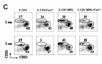
89 FIGURE
90 FIGURE 2.2, Continued 75
91 Figure 2.3. ELISpot analysis of anti-sm ASCs in 2-12H, 2-12H/Fas lpr, and 2-12HMRL/Fas lpr mice. A. Spleen cells were stained for expression of CD19, CD138, and Sm. The CD138-expression of CD19 +, Sm + cells is shown. Boxes indicate the CD138 int and CD138 hi populations. The percent of each population is provided. Data are representative of 3 or more mice of each strain. The percentages of CD138 int B cells are: 17%±2.4% for 2-12H, 14%±3.1% for 2-12H/Fas lpr, 7.0%±1.1% for 2-12H MRL, and 7.7%±0.50% for 2-12H MRL/Fas lpr. The percentages of CD138 hi B cells are: 0.13%±0.07% for 2-12H, 0.09%±0.01% for 2-12H/Fas lpr, 1.4%±0.46% for 2-12H MRL, and 1.9%±0.66% for 2-12H MRL/Fas lpr. B. Size, IgM expression, and CXCR4 expression are shown for anti-sm CD138 int and CD138 hi B cells from mice of the indicated strains. The gates are illustrated in A. Data are representative of 3 or more mice. C. Spleen cells were stained for CD19, CD138, CD21, and CD23. The gating scheme used to sort the FO, MZ and CD138 + cells is indicated using 2-12H spleen cells. The top histogram shows the identification of CD19 +, CD138 + cells sorted for ELISpot analysis. The CD19 + CD138 - cells were further fractionated by CD21 and CD23 expression to identify FO and MZ B cells as shown. D. The number of small and large anti-sm ELISpots was determined for each of the indicated cell populations. The definition of large and small ELISpots is given in the Materials and Methods. The average size of small ELISpots characteristic of MZ and FO B cells is 2 x 10⁻³ ± 4 x 10⁻⁴ mm 2, and the average size of the large ELISpots characteristic of CD138 + pre-pcs is 1.7 x 10⁻² ± 4 x 10⁻⁴ mm 2. Examples of the small FO/MZ-like spots and large CD138 + pre- PC ELISpots are shown. The number of FO anti-sm ELISpots per 10 5 cells in 2-12H and 2-12H/Fas lpr mice is 23.3 ± 2.36 and 3.51 ± 2.48, respectively (p<0.001). The number of MZ anti-sm ELISpots per 10 5 cells in 2-12H and 2-12H/Fas lpr mice is 183 ± 19.2 and 22.5 ± 10.9, respectively (p=0.002). The number of CD138 + pre-pc anti-sm ELISpots per 10 5 cells from 2-12H, 2-12H/Fas lpr, and 2-12H MRL/Fas lpr mice is 26.0 ± 5.6, 1540 ± 135, and 7600 ± 265, respectively (p<0.01 for all comparisons). ND: none detected. 76

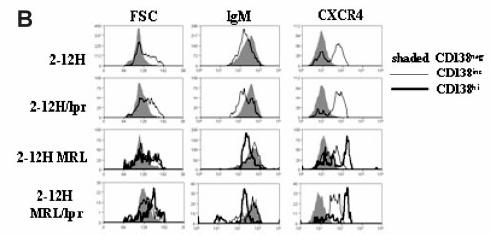
92 FIGURE


93 FIGURE 2.3, Continued 78
94 Figure 2.4. Peritoneal anti-sm B-1 cells in 2-12H Tg, 2-12H MRL, 2-12H/Fas lpr, and 2-12H MRL/Fas lpr mice. A. Phenotypic analysis of peritoneal B cells from 2-12H Tg, 2-12H MRL, 2-12H/Fas lpr, and 2-12H MRL/Fas lpr mice. Peritoneal cells from 1- month-old and 3-month-old mice were stained with CD19, IgM, CD11b, and Sm. Anti- Sm B-1 cells are boxed. All histograms are gated on CD19 + B cells. B. Analysis of total B-1 cell numbers (upper and lower graphs) and anti-sm B cell numbers (middle) for mice between 1 and 6 months of age is shown. 79

95 FIGURE
96 FIGURE 2.5 Figure 2.5. Anti-Sm B-1 cell activation in Fas lpr mice after peritoneal cell transfer. A. 2-12H/Fas lpr peritoneal cells were stained for CD19 and CD11b, and the CD19 + CD11b + B-1 cells and CD19 + CD11b - B-2 cells were sorted and transferred to Fas lpr recipient mice. Two weeks after transfer anti-sm ASCs in the bone marrow, spleen, MLN, and LP were quantified by ELISpot. Asterisks indicate that differences (p<0.05) between wt and Fas lpr recipients. ND: none detected. B. Unsorted peritoneal cells from 2-12H/Fas lpr mice were transferred to wild type (wt) or Fas lpr recipient mice. ELISA was used to measure serum IgM a+ anti-sm (inset) and ELISpot was used to quantify anti-sm ASCs from BM, spleen, MLN, and LP. C. The same as B except that 2-12H peritoneal cells were transferred to wt or Fas lpr recipients. 81
 are boxed and the percent of total CD19 + B cells is indicated. B. The number of PtCspecific B-1 cells present in the peritoneal cavity of 6-1 and 6-1/Fas lpr between 1 and 6 months is graphed.")
97 FIGURE 2.6 Figure 2.6. The effect of Fas lpr on PtC-specific B cells. A. Comparison of 6-1 and 6-1/ Fas lpr peritoneal B cells for liposome binding and CD43 and CD5 expression. Histograms are gated on CD19 + B cells. PtC-specific B-1 cells (CD43 + and CD5 + ) are boxed and the percent of total CD19 + B cells is indicated. B. The number of PtCspecific B-1 cells present in the peritoneal cavity of 6-1 and 6-1/Fas lpr between 1 and 6 months is graphed. PtC-specific B-1 cells were gated as shown in A. C. Number of IgM a ASCs in wt and Fas lpr recipient mice 2 weeks after transfer of 6-1 peritoneal cells. 82
98 Figure 2.7. Apoptotic cells in wt and Fas lpr mice. A. Flow cytometry analysis for the presence of apoptotic cells in the spleens and MLNs of wt and Fas lpr mice by FITC- VAD-FMK staining is shown. Representative histograms for wt and Fas lpr spleen cells are shown (top). Each histogram is gated on lymphocytes and the percent VAD-FMK + is provided. The graph below shows the average ± SEM of percent apoptotic lymphocytes in the spleen and MLN of mice at 2 months and 3 months of age (n=3). ** indicates a significant difference with wt (P<0.01). B. TUNEL positive cell distributions in spleen and MLN of both wt and Fas lpr mice. Both low (10X) and high (40X) magnification are shown. Boxes indicate the areas of higher magnification. WP: white pulp; RP: red pulp; GC: germinal center; F: follicle. C. Macrophages from wt, Fas lpr, MRL/Fas lpr, and mertk kd mice were incubated with apoptotic thymocytes for 60 min and the percent of phagocytized thymocytes determined by fluorescent microscopy. Controls are the percentage of live thymocytes (non-apoptotic) phagocytized. The graph is representative of four experiments and shows the average ± SEM of 3 mice per strain at both 2 months and 3 months of age. * P<0.05 in comparison to wt. 83

99 FIGURE
100 F. REFERENCES 1. Kono DH, Theofilopoulos AN. Genetics of systemic autoimmunity in mouse models of lupus. Int Rev Immunol. 2000;19: Eisenberg RA, Tan EM, Dixon FJ. Presence of anti-sm reactivity in autoimmune mouse strains. J Exp Med. 1978;147: Kakkanaiah VN, Sobel ES, MacDonald GC, Cheek RL, Cohen PL, Eisenberg RA. B cell genotype determines the fine specificity of autoantibody in lpr mice. J Immunol. 1997;159: Watanabe-Fukunaga R, Brannan CI, Copeland NG, Jenkins NA, Nagata S. Lymphoproliferation disorder in mice explained by defects in Fas antigen that mediates apoptosis. Nature. 1992;356: Rieux-Laucat F, Le Deist F, Hivroz C, et al. Mutations in Fas associated with human lymphoproliferative syndrome and autoimmunity. Science. 1995;268: Fisher GH, Rosenberg FJ, Straus SE, et al. Dominant interfering Fas gene mutations impair apoptosis in a human autoimmune lymphoproliferative syndrome. Cell. 1995;81: Theofilopoulos AN, Dixon FJ. Murine models of systemic lupus erythematosus. Adv Immunol. 1985;37: Cohen PL, Eisenberg RA. Lpr and gld: single gene models of systemic autoimmunity and lymphoproliferative disease. Annu Rev Immunol. 1991;9: Santulli-Marotto S, Retter MW, Gee R, Mamula MJ, Clarke SH. Autoreactive B cell regulation: peripheral induction of developmental arrest by lupus-associated autoantigens. Immunity. 1998;8: Borrero M, Clarke SH. Low-affinity anti-smith antigen B cells are regulated by anergy as opposed to developmental arrest or differentiation to B-1. J Immunol. 2002;168: Culton DA, O'Conner BP, Conway KL, et al. Early pre-plasma cells define a tolerance checkpoint for autoreactive B cells. J Immunol. 2006;In press. 12. Santulli-Marotto S, Qian Y, Ferguson S, Clarke SH. Anti-Sm B cell differentiation in Ig transgenic MRL/Mp-lpr/lpr mice: altered differentiation and an accelerated response. J Immunol. 2001;166:
101 13. Xu H, Li H, Suri-Payer E, Hardy RR, Weigert M. Regulation of anti-dna B cells in recombination-activating gene- deficient mice. J Exp Med. 1998;188: Martin F, Kearney JF. B-cell subsets and the mature preimmune repertoire. Marginal zone and B1 B cells as part of a "natural immune memory". Immunol Rev. 2000;175: Grimaldi CM, Michael DJ, Diamond B. Cutting edge: expansion and activation of a population of autoreactive marginal zone B cells in a model of estrogeninduced lupus. J Immunol. 2001;167: Thien M, Phan TG, Gardam S, et al. Excess BAFF rescues self-reactive B cells from peripheral deletion and allows them to enter forbidden follicular and marginal zone niches. Immunity. 2004;20: Mackay F, Woodcock SA, Lawton P, et al. Mice transgenic for BAFF develop lymphocytic disorders along with autoimmune manifestations. J Exp Med. 1999;190: Batten M, Groom J, Cachero TG, et al. BAFF mediates survival of peripheral immature B lymphocytes. J Exp Med. 2000;192: Watanabe N, Ikuta K, Nisitani S, Chiba T, Honjo T. Activation and differentiation of autoreactive B-1 cells by interleukin 10 induce autoimmune hemolytic anemia in Fas-deficient antierythrocyte immunoglobulin transgenic mice. J Exp Med. 2002;196: Mohan C, Morel L, Yang P, Wakeland EK. Genetic dissection of systemic lupus erythematosus pathogenesis: Sle2 on murine chromosome 4 leads to B cell hyperactivity. J Immunol. 1997;159: Xu Z, Butfiloski EJ, Sobel ES, Morel L. Mechanisms of peritoneal B-1a cells accumulation induced by murine lupus susceptibility locus Sle2. J Immunol. 2004;173: Qian Y, Wang H, Clarke SH. Impaired clearance of apoptotic cells induces the activation of autoreactive anti-sm marginal zone and B-1 B cells. J Immunol. 2004;172: Scott RS, McMahon EJ, Pop SM, et al. Phagocytosis and clearance of apoptotic cells is mediated by MER. Nature. 2001;411: Casciola-Rosen LA, Anhalt G, Rosen A. Autoantigens targeted in systemic lupus erythematosus are clustered in two populations of surface structures on apoptotic keratinocytes. J Exp Med. 1994;179:
102 25. Arnold LW, Pennell CA, McCray SK, Clarke SH. Development of B-1 cells: segregation of phosphatidyl choline-specific B cells to the B-1 population occurs after immunoglobulin gene expression. J Exp Med. 1994;179: Qian Y, Santiago C, Borrero M, Tedder TF, Clarke SH. Lupus-specific antiribonucleoprotein B cell tolerance in nonautoimmune mice is maintained by differentiation to B-1 and governed by B cell receptor signaling thresholds. J Immunol. 2001;166: Mercolino TJ, Arnold LW, Hawkins LA, Haughton G. Normal mouse peritoneum contains a large population of Ly-1+ (CD5) B cells that recognize phosphatidyl choline. Relationship to cells that secrete hemolytic antibody specific for autologous erythrocytes. J Exp Med. 1988;168: Pop SM, Wong CP, Culton DA, Clarke SH, Tisch R. Single cell analysis shows decreasing FoxP3 and TGFbeta1 coexpressing CD4+CD25+ regulatory T cells during autoimmune diabetes. J Exp Med. 2005;201: Mandik-Nayak L, Seo SJ, Sokol C, Potts KM, Bui A, Erikson J. MRL-lpr/lpr mice exhibit a defect in maintaining developmental arrest and follicular exclusion of anti-double-stranded DNA B cells. J Exp Med. 1999;189: Li Y, Li H, Ni D, Weigert M. Anti-DNA B Cells in MRL/lpr Mice Show Altered Differentiation and Editing Pattern. J Exp Med. 2002;196: Ahn JH, Park SM, Cho HS, et al. Non-apoptotic signaling pathways activated by soluble Fas ligand in serum-starved human fibroblasts. Mitogen-activated protein kinases and NF-kappaB-dependent gene expression. J Biol Chem. 2001;276: Wajant H, Pfizenmaier K, Scheurich P. Non-apoptotic Fas signaling. Cytokine Growth Factor Rev. 2003;14: Rothstein TL, Wang JK, Panka DJ, et al. Protection against Fas-dependent Th1- mediated apoptosis by antigen receptor engagement in B cells. Nature. 1995;374: Rathmell JC, Cooke MP, Ho WY, et al. CD95 (Fas)-dependent elimination of self-reactive B cells upon interaction with CD4+ T cells. Nature. 1995;376: Seo SJ, Fields ML, Buckler JL, et al. The impact of T helper and T regulatory cells on the regulation of anti-double-stranded DNA B cells. Immunity. 2002;16: Sakic B, Maric I, Koeberle PD, et al. Increased TUNEL staining in brains of autoimmune Fas-deficient mice. J Neuroimmunol. 2000;104:
103 37. Evan GI, Vousden KH. Proliferation, cell cycle and apoptosis in cancer. Nature. 2001;411: Kuan AP, Cohen PL. p53 is required for spontaneous autoantibody production in B6/lpr lupus mice. Eur J Immunol. 2005;35: Botto M, Dell'Agnola C, Bygrave AE, et al. Homozygous C1q deficiency causes glomerulonephritis associated with multiple apoptotic bodies. Nat Genet. 1998;19: Hanayama R, Tanaka M, Miyasaka K, et al. Autoimmune disease and impaired uptake of apoptotic cells in MFG-E8-deficient mice. Science. 2004;304: Mahoney JA, Rosen A. Apoptosis and autoimmunity. Curr Opin Immunol. 2005;17: Herrmann M, Voll RE, Zoller OM, Hagenhofer M, Ponner BB, Kalden JR. Impaired phagocytosis of apoptotic cell material by monocyte-derived macrophages from patients with systemic lupus erythematosus. Arthritis Rheum. 1998;41: Baumann I, Kolowos W, Voll RE, et al. Impaired uptake of apoptotic cells into tingible body macrophages in germinal centers of patients with systemic lupus erythematosus. Arthritis Rheum. 2002;46: Potter PK, Cortes-Hernandez J, Quartier P, Botto M, Walport MJ. Lupus-prone mice have an abnormal response to thioglycolate and an impaired clearance of apoptotic cells. J Immunol. 2003;170: Zurier RB. Reduction of phagocytosis and lysosomal enzyme release from human leukocytes by serum from patients with systemic lupus erythematosus. Arthritis Rheum. 1976;19: Rescigno M, Piguet V, Valzasina B, et al. Fas engagement induces the maturation of dendritic cells (DCs), the release of interleukin (IL)-1beta, and the production of interferon gamma in the absence of IL-12 during DC-T cell cognate interaction: a new role for Fas ligand in inflammatory responses. J Exp Med. 2000;192: Guo Z, Zhang M, Tang H, Cao X. Fas signal links innate and adaptive immunity by promoting dendritic-cell secretion of CC and CXC chemokines. Blood. 2005;106: Kataoka T, Budd RC, Holler N, et al. The caspase-8 inhibitor FLIP promotes activation of NF-kappaB and Erk signaling pathways. Curr Biol. 2000;10:
104 49. Hu WH, Johnson H, Shu HB. Activation of NF-kappaB by FADD, Casper, and caspase-8. J Biol Chem. 2000;275: Ogden CA, Pound JD, Batth BK, et al. Enhanced apoptotic cell clearance capacity and B cell survival factor production by IL-10-activated macrophages: implications for Burkitt's lymphoma. J Immunol. 2005;174: Popi AF, Lopes JD, Mariano M. Interleukin-10 secreted by B-1 cells modulates the phagocytic activity of murine macrophages in vitro. Immunology. 2004;113: Reap EA, Sobel ES, Cohen PL, Eisenberg RA. Conventional B cells, not B-1 cells, are responsible for producing autoantibodies in lpr mice. J Exp Med. 1993;177: Wither JE, Roy V, Brennan LA. Activated B cells express increased levels of costimulatory molecules in young autoimmune NZB and (NZB x NZW)F(1) mice. Clin Immunol. 2000;94: Hayakawa K, Hardy RR, Honda M, Herzenberg LA, Steinberg AD. Ly-1 B cells: functionally distinct lymphocytes that secrete IgM autoantibodies. Proc Natl Acad Sci U S A. 1984;81: Hayakawa K, Hardy RR, Parks DR, Herzenberg LA. The "Ly-1 B" cell subpopulation in normal immunodefective, and autoimmune mice. J Exp Med. 1983;157: Murakami M, Tsubata T, Okamoto M, et al. Antigen-induced apoptotic death of Ly-1 B cells responsible for autoimmune disease in transgenic mice [see comments]. Nature. 1992;357: Martin F, Oliver AM, Kearney JF. Marginal zone and B1 B cells unite in the early response against T- independent blood-borne particulate antigens. Immunity. 2001;14: Attanavanich K, Kearney JF. Marginal zone, but not follicular B cells, are potent activators of naive CD4 T cells. J Immunol. 2004;172:
105 CHAPTER 3: ANTI-Sm FOLLICULAR, MARGINAL ZONE, AND EARLY PRE-PC B CELL ACTIVATION IN Fas lpr MICE
106 A. ABSTRACT Autoreactive splenic B cells specific for the Smith (Sm) ribonucleoprotein antigen belong to multiple subsets: follicular (FO), marginal zone (MZ), and pre-plasma cell (pre-pc). However, anti-sm B cells are not activated to become antibody secreting cells (ASCs) in non-autoimmune mice, but are activated in Fas lpr, MRL, and MRL/Fas lpr mice. To identify the anti-sm B cells activated in autoimmune mice, we performed adoptive transfer experiments of anti-sm Fas wt B cells into Fas lpr recipient mice. We find that both anti-sm FO and MZ B cells become early pre-pcs and that they are blocked from activation by a B cell-extrinsic mechanism. However, anti-sm MZ and pre-pcs give rise to short-lived and immediate ASC responses, while anti-sm FO B cells give rise to a delayed and long-term ASC response that is restricted to the spleen. In addition, anti-sm FO B cells undergo extensive proliferation to generate a memory B cell-like population in the spleen. Thus, the activation of MZ and FO B cells in this autoimmune response mirrors the activation of MZ and FO B cells in response to foreign antigen, indicating that the activation programs inherent in B cells of each subset are maintained in autoimmunity. 91
107 B. INTRODUCTION The mature, splenic B cell populations in mice, consisting of follicular (FO), marginal zone (MZ), and pre-plasma cells (pre-pcs), have distinct phenotypes and functions, suggesting their regulation and potential contributions to disease may be different. FO B cells are recirculating, naïve B cells that give rise to primarily T- dependent antigens involving germinal center formation, where somatic hypermutation, H chain class switch, and affinity maturation occur. The products of germinal center reactions are long-lived plasma cells (PCs) that localize primarily to the bone marrow and recirculating memory B cells[1, 2]. MZ B cells, on the other hand, are noncirculating antigen-experienced B cells that give rise to primarily to T-independent type 2 (TI-2) responses[3-7]. These cells have a pre-activated phenotype and thus are able to rapidly differentiate to PCs upon antigen exposure[4, 8]. Upon activation they migrate to the T-B border where they proliferate and quickly differentiate to short-lived PCs that remain in the spleen[4, 8]. We have previously described an early pre-pc subset of B cells in mice[9]. They are defined by expression of CD19, B220, and IgM at levels typical of B cells, but to also express the PC-specific marker CD138 (syndecan) at intermediate levels (CD138 int ). Like MZ B cells, they are positively selected, but have a more rapid turnover rate (~3 weeks). Intriguingly, a high frequency of pre-pcs in nonautoimmune mice are autoreactive suggesting they could have a prominent role in autoantibody production in autoimmunity[9]. The role of B cells of these subsets in autoimmunity has not been clearly elucidated. Many groups have highlighted the involvement of MZ B cells, and their 92
108 close peritoneal counterpart, B-1 B cells, in the development of autoimmunity[10]. Our analysis of B cells specific for the Sm ribonucleoprotein autoantigen using the Ig H chain Tg mice (2-12H) has indicated that they are present in all mature B cell subsets, including those (MZ, B-1, and pre-pc) that are antigen selected. FO B cells appear to be anergic and regulated by both intrinsic and extrinsic mechanisms. These B cells respond poorly to LPS and their response to LPS is further repressed by production of IL-6, CD40L, and TNFα by DCs and macrophages[10-12]. In contrast, anti-sm MZ and B-1 cells appear to be functional, and our analysis of anti-sm pre-pcs suggests that they are short-lived (~7 days), but how they are regulated has yet to be determined[9]. Nevertheless, the presence of both positively and negatively selected anti-sm B cells in non-autoimmune mice does not result in autoantibody production, indicating that tolerance to Sm is maintained and that even positively selected functional B cells are not normally activated[10, 13]. Tolerance to Sm is lost in 2-12H mice that carry the lpr mutation of the proapoptosis gene Fas (Fas lpr ), or that have been backcrossed to the autoimmune MRL background[10]. Which B cells are activated in these mice is not clearly understood. Anti-Sm pre-pcs overcome the tolerance checkpoint that prevents their differentiation to PCs indicating that they are involved, but it is not known whether FO or MZ B cells are precursors to pre-pcs. Both anti-sm MZ and B-1 B cells decrease in number between 2 and 3 months of age, which corresponds to the appearance of anti-sm in the serum, suggesting that both subsets are activated in autoimmune mice. Transfer of 2-12H B-1 B cells to Fas lpr mice confirms the activation of anti-sm B-1 B cells which are activated to form ASCs in the mesenteric lymph node and lamina propria of the gut[10]. Whether 93
109 MZ and FO B cells are also activated is not known, and nor is the nature of the response each cell type generates known. In this report we demonstrate that both FO and MZ B cells can differentiate to pre-pcs and that these cells are functional and blocked in their activation in nonautoimmune mice by a pre-pc-extrinsic mechanism. We also demonstrate that FO, MZ, and pre-pcs are activated in autoimmune Fas lpr mice. However, the responses generated by the B cells of each subset follow distinct activation programs that parallel their responses to foreign antigen. Thus, the activation programs inherent to each cell type are not altered in responses to self-antigen. 94
110 C. MATERIALS AND METHODS Mice Fas lpr and 2-12H Tg mice have been previously described[13-15]. The 2-12H Tg mice have been backcrossed 8 times to the C57BL/6 (B6) background. The 2-12H transgene was identified by PCR of tail genomic DNA as previously described. Animals were housed and bred in a conventional facility at the University of North Carolina at Chapel Hill (Chapel Hill, NC) and all animal experiments were carried out with institutional IACUC approval. Flow Cytometry Cells were stained as previously described and analyzed at the University of North Carolina at Chapel Hill Flow Cytometry Facility (Chapel Hill, NC) using a FACSCalibur (BD Biosciences) or CyAn (DakoCytomation, Denmark)[15, 16]. The antibodies specific for IgM a (DS-1), CD19 (1D3), B220 (RA3-6B2), CD21 (7G6), CD23 (B3B4), and CD138 (281-2) were conjugated to FITC, PE, biotin, APC, or APC-Cy7 (Pharmingen, San Diego, CA). For the detection of anti-sm B cells, we biotinylated Sm (SMA-3000; Immunovision, Springdale, AR) as previously described. Biotinylated reagents were revealed with streptavidin conjugated to either PerCP or APC (Pharmingen). Data were analyzed using Summit software (DakoCytomation). All data represent cells that fall within the lymphocyte gate determined by forward and 90 light scatter cells per sample were analyzed. 95
111 Cell sorting 2-12H Tg splenocytes were stained for B220, CD21, CD23, and CD138 as described above for cell sorting of FO B cells, MZ B cells and early pre-pcs for ELISpot analysis and adoptive transfer experiments. MZ B cells were sorted as B220 +, CD21 hi, CD23 -/lo CD138 - cells, FO B cells were sorted as B220 +, CD21 low, CD23 +, CD138 - cells, and early pre-pcs were sorted as B220 +, CD138 + cells. The cells were sorted using a MoFlo high speed sorter (DakoCytomation). Sorted populations were >90% pure as determined by reanalysis. CFSE labeling of sorted cells In denoted experiments, cells were CFSE labeled prior to adoptive transfer via manufacturer s protocol (CellTrace CFSE Cell Proliferation Kit, Invitrogen, San Diego, CA). A concentration of 10µM CFSE dye was used due to the longevity of our adoptive transfer experiments. B cell subset adoptive transfers 2-12H Tg FO, MZ, and early pre-pc populations were sorted as described above. 1x10 6 sorted cells in PBS were injected i.v. into either non-transgenic Fas wt or Fas lpr recipients. On days 3, 4, 12, or 30 post-transfer, cells from spleen and bone marrow were harvested for ELISpot and flow cytometry analysis. Donor origin cells were identifiable through their expression of IgM a, as they were transferred to recipients with IgM b B cells. 96
112 ELISpot assay ELISpots were performed as previously described using plates (Millipore, Billerica, MA) that we coated with 10 U/well Sm Ag (Immunovision, Springdale, AR) or anti-igm (Southern Biotech, Birmingham, AL)[9, 10]. Cells were resuspended in HL-1 medium (Cambrex) supplemented with 1% L-glutamine and 1% penicillin/streptomycin prior to plating. For sorted early pre-pc ex vivo stimulations, 5x10 3 cells were plated per well and 10 ng/ml IL-4 (Peprotech, Rocky Hill, NJ), 10 µg/ml anti-cd40 (Pharmingen), 30 µg/ml LPS (Invivogen, San Diego, CA), or 1 µg/ml CpG (Invivogen) were added to designated wells prior to their 72 hour incubation. For spleen and BM samples harvested after adoptive transfer experiments, approximately 5x 10 6 cells were plated per well and incubated for 24 hours. Secreted 2-12H-specific anti-sm or IgM was detected by using biotin-labeled anti-igm a Ab and streptavidin-hrp (Pharmingen), while secreted antibodies from non-tg mice were detected by using biotin-labeled anti-igm b Ab and streptavidin-hrp (Pharmingen). Incubation with the developing substrate 3-amino-9- ethylcarbazole (Sigma-Aldrich, St. Louis, MO) allowed for ASC detection. The plates were scanned and analyzed using the Immunospot ELISpot analyzer (Cellular Technology Ltd., Cleveland, OH). Transcription Factor Staining Cells were prepared as previously described before being fixed with 1.5% paraformaldehyde (Electron Microscopy Sciences, Hatfield, PA) in PBS for 10 minutes 97
113 at 4 C. After fixation, cells were permeabilized with methanol for 30 minutes at 4 C. Cells were then stained for both surface markers and transcription factors for 1 hour at room temperature. Pax-5(C20), XBP-1 (M-186), and IRF-4 (M-17) [Santa Cruz Biotechnology, Santa Cruz, CA] and their isotype controls were each labeled with Zenon technology per manufacturer s protocol (Invitrogen, San Diego, CA). Cells were then washed with RPMI 3 times for 10 minutes each before running on the CyAn (DakoCytomation). Statistical analysis The independent and paired Student s t tests were used to evaluate significance. A value of p<0.05 was considered significant. 98
114 D. RESULTS Anti-Sm pre-pcs are functional and regulated by extrinsic mechanisms. We previously demonstrated that anti-sm early pre-pcs from 2-12H mice are antigenselected, but are regulated to prevent their differentiation to ASCs[9]. As previously reported, neither CD138 neg nor CD138 int B cells from 2-12H Tg mice spontaneously secrete anti-sm[9]. However, significantly more CD138 int early pre-pcs than CD138 neg B cells from non-tg mice spontaneously secrete IgM, suggesting that the block in differentiation of anti-sm CD138 int pre-pcs is due to their auto specificity[9]. Several mechanisms may explain regulation of anti-sm pre-pcs. Regulation may be intrinsic, such as by the active mechanisms of anergy or ongoing cell death. This is suggested by their short half-life relative to non-tg pre-pcs (7 days vs. 21 days), and the higher frequency of anti-sm pre-pcs staining with the caspase inhibitor VAD-FMK (17% vs. 4%)[9]. Alternatively, regulation may be through an extrinsic mechanism such as the lack of an activating signal or the presence of a negative signal. To discriminate between extrinsic or intrinsic regulation, we assessed the ability of 2-12H pre-pcs to be activated in vitro. B cells (B220 +, CD138 neg ) and early pre-pcs (B220 + CD138 int ) were sorted from 2-12H Tg mice and cultured in the presence or absence of various B cell activators. Purities of both populations were consistently greater than 92% (Fig. 3.1A). Stimulation with anti-cd40 and rmil-4 induced an increase in IgM ASC differentiation by 2-12H CD138 int pre-pcs, which was significantly greater than the increase by CD138 neg B cells, but comparable to that exhibited by CD138 int non-tg pre-pcs (Fig. 3.1B, top middle). Despite the fact that 2-12H CD138 int responded to anti-cd40 and 99
115 rmil-4 better than CD138 neg B cells, the increase in anti-sm ASCs was similar (Fig. 3.1B, top middle). Thus, anti-sm pre-pcs are responsive to activation signals that mimic T cell help. Anti-Sm pre-pcs were also responsive to TLR agonists. Non-Tg early pre-pcs were ~4 times more responsive to LPS than CD138 neg B cells indicating that pre-pcs were more responsive to LPS than B cells (Fig. 3.1B, bottom middle). 2-12H CD138 int and CD138 neg B cells gave rise to similar frequencies of IgM ASCs. Unlike their responsiveness to LPS, CD138 int and CD138 neg B cells are equally responsive to CpG. In addition, 2-12H pre-pcs gave rise to significantly higher frequencies of anti-sm ASCs after LPS activation than 2-12H CD138 neg B cells. In contrast, CpG stimulation induced similar frequencies of IgM ASCs from CD138 neg and CD138 int B cells and non- Tg and 2-12H B cell populations were not different. However, CpG activation induced significantly more anti-sm ASCs by 2-12H CD138 int pre-pcs than 2-12H CD138 neg B cells (Fig. 3.1B, bottom). Interestingly, responsiveness to CpG by anti-sm pre-pcs was much greater than to LPS or anti-cd40/rmil-4, and a much higher proportion of the activated 2-12H pre-pcs were anti-sm ASCs in response to CpG (~50%) than to LPS (~7%) or anti-cd40/rmil-4 (~14%). Thus, CpG is a potent activator of anti-sm pre- PCs. Taken together, the TLR agonist and anti-cd40/il-4 stimulations indicate that despite the differences in spontaneous activation, half-life, and frequency of apoptosis, 2-12H pre-pcs and 2-12H anti-sm pre-pcs are functional. These data are inconsistent with B cell-intrinsic regulation by anergy or cell death, suggesting this block of early pre-pc differentiation is through a B cell-extrinsic mechanism. 100
116 To examine this issue in vivo, we adoptively transferred sorted 2-12H CD138 int pre-pcs into Fas wt and Fas lpr mice. 2-12H/Fas lpr mice develop a spontaneous anti-sm response between 2 and 3 months of age[10]. Donor pre-pcs were detected at high frequencies (66±3% and 59±7% of number injected) by flow cytometry in Fas wt recipient spleens at 3 and 12 days post transfer (Table 3.1). However anti-sm ASCs were not detected at either time point (Fig. 3.1C and Table 3.1). At 30 days posttransfer, we detected neither donor B cells nor anti-sm ASCs (Table 3.1), and at no time were donor anti-sm ASCs or B cells detected in the bone marrow (data not shown). In contrast, we observed rapid ASC differentiation by donor B cells in Fas lpr recipients. As in Fas wt recipients, a high frequency of donor pre-pcs were detected in the spleen at both 3 and 12 days (70±8% and 74±3% of number injected), but unlike in Fas wt recipients, 93% of donor cells in the spleens of Fas lpr recipients were ASCs by day 3 (Fig. 3.1C and Table 3.1). Anti-Sm ASCs are not detectable by day 12 and 30 (Fig. 3.1C and Table 3.1), and as with Fas wt recipients, there were no detectable donor origin cells or donor origin ASCs in the BM at any time point (data not shown). Thus, anti-sm pre-pcs are not intrinsically defective in their ability to be activated, consistent with our in vitro findings. Anti-Sm FO and MZ B cells can both acquire an early pre-pc phenotype. To identify the cellular precursors of anti-sm pre-pcs, we sorted 2-12H FO (CD23 pos CD21 int ) and MZ (CD23 neg CD21 hi ) B cells, as illustrated in Figure 3.2A, and adoptively transferred them to non-tg recipients. After 4 days recipient splenocytes were analyzed by flow cytometry to assess donor cell (IgM a+ ) expression of the early 101
117 pre-pc marker CD138. A significantly higher percentage of transferred 2-12H Tg MZ B cells (49±4% of injected) than FO B cells (21±3% of injected) were detected in the spleen (Table 3.2), and this percentage decreased somewhat at 12 days post-transfer (34±2% of MZ B cells injected, 15±1% of FO B cells injected) (Table 3.2). Neither subset was detected 30 days-post transfer (Table 3.2). IgM a+ donor origin cells from both the FO and MZ B cell subsets were heterogeneous for Sm affinity, each possessing both Sm hi and Sm lo binders (Fig. 3.2C and D). Interestingly, all of the Sm hi donor cells in both FO and MZ recipient mice had acquired CD138 expression, while the Sm lo B cells of both subtypes remained predominantly CD138 neg (Fig. 3.2C and D). Thus, both MZ and FO B cells can differentiate to early pre-pcs, and the ability for those of 2-12H origin to differentiate is dependent on the ability to bind Sm. Both anti-sm MZ and FO B cells are activated in autoimmune mice, but follow different activation programs. To determine whether anti-sm MZ and FO B cells contribute to the anti-sm response in Fas lpr mice, we transferred sorted 2-12H FO and MZ B cells to Fas wt and Fas lpr mice. At 4 and 12 days post-transfer into Fas wt mice, neither anti-sm FO nor anti-sm MZ B cells became ASCs (Fig. 3.3A and 3.4A), although cells of each subset were detected in the spleen at 4 and 12 days post-transfer (Table 3.2). After 30 days, no transferred cells from either population were detected in the spleen or the BM (Table 3.2 and data not shown) and no anti-sm ASCs of donor origin were present in these tissues (Fig. 3.3A, 3.4A, and data not shown). In contrast to Fas wt recipients, Fas lpr recipients of 2-12H FO and MZ B cells developed anti-sm ASCs. Nearly two-thirds of transferred 2-12H Tg MZ B cells were detected in the spleen on day 4, and of these, ~72% had became anti-sm ASCs (Table 102
118 3.2 and Fig. 3.3A). The anti-sm ASCs were gone by day 12, even though there was little change in the number of donor B cells present in the spleen at this time (Table 3.2 and Fig. 3.3A). As expected, transferred 2-12H MZ B cells upregulated CD138 by day 4 and retained this expression on day 12, despite their inability to secrete antibody. To assess the extent of proliferation of these cells after transfer, sorted B cells were labeled with CFSE before transfer. As shown in Fig. 3B, transferred MZ B cells underwent extensive proliferation, as seen by a ~100-fold decrease in CFSE levels by day 4. There was no further dilution of CFSE on day 12 indicating that all of the proliferation of anti- Sm MZ B cells occurred within the first few days after transfer. No donor-derived B cells were detected in the spleens or bone marrow of recipient mice on day 30 (data not shown). Since the donor derived pre-pcs in Fas lpr recipient mice were present in relatively equal numbers on days 4 and 12 post transfer, but were only ASCs on day 4, we examined the phenotype of these cells for differences in size, granularity, and transcriptional program that could explain for the lack of antibody secretion on day 12. At 4 days post-transfer of anti-sm MZ B cells, donor-derived B cells were larger (FSC) and more granular (SSC) than non-transferred MZ B cells, and had a transcription program consistent with PC differentiation (Fig. 3.3C). Pax5 is a B cell specific transcription factor that is downregulated upon PC differentiation, and XBP-1 and IRF-4 are PC-specific transcription factors that are required for PC differentiation[17-22]. As shown in Fig. 3.3C, Pax5 expression was downregulated, while XBP-1 and IRF-4 levels were elevated compared to non-transferred MZ B cells. At 12 days post transfer, donor B cells had further increased in size, but had decreased in granularity compared to non- 103
119 transferred MZ B cells (Fig. 3.3C). These cells remained low in PAX-5 expression and high in IRF-4 expression (Fig. 3.3C). However, while about half of the donor cells increased XBP-1 expression, the other half decreased IRF-4 expression (Fig. 3.3C). Thus, with the exception of granularity and XBP-1 expression, donor-derived B cells on day 12 largely retained a PC-specific pattern of transcription factor expression, despite the fact that they are not ASCs. Since donor cells were not present on day 30, we examined VAD-FMK staining to determine whether the donor B cells were undergoing apoptosis. Supporting our hypothesis, a larger percentage of donor B cells were VAD- FMK + on day 12 than on day 4 suggesting that the lack of antibody secretion is due to ongoing apoptosis (data not shown). We conclude that anti-sm MZ B cells undergo an immediate burst of cell division and differentiate rapidly to pre-pcs and short-lived ASCs in Fas lpr mice. Anti-Sm FO B cells exhibit a delayed ASC response when transferred to Fas lpr mice. Despite the presence of large numbers of donor B cells in recipient spleens (~56% of injected FO B cells) on day 4, no anti-sm ASCs were detected (Table 3.2 and Fig. 3.4A). However, we detected anti-sm ASCs on day 12 in numbers ~4 times larger than those derived from transferred 2-12H MZ B cells on day 4 (Fig. 3.3A vs. Fig. 3.4A). In addition, we detected anti-sm ASCs in the spleens on day 30, although their number had decreased by about half. Donor FO B cells underwent extensive proliferation after transfer. At day 4, the proliferation of FO B cells resembled that for MZ B cells based on CFSE dilution, but in contrast to MZ B cells, which did not proliferate after day 4, FO B cells continued to proliferate extensively after day 4 and lost all CFSE. The number of donor B cells detected in the spleen by flow cytometry 104
120 amounted to 1600% of that transferred (Table 3.2). Thus, although FO B cells generated a larger number of anti-sm ASCs on day 12 than MZ B cells on day 4, this represented a much smaller proportion of the total donor cell number (0.27% vs. 72%) (Table 3.2). We did not detect B cells of donor origin on day 30 post-transfer, despite the presence of ASCs (data not shown and Fig. 3.4A), and we did not detect anti-sm ASCs in the bone marrow at any time point (data not shown). Similar to the 2-12H MZ B cell transfers (Fig. 3.3B), a high frequency of donor anti-sm FO B cells acquired CD138 expression by day 4 (~77%), but only ~7% of donor B cells expressed CD138 on day 12 (Fig. 3.4B). This suggests that the majority of B cells generated after day 12 are likely to be memory B cells. Consistent with the initial appearance of ASCs on day 12, the donor B cells on day 4 were similar to non-transferred 2-12H FO B cells in size, granularity and transcription factor expression (Fig. 3.4C). However, by day 12, the transferred cells had increased in size, decreased Pax5 expression, and increased XBP-1 and IRF-4 expression. Interestingly, although the majority of donor B cells have decreased Pax5 and increased IRF-4, a small subset has even lower Pax5 and higher IRF-4 (Fig. 3.4C). This corresponds roughly in size to the size of the CD138 int population. Thus, the majority of donor B cells on day 12 had acquired a more PC-like phenotype even though the majority did not express CD138 and were not ASCs. We conclude that anti-sm FO B cells contribute to the anti-sm response in Fas lpr mice, but that they contribute to a delayed and long-term response compared to anti-sm MZ B cells. In addition, these cells give rise to a large memory-like B cell population. This parallels the responses by 105
121 FO and MZ B cells to foreign antigens indicating that anti-sm FO and MZ B cells follow their normal activation programs in response to self-antigens[23]. 106
122 E. DISCUSSION We directly demonstrate the activation of B cells of the three mature B cell subsets in the spleens of Fas lpr mice; FO, MZ, and pre-pc. This activation involves overcoming at least two tolerance checkpoints, B cell anergy (FO) and the early pre-pc checkpoint (MZ and pre-pc). The B cells of these subsets differ in their activation programs and therefore make different contributions to autoimmunity, correlating with their functions in response to archetypal foreign antigens[23]. These data have implications for the development of therapeutic approaches to block activation of autoreactive B cells. Our analysis of 2-12H anti-sm B cells has indicated that the pre-pc stage is a tolerance checkpoint for anti-sm B cells[9]. Tolerance may be due to an intrinsic mechanism such as anergy or an ongoing cell death program, or the B cells may be regulated by an extrinsic mechanism, such as by cytokine-induced repression[11, 12] or the absence of an essential activating signal. The short half-life and high frequency of apoptosis of 2-12H pre-pcs compared to non-tg pre-pcs supports an ongoing death mechanism[9]. To address these issues, we stimulated sorted pre-pcs with two TLR agonists, LPS and CpG and with anti-cd40 and IL-4 to mimic T cell help. We find that non-tg and 2-12H pre-pcs are activated by all stimuli essentially equally well. This differs from the responsiveness of anergic 2-12H/Vκ8 FO B cells or of anergic B cells of multiple other autoantigen specificities[24-27]. Thus, we conclude that anti-sm pre-pcs are not anergic, and therefore likely regulated by extrinsic mechanisms. The ability of anti-sm pre-pcs to become ASCs after transfer to Fas lpr mice substantiates this. The 107
123 mechanism of regulation could be the lack of a positive signal or the presence of a negative signal. Repression by IL-6, TNFα, or CD40L secreted by DCs or macrophages is unlikely to be involved in pre-pc repression in the steady state[11, 12]. Although pre- PCs are susceptible to repression by IL-6 (M.A. Kilmon and B.J. Vilen, personal communication), this only acts to repress activation induced by LPS or another TLR agonist. In the absence of infection, pre-pcs are presumably not exposed to any of these cytokines. Whatever the mechanism of activation arrest, we conclude that anti-sm pre- PCs are functional and poised to become ASCs. Interestingly, these cells are more responsive to CpG than to either LPS or anti- CD40/IL-4, suggesting that they would be sensitive to activation by antigens that include CpG motifs. Marshak-Rothstein and colleagues have demonstrated that rheumatoid factor B cells and anti-dna B cells are activated by antigen that simultaneously provide a BCR and TLR9 (CpG) signal[28-31]. Thus, pre-pcs may be very susceptible to activation by self-antigens that include a CpG motif for TLR9 stimulation, and may be the dominate cell type activated in vivo by this mechanism. The precursor cells of pre-pcs can be either FO or MZ B cells, as adoptive transfers indicate that essentially all transferred FO and MZ B cells become CD138 int. In the case of MZ B cells, they also decrease CD21 expression and increase CD23 expression, characteristic of pre-pcs. However, we believe that only MZ B cells contribute to the pre-pc population in unmanipulated mice. First, pre-pcs are not present in mice that do not have a MZ B cell population, even though they have FO B cell populations. For example, pre-pcs are not present in either 2-12H/Vκ8 mice, which generate anergic anti-sm FO B cells, or in young anti-hel MD4 mice (<4 mos), which 108
124 only have anti-hel FO B cells (unpublished observations). Older MD4 mice (>4 mos) acquire a MZ B cell population, and also acquire a pre-pc population. Secondly, we show here, that anti-sm pre-pcs exclusively generate a short-lived, immediate ASC response when transferred to Fas lpr mice. Even though anti-sm FO B cells become pre- PCs after transfer to Fas lpr mice, they exclusively generate a delayed ASC response after transfer to Fas lpr mice. These data argue that anti-sm FO B cells contribute little to the pre-pc population in 2-12H mice. Thirdly, in Chapter 4, we demonstrate that DCs induce MZ B cell activation to become pre-pcs both in vivo and in vitro, but that they do not have this ability with FO B cells. Thus, we consider it likely that, although anti-sm B cells of both subsets can become pre-pcs, in the steady state MZ B cells are the primary source of pre-pcs in 2-12H mice. In addition to the activation of pre-pcs in Fas lpr mice, MZ and FO B cells are activated. Consistent with their precursor-product relationship, the activation of MZ B cells closely follows that of pre-pcs. This similarity between pre-pcs and MZ B cells is consistent with our evidence that anti-sm MZ B cells are precursors to anti-sm pre-pcs. Interestingly, although they are not ASCs, the pre-pcs derived from transferred MZ B cells are present on day 12. This suggests that pre-pcs can cease antibody secretion, consistent with changes in IRF-4 expression. The high frequency of apoptosis among these cells suggests that they may also be undergoing deletion. Thus, the MZ B cell response parallels classic T-independent MZ B cell responses to foreign antigen[23]. In contrast to MZ B cells and pre-pcs, FO B cells generate a delayed, but longlived ASC response. Anti-Sm ASCs are detected on day 12 and are still detected on day 30. Surprisingly, the response was exclusive to the spleen, as no ASCs were detected at 109
125 any time in the bone marrow, the normal site of long-lived ASCs[2, 32, 33]. Accompanying ASC differentiation of FO B cells is extensive proliferation and the generation of a CD138 neg B cell population. These cells may be memory B cells of germinal center origin. However, their transcription factor profile is more characteristic of PCs than memory B cells, particularly the low Pax5 expression[20, 21, 34, 35], and they are not present in the spleen on day 30, as would be expected of long-lived memory B cells. In future experiments it will be important to determine what these cells are. Thus, anti-sm FO B cells generate a long-lived response, typical of T-dependent responses by FO B cells specific for foreign antigens[23]. However, this differs from foreign antigen responses in at least two regards; absence of bone marrow involvement and disappearance of the putative memory B cell population. These data suggest that two anti-sm responses occur simultaneously in autoimmune mice. The first is from MZ B cells that differentiate to pre-pcs and then short-lived ASCs. By continual replacement of MZ B cells and exposure to self-antigen, this response could provide a persistent supply of short-lived, anti-sm ASCs. However, as these ASCs would not be GC products and are unlikely to have undergone affinity maturation, they would produce only low-affinity, anti-sm IgM antibodies that are likely to be non-pathogenic. The second response is from anti-sm FO B cells that differentiate into long-lived, anti-sm ASCs. They may form germinal centers and thus, the ASCs would be somatically mutated and more likely to produce high affinity, pathogenic IgG antibodies. Interestingly, analysis of anti-sm hybridomas from MRL/Fas lpr mice supports our two response theory as we have identified both heavily somatically mutated IgG hybridomas, as well as clonal populations of IgM hybridomas that are unmutated. 110
126 Unmated and clonally-related IgM hybridomas are rare among hybridomas specific for foreign antigens. Since the ASCs of FO B cell origin are likely to have the greatest impact on disease, a better understanding of this response and its differences from responses to foreign antigens will be important to design therapeutic approaches to interfere with FO B cell activation. 111
127 Figure 3.1. Anti-Sm pre-pcs are functional and regulated by extrinsic mechanisms. A. Sort scheme and post-sort purities depicting the CD138 neg and CD138 int populations isolated for experiments shown in B and C. Cells are gated on lymphocytes and sorted by a MoFlo cell sorter. Percentages on representative post-sort purity histograms show cell frequencies falling into their respective gates, with sort purities >93%. B. IgM and anti-sm ASCs formed from sorted Non-Tg and 2-12H CD138 neg and CD138 int populations were detected 72 hours after plating. Cells were cultured with media alone (top panel), 10µg/mL anti-cd40 and 10ng/mL IL-4 (top middle panel), 30µg/mL LPS (bottom middle panel), and 1µg/mL CpG (bottom panel). Each circle represents cells sorted from one mouse, and significant differences are indicated on each graph. C. Donor-origin anti-sm ASCs were detected by ELISpot assay 3 or 12 days post-transfer to Fas wt or Fas lpr recipients, as indicated. The asterisk (*) represents significance (p<0.001) as compared to background spots (n=at least 3 per recipient group). 112
128 FIGURE
129 FIGURE 3.1, Continued 114
130 FIGURE 3.1, Continued 115
131 TABLE 3.1 Table 3.1. Donor cell detection and ASC secretion in the spleen after 2-12H early pre-pc transfers. Transferred cells were assessed in recipient mice at the designated time points by flow cytometry (% detected in spleen) and ELISpots (% of detected cells that are ASCs). Donor cells were identified through IgM a antibodies for both flow cytometry and ELISpot assays, as recipient B cells were all IgM b. Numbers shown are percentages ± standard error and n=at least 3 per recipient group. 116
132 Figure 3.2. Anti-Sm follicular (FO) and marginal zone (MZ) B cells can both acquire an early pre-pc phenotype. A. Sort scheme and post-sort purities depicting the FO (B220 +, CD21 low, CD23 + ) and MZ (B220 +, CD21 hi, CD23 -/lo ) populations isolated for adoptive transfer experiments in Figures Cells are gated on lymphocytes and the above parameters and excluded CD138-expressing cells, and were sorted by a MoFlo cell sorter. Percentages on representative post-sort purity histograms show cell frequencies falling into their respective gates, with sort purities >94%. B. Representative histogram of a Fas wt mouse that did not receive any IgM a cells, showing background levels of IgM a staining. Cells are gated on lymphocytes only. C and D. Cell surface staining of Fas wt recipient splenocytes that received 2-12H FO (C) or 2-12H MZ (D) B cells 4 days prior. Percentages shown are of total B cells (despite showing the B cell negative population for comparison) and representative histograms are displayed. 117
133 FIGURE
134 Figure 3.3. Anti-Sm MZ are activated in autoimmune mice to induce a rapid, short-lived ASC response. A. Donor-origin anti-sm ASCs were detected by ELISpot assay 4 or 12 days post-transfer to Fas wt or Fas lpr recipients, as indicated. Day 30 data was not displayed, as no ASCs were detected. The asterisk (*) represents significance (p<0.001) as compared to background spots (n=at least 3 per recipient group). Error bars indicate standard error. B. Cell surface staining of Fas lpr control splenocytes (top row, left column) and Fas lpr 2-12H MZ transfer recipients on days 4 (middle) and 12 (bottom) post-transfer. As described before, all transferred MZ B cells were CD138 neg (top row, middle column) and were now CFSE-labeled prior to transfer (top row, right column). While all histograms are gated on lymphocytes, the histograms in the middle and right columns (for day 4 and 12 stains) are further gated on the donor-origin (IgM a+ ) cells only, as depicted in the left columns. Percentages displayed indicate percentage of B cells (left column) and percentage of donor-origin cells (middle column). C. FSC, SSC, and transcription factor expression by non-transferred 2-12H MZ B cells (shaded) and 2-12H MZ B cells transferred to Fas lpr mice(black line). Representative histograms are shown (n=3). 119
135 FIGURE
136 FIGURE 3.3, Continued 121
137 Figure 3.4. FO B cells are activated in autoimmune mice to induce a delayed, longlived ASC response. A. Donor-origin anti-sm ASCs were detected by ELISpot assay 4, 12, or 30 days post-transfer to Fas wt or Fas lpr recipients, as indicated. The asterisk (*) represents significance (p<0.001) as compared to background spots (n=at least 3 per recipient group). Error bars indicate standard error. B. Cell surface staining of Fas lpr control splenocytes (top row, left column) and Fas lpr 2-12H FO transfer recipients on days 4 (middle) and 12 (bottom) post-transfer. As described before, all transferred FO B cells were CD138 neg (top row, middle column) and were now CFSE-labeled prior to transfer (top row, right column). While all histograms are gated on lymphocytes, the histograms in the middle and right columns (for day 4 and 12 stains) are further gated on the donor-origin (IgM a+ ) cells only, as depicted in the left columns. Percentages displayed indicate percentage of B cells (left column) and percentage of donor-origin cells (middle column). C. FSC, SSC, and transcription factor expression by nontransferred 2-12H FO B cells (shaded) and 2-12H FO B cells transferred to Fas lpr mice(black line). Representative histograms are shown. 122
138 FIGURE
139 FIGURE 3.4, Continued 124
140 TABLE 3.2 Table 3.2. Donor cell detection and ASC secretion in the spleen after 2-12H MZ and FO transfers. Transferred cells were assessed in recipient mice at the designated time points by flow cytometry (% detected in spleen) and ELISpots (% of detected cells that are ASCs). Donor cells were identified through IgM a antibodies for both flow cytometry and ELISpot assays, as recipient B cells were all IgM b. Numbers shown are percentages ± standard error and n=at least 3 per recipient group. 125
141 F. REFERENCES 1. McHeyzer-Williams, L.J., D.J. Driver, and M.G. McHeyzer-Williams, Germinal center reaction. Curr Opin Hematol, (1): p McHeyzer-Williams, M.G., B cells as effectors. Curr Opin Immunol, (3): p Cyster, J.G., B cells on the front line. Nat Immunol, (1): p Martin, F. and J.F. Kearney, Marginal-zone B cells. Nat Rev Immunol, (5): p Martin, F., A.M. Oliver, and J.F. Kearney, Marginal zone and B1 B cells unite in the early response against T-independent blood-borne particulate antigens. Immunity, (5): p Martin, F. and J.F. Kearney, B-cell subsets and the mature preimmune repertoire. Marginal zone and B1 B cells as part of a "natural immune memory". Immunol Rev, : p Lopes-Carvalho, T., J. Foote, and J.F. Kearney, Marginal zone B cells in lymphocyte activation and regulation. Curr Opin Immunol, (3): p Gunn, K.E. and J.W. Brewer, Evidence that marginal zone B cells possess an enhanced secretory apparatus and exhibit superior secretory activity. J Immunol, (6): p Culton, D.A., et al., Early preplasma cells define a tolerance checkpoint for autoreactive B cells. J Immunol, (2): p Qian, Y., et al., Autoreactive MZ and B-1 B-cell activation by Faslpr is coincident with an increased frequency of apoptotic lymphocytes and a defect in macrophage clearance. Blood, (3): p Kilmon, M.A., et al., Low-affinity, Smith antigen-specific B cells are tolerized by dendritic cells and macrophages. J Immunol, (1): p Kilmon, M.A., et al., Macrophages prevent the differentiation of autoreactive B cells by secreting CD40 ligand and IL-6. Blood, Santulli-Marotto, S., et al., Autoreactive B cell regulation: peripheral induction of developmental arrest by lupus-associated autoantigens. Immunity, (2): p
142 14. Santulli-Marotto, S., et al., Anti-Sm B cell differentiation in Ig transgenic MRL/Mp-lpr/lpr mice: altered differentiation and an accelerated response. J Immunol, (8): p Qian, Y., H. Wang, and S.H. Clarke, Impaired clearance of apoptotic cells induces the activation of autoreactive anti-sm marginal zone and B-1 B cells. J Immunol, (1): p Qian, Y., et al., Lupus-specific antiribonucleoprotein B cell tolerance in nonautoimmune mice is maintained by differentiation to B-1 and governed by B cell receptor signaling thresholds. J Immunol, (4): p Fanzo, J.C., et al., Loss of IRF-4-binding protein leads to the spontaneous development of systemic autoimmunity. J Clin Invest, (3): p Iwakoshi, N.N., A.H. Lee, and L.H. Glimcher, The X-box binding protein-1 transcription factor is required for plasma cell differentiation and the unfolded protein response. Immunol Rev, : p Turner, C.A., Jr., D.H. Mack, and M.M. Davis, Blimp-1, a novel zinc fingercontaining protein that can drive the maturation of B lymphocytes into immunoglobulin-secreting cells. Cell, (2): p Calame, K.L., Plasma cells: finding new light at the end of B cell development. Nat Immunol, (12): p Shapiro-Shelef, M., et al., Blimp-1 is required for maintenance of long-lived plasma cells in the bone marrow. J Exp Med, (11): p Sciammas, R., et al., Graded expression of interferon regulatory factor-4 coordinates isotype switching with plasma cell differentiation. Immunity, (2): p Allman, D., B. Srivastava, and R.C. Lindsley, Alternative routes to maturity: branch points and pathways for generating follicular and marginal zone B cells. Immunol Rev, : p Borrero, M. and S.H. Clarke, Low-affinity anti-smith antigen B cells are regulated by anergy as opposed to developmental arrest or differentiation to B-1. J Immunol, (1): p Goodnow, C.C., et al., Altered immunoglobulin expression and functional silencing of self-reactive B lymphocytes in transgenic mice. Nature, (6184): p Adams, E., A. Basten, and C.C. Goodnow, Intrinsic B-cell hyporesponsiveness accounts for self-tolerance in lysozyme/anti-lysozyme double-transgenic mice. Proc Natl Acad Sci U S A, (15): p
143 27. Fulcher, D.A. and A. Basten, Reduced life span of anergic self-reactive B cells in a double-transgenic model. J Exp Med, (1): p Viglianti, G.A., et al., Activation of autoreactive B cells by CpG dsdna. Immunity, (6): p Marshak-Rothstein, A., Toll-like receptors in systemic autoimmune disease. Nat Rev Immunol, (11): p Lau, C.M., et al., RNA-associated autoantigens activate B cells by combined B cell antigen receptor/toll-like receptor 7 engagement. J Exp Med, (9): p Marshak-Rothstein, A., et al., Comparison of CpG s-odns, chromatin immune complexes, and dsdna fragment immune complexes in the TLR9-dependent activation of rheumatoid factor B cells. J Endotoxin Res, (4): p McHeyzer-Williams, M.G. and R. Ahmed, B cell memory and the long-lived plasma cell. Curr Opin Immunol, (2): p O'Connor, B.P., et al., The rise and fall of long-lived humoral immunity: terminal differentiation of plasma cells in health and disease. Immunol Rev, : p Wakatsuki, Y., et al., The B cell-specific transcription factor BSAP regulates B cell proliferation. J Exp Med, (4): p Tarlinton, D., B-cell memory: are subsets necessary? Nat Rev Immunol, (10): p
144 CHAPTER 4: DENDRITIC CELL ACTIVATION OF ANTI-Sm MARGINAL ZONE B CELLS
145 A. ABSTRACT Anti-Smith (Sm) B cells are positively selected into the marginal zone (MZ) B cell subset, but whereas their differentiation to plasma cells (PCs) is blocked in nonautoimmune mice, they bypass this block in autoimmune mice. Dendritic cells (DCs) can activate MZ B cells specific for a foreign antigen, and, since they efficiently phagocytize apoptotic cells, are a potential source of Sm antigen. We therefore tested whether DCs can activate of anti-sm MZ B cells. Using anti-sm Tg mice (2-12H), we demonstrate that immature bone marrow-derived (BMDCs) and ex vivo DCs that have phagocytized apoptotic cells induce anti-sm MZ B cell differentiation to PCs in vivo and in vitro. DCs activate MZ B cells to become non-secreting pre-pcs, but require T cells to induce pre-pc to PC differentiation. B cell activation is antigen-specific, cell contactdependent, and dependent on IL-1β, CD40L, and BAFF production. Interestingly, TNFα treatment of BMDCs abrogates the response. T cell help is non-specific, as 2-12H, non-tg, and OVA TCR Tg T cells are equally effective. BMDCs from autoimmune Fas-deficient mice (Fas lpr ) are able to activate anti-sm MZ B cells independent of apoptotic cell phagocytosis and regardless of TNF-α treatment. Restriction to the immature DC stage prevents concomitant T cell activation and limiting the B cell response to being short-lived in non-autoimmune mice. In contrast, the lack of restriction of autoimmune Fas lpr DCs may result in concomitant cognate T cell activation, potentially resulting in a long-lived T cell-dependent response. These data suggest a possible mechanism for long-lived anti-self responses in SLE. 130
146 B. INTRODUCTION Dendritic cells (DCs) are professional antigen presenting cells that provide a crucial link between innate and acquired immunity, as they are able to both initiate and dictate immune responses[1]. DCs perform a variety of functions at different stages of an immune response, and their abilities are largely dependent upon their activation status. Immature DCs have not upregulated co-stimulatory molecules, and thus, are unable to induce T cell activation, yet, importantly, immature DCs are very efficient at phagocytosis[2-5]. On the other hand, mature DCs are defined by co-stimulatory molecule upregulation and pro-inflammatory cytokine secretion, and therefore are able to activate T cells and initiate an adaptive immune response[4-9]. Further complexity in the interplay between DCs and acquired immune responses comes from the fact that there are many different DC subsets that are also phenotypically and functionally distinct. While DC-mediated T cell activation has garnered much focus over the years, recent studies have revealed direct DC interactions with B cells. DCs have been shown to play a role in Ig class-switch, B cell activation, and B cell antibody production[10, 11]. Germain and colleagues demonstrated through 2-photon intravital imaging that antigen-bearing DCs that come in direct contact with B cells results in B cell Ca 2+ signaling, antigen acquisition, and extrafollicular accumulation[12]. Kearney and colleagues demonstrated DC involvement in response to a TI-2 antigen by showing that blood DCs transport bacteria to the MZ where they activate marginal zone (MZ) B 131
147 cells[13]. They also showed that this activation is dependent on BAFF[13]. Moreover, Vilen and colleagues showed a DC-mediated repression of anergic, autoreactive B cells that requires direct cell contact and secretion of the cytokines IL-6 and TNFα[14, 15]. Thus, a picture is emerging of DCs having a direct role in B cell activations. In addition, DCs also have a role in the development of autoimmunity both in mice and human diseases, including systemic lupus erythematosus (SLE). Vilen and colleagues have recently demonstrated that DCs from lupus-prone MRL/Fas lpr mice are defective in IL-6 mediated repression of autoreactive B cells, suggesting that the loss of this tolerance mechanism may contribute to disease in these mice[16]. Additionally, CD4 + T cells in lupus-prone MRL/Fas lpr were shown to be activated by B cells and DCs in a fixed-light chain transgenic mouse model[17]. One of the critical factors secreted by DCs for B cell activation and survival is B cell activating factor (BAFF). BAFF over-production is associated with autoimmunity in mice, as transgenic mice with excess BAFF, or mice that receive recombinant BAFF, have increased mature B cell numbers and develop autoantibodies[18-22]. BAFF over-expression is also a characteristic of human SLE[23, 24]. Thus, DCs are able to directly activate B cells and this cellular relationship may be dysregulated in autoimmune diseases. The ability of immature DCs to phagocytize apoptotic cells (ACs) suggests they may be a source of antigen for the regulation and activation of autoreactive B cells in SLE. Several SLE-associated antigens, including DNA, histones, La, Ro, and Sm have been shown to localize on the outer surface of ACs[25, 26]. We and others have demonstrated a transient anti-dna and anti-sm response after AC immunization of nonautoimmune mice, indicating that ACs can activate some autoreactive B cells[26]. 132
148 Therefore efficient AC ingestion by phagocytes may be crucial to the maintenance of tolerance. Indeed, defects in AC clearance can result in the development of autoimmunity[27]. To investigate autoreactive B cell regulation, we developed 2-12H transgenic mice expressing an anti-sm heavy chain transgene[28]. Sm is a component of a ribonucleoprotein complex involved in RNA splicing and the development of pathogenic, anti-sm autoantibodies is unique to SLE. 2-12H B cells are both positively and negatively selected, yet they are not activated to spontaneously secrete autoantibody in these non-autoimmune mice[28]. Three distinct, mature anti-sm B cell subsets exist in the murine 2-12H spleen: follicular (FO), marginal zone (MZ), and pre-plasma cell (PC) B cells[28]. Anti-Sm FO B cells are anergic, while anti-sm MZ B cells are positively selected and functionally responsive to TLR and AC stimulation[27, 28]. The anti-sm pre-pcs are also positively selected and activated, yet they fail to terminally differentiate into PCs, suggesting the presence of a tolerance checkpoint at this stage [29]. The presence of functional anti-sm MZ B cells and the demonstrated ability of DCs to both phagocytose ACs and induce MZ B cell activation raises the possibility that DCs could be involved in the activation of anti-sm MZ B cells. Indeed we have shown that MZ B cells are progressively lost early in the development of SLE, coincident with the appearance of serum anti-sm and anti-sm ASCs in the spleen[27]. We show here that immature apoptotic cell-pulsed DCs activate anti-sm B cells in an antigen-specific, T cell-dependent manner. Anti-Sm MZ B cells are primarily responsible for this loss of tolerance, as they are depleted upon in vivo AC-pulsed DC immunization and become antibody secreting cells (ASCs) upon in vitro co-culture. We 133
149 show that this mechanism requires IL-1β, BAFF, and CD40L, and that DC maturation abrogates it. Interestingly, DCs derived from autoimmune-prone mice deficient in Fas (Fas lpr ) activate anti-sm MZ B cells regardless of AC-pulsing and maturation status, revealing a mechanism by which tolerance may be lost in autoimmunity. The restricted ability of immature, non-autoimmune DCs to activate anti-sm MZ B cells after AC phagocytosis is likely to prevent concomitant T cell activation and to induce only a short-lived B cell response. In contrast, the loss of these restrictions by Fas lpr DCs is likely to allow concomitant T cell activation, resulting in long-lived, T cell-dependent responses. These data not only demonstrate a new role for immature DCs that have ingested ACs but also provide a possible mechanism for long-lived, anti-self responses in SLE. 134
150 C. MATERIALS AND METHODS Mice C57BL/6 and Fas lpr mice were used to harvest bone marrow (BM) from which dendritic cells (DCs) were derived. 2-12H Tg mice have been previously described, yet the 2-12H Tg mice have now been backcrossed 8 times to the C57BL/6 background[28]. The 2-12H transgene was identified by PCR of tail genomic DNA as previously described[28]. Tg(IghelMD4)4Ccg (MD4 mice), Tg(TcraTcrb)426Cbn (OT-II mice), and Tnfsf13b (BAFFko mice) mice used are previously described. Animals were housed and bred in a conventional facility at the University of North Carolina at Chapel Hill (Chapel Hill, NC) and all animal experiments were carried out with institutional IACUC approval. Apoptotic cells (ACs) Apoptotic cells were prepared as described by Mevorach et al. with slight modification. Briefly, thymocytes were prepared from young mice (4 6 wks) and irradiated at 600 rads[30]. The irradiated thymocytes were then cultured overnight, washed, and either (a) added to DC cultures at a ratio of 10 ACs: 1 DC or (b) injected into 2-12H mice (1-10 x 10 7 ). Bone marrow-derived dendritic cell (BMDC) cultures Bone marrow was harvested from femurs and tibias of C57BL/6, Fas lpr, or BAFFko mice, depending on the experiment. Single-cell suspensions were RBC lysed and cultured in 6-well ultra low attachment plates (Corning, Lowell, MA) in the presence of 135
151 10ng/mL GM-CSF (Peprotech, Rocky Hill, NJ) and 10ng/mL IL-4 (Peprotech) for 5 days. The immature BMDCs at day 5 (CD11c +, CD86 lo, I-A lo ) were incubated with 10X more ACs for 24 hours for designated treatments only. Non-ingested apoptotic cells were removed prior to injections or cultures via Lympholyte M density cell separation medium per manufacturer s protocol (Cedarlane, Canada). Additionally, for TNF-α treated groups, 10ng/mL TNF-α (Peprotech) was added for 24 hours after AC pulsing. In vivo CD4 and CD8 T cell depletion Animals were depleted simultaneously of CD4 + and CD8 + cells by i.p. injection of 500 µg of anti-cd4 (GK1.5 hybridoma) and 500 µg of anti-cd8 (2.43 hybridoma). Antibodies were injected on days -4 and -1 before dendritic cell transfers (Day 0) into 2-12H Tg mice. Control mice received PBS as a control. The depletion efficiency was validated by flow cytometry analysis of splenocytes using anti-cd4-fitc (Pharmingen, San Diego, CA) and anti-cd8-pe (Pharmingen). 97% of the relevant cell subsets were depleted. BMDC transfers to recipient mice After BMDCs were ready for use and had undergone necessary treatments as described above, cells were washed 3X with PBS before intravenous injection of 1x10 6 BMDCs/mouse. Spleens and BM (data not shown) were harvested for ELISpot and flow cytometry analysis after 6 days. Phagocytosis assay 136
152 BMDCs derived from either C57BL/6 or Fas lpr mice, as outlined above, were assessed for apoptotic cell ingestion as previously described[27, 31]. B cell and CD4 + T cell purifications Splenic CD19 + B cells and CD4 + T cells were individually isolated from 2-12H spleen, unless otherwise noted (Fig. 4C), by negative selection per the manufacturer s protocol to a purity >97% (Stem Cell Technologies, Canada). Cells were washed 3X prior to use in the co-culture systems. Anti-Sm ELISpot assay Anti-Sm ELISpots were performed as previously described using plates (Millipore, Billerica, MA) which we coated with 10 U/well Sm Ag (Immunovision, Springdale, AR)[27, 29]. Cells were resuspended in HL-1 medium (Cambrex) supplemented with 1% L-glutamine and 1% penicillin/streptomycin prior to plating. For analysis of splenocytes 6 days after BMDC injections, 5x10 5 cells were plated per well and serially diluted across the plate before incubating for 24 hours. For the in vitro co-culture system, approximately 4.5x 10 4 purified B cells, 4.5x10 3 purified CD4 + T cells, and 4.5x10 3 DCs were plated per well after optimizing the response as detailed in Figure 4A. T cells were purified from 2-12H splenocytes, unless otherwise noted (Fig. 4C). Coculture experiments were incubated for 72 hours before detection of secretion. One exception is in the sorted B cell populations in vitro co-culture experiment where 1x10 4 B cells were plated per well due to smaller B cell yields, with the identical 10 fold-less number of T cells and DCs, and incubated for 72 hours. Secreted anti-sm was detected 137
153 by using biotin-labeled anti-igm a Ab and streptavidin-hrp (Pharmingen), followed with the developing substrate 3-amino-9-ethylcarbazole (Sigma-Aldrich, St. Louis, MO). The plates were scanned and analyzed using the Immunospot ELISpot analyzer (Cellular Technology Ltd., Cleveland, OH ). BAFF ELISpot The BAFF ELISpot was adapted from the ELISA protocol developed by Apotech (Switzerland). 1x10 5 BMDCs were plated on ELISpot plates coated with monoclonal BAFF (5A8, Apotech) to assess spontaneous BAFF secretion, while the same cell numbers as outlined above were used to measure BAFF secretion in the co-culture environment. Secreted BAFF was detected by using biotinylated monoclonal BAFF antibody (1C9, Apotech) and streptavidin-hrp (Pharmingen), followed with the developing substrate 3-amino-9-ethylcarbazole (Sigma-Aldrich). The plates were scanned and analyzed using the Immunospot ELISpot analyzer (Cellular Technology Ltd.). Flow Cytometry Cells were stained as previously described and analyzed at the University of North Carolina at Chapel Hill Flow Cytometry Facility (Chapel Hill, NC) using a FACSCalibur (BD Biosciences) or CyAn (DakoCytomation, Denmark). The antibodies specific for CD80 (16-10A1), CD86 (GL-1), I-Ab ( ), CD40 (3/23), CD19 (1D3), B220 (RA3-6B2), CD21(7G6), CD23 (B3B4), CD138 (281-2), CD11c (HL3), CD11b 138
154 (M1/70), and Gr.1(Gr-1) were conjugated to FITC, PE, biotin, APC, or APC-Cy7 (Pharmingen). For the detection of anti-sm B cells, we biotinylated Sm (SMA-3000; Immunovision) as previously described. Biotinylated reagents were revealed with streptavidin conjugated to either PerCP or APC (Pharmingen). Data were analyzed using Summit software (DakoCytomation). All data represent cells that fall within the lymphocyte gate determined by forward and 90 light scatter cells per sample were analyzed. Cell sorting For B cells, 2-12H Tg splenocytes were stained for B220, CD21, CD23, and CD138 as described above for cell sorting of each subset for ELISpot co-culture experiments. MZ B cells are B220 +, CD21 hi, CD23 -/lo CD138 - cells, FO B cells are B220 +, CD21 low, CD23 +, CD138 - cells, and early pre-pcs are B220 +, CD138 + cells. For ex vivo DCs, C57BL/6 or Fas lpr splenocytes were stained for CD11c, CD11b, Gr.1, and B220 for cell sorting of each subset for ELISpot co-culture experiments. Myeloid (My) DCs are CD11c +, CD11b +, lymphoid (Ly) DCs are CD11c +, CD11b -, Gr.1 -, B220 -, and plasmacytoid DCs (PDC) are CD11c +, CD11b -, Gr.1 +, B The cells were sorted using a MoFlo high speed sorter (DakoCytomation). Sorted populations were >90% pure as determined by reanalysis. In vitro co-culture reagents The following is a list of neutralizing antibody concentrations and add-back reagent concentrations for co-culture ELISpot experiments: 10 µg/ml anti-cd40l 139
155 (Pharmingen), 2.5 µg/ml anti-il-1β (R&D Systems, Minneapolis, MN), 10 µg/ml TACI-Ig (R&D Systems), 200 ng/ml hubaff(peprotech), and 10pg/mL-100ng/mL mil-1β (R&D). Statistical analysis The independent and paired Student s t tests were used to evaluate significance. A value of p<0.05 was considered significant. 140
156 D. RESULTS Apoptotic cell (AC)-pulsed BMDCs induce anti-sm antibody secreting cell (ASC) formation in vivo. We previously demonstrated that AC immunization of 2-12H Tg mice induces a transient anti-sm autoantibody response[30]. The mechanism by which this occurs, however, had not been investigated. The decline in the number of MZ B cells in response to AC immunization suggested that MZ B cells were activated by apoptotic cells (unpublished observation). Since MZ B cells can be activated by DCs bearing antigen, we investigated whether DCs are involved in the activation of ACspecific B cells. BMDCs were grown in GM-CSF and IL-4 for 5 days, cultured for an additional 24 hrs with or without apoptotic thymocytes, and iv injected into 2-12H mice. As shown in Fig. 1A, AC-pulsed BMDCs induced a significant increase in the number of splenic anti-sm ASCs by day 6. In contrast, non-pulsed BMDCs did not induce anti- Sm ASCs. ACs were separated from the BMDCs before iv injection by passage over a lympholyte column, but to control for the possibility that B cell activation was the result of non-phagocytized ACs, we gave mice a dose of ACs equivalent to either 1x or 10x the amount used to pulse a dose of BMDCs. As shown in Fig 1A, ACs induced significantly fewer anti-sm ASCs than AC-pulsed DCs. Thus, uptake by BMDCs enhances the ability of ACs to activate anti-sm B cells. Flow cytometry indicated that both ACpulsed and unpulsed BMDCs expressed low levels of CD80, CD86, and MHCII, consistent with an immature phenotype (Fig. 1B). AC-pulsed BMDCs treated for 24 hrs with TNF-α before injection induced DC maturation (Fig. 1B) but ablated anti-sm ASC differentiation (Fig. 1A). BMDC-induced B cell activation appeared to be antigen- 141
157 specific, since AC-pulsed BMDCs did not induce anti-hel ASCs when injected into anti-hel MD4 mice, although BMDCs induced a robust anti-hel ASC response when pulsed with HEL (Fig. 1C). Thus, we conclude that immature, but not mature, ACpulsed BMDCs induce anti-sm B cell activation in non-autoimmune mice by an antigenspecific mechanism. Since AC phagocytosis induces tolerogenic DCs [3, 5], and since 2-12H mice lack functional anti-sm T cells[32], we expected that anti-sm ASC formation was T cell independent. To confirm this, 2-12H recipient mice were depleted of both CD4 and CD8 T cells by anti-cd4 and anti-cd8 treatment prior to BMDC transfer. Recipient mice had fewer than 1% T cells in their spleens after treatment (data not shown). However, contrary to expectation, T cell depletion ablated the anti-sm response induced by AC-pulsed BMDCs (Fig. 1D). Thus, the anti-sm ASC response induced by immature AC-pulsed BMDCs is T cell dependent. AC-pulsed BMDCs affect B cell activation at two stages. To identify the B cells activated by AC-pulsed BMDCs, we examined the splenic B cell compartments in 2-12H Tg mice 6 days-post transfer for changes in subset size and B cell phenotype. We observed no changes in the total number of splenic anti-sm FO B cells (2.9 x 10 7 ± 1 x 10 6 ) in response to BMDC treatment (Fig. 2A). However, there was an ~50% decrease in both frequency (p<0.01) and number (p<0.001) of anti-sm MZ B cells in response to the AC-pulsed BMDCs, but not in response to unpulsed BMDCs or TNF-α activated BMDCs (Fig. 2B). In addition, we observed a significant increase in the frequency (p<0.001) and number (p<0.001) of anti-sm early pre-pcs in response to AC-pulsed 142
158 BMDCs, but not in response to unpulsed BMDCs or TNF-α treated BMDCs (Fig. 2C). Differentiation beyond the tolerance checkpoint at the early pre-pc stage was evident by the increase in cytoplasmic IgM in pre-pcs in mice that received AC-pulsed BMDCs, but not any of the other treatments (Fig. 2E). Anti-Sm MZ B cells rapidly differentiate into early pre-pcs upon transfer to Fas lpr mice (Chapter 3), which supports our current findings linking these two splenic subsets. Thus, AC-pulsed BMDCs induce differentiation of anti-sm MZ B cells to pre-pcs. They also act at a second stage in overcoming the tolerance checkpoint at the pre-pc to ASC transition, since anti-sm pre- PCs from non-autoimmune mice are not ASCs [29]. To determine where in the activation pathway T cells are required, we analyzed the changes in B cell subsets in T cell depleted 2-12H mice after transfer of AC-pulsed BMDCs. As shown in Fig. 2D, the loss of MZ B cells and the increase in pre-pc numbers occurred in the absence of T cells. However, unlike T cell-sufficient mice treated with AC-pulsed BMDCs, the anti-sm pre-pcs in T cell-deficient mice treated with AC-pulsed BMDCs exhibited no increase in cytoplasmic IgM (Fig. 2E). Thus, BMDCs induce the activation of anti-sm MZ B cells to become pre-pcs, but T cells are required for bypass of the early pre-pc tolerance checkpoint. Fas-deficient BMDC activation of anti-sm B cells is not limited by ingestion of ACs or to the immature stage. Since tolerance to Sm is lost in 2-12H/Fas lpr mice[27], we determined whether Fas wt and Fas lpr BMDCs differed in their ability to activate anti-sm B cells. As shown in Fig. 3A, unlike with Fas wt BMDCs, the ability of Fas lpr BMDCs to activate anti-sm B cells was not limited by AC-pulsing or to the immature DC stage. 143
159 Unpulsed Fas lpr BMDCs and TNF-α treated Fas lpr BMDCs induced anti-sm ASC formation, although the greatest response was by AC-pulsed Fas lpr BMDCs. This difference between Fas wt and Fas lpr BMDCs was not due to a difference in maturation stage, as analysis of co-stimulatory molecule expression confirms that, like Fas wt BMDCs, AC-pulsed Fas lpr BMDCs had an immature phenotype, and that treatment with TNF-α induced a mature phenotype (Fig. 3B). Neither were Fas wt and Fas lpr BMDCs different in their ability to phagocytize ACs, as shown using an in vitro phagocytosis assay (Fig. 3C). In vitro activation of anti-sm B cells by BMDCs recapitulates in vivo activation. To further investigate the mechanism of anti-sm B cell activation by AC-pulsed BMDCs, we developed an in vitro B cell activation assay. AC-pulsed Fas wt BMDCs induced a modest increase in anti-sm ASC in vitro, which could be titrated to background levels with increasing B:DC ratios (Fig. 4A). However, there was no difference between ACpulsed and unpulsed BMDCs in their ability to induce anti-sm ASCs. In contrast, there was a dramatic induction of anti-sm ASCs in the presence of 2-12H T cells. This increase was dependent on AC-pulsing and was abrogated by TNF-α treatment of the BMDCs (Fig. 4A). Fas lpr BMDCs induce anti-sm ASCs in vitro, and, as seen in vivo (Fig. 3A), their ability to activate anti-sm B cells is independent of AC pulsing and occurs after TNF-α treatment (Fig. 4B). Thus, the in vitro activation of anti-sm B cells by Fas wt and Fas lpr BMDCs recapitulates their ability to activate anti-sm B cells in vivo. 144
160 To further delineate the role of T cells in anti-sm B cell activation, we performed the in vitro activation of anti-sm B cells using T cells from different sources. Non-Tg T cells and 2-12H T cells were equally able to activate anti-sm B cells. Moreover, OVAspecific T cells from OT-II TCR Tg mice were as effective as 2-12H T cells (Fig. 4C). Since T cells specific for Sm should be rare in non-tg mice and rarer still in OVA TCR Tg mice, we conclude that the contribution of T cells to the activation of anti-sm B cells is independent of their antigen specificity, and that cognate T cell help is not required. BMDCs activate only anti-sm MZ B cells in vitro. To determine the identity of the B cells activated by AC-pulsed DCs, we sorted FO, MZ, and pre-pcs from 2-12H spleens and cultured them with T cells and BMDCs. As shown in Fig. 5A, anti-sm MZ B cells differentiated to ASCs, but not FO B cells or pre-pcs. In addition, only MZ B cells were induced to become ASCs by Fas lpr BMDCs (Fig. 5B). Thus, the ability of DCs to induce an anti-sm response is limited to the MZ B cell subset. Ex vivo DCs can activate anti-sm B cells. To determine whether splenic DC subsets can activate anti-sm B cells, we sorted myeloid (CD11b +, CD11c + ), lymphoid (CD11b -, CD11c +, Gr.1 -, B220 - ), and plasmacytoid (CD11b -, CD11c +, Gr.1 +, B220 + ) DCs from Fas wt and Fas lpr mice (Fig. 5C, left) and used them to activate anti-sm B cells in vitro. As shown in Fig. 5D, AC-pulsed myeloid and lymphoid DCs from Fas wt mice induced anti-sm ASCs, while their non-pulsed counterparts did not. Moreover, myeloid and lymphoid DC subsets from Fas lpr mice also induced anti-sm ASCs (Fig. 5C, right), and in line with our findings with Fas lpr BMDCs, activation did not require AC-pulsing and 145
161 occurred after TNF-α treatment (Fig. 5C, right). One notable difference between ex vivo Fas wt DCs and BMDCs is that TNF-α treatment does not abrogate anti-sm B cell activation by myeloid Fas wt DCs, as it does with Fas wt BMDCs (Fig. 1), and in this way, they resemble Fas lpr DCs. Neither Fas wt nor Fas lpr plasmacytoid DC subsets induced anti-sm ASCs (Fig. 5C, right). Thus, ex vivo myeloid and lymphoid splenic DCs can activate anti-sm B cells. Cell contact is required for optimal anti-sm B cell activation by AC-pulsed BMDCs. To identify whether the anti-sm MZ B cell activation required cell contact, we performed transwell assays. Cells were cultured at the same ratios used above with the B cells consistently in the lower chamber. Cells were cultured as depicted in Fig. 6A for 3 days before ELISpot analysis. As shown in Fig. 6A, co-culture of any of the two cell types with one another, for both Fas wt and Fas lpr BMDCs, slightly increases anti-sm ASC formation. However, the most efficient anti-sm ASC response is generated when B cells, T cells, and AC-pulsed BMDCs are all cultured together. Thus, contact by B, T, and DCs is required for optimal anti-sm ASC formation. Cytokines are involved in anti-sm ASC formation by AC-pulsed BMDC. To identify the cytokines required for anti-sm ASC formation, we added cytokine-specific inhibitory antibodies to the anti-sm B cell activation cultures. A significant reduction in anti-sm ASCs by Fas wt and Fas lpr BMDCs was noted after neutralization of CD40L, and interleukin (IL)-1β (Fig. 6B). Neutralization of multiple other cytokines (IL-2, IL-4, IL- 5, IL-6, IL-21, GM-CSF, and IFN-γ) had no effect on ASC formation (data not shown). 146
162 The role of BAFF was tested using BAFF-deficient BMDCs. As shown in Fig. 6C, BAFF-deficient BMDCs were unable to induce anti-sm ASCs. Since BAFFdeficient Fas lpr DCs are not available, we neutralized BAFF with a decoy receptor (TACI-Ig). BAFF neutralization reduced activation by Fas lpr DCs, but seemingly had no effect on activation by Fas wt BMDCs (Fig. 6C). One explanation for the inability of TACI-Ig to block activation by Fas wt DCs despite the inability of BAFF-deficient Fas wt DCs to activate anti-sm B cells, is that Fas wt DCs secrete more BAFF than do Fas lpr DCs. To test this, we activated BMDCs as before and used them in a BAFF ELISpot assay to determine the frequency of BAFF-secreting BMDCs. Fig. 6D shows that a higher frequency of Fas wt BMDCs secrete BAFF when co-cultured with T and B cells than when cultured alone, and the frequency was highest after AC-pulsing. The effect of AC-pulsing on BAFF production, however, was lost after TNF-α activation. Thus, ACpulsing induces BAFF-secretion by BMDCs and TNF-α blocks BAFF secretion providing a possible explanation for why AC-pulsing is required for anti-sm ASC formation and why this function is lost after TNF-α treatment. Similar to Fas wt BMDCs, the frequency of BAFF-secreting Fas lpr BMDCs was increased by co-culturing with T and B cells, but in contrast to Fas wt BMDCs, ACpulsing had no effect on the frequency of BAFF-secreting Fas lpr BMDCs (Fig. 6D). Thus, Fas lpr DCs are defective in AC-induced BAFF secretion, resulting in lower BAFF levels and providing an explanation for the inhibition by TACI-Ig. Moreover, this result suggests that another factor(s) is required for activation of anti-sm B cells by Fas lpr BMDCs. 147
163 Exogenous BAFF and IL-1β can replace AC-pulsing of DCs. To determine whether IL-1β and BAFF are sufficient to replace AC-pulsing or DCs altogether, we added these cytokines with or without unpulsed BMDCs to cultured 2-12H B and T cells. Both IL- 1β and BAFF induced anti-sm ASCs in cultures of B and T cells, and in combination the effects of IL-1β and BAFF were additive (Fig. 7, first bar in each group). However, in the absence of DCs, activation is significantly suboptimal (Fig. 7). The addition of either cytokine to cultures of B, T, and unpulsed BMDCs increased the frequency of anti-sm ASCs, and the combination of the two cytokines induced the frequency of anti- Sm ASCs to be equal to that induced by AC-pulsed BMDCs (Fig. 7, second bar in each group). Thus, BAFF and IL-1β, in the presence of unpulsed BMDCs, are sufficient to replace AC-pulsing of BMDCs. The addition of IL-1β and BAFF (or both) to cultures containing B, T and AC-pulsed BMDCs increases the frequency of anti-sm ASCs still further (data not shown). Unpulsed Fas lpr BMDCs alone are more potent in the activation of anti-sm B cells than Fas wt BMDCs (Fig. 7, third bar in each group), as seen before (Fig. 1), but exogenously added IL-1β and BAFF increase this activation significantly. Significantly, BAFF alone induces a much greater ASC response by Fas lpr BMDCs than by Fas wt BMDCs, indicating that BAFF has a more potent effect on activation of anti-sm B cells by Fas lpr BMDCs than on activation by Fas wt BMDCs. 148
164 E. DISCUSSION We show here that AC-pulsed DCs can activate anti-sm MZ B cells. This activation is T cell-dependent, cell contact-dependent, and cytokine-dependent. Activation is antigen specific since AC-pulsed DCs do not activate anti-hel B cells after transfer to MD4 mice. Antigen specificity likely comes from phagocytized apoptotic cells, as apoptotic cells expose many nuclear antigens targeted in lupus including Sm[25, 30]. Moreover, Sm is present on the surface of immature DCs and is increased after apoptotic cell phagocytosis (D. Gutches and B. Vilen, personal communication). DCs are separated from free apoptotic cells before i.v. injection or in vitro culture, arguing against the possibility that free apoptotic cells are responsible for anti-sm B cell activation. In addition, i.v. injection of 10-times the number of ACs used to pulse a dose of DCs is not sufficient to induce an equal anti-sm response in vivo. These data establish that DCs can induce the activation of autoreactive MZ B cells specific for antigens exposed on the surface of apoptotic cells. It is surprising that anti-sm B cell activation is T cell dependent, since MZ B cells are commonly associated with T-independent responses and since apoptotic cell phagocytosis induces tolerogenic DCs[3-5, 33-36]. Tolerogenic DCs induce antigenspecific T cells to undergo a proliferative burst before undergoing apoptosis[5]. Tolerogenic DCs have an immature phenotype characterized by low expression of coactivation molecules, CD80, CD86, CD40, and MHCII, which we confirm to be the case with the DCs used in the activation of anti-sm B cells[5]. Thus, we consider it unlikely that DCs present antigens via MHCII to T cells to provide cognate T cell help for B cell 149
165 activation. Corroborating this conclusion are the observations that 2-12H, non-tg, and OVA-specific T cells are equally able to activate anti-sm B cells. Further, TNF-α activation of DCs following apoptotic cell phagocytosis results in the abrogation of the anti-sm B cell response in vivo and in vitro, indicating that DCs expressing of high levels of MHCII and other co-stimulatory molecules are unable to activate anti-sm B cells, opposite of that expected for cognate T cell help. How T cells contribute to anti- Sm MZ B cell activation has yet to be determined, but, as discussed below, one possibility is that activated T cells provide FasL and CD40L to DCs or B cells. The B cells targeted for activation by AC-pulsed DCs are MZ B cells. We observed no activation of FO B cells, although we cannot exclude the possibility that they require more time for activation. MZ B cells are depleted and pre-pcs are expanded in vivo after AC-pulsed DC injection, but not when mice are given DCs that do not induce anti-sm B cell activation. In addition, only sorted MZ B cells are activated in vitro. We have established that anti-sm MZ B cells can become pre-pcs in vivo (manuscript in preparation), and in the in vitro activation cultures used here, MZ B cells become CD138 int (data not shown), a defining characteristic of pre-pcs. FO B cells may be intrinsically refractory from activation by DCs for lack of a critical receptor for DC-mediated activation. Alternatively, the inability of anti-sm FO B cells to be activated may be because these cells are anergic [37] and intrinsically refractory to activation. In contrast, our analysis of 2-12H anti-sm MZ B cells suggests that they are functional. Anti-Sm pre-pcs are not activated directly even though they too appear to be functional [29] and are largely derived from MZ B cells (manuscript in preparation). 150
166 Perhaps the interaction of AC-pulsed DCs with anti-sm MZ B cells provides a critical signal for induced pre-pcs to respond to additional signals required for ASC formation. Our findings indicate that AC-pulsed DCs act at two stages in B cell activation. The first is at the MZ B cell stage to induce differentiation to the pre-pc stage. This is evident by the in vivo decrease in MZ B cell numbers and increase in pre-pc numbers after AC-pulsed DC injection, and by the pre-pc differentiation by MZ B cells in vitro (data not shown). We suggest that this stage of activation is T-independent since T cell depleted 2-12H mice undergo this stage of activation after AC-pulsed DC injection. The presence of Ag on the surface of DCs is likely crucial, but whether additional signals are provided by the DC is unknown. The second stage of activation is at the pre-pc to ASC transition. This step is T-cell dependent, as seen from that lack of ASC differentiation in vivo in T cell depleted mice, and breaches the tolerance checkpoint that blocks activation in non-autoimmune mice. Since T cells are required for inducing a high frequency of BAFF-producing DCs (Fig. 6), we speculate that BAFF is required for the pre-pc to PC transition. These results parallel the activation of MZ B cells by foreign antigens captured by DCs[13]. In addition, this two-step activation program of anti-sm MZ B cells by DCs is reminiscent of a study by Banchereau and colleagues that demonstrates how human plasmacytoid DCs (pdcs) mediate plasma cell differentiation through there production of interferonα/β (IFNα/β) and IL-6[38, 39]. Their study shows that IFNα/β generates activated pre-pcs, but does not induce them to become ASCs, and that IL-6 is required for ASC differentiation[39]. Our findings indicate a requirement for IL-1β secretion by DCs for the activation of anti-sm MZ B cells. IL-1β secretion may be induced by Fas-FasL or CD40-CD40L 151
167 interactions between DCs and activated T cells, which express elevated levels of FasL and CD40L[9, 40-43]. IL-1β induces, in an autocrine fashion, DC activation and cytokine secretion[42]. Apoptotic cell phagocytosis likely blocks IL-1β induced upregulation of co-stimulatory molecules, but may induce the secretion of other cytokines such as BAFF. We are not clear whether we have identified all of the cytokines involved in the activation of these B cells. However, add-back experiments indicate that we have likely identified the principal cytokines, since the addition of IL-1β and BAFF to DCs that have not been pulsed with apoptotic cells generate a response that is equivalent to that induced by AC-pulsed DCs alone. These data expand our understanding of how B cells are involved in the regulation of autoreactive B cell activation. Vilen and colleagues recently demonstrated that DCs repress the activation of anergic FO B cells by LPS[14, 15]. IL-6 and TNF-α produced by activated LPS-activated DCs repress the ability of these B cells in vitro. Thus, responses to LPS-producing bacteria that lead to DC activation and could lead to polyclonal activation of autoreactive B cells cannot occur. Our findings indicate that DCs can induce the activation of functional anti-sm B cells. However, this ability is restricted to the immature DC stage and is restricted to MZ B cells. Non-specific polyclonal activation by LPS-producing bacteria cannot occur, because this would result in DC maturation. An important consequence of anti-sm MZ B cell activation by only immature DCs is that concomitant T cell activation will not occur[5]. This block in T cell activation would prevent the formation of germinal centers, which can occur with MZ B cells that receive cognate T cell help, and consequently, the production of a longlived, affinity matured anti-sm response. We predict that injection of apoptotic cells 152
168 into 2-12H mice, which generates a short-lived response, activates anti-sm MZ B cells after the apoptotic cells are captured by DCs[30]. Marginal zone DCs are a likely DC subset to be involved, since they are excellent at capturing apoptotic cells in vivo. Consistent with this, we see a depletion of MZ B cells in 2-12H mice after injection of apoptotic cells (unpublished observation). The restricted ability of DCs to activate anti-sm B cells is lost in DCs from Fas lpr mice. DCs no longer require apoptotic cell phagocytosis, and maturation with TNF-α does not abrogate the ability to activate anti-sm MZ B cells. It should be noted that, as with Fas wt DCs, optimal activation occurs with AC-pulsed Fas lpr DCs. Thus, Fas signaling plays a critical role in the proper regulation of autoreactive B cells. We suggest that Fas signals provide a negative signal to DCs that apoptotic cell phagocytosis overcomes. Fas lpr DCs do not acquire the ability to activate non-mz B cells and thus the target B cells for DC-mediated activation are unchanged. However, we have demonstrated that FO B cells are activated in Fas lpr mice (manuscript in preparation) and whether DCs indirectly induce anti-sm FO B cell activation cannot be excluded. A critical question that remains to be resolved is how Fas signals regulate DCmediated activation of anti-sm B cells. Fas signals induce DC activation and IL-1β secretion[9, 41, 44]. IL-1β induces upregulation of co-stimulatory molecules and cytokine secretion, and we have shown that IL-1β secretion is required for the ability of Fas lpr DCs to activate anti-sm B cells. Thus, we deduce that Fas lpr DCs secrete IL-1β despite the absence of Fas. Other signals are likely involved, such as from CD40 binding to CD40L on T cells. Constitutively high BAFF secretion could explain the loss of regulation, but we find no evidence to support this. The frequency of BAFF secreting 153
169 Fas lpr DCs is quite similar to that of Fas wt DCs. The one exception is that AC-pulsed Fas lpr DCs do not increase to the very high levels seen with AC-pulsed Fas wt DCs. Thus, the ability of Fas lpr DCs to constitutively activate anti-sm B cells is unlikely related to BAFF secretion. However, cell surface BAFF is increased on DCs given apoptotic cells[45] and thus, it is possible that surface BAFF is upregulated to higher levels on Fas lpr DCs than on Fas wt DCs, regardless of whether they have phagocytized apoptotic cells or been treated with TNF-α. Regardless of the mechanism, the constitutive ability of Fas lpr DCs to activate anti-sm B cells provides new insights into autoantibody production. Fas lpr DCs are able to constitutively activate anti-sm B cells, as they do not require increased apoptotic cell loads, and more importantly, activation will occur even after they become mature. Consequently, concomitant anti-sm B and T cell activation could occur. MZ B cells can form germinal centers when appropriate T cell help is available[46], and thus, rather than generate short-lived responses, as with Fas wt DCs, Fas lpr DCs could generate long-lived responses that are accompanied by affinity maturation, H chain class switch, memory B cell formation, and long-lived bone marrow plasma cells. This would be a fundamentally different response from that in Fas wt mice, since decreased apoptotic cell loads cannot turn off the response, and high affinity IgG autoantibodies that are more likely to be pathogenic will be formed. In summary, we have shown that anti-sm B cells are activated in vivo and in vitro by immature DCs by a BAFF-dependent mechanism. In non-autoimmune mice, activation is dependent on apoptotic cell phagocytosis and an immature DC phenotype. In addition, activation is limited to MZ B cells. These restrictions prevent the 154
170 concomitant activation of anti-sm T cells, thereby limiting the scope of the response to being short-lived and self-limiting. In contrast, in Fas lpr autoimmune mice, anti-sm MZ B cell activation is not restricted. It occurs in the absence of apoptotic cell phagocytosis and regardless of the DC maturation stage. Consequently, concomitant anti-sm B and T cells will likely occur, generating a germinal center reaction and long-lived memory and plasma cells that produce pathogenic, high affinity IgG autoantibodies. Thus, these data identify a new mechanism for autoreactive B cell activation and provides additional evidence that DC-B cell interactions are critical in this process. 155
171 Figure 4.1. Apoptotic cell (AC)-pulsed BMDCs induce anti-sm antibody secreting cell (ASC) formation in vivo. A,D. Splenic anti-sm ASCs detected by ELISpot assay 6 days post-transfer of ACs or each BMDC group (see Materials and Methods section) into (A) 2-12H mice (n=at least 16 per group) or (D) T cell-depleted 2-12H mice (n=4 per group). The asterisk (*) denotes significance of that group over background and over all of the other injection groups. Error bars indicate standard error. B. Overlays of cell surface activation marker staining comparing AC-pulsed (open) and un-pulsed (shaded) BMDCs. Representative histograms are displayed. C. Splenic anti-hel ASCs detected by ELISpot assay 6 days post-transfer of ACs or each BMDC group (see Materials and Methods section) into HEL-transgenic MD4 mice. The asterisk (*) denotes significance of that group over background and over all of the other injection groups. Error bars indicate standard error. n=5 per group. 156
172 FIGURE
173 Figure 4.2. AC-pulsed BMDCs affect B cell activation at two stages. A, B, C. Anti- Sm B cell population frequencies (left) and absolute numbers (right) as detected by flow cytometry 6 days post-transfer of each BMDC group into 2-12H mice. The anti-sm FO subset (CD23 + CD21 int ) is displayed in A, the anti-sm MZ subset (CD23 lo/int CD21 hi ) is displayed in B, and the anti-sm early pre-pc subset (CD19 + CD138 int ). All histograms are gated on lymphocytes and Sm and those displayed in A and B are gated on CD19 + cells. The percentages represent the percentage of anti-sm B cells. The asterisk (*) denotes significance (p<0.001). D. Anti-Sm MZ B cell (left) and early pre-pc (right) absolute numbers as detected by flow cytometry 6 days post-transfer of each BMDC group into T cell-depleted mice. The asterisk (*) denotes significance (p<0.001). E. Overlay of anti-sm early pre-pc intracellular IgM levels as detected by flow cytometry. Cells are gated on lymphocytes, CD19, Sm, and CD138. Representative samples show intracellular IgM levels of early pre-pcs after PBS was injected into 2-12H mice (shaded), AC-pulsed BMDCs were injected into T cell-depleted 2-12H mice (black line), and AC-pulsed BMDCs were injected into 2-12H mice (dotted-line). n=4 per group 158
174 FIGURE
175 FIGURE 4.2, Continued 160
176 Figure 4.3. Fas-deficient BMDC activation of anti-sm B cells is not limited by ingestion of ACs or to the immature stage. A. Splenic anti-sm ASCs detected by ELISpot assay 6 days post-transfer of ACs or each BMDC group (see Materials and Methods section) into 2-12H mice (n=4 per group). B6 BMDC injections (black bars) are shown for comparison to the Fas lpr BMDC injections (checkered bars). Significant differences between B6 and Fas lpr BMDC injections are denoted with p values. Error bars indicate standard error. B. Overlays of cell surface activation marker staining comparing AC-pulsed (open) and un-pulsed (shaded) Fas lpr BMDCs. Representative histograms are displayed. C. BMDCs from B6 and Fas lpr mice were incubated with apoptotic thymocytes for 60 min and the percent of phagocytized thymocytes determined by fluorescent microscopy. The graph is representative of two experiments and shows the average ± SEM of 3 mice per strain. 161
177 FIGURE
178 Figure 4.4. In vitro activation of anti-sm B cells by BMDCs recapitulates in vivo activation. A. Anti-Sm ASCs were detected by ELISpot assay after 72 hours of culture, as described in Materials and Methods. Varying ratios of B cells:t cells:dcs were used to initially optimize the assay, as shown below the graph, and all subsequent in vitro experiments used the cell numbers outlined in Materials and Methods. Fold increase in anti-sm secretion, compared to 2-12H B cells cultured alone, is displayed and un-pulsed (open bars) and AC-pulsed (black bars) BMDC treatments are compared. B. In vitro assay performed as in 4.4A with the addition of Fas lpr BMDCs; un-pulsed (gray) and AC-pulsed (checkered) Fas lpr BMDCs are each displayed. C. Anti-Sm ASCs were detected by ELISpot assay to compare the efficacy of 2-12H, Non-Tg, and OT-II TCR Tg T cells in this assay. T cells of each mouse strain were individually purified as outlined in Materials and Methods and cultured with either B cells only (open bars) or B cells and AC-pulsed B6 BMDCs. The asterisk (*) denotes significance (p<0.001) and n=3 per T cell strain. 163
179 FIGURE
180 Figure 4.5. Cell sorting of splenic B cell and splenic DC populations demonstrates the primary responding subsets involved in anti-sm B cell activation by BMDCs in vitro. A and B. FO (B220 + CD23 + CD21 int ), MZ (B220 + CD23 low/int CD21 hi ), and early pre-pc (B220 + CD138 int ) B cells were sorted from 2-12H spleens and put into culture for ELISpot analysis as described above for 2-12H bulk populations. Each subset was cultured with 10-fold less 2-12H T cells and 10-fold less of each B6 (A) and Fas lpr (B) BMDC treatment. The asterisk (*) denotes significance (p<0.001). n=3 per B cell subset. C. Splenic myeloid (CD11b +, CD11c + ), lymphoid (CD11b -, CD11c +, Gr.1 -, B220 - ), and plasmacytoid (CD11b -, CD11c +, Gr.1 +, B220 + ) DCs were sorted from C57BL/6 and Fas lpr spleens and put into culture with 2-12H T cells and bulk 2-12H B cells for ELISpot analysis as described above for BMDC cultures. The gates used to sort myeloid (My), lymphoid (Ly), and plasmacytoid (PDC) DCs are outlined on the top and anti-sm ASCs created after 72 hours are displayed on the bottom. n=5. 165
181 FIGURE

182 FIGURE 4.5, Continued 167
183 Figure 4.6. Both cell contact and secreted cytokines are required for optimal anti- Sm B cell activation by AC-pulsed BMDCs. A. Anti-Sm ASCs were detected by ELISpot after transwell cultures, as described in Materials and Methods. Labels beneath the graph indicate the localization of each cell type that correlates to that particular amount of secretors, with B cells consistently on the bottom closest to the ELISpot capture membrane. Cultures were performed with un-pulsed (open bars) and AC-pulsed (black bars) BMDCs from C57BL/6 DCs (top graph) and Fas lpr mice (bottom graph). B. In vitro ELISpot assay was performed as above, yet neutralizing anti-cd40l (10 µg/ml) and anti-il-1β (2.5 µg/ml) were individually added at the start of culture, as indicated. All DC cultures exhibited significant (p<0.001) reduction in anti-sm ASC production in response to both neutralizing antibodies. C. Anti-Sm ELISpot assays altering BAFF production were performed as described BAFFko BMDCs replaced the B6 BMDCs in the middle columns while TACI-Ig (10 µg/ml ), a BAFF agonist, was added to the cultures represented by the right columns. The asterisk (*) denotes significance (p<0.001) in comparison to the same treatment of control in vitro cultures, shown by left columns. (n=3) D. A BAFF ELISpot assay was adapted from the BAFF ELISA developed by Apotech, as detailed in Materials and Methods. BAFF-secreting C57BL/6 and Fas lpr BMDCs were detected after 72 hrs. Spontaneous BAFF secretion was assessed, as well as BAFF secretion of BMDCs in response to co-cultures with 2-12H B cells and T cells. All BMDC treatments induced significantly more (p<0.001) BAFFsecretors when co-cultured with B and T cells than individually, the highest of which in response to AC-pulsed C57BL/6 BMDCs. 168
184 FIGURE
185 FIGURE 4.6, Continued 170
186 FIGURE 4.7 Figure 4.7. Exogenous BAFF and IL-1β can replace AC-pulsing in anti-sm ASC induction by BMDCs. An in vitro ELISpot assay was performed as above with C57BL/6 and Fas lpr un-pulsed BMDCs to determine whether exogenous BAFF, IL-1β, or both can mimic the AC-pulsing BMDC-induced anti-sm response. Recombinant murine IL-1β (100ng/mL), human recombinant BAFF (200 ng/ml), or both were added at the start of culture and anti-sm ELISpots were performed after 72 hrs. The asterisk (*) denotes a significant increase in anti-sm ASCs when B6 BMDCs were cultured with both cytokines, as compared to no added cytokines. Importantly, this number of ASCs correlates with the number of anti-sm ASCs induced by AC-pulsed BMDCs. 171
187 F. REFERENCES 1. Banchereau, J. and R.M. Steinman, Dendritic cells and the control of immunity. Nature, (6673): p Rutella, S., S. Danese, and G. Leone, Tolerogenic dendritic cells: cytokine modulation comes of age. Blood, (5): p Stuart, L.M., et al., Inhibitory effects of apoptotic cell ingestion upon endotoxindriven myeloid dendritic cell maturation. J Immunol, (4): p Steinman, R.M., et al., The induction of tolerance by dendritic cells that have captured apoptotic cells. J Exp Med, (3): p Steinman, R.M., D. Hawiger, and M.C. Nussenzweig, Tolerogenic dendritic cells. Annu Rev Immunol, : p Sparwasser, T., et al., Bacterial CpG-DNA activates dendritic cells in vivo: T helper cell-independent cytotoxic T cell responses to soluble proteins. Eur J Immunol, (12): p Boule, M.W., et al., Toll-like receptor 9-dependent and -independent dendritic cell activation by chromatin-immunoglobulin G complexes. J Exp Med, (12): p Kalergis, A.M. and J.V. Ravetch, Inducing tumor immunity through the selective engagement of activating Fcgamma receptors on dendritic cells. J Exp Med, (12): p Guo, Z., et al., Fas ligation induces IL-1beta-dependent maturation and IL- 1beta-independent survival of dendritic cells: different roles of ERK and NFkappaB signaling pathways. Blood, (13): p Gerloni, M., D. Lo, and M. Zanetti, DNA immunization in relb-deficient mice discloses a role for dendritic cells in IgM-->IgG1 switch in vivo. Eur J Immunol, (2): p MacPherson, G., N. Kushnir, and M. Wykes, Dendritic cells, B cells and the regulation of antibody synthesis. Immunol Rev, : p Qi, H., et al., Extrafollicular activation of lymph node B cells by antigen-bearing dendritic cells. Science, (5780): p Balazs, M., et al., Blood dendritic cells interact with splenic marginal zone B cells to initiate T-independent immune responses. Immunity, (3): p
188 14. Kilmon, M.A., et al., Low-affinity, Smith antigen-specific B cells are tolerized by dendritic cells and macrophages. J Immunol, (1): p Kilmon, M.A., et al., Macrophages prevent the differentiation of autoreactive B cells by secreting CD40 ligand and IL-6. Blood, Gilbert, M.R., et al., Dendritic cells from lupus-prone mice are defective in repressing immunoglobulin secretion. J Immunol, (8): p Beaudette-Zlatanova, B.C., et al., B cells and dendritic cells from V kappa 8 light chain transgenic mice activate MRL-lpr/gld CD4+ T cells. J Immunol, (1): p Gross, J.A., et al., TACI and BCMA are receptors for a TNF homologue implicated in B-cell autoimmune disease. Nature, (6781): p Khare, S.D., et al., Severe B cell hyperplasia and autoimmune disease in TALL-1 transgenic mice. Proc Natl Acad Sci U S A, (7): p Mackay, F., et al., Mice transgenic for BAFF develop lymphocytic disorders along with autoimmune manifestations. J Exp Med, (11): p Parry, T.J., et al., Pharmacokinetics and immunological effects of exogenously administered recombinant human B lymphocyte stimulator (BLyS) in mice. J Pharmacol Exp Ther, (2): p Moore, P.A., et al., BLyS: member of the tumor necrosis factor family and B lymphocyte stimulator. Science, (5425): p Zhang, X., et al., BAFF supports human B cell differentiation in the lymphoid follicles through distinct receptors. Int Immunol, (6): p Cheema, G.S., et al., Elevated serum B lymphocyte stimulator levels in patients with systemic immune-based rheumatic diseases. Arthritis Rheum, (6): p Qian, Y., et al., Lupus-specific antiribonucleoprotein B cell tolerance in nonautoimmune mice is maintained by differentiation to B-1 and governed by B cell receptor signaling thresholds. J Immunol, (4): p Qian, Y., H. Wang, and S.H. Clarke, Impaired clearance of apoptotic cells induces the activation of autoreactive anti-sm marginal zone and B-1 B cells, in J Immunol Qian, Y., et al., Autoreactive MZ and B-1 B-cell activation by Faslpr is coincident with an increased frequency of apoptotic lymphocytes and a defect in macrophage clearance. Blood, (3): p
189 28. Santulli-Marotto, S., et al., Autoreactive B cell regulation: peripheral induction of developmental arrest by lupus-associated autoantigens. Immunity, (2): p Culton, D.A., et al., Early preplasma cells define a tolerance checkpoint for autoreactive B cells. J Immunol, (2): p Qian, Y., H. Wang, and S.H. Clarke, Impaired clearance of apoptotic cells induces the activation of autoreactive anti-sm marginal zone and B-1 B cells. J Immunol, (1): p Scott, R.S., et al., Phagocytosis and clearance of apoptotic cells is mediated by MER. Nature, (6834): p Yan, J. and M.J. Mamula, Autoreactive T cells revealed in the normal repertoire: escape from negative selection and peripheral tolerance. J Immunol, (7): p Martin, F. and J.F. Kearney, B-cell subsets and the mature preimmune repertoire. Marginal zone and B1 B cells as part of a "natural immune memory". Immunol Rev, : p Lopes-Carvalho, T., J. Foote, and J.F. Kearney, Marginal zone B cells in lymphocyte activation and regulation. Curr Opin Immunol, (3): p Cyster, J.G., B cells on the front line. Nat Immunol, (1): p Martin, F., A.M. Oliver, and J.F. Kearney, Marginal zone and B1 B cells unite in the early response against T-independent blood-borne particulate antigens. Immunity, (5): p Borrero, M. and S.H. Clarke, Low-affinity anti-smith antigen B cells are regulated by anergy as opposed to developmental arrest or differentiation to B-1. J Immunol, (1): p Jego, G., et al., Dendritic cells control B cell growth and differentiation. Curr Dir Autoimmun, : p Jego, G., et al., Plasmacytoid dendritic cells induce plasma cell differentiation through type I interferon and interleukin 6. Immunity, (2): p Wesa, A. and A. Galy, Increased production of pro-inflammatory cytokines and enhanced T cell responses after activation of human dendritic cells with IL-1 and CD40 ligand. BMC Immunol, : p Ashany, D., et al., Dendritic cells are resistant to apoptosis through the Fas (CD95/APO-1) pathway. J Immunol, (10): p
190 42. Luft, T., et al., IL-1 beta enhances CD40 ligand-mediated cytokine secretion by human dendritic cells (DC): a mechanism for T cell-independent DC activation. J Immunol, (2): p Companjen, A.R., et al., CD40 ligation-induced cytokine production in human skin explants is partly mediated via IL-1. Int Immunol, (6): p Ma, Y., et al., Fas ligation on macrophages enhances IL-1R1-Toll-like receptor 4 signaling and promotes chronic inflammation. Nat Immunol, (4): p Diaz-de-Durana, Y., et al., TACI-BLyS signaling via B-cell-dendritic cell cooperation is required for naive CD8+ T-cell priming in vivo. Blood, (2): p Song, H. and J. Cerny, Functional heterogeneity of marginal zone B cells revealed by their ability to generate both early antibody-forming cells and germinal centers with hypermutation and memory in response to a T-dependent antigen. J Exp Med, (12): p
191 CHAPTER 5: CONCLUSIONS AND FUTURE DIRECTIONS
192 Understanding how autoreactive B cells are regulated and how these mechanisms are compromised in autoimmunity is important for treatment of autoimmune diseases. With this goal in mind, I have focused my studies throughout my graduate school training on elucidating how autoreactive B cell tolerance is lost. Particularly, I have concentrated on autoreactive B cells specific for an anti-nuclear antigen, Smith (Sm), which is unique for and diagnostic of systemic lupus erythematosus, or SLE. The proapoptosis gene Fas has been implicated in both human and murine autoimmune diseases, and thus Fas lpr mice, which possess a mutation in this gene, were the predominant murine strain on which I concentrated. The data presented in my dissertation focuses on mechanisms of anti-sm B cell activation in mice, which has important implications for understanding human SLE initiation and progression. While my studies have offered insight into basic B cell activation and plasma cell (PC) differentiation, they have more importantly contributed to the understanding of these processes in autoreactive B cells. In Chapter 2, I demonstrate that tolerance to Sm is lost in 2-12H mice with autoimmune backgrounds or susceptibility genes. Fas lpr mice, which do not typically develop an anti-sm response, acquired elevated anti-sm titers and developed anti-sm ASCs in the bone marrow, spleen, mesenteric lymph node and lamina propria. I demonstrate that B-1 B cells are activated to produce ASCs in the mesenteric lymph node and lamina propria and that the early pre-pc tolerance checkpoint is bypassed. Moreover, marginal zone (MZ) B cell numbers, like B-1 B cell numbers, decrease, concurrent with an increase in serum anti-sm, suggesting that anti-sm MZ B cells are also activated. The activation of anti-sm MZ and B-1 B cells in Fas-deficient mice adds 177
193 to a growing body of evidence that MZ and B-1 B cells are involved in autoimmunity. Understanding what mechanisms are inducing the activation of these anti-sm B cell subsets, as well as determining whether other subsets are involved in this Fas lpr -induced anti-sm response, are important questions that I have begun to address in the latter two chapters of my dissertation. Coincident with the activation of these anti-sm subsets in 2-12H/Fas lpr mice, we demonstrate a macrophage defect in AC ingestion and subsequent accumulation of Smbearing ACs in the spleen and lymph nodes of Fas lpr mice. Defective clearance of ACs is a recurring feature in autoimmunity and targeted impairment of AC clearance invariably results in autoimmunity. Defective phagocytosis of ACs is also a feature of human lupus. We believe this defect and increase in antigenic load may be contributing factors to the activation of anti-sm B cells and the development of disease. How Fas lpr induces this macrophage defect remains to be investigated. Serum from lupus patients significantly inhibits efficient phagocytosis by normal human neutrophils and thus there may be a secreted factor in Fas lpr mice that diminishes macrophage phagocytosis of ACs. Although incompletely characterized, Fas has non-apoptotic functions, such as cytokine production, induction of DC maturation and survival, and chemokine production involving multiple cell types, thus there are many mechanisms by which this macrophage defect could be induced, warranting further investigation. In, Chapter 3 I directly demonstrate the involvement of B cells of the follicular, (FO), MZ, and early pre-pc subsets in the anti-sm response in Fas lpr mice. Through adoptive transfer experiments of 2-12H splenic B cells into Fas wt and Fas lpr recipients, I 178
194 identify that both anti-sm FO and MZ B cells can become early pre-pcs and that all three subsets are activated in Fas lpr mice. However, the anti-sm subsets undergo distinct activation programs, thus each making different contributions to disease. Anti-Sm pre- PCs, which are functional, generate an immediate, short-lived anti-sm ASC response that is limited to the spleen. I show that anti-sm MZ B cells undergo an immediate burst of cell division and differentiate rapidly to pre-pcs and short-lived ASCs in Fas lpr mice. Anti-Sm FO B cells also contribute to the anti-sm response in Fas lpr mice, however they contribute to a delayed and long-term response compared to anti-sm MZ B cells. While these anti-sm FO and MZ B cell responses in Fas lpr mice parallel the responses by FO and MZ B cells to foreign antigens, there is much more to be learned about autoreactive B cell activation. The rapid differentiation and short-lived ASC formation from anti-sm MZ B cells suggests an extrafollicular, most likely T cellindependent response, which is consistent with the paradigm of MZ responses to bloodbourne pathogens. I propose FO B cells, which exhibit a delayed, long-lived ASC response in these Fas lpr transfers, may undergo germinal center reactions, requiring T cell-help, in response to self-antigens. Current efforts are focusing on acquiring immunohistochemistry images of the spleen to identify where the transferred cells are localizing throughout their responses, perhaps providing a glimpse at whether these anti- Sm subsets are extrafollicular and/or forming germinal center foci. To corroborate and strengthen those findings, anti-sm FO, MZ, and early pre-pc B cell transfers should be performed into T cell-depleted Fas lpr recipients to fully understand the T cell requirements for each of these responses. Understanding the nature of the response 179
195 elicited by each anti-sm subset is of utmost importance. These data provide provoking insights into subset-specific activation programs of autoreactive B cells. In Chapter 4, I describe a role for DCs in anti-sm B cell activation. Dendritic cells (DCs) can activate MZ B cells that are specific for a foreign antigen, and, since they efficiently phagocytize apoptotic cells, a source of Sm antigen, I investigated whether DCs can activate anti-sm MZ B cells. I demonstrate a role for immature, ACpulsed DCs in activating anti-sm MZ B cells in a T cell-dependent manner. In these AC-pulsed DC studies, anti-sm MZ B cells differentiate into early pre-pcs (corroborating my findings in Chapter 3) in the absence of T cells. However, I demonstrate that T cells are required for the subsequent early pre-pc activation and ASC formation, as intracellular IgM is not upregulated in the early pre-pc population and anti-sm ASCs are not formed in response to AC-pulsed DC injections into T celldepleted 2-12H mice. Interestingly, this response is abrogated when DCs are pre-treated with TNF-α, suggesting non-autoimmune DCs possess a regulatory mechanism that prevents autoreactive B cell activation in a proinflammatory environment. We also demonstrate IL-1β and BAFF are both required for this MZ B cell activation. Fas lpr DCs, on the other hand, activate anti-sm MZ B cells regardless of maturation and AC phagocytosis, suggesting they are unrestricted in their anti-sm B cell activation. We propose non-autoimmune, immature AC-pulsed DCs are able to induce controlled, short-lived anti-sm secretion under appropriate environmental conditions. We suggest Fas lpr autoimmune-prone DCs, on the other hand, elicit concomitant T cell activation as they are not restricted in their activation potential, thus promoting chronic, 180
196 long-lived B cell responses. These studies provide another mechanism by which anti- Sm B cells are activated in disease, as we hypothesize the Fas lpr DCs are contributing to the induction of chronic, long-lived B cell responses. Current and future efforts are focusing on elucidating the mechanism through which this anti-sm MZ B cell activation is occurring. While we have identified several important mediators, many questions remain to be resolved, the most important of which is how Fas signals regulate DC-mediated activation of anti-sm B cells. We have shown AC-pulsing is necessary for this DC-mediated anti-sm MZ B cell activation, suggesting a BCR signal and/or TLR signal may be involved, although AC ingestion is most likely affecting DC function as well. Moreover, we demonstrate that CD40 and Fas signals are also a requirement and propose that one or both of these pathways are involved in the DCs production of another necessary component, IL-1β. IL-1β, known to work in an autocrine fashion and induce production of downstream effectors in DCs, including BAFF, is integral and may dictate BAFF secretion, providing a potential link between these identified players. Deciphering how CD40L, IL-1β, BAFF, and Fas are interconnected and working in concert to induce anti-sm B cell activation and how this is dysregulated in autoimmunity will contribute greatly to the autoimmunity field. As over-production of BAFF and mutations in Fas have each been associated with SLE patients, understanding how these factors induce autoreactive B cell activation will provide much insight into some components of this complex, systemic disease. In addition, while we believe we have identified the primary players, we can not exclude that there are other mediators involved in this response. These data identify a new 181
197 mechanism for autoreactive B cell activation and provide additional evidence that DC-B cell interactions are critical in this process. 182
198 APPENDIX 1: EARLY PRE-PLASMA CELLS DEFINE A TOLERANCE CHECKPOINT FOR AUTOREACTIVE B CELLS This research was originally published in Journal of Immunology. Culton DA, O Conner BP, Conway KL, Diz R, Rutan J, Vilen BJ, and Clarke SH. Early pre-plasma cells define a tolerance checkpoint for autoreactive B cells. Journal of Immunology. 2006; 176: The American Association of Immunologists, Inc.
199 A ABSTRACT Ab-secreting plasma cells (PCs) are the effectors of humoral immunity. In this study, we describe regulation of autoreactive B cells specific for the ribonucleoprotein Smith (Sm) at an early pre-pc stage. These cells are defined by the expression of the PC marker CD138 and normal levels of CD19 and B220. They are present at a high frequency in normal mouse spleen and bone marrow, are Ag dependent, and are located predominantly along the T cell-b cell border and near bridging channels. Anti-Sm pre- PCs also occur at a high frequency in nonautoimmune mice and show additional phenotypic characteristics of PC differentiation. However, while some of these pre-pcs are Ab-secreting cells, those specific for Sm are not, indicating regulation. Consistent with this, anti-sm pre-pcs have a higher turnover rate and higher frequency of cell death than those that do not bind Sm. Regulation of anti-sm pre-pcs occurs upstream of the transcriptional repressor, B lymphocyte-induced maturation protein-1, expression. Regulation at this stage is overcome in autoimmune MRL/lpr mice and is accompanied by an altered B lymphocyte stimulator receptor profile. These data reveal a new B cell tolerance checkpoint that is overcome in autoimmunity. 184
200 B. INTRODUCTION Humoral immune responses to foreign Ag involve differentiation of mature B cells to Ab-secreting cells (ASCs) or plasma cells (PCs) (1). Responses to both T- independent (TI) and T-dependent (TD) Ags result in the formation of short-lived PCs and TD Ags give rise to long-lived PCs that derive from germinal centers (2, 3). Numerous changes are associated with PC differentiation, including the loss of surface IgM, CD19, and MHC class II, the production of secretory IgM, and the up-regulation of the PC marker CD138 (syndecan-1) (1). Recently, PCs have been found to up-regulate the B lymphocyte stimulator (BlyS) receptor, B cell maturation Ag (BCMA), and chemokine receptor CXCR4, which are necessary for PC survival and homing to the bone marrow (BM), respectively (4, 5, 6, 7, 8, 9, 10, 11). PC differentiation is largely controlled by the transcriptional repressor B lymphocyte-induced maturation protein-1 (Blimp-1) (12). Direct targets of Blimp-1 repression are c-myc, Pax5 (encoding B cell-specific activator protein), MHC CIITA, SpiB, and Id3 (12, 13). Blimp-1 is also important for induction of X box-binding protein 1 (XBP-1), the only other transcription factor required for PC differentiation (14, 15). Thus, Blimp-1 plays a critical role in PC differentiation by inducing cell cycle arrest, induction of Ig secretion, and inhibition of germinal center function (12). The cellular differentiation of B cells to PCs has not been completely elucidated, but recently several intermediate stages of PC differentiation have been described (6, 7, 14, 16, 17, 18). These intermediates differ in function and phenotype exhibiting characteristics of mature B cells and PCs (B220 low, CD19 +, surface Ig low, CD138 +, high 185
201 intracellular Ig +, and Ig secretion). It is not clear how the different intermediates relate to one another and whether they all exist on a single differentiative pathway. Multiple mechanisms of B cell regulation, including central deletion, receptor editing, peripheral deletion, and anergy (19, 20, 21, 22, 23, 24, 25, 26, 27, 28), block anti-self B cell differentiation to ASCs thereby preventing autoimmunity. Several of these mechanisms regulate B cells specific for Smith (Sm), a ribonucleoprotein uniquely targeted in systemic lupus erythematosus (SLE). In transgenic (Tg) mice that posses an anti-sm H chain rearrangement (2-12H), anti-sm B cells comprise 30 50% of peripheral B cells and are present as transitional, follicular (FO), and marginal zone (MZ) B cells in the spleen and as B-1 cells in the peritoneum (29, 30). Despite the large number of anti- Sm B cells, serum anti-sm in 2-12H Tg mice is no different from non-tg mice indicating that tolerance is maintained and PC differentiation prohibited (29). Anti-Sm B cells are regulated by developmental arrest, anergy, and ignorance after differentiation to MZ and B-1 B cells (29, 30, 31, 32). In this study, we describe a new regulatory checkpoint that occurs after activation at an early pre-pc stage. Regulation of autoreactive B cells at a pre-pc stage prompts new considerations regarding the fundamentals of B cell activation and early PC differentiation in the context of autoimmunity and tolerance. 186
202 C. MATERIALS AND METHODS Mice 2-12H Tg, 2-12H Tg MRL/lpr, MD4 and MD4 x ML5 mice have been previously described (24, 29, 33) and were housed and bred in a conventional facility at the University of North Carolina (Chapel Hill, NC). Screening of 2-12H mice for the transgene was performed by PCR analysis of tail genomic DNA as previously described (29). All mice were 2 8 mo of age at the time of analysis. MD4 and MD4 x ML5 mice were the gift of P. Oliver (National Jewish Medical and Research Center, Denver, CO) and K. Hippen (University of Minnesota Medical School, Minneapolis, MN). Flow cytometry Single-cell suspensions of splenocytes were prepared and RBCs were lysed using an RBC lysis solution (1 M Tris, 0.15 M ammonium chloride, and 0.1 M EDTA). All staining was done in RPMI 1640 medium (HyClone) containing 3.0% bovine calf serum (HyClone) as described (30). FcRs were blocked with mab 2.4G2 for 20 min at 4 C. Cells were analyzed at the University of North Carolina Flow Cytometry Facility (Chapel Hill, NC) using a FACSCalibur (BD Biosciences). The Abs specific for CD19 (1D3), CD138 (281-2), IgM (II/41), B220 (RA3-6B2), CD21 (7G6), CD23 (B3B4), CD80 (16-10A1), CXCR4 (2B11), and CXCR5 (2G8) were obtained from BD Pharmingen and were labeled with FITC, PE, allophycocyanin, biotin, or Alexa 647. For the detection of apoptotic cells, CaspACE FITC-VAD-FMK was used as described by the manufacturer (Promega). For the identification of anti-sm B cells, we used Sm (SMA-3000; Immunovision) that was biotinylated in our laboratory as described 187
203 previously (29). For the detection of biotinylated probes we used streptavidin-fitc, streptavidin-percp (BD Pharmingen), or streptavidin-allophycocyanin (BD Pharmingen). Data were analyzed using Summit software (DakoCytomation). All data represent cells that fell within the lymphocyte gate determined by forward and 90 light scatter. One to 5 x 10 5 cells per sample were analyzed. All contour plots are 5% probability. Immunohistochemistry Freshly isolated spleens were embedded in Tissue-Tek OCT (Sakura Finetek) and frozen in 2-methyl-butane and liquid nitrogen. Six-micrometer spleen sections were prepared and fixed for 5 min in acetone:meoh at 20 C before staining. Sections were blocked for 1 h with Superblock Blocking buffer in PBS (1:1 ratio) containing 2.4G2 anti-fc R Ab. Slides were rinsed and stained at room temperature for 2 h with anti-cd138-pe, IgM-Alexa 350 (Molecular Probes), anti-cd3-allophycocyanin (BD Pharmingen), anti- CD11b-biotin, and anti-cd11c-biotin (ebioscience) diluted in blocking buffer. CD11b and CD11c staining was revealed by streptavidin Alexa 488 (Molecular Probes). Stained slides were rinsed with PBS and coverslips mounted in FluorSave mounting media (Calbiochem). Analysis was performed using a digital deconvolusion microscope (Intelligent Imaging Innovations (3I)). Images were collected and analyzed using Slidebook software (3I). IC IgM detection Cell surface proteins were stained as described above. During the staining of cell surface proteins, surface Ig was blocked using unlabeled anti-igm (II/41; BD Pharmingen) at 2 188
204 µg/10 6 cells for 20 min at 4 C. After washing with RPMI 1640, cells were washed once with PBS and fixed with 1% paraformaldehyde in PBS (200 µl/10 6 cells for 10 min at 4 C). After fixation, cells were permeabilized with saponin buffer (0.05% in PBS containing 0.5% BSA) using 100 µl/10 6 cells for 30 min at 4 C. Cells were then stained with anti-igm-fitc (II/41; BD Pharmingen) in saponin buffer using 50 µl/10 6 cells at a previously determined optimal concentration for 30 min at 4 C. Cells were washed twice with saponin buffer and once with RPMI 1640 before analysis in RPMI 1640 as described above. Cell sorting For cell-sorting experiments, spleen cells were stained with Ab as described for each experiment and sorted on a MoFlo high-speed sorter (DakoCytomation). Sorted populations were >90% pure as determined by reanalysis. Ex vivo ELISPOT ELISPOT was performed as previously described (30) using ImmunoSpot Mid Plates (Cellular Technology) were coated with 10 U/well Sm Ag (Immunovision), anti-igm (Southern Biotechnology Associates), or anti-ig (Southern Biotechnology Associates). Sorted cells were resuspended in HL-1 medium (BioWhittaker) supplemented with 1% L- glutamine and 1% penicillin/streptomycin; 1 x 10 5 cells were added to each well and 1/2 serial dilutions were made across the plate. Spots were detected using biotin-labeled anti- IgM a or IgM b Ab (BD Pharmingen), streptavidin-hrp (BD Pharmingen), or anti-igg HRP (Southern Biotechnology Associates) and developed with 3-amino-9-189
205 ethylcarbazole (Sigma-Aldrich) in 3-amino-9-ethylcarbazole buffer. The plates were scanned and analyzed using an ELISPOT Analyzer (Cellular Technology). BrdU incorporation Mice were given 0.5 mg/ml BrdU (Sigma-Aldrich) with 1 mg/ml dextrose in their drinking water continuously for 1 wk and spleens cells were prepared for flow cytometry as described above. Staining for BrdU was done as described by Allman et al. (34) using anti-brdu-fitc (BD Biosciences). Cell cycle analysis Cells were stained with CD19-allophycocyanin, CD138-PE, and Sm and were sorted as described above. The cells were then fixed with 70% ethanol and stained with a buffer containing 100 µg/ml propidium iodide and 250 µg/ml RNase A (Boehringer Mannheim) overnight at 4 C. The DNA content was analyzed by FACSCalibur, as described above. Real time RT-PCR CD138, CD138 int, and CD138 high CD19 + B cells were positively selected to a purity of >90% via FACS sorting using Ab combinations against 2-12Tg BCR, CD138, and CD19. Total RNA was isolated from the purified cell samples by the TRIzol method (Invitrogen Life Technologies) followed by a DNase-I treatment step. One microgram of DNA-free RNA was reverse-transcribed to cdna using Moloney murine leukemia virus reverse transcriptase (Invitrogen Life Technologies). Real-time PCR was performed with the Absolute SYBR Green Mix (ABgene) on an ABI Prism 7900HT sequence detection system (Applied Biosystems). Amplification conditions were as follows: 50 C for 2 min, 190
206 95 C for 15 min, followed by 40 cycles of 95 C for 15 s, 56 C for 30 s, and 72 C for 15 s. Real-time primers were designed using Primer3 software, with a specified amplicon length between 100 and 250 bp. Primers for the control gene mouse actin were as follows: forward, 5'-AGGGCTATGCTCTCCCTCAC-3' and reverse, 5'- CTCTCAGCTGTGGTGGTGAA-3'. Other primers used were as follows: BCMA, forward, 5'-ATCTTCTTGGGGCTGACCTT-3' and reverse, 5'- CTTTGAGGCTGGTCCTTCAG-3'; B cell-activating factor receptor (BAFF-R), forward, 5'-CCCCAGACACTTCAGAAGGA-3' and reverse, 5'- AGGTAGGAGCTGAGGCATGA-3'; transmembrane activator and calcium modulator and cyclophilin ligand interactor (TACI), forward, 5'-GTGTGGCCACTTCTGTGAGA- 3' and reverse, 5'-CTGGTGCCTTCCTGAGTTGT-3'; Blimp-1, forward, 5'- TGTTGGATCTTCTCTTGGAAAA-3' and reverse, 5'- GTGTAAAGTAGACTGCCTTGA3'; CD138, forward, 5'- CTCACTAGGCTCCCACTTGC-3' and reverse 5'- ATGCAAGAAACCCTTTGCAC-3'. Relative expression of RNA was determined via: relative expression = 2 (CT) x 1000, where CT = (cycle threshold (CT) gene of interest) (CT -actin in experimental sample) (CT gene of interest CT -actin in a no-template control sample). Statistical p values were determined via a two-tailed paired t test. Real-time SYBR-green dissociation curves show one species of amplicon for each primer combination (data not shown). Agarose gels (1.5%) show a single PCR product of the appropriate size (data not shown). Statistical analysis The paired Student s t test and the independent Student s t test were used to assess the significance of the observed differences. A value of p < 0.05 was considered significant. 191
207 D. RESULTS Anti-Sm B cells are present at a CD138 int B cell stage We have previously shown that immunization of 2-12H mice with apoptotic cells or small nuclear ribonucleoproteins induces an anti-sm response and anti-sm B cells are activated upon transfer to mice deficient in clearance of apoptotic cells (29, 30). To understand the activation of anti-sm B cells, we looked for the presence of pre-pcs and PCs in the spleens of 2-12H and 2-12H MRL/lpr mice using CD138 as a marker (1, 5, 6, 7, 14, 16, 17, 18). Few CD19 /low, CD138 + PCs were present in the spleens of non-tg and 2-12H mice. However, a population of B cells expressing intermediate levels of CD138 (CD138 int ), and normal levels of CD19 and B220, was observed (Fig. 1A, and data not shown). They are distinct from pre-pcs described previously that have high levels of CD138 and low levels of CD19 and B220 (6, 7, 14, 18). The CD138 int B cells comprise 20% of the anti-sm CD19 + cells in 2-12H mice and 20% of non-sm binding B cells from 2-12H and non-tg mice (Fig. 1B). Most were CD23 + and CD21 int similar to FO B cells, although a small subset were CD23 low and CD21 high similar to MZ B cells (Fig. 1C). Thus, a large fraction of normal mouse B cells express the PC marker CD138. Interestingly, autoreactive B cells are enriched in the CD138 int population of non-tg mice; 20% and 13% CD138 int and CD138 non-tg B cells, respectively, were anti-sm, a nearly 2-fold enrichment in the CD138 int population (Fig. 1D and Table I). Thus, autoreactive B cells of normal mice are enriched in the CD138 int population and a substantial fraction of CD138 int cells are autoreactive. CD138 int B cells were also present in the BM (Fig. 1, E and F). Twenty-five to 30% of mature (IgD + and/or CD23 + ) B cells 192
208 were CD138 int, and in 2-12H mice this population included anti-sm B cells. Thus, significant numbers of CD138 int B cells are present in the BM and spleen. To determine whether Ag stimulation is required for anti-sm CD138 int B cell differentiation, we compared the expression levels of activation markers on CD138 and CD138 int B cells. Both flow cytometry and light microscopy of Wright-Giemsa-stained cells indicated that the CD138 int or CD138 B cells of non-tg did not differ in size or granularity (Fig. 1C, first column, and data not shown). However, the anti-sm CD138 int B cells from 2-12H mice were significantly larger and more granular by flow cytometry, and had significantly lower surface IgM levels than CD138 B cells (Fig. 1C). In addition, chemokine receptor expression levels were different; CXCR4 was significantly increased and CXCR5 was significantly decreased on anti-sm CD138 int compared with CD138 B cells (Fig. 1C). The differences in CD80 expression were not significant, although the pattern of expression was different (Fig. 1C). In contrast to the phenotypic differences between anti-sm CD138 and CD138 int B cells of 2-12H mice, CD138 and CD138 int B cells of non-tg mice differed only in IgM and CXCR5 expression levels (Fig. 1C). No differences were seen in CD86, MHC class II, peanut agglutinin (PNA), and GL7 expression between CD138 and CD138 int B cells from mice of either strain (data not shown). Thus, the differences between the anti-sm CD138 and CD138 int B cells of 2-12H mice suggest that the latter have encountered Ag, consistent with a model in which they have begun PC differentiation. To more definitively address the requirement for Ag, we determined whether Ag was required for the development of CD138 int B cells specific for hen egg lysozyme (HEL). As shown in Fig. 1, B and G, there was a significantly (p = ) higher 193
209 frequency of anti-hel CD138 int B cells in the presence of Ag (10%; MD4 x ML5 mice) than in the absence of Ag (MD4 only mice; 0.70%). CD138 staining is unlikely to be artifactual; real-time PCR shows that CD138 mrna expression was higher in sorted CD138 int cells than CD138 cells (Fig. 1E), and the CD138 int B cells were significantly larger and more granular (p < 0.01) than CD138 B cells (Fig. 1E). Anti-HEL B cells were transitional, FO, and MZ in similar proportions in the presence or absence of HEL (data not shown) indicating that changes in subset distribution caused by the presence of Ag cannot explain the increase in CD138 int B cell frequency. To assess whether a BCR signal is similarly required for CD138 int B cell differentiation in non-tg mice, we generated mice with low levels of CD19 (CD19 +/ ), a positive regulator of BCR signaling. As shown in Fig. 1B, there was a significant decrease in the frequency of CD138 int B cells suggesting the requirement for a BCR signal. Thus, CD138 int B cell differentiation is Ag dependent, and self-ag can induce autoreactive B cells to differentiate to the CD138 int stage in nonautoimmune mice. Pre-PCs are located in follicles primarily near bridging channels and the T cell areas Activated B cells typically migrate within follicles (9) and we therefore determined the location of CD138 int B cells in the spleen by immunohistochemistry. Because multiple cell types express CD138, sections were stained for CD138 and for CD3, CD11b, CD11c, and IgM to discriminate B cells from T cells, macrophages, and dendritic cells (DCs). In non-tg and 2-12H mice, CD138 int B cells were located primarily in follicles (Fig. 2, A and B), and were rare in the MZ and red pulp. Within the follicle, the CD138 int B cells were generally clustered, and were more likely to be found 194
210 near the border with the T cell-rich periarteriolar lymphoid sheath (PALS) and bridging channels. In addition, the CD138 int B cells proximal to the PALS border and bridging channels tended to stain more intensely for CD138 than those located more distally. Location in these areas allowed for proximity of some CD138 int B cells to T cells and DCs. These data suggest that CD138 int B cells migrate toward the T cell region and bridging channels. This is similar to migration induced by TI Ags (35). Anti-Sm B cells are regulated at the CD138 int B cell stage To further test the possibility that CD138 int B cells were pre-pcs, they were examined for intracellular IgM (IC IgM) and Ab secretion (6, 16, 17). In both 2-12H and non-tg mice a higher frequency of CD138 int than CD138 B cells were IC IgM high (Fig. 3, A and B) suggestive of PC differentiation. Moreover, a higher frequency of CD138 int than CD138 B cells of non-tg mice secreted Ab (IgM or IgG) according to an ELISPOT assay (Fig. 3C). Although CD138 int B cells constitute a minority of the B cell population, they contribute 8-fold more IgM and IgG ASCs than CD138 B cells (Table II). Similarly, a higher frequency of CD138 int than CD138 B cells of 2-12H mice were ASCs, although the difference was considerably smaller than in non-tg mice. The frequency of IgM ASCs from non-tg and 2-12H mice was low among CD138 int B cells (<1%) (Fig. 3C), but this is consistent with the low frequency IC IgM high B cells (<3%) (Fig. 3B) and with their early pre-pc phenotype. Most significantly, there was no difference between 2-12H CD138 int and CD138 B cells in the frequency of anti-smsecreting cells (Fig. 3C and Table II). There was also no difference in the number anti- HEL CD138 and CD138 int ASCs from anti-hel mice (data not shown). Because anti- Sm ASCs were largely absent, we presumed that the IgM ASCs among CD138 int B cells 195
211 of 2-12H mice had either a low affinity for Sm or did not bind Sm. The low frequency of IgM ASCs from 2-12H mice is seemingly at odds with the high frequency of splenic B cells falling outside of the Sm-binding gate (Fig. 1A). However, many 2-12H B cells show weak staining with Sm (Fig. 1A) and are likely to have low affinity anti-sm BCRs. As a result, they too may be subject to regulation resulting in the low frequency of IgM ASCs observed. Thus, while some non-sm CD138 int B cells had become ASCs, consistent with their early pre-pc phenotype, the autoreactive anti-sm and anti-hel CD138 int B cells had not. Anti-Sm CD138 int B cells are not regulated in autoimmune 2-12H Tg MRL/lpr mice To determine how anti-sm CD138 int B cells were regulated differently in autoimmune mice, we examined 2-12H MRL/lpr mice. Both Sm binding and non-sm binding CD138 int B cells were present in the spleens of 2-12H MRL/lpr mice (Fig. 4, A and B), but they were present at a lower frequency than in nonautoimmune mice (Fig. 1, A and B). As with nonautoimmune mice, a large fraction of CD138 int B cells from non- Tg MRL/lpr mice were anti-sm (Fig. 4C), and the anti-sm B cells were enriched (3-fold) in this population relative to the CD138 population (35 vs 11%; Fig. 4C and Table I). Compared with CD138 B cells, anti-sm CD138 int B cells had lower IgM levels, increased CXCR4, and decreased CXCR5 levels, and most had high levels of CD80 (Fig. 4D). These expression differences were more pronounced than those in nonautoimmune mice (compare Fig. 4D with 1C). There was no difference in PNA or GL-7 expression (data not shown). Similar results were observed with non-sm binding CD138 and CD138 int B cells (data not shown). As in nonautoimmune mice, some CD138 int B cells were IC IgM high and ASCs, but the frequencies in both MRL/lpr and 2-12H MRL/lpr 196
212 mice (IC IgM high : 10 30%; ASCs: %) were higher than in their nonautoimmune counterparts (IC IgM high : 2 3%, ASCs: <1%) (Fig. 4, E and F). Most significantly, the frequency of anti-sm ASCs among CD138 int B cells of 2-12H MRL/lpr mice was higher than among anti-sm CD138 B cells (p < 0.05; Fig. 4F). The average number of anti-sm ASCs among CD138 int from 2-12 MRL/lpr was 4129 ± 1557 per 10 6 B cells (range: 0 12,687/10 6 ; n = 11; Fig. 4F), but was 155 ± 54 per 10 6 B cells in 2-12H mice (range:0 196; n = 8; p = 0.045; Fig. 4C). Thus, the anti-sm CD138 int cells of MRL/lpr mice showed a more activated and differentiated phenotype than those of nonautoimmune mice, with a higher frequency having become ASCs. Anti-Sm B cells of MRL/lpr mice had also differentiated to a CD138 high stage in both MRL/lpr and 2-12H MRL/lpr mice. These cells were also CD19 low and B220 low (Fig. 4, A and B, and data not shown), and in 2-12H MRL/lpr mice this population included anti-sm B cells (Fig. 4, A and B). Their frequency was significantly higher in 2-12H MRL/lpr mice than in 2-12H mice where they were barely detectable (0.66 ± 0.13% and 0.13 ± 0.025%, respectively; p = ; see legend to Fig. 4A). Their phenotype resembled that of the late pre-pcs described previously (6, 14). Consistent with a later stage of differentiation, the anti-sm CD138 and CD138 high B cells were generally more different in the expression levels of IgM, CD80, CXCR4, and CXCR5 than were anti-sm CD138 and CD138 int B cells, although not all differences were statistically significant, owing in part to greater heterogeneity in expression levels between mice (Fig. 4D). CD138 high B cells did not show increased expression of either PNA or GL-7. A higher proportion of CD138 high than CD138 int B cells were IC IgM high and ASCs (Fig. 4, E and F). The frequency of anti-sm ASCs among CD138 high B cells was also higher than 197
213 among CD138 B cells (Fig. 4, E and F). This frequency is lower than the frequency of IgM ASCs consistent with the fact that not all B cells are anti-sm. The CD138 int and CD138 high B cells contributed times the number of IgM and IgG ASCs, and 3 times the number of anti-sm ASCs, as CD138 B cells (Table II). Based on a postsort analysis, <1% of the CD138 int ASCs could be attributed to contaminating CD138 high B cells and even fewer could be due to contaminating CD138 + CD19 PCs (data not shown). Thus, anti-sm CD138 int pre-pcs of MRL/lpr mice differentiated to CD138 high pre-pcs and PCs. Immunohistochemistry analysis indicated that the distribution of CD138 + B cells in MRL/lpr is similar to that in nonautoimmune mice. However, there are significant architectural differences in the white pulp of MRL/lpr mice compared with nonautoimmune mice. A MZ region rich in B cells surrounded the follicle and PALS (Fig. 2, C and D). B cells were also located in a follicle that is often separated from the MZ by an infiltration of T cells into the marginal sinus (Fig. 2, C and D). Staining with PNA did not reveal germinal centers (data not shown). Despite these differences, as in nonautoimmune mice, CD138 + B cells were located primarily in the follicle and were rare in the encircling MZ and in the red pulp. In the follicles, B cells staining brightly and weakly for CD138 were evident, consistent with the flow cytometry analysis. Thus, the CD138 + B cells of MRL/lpr mice are located primarily in follicles. Anti-Sm CD138 int B cells progress toward apoptosis To understand the regulation of anti-sm CD138 int B cells in nonautoimmune mice, we examined cell cycle, half-life, and cell death. The absence of anti-sm ASCs 198
214 among CD138 int B cells of 2-12H Tg mice could be due to their elimination before becoming competent to secrete Ab. A BrdU incorporation assay indicated that anti-sm CD138 int B cells of nonautoimmune mice incorporated BrdU more rapidly than nonautoreactive CD138 int B cells (Fig. 5A). Moreover, few anti-sm and non-sm CD138 int B cells were in cycle (Fig. 5B). Cell division was unlikely to occur before expression of CD138 because <1% of CD138 B cells were in cycle (data not shown). Thus, in the absence of proliferation of anti-sm CD138 and CD138 int B cells, the BrdU incorporation rate would reflect the 50% turnover rate of the cells, estimated to be 7 days for anti-sm CD138 int B cells and 21 days for non-sm binding CD138 int B cells from nonautoimmune mice. Consistent with this difference in turnover rate, a high frequency (17%) of anti-sm CD138 int B cells were undergoing apoptosis, based on staining with the caspase inhibitor VAD-FMK (Fig. 5C). In contrast, only 4% of non-sm CD138 int B cells were apoptotic. Some anti-sm CD138 B cells may be eliminated before they reach the CD138 int stage, because a high frequency of these cells were VAD-FMK + (12%) (Fig. 5C). These data indicate that anti-sm CD38 int B cells of nonautoimmune mice do not enter cell cycle and have a rapid turnover rate at least partly due to cell death, while the non-sm CD138 int B cells also do not enter cell cycle but have a relatively long turnover rate. An analysis of anti-sm CD138 int B cells of MRL/lpr mice showed evidence of both entry into cell cycle and apoptosis. The anti-sm CD138 int B cells of MRL/lpr mice had a comparable BrdU incorporation rate to those in nonautoimmune mice (Fig. 5A). This rate was similar to those of their non-sm binding counterparts (Fig. 5A). This high incorporation rate for anti-sm CD138 int B cells was likely due to cell division in the 199
215 spleen, because 3% of anti-sm and 7% of non-sm CD138 int B cells were in cycle (Fig. 5B). This proliferation precluded an estimation of half-life. Also, a high frequency of anti-sm CD138 int B cells in 2-12H MRL/lpr was VAD-FMK + (22%). Thus, some anti- Sm B cells undergo apoptosis after they reach the CD138 int stage (Fig. 5C), as they do in nonautoimmune mice, while others must continue differentiation to the CD138 high and PC stages. Autoreactive CD138 int B cell regulation and Blimp-1 transcription To understand the molecular basis of CD138 int B cell regulation, we examined Blimp-1 mrna expression by real-time PCR. Blimp-1 mrna levels were significantly higher in nonautoreactive CD138 int B cells than CD138 B cells of nonautoimmune mice (Fig. 6), consistent with the observation that some CD138 int B cells were IC IgM high and were ASCs (Fig. 4C). However, anti-sm CD138 int B cells of nonautoimmune mice, which were not ASCs, did not have elevated Blimp-1 mrna levels (Fig. 6). Thus, regulation occurred upstream of Blimp-1 transcription. Blimp-1 mrna levels were significantly elevated in anti-sm CD138 int B cells of MRL/lpr mice, and higher still in the anti-sm CD138 high pre-pcs (Fig. 6) consistent with the progressive increase in frequency of cells that were IC IgM high and ASCs. Interestingly, Blimp-1 levels were higher in anti-hel CD138 int B cells than anti-hel CD138 B cells. Because the anti- HEL CD138 int B cells were not ASCs (data not shown), these data indicate that regulation occurred downstream of Blimp-1 expression. Thus, regulation of autoreactive CD138 int B cells can occur before or after Blimp-1 expression. Differential BlyS receptor expression in CD138 int B cells 200
216 BlyS receptors affect the longevity of pre-pcs (35, 36) and thus could influence the life span of anti-sm CD138 int B cells. Using real-time PCR, we determined the relative mrna expression levels in CD138 int and CD138 B cells of the three BlyS receptors, BAFF-R, TACI, and BCMA. As shown in Fig. 7, CD138 int B cells from non- Tg mice had not up-regulated BAFF-R mrna, but had up-regulated TACI mrna, a negative regulator of cell survival, and BCMA mrna, a prosurvival receptor for pre- PCs. Interestingly, the anti-sm CD138 int B cells of 2-12H mice had up-regulated BAFF- R and TACI mrna, but had not up-regulated BCMA mrna, while anti-sm CD138 int B cells from MRL/lpr mice had up-regulated BAFF-R, TACI, and BCMA mrnas, and to a much higher level than in nonautoimmune mice. Thus, BCMA mrna up-regulation correlates with anti-sm CD138 int B cell differentiation to ASCs, suggesting a role for BCMA in their regulation. 201
217 E. DISCUSSION In this study, we describe an early pre-pc population of B cells in the BM and spleen of normal mice. They are defined by the expression of intermediate levels of CD138, but normal levels of CD19 and B220. Some are IC IgM high and ASCs. These pre-pcs do not correspond to any other recognized B cell population; based on CD21 and CD23 staining, most resemble FO B cells and some MZ B cells, but the large majority of FO and MZ B cells are CD138. Autoreactive B cells appear to be enriched in this population and a large fraction of them are autoreactive. In nonautoimmune 2-12H mice a large anti-sm CD138 int population is present, but these cells exhibit a more differentiated phenotype than the non-sm-binding CD138 int B cells of non-tg mice. These data suggest that autoreactive B cells in normal mice have been activated and have begun differentiation toward the PC stage. A number of findings suggest that the activation of CD138 int B cells is Ag driven. This includes the cell surface phenotype and increase in size and granularity relative to CD138 B cells, the lower frequency of CD138 int B cells in CD19 +/ than CD19 +/+ mice, and the near absence of anti-hel CD138 int B cells in mice that lack HEL. In addition, the location of CD138 int pre-pcs primarily in the follicles proximal to the PALS and bridging channels is suggestive of the B cell migration induced by Ag activation (9). This possible migration is consistent with the chemokine receptor expression changes observed. These data argue that CD138 int B cell differentiation is Ag dependent. The Ags responsible for CD138 int B cell differentiation in normal mice are unknown. The analysis of anti-hel mice and the fact that a large proportion (20%) of 202
218 CD138 int B cells of non-tg mice are anti-sm argues that self-ags are involved. In fact, the bulk of these cells may be specific for self-ags and therefore there may be continual pressure to generate CD138 int B cells because of the ubiquitous presence of Ag. The distribution of these cells near bridging channels is suggestive of activation by TI Ags, which would be consistent with self-ag involvement (35). It remains unresolved whether foreign Ags are involved. The autoantibodies produced by these cells may have a normal physiological role and their differentiation to the brink of PC differentiation would ensure a rapid response. Our previous finding that anti-sm B cells are selected into the MZ and B-1 subsets (30, 31) reinforces the idea of a physiological role for anti-sm B cells. For the anti-sm CD138 int B cells, the activating Ag may be Sm itself. Sm is exposed on apoptotic cells and immunization with apoptotic cells and soluble Sm induces an anti-sm response (30). In addition, mice defective in apoptotic cell clearance develop chronically high titers of anti-sm (30). Thus, the high rate of lymphocyte turnover by apoptosis in the spleen may provide a continuous source of Ag to drive CD138 int B cell differentiation. Important questions regarding the CD138 int population in non-tg mice are yet to be answered. For example, why do CD138 int B cells of non-tg mice and the anti-sm CD138 int B cells of 2-12H mice differ in phenotype (size, granularity, and surface IgM and chemokine receptor expression), and why are there so few CD138 high B cells in non- Tg mice, given the large number of CD138 int B cells? There are several possible explanations. 1) Many may be autoreactive and regulated to prevent further differentiation. That 20% of non-tg CD138 int B cells are anti-sm supports this possibility. 2) They may serve as a reservoir of cells capable of rapid PC differentiation 203
219 upon encounter with foreign or self-ags. Along these lines, an interesting possibility is that these cells can move back and forth between the CD138 and CD138 int populations. Fujita et al. (37) have demonstrated that the PC transcription program can be reversed and that PCs can reacquire a B cell transcription program and a B cell phenotype. Thus, the CD138 and CD138 int populations may exist in dynamic equilibrium controlled by fluctuations in self-ag concentration. 3) Non-Tg CD138 int B cells may in general have low affinity BCRs for their cognate Ag or are exposed to Ag at concentrations suboptimal for driving differentiation to the CD138 high stage. This may result in increased CD138 expression, but not the other changes seen in 2-12H mice. The 2-12H and MD4 x ML5 (anti-hel/hel) mice may reveal a more extreme phenotype because of a high affinity of their BCRs for self-ag. 4) CD138 int B cells from non-tg mice may also exit the spleen and complete their differentiation to PCs elsewhere, although whether these cells can exit the spleen is unknown. CD138 int B cells are present in the bone marrow and may derive from splenic B cells, although it cannot be excluded that they arise in situ from newly developed B cells or from recirculating mature CD138 B cells. The pre-pcs described here appear to precede in differentiation the previously described PC intermediates. PC differentiation involves multiple intermediates whose relationship to each other is ill-defined. There are likely to be multiple pathways for PC differentiation, dictated by several factors, not the least of which is whether the Ag is TI or TD. Several groups have begun to unravel this complexity. Underhill et al. (6), using P- and E-selectin-deficient mice, have proposed a stepwise scheme of PC differentiation. According to this scheme, activated B cells progressively acquire increasing levels of 204
220 CD138 and decreasing levels of B220 to finally arrive at B220 PCs, which are either CD138 int or CD138 high. Based on this scheme, the CD138 int population we have described would likely precede the earliest (CD138 high ) population identified by Underhill et al. (6). There are also similarities between the CD138 int B cells we have described and pre-pcs induced by TD and TI stimulation. Murine mammary tumor virus immunization generates CD138 int and CD138 high cells (7). They too have up-regulated CXCR4 and down-regulated CXCR5. Likewise, TI stimulation with LPS induces CD138 int/high B cells that retain surface IgM. A more comprehensive comparison will be required to understand how these populations are related. Unfortunately, these comparisons provide little insight into the nature of the Ag responsible for the CD138 int B cells in unimmunized mice. The CD138 int cells in nonautoimmune mice are GL7 (data not shown) and the immunohistochemistry analysis indicates that they are not in germinal centers (Fig. 2), pointing to activation by TI Ags. Our data demonstrate that the CD138 int B cell stage is a checkpoint for the regulation of autoreactive B cells. A higher frequency of CD138 int B cells from both non- Tg and 2-12H mice are IC IgM high cells than are their CD138 counterparts, but only the non-sm binding CD138 int B cells become ASCs. The up-regulation of Blimp-1 by non- Sm, but not anti-sm, CD138 int B cells would account for this (12, 13). We also find that these cells have not up-regulated XBP-1 or down-regulated PAX-5 (H. Wang, and S. H. Clarke, manuscript in preparation). The up-regulation of XBP-1 and down-regulation of PAX-5 are important to PC differentiation (15, 38). A CD138 int population is present in 205
221 mice lacking Blimp-1 (14), confirming that Blimp-1 is not required for CD138 int B cell differentiation. However, Blimp-1 transcription is up-regulated by anti-hel B cells (Fig. 5) despite the fact that they do not progress to the ASC stage (data not shown). Thus, a block in PC differentiation can occur at different molecular checkpoints in differentiation. That anti-sm and anti-hel B cells differ in this regard suggests that the self-ag-induced signals control the molecular checkpoint. Why anti-sm CD138 int B cells fail to up-regulate Blimp-1 is unknown. Bcl-6 represses Blimp-1 transcription, and strong BCR signals can repress Bcl-6, thereby allowing Blimp-1 transcription (39, 40). This model is presented in the context of a germinal center reaction, for which there is no evidence in 2-12H Tg mice, and how Blimp-1 transcription is up-regulated in a TI response is not known. A signal necessary for Blimp-1 transcription, such as a T cell- or DC-derived signal (35, 36), may be lacking, or anti-sm CD138 int B cells may receive a signal that blocks Blimp-1 transcription. For example, anti-sm pre-pcs may be susceptible to IL-6-mediated repression by DCs, as described for anergic anti-sm B cells (41), or to repression by regulatory T cells (42). Alternatively, continuous BCR stimulation may lead to BCR desensitization (43) or sustained ERK phosphorylation (44), either of which might block Blimp-1 transcription. Finally, autoreactive CD138 int B cells may be eliminated before Blimp-1 up-regulation, consistent with the rapid turnover rate and high frequency of apoptosis among anti-sm CD138 int B cells in nonautoimmune mice. The subset origin of CD138 int B cells has yet to be determined. The CD23 and CD21 expression by anti-sm and non-sm CD138 int B cells (Fig. 1C) and their location 206
222 primarily in follicles (Fig. 2) suggests that in nonautoimmune mice most derive from FO B cells. However, because CD23 and CD21 expression levels are altered by activation and MZ B cells migrate toward the PALS upon activation (35), these findings do not exclude or include B cells of any subset as direct precursors. Interestingly, the presence of these cells in MD4 x ML5 mice (Fig. 1G) indicates that the same BCR that mediates anergy also mediates CD138 int B cell differentiation. One interpretation of this is that anergic B cells can be activated to become CD138 int pre-pcs, but a more interesting possibility is that immature or transitional B cells differentiate to either an anergic FO B cell or a CD138 int pre-pc depending on the signal they receive. The difference in signal may be the strength of the BCR signal, or the transduction of another signal, such as through a TLR or from DCs (45). The latter hypothesis is attractive because it does not require the activation of anergic B cells. Comparison of the BlyS receptor expression patterns in Sm- and non-sm-binding CD138 int B cells may provide clues to the regulation of anti-sm CD138 int B cells. BlyS and its receptors BAFF-R, TACI, and BCMA are key players in B cell development and regulation (46) and have different functions (4, 47, 48). They also differ in ability to bind BlyS and the related cytokine a proliferation-inducing ligand (APRIL); BlyS binds all three receptors, while APRIL binds only TACI and BCMA. It is intriguing that BCMA expression is up-regulated by CD138 int B cells of non-tg mice and by anti-sm CD138 int B cells of MRL/lpr mice, but not by those of 2-12H Tg mice. Because BCMA is critical for long-term PC survival in part by up-regulating the antiapoptotic gene Mcl-1 (4), BCMA expression could account for the shortened half-life of anti-sm CD138 int B cells in nonautoimmune mice and survival and continued differentiation in MRL/lpr mice. 207
223 High TACI levels on anti-sm CD138 int B cells could also contribute to their short halflife. TACI is a negative regulator of BlyS signaling, and TACI-deficient mice develop an SLE-like disease, suggesting that TACI has a role in negative selection of autoreactive B cells (48). To our knowledge, this is the first example of TACI up-regulation by autoreactive B cells. TACI, in the absence of BCMA, may provide a predominantly negative BlyS signal. Although BAFF-R is also up-regulated on anti-sm CD138 int B cells, which could deliver a survival signal (49), non-tg CD138 int B cells have a long half-life in the absence of BAFF-R up-regulation, suggesting that it is not required. Interestingly, APRIL could provide an unopposed negative signal to anti-sm CD138 int B cells, because it binds TACI, but not the BAFF-R. In this model, anti-sm CD138 int B cells of 2-12H MRL/lpr mice would receive signals through both TACI and BCMA, which may be sufficient to prolong survival. This speculation highlights the need to understand how BlyS receptor signals are integrated and whether BlyS and APRIL are involved in regulation. Anti-Sm B cells from MRL/lpr mice overcome the CD138 int tolerance checkpoint. Many have differentiated to the more PC-like CD138 high stage and have become ASCs. The CD138 high B cells have lower levels of IgM and other cell surface molecules and increased granularity and size compared with CD138 int B cells (Fig. 4). They are similar to late pre-pcs described previously (6, 14). In addition, CD138 int and CD138 high B cells have progressively increased Blimp-1 expression and increased XBP-1 expression and decreased PAX-5 expression (Fig. 6 and H. Wang and S. H. Clarke, manuscript in preparation). The immunohistochemistry analysis suggests that CD138 int B cells of MRL/lpr mice are located predominantly in follicles, as they are in 208
224 nonautoimmune mice. There is no evidence that they are formed in germinal centers. However, it is possible that many are formed in a TI response, as we speculate for nonautoimmune mice, while a few are activated in a TD response and in germinal centers. The numbers of the cells activated in germinal centers may be too few for detection by the methods used here. Anti-Sm T cells are present in 2-12H MRL/lpr mice (50), and thus T cell-derived signals, whether in germinal centers or not, may be involved in driving differentiation of anti-sm CD138 int B cells to become ASCs. Consistent with this, nonautoimmune anti-sm CD138 int B cells can be induced in vitro to become ASCs by anti-cd40 + IL-4 stimulation (data not shown). Other signals may also be important to overcoming tolerance at this checkpoint. Increased levels of serum BlyS have been described in autoimmune NZBWF 1 and MRL/lpr mice (51), and in SLE patients (52), and thus BlyS could be important to CD138 int B cell survival and differentiation due to increased BCMA expression. Increased levels of apoptotic cells (a source of Sm Ag) (53, 54), different cytokine profiles (10), and DC activation could also contribute to overcoming this checkpoint in MRL/lpr mice. Fluctuations in any of these factors could affect the rate of ASC differentiation and explain the considerable variability in anti-sm ASC numbers among CD138 int B cells in 2-12 MRL/lpr mice. Precisely how the early pre-pc checkpoint is overcome in MRL/lpr mice will be an important area for further investigation. Regulation at an early pre-pc stage prompts new considerations of how B cell tolerance is lost. The anti-sm CD138 int cells in 2-12H mice have been activated and appear to be on the verge of becoming ASCs. The conditions under which these cells can be activated in vivo have yet to be determined. The expression of Blimp-1 and BlyS 209
225 receptors are potentially critical factors in maintaining these cells at the CD138 int stage in normal mice. Thus, there may be an important interface between anti-self BCR stimulation, BlyS/APRIL signaling, and Blimp-1 transcription in determining an appropriate balance between tolerance and activation. Understanding the molecular mechanism of how Blimp-1 and BlyS receptors are regulated in autoreactive B cells may identify new targets for therapeutic intervention in autoimmune diseases. 210
226 FIGURE A1.1. CD138 int cells are present at a high frequency in the spleens and BM of non-tg and 2-12H mice. A, CD138 expression by splenic B cells. Top row, CD19 and CD138 staining of total splenic lymphocytes in non-tg and 2-12H littermates as determined by forward scatter (FSC) and side scatter (SSC). The right histogram is a representative isotype (IgG2a, ) control stain (1.19 ± 0.25% (n = 4) falling within the CD19 +, CD138 + quadrant). Middle row, Shown are histograms for IgM and Sm staining of CD19 + B cells from non-tg and 2-12H mice. The indicated gate is that for the Sm + population analyzed in the bottom row. Bottom row, Shown is the CD138 expression of gated Sm + and Sm B cells from 2-12H mice and total B cells from non-tg mice. The percentage of CD19 + B cells that are CD138 int is provided. B, Frequency of CD19 + CD138 int B cells among CD19 + B cells. Each symbol represents a single mouse and a horizontal line marks the mean. Absolute numbers of CD138 int B cells are 5.48 x 10 6 ± 1.16 x 10 6 (n = 8) for 2-12H anti-sm, 8.19 x 10 6 ± 1.77 x 10 6 (n = 8) for 2-12H non- Sm, and 2.37 x 10 7 ± 6.47 x 10 6 (n = 6) for non-tg mice, 4.91 x 10 6 ± (Figure legend continues) 4.66 x 10 5 (n = 4) for CD19 +/ mice, 2.7 x 10 5 ± 1.63 x 10 5 (n = 8) in MD4 mice, and 1.20 x 10 6 ± 4.76 x 10 5 (n = 11) in MD4 x ML5 mice. C, Activation marker expression by CD19 + CD138 B cells (shaded) and CD19 + CD138 int (black line) from non-tg and 2-12H mice. Representative histograms are shown. Values of p are given for the differences between the CD138 int and CD138 cells. D, Sm binding by CD138 int B cells. Histograms are gated on CD138 int B cells from the indicated mice as illustrated in A (upper right quadrant of top row). The percentage of anti-sm CD138 int B cells is provided. The average percentage for non-tg and 2-12H mice is given in Table I. E, CD138 expression by BM B cells. Top row, Histograms are gated on CD19 + cells to identify the recirculating IgD + CD23 + B cells. Middle row, Sm binding by gated IgD + CD23 + B cells. Bottom row, CD138 expression by the indicated B cell subsets. The percentage of gated B cells that are CD138 + is given. F, Frequency of CD138 int B cells among the recirculating IgD + CD23 + B cells of the BM. Each symbol represents a single mouse and a horizontal line marks the mean. G, CD19 + CD138 int B cells are absent in anti-hel MD4 mice, but present in anti-hel/hel MD4 x ML5 mice. Frequency of CD138 int B cells is given as percent of CD19 + B cells. The FSC and SSC for CD138 (shaded) and CD138 int (solid line) B cells are shown along with the p values for the differences in mean fluorescence intensities. The gating for CD138 int cells is indicated in the top histograms. The CD138 B cells were all CD19 + cells that fell outside the CD138 int gate. Lower graph shows real-time PCR results for CD138 expression using sorted CD138 and CD138 int B cells from MD4 and MD4 x ML5 mice. 211
227 FIGURE A
228 FIGURE A1.2. Immunofluorescence analysis of nonautoimmune and autoimmune mice. Spleens were sectioned and stained with anti-cd138 and anti-igm, anti-cd3, anti- CD11b, and anti-cd11c to identify B cells, T cells, macrophages, and DCs, respectively. A, Non-Tg: A representative follicle at x10 magnification is shown in panel 1. B cells are blue, CD138 is red, and macrophages/dcs are green. The locations and corresponding photos for the higher magnifications (x40) are indicated. Panels 2 and 4 are stained identically to A1, but in panels 3 and 5, T cells are blue and B cells are not shown. White carets identify examples of CD138 + B cells. Panels 2 and 3 show a clustering of CD138 int B cells near a bridging channel (BC). Note that the CD138 + B cells are more frequent near the PALS and bridging channel. Panels 4 and 5 show an area of the follicle away from the bridging channel in which CD138 + B cells are infrequent. B, 2-12H: Panels 1 3 are as described for the corresponding photos in A. Panels 4 6 are higher magnification (x64) of the indicated areas indicated in panel 2. Panels 4 and 6 show B cells (blue), but not T cells; panels 5 and 7 show T cells (blue), but not B cells. Panels 2 6 show an area near a bridging channel. Note the greater concentration of CD138 + B cells near the PALS and bridging channel. White carets identify representative CD138 + B cells. The yellow carrot shows an example of a T cell near a cluster of CD138 + B cells. C, MRL/lpr: Panels 1 and 2 show a representative follicle at x10 magnification. Panel 1 shows B cells (blue), macrophages/dcs (green), and T cells (red). In this view, T cells have infiltrated into the marginal sinus. Panel 2 shows the same follicle with B cells (blue), macrophages/dcs (green), and CD138 (red). Note that many T cells are CD Panels 3 and 4 show the MZ area (x40 magnification), and panels 5 and 6 show a follicle (x40 magnification). Panels 3 and 5 show B cells (blue) but not T cells; panels 4 and 6 show T cells (blue), but not B cells. D, 2-12H MRL/lpr. Same as C. Panels 3 and 4 show the follicle; panels 5 and 6 show the MZ. 213

229 FIGURE A
230 FIGURE A1.3. A subset of CD138 int B cells are IC IgM high and secrete Ab, but anti- Sm CD138 int B cells do not secrete Ab. A, Surface and IC IgM on non-tg CD138 and CD138 int B cells. Nonpermeabilized and isotype controls do not show IC IgM staining (top row). CD138 and CD138 int B cells were gated as indicated (bottom row; left histogram) and the percentage of IC IgM + B cells that are IC IgM high determined (second row; middle and right histograms) and displayed in B. The bottom and top gates indicate the IC IgM low and IC IgM high populations, respectively. B, The frequency of IC IgM high cells among CD138 and CD138 int B cells from non-tg and 2-12H mice. The horizontal line marks the mean frequency. C, The number of ASCs per 10 6 sorted CD138 or CD138 int B cells detected by ELISPOT. IgM and IgG ELISPOT assays were used for analysis of non-tg mice, and IgM and anti-sm ELISPOT assays were used for analysis of 2-12H mice. The total number of ASCs per spleen is presented in Table II. 215
231 FIGURE A
232 FIGURE A1.4. Anti-Sm CD138 int B cells are ASCs in autoimmune mice. A, CD19 + CD138 int and CD19 + CD138 high B cells are present in non-tg MRL/lpr mice and as anti-sm and non-sm B cells in 2-12H MRL/lpr mice. T cells from MRL/lpr mice express CD138 and have been excluded from the histograms of the top row by anti-cd3 staining and gating on the CD3-negative cells. The histograms of the middle row are gated on CD19 + lymphocytes, and the bottom row on the indicated Sm-binding or non- Sm-binding populations. B, Frequency of CD19 + CD138 int and CD19 low CD138 high B cells as a percentage of CD19 + B cells. Each symbol represents a single mouse, and the horizontal line marks the mean frequency. The number of CD19 + CD138 int B cells is as follows: 9.48 x 10 6 ± 3.55 x 10 6 for non-tg MRL/lpr (frequency: 6.8 ± 2.5% of CD19 + B cells); 2.12 x 10 6 ± 1.49 x 10 6 for 2-12H MRL/lpr anti-sm (frequency: 3.0 ± 2.1% of CD19 + B cells); and 2.67 x 10 6 ± 1.31 x 10 6 for 2-12H MRL/lpr non-sm (frequency: 3.8 ± 1.8% of CD19 + B cells). The number of CD19 low CD138 high cells are as follows: 1.46 x 10 6 ± 8.46 x 10 5 for non-tg MRL/lpr (frequency: 1.04 ± 0.174% of CD19 + B cells); 1.48 x 10 5 ± 1.20 x 10 5 for 2-12H MRL/lpr anti-sm (frequency: 0.66 ± 0.130% of CD19 + B cells); and 4.27 x 10 5 ± 3.52 x 10 5 for 2-12H MRL/lpr non-sm (frequency: ± 0.180% of CD19 + B cells). The number and frequency of CD138 int from 2-12H nonautoimmune mice is given in Fig. 1. The number of anti-sm CD138 high B cells in 2-12H mice is 4.24 x 10 4 ± 8.33 x 10 3 (frequency: ± % of CD19 + B cells). The number of CD138 high B cells in nonautoimmune non-tg mice is 2.92 x 10 5 ± 5.78 x 10 4 (frequency: 0.23 ± 0.047% of CD19 + B cells) (using gates (Figure legend continues) identical to those used for 2-12H mice). The frequency of anti-sm CD138 high B cells in 2-12H and 2-12H MRL/lpr mice differs significantly (p = ). C, Sm binding by CD138 int B cells. Histograms are gated on CD138 int B cells from the indicated mice as illustrated in A (upper right quadrant of top row). The percentage of anti-sm CD138 int B cells is provided. The average percentage for non-tg and 2-12H mice is given in Table I. D, FSC, SSC, and activation marker expression for CD19 + CD138 (shaded), CD19 + CD138 int (thin black line), and CD19 low CD138 high (thick black line) B cells from 2-12H Tg MRL/lpr mice. Representative histograms are shown. Anti-IgM levels on T cells are shown as a negative control for IgM expression in the first panel. The p values for the differences between CD138 and CD138 int (top value) and between CD138 int and CD138 high (bottom value) are given. E, The frequency of IC IgM high cells among CD138, CD138 int, and CD138 high B cells from non-tg and 2-12H Tg MRL/lpr mice. The horizontal line marks the mean frequency. The p values for the relevant comparisons are shown. F, ASCs among 10 6 sorted CD138, CD138 int, and CD138 high B cells from non- Tg MRL/lpr and 2-12H MRL/lpr mice. *, Statistical significance from CD138 B cells (p < 0.05). The ELISPOT assays were as described for Fig. 1. Note that the scale is different from that for Fig. 3C. 217
233 FIGURE A
234 FIGURE A1.5. Anti-Sm CD138 int B cells have a high turnover rate and a high frequency are undergoing apoptosis. A, Seven-day BrdU incorporation by CD138 ( ) and CD138 int ( ) B cells from non-tg, 2-12H, and 2-12H MRL/lpr mice (n = 4). Error bars indicate SD. On the right are representative histograms to illustrate the gatings used to measure the frequency CD138 and CD138 int populations that have incorporated BrdU. B, Cell cycle analysis of CD138 int B cells based on propidium iodide (PI) incorporation. The frequency of PI + cells in the G 2 /S gate is shown for each population. Representative of two independent experiments using cells pooled from three mice. C, Detection of early apoptosis using VAD-FMK staining of CD138 ( ) and CD138 int ( ) B cells in non-tg, 2-12H, and 2-12H MRL/lpr mice (n = 6). Error bars indicate SD. On the right are representative histograms illustrating the gates for VAD-FMK and CD138 staining used to generate the frequency of apoptotic cells. Values of p are for the comparison between the indicated CD138 int populations. 219
235 FIGURE A
236 FIGURE A1.6 FIGURE A1.6. Blimp-1 mrna is up-regulated in CD138 int B cells that secrete Ab. Shown are Blimp-1 mrna levels from sorted CD138 ( ) and CD138 int ( ) B cells from non-tg mice and sorted anti-sm CD138 and CD138 int B cells from 2-12H and 2-12H MRL/lpr mice. The right panel shows Blimp-1 mrna levels in CD138 int ( ) and CD138 high ( ) B cells in 2-12H MRL/lpr. In the lower graph are relative Blimp-1 levels from sorted CD138 and CD138 int B cells from MD4 and MD4 x ML5 mice. Data are representative of two independent experiments using pooled mice. Values of p are shown for the indicated comparisons. 221
237 FIGURE A1.7 FIGURE A1.7. Differential expression of BAFF-R, TACI, and BCMA on anti-sm and non-tg CD138 int B cells. BAFF-R, TACI, and BCMA mrna levels in CD138 ( ) and CD138 int ( ) non-tg B cells and anti-sm B cells from 2-12H and 2-12H MRL/lpr mice. Data are representative of two independent experiments using pooled mice. Values of p are shown for the indicated comparisons. 222
238 F. REFERENCES 1. Calame, K. L., K. I. Lin, C. Tunyaplin Regulatory mechanisms that determine the development and function of plasma cells. Annu. Rev. Immunol. 21: MacLennan, I. C., K. M. Toellner, A. F. Cunningham, K. Serre, D. M. Sze, E. Zuniga, M. C. Cook, C. G. Vinuesa Extrafollicular antibody responses. Immunol. Rev. 194: O Connor, B. P., M. W. Gleeson, R. J. Noelle, L. D. Erickson The rise and fall of long-lived humoral immunity: terminal differentiation of plasma cells in health and disease. Immunol. Rev. 194: O Connor, B. P., V. S. Raman, L. D. Erickson, W. J. Cook, L. K. Weaver, C. Ahonen, L. L. Lin, G. T. Mantchev, R. J. Bram, R. J. Noelle BCMA is essential for the survival of long-lived bone marrow plasma cells. J. Exp. Med. 199: Tarte, K., F. Zhan, J. De Vos, B. Klein, J. Shaughnessy, Jr Gene expression profiling of plasma cells and plasmablasts: toward a better understanding of the late stages of B-cell differentiation. Blood 102: Underhill, G. H., K. P. Kolli, G. S. Kansas Complexity within the plasma cell compartment of mice deficient in both E- and P-selectin: implications for plasma cell differentiation. Blood 102: Wehrli, N., D. F. Legler, D. Finke, K. M. Toellner, P. Loetscher, M. Baggiolini, I. C. MacLennan, H. Acha-Orbea Changing responsiveness to chemokines allows medullary plasmablasts to leave lymph nodes. Eur. J. Immunol. 31: Hargreaves, D. C., P. L. Hyman, T. T. Lu, V. N. Ngo, A. Bidgol, G. Suzuki, Y. R. Zou, D. R. Littman, J. G. Cyster A coordinated change in chemokine responsiveness guides plasma cell movements. J. Exp. Med. 194: Cyster, J. G., K. M. Ansel, V. N. Ngo, D. C. Hargreaves, T. T. Lu Traffic patterns of B cells and plasma cells. Adv. Exp. Med. Biol. 512: Cassese, G., S. Arce, A. E. Hauser, K. Lehnert, B. Moewes, M. Mostarac, G. Muehlinghaus, M. Szyska, A. Radbruch, R. A. Manz Plasma cell survival is mediated by synergistic effects of cytokines and adhesiondependent signals. J. Immunol. 171:
239 11. Hauser, A. E., G. F. Debes, S. Arce, G. Cassese, A. Hamann, A. Radbruch, R. A. Manz Chemotactic responsiveness toward ligands for CXCR3 and CXCR4 is regulated on plasma blasts during the time course of a memory immune response. J. Immunol. 169: Lin, K. I., C. Tunyaplin, K. Calame Transcriptional regulatory cascades controlling plasma cell differentiation. Immunol. Rev. 194: Shaffer, A. L., K. I. Lin, T. C. Kuo, X. Yu, E. M. Hurt, A. Rosenwald, J. M. Giltnane, L. Yang, H. Zhao, K. Calame, L. M. Staudt Blimp-1 orchestrates plasma cell differentiation by extinguishing the mature B cell gene expression program. Immunity 17: Shapiro-Shelef, M., K. I. Lin, L. J. McHeyzer-Williams, J. Liao, M. G. McHeyzer-Williams, K. Calame Blimp-1 is required for the formation of immunoglobulin secreting plasma cells and pre-plasma memory B cells. Immunity 19: Iwakoshi, N. N., A. H. Lee, P. Vallabhajosyula, K. L. Otipoby, K. Rajewsky, L. H. Glimcher Plasma cell differentiation and the unfolded protein response intersect at the transcription factor XBP-1. Nat. Immunol. 4: Angelin-Duclos, C., G. Cattoretti, K. I. Lin, K. Calame Commitment of B lymphocytes to a plasma cell fate is associated with Blimp-1 expression in vivo. J. Immunol. 165: Soro, P. G., A. P. Morales, M. J. Martinez, A. S. Morales, S. G. Copin, M. A. Marcos, M. L. Gaspar Differential involvement of the transcription factor Blimp-1 in T cell-independent and -dependent B cell differentiation to plasma cells. J. Immunol. 163: O Connor, B. P., M. Cascalho, R. J. Noelle Short-lived and long-lived bone marrow plasma cells are derived from a novel precursor population. J. Exp. Med. 195: Goodnow, C. C., J. G. Cyster, S. B. Hartley, S. E. Bell, M. P. Cooke, J. I. Healy, S. Akkaraju, J. C. Rathmell, S. L. Pogue, K. P. Shokat Selftolerance checkpoints in B lymphocyte development. Adv. Immunol. 59: Gay, D., T. Saunders, S. Camper, M. Weigert Receptor editing: an approach by autoreactive B cells to escape tolerance. J. Exp. Med. 177:
240 21. Nemazee, D., M. Weigert Revising B cell receptors. J. Exp. Med. 191: Hartley, S. B., J. Crosbie, R. Brink, A. B. Kantor, A. Basten, C. C. Goodnow Elimination from peripheral lymphoid tissues of self-reactive B lymphocytes recognizing membrane-bound antigens. Nature 353: Lam, K. P., K. Rajewsky Rapid elimination of mature autoreactive B cells demonstrated by Cre-induced change in B cell antigen receptor specificity in vivo. Proc. Natl. Acad. Sci. USA 95: Goodnow, C. C., J. Crosbie, S. Adelstein, T. B. Lavoie, S. J. Smith-Gill, R. A. Brink, H. Pritchard-Briscoe, J. S. Wotherspoon, R. H. Loblay, K. Raphael, et al Altered immunoglobulin expression and functional silencing of self-reactive B lymphocytes in transgenic mice. Nature 334: Mandik-Nayak, L., A. Bui, H. Noorchashm, A. Eaton, J. Erikson Regulation of anti-double-stranded DNA B cells in nonautoimmune mice: localization to the T-B interface of the splenic follicle. J. Exp. Med. 186: Nguyen, K. A., L. Mandik, A. Bui, J. Kavaler, A. Norvell, J. G. Monroe, J. H. Roark, J. Erikson Characterization of anti-single-stranded DNA B cells in a non-autoimmune background. J. Immunol. 159: Roark, J. H., A. Bui, K. A. Nguyen, L. Mandik, J. Erikson Persistence of functionally compromised anti-double-stranded DNA B cells in the periphery of non-autoimmune mice. Int. Immunol. 9: Cyster, J. G., S. B. Hartley, C. C. Goodnow Competition for follicular niches excludes self-reactive cells from the recirculating B-cell repertoire. Nature 371: Santulli-Marotto, S., M. W. Retter, R. Gee, M. J. Mamula, S. H. Clarke Autoreactive B cell regulation: peripheral induction of developmental arrest by lupus-associated autoantigens. Immunity 8: Qian, Y., H. Wang, S. H. Clarke Impaired clearance of apoptotic cells induces the activation of autoreactive anti-sm marginal zone and B-1 B cells. J. Immunol. 172: Qian, Y., C. Santiago, M. Borrero, T. F. Tedder, S. H. Clarke Lupusspecific antiribonucleoprotein B cell tolerance in nonautoimmune mice is maintained by differentiation to B-1 and governed by B cell receptor signaling thresholds. J. Immunol. 166:
241 32. Borrero, M., S. H. Clarke Low-affinity anti-smith antigen B cells are regulated by anergy as opposed to developmental arrest or differentiation to B-1. J. Immunol. 168: Santulli-Marotto, S., Y. Qian, S. Ferguson, S. H. Clarke Anti-Sm B cell differentiation in Ig transgenic MRL/Mp-lpr/lpr mice: altered differentiation and an accelerated response. J. Immunol. 166: Allman, D. M., S. E. Ferguson, V. M. Lentz, M. P. Cancro Peripheral B cell maturation. II. Heat-stable antigen high splenic B cells are an immature developmental intermediate in the production of long-lived marrow-derived B cells. J. Immunol. 151: Martin, F., A. M. Oliver, J. F. Kearney Marginal zone and B1 B cells unite in the early response against T-independent blood-borne particulate antigens. Immunity 14: Avery, D. T., S. L. Kalled, J. I. Ellyard, C. Ambrose, S. A. Bixler, M. Thien, R. Brink, F. Mackay, P. D. Hodgkin, S. G. Tangye BAFF selectively enhances the survival of plasmablasts generated from human memory B cells. J. Clin. Invest. 112: Fujita, N., D. L. Jaye, C. Geigerman, A. Akyildiz, M. R. Mooney, J. M. Boss, P. A. Wade MTA3 and the Mi-2/NuRD complex regulate cell fate during B lymphocyte differentiation. Cell 119: Lin, K. I., C. Angelin-Duclos, T. C. Kuo, K. Calame Blimp-1- dependent repression of Pax-5 is required for differentiation of B cells to immunoglobulin M-secreting plasma cells. Mol. Cell. Biol. 22: Shaffer, A. L., X. Yu, Y. He, J. Boldrick, E. P. Chan, L. M. Staudt BCL-6 represses genes that function in lymphocyte differentiation, inflammation, and cell cycle control. Immunity 13: Reljic, R., S. D. Wagner, L. J. Peakman, D. T. Fearon Suppression of signal transducer and activator of transcription 3-dependent B lymphocyte terminal differentiation by BCL-6. J. Exp. Med. 192: Kilmon, M. A., J. A. Rutan, S. H. Clarke, B. J. Vilen Low-affinity, Smith antigen-specific B cells are tolerized by dendritic cells and macrophages. J. Immunol. 175: Hori, S., T. Takahashi, S. Sakaguchi Control of autoimmunity by naturally arising regulatory CD4 + T cells. Adv. Immunol. 81:
242 43. Vilen, B. J., T. Nakamura, J. C. Cambier Antigen-stimulated dissociation of BCR mig from Ig- /Ig- : implications for receptor desensitization. Immunity 10: Rui, L., C. G. Vinuesa, J. Blasioli, C. C. Goodnow Resistance to CpG DNA-induced autoimmunity through tolerogenic B cell antigen receptor ERK signaling. Nat. Immunol. 4: Leadbetter, E. A., I. R. Rifkin, A. Marshak-Rothstein Toll-like receptors and activation of autoreactive B cells. Curr. Dir. Autoimmun. 6: Zhou, T., J. Zhang, R. Carter, R. Kimberly BLyS and B cell autoimmunity. Curr. Dir. Autoimmun. 6: Hsu, B. L., S. M. Harless, R. C. Lindsley, D. M. Hilbert, M. P. Cancro Cutting edge: BLyS enables survival of transitional and mature B cells through distinct mediators. J. Immunol. 168: Seshasayee, D., P. Valdez, M. Yan, V. M. Dixit, D. Tumas, I. S. Grewal Loss of TACI causes fatal lymphoproliferation and autoimmunity, establishing TACI as an inhibitory BLyS receptor. Immunity 18: Cancro, M. P., S. H. Smith Peripheral B cell selection and homeostasis. Immunol. Res. 27: Yan, J., M. J. Mamula Autoreactive T cells revealed in the normal repertoire: escape from negative selection and peripheral tolerance. J. Immunol. 168: Gross, J. A., J. Johnston, S. Mudri, R. Enselman, S. R. Dillon, K. Madden, W. Xu, J. Parrish-Novak, D. Foster, C. Lofton-Day, et al TACI and BCMA are receptors for a TNF homologue implicated in B-cell autoimmune disease. Nature 404: Roschke, V., S. Sosnovtseva, C. D. Ward, J. S. Hong, R. Smith, V. Albert, W. Stohl, K. P. Baker, S. Ullrich, B. Nardelli, et al BLyS and APRIL form biologically active heterotrimers that are expressed in patients with systemic immune-based rheumatic diseases. J. Immunol. 169: Casciola-Rosen, L. A., G. Anhalt, A. Rosen Autoantigens targeted in systemic lupus erythematosus are clustered in two populations of surface structures on apoptotic keratinocytes. J. Exp. Med. 179:
243 54. Rosen, A., L. Casciola-Rosen Autoantigens as substrates for apoptotic proteases: implications for the pathogenesis of systemic autoimmune disease. Cell Death Differ. 6:
244 APPENDIX 2: EBV LATENT MEMBRANE PROTEIN 2A INDUCES AUTOREACTIVE B CELL ACTIVATION AND TLR HYPERSENSITIVITY This research was originally published in Journal of Immunology. Wang H, Nicholas MW, Conway KL, Sen P, Diz R, Tisch RM, and Clarke SH. EBV latent membrane protein 2A induces autoreactive B cell activation and TLR hypersensitivity. Journal of Immunology. 2006; 177: The American Association of Immunologists, Inc.
245 A. ABSTRACT EBV is associated with systemic lupus erythematosus (SLE), but how it might contribute to the etiology is not clear. Since EBV-encoded latent membrane protein 2A (LMP2A) interferes with normal B cell differentiation and function, we sought to determine its effect on B cell tolerance. Mice transgenic for both LMP2A and the Ig transgene 2-12H specific for the ribonucleoprotein Smith (Sm), a target of the immune system in SLE, develop a spontaneous anti-sm response. LMP2A allows anti-sm B cells to overcome the regulatory checkpoint at the early preplasma cell stage by a self-agdependent mechanism. LMP2A induces a heightened sensitivity to TLR ligand stimulation, resulting in increased proliferation or Ab-secreting cell differentiation or both. Thus, we propose a model whereby LMP2A induces hypersensitivity to TLR stimulation, leading to activation of anti-sm B cells through the BCR/TLR pathway. These data further implicate TLRs in the etiology of SLE and suggest a mechanistic link between EBV infection and SLE. 230
246 B. INTRODUCTION Systemic lupus erythematosus (SLE) is a chronic autoimmune disorder characterized by the production of autoantibodies and multiorgan inflammatory damage. Both genetic and environmental factors contribute to the etiology of SLE, but a clear understanding of how these factors contribute to disease is still lacking. Among the environmental risk factors, EBV is the most closely associated with SLE. SLE patients have higher titers of anti-ebv Abs than control populations (1, 2, 3, 4), and EBV infection is more common among juvenile and adult SLE patients than among control populations (5, 6). Some cases of SLE appear to directly result from acute EBV infection (4, 7) or reactivation of EBV (8). In addition, SLE patients have a 40-fold higher EBV viral load in peripheral blood leukocytes than control populations due to poor cytotoxic T cell (CTL) responses (9) and a higher frequency of infected B cells (10). Experimental studies support a role for EBV in the induction of SLE. EBV induces infectious mononucleosis causing B cell activation and autoantibody production (11). Some Abs elicited by EBV cross-react with nuclear Ags, suggesting that antigenic mimicry is involved in the appearance of certain autoantibodies (12, 13, 14). This possibility is strengthened by the observation that EBV-encoded protein, EBNA-1, contains linear epitopes similar to those of common self-antigenic targets in SLE, the B/B' and D peptides of ribonucleoprotein Smith (Sm) (15) and the Ro protein (16). Moreover, animal immunizations with plasmids for in vivo expression of EBNA-1 peptides cross-reactive with Sm and Ro elicit anti-sm and anti-ro responses that are characterized by epitope spreading. The spreading involves other regions of the autoantigen, as well as additional nuclear Ags, including dsdna, and is associated with 231
247 the development of clinical symptoms of SLE (16, 17, 18). Thus, EBV Ags could induce anti-nuclear Abs in SLE, providing a basis for EBV as an etiologic factor. EBV exists as a lifelong latent B cell infection in >90% of the world s population. The latent membrane protein (LMP) 2A plays a critical role in regulating viral latency by mimicking a constitutively activated BCR (19). The cytoplasmic domain of LMP2A contains ITAMs, the same signaling domains found in BCR subunits Ig and Ig. This allows LMP2A to recruit the protein tyrosine kinases Lyn and Syk, sequestering them from the BCR and thereby inhibiting BCR signal transduction (20, 21). It also activates the PI3K/Akt pathway, which enhances survival by increasing Bclx L expression (19). A LMP2A transgene interferes with B cell development by permitting B cells that lack a BCR to leave the bone marrow and migrate to the spleen (22), indicating that it provides survival signals independent of the BCR. The effects of LMP2A on B cell development and activation raise the possibility that it affects the regulation of autoreactive B cells. Recently, LMP2A expression has been shown to affect anergy induction (23). In this report, we sought to determine whether LMP2A affects the regulation of B cells specific for an Ag characteristically targeted in SLE. We have investigated the regulation of anti-sm B cells in mice transgenic (Tg) for an anti-sm H chain, 2-12H (24, 25). 2-12H Tg mice develop a high frequency of anti-sm B cells, some of which are negatively regulated by developmental arrest and anergy (24). Other anti-sm B cells are activated in 2-12H mice and begin plasma cell (PC) differentiation, but arrest at an early pre-pc stage (26). Differentiative arrest occurs before the up-regulation of the transcription factor B lymphocyte maturation protein 1 (Blimp-1), which is required for PC differentiation (27). However, 232
248 these anti-sm pre-pcs differentiate to Ab-secreting PCs on the autoimmune MRL/lpr background (26). Thus, in 2-12H mice, anti-sm B cell regulation occurs at multiple checkpoints and both before and after activation. Since LMP2A affects BCR signaling and B cell differentiation, we examined whether it interferes with tolerance to SLE-specific Ags leading to autoantibody production. Using mice with both the anti-sm 2-12H transgene and the EBV-derived LMP2A transgene, we demonstrate that EBV contributes to autoantibody production by allowing anti-sm B cells to bypass the pre-pc tolerance checkpoint. Moreover, LMP2A increases the reactivity and sensitivity of B cells to TLR activation. We propose that LMP2A facilitates autoreactive B cell activation by the BCR/TLR pathway through enhancement reactivity to TLR ligands. 233
249 C. MATERIALS AND METHODS Mice Anti-Sm 2-12H Tg mice and LMP2A TgE mice have been described previously (22, 24). Both 2-12H and LMP2A Tg mice are on a mixed background of C57BL/6 and CB H/LMP2A double Tg mice were generated by crossing 2-12H with LMP2A Tg mice. Offspring carrying transgenes were identified by tail genomic DNA as described previously (22, 24). All animal protocols were approved by the University of North Carolina Institutional Animal Care and Use Committee. Flow cytometry Cells were prepared and stained as reported previously (28). Fluorochrome-conjugated or biotinylated mabs specific for mouse B220, IgM, IgM a, IgM b, CD43, CD5, CD19, CD23, CD21/CD35, CD138, and MHC class II (I-A b ) were obtained from BD Pharmingen. For staining of TLRs, splenic B cells were fixed in 1.5% paraformaldehyde and permeabilized with methanol according to standard protocols. The cells were stained with CD19-FITC (ebioscience) and IgM-allophycocyanin (BD Pharmingen) plus one of the following: anti-tlr4-biotin (ebioscience), anti-tlr9-fitc (Imgenex), or anti- RP105-biotin (ebioscience). PerCP-labeled streptavidin was used as a secondary reagent for biotinylated Abs. Cells were analyzed using a FACSCalibur (BD Biosciences). Data were analyzed by WinMDI software (The Scripps Institute). In some experiments, anti- µ-stimulated cells were fixed with 1% formaldehyde and methanol, followed by staining with PerCP-labeled anti-b220 and Alexa 647-labeled anti-phospho-erk(p44/42) Ab (Cell Signaling Technology) according to a previous protocol (29). The level of 234
250 phospho-erk was detected by flow cytometry on gated B220 + B cells. Cells pretreated with 20 µm of a phosphor-erk inhibitor, PD98059 (Calbiochem), for 20 min at 37 C were used as a control. For cell sorting experiments, splenic cells were stained with Abs as described in each experiment and sorted on a MoFlo high-speed sorter (DakoCytomation). Sorted populations were >90% pure as determined by reanalysis. ELISA and ELISPOT Quantitation of anti-sm Abs and total IgM in mouse serum was done by ELISA as previously described (24). Quantitation of anti-sm secreting Ab-secreting cells (ASCs) was performed using ex vivo ELISPOT assays described elsewhere (30). Real-time PCR Total RNA from sorted CD138, CD138 int, and CD138 high B cells was isolated using a RNeasy Mini kit (Qiagen) according to the manufacturer s instructions. A DNA digestion step is included before elution of RNA. Approximately 200 ng of total RNA in 20 µl was reverse-transcribed with SuperScript II reverse transcriptase and random hexamer primers (Invitrogen Life Technologies). The PCR was performed in an ABI Prism 7000 Sequence Detection System with the SYBR Green PCR Master Mix reagents (Applied Biosystems). The cdnas were serially diluted (50/10/5/1) to verify the efficiency of the PCR to amplify Blimp-1, X-box binding protein 1 (XBP-1), PAX-5, and the housekeeping gene hypoxanthine phosphoribosyltransferase (HPRT) transcripts. The PCR products were revealed by gel electrophoresis to verify the specificity of the PCR. Calculations were performed with the ABI Prism 7000 SDS software and using HPRT as 235
251 an internal control. Quantification was performed relative to the expression levels of non- Tg CD138 cells. PCR primer sequences were as follows: Blimp-1 forward, 5'- TGTTGGATCTTCTCTTGGAAAA-3'; Blimp-1 reverse, 5'- GTGTAAAGTAGACTGCCTTGA-3'; XBP-1 forward, 5'- GGTGCAGGCCCAGTTGTC-3'; XBP-1 reverse, 5'-TCCAGAATGCCCAAAAGGAT- 3'; PAX-5 forward, 5'-CAGCACTACTCTGACATCTT-3'; PAX-5 reverse, 5'- GTTGGCTTTCATGTCATCC-3'; HPRT forward, 5'- GTTCTTTGCTGACCTGCTGGAT-3'; and HPRT reverse, 5'- GTCCCCCGTTGACTGATCAT-3'. EMSA and Western blotting Splenic B cells were purified using a Mouse B cell Recovery Column kit (Cedarlane Laboratories) according to manufacturer s instruction with a slight modification. The purity of B cells is monitored by flow cytometry and was always 90%. Nuclear and cytoplasmic extracts were prepared as described previously (31). EMSA was performed as described using 32 P-labeled dsdna probes containing NF- B binding sites (32). Bands were visualized using a PhosphorImager (Molecular Dynamics). For I B Western blotting, 40 µg of cytoplasmic extract was resolved by SDS-PAGE using a 10% gel. For phosphorylation Western blotting, 3 x 10 6 of purified B cells were stimulated with 20 µg/ml goat F(ab') 2 anti-mouse µ Abs for 3 and 12 min at 37 C. After lysis, the cell lysate was resolved by 10% gels. Proteins were transferred to nitrocellulose membrane and detected with Abs against I B, I B, I B, ERK, phospho-erk (Santa Cruz Biotechnology), phosphotyrosine (Ab-2; Oncogene Research Products), and -actin 236
252 (Sigma-Aldrich), followed by an HRP-labeled secondary Abs. The bands were visualized using ECL development reagents (Amersham Biosciences). Proliferation assay Column-purified B cells were labeled with CFSE (Molecular Probes) at a concentration of 1 µm for 10 min at room temperature. The cells were then cultured in 10% FBS/RPMI 1640 at 1 x 10 6 /ml in 24-well plates in the presence and absence of LPS (InvivoGen), imiquimod-r837 (InvivoGen), and CpG oligonucleotides (ODNs) (InvivoGen) at concentrations as indicated for 3 days. The cells were then stained with annexin V-Cy5 or 7-aminoactinomycin D (BD Pharmingen) and analyzed by a FACSCalibur. Ab secretion assay A total of 2 x 10 5 of purified B cells was cultured in 96-well plates in 200 µl of RPMI 1640 medium for 3 or 5 days. CpG ODNs, imiquimod, or LPS was added at different concentrations as indicated. The supernatant was analyzed by ELISA for anti-sm and total IgM. Statistical analysis The Student t test was used to assess the significance of the differences between groups. A value of p < 0.05 was considered significant. 237
253 D. RESULTS LMP2A induced a spontaneous anti-sm response To determine whether LMP2A disrupts anti-sm B cell regulation, we generated mice carrying both the LMP2A and 2-12H transgenes. As shown in Fig. 1A, 2-12H/LMP2A mice produced significantly higher levels of serum anti-sm than 2-12H and non-tg controls, indicating that LMP2A-expressing anti-sm B cells are activated in otherwise nonautoimmune mice. Control 2-12H mice had higher levels of serum anti-sm than non-tg littermates. This result differs from those of earlier studies, which found that Tg mice had levels similar to non-tg mice (24, 33), and could be due to a subclinical infection or to background gene differences resulting from continued backcrossing to C57BL/6 mice. Total IgM levels in 2-12H/LMP2A mice did not differ from those of 2-12H Tg or non-tg controls (Fig. 1A), suggesting that anti-sm production was selective. Consistent with increased serum anti-sm, there were significantly higher numbers of anti-sm ASCs in the bone marrow and spleens of 2-12H/LMP2A mice compared with control mice. LMP2A induced an expansion of the splenic B cell population and a contraction of the peritoneal B cell population In keeping with previous studies (22), we found that the population of immature IgM + B cells was markedly reduced in the bone marrow of LMP2A Tg mice (data not shown) and that they were replaced by an aberrant population of µh chain-negative B lineage cells that survive by virtue of a constitutive, LMP2A-directed survival signal. In contrast, IgM + B cell differentiation was not reduced in the marrow of 2-12H/LMP2A Tg mice since all B cells express the 2-12H transgene (data not shown). Further studies 238
254 showed that the splenic B cell population of 2-12H/LMP2A mice was 3-fold larger than that of 2-12H mice (Table I) and that, consistent with previous reports (22, 34), there were significantly fewer splenic B cells in LMP2A Tg mice compared with their non-tg littermates. Between 50 and 80% of the CD19 + cells in the spleens of LMP2A Tg mice were IgM, whereas almost all CD19 + B cells in spleens of 2-12H/LMP2A mice were IgM + due to the expression of the 2-12H transgene (Fig. 2A). The frequency of splenic anti-sm B cells in 2-12H and 2-12H/LMP2A mice was similar, while the number of anti- Sm B cells was higher in 2-12H/LMP2A mice, commensurate with the increased number of B cells (Fig. 2A and Table I). To determine whether B cell differentiation in 2-12H mice was altered by LMP2A, we examined B cells for CD23 and CD21 expression to discriminate transitional, follicular (FO), and marginal zone (MZ) B cells (Fig. 2A and Table I). We also examined expression of CD5 and CD43 because splenic B cells of LMP2A mice were reported to have a CD5 + B-1 phenotype (34). Thus, it was unexpected that most LMP2A splenic B cells showed an ambiguous phenotype. Most were CD23 + CD21 int, similar to FO B cells, and some were CD23 low/ CD21 high, similar to MZ B cells (Fig. 2B). LMP2A splenic B cells had a modestly elevated level of CD5 expression compared with B cells from non-tg mice, but CD43 expression was high mimicking the CD43 level on B-1 cells. The phenotype of splenic B cells from 2-12H/LMP2A Tg mice was similar to that seen in LMP2A mice, although CD5 expression was not increased and CD43 expression was somewhat lower (Fig. 2A and data not shown). This CD23 low CD43 + CD5 +/ phenotype was previously observed with B cells that had received 239
255 a partial signal for B-1 cell differentiation (35), suggesting that they might be arrested at an intermediate step in B-1 differentiation. The number of peritoneal B cells was significantly reduced in LMP2A and 2-12H/LMP2A mice, although this effect of LMP2A expression was partially ameliorated by the 2-12H transgene (Fig. 2B and Table II). We also found that LMP2A had significant effects on anti-sm B cells. The majority of 2-12H and 2-12H/LMP2A anti-sm B cells had the CD5 + CD43 + CD11b + phenotype of B-1 cells (33) (Fig. 2B), but those in 2-12H/LMP2A mice had lower levels of CD5 and CD43 (Fig. 2B). Anti-Sm B cell regulation at the pre-pc stage was bypassed We recently identified a checkpoint in B cell regulation at an early pre-pc stage (IgM + CD138 int ), beyond which anti-sm B cells do not mature in nonautoimmune mice (26). However, anti-sm B cells on the autoimmune MRL/lpr background can bypass this checkpoint to become CD138 high pre-pcs and fully mature PCs (26). To assess the effect of LMP2A on regulation at the IgM + CD138 int checkpoint, we examined pre-pc differentiation in 2-12H/LMP2A mice. As shown in Fig. 3A and Table I, 2-12H/LMP2A mice had 6-fold more CD138 int and 10-fold more CD138 high pre-pcs than 2-12H mice. Similar increases were evident in anti-sm CD138 int and CD138 high pre-pcs. As noted above, LMP2A mice have significantly fewer B cells than the double Tg mice and therefore had fewer CD138 int B cells than non-tg mice (Fig. 3A and Table I). Despite reduced splenic B cells, LMP2A mice had a larger number of CD138 high pre-pcs than non-tg control mice (Table I). The anti-sm CD138 int and CD138 high B cells were IgM low and were larger and more granular than CD138 B cells (Fig. 3B), all features shared 240
256 with anti-sm pre-pcs in 2-12H MRL/lpr mice (26). Thus, expression of LMP2A enhanced the differentiation of anti-sm B cells to the CD138 high pre-pc stage. An anti-sm ELISPOT assay was used to determine whether anti-sm B cells and pre-pcs were ASCs. Sorted CD138 B cells, CD138 int, and CD138 high pre-pcs were compared for the frequency of anti-sm ASCs. As shown in Fig. 3C, the frequency of anti-sm ASCs among CD138 B cells of both 2-12H and 2-12H/LMP2A mice was low. In contrast, there was a progressive increase in anti-sm ASCs among CD138 int and CD138 high pre-pcs of 2-12H/LMP2A, but only a small increase among CD138 int pre-pcs of 2-12H mice. Thus, anti-sm pre-pcs of 2-12H/LMP2A mice differentiated to ASCs. In keeping with their differentiation to ASCs, CD138 + B cells from 2-12H/LMP2A mice exhibited changes in gene expression characteristic of normal PC differentiation. As determined by real-time PCR, Blimp-1 mrna levels increased progressively as anti-sm B cells differentiated from CD138 to CD138 int and CD138 high pre-pcs (Fig. 4). Blimp-1 activates XBP-1, which controls a variety of PC-specific functions, and represses PAX-5, which drives the expression of a series of B cell-specific genes. XBP-1 mrna levels mirrored the progressive increase in Blimp-1 expression, whereas PAX-5 transcripts progressively decreased during PC differentiation (Fig. 4). LMP2A mice showed the same pattern of Blimp-1 and PAX5 expression, whereas CD138 and CD138 int B cells of 2-12H and non-tg mice were unchanged in their expression of these genes (Fig. 4). LMP2A had no apparent effect on 2-12H BCR stimulation LMP2A is reported to severely impair the ability of B cells to signal through the BCR (20, 36). The observation that anti-sm B cells from 2-12H/LMP2A mice can differentiate to ASCs suggests that BCR signaling in these animals is relatively intact. To 241
257 evaluate this possibility, we examined BCR signaling in 2-12H/LMP2A and control mice. In comparison to B cells from non-tg mice, few B cells from 2-12H Tg mice proliferated in response to stimulation with anti-igm (68 vs 19%, respectively). The failure of the majority of 2-12H B cells to proliferate in response to BCR ligation is consistent with anergy as a major mechanism of anti-sm B cell regulation (24, 25). 2-12H/LMP2A B cells had a higher background proliferative rate than B cells from 2-12H mice (4 vs 10%; p < 0.01) and mice from the other two strains (5 6%; p < 0.01) (Fig. 5A). However, while BCR ligation induced an 5-fold increase in proliferation of 2-12H B cells (p < 0.05), it did not induce significantly higher proliferation of 2-12H/LMP2A B cells. LMP2A B cells did not proliferate in response to BCR ligation by anti-igm, which is consistent with the observations that many were BCR negative and that LMP2A interferes with BCR signaling (20, 36). Thus, for B cells of 2-12H Tg mice, LMP2A induced a higher background proliferation but inhibited anti-bcr-induced proliferation. The poor proliferative response of 2-12H/LMP2A B cells to BCR cross-linking would appear to contradict the selective increase in serum anti-sm Ab and number of anti-sm CD138 high ASCs in 2-12H/LMP2A mice, both of which are indicative of B cell activation and differentiation in these animals. To compare BCR signaling by 2-12H/LMP2A and 2-12H B cells, we examined both BCR ligation-induced intracellular protein tyrosine phosphorylation and ERK phosphorylation. Splenic B cells from 2-12H and 2-12H/LMP2A mice responded to anti-igm stimulation with similar overall protein tyrosine phosphorylation and ERK-specific phosphorylation (Fig. 5, B and C), and this response was similar to that of non-tg B cells. There was little evidence of protein tyrosine phosphorylation in BCR-activated LMP2A B cells (Fig. 5B), and the level of 242
258 ERK phosphorylation was weak (Fig. 5, C E), particularly ERK1 phosphorylation (Fig. 5C). This difference is likely due to low percentage of BCR-expressing B cells in these mice (Fig. 2A). Interestingly, B cells from 2-12H and 2-12H/LMP2A mice had higher basal intracellular protein tyrosine phosphorylation and ERK phosphorylation than B cells from non-tg mice (Fig. 5, B and C), suggestive of chronic BCR engagement (37). Thus, although BCR ligation-induced signaling in 2-12H/LMP2A B cells is equal to or greater than that seen in non-tg B cells, proliferation is not significantly induced. LMP2A-expressing B cells exhibit constitutive NF-κB activation Longnecker and colleagues (23) showed that NF-κB is constitutively activated in B cells expressing LMP2A. Since NF-κB is involved in B cell activation and differentiation, we examined resting 2-12H/LMP2A B cells for evidence of NF-κB activation and/or IκB degradation. Fractionated cytoplasmic and nuclear proteins were examined for the levels of the NF-κB cytoplasmic inhibitors IκBα,β, and ε and the amount of nuclear NF-κB DNA binding activity. As expected, B cells from 2-12H/LMP2A mice had significantly more nuclear NF-κB activity and less cytoplasmic IκB inhibitors than B cells from 2-12H mice (Fig. 5, F and G). A similar difference was seen in comparisons of B cells from LMP2A and non-tg mice. Thus, B cells from 2-12/LMP2A mice exhibited constitutive activation of NF-κB. LMP2A-expressing B cells were hypersensitive to TLR activation TLR signaling has been suggested to be important to the activation of autoreactive B cells in autoimmunity (38, 39). Since NF-κB is activated by TLR signaling, we hypothesized that LMP2A affects TLR activation. Thus, we examined the 243
259 responsiveness of B cells from 2-12H/LMP2A and control mice to stimulation through TLR4/RP105 by LPS, TLR9 by hypomethylated CpG ODNs, and TLR7 by imiquimod. As shown in Fig. 6A, B cells from both 2-12H and 2-12H/LMP2A mice proliferated equally well at high concentrations (1 µg/ml) of CpG ODNs, whereas B cells from 2-12H/LMP2A mice exhibited increased proliferation compared with 2-12H B cells in response to a low CpG ODN concentration (0.1 µg/ml) (p < 0.01). B cells from LMP2A mice showed a similar hypersensitivity to CpG ODN stimulation when compared with B cells from non-tg mice (Fig. 6A). In contrast, we did not observe a statistically significant difference in proliferation between 2-12H/LMP2A and 2-12H in response to LPS or imiquimod (Fig. 6A). Thus, LMP2A enhanced the sensitivity of B cells to activation by TLR9. In contrast to proliferation, LMP2A enhanced Ab secretion by all three TLR ligands. TLR simulation of B cells from 2-12H/LMP2A mice with CpG ODNs, imiquimod, and LPS secreted 2- to 3-fold more IgM than B cells from non-tg mice and 7-fold more IgM than B cells from 2-12H Tg mice (Fig. 6B). For CpG ODNs and LPS, increased secretion was seen at concentrations suboptimal for the activation of non-tg B cells. Much of the Ab produced in response to these stimuli was anti-sm in cultures of B cells from 2-12H/LMP2A and 2-12H Tg mice but not in cultures of B cells from non-tg. Thus, LMP2A induces a heightened sensitivity to CpG ODN- and LPSinduced ASC differentiation by autoreactive B cells. LMP2A affected TLR expression To determine whether LMP2A alters TLR expression, we detected the expression of LPS receptors, TLR4 and RP105, and the CpG-ODN receptor, TLR9, by flow cytometry. As shown in Fig. 7, B cells from 2-12H/LMP2A and LMP2A Tg mice 244
260 expressed slightly higher levels of TLR4 than B cells from normal and 2-12H mice but considerably higher levels of RP105, a homolog of TLR4. TLR9 expression was also slightly increased on B cells expressing LMP2A (Fig. 7). IgM + B cells from LMP2A Tg mice have increased expression of each of these receptors compared with IgM B cells from the same mice, supporting a functional role for the BCR in regulation of TLR expression in LMP2A-expressing B cells. Thus, LMP2A induces a BCR-dependent increase in TLR expression on B cells. 245
261 E. DISCUSSION This study demonstrates that the EBV gene, LMP2A, induces a spontaneous anti- Sm response in nonautoimmune mice and induces a loss of tolerance at the CD138 int pre- PC checkpoint. Sorted anti-sm CD138 int pre-pcs of 2-12H/LMP2A mice become ASCs and differentiate into CD138 high PCs, resulting in an increased frequency relative to B cells of 2-12H mice (Fig. 3A and Table I). Differentiation to the PC stage could be inferred from the high frequency of anti-sm ASCs in the bone marrow (Fig. 1B), the residence of long-lived PCs in normal mice (40). While the CD138 int B cells of 2-12H mice exhibit a gene expression profile of a B cell (PAX-5 +, Blimp-1, XBP-1 ), the same cells of 2-12H/LMP2A mice exhibit a profile consistent with differentiation to a PC (PAX-5 -/low, Blimp-1 +, XBP-1 + ) (Fig. 4). B cells from LMP2A mice showed a similar increase in differentiation to the CD138 high pre-pc stage and changes in gene expression. Thus, the block in expression of PC-specific genes that occurs in B cells from nonautoimmune mice was lost in B cells from 2-12H/LMP2A mice. These changes in differentiation and gene expression mirrored the changes observed in B cells from autoimmune 2-12H MRL/lpr mice (26). Whether LMP2A disrupts other regulatory checkpoints in this model, such as anergy and developmental arrest, has yet to be systematically addressed. Longnecker and colleagues (23) reported that expression of LMP2A prevented induction of anergy to soluble hen egg lysozyme, suggesting that multiple checkpoints are affected. An important question raised by these findings is whether tolerance is lost by an Ag-dependent or -independent mechanism. A role for Ag is possible because 2-12H/LMP2A B cells can signal in response to BCR ligation similar to those from 2-12H 246
262 Tg mice (Fig. 5B). Since all B cells in 2-12H/LMP2A mice expressed the LMP2A transgene, the presence of naive splenic B cells suggests the need for additional signals. Moreover, there was a disproportionate increase in serum anti-sm; 2-12H/LMP2A mice had 5-fold higher level of serum anti-sm Abs than 2-12H mice, even though the levels of total serum IgM were similar. Thus, while these data suggest Ag dependence, a definitive answer will require a model system in which Ag can be removed. The hypersensitivity of B cells from 2-12H/LMP2A mice to TLR-induced differentiation to ASCs suggests a possible mechanism for the anti-sm response in these mice. Marshak-Rothstein and colleagues (38) identified a BCR/TLR9-dependent activation pathway for autoreactive B cells, in which activation of B cells producing rheumatoid factor or anti-dna occurs when both the BCR and TLR9 are engaged (38, 39). Whether TLR signaling is required for anti-sm B cell activation, and if so, which TLR is involved, is not known, although a recent study demonstrates that TLR9 and TLR3 are not required for an anti-sm response by MRL/lpr mice (41). We suggest that the sensitivity to TLR-induced activation caused by LMP2A expression increases the likelihood that anti-sm B cells and possibly other autoreactive B cells such as anti-dna and rheumatoid factor B cells will be activated through a BCR/TLR pathway. Since activation by this pathway requires a BCR signal, it would be consistent with the evidence for Ag dependence. Experiments to test this hypothesis in mice that lack functional TLRs are underway. A role for LMP2A in anti-sm B cell activation is not exclusive of the antigenic mimicry hypothesis (16, 17, 18) and in fact could facilitate autoreactive B cell activation by EBV Ags. The activation and increased sensitivity of 2-12H/LMP2A B cells to TLR 247
263 stimulation is unlikely to be explained by changes in receptor levels alone. TLR-induced proliferation and secretion do not necessarily correlate (e.g., LPS; Fig. 6) and neither do changes in sensitivity and receptor level (Fig. 7). Thus, we suggest that a more likely explanation for the increased reactivity by LMP2A-expressing B cells is that LMP2A induces a change in TLR signaling. LMP2A expression causes constitutive activation of the Ras/PI3K/Akt pathway (19, 42, 43) and of NF-κB (23). Since TLRs, including TLR4, TLR7, and TLR9, also trigger the PI3K/Akt pathway leading to increased NF-κB activation (44, 45), activation of this pathway by LMP2A may reduce the TLR signaling threshold. The increase in Ras activity, which activates the Raf/MEK/ERK pathway in addition to the PI3K/Akt pathway, may be responsible for the up-regulation of RP105 and TLR4 expression. This is based on the understanding that PMA, a protein kinase C activator that triggers the MEK/ERK cascade, dramatically increases the expression of RP105 but increases TLR4 and TLR9 expression only modestly (46). Whether these findings reflect the action of LMP2A in EBV-infected human B cells remains to be tested. There are important differences between this model system and EBV-infected human B cells that may affect the action of LMP2A in human B cells; whereas in humans EBV infects a small number of mature B cells (9, 47), LMP2A is expressed in all LMP2A Tg B cells throughout differentiation because LMP2A expression is under the control of an Eµ enhancer and promoter (22). Hypersensitivity to TLR ligands by autoreactive B cells may require that LMP2A be expressed before anergy induction. Since this is unlikely to occur in humans, EBV-infected B cells may not be hypersensitive to TLR ligand stimulation. Alternatively, TLR hypersensitivity may be independent of the functional status of the B cell, and thus, LMP2A may have a 248
264 role in human autoimmune disease. In this case, based on the data presented here, multiple TLRs may be affected, resulting in responses to multiple autoantigens characteristic of SLE. It should be noted that LPS is unlikely to play a role because human B cells respond poorly to LPS, as TLR4 expression is low (48). We have focused on an SLE-specific autoantigen, but since this mechanism could act on B cells of specific for autoantigens targeted in other autoimmune diseases, EBV could have a role in the etiology of multiple autoimmune diseases such as Sjögren s syndrome (49), rheumatoid arthritis (50), multiple sclerosis (51), and autoimmune hepatitis (52). Increased sensitivity to TLR ligands may facilitate EBV latency. EBV infects naive B cells in the oral cavity and induces memory B cell differentiation (reviewed in Ref. 53), due in part to the ability of LMP1 and LMP2A to mimic CD40 and BCR signals, respectively (19, 54). Once established as memory B cells, EBV can exist as a latent infection for as long as the lifetime of the individual. LMP2A s ability to increase sensitivity to TLR stimulation, as suggested here, may affect this process in two ways. First, it could facilitate the activation of naive B cells because TLR stimulation is required for efficient activation of naive human B cells (55). Second, increased TLR sensitivity may provide a survival advantage to EBV-infected memory B cells because periodic TLR stimulation may be required for long-term survival of memory B cells (56). Since EBV infection is present in >90% of the population worldwide, additional genetic and environmental factors are obviously required for chronic autoantibody production and development of SLE (57, 58). One relevant factor that could account for a role in SLE is the frequency of EBV-infected B cells. SLE patients have a higher EBV 249
265 load (9) and increased frequency of infected B cells compared with healthy controls (10). We have previously shown (59) that the frequency of anti-sm B cells affects the penetrance of the anti-sm response in murine SLE. Thus, a high EBV infection frequency in humans could increase the chances for autoreactive B cell activation through a hypersensitive BCR/TLR activation pathway, particularly if, as indicated, genetic predisposition leads to an increase in autoreactive B cells in the periphery (60). A question raised by the LMP2A-driven activation of autoreactive B cells is whether EBV-infected B cells are in sufficient number to account for autoantibody production in disease, even taking into account the higher frequency of EBV-infected B cells in SLE (10). This possibility cannot be ruled out given extrafollicular (61) and germinal center expansion of autoreactive B cells. EBV-infected PCs have been thought to undergo lysis due to the activation of the EBV lytic genes and virus production, casting doubt on the possibility of EBV-infected B cells generating serum autoantibody. However, recent evidence suggests that as many as 80% of infected PCs do not activate the lytic genes and are not replicating virus (62). Thus, a response by EBV-infected B cells may be able to generate autoantibody production. Alternatively, the activation of autoreactive EBV-infected B cells could also lead to the activation of uninfected B cells through epitope spreading (63, 64), a mechanism that does not require PC differentiation. By this mechanism, the development of a response to one Ag (e.g., Sm B/B') could lead to the activation of B cells specific for other epitopes on the same Ag, epitopes on associated proteins, and even DNA (64). Thus, an initial response by EBV-infected B cells could lead to an autoimmune response composed of both EBV-infected and uninfected B cells. 250
266 In summary, we report that LMP2A induces an anti-sm B cell response, increases B cell sensitivity to TLR activation, and allows ASC differentiation by otherwise regulated B cells. We suggest that alterations in TLR ligand reactivity and sensitivity increase the likelihood autoreactive B cell activation through a BCR/TLR activation pathway. This may have implications for multiple autoimmune diseases in which an association with EBV is indicated. The targeting of therapeutic strategies to the pathways activated by LMP2A may prove useful for the treatment of these diseases, as well as EBV-associated B cell tumors and lymphoproliferative disorders. 251
267 FIGURE A2.1 FIGURE A2.1. Activation of anti-sm B cells in 2-12H/LMP2A mice. A, Serum anti- Sm IgM and total IgM levels were measured by ELISA. Each symbol represents an individual mouse (age 2 12 mo). The amount of anti-sm IgM was significantly higher in 2-12H/LMP2A mice than in 2-12H mice (p < ). The total IgM level was significantly lower in LMP2A mice than other groups (p < 0.05). B, The number of Smspecific ASCs in bone marrow (BM), spleen, and mesenteric lymph node (MLN) in mice of the indicated strains. ASCs were detected by a Sm-specific ELISPOT assay. Four to five mice from each group (means ± SEM) have been examined. 252
268 FIGURE A2.2 FIGURE A2.2. LMP2A significantly affects B cell phenotype analysis. A, Splenic cells from each group were stained with indicated Abs to identify B cell subsets. The histograms in the first column are gated on lymphocytes, and those of the remaining columns are gated on B cell subsets as illustrated. The single parameter overlays are gated on CD19 + IgM + cells. B, Analysis of peritoneal B cells. The histograms of the top row are gated on lymphocytes, and the single parameter histograms are gated on the anti- Sm B cells as shown. Single parameter overlays are gated on CD19 + IgM + anti-sm B cells. These analyses of bone marrow, splenic, and peritoneal B cells are representative of three or more experiments. MZ, marginal zone B cells; Tr, transitional B cells; and FO, follicular B cells. 253
269 FIGURE A2.3 FIGURE A2.3. Analysis of CD138-expressing B cells indicates that LMP2A allows differentiation beyond the pre-pc checkpoint. A, CD138 int and CD138 high B cells in LMP2A and nonexpressing mice. Splenic cells were stained with Sm and Abs to CD19, IgM, and CD138. Percentages of CD138 int and CD138 high B cells are indicated. The dot plots of the top and middle rows are gated on lymphocytes, and those on the bottom row are gated on anti-sm B cells, as indicated. The data are representative of four to six mice analyzed. B, Forward (FSC) and side (SSC) scatter and surface IgM are compared for CD138 (shaded) and CD138 + B cells (line). C, ELISPOT analysis of sorted CD138, CD138 int, and CD138 high B cells to determine anti-sm ASC frequency. The CD138 + B cells were sorted using the gates depicted in A. The CD138 B cells used were all remaining B cells. Shown for each group (n = 4) is the average number (± SEM) of Smspecific ASCs per million B cells. *, p < 0.05 and **, p < compared with CD138 B cells, respectively. 254

270 FIGURE A2.4 FIGURE A2.4. Real-time PCR comparison of Blimp-1, XBP-1, and PAX-5 expression in sorted B cell subpopulations indicates differentiation beyond the pre- PC tolerance checkpoint. Splenic B220 + CD138, CD138 int, and CD138 high B cell populations were sorted from multiple mice of each group based on gates depicted in Fig. 3A. CD138 + cells from non-tg and LMP2A Tg mice were sorted to include both CD138 int and CD138 high subsets. Total RNA was extracted and reverse transcribed to cdna, followed by real-time PCR to amplify Blimp-1, XBP-1, and PAX-5 as well as HPRT. Data are relative amount of mrna to non-pc cells that were set to 1.0. Data are means ± SEM of triplicate assays. One of two independent experiments with similar results is shown. 255
271 FIGURE A H/LMP2A B cells do not proliferate in response to BCR signals but are nevertheless BCR signaling competent. A, Column-purified splenic B cells were CFSE labeled and stimulated with 10 µg/ml F(ab') 2 anti-mouse µ. After 3 days, cultured cells were analyzed by flow cytometry to determine CFSE content of CD19 + viable B cells. Data are means ± SEM of two to four mice in each group. *, p < 0.05 compared with nonstimulated controls. B, Protein tyrosine phosphorylation. Columnpurified B cells from mice of each line were stimulated with F(ab') 2 anti-µ for 3 and 12 min. Cytoplasmic proteins were resolved by SDS-PAGE and immunoblotted with antiphosphotyrosine (ptyr) Abs. The molecular weights of protein bands are indicated on the right. Data are representative of two independent experiments. C, ERK phosphorylation was determined by Western blot analysis. The membranes from B were sequentially stripped and reprobed with anti-phospho-erk (top panel) and anti-erk (lower panel) Abs. The ratios of the intensities of phospho-erk over total ERK are given at the bottom. D, ERK phosphorylation was also assessed by flow cytometry. Purified B cells from each strain were stimulated as in B, fixed with formaldehyde, and treated with methanol. Cells were then stained with B220-PerCP and phospho-erk- Alexa 647 Abs and analyzed by flow cytometry. Shown is the perk staining on gated B220 + B cells. Bottom panel, The ERK phosphorylation for 2-12H/LMP2A B cells 3 min after anti-igm stimulation in the presence or absence of the inhibitor of ERK phosphorylation PD98059 as a control for the specificity of the anti-perk Ab in this assay. E, The fold change over unstimulated cells (means ± SD of two independent experiments) in the mean fluorescence intensity (MFI) of the cells in D is presented. F, NF- B DNA binding activity assay. Nuclear extracts from purified B cells were analyzed by EMSA. Lower panel, Oct-1 DNA binding acting as a control for protein loading. G, I B expression. Cytoplasmic protein extracts from E were analyzed by immunoblotting using Abs recognizing I B,,, and actin. The numbers at the bottom are densities of the band. Data are representative of two independent experiments. 256

272 FIGURE A
273 FIGURE A2.6. LMP2A induces B cell hyperresponsiveness to CpG ODN, LPS, and imiquimod. A, CpG ODN induced proliferation. Column-purified B cells were labeled with CFSE, stimulated with CpG ODN, LPS, or imiquimod at the indicated concentrations for 3 days, and analyzed by flow cytometry to determine the CFSE content of CD19 + B cells. The number of cell divisions is indicated on the top of each panel. The numbers (means ± SEM of three to five mice) indicated in each histogram are percentages of cells that undergo more than two divisions. All cells are gated on live cells (annexin V negative). Values of p < 0.01 compared with non-tg ( * ) or 2-12H Tg ( ** ) mice. B, Ab secretion by CpG ODN, imiquimod, or LPS-stimulated cells. Purified B cells stimulated with different concentrations of CpG ODN for 5 days (top panel) or imiquimod and LPS for 3 days (lower panel). The total IgM (left panel) and anti-sm IgM (right panel) in the supernatant were measured by ELISA. Data are means ± SEM of triplicate assays. Data are representative of three independent experiments. *, p < compared with non-tg mice. **, p < compared with 2-12H and non-tg mice. 258

274 FIGURE A

275 FIGURE A2.6, Continued 260
 and LMP2A (line) mice. The second column is a comparison of expression by B cells from 2-12H (shaded) and 2-12H/LMP2A (line).")
276 FIGURE A2.7 FIGURE A2.7. LMP2A induces higher TLR expression levels. One parameter histograms are shown for TLR4, RP105, and TLR9 expression. The first column is a comparison of B cells from non-tg (shaded) and LMP2A (line) mice. The second column is a comparison of expression by B cells from 2-12H (shaded) and 2-12H/LMP2A (line). Non-Tg and 2-12H B cells do not differ in expression of these receptors. The third column is a comparison of non-tg CD19 + B cells (shaded) with IgM + (thin line) and IgM (thick line) B cells of LMP2A mice. The data are representative of three mice of each strain. 261
Adaptive Immunity: Humoral Immune Responses
 MICR2209 Adaptive Immunity: Humoral Immune Responses Dr Allison Imrie 1 Synopsis: In this lecture we will review the different mechanisms which constitute the humoral immune response, and examine the antibody
The Adaptive Immune Response. B-cells
 The Adaptive Immune Response B-cells The innate immune system provides immediate protection. The adaptive response takes time to develop and is antigen specific. Activation of B and T lymphocytes Naive
Structure and Function of Antigen Recognition Molecules
 MICR2209 Structure and Function of Antigen Recognition Molecules Dr Allison Imrie allison.imrie@uwa.edu.au 1 Synopsis: In this lecture we will examine the major receptors used by cells of the innate and
T-cell activation T cells migrate to secondary lymphoid tissues where they interact with antigen, antigen-presenting cells, and other lymphocytes:
 Interactions between innate immunity & adaptive immunity What happens to T cells after they leave the thymus? Naïve T cells exit the thymus and enter the bloodstream. If they remain in the bloodstream,
Antigen-Independent B-Cell Development Bone Marrow
 Antigen-Independent B-Cell Development Bone Marrow 1. DNA rearrangements establish the primary repertoire, creating diversity 2. Allelic exclusion ensures that each clone expresses a single antibody on
Immunobiology 7. The Humoral Immune Response
 Janeway Murphy Travers Walport Immunobiology 7 Chapter 9 The Humoral Immune Response Copyright Garland Science 2008 Tim Worbs Institute of Immunology Hannover Medical School 1 The course of a typical antibody
Chapter 11. B cell generation, Activation, and Differentiation. Pro-B cells. - B cells mature in the bone marrow.
 Chapter B cell generation, Activation, and Differentiation - B cells mature in the bone marrow. - B cells proceed through a number of distinct maturational stages: ) Pro-B cell ) Pre-B cell ) Immature
The Adaptive Immune Responses
 The Adaptive Immune Responses The two arms of the immune responses are; 1) the cell mediated, and 2) the humoral responses. In this chapter we will discuss the two responses in detail and we will start
General Overview of Immunology. Kimberly S. Schluns, Ph.D. Associate Professor Department of Immunology UT MD Anderson Cancer Center
 General Overview of Immunology Kimberly S. Schluns, Ph.D. Associate Professor Department of Immunology UT MD Anderson Cancer Center Objectives Describe differences between innate and adaptive immune responses
Helper-T-cell regulated B-cell differentiation. Phase I begins at the site of infection with acute inflammation that leads to the activation and
 1 2 Helper-T-cell regulated B-cell differentiation. Phase I begins at the site of infection with acute inflammation that leads to the activation and emigration of DCs to the T- cell zones of the lymph
Adaptive Immunity: Specific Defenses of the Host
 17 Adaptive Immunity: Specific Defenses of the Host SLOs Differentiate between innate and adaptive immunity, and humoral and cellular immunity. Define antigen, epitope, and hapten. Explain the function
1. Overview of Adaptive Immunity
 Chapter 17A: Adaptive Immunity Part I 1. Overview of Adaptive Immunity 2. T and B Cell Production 3. Antigens & Antigen Presentation 4. Helper T cells 1. Overview of Adaptive Immunity The Nature of Adaptive
Chapter 11. B cell generation, Activation, and Differentiation. Pro-B cells. - B cells mature in the bone marrow.
 Chapter B cell generation, Activation, and Differentiation - B cells mature in the bone marrow. - B cells proceed through a number of distinct maturational stages: ) Pro-B cell ) Pre-B cell ) Immature
TCR, MHC and coreceptors
 Cooperation In Immune Responses Antigen processing how peptides get into MHC Antigen processing involves the intracellular proteolytic generation of MHC binding proteins Protein antigens may be processed
CELL BIOLOGY - CLUTCH CH THE IMMUNE SYSTEM.
 !! www.clutchprep.com CONCEPT: OVERVIEW OF HOST DEFENSES The human body contains three lines of against infectious agents (pathogens) 1. Mechanical and chemical boundaries (part of the innate immune system)
Adaptive immune responses: T cell-mediated immunity
 MICR2209 Adaptive immune responses: T cell-mediated immunity Dr Allison Imrie allison.imrie@uwa.edu.au 1 Synopsis: In this lecture we will discuss the T-cell mediated immune response, how it is activated,
Question 1. Kupffer cells, microglial cells and osteoclasts are all examples of what type of immune system cell?
 Abbas Chapter 2: Sarah Spriet February 8, 2015 Question 1. Kupffer cells, microglial cells and osteoclasts are all examples of what type of immune system cell? a. Dendritic cells b. Macrophages c. Monocytes
Introduction. Abbas Chapter 10: B Cell Activation and Antibody Production. General Features. General Features. General Features
 Introduction Abbas Chapter 10: B Cell Activation and Antibody Production January 25, 2010 Children s Mercy Hospitals and Clinics Humoral immunity is mediated by secreted antibodies (Ab) Ab function to
B cell activation and antibody production. Abul K. Abbas UCSF
 1 B cell activation and antibody production Abul K. Abbas UCSF 2 Lecture outline B cell activation; the role of helper T cells in antibody production Therapeutic targeting of B cells 3 Principles of humoral
Defensive mechanisms include :
 Acquired Immunity Defensive mechanisms include : 1) Innate immunity (Natural or Non specific) 2) Acquired immunity (Adaptive or Specific) Cell-mediated immunity Humoral immunity Two mechanisms 1) Humoral
Third line of Defense
 Chapter 15 Specific Immunity and Immunization Topics -3 rd of Defense - B cells - T cells - Specific Immunities Third line of Defense Specific immunity is a complex interaction of immune cells (leukocytes)
Follicular Lymphoma. ced3 APOPTOSIS. *In the nematode Caenorhabditis elegans 131 of the organism's 1031 cells die during development.
 Harvard-MIT Division of Health Sciences and Technology HST.176: Cellular and Molecular Immunology Course Director: Dr. Shiv Pillai Follicular Lymphoma 1. Characterized by t(14:18) translocation 2. Ig heavy
Putting it Together. Stephen Canfield Secondary Lymphoid System. Tonsil Anterior Cervical LN s
 Putting it Together Stephen Canfield smc12@columbia.edu Secondary Lymphoid System Tonsil Anterior Cervical LN s Axillary LN s Mediastinal/Retroperitoneal LN s Thoracic Duct Appendix Spleen Inguinal LN
Immunology Lecture 4. Clinical Relevance of the Immune System
 Immunology Lecture 4 The Well Patient: How innate and adaptive immune responses maintain health - 13, pg 169-181, 191-195. Immune Deficiency - 15 Autoimmunity - 16 Transplantation - 17, pg 260-270 Tumor
Chapter 1. Chapter 1 Concepts. MCMP422 Immunology and Biologics Immunology is important personally and professionally!
 MCMP422 Immunology and Biologics Immunology is important personally and professionally! Learn the language - use the glossary and index RNR - Reading, Note taking, Reviewing All materials in Chapters 1-3
How plasma cells develop. Deutsches Rheuma Forschungs Zentrum, Berlin Institut der Leibniz Gemeinschaft
 How plasma cells develop Deutsches Rheuma Forschungs Zentrum, Berlin Institut der Leibniz Gemeinschaft 1 Plasma cells develop from activated B cells Toll Like Receptor B Cell Receptor B cell B cell microbia
Introduction. Introduction. Lymphocyte development (maturation)
") Introduction Abbas Chapter 8: Lymphocyte Development and the Rearrangement and Expression of Antigen Receptor Genes Christina Ciaccio, MD Children s Mercy Hospital January 5, 2009 Lymphocyte development
Introduction Abbas Chapter 8: Lymphocyte Development and the Rearrangement and Expression of Antigen Receptor Genes Christina Ciaccio, MD Children s Mercy Hospital January 5, 2009 Lymphocyte development
PLAN. Réponses B thymodépendantes et thymoindépendantes. B cell development and classification. B cell activation. Thymodependent B(2) cell response
 cell response") Réponses B thymodépendantes et thymoindépendantes PLAN B cell development and classification B cell activation Thymodependent B(2) cell response BMC 423 (IF) - 2007 Antonino Nicoletti Thymo-independent
Réponses B thymodépendantes et thymoindépendantes PLAN B cell development and classification B cell activation Thymodependent B(2) cell response BMC 423 (IF) - 2007 Antonino Nicoletti Thymo-independent
Third line of Defense. Topic 8 Specific Immunity (adaptive) (18) 3 rd Line = Prophylaxis via Immunization!
 (18) 3 rd Line = Prophylaxis via Immunization!") Topic 8 Specific Immunity (adaptive) (18) Topics - 3 rd Line of Defense - B cells - T cells - Specific Immunities 1 3 rd Line = Prophylaxis via Immunization! (a) A painting of Edward Jenner depicts a cow
Topic 8 Specific Immunity (adaptive) (18) Topics - 3 rd Line of Defense - B cells - T cells - Specific Immunities 1 3 rd Line = Prophylaxis via Immunization! (a) A painting of Edward Jenner depicts a cow
Allergy and Immunology Review Corner: Chapter 1 of Immunology IV: Clinical Applications in Health and Disease, by Joseph A. Bellanti.
 Allergy and Immunology Review Corner: Chapter 1 of Immunology IV: Clinical Applications in Health and Disease, by Joseph A. Bellanti. Chapter 1: Overview of Immunology Prepared by David Scott, MD, Scripps
PBS Class #2 Introduction to the Immune System part II Suggested reading: Abbas, pgs , 27-30
 PBS 803 - Class #2 Introduction to the Immune System part II Suggested reading: Abbas, pgs. 15-25, 27-30 Learning Objectives Compare and contrast the maturation of B and T lymphocytes Compare and contrast
κ λ Antigen-Independent B-Cell Development Bone Marrow Ordered Rearrangement of Ig Genes During B-Cell Development in the Bone Marrow
 Antigen-Independent B-Cell Development Bone Marrow 1. DNA rearrangements establish the primary repertoire, creating diversity 2. Allelic exclusion ensures that each clone expresses a single antibody on
Effector T Cells and
 1 Effector T Cells and Cytokines Andrew Lichtman, MD PhD Brigham and Women's Hospital Harvard Medical School 2 Lecture outline Cytokines Subsets of CD4+ T cells: definitions, functions, development New
Examples of questions for Cellular Immunology/Cellular Biology and Immunology
 Examples of questions for Cellular Immunology/Cellular Biology and Immunology Each student gets a set of 6 questions, so that each set contains different types of questions and that the set of questions
ACTIVATION AND EFFECTOR FUNCTIONS OF CELL-MEDIATED IMMUNITY AND NK CELLS. Choompone Sakonwasun, MD (Hons), FRCPT
, FRCPT") ACTIVATION AND EFFECTOR FUNCTIONS OF CELL-MEDIATED IMMUNITY AND NK CELLS Choompone Sakonwasun, MD (Hons), FRCPT Types of Adaptive Immunity Types of T Cell-mediated Immune Reactions CTLs = cytotoxic T lymphocytes
ACTIVATION AND EFFECTOR FUNCTIONS OF CELL-MEDIATED IMMUNITY AND NK CELLS Choompone Sakonwasun, MD (Hons), FRCPT Types of Adaptive Immunity Types of T Cell-mediated Immune Reactions CTLs = cytotoxic T lymphocytes
chapter 17: specific/adaptable defenses of the host: the immune response
 chapter 17: specific/adaptable defenses of the host: the immune response defense against infection & illness body defenses innate/ non-specific adaptable/ specific epithelium, fever, inflammation, complement,
ACTIVATION OF T LYMPHOCYTES AND CELL MEDIATED IMMUNITY
 ACTIVATION OF T LYMPHOCYTES AND CELL MEDIATED IMMUNITY The recognition of specific antigen by naïve T cell induces its own activation and effector phases. T helper cells recognize peptide antigens through
Immunology for the Rheumatologist
 Immunology for the Rheumatologist Rheumatologists frequently deal with the immune system gone awry, rarely studying normal immunology. This program is an overview and discussion of the function of the
COURSE: Medical Microbiology, MBIM 650/720 - Fall TOPIC: Antigen Processing, MHC Restriction, & Role of Thymus Lecture 12
 COURSE: Medical Microbiology, MBIM 650/720 - Fall 2008 TOPIC: Antigen Processing, MHC Restriction, & Role of Thymus Lecture 12 FACULTY: Dr. Mayer Office: Bldg. #1, Rm B32 Phone: 733-3281 Email: MAYER@MED.SC.EDU
Ig light chain rearrangement: Rescue pathway
 B Cell Development Ig light chain rearrangement: Rescue pathway There is only a 1:3 chance of the join between the V and J region being in frame Vk Jk Ck Non-productive Rearrangement Light chain has a
LESSON 2: THE ADAPTIVE IMMUNITY
 Introduction to immunology. LESSON 2: THE ADAPTIVE IMMUNITY Today we will get to know: The adaptive immunity T- and B-cells Antigens and their recognition How T-cells work 1 The adaptive immunity Unlike
Innate immunity. Abul K. Abbas University of California San Francisco. FOCiS
 1 Innate immunity Abul K. Abbas University of California San Francisco FOCiS 2 Lecture outline Components of innate immunity Recognition of microbes and dead cells Toll Like Receptors NOD Like Receptors/Inflammasome
Development of B and T lymphocytes
 Development of B and T lymphocytes What will we discuss today? B-cell development T-cell development B- cell development overview Stem cell In periphery Pro-B cell Pre-B cell Immature B cell Mature B cell
Medical Virology Immunology. Dr. Sameer Naji, MB, BCh, PhD (UK) Head of Basic Medical Sciences Dept. Faculty of Medicine The Hashemite University
 Head of Basic Medical Sciences Dept. Faculty of Medicine The Hashemite University") Medical Virology Immunology Dr. Sameer Naji, MB, BCh, PhD (UK) Head of Basic Medical Sciences Dept. Faculty of Medicine The Hashemite University Human blood cells Phases of immune responses Microbe Naïve
Medical Virology Immunology Dr. Sameer Naji, MB, BCh, PhD (UK) Head of Basic Medical Sciences Dept. Faculty of Medicine The Hashemite University Human blood cells Phases of immune responses Microbe Naïve
Macrophage Activation & Cytokine Release. Dendritic Cells & Antigen Presentation. Neutrophils & Innate Defense
 Macrophage Activation & Cytokine Release Dendritic Cells & Antigen Presentation Neutrophils & Innate Defense Neutrophils Polymorphonuclear cells (PMNs) are recruited to the site of infection where they
T Cell Effector Mechanisms I: B cell Help & DTH
 T Cell Effector Mechanisms I: B cell Help & DTH Ned Braunstein, MD The Major T Cell Subsets p56 lck + T cells γ δ ε ζ ζ p56 lck CD8+ T cells γ δ ε ζ ζ Cα Cβ Vα Vβ CD3 CD8 Cα Cβ Vα Vβ CD3 MHC II peptide
SINGLE CHOICE. 5. The gamma invariant chain binds to this molecule during its intracytoplasmic transport. A TCR B BCR C MHC II D MHC I E FcγR
 A Name: Group: SINGLE CHOICE 1. Which is the most important ligand of TLR5? A endospore B flagellin C polysaccharide capsule D DNA E pilus 2. The antibody-binding site is formed primarily by... A the constant
Chapter 17B: Adaptive Immunity Part II
 Chapter 17B: Adaptive Immunity Part II 1. Cell-Mediated Immune Response 2. Humoral Immune Response 3. Antibodies 1. The Cell-Mediated Immune Response Basic Steps of Cell-Mediated IR 1 2a CD4 + MHC cl.
All animals have innate immunity, a defense active immediately upon infection Vertebrates also have adaptive immunity
 1 2 3 4 5 6 7 8 9 The Immune System All animals have innate immunity, a defense active immediately upon infection Vertebrates also have adaptive immunity Figure 43.2 In innate immunity, recognition and
Chapter 23 Immunity Exam Study Questions
 Chapter 23 Immunity Exam Study Questions 1. Define 1) Immunity 2) Neutrophils 3) Macrophage 4) Epitopes 5) Interferon 6) Complement system 7) Histamine 8) Mast cells 9) Antigen 10) Antigens receptors 11)
Scott Abrams, Ph.D. Professor of Oncology, x4375 Kuby Immunology SEVENTH EDITION
 Scott Abrams, Ph.D. Professor of Oncology, x4375 scott.abrams@roswellpark.org Kuby Immunology SEVENTH EDITION CHAPTER 13 Effector Responses: Cell- and Antibody-Mediated Immunity Copyright 2013 by W. H.
WHY IS THIS IMPORTANT?
 CHAPTER 16 THE ADAPTIVE IMMUNE RESPONSE WHY IS THIS IMPORTANT? The adaptive immune system protects us from many infections The adaptive immune system has memory so we are not infected by the same pathogen
Blood and Immune system Acquired Immunity
 Blood and Immune system Acquired Immunity Immunity Acquired (Adaptive) Immunity Defensive mechanisms include : 1) Innate immunity (Natural or Non specific) 2) Acquired immunity (Adaptive or Specific) Cell-mediated
Clinical Basis of the Immune Response and the Complement Cascade
 Clinical Basis of the Immune Response and the Complement Cascade Bryan L. Martin, DO, MMAS, FACAAI, FAAAAI, FACOI, FACP Emeritus Professor of Medicine and Pediatrics President, American College of Allergy,
White Blood Cells (WBCs)
") YOUR ACTIVE IMMUNE DEFENSES 1 ADAPTIVE IMMUNE RESPONSE 2! Innate Immunity - invariant (generalized) - early, limited specificity - the first line of defense 1. Barriers - skin, tears 2. Phagocytes - neutrophils,
YOUR ACTIVE IMMUNE DEFENSES 1 ADAPTIVE IMMUNE RESPONSE 2! Innate Immunity - invariant (generalized) - early, limited specificity - the first line of defense 1. Barriers - skin, tears 2. Phagocytes - neutrophils,
Principles of Adaptive Immunity
 Principles of Adaptive Immunity Chapter 3 Parham Hans de Haard 17 th of May 2010 Agenda Recognition molecules of adaptive immune system Features adaptive immune system Immunoglobulins and T-cell receptors
Immune Regulation and Tolerance
 Immune Regulation and Tolerance Immunoregulation: A balance between activation and suppression of effector cells to achieve an efficient immune response without damaging the host. Activation (immunity)
The T cell receptor for MHC-associated peptide antigens
 1 The T cell receptor for MHC-associated peptide antigens T lymphocytes have a dual specificity: they recognize polymporphic residues of self MHC molecules, and they also recognize residues of peptide
The Adaptive Immune Response. T-cells
 The Adaptive Immune Response T-cells T Lymphocytes T lymphocytes develop from precursors in the thymus. Mature T cells are found in the blood, where they constitute 60% to 70% of lymphocytes, and in T-cell
The development of T cells in the thymus
 T cells rearrange their receptors in the thymus whereas B cells do so in the bone marrow. The development of T cells in the thymus The lobular/cellular organization of the thymus Immature cells are called
The Development of Lymphocytes: B Cell Development in the Bone Marrow & Peripheral Lymphoid Tissue Deborah A. Lebman, Ph.D.
 The Development of Lymphocytes: B Cell Development in the Bone Marrow & Peripheral Lymphoid Tissue Deborah A. Lebman, Ph.D. OBJECTIVES 1. To understand how ordered Ig gene rearrangements lead to the development
محاضرة مناعت مدرس المادة :ا.م. هدى عبدالهادي علي النصراوي Immunity to Infectious Diseases
 محاضرة مناعت مدرس المادة :ا.م. هدى عبدالهادي علي النصراوي Immunity to Infectious Diseases Immunity to infection depends on a combination of innate mechanisms (phagocytosis, complement, etc.) and antigen
Immunology - Lecture 2 Adaptive Immune System 1
 Immunology - Lecture 2 Adaptive Immune System 1 Book chapters: Molecules of the Adaptive Immunity 6 Adaptive Cells and Organs 7 Generation of Immune Diversity Lymphocyte Antigen Receptors - 8 CD markers
Hematopoiesis. Hematopoiesis. Hematopoiesis
 Chapter. Cells and Organs of the Immune System Hematopoiesis Hematopoiesis- formation and development of WBC and RBC bone marrow. Hematopoietic stem cell- give rise to any blood cells (constant number,
all of the above the ability to impart long term memory adaptive immunity all of the above bone marrow none of the above
 1. (3 points) Immediately after a pathogen enters the body, it faces the cells and soluble proteins of the innate immune system. Which of the following are characteristics of innate immunity? a. inflammation
T cell maturation. T-cell Maturation. What allows T cell maturation?
 T-cell Maturation What allows T cell maturation? Direct contact with thymic epithelial cells Influence of thymic hormones Growth factors (cytokines, CSF) T cell maturation T cell progenitor DN DP SP 2ry
Autoimmunity. Autoimmunity arises because of defects in central or peripheral tolerance of lymphocytes to selfantigens
 Autoimmunity Autoimmunity arises because of defects in central or peripheral tolerance of lymphocytes to selfantigens Autoimmune disease can be caused to primary defects in B cells, T cells and possibly
Andrea s SI Session PCB Practice Test Test 3
 Practice Test Test 3 READ BEFORE STARTING PRACTICE TEST: Remember to please use this practice test as a tool to measure your knowledge, and DO NOT use it as your only tool to study for the test, since
Stage I Stage II Stage III Stage IV
 Harvard-MIT Division of Health Sciences and Technology HST.176: Cellular and Molecular Immunology Course Director: Dr. Shiv Pillai Stage I Stage II Stage III Stage IV Receptor gene Rearrangement Elimination
Introduction to Immunology Part 2 September 30, Dan Stetson
 Introduction to Immunology Part 2 September 30, 2016 Dan Stetson stetson@uw.edu 441 Lecture #2 Slide 1 of 26 CLASS ANNOUNCEMENT PLEASE NO TREE NUTS IN CLASS!!! (Peanuts, walnuts, almonds, cashews, etc)
Introduction to Immunology Lectures 1-3 by Bellur S. Prabhakar. March 13-14, 2007
 Introduction to Immunology Lectures 1-3 by Bellur S. Prabhakar. March 13-14, 2007 TheComponents Of The Immune System and Innate Immunity: Ref: Immunobiology-5 th edition. Janeway et al. Chapters-1 & 2.
Adaptive Immune System
 Short Course on Immunology Adaptive Immune System Bhargavi Duvvuri Ph.D IIIrd Year (Immunology) bhargavi@yorku.ca Supervisor Dr.Gillian E Wu Professor, School of Kinesiology and Health Sciences York University,
Innate immunity (rapid response) Dendritic cell. Macrophage. Natural killer cell. Complement protein. Neutrophil
 Dendritic cell. Macrophage. Natural killer cell. Complement protein. Neutrophil") 1 The immune system The immune response The immune system comprises two arms functioning cooperatively to provide a comprehensive protective response: the innate and the adaptive immune system. The innate
1 The immune system The immune response The immune system comprises two arms functioning cooperatively to provide a comprehensive protective response: the innate and the adaptive immune system. The innate
MICROBIO320 EXAM 1-Spring 2011 Name True/False (1 point each) T 2. T cell receptors are composed of constant and variable regions.
 T 2. T cell receptors are composed of constant and variable regions.") True/False (1 point each) T 1. Mature T cells (lymphocytes) bind only to processed antigen. T 2. T cell receptors are composed of constant and variable regions. F 3. Natural Killer cells do not proliferate
True/False (1 point each) T 1. Mature T cells (lymphocytes) bind only to processed antigen. T 2. T cell receptors are composed of constant and variable regions. F 3. Natural Killer cells do not proliferate
The Immune System. These are classified as the Innate and Adaptive Immune Responses. Innate Immunity
 The Immune System Biological mechanisms that defend an organism must be 1. triggered by a stimulus upon injury or pathogen attack 2. able to counteract the injury or invasion 3. able to recognise foreign
Immunological Tolerance
 Immunological Tolerance Introduction Definition: Unresponsiveness to an antigen that is induced by exposure to that antigen Tolerogen = tolerogenic antigen = antigen that induces tolerance Important for
Immune System AP SBI4UP
 Immune System AP SBI4UP TYPES OF IMMUNITY INNATE IMMUNITY ACQUIRED IMMUNITY EXTERNAL DEFENCES INTERNAL DEFENCES HUMORAL RESPONSE Skin Phagocytic Cells CELL- MEDIATED RESPONSE Mucus layer Antimicrobial
Adaptive Immunity. PowerPoint Lecture Presentations prepared by Mindy Miller-Kittrell, North Carolina State University C H A P T E R
 PowerPoint Lecture Presentations prepared by Mindy Miller-Kittrell, North Carolina State University C H A P T E R 16 Adaptive Immunity The Body s Third Line of Defense Adaptive Immunity Adaptive immunity
B Lymphocyte Development and Activation
 Harvard-MIT Division of Health Sciences and Technology HST.176: Cellular and Molecular Immunology Course Director: Dr. Shiv Pillai 09/26/05; 9 AM Shiv Pillai B Lymphocyte Development and Activation Recommended
INTRINSIC MECHANISMS OF REGULATION OF ANTI-Sm B CELL ANERGY
 INTRINSIC MECHANISMS OF REGULATION OF ANTI-Sm B CELL ANERGY Ramiro E. Diz A dissertation submitted to the faculty of the University of North Carolina at Chapel Hill in partial fulfillment of the requirements
The Immune System: Innate and Adaptive Body Defenses Outline PART 1: INNATE DEFENSES 21.1 Surface barriers act as the first line of defense to keep
 The Immune System: Innate and Adaptive Body Defenses Outline PART 1: INNATE DEFENSES 21.1 Surface barriers act as the first line of defense to keep invaders out of the body (pp. 772 773; Fig. 21.1; Table
Properties & Overview of IRs Dr. Nasser M. Kaplan JUST, Jordan. 10-Jul-16 NM Kaplan 1
 Properties & Overview of IRs Dr. Nasser M. Kaplan JUST, Jordan 10-Jul-16 NM Kaplan 1 Major components of IS & their properties Definitions IS = cells & molecules responsible for: 1- Physiologic; protective
Antigen Presentation and T Lymphocyte Activation. Abul K. Abbas UCSF. FOCiS
 1 Antigen Presentation and T Lymphocyte Activation Abul K. Abbas UCSF FOCiS 2 Lecture outline Dendritic cells and antigen presentation The role of the MHC T cell activation Costimulation, the B7:CD28 family
Antigen Presentation to Lymphocytes
 Antigen Presentation to Lymphocytes Jiyang O Wang, Chiba Cancer Center Research Institute, Chiba, Japan Takeshi Watanabe, Medical Institute of Bioregulation, Kyushu University, Fukuoka, Japan Antigen presentation
The Immune System is the Third Line of Defense Against Infection. Components of Human Immune System
 Chapter 17: Specific Host Defenses: The Immune Response The Immune Response Immunity: Free from burden. Ability of an organism to recognize and defend itself against specific pathogens or antigens. Immune
Supplemental Table I.
 Supplemental Table I Male / Mean ± SEM n Mean ± SEM n Body weight, g 29.2±0.4 17 29.7±0.5 17 Total cholesterol, mg/dl 534.0±30.8 17 561.6±26.1 17 HDL-cholesterol, mg/dl 9.6±0.8 17 10.1±0.7 17 Triglycerides,
VMC-221: Veterinary Immunology and Serology (1+1) Question Bank
 Question Bank") VMC-221: Veterinary Immunology and Serology (1+1) Objective type Questions Question Bank Q. No. 1 - Fill up the blanks with correct words 1. The British physician, who developed the first vaccine against
VMC-221: Veterinary Immunology and Serology (1+1) Objective type Questions Question Bank Q. No. 1 - Fill up the blanks with correct words 1. The British physician, who developed the first vaccine against
Effector mechanisms of cell-mediated immunity: Properties of effector, memory and regulatory T cells
 ICI Basic Immunology course Effector mechanisms of cell-mediated immunity: Properties of effector, memory and regulatory T cells Abul K. Abbas, MD UCSF Stages in the development of T cell responses: induction
Adaptive Immunity. PowerPoint Lecture Presentations prepared by Mindy Miller-Kittrell, North Carolina State University C H A P T E R
 CSLO7. Describe functions of host defenses and the immune system in combating infectious diseases and explain how immunizations protect against specific diseases. PowerPoint Lecture Presentations prepared
Chapter 22: The Lymphatic System and Immunity
 Bio40C schedule Lecture Immune system Lab Quiz 2 this week; bring a scantron! Study guide on my website (see lab assignments) Extra credit Critical thinking questions at end of chapters 5 pts/chapter Due
Immunology 2017: Lecture 12 handout. Secondary lymphoid organs. Dr H Awad
 Immunology 2017: Lecture 12 handout Secondary lymphoid organs Dr H Awad INTRODUCTION So far we discussed the cells of the immune system and how they recognize their antigens and get stimulated. The number
Allergy and Immunology Review Corner: Chapter 19 of Immunology IV: Clinical Applications in Health and Disease, by Joseph A. Bellanti, MD.
 Allergy and Immunology Review Corner: Chapter 19 of Immunology IV: Clinical Applications in Health and Disease, by Joseph A. Bellanti, MD. Chapter 19: Tolerance, Autoimmunity, and Autoinflammation Prepared
Immunodermatology. Danielle Tartar, MD, PhD Assistant Clinical Professor Co-Director of Inpatient Dermatology University of California - Davis
 Immunodermatology Danielle Tartar, MD, PhD Assistant Clinical Professor Co-Director of Inpatient Dermatology University of California - Davis Outline Adaptive Immunity: T and B cell development, activation,
7/6/2009. The study of the immune system and of diseases that occur as a result of inappropriate or inadequate actions of the immune system.
 Diseases of Immunity 2009 CL Davis General Pathology Paul W. Snyder, DVM, PhD Purdue University Acknowledgements Pathologic Basis of Veterinary Disease, 4 th Ed Veterinary Immunology, An Introduction 8
The Immune System All animals have innate immunity, a defense active immediately
 The Immune System All animals have innate immunity, a defense active immediately upon infection Vertebrates also have adaptive immunity Figure 43.2 INNATE IMMUNITY (all animals) Recognition of traits shared
Physiology Unit 3. ADAPTIVE IMMUNITY The Specific Immune Response
 Physiology Unit 3 ADAPTIVE IMMUNITY The Specific Immune Response In Physiology Today The Adaptive Arm of the Immune System Specific Immune Response Internal defense against a specific pathogen Acquired
11/25/2017. THE IMMUNE SYSTEM Chapter 43 IMMUNITY INNATE IMMUNITY EXAMPLE IN INSECTS BARRIER DEFENSES INNATE IMMUNITY OF VERTEBRATES
 THE IMMUNE SYSTEM Chapter 43 IMMUNITY INNATE IMMUNITY EXAMPLE IN INSECTS Exoskeleton made of chitin forms the first barrier to pathogens Digestive system is protected by a chitin-based barrier and lysozyme,
Defense mechanism against pathogens
 Defense mechanism against pathogens Immune System What is immune system? Cells and organs within an animal s body that contribute to immune defenses against pathogens ( ) Bacteria -Major entry points ;open
M.Sc. III Semester Biotechnology End Semester Examination, 2013 Model Answer LBTM: 302 Advanced Immunology
 Code : AS-2246 M.Sc. III Semester Biotechnology End Semester Examination, 2013 Model Answer LBTM: 302 Advanced Immunology A. Select one correct option for each of the following questions:- 2X10=10 1. (b)