Flow Cytometry. Hanan Jafar (2017)
|
|
|
- Adela Rose
- 5 years ago
- Views:
Transcription

1 1 Flow Cytometry Flow cytometry is a popular laser-based technology to analyze the characteristics of cells or particles. It is predominantly used to measure fluorescence intensity produced by fluorescent-labeled antibodies detecting proteins, or ligands that bind to specific cell-associated molecules. When a cell suspension is run through the cytometer, sheath fluid is used to hydrodynamically focus the cell suspension through a small nozzle. The tiny stream of fluid takes the cells past the laser light one cell at a time. Light scattered from the cells or particles is detected as they go through the laser beam. A detector in front of the light beam measures forward scatter (FS) and several detectors to the side measure side scatter (SS). Fluorescence detectors measure the fluorescence emitted from positively stained cells or particles. Cells or particles passing through the beam scatter light, which is detected as FS and SS. FS correlates with cell size and SS is proportional to the granularity of the cells. In this manner, cell populations can often be distinguished based on differences in their size and granularity alone.


2 2 Example_1 A useful example of flow cytometry is when running blood samples on the flow cytometer. Larger and more granular granulocyte cells produce a large population with high SS and FS. Monocytes are large cells, but not so granular, so these produce a separate population with high FS but lower SS. Smaller lymphocytes and lymphoblasts produce a separate population with less FS. They are not granular cells, so also have low SS. Therefore, these cells can be separated into different populations based on their FS and SS alone.
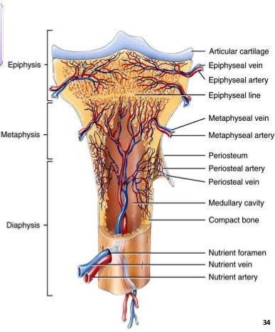
3 3 Bone Marrow Histology The term bone marrow (BM) refers to the tissue occupying the cavities under the cortex within the honeycomb of trabecular bone. The BM is composed of: The parenchyme: the hemopoietic cells including precursors and mature cells of the erythroid, myeloid and megakaryocytic lineages, i.e. various stages of red cells, white cells, megakaryocytes, lymphocytes, plasma cells and mast cells The stroma: fat cells, histiocytes/macrophages, fibroblasts, blood vessels and intercellular matrix. Bone marrow structure The constituents of the normal BM are closely packed within a hard bony container. Hemopoiesis occurs in the intertrabecular space within marrow cavities. The bony trabeculae (cancellous bone) are lined by endosteum, osteoblasts and osteoclasts. The stromal elements form an extensive, closely woven network in which the hematopoietic precursors are embedded, attached in various ways and to different components by the adhesive proteins and by other cells, such as the central macrophages in the erythroid islands. The blood supply to the BM consists of two systems: periosteal arteries, which give off branches to the BM after they penetrate the bone, and nutrient arteries. Blood drains from the BM cavity through central veins.
4 4
5 5 The microvasculature of the BM comprises a network of sinusoids Hemopoiesis only occurs in the interstital space between these sinusoids, thereby ensuring that hemopoietic progenitors are located close to the blood supply. Marrow architecture The outermost elements of the biopsy are composed of collagenous periosteal connective tissue, followed by a zone of cartilage or cortical bone (depending on the age of the patient). After this the bone breaks up into a meshwork of trabeculae, between which are the intertrabecular spaces. Hemopoietic cells are present within these intertrabecular spaces and are supported by fat cells, stromal cells, histocytes, extracellular matrix and blood vessels The hemopoietic cells are located within the intertrabecular spaces. The intertrabecular areas can be divided into three zones which contain different hemopoietic cell types : Endosteal or paratrabecular zone: immediately adjacent to the trabecular bone and composed predominantly of myeloid precursor cells Intermediate zone: contains erythroid colonies and maturing myeloid cells Central zone: in the center of the intertrabecular space. In addition to erythroid cells and maturing myeloid cells, this contains sinusoids and megakaryocytes

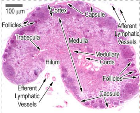
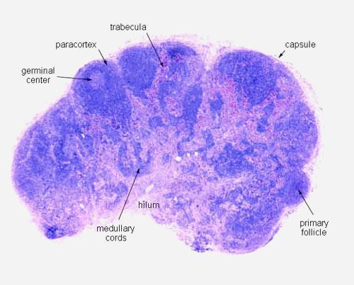
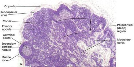
6 6 Histology of lymph nodes The nodes are covered by a capsule of dense connective tissue, and have capsular extensions, of connective tissue, called the trabeculae, which provide support for blood vessels entering into the nodes. Lymph, containing micro-organisms, soluble antigens, antigen presenting cells, and a few B-cells, enters the lymph node via afferent lymphatic vessels which enter the subcapsular sinus. It then runs through cortical sinuses into medullary sinuses and leaves through the efferent lymphatic vessels, at the Hilium as efferent lymph. This contains lots of T-lymphocytes, B-lymphocytes, plasma cells and antibody. All the sinuses are lined by a discontinuous layer of simple squamous endothelium, and they also contain lymphocytes and macrophages. Reticular fibers provide additional support to the matrix/stroma. The cortex is divided into an outer and an inner cortex (paracortex).

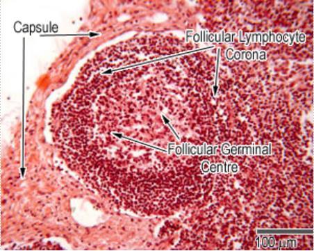
7 7 The outer cortex has lymphatic nodules that mostly contain B-cells. Small lymphocytes sit in the spaces between the reticular fiber meshwork in the cortex. The lighter staining areas are germinal centers, where the B-cells proliferate into antibody secreting plasma cells. Macrophages are also present in these regions, together with dendritic cells, and some T-cells. Both the macrophages, and the dendritic cells trap antigens and present them on their surfaces to B-cells. The inner cortex contains mostly T-cells. The deep cortical, and medullary cords contain B-cells and plasma cells. Most of the lymphocytes enter the lymph nodes via blood vessels, and about 10% enter through the lymph. The structure of the post-capillary venule, in the deep cortex (paracortex) is unusual in that it is not lined by simple squamous epithelium, but by a simple cuboidal epithelium. These are called high endothelial venules (HEVs). Lymphocytes recognize and adhere to these endothelial cells, and squeeze

8 8 through them into the deep cortical regions of the nodes. This region of the lymph has lots of T-cells, as well as the antigen presenting dendritic cells. T-cells entering here become activated in the cortex, between lymphoid follicles.
9 9
10 10

11 11 Mucosa-Associated Lymphoid Tissue The mucosal lining of the alimentary canal and airways is in many ways specialized to facilitate the exchange of substances between the external environment and the body. Unfortunately, these specialization do not just apply e.g. to components of the digested food but also pathogens. This is combined with excellent living conditions for bacteria in parts of the alimentary canal - in particular the ileum and the colon. Lymphoid tissue located beneath the mucosal epithelia, mucosa-associated lymphoid tissue (MALT), protects the body against pathogens that may enter the body via the mucosa. The importance of this task is reflected in the mass of the MALT, which corresponds to the combined mass of the other lymphoid organs and tissues. The task that the immune cells of the MALT have to accomplish is different from that of other parts of the immune system. We do need a defense against pathogens, but it would not be a good idea to mount an immune response against components of the food. Immune cell activation therefore differs between the MALT and other lymphoid tissues. This difference is mediated by different receptors expressed by immune cells of the MALT and by different substances which they release upon contact with an antigen. Because of their specific functions, immune cells of the MALT do not mingle with other immune cells. Epithelial cells of the vessels supplying the MALT express specific receptors which are recognized by MALT immune cells and allow their homing to the MALT during recirculation. Lastly, MALT plasma cells produce a secretable form of antibodies, immunoglobulin type A dimers, which can be taken up by epithelial cells and then released onto the epithelial surface. Specialization of MALT immune cells occur at the molecular level. In routine


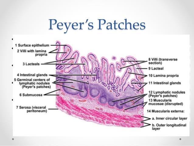
12 12 histological preparations, immune cells of the MALT look pretty much like immune cells of other lymphoid tissues. Often MALT consists of small accumulations of lymphoid cells or one to a few lymph follicles beneath the epithelium and possibly extending into the submucosa. The tonsils and Peyer's patches are large accumulations of lymphoid tissue with associated specializations of the epithelium.
. The tonsils have many invaginations which form blind crypts.")
13 13 Histology of Tonsils Tonsils are large non-encapsulated (or partially encapsulated) masses of lymphoid tissue, that lie in the walls of the pharynx and nasopharynx and at the base of the tongue. The luminal surface of the tonsils are covered with a stratified squamous epithelium (in common with the oral epithelia). The tonsils have many invaginations which form blind crypts. Below the epithelium, there are many lymphoid follicles beneath which have germinal centers like the lymph nodes. The epithelial cells are able to phagocytose bacteria, and transfer them to macrophages, which then present the foreign antigens to B-cells, which are activated (with the help of T cells). The activated cells mostly secrete IgA type antibodies, which are secreted locally.

14 14 Histology of Peyer's patches Small accumulations of lymphocytes or solitary lymph follicles are found scattered in beneath the epithelium throughout the gastrointestinal tract. However, the most prominent accumulations occur in the ileum and appendix in the form of Peyer's patches. In the ileum, they form dome-shaped protrusions into the lumen. Beneath the epithelial lining of the domes, Peyer's patches extend from the lamina propria to the submucosa. Within Peyer's patches, lymph follicles with germinal centers are typically located deep in the submucosa. The epithelium in contact with the lymphoid tissue is specialised to facilitate the contact of antigens with cells of the immune system. The epithelium appears columnar and contains cells with deeply invaginated basal surfaces - microfold cells or M-cells. Immune system cells can enter these invaginations (intraepithelial pockets) where they are exposed to materials which have been endocytosed by the epithelial cells and then released into the invaginations. Goblet cells are rare or absent in the epithelium which covers the domes.

15 15
16 16
Flow Cytometry. What is flow cytometry?
 Flow Cytometry Flow Cytometry What is flow cytometry? Flow cytometry is a popular laser-based technology to analyze the characteristics of cells or particles. It is predominantly used to measure fluorescence
The peripheral (secondary) lymphoid tissues
 lymphoid tissues") The peripheral (secondary) lymphoid tissues The peripheral (secondary) lymphoid tissues : are the lymph nodes, spleen, Mucosal associated lymphoid tissue (MALT). All secondary lymphoid organs have one
The peripheral (secondary) lymphoid tissues The peripheral (secondary) lymphoid tissues : are the lymph nodes, spleen, Mucosal associated lymphoid tissue (MALT). All secondary lymphoid organs have one
Lymphoid Organs. Dr. Sami Zaqout. Dr. Sami Zaqout IUG Faculty of Medicine
 Lymphoid Organs Dr. Sami Zaqout Cells of the Immune System Lymphocytes Plasma cells Mast cells Neutrophils Eosinophils Cells of the mononuclear phagocyte system Distribution of cells of the immune system
Lymph I: The Peripheral Lymph System
 Lymph I: The Peripheral Lymph System Peripheral = Secondary Primary Immune Organs = bone marrow, thymus Site of maturation of cells of the immune system Secondary Immune Organs = Nodes, MALT, spleen Filter
Human Anatomy and Physiology - Problem Drill 20: Immunity and the Lymphatic System
 Human Anatomy and Physiology - Problem Drill 20: Immunity and the Lymphatic System Question No. 1 of 10 The lymphatic system is formed early during human development. Which of the following statements
Sinusoids and venous sinuses
 LYMPHOID SYSTEM General aspects Consists of organs that are made of lymphoid tissue; Immune defense Breakdown of red blood cells. 1 Sinusoids In place of capillaries Endothelium; often fenestrated More
A Rough look at the tonsils and adenoids, for Bonny Peppa!
 A Rough look at the tonsils and adenoids, for Bonny Peppa! tonsils (two oval masses in the back of the throat) Lymphoid organs include: adenoids (two glands located at the back of the nasal passage) appendix
LYMPHOID ORGANS. Dr. Iram Tassaduq
 LYMPHOID ORGANS Dr. Iram Tassaduq COMPONENTS OF IMMUNE SYSTEM Lymphocytes Diffuse Lymphatic Tissue Lymphatic Nodules Lymph node Spleen Bone marrow Thymus Functions of Immune System Has the ability to distinguish
8: Lymphatic vessels and lymphoid tissue. nur
 8: Lymphatic vessels and lymphoid tissue nur Lymphatic vascular system Functions return to the blood extracellular fluid (Lymph) from connective tissue spaces. ensures the return of water, electrolytes
Chapter10 Immune system
 Chapter10 Immune system Lyu Zhengmei Department of Histology and Embryology, Anhui Medical University Ⅰ.General Introduction Function ------ Defense The human body immune system has the ability to distinguish
Immune - lymphatic system
 Immune system - organisation: Immune - lymphatic system - histology & embryology organised lymphoid structures cell components lymphocytes event. lymphatic follicles accessory cells monocytes-macrophages
PBS Class #2 Introduction to the Immune System part II Suggested reading: Abbas, pgs , 27-30
 PBS 803 - Class #2 Introduction to the Immune System part II Suggested reading: Abbas, pgs. 15-25, 27-30 Learning Objectives Compare and contrast the maturation of B and T lymphocytes Compare and contrast
2/19/2018. Lymphatic System and Lymphoid Organs and Tissues. What is Lymph?
 Lymphatic System and Lymphoid Organs and Tissues Lymphatic system a transport system for tissue fluids 1. elaborate network of one-way drainage vessels returning lymph to systemic circulation 2. Lymph:
Lymphatic System and Immunity. Lymphatic System
 Lymphatic System and Immunity Lymphatic System Lymphatic System High hydrostatic pressure in the arterioles and capillaries at the arterial part of the circulation leads to move plasma fluid from the capillaries
The Lymphatic System
 The Lymphatic System The Lymphatic Systems Overview General Functions Organization Components Lymphatic System General Functions Transportation Excess fluid from capillary exchange Fats & fat soluble vitamins
LECTURE 12: MUCOSAL IMMUNITY GUT STRUCTURE
 LECTURE 12: MUCOSAL IMMUNITY GUT STRUCTURE - Small intestine in humans is around 3-4 metres long - Internal surface of the small intestines are lined by villi o Villi are composed of absorptive cells (epithelial/enterocytes)
the liver and spleen. There they will proliferate and differentiate along the various leucocyte lines. Later, bone marrow becomes the predominant
 Chapter 10 Lymphatic System 10.1. General Comments The primary functions of lymphoid organs are protective or immunologic in nature. They are the source of immunocompetent cells which are capable of neutralizing
Chapter 21 The Lymphatic System Pearson Education, Inc.
 Chapter 21 The Lymphatic System Overview of the Lymphatic System The Lymphatic System Protects us against disease Lymphatic system cells respond to: Environmental pathogens Toxins Abnormal body cells,
The Lymphoid System Pearson Education, Inc.
 23 The Lymphoid System Introduction The lymphoid system consists of: Lymph Lymphatic vessels Lymphoid organs An Overview of the Lymphoid System Lymph consists of: Interstitial fluid Lymphocytes Macrophages
3/17/2014. The Lymphatic System. Lymphatic System Overview Lymphatic Vessels and Flow of Lymph Lymphoid Cells, Tissues, and Organs
 The Lymphatic System Lymphatic System Overview Lymphatic Vessels and Flow of Lymph Lymphoid Cells, Tissues, and Organs Overview of the Lymphatic System Slide 2 Major Components of the Lymphatic System
LYMPH GLAND. By : Group 1
 LYMPH GLAND By : Group 1 ANATOMY LYMPH NODE Lymphatic Organs Red bone marrow Thymus gland Lymph nodes Lymph nodules Spleen Primary organs Secondary organs Lymph Nodes Firm, smooth-surfaced, bean-shaped
HISTOLOGY OF THE RESPIRATORY SYSTEM I. Introduction A. The respiratory system provides for gas exchange between the environment and the blood. B.
 HISTOLOGY OF THE RESPIRATORY SYSTEM I. Introduction A. The respiratory system provides for gas exchange between the environment and the blood. B. The human respiratory system may be subdivided into two
Lymphoid tissue. 1. Central Lymphoid tissue. - The central lymphoid tissue (also known as primary) is composed of bone morrow and thymus.
 is composed of bone morrow and thymus.") 1. Central Lymphoid tissue Lymphoid tissue - The central lymphoid tissue (also known as primary) is composed of bone morrow and thymus. Bone Morrow - The major site of hematopoiesis in humans. - Hematopoiesis
1. Central Lymphoid tissue Lymphoid tissue - The central lymphoid tissue (also known as primary) is composed of bone morrow and thymus. Bone Morrow - The major site of hematopoiesis in humans. - Hematopoiesis
Chapter 2 (pages 22 33): Cells and Tissues of the Immune System. Prepared by Kristen Dazy, MD, Scripps Clinic Medical Group
: Cells and Tissues of the Immune System. Prepared by Kristen Dazy, MD, Scripps Clinic Medical Group") Allergy and Immunology Review Corner: Cellular and Molecular Immunology, 8th Edition By Abul K. Abbas, MBBS; Andrew H. H. Lichtman, MD, PhD; and Shiv Pillai, MBBS, PhD. Chapter 2 (pages 22 33): Cells and
Allergy and Immunology Review Corner: Cellular and Molecular Immunology, 8th Edition By Abul K. Abbas, MBBS; Andrew H. H. Lichtman, MD, PhD; and Shiv Pillai, MBBS, PhD. Chapter 2 (pages 22 33): Cells and
3- Antigen-presenting cells, which are found, in addition to lymphoid tissue, in skin.
 Lymphoid System Dr.firdous It consists of organs, whose tissues and cells play an important role in immunity; a protective response of the internal environment of the body against microorganisms and foreign
Immunology 2017: Lecture 12 handout. Secondary lymphoid organs. Dr H Awad
 Immunology 2017: Lecture 12 handout Secondary lymphoid organs Dr H Awad INTRODUCTION So far we discussed the cells of the immune system and how they recognize their antigens and get stimulated. The number
The Lymphatic System
 94 The Lymphatic System Lymph Lymph is the name for tissue fluid that enters lymph capillaries. Filtration in capillaries creates tissue fluid, most of which returns almost immediately to the blood in
Epithelia will be discussed according to the following scheme: Type Number of layers Shape Line drawing. Squamous Cuboidal Columnar
 Epithelia Epithelia will be discussed according to the following scheme: Type Number of layers Shape Line drawing Simple Squamous Cuboidal Columnar Covering and Lining epithelium Pseudostratified Stratified
LYMPHATIC ANATOMY LAB. BIO 139 ANATOMY AND PHYSIOLOGY II MARY CATHERINE FLATH, Ph.D.
 LYMPHATIC ANATOMY LAB BIO 139 ANATOMY AND PHYSIOLOGY II MARY CATHERINE FLATH, Ph.D. THE LYMPHATIC SYSTEM ORGANS PRIMARY BONE MARROW THYMUS SECONDARY LYMPH NODES SPLEEN FUNCTIONS CONTROL DISEASE TRANSPORT
General Structure of Digestive Tract
 Dr. Nabil Khouri General Structure of Digestive Tract Common Characteristics: Hollow tube composed of a lumen whose diameter varies. Surrounded by a wall made up of 4 principal layers: Mucosa Epithelial
Paper No.: 10: Immunology Module : 04: Phylogeny and Ontogeny of immune system: Organization and structure of immune system.
 Paper No.: 10: Module : 04: Phylogeny and Ontogeny of immune system: Organization and Development Team Principal Investigator: Co-Principal Investigator: Paper Coordinator: Content Writer: Content Reviewer:
Lymphoid System: cells of the immune system. Answer Sheet
 Lymphoid System: cells of the immune system Answer Sheet Q1 Which areas of the lymph node have most CD3 staining? A1 Most CD3 staining is present in the paracortex (T cell areas). This is towards the outside
ANATOMY & PHYSIOLOGY ONLINE COURSE - SESSION 11 THE LYMPHATIC SYSTEM AND IMMUNITY
 ANATOMY & PHYSIOLOGY ONLINE COURSE - SESSION 11 THE LYMPHATIC SYSTEM AND IMMUNITY Functions of the Lymphatic System The lymphatic system has three primary functions. First of all, it returns excess interstitial
Sample Exam Biology 2050 Circulatory and Lymphatic Systems
 Sample Exam Biology 2050 Circulatory and Lymphatic Systems Note: Not all of the lymphatic system will be on the actual exam next Monday so disregard any questions that deal with something that wasn t covered
Introduction to Lesson 4 - The Lymphatic System
 Introduction to Lesson 4 - The Lymphatic System Your circulatory system is not your body s only vascular transport system. Closely associated with the blood vessels of the circulatory system is the lymphatic
KEY - Sample Exam Biology 2050 Circulatory and Lymphatic Systems - KEY
 KEY - Sample Exam Biology 2050 Circulatory and Lymphatic Systems - KEY Note: Not all of the lymphatic system will be on the actual exam next Monday so disregard any questions that deal with something that
For more information about how to cite these materials visit
 Author(s): Matthew Velkey, 2009 License: Unless otherwise noted, this material is made available under the terms of the Creative Commons Attribution Non-commercial Share Alike 3.0 License: http://creativecommons.org/licenses/by-nc-sa/3.0/
ANATOMY & PHYSIOLOGY II
 ANATOMY & PHYSIOLOGY II THE BODY SYSTEMS Anatomy & Physiology II The Body Systems Michelle Cochrane 2014 All rights reserved. This material is subject to copyright and may not be reprinted or reproduced
Mucosal Immunology Sophomore Dental and Optometry Microbiology Section I: Immunology. Robin Lorenz
 Mucosal Immunology Sophomore Dental and Optometry Microbiology Section I: Immunology Robin Lorenz rlorenz@uab.edu Why do we Need to Understand How the Mucosal Immune System Works? The mucosa is the major
Lymphoid Organs and Lymphocyte Trafficking. Dr. Issa Abu-Dayyeh
 Lymphoid Organs and Lymphocyte Trafficking Dr. Issa Abu-Dayyeh Invader recognition Where does invader recognition take place?? Secondary lymphoid organs: Lymph nodes Spleen Mucosal-associated lymphoid
Slide 154: Pancreas, H&E
 Slide 154: Pancreas, H&E the pancreas, located adjacent to the duodenum, is a mixed exocrine and endocrine gland; it is usually readily identifiable by the presence of the interspersed endocrine pancreatic
Small intestine. Small intestine
 General features Tubular organ longest part; 5-6 m most of chemical digestion absorption of nutrients reabsorption of H2O occurs. Two structural features; maximize the lumenal surface area villi microvilli
Chapt 21: The Lymphatic and Immune Systems
 Chapt 21: The Lymphatic and Immune Systems Goals 1. Discuss the organization of the lymphatic system, including the vessels, principal lymph nodes, thymus, and spleen 2. Explain the relationship between
Lymphatic and Immune Systems
 Lymphatic and Immune www.vastaccess.com 2 Specialized component of circulatory system Lymphatic system functions: Maintenance of internal fluid balance Immunity Lymph derived from blood and tissue fluid
CELLS, ORGANS, AND MOLECULES: ANATOMY AND PHYSIOLOGY OF THE IMMUNE SYSTEM THE CELLS OF THE BLOOD FROM AN IMMUNOLOGICAL PERSPECTIVE.
 CELLS, ORGANS, AND MOLECULES: ANATOMY AND PHYSIOLOGY OF THE IMMUNE SYSTEM THE CELLS OF THE BLOOD FROM AN IMMUNOLOGICAL PERSPECTIVE. Erythrocytes: Red blood cells (~5 x 10 6 / L) (~5 x 10 12 /L) of blood.
Epithelium. Four primary tissue types:
 Epithelium Four primary tissue types: Epithelial (covering) Connective (support) Nervous (control) Muscular (movement) Smooth muscle Cardiac muscle Skeletal muscle 1 Epithelial Tissue Features Epithelial
BONE TISSUE. Dr. Heba Kalbouneh Associate Professor of Anatomy and Histology
 BONE TISSUE Dr. Heba Kalbouneh Associate Professor of Anatomy and Histology BONE FUNCTION Support Protection (protect internal organs) Movement (provide leverage system for skeletal muscles, tendons, ligaments
Hematopoiesis. Hematopoiesis. Hematopoiesis
 Chapter. Cells and Organs of the Immune System Hematopoiesis Hematopoiesis- formation and development of WBC and RBC bone marrow. Hematopoietic stem cell- give rise to any blood cells (constant number,
Body Tissues Pearson Education, Inc.
 Body Tissues Tissues Groups of cells with similar structure and function Four primary types: Epithelial tissue (epithelium).1 Connective tissue.2 Muscle tissue.3 Nervous tissue.4 Epithelial Tissues Locations:
Lymphatic System. Where s your immunity idol?
 Lymphatic System Where s your immunity idol? Functions of the Lymphatic System Fluid Balance Drains excess fluid from tissues Lymph contains solutes from plasma Fat Absorption Lymphatic system absorbs
Most abundant and widely distributed tissues in the body Binds, support, and strengthen body tissues, protect and insulate internal organ, serve as
 Connective tissue Most abundant and widely distributed tissues in the body Binds, support, and strengthen body tissues, protect and insulate internal organ, serve as major transport system, compartmentalizes
Lymphatic System. The most important functions of the lymphatic system are: Maintenance of fluid balance in the internal environment
 Lymphatic System Lymphatic System The lymphatic system is a complex network of connective tissue that is composed of: Lymphoid organs Lymph nodes Lymph ducts Lymph vessels Lymph capillaries Lymphatic System
What is histology? HISTOLOGY
 Introduction to Histology What is histology? HISTOLOGY histo = tissue ogy = study So HISTOLOGY = the study of tissues! What is a TISSUE? Tissues are groups of cells with specialized structural and functional
Returns fluids that leaked from blood vessels back to blood Consists of three parts
 Lymphatic System Returns fluids that leaked from blood vessels back to blood Consists of three parts 1. Network of lymphatic vessels (lymphatics) 2. Lymph fluid in vessels 3. Lymph cleanse lymph 1 Lymphoid
The Lymphatic and Immune Systems
 PowerPoint Lecture Slides prepared by Leslie Hendon University of Alabama, Birmingham C H A P T E R 21 Part 1 The Lymphatic and Immune Systems The Lymphatic and Immune Systems Lymphatic system Main function
Compact bone; Many parallel Haversian canals contain: small blood vessels. very small nerve. Interconnected by Volkmann s canals.
 Special characteristics of COMPACT BONE (dense bone) Thick; well vascularized Osteocytes and lamellae Concentric rings around blood vessels Most bones: outer compact bone inner spongy bone Marrow cavity
Putting it Together. Stephen Canfield Secondary Lymphoid System. Tonsil Anterior Cervical LN s
 Putting it Together Stephen Canfield smc12@columbia.edu Secondary Lymphoid System Tonsil Anterior Cervical LN s Axillary LN s Mediastinal/Retroperitoneal LN s Thoracic Duct Appendix Spleen Inguinal LN
Tissue Outline (chapter 4) Tissues group of cells that perform structural and roles. List the 4 types:
 Tissues group of cells that perform structural and roles. List the 4 types:") Tissue Outline (chapter 4) Tissues group of cells that perform structural and roles. List the 4 types: 1. 2. 3. 4. I. Epithelial Tissue covers all the surfaces, inside & out. Are the major tissues of,
Tissue Outline (chapter 4) Tissues group of cells that perform structural and roles. List the 4 types: 1. 2. 3. 4. I. Epithelial Tissue covers all the surfaces, inside & out. Are the major tissues of,
Chapter Lymphatic Cells, Lymphatic Tissues, and Lymphatic Organs
 Chapter 22.2 Lymphatic Cells, Lymphatic Tissues, and Lymphatic Organs Lymphatic Cells These are the cells that play a key roll in the structure and function of the immune system. We have already introduced
Histo lab 7. Special connective tissue is derived from the mesoderm (mesenchyme).
.") Histo lab 7 Special connective tissue is derived from the mesoderm (mesenchyme). If we have high density of fibers, we call it dense connective tissue. (Fibers are more than the ground substance). If we
Histo lab 7 Special connective tissue is derived from the mesoderm (mesenchyme). If we have high density of fibers, we call it dense connective tissue. (Fibers are more than the ground substance). If we
SOPHOMORE DENTAL/OPTOMETRY MICROBIOLOGY SECTION: IMMUNOLOGY CELLS AND ORGANS OF THE IMMUNE SYSTEM
 SOPHOMORE DENTAL/OPTOMETRY MICROBIOLOGY SECTION: IMMUNOLOGY CELLS AND ORGANS OF THE IMMUNE SYSTEM Lecturer: Dr. John F Kearney 934-6557 jfk@uab.edu Objectives: To learn: 1) the basic cell types that make
Epithelial Lecture Test Questions
 Epithelial Lecture Test Questions 1. Which of the following free surfaces lack(s) epithelia: a. lung alveoli (air sacs) b. hard palate c. joint cavities d. abdominal cavity e. salivary gland ducts 2. Which
Formation of Blood Cells
 Hematopoiesis Lecture Objectives Name organs responsible for hematopoiesis in the fetus. List the developmental stages of hematopoiesis both prenatally and postnatally. Outline the major steps of post
Connective Tissue. Found everywhere in the body. Most abundant and widely distributed. Never exposed to the outside environment.
 Connective Tissue Found everywhere in the body. Most abundant and widely distributed. Never exposed to the outside environment. Connective Tissue Functions Binding and support Protection Insulation Transportation
Organs Histology D. Sahar AL-Sharqi. Respiratory system
 Respiratory system The respiratory system provides for exchange of O2 and CO2 to and from the blood. Respiratory organs include the lungs and a branching system of bronchial tubes that link the sites of
Basic Histology. By Mrs. Bailey
 Basic Histology By Mrs. Bailey Primary Tissues 1. Epithelial Tissue 2. Connective Tissue 3. Muscle Tissue 4. Nervous Tissue Very cellular Supported by underlying connective tissue Epithelial & connective
Section 1.1: What is the function of digestion?
 Section 1.1: What is the function of digestion? When you have completed this section, you should be able to: Describe the overall function of the GI tract. Describe the processes involved in digestion.
Molecular and Cellular Basis of Immune Protection of Mucosal Surfaces
 Molecular and Cellular Basis of Immune Protection of Mucosal Surfaces Department of Biologic & Materials Sciences School of Dentistry University of Michigan Ann Arbor, Michigan 48109-1078 1 Image quality
Unit I Problem 9 Histology: Basic Tissues of The Body
 Unit I Problem 9 Histology: Basic Tissues of The Body - What is the difference between cytology and histology? Cytology: it is the study of the structure and functions of cells and their contents. Histology:
Basic Tissue Types and Functions
 Tissues Histology Basic Tissue Types and Functions 1) Epithelial tissue covering 2) Connective tissue support 3) Muscle tissue movement 4) Nervous tissue control Epithelial Tissue 1) Covers a body surface
GENERAL ANATOMY OF THE IMMUNE SYSTEM
 GENERAL ANATOMY OF THE IMMUNE SYSTEM 1 THE IMMUNE SYSTEM - A SYSTEM WHICH CONTROLS PRESERVE GENETIC INTEGRITY OF THE ORGANISM. BODILY FUNCTIONS PROTECTION OF ANTIGENS CALLED IMMUNITY (FROM THE LATIN WORD
MCAT Biology - Problem Drill 16: The Lymphatic and Immune Systems
 MCAT Biology - Problem Drill 16: The Lymphatic and Immune Systems Question No. 1 of 10 1. Which of the following statements about pathogens is true? Question #01 (A) Both viruses and bacteria need to infect
Blood. Hematopoietic Tissue
 Blood Hematopoietic Tissue Is a type of connective tissue in which its cells are suspended in a circulating fluid. Erythrocytes+ leukocytes + platelets (thrombocytes) =formed elements of blood. These formed
DIGESTIVE TRACT ESOPHAGUS
 DIGESTIVE TRACT From the lower esophagus to the lower rectum four fundamental layers comprise the wall of the digestive tube: mucosa, submucosa, muscularis propria (externa), and adventitia or serosa (see
Urinary Anatomy. Lab 40. Kidneys. Nephrons. Renal Corpuscle
 Urinary Anatomy Lab 40. Urinary Anatomy and Kidney Dissection Kidneys: filters blood, produces urine Ureters: convey urine to bladder Bladder: holding tank Urethra: carries urine to the outside for elimination
Lab 1 ANIMAL TISSUES
 Lab 1 ANIMAL TISSUES Levels of Organization Animals are multicellular heterotrophs whose cells lack cell walls. Most animals exhibit a hierarchical level of organization: Cells are organized into tissues
TISSUES. Objectives. Tissues
 TISSUES Objectives Introduce the four major types of tissues Describe the general characteristics and functions of epithelial & connective tissue Name the major types of epithelial & connective tissues
Chapter 16 Lymphatic System and Immunity. Lymphatic Pathways. Lymphatic Capillaries. network of vessels that assist in circulating fluids
 Chapter 16 Lymphatic System and Immunity network of vessels that assist in circulating fluids closely associated with the cardiovascular system transports excess fluid away from interstitial spaces transports
Prelab #4 BLOOD; BONE MARROW; RESPIRATORY; INTEGUEMENT Page 1
 Prelab #4 BLOOD; BONE MARROW; RESPIRATORY; INTEGUEMENT Page 1 Blood Slide 101 This a classic slide of blood cells using a Wright stain. Inspect red blood cells and their appearance. Note the approximate
Biology. Dr. Khalida Ibrahim
 Dr. Khalida Ibrahim Biology Histology: Histology: is the study of the tissues of the body. Tissue: group of similar cells combined to perform a common function. The human body is composed of only 4 basic
HISTOLOGY VIRTUAL LABORATORY GASTROINTESTINAL SYSTEM
 HISTOLOGY VIRTUAL LABORATORY GASTROINTESTINAL SYSTEM LIP (Slides GI 1, 2) Identify the outer portion lined by stratified squamous (keratinized) epithelium. Note the hair follicles and sebaceous glands
Tissues 10/21/2016. Epithelial Tissue
 Tissues This is a generalized cell diagram. It shows the anatomy of a cell, but most cells do not actually look like this. Cells can have a wide variety of shapes and sizes, depending on their function.
Prepared By Student. Dania Abed Al-majeed. Rahma Raad Hanna. Balqees Mohammed Aasim. Dania Hisham. Rasha Rafiee
 Prepared By Student Rahma Raad Hanna Balqees Mohammed Aasim Dania Hisham Dania Abed Al-majeed Rasha Rafiee Epithelia Epithelia can be derived from ectoderm, mesoderm or endoderm -ectoderm gives rise to
The Tissue Level of Organization
 Tissue The Tissue Level of Organization Chapter 3 Definition an aggregation of cells in which each cooperates with all others in the performance of a given function Examples of general functions Movement
Connective Tissue. Consists of two basic elements: Cells and Extra-cellular matrix
 Connective Tissue Consists of two basic elements: Cells and Extra-cellular matrix True Connective Tissue Cells Fibroblasts: Secrete both fibers and ground substance of the matrix (wandering) Macrophages:
Sca-1-positive cells can reconstitute a 950 rad (9.5 Gy)-irradiated mouse instead of more than 10,000 unpurified BM cells.
-irradiated mouse instead of more than 10,000 unpurified BM cells.") Immunology Dr. John J. Haddad Cells and Organs of the Immune System Chapter 2 Human hematopoiesis The process begins in yolk sac of embryo is 1 st week. In month 3, stem cells migrate to fetal liver and
Immunology Dr. John J. Haddad Cells and Organs of the Immune System Chapter 2 Human hematopoiesis The process begins in yolk sac of embryo is 1 st week. In month 3, stem cells migrate to fetal liver and
Histology. Histology. Tissue - Four main tissues in body. 1. Epithelial tissue an epithelium; plural: epithelia. Function. Location.
 Histology Histology Tissue Four main tissues in body 1. Epithelial tissue an epithelium; plural: epithelia Function Location Characteristics Example 2. Connective tissue Function Location Characteristics
Bio& 241 Unit 1 / Lecture 4
 Bio& 241 Unit 1 / Lecture 4 Connective Tissue Consists of two basic elements: Cells and Extra-cellular matrix 1 True Connective Tissue Cells Fibroblasts: Secrete both fibers and ground substance of the
D1120 Connective Tissue and Muscle Laboratory Module. 1) Connective tissue
 Connective tissue") D1120 Connective Tissue and Muscle Laboratory Module 1) Connective tissue Objectives: 1) identify the components (cells, fibres) present in "ordinary" connective tissue 2) differentiate the three types
D1120 Connective Tissue and Muscle Laboratory Module 1) Connective tissue Objectives: 1) identify the components (cells, fibres) present in "ordinary" connective tissue 2) differentiate the three types
The Lymphatic System and Body Defenses
 12 PART A The Lymphatic System and Body Defenses PowerPoint Lecture Slide Presentation by Jerry L. Cook, Sam Houston University ESSENTIALS OF HUMAN ANATOMY & PHYSIOLOGY EIGHTH EDITION ELAINE N. MARIEB
HISTOLOGY. Simple squamal lungs
 HISTOLOGY Lab Objectives: Students should be able to... 1. Visually identify each class of tissue and examples within each class 2. Indicate the location (in the human body and/or organ) and function of
Lab activity manual - Histology of the digestive system. Lab activity 1: esophagus stomach - small intestines
 Lab activity manual - Histology of the digestive system Jeanne Adiwinata Pawitan Prerequisite: Histology of the 4 basic tissues In this module we learn about the histology of the digestive system, from
NOTES: CH 40 Introduction to Human Anatomy & Physiology
 NOTES: CH 40 Introduction to Human Anatomy & Physiology THE HUMAN BODY Anatomy Physiology (= structures) (= functions or processes) Characteristics of LIFE: 1) Made up of 1 or more CELLS. 2) Obtain and
Urinary system. Urinary system
 Distal convoluted tubule (DCT) Highly coiled, ~ 5 mm in length Last part of the nephron. Wall; simple cuboidal epithelium Less metabolically active than the PCT no brush border light eosinophilic cytoplasm
A. Incorrect! Axons covey messages from the cell body of the neuron. D. Correct! Dendrites convey messages to the cell body of the neuron.
 CLEP Biology - Problem Drill 14: Animal Form No. 1 of 10 1. The branches of a neuron receiving information from another cell and which transmit the message to the cell body are called? (A) (B) (C) (D)
Study of different tissues Abnormal cells and tissues can be compared to normal tissues to identify disease, such as cancer Being able to know and
 CHAPTER 4 Study of different tissues Abnormal cells and tissues can be compared to normal tissues to identify disease, such as cancer Being able to know and recognize normal tissues under the microscope
Histology 101! !! Name:! Block: Identify and describe the functions of major tissue types including their subclasses and varieties!
 Histology 101 Identify and describe the functions of major tissue types including their subclasses and varieties Name: Block: "1 Introduction to Tissues Histology Notes Tissue (living fabric) : groups
1. Lymphatic vessels recover about of the fluid filtered by capillaries. A. ~1% C. ~25% E. ~85% B. ~10% D. ~50%
 BIOL2030 Huaman A&P II -- Exam 3 -- XXXX -- Form A Name: 1. Lymphatic vessels recover about of the fluid filtered by capillaries. A. ~1% C. ~25% E. ~85% B. ~10% D. ~50% 2. Special lymphatic vessels called
Histology Urinary system
 Histology Urinary system Urinary system Composed of two kidneys, two ureters, the urinary bladder, and the urethra, the urinary system plays a critical role in: 1- Blood filtration,(filtration of cellular