The Envelope Gene of Transmitted HIV-1 Resists a Late IFNγ-Induced Block. Address correspondence to Sam J. Wilson,
|
|
|
- Darren Hodge
- 6 years ago
- Views:
Transcription
1 JVI Accepted Manuscript Posted Online 18 January 2017 J. Virol. doi: /jvi Copyright 2017 Rihn et al. This is an open-access article distributed under the terms of the Creative Commons Attribution 4.0 International license The Envelope Gene of Transmitted HIV-1 Resists a Late IFNγ-Induced Block Suzannah J. Rihn a, Toshana L. Foster b, Idoia Busnadiego a, Muhamad Afiq Aziz a, Joseph Hughes a, Stuart J. D. Neil b and Sam J. Wilson a a MRC University of Glasgow Centre for Virus Research, Institute of Infection, Inflammation and Immunity, University of Glasgow, UK b Department of Infectious Diseases, King s College London Faculty of Life Sciences and Medicine, Guy s Hospital, London SE1 9RT, UK Running Head: HIV-1 Interferon Resistance Governed by env Address correspondence to Sam J. Wilson, sam.wilson@glasgow.ac.uk Abstract: 196 words Importance: 150 words Text: 7827 words 19
2 Abstract Type I interferon (IFN) signaling engenders an antiviral state that likely plays an important role in constraining HIV-1 transmission and contributes to defining subsequent AIDS pathogenesis. Type II IFN (IFNγ) also induces an antiviral state but is often primarily considered to be an immunomodulatory cytokine. We report that IFNγ stimulation can induce an antiviral state that can be both distinct from that of type I interferon, and can potently inhibit HIV-1 in primary CD4+ T cells and a number of human cell lines. Strikingly, we find that transmitted/founder (TF) HIV-1 viruses can resist a late block that is induced by type II IFN, and the use of chimeric IFNγsensitive/resistant viruses indicates that interferon-resistance maps to the env gene. Simultaneously, in vitro evolution also revealed that just a single amino acid substitution in envelope can confer substantial resistance to IFN-mediated inhibition. Thus, the env gene of transmitted HIV-1 confers resistance to a late block that is phenotypically distinct from those previously described to be resisted by env, and is therefore mediated by unknown IFNγ-stimulated factor(s) in human CD4+ T cells and cell lines. This important unidentified block could play a key role in constraining HIV-1 transmission Importance The human immune system can hinder invading pathogens through interferon (IFN) signaling. One consequence of this signaling is that cells enter an 'antiviral state', increasing the levels of hundreds of defenses that can inhibit the replication and spread of viruses. The majority of HIV-1 infections result from a single virus particle (the transmitted/founder) that makes it past these defenses and colonizes the host. Thus, 2
3 the founder virus is hypothesized to be a relatively interferon-resistant entity. Here we show that certain HIV-1 envelope genes have the unanticipated ability to resist specific human defenses mediated by different types of interferons. Strikingly, the envelope gene from a founder HIV-1 virus is far better at evading these defenses than the corresponding gene from a common HIV-1 lab strain. Thus, these defenses could play a role in constraining the transmission of HIV-1, and may select for transmitted viruses that are resistant to this IFN-mediated inhibition Introduction Humans have evolved to possess a diverse arsenal of antiretroviral defenses. Despite this, the human immune system is unable to eradicate an established human immunodeficiency virus (HIV) infection. Following infection, individuals mount vigorous immune responses targeting HIV-1 (1). While these responses are unable to clear systemic HIV-1 infection, the timing and magnitude of the innate and acquired immune responses still play a key role in shaping disease progression and pathogenesis. A major component of innate immunity is the interferon (IFN) system, which is comprised of a diverse family of related cytokines that for humans includes 17 type I IFNs (13 IFNα subtypes, as well as IFN β, κ, ε and ω), one type II IFN (IFNγ) and 4 type III IFNs (IFNλ1-4). Research into how IFNs inhibit retroviruses has largely focused on type I IFNs as type I/III IFNs are often perceived as the major antiviral IFNs, whereas IFNγ is frequently considered solely as an immunomodulatory player (2). To this end, it is now well established that type I IFNs inhibit infection and replication of HIV-1 and related primate lentiviruses both in vitro (3-12) and in vivo (13) (recently reviewed by 3
4 Doyle et al., (2)). Notably, HIV-1 infected individuals treated with IFNα experience significant, albeit transient, reductions in viral loads (13). Similarly, rhesus macaques administered IFNα can resist simian immunodeficiency virus (SIV) infection (14). In addition, transmitted HIV-1 is proposed to be relatively IFN-resistant (15, 16) (although this is not universally observed (17)). Despite this, IFNs are not always beneficial to the host, and repeated IFN administration in primate models (14), or persistent stimulation in chronically infected patients, is associated with poorer clinical outcome (18, 19). Thus, although IFN responses do not eradicate systemic HIV-1, there is great interest in understanding how IFNs might shape susceptibility to HIV-1 infection and subsequent progression to AIDS. Over the last decade, much of the attention paid to the ability of type I IFNs to inhibit HIV-1 has focused on restriction factors, including TRIM5/TRIMCyp (20, 21), APOBEC3s (22), Tetherin/BST2 (23) and SAMHD1 (24, 25). These interferonstimulated genes (ISGs) represent powerful barriers that primate lentiviruses must evade or overcome in order to thrive within human populations (26), and even successful viruses do not always completely escape inhibition by these factors (27). Alongside the restriction factors, a growing number of other ISGs have been identified as being capable of inhibiting HIV-1, but are not fully evaded or antagonized in natural settings. These resistance factors include IFITMs (28-30), GBP5 (31) and Mx2/MxB (32, 33). Importantly, these known resistance factors, along with the established restriction factors, still cannot fully explain the IFN-mediated inhibition of HIV-1 observed in vitro (2). Thus, there is great interest in understanding the molecular details of how 4
5 IFNs might constrain HIV transmission, acute viral replication, pathogenesis or even the pandemic potential of geographically restricted HIVs (13-16, 30, 34, 35). Despite this predominant focus on type I IFNs and type I ISGs, reports in the last century demonstrated that IFNγ treatment can also confer substantial antiretroviral activity in vitro (5, 9, 36, 37). Recently, this concept has been revisited with the observations that some antiretroviral ISGs, such as GBP5 and IDO1, are most strongly upregulated by IFNγ (31, 38). Although the antiretroviral potential of IFNγ has been reported, and patients mount robust IFNγ responses following HIV-1 infection (1), the clinical significance of these observations is currently unclear. Here we show that IFNγ has anti-hiv-1 activity in primary CD4+ T cells and a number of common cell lines, and can induce strong early and late blocks to HIV-1 replication. IFNγ can induce a divergent antiviral state from type I IFNs, and potent IFNγ-induced early block(s) to HIV-1 infection can be entirely independent of Mx2. Surprisingly, not all HIV viruses are equally susceptible to IFNγ-mediated inhibition, and certain HIV-1 and HIV-2 viruses can resist inhibition. Crucially, HIV-1 transmitted/founder (TF) viruses are strikingly resistant to a late IFNγ-stimulated block. Using two independent approaches, we map IFN-resistance to the HIV-1 env gene. Notably, a single amino acid (aa) substitution in envelope conferred substantial resistance to IFN-mediated inhibition. Thus, the env gene from transmitted HIV-1 can resist unidentified IFN-induced factor(s) and this ability has been lost in multiple common lab strains. The potent inhibition observed here suggests that type II IFN may play an underappreciated role in limiting HIV-1 transmission and disease progression, in a fashion similar to type I IFNs. 5
6 Materials and Methods Cell lines We assembled a panel of cell lines that are commonly used in HIV-1 research and/or were readily available from the UK National Institute of Biological Standards and Control (NIBSC). Jurkat, THP-1, HEK 293T, TE671 (RD or TE671/RD), and TZM-bl cell lines were a generous gift from Paul Bieniasz. MONOMAC6 cells were a generous gift from Mark Marsh and A549 cells were a generous gift from Ben Hale. The following cell lines were obtained from the UK NIBSC (repository reference indicated in parentheses): Molt 4 Clone 8 (ARP052), H9 (ARP001), HUT78 (ARP002), MOLT3 (ARP010), MOLT4 (ARP011), U937 (ARP012), HL60 (ARP030), PM1 (ARP057), AA-2 (ARP054), KARPAS 45 (ARP032) and U87 (U87.MG, ARP 043). Human suspension cell lines were maintained in RPMI supplemented with 9% FCS and gentamycin, with the exception of MONOMAC6 cells whose culture medium was also supplemented with NEAA and OPI. The remaining cell lines were cultured in DMEM supplemented with 9% FCS and gentamycin. All cell lines were tested for mycoplasma contamination, and the KARPAS 45 cell line tested positive for mycoplasma at the end of this study and should be considered as mycoplasma positive throughout this study. The CCR5+ MT4-R5-LTR- GFP indicator cells were described previously and contain a cassette in which hrgfp expression is driven by the HIV-1 LTR (38, 39). PMA-treated THP-1 cells were seeded with 30 ng/ml of PMA in RPMI plus 9% FCS and gentamycin overnight, and washed the following morning (~16h later) to remove the PMA. 6
7 Primary Cells As described previously (30), human primary CD4+ T cells were obtained from peripheral blood mononuclear cells (PBMCs) from healthy human donors. Ethical approval for primary cell work was granted by King s College London Infectious Disease BioBank Local Research Ethics Committee (under the authority of the Southampton and South West Hampshire Research Ethics Committee approval REC09/H0504/39), approval number SN-1/6/7/9. In brief, PBMCs were isolated by density gradient centrifugation through Lymphoprep (Axis-Shield) and CD4+ T cells obtained by negative selection using the Dynabeads Untouched Human CD4 T Cells kit (Life Technologies) using the manufacturer s instructions. Flow cytometry for CD4 was used to assess the purity of the isolated cell population, which was reproducibly >95%. Cells were cultured with RPMI supplemented with 10% FCS, 20μg/ml gentamycin, and 30 U/ml recombinant IL-2 (PeproTech), and were then activated within 48 hours using Dynabeads Human T- Activator CD3/CD28 beads (CD3/CD28 Dynabeads, Invitrogen) using the manufacturer s instructions. Cells were typically resuspended in flasks with the CD3/CD28 Dynabeads at 1:5 bead:cell ratio (cells at 1x10 6 cells/ml). Cells were washed and resuspended in fresh RPMI-IL-2 supplemented media prior to infection or postactivation analysis. Where indicated, cells were treated with IFNγ (1000 U/ml, human interferon gamma, Thermo Fisher, PHC4031) for 24 hours before virus infection Primate lentiviruses 7
8 Lentivirus stocks (Table 1) were all generated through transient transfection of HEK 293T cells in the presence/absence of pcmv-vsv-g using polyethylenimine (PEI). The following proviral clones were used: pnl4-3 (M ) (40); prod10 (KY272752) (41); pjk7312a (L36874) (42); pcmo2.5 (AY ) (43); as well as replication competent GFP-encoding pnhg (JQ585717) (44, 45); and the following HIV-1 strains that were obtained from the NIH AIDS reagents program (catalog numbers indicated in parentheses): pnl(ad8) (11346), pyk-jrcsf (JRCSF, 2708), p89.6 (3552), pzm249m-1 (12260) and a panel of full length transmitted/founder (TF) HIV-1 infectious molecular clones (11919). In addition to chimeric/mutant proviral clones, env minus GFP-encoding NHGΔenv-GFP (44, 46) and phiv-2 ROD Δenv-GFP (33) were also used. In all cases, supernatants were harvested at ~48 h post-transfection and clarified using a 0.45 μm filter Vesicular Stomatitis Virus (VSV) VSV-GFP (Indiana serotype) competent to undergo a single round of infection but not encoding the VSV-G envelope (rvsv-δg-gfp decorated with VSV-G expressed in trans) was used (47). Virus stocks were generated through transfection of HEK293T using PEI (PolySciences) with 2μg of VSV-G plasmid. The following day, the cells were infected with rvsv-δg-gfp at MOI 1. Progeny VLP stocks were harvested at 24 hours postinfection and clarified using a 0.45 μm filter Plasmid construction of chimeric/mutant viruses 8
9 Chimeric proviral molecular clones between pnl4-3 (M ) and pch040 (NIH AIDS Reagent Program 11919) were constructed first by introducing an MluI restriction site outside of the proviral sequence in pnl4-3 (in the 3' host genomic sequence) by inserting the following complementary oligonucleotides inside of the unique NcoI site (5'-CAT GTA CGC GTA AGC TTA-3' & 5'-CAT GTA AGC TTA CGC GTA-3'). The resulting pnl4-3δncoi+mlui was used as a backbone to receive BssHII- ApaI (pnl-040-ba), ApaI-EcoRI (pnl-040-ae), EcoRI-BlpI (pnl-040-eb) or BlpI-MluI (pnl-040-bm) fragments from pch040. To generate pnl(ch040) and pnl(ch040 BamHI ), overlap extension PCR was used to weave together the amplified sequence of pnl4-3 between the unique EcoRI site and the start codon of env (5'-GAT ACT TGG GCA GGA GTG GAA GCC ATA ATA AGA ATT CTG C-3' & 5'-CCT GAT CCC CAT CAC TCT CAT TGC CAC TGT CTT CTG CTC TTT CTA TTA G-3') with the PCR amplified region of pch040 between the env start codon and the BlpI site (5'-GAG CAG AAG ACA GTG GCA ATG AGA GTG ATG GGG ATC AGG AAG AAT TAT C-3' & 5'-TAT TGC TAC TTG TGA TTG CTC CAT G-3') or the amplified region of pch040 between the start codon and the BamHI site in the NL4-3 env (5'-GAG CAG AAG ACA GTG GCA ATG AGA GTG ATG GGG ATC AGG AAG AAT TAT C-3' & 5'-GAG AGA GGA TCC GTT CAC TAA TGG ATC GGA TCT G- 3'). CH040 has no BamHI site in env and this site was specified in the oligonucleotide sequence (italicized). PCRs were completed using PfuTurbo (Agilent) and inserted into pnl4-3δncoi+mlui using EcoRI and BlpI for pnl(ch040), and EcoRI and BamHI for pnl(ch040 BamHI ). 9
10 To generate the T192M mutant virus, overlap extension PCR was used to weave a region of pnhg (JQ585717) amplified using oligonucleotides 5'-GAT ACT TGG GCA GGA GTG GAA GCC ATA ATA AGA ATT CTG C-3' & 5'-GTA ATG ACT GAG GTG TTA CAA CTT GTC AAC ATA TAG CTG GTA GTA TCA TTA TCT ATT GG-3' together with a region of pnhg amplified using oligonucleotides 5'-CCA ATA GAT AAT GAT ACT ACC AGC TAT ATG TTG ACA AGT TGT AAC ACC TCA GTC ATT AC-3' & 5'- AGA GAG GCG GCC GCT TAT AGC AAA ATC CTT TCC AAG CCC-3' (italicized nucleotides specify the C6795T/T192M mutation). To generate the T192R mutant virus, overlap extension PCR was also used to weave a region of pnhg (JQ585717) amplified using oligonucleotides 5'-GAT ACT TGG GCA GGA GTG GAA GCC ATA ATA AGA ATT CTG C-3' & 5'-GTA ATG ACT GAG GTG TTA CAA CTT GTC AAC CTA TAG CTG GTA GTA TCA TTA TCT ATT GG-3' together with a region of pnhg amplified using oligonucleotides 5'-CCA ATA GAT AAT GAT ACT ACC AGC TAT AGG TTG ACA AGT TGT AAC ACC TCA GTC ATT AC-3' & 5'-AGA GAG GCG GCC GCT TAT AGC AAA ATC CTT TCC AAG CCC-3' (italicized nucleotides specify the C6795G/T192R mutation). The PCRs were completed using PfuTurbo (Agilent) and inserted into pnhg using EcoRI and BamHI. To generate CH040 and NL4-3 env chimeras, overlap extension PCR was used to weave together a fragment of NL(CH040) amplified using 5'-GAT ACT TGG GCA GGA GTG GAA GCC ATA ATA AGA ATT CTG C-3' paired with either 5'-TTG GGG TCT GTG GGT ACA CAG GCG TGT GTG GCC CAA ACA TTA TGT G-3' (NL(CH )), 5'-TTG TGC TGA TAT TGA AAG AGC AGT TTT TTA CTT CTC CCT TCT CCA TC-3' (NL(CH )), 5'-TGA GTT GAT ACT ACT GGC CTA ATT CCA TGT GTA 10
11 CAT TGT ACT GTG-3' (NL(CH )), 5'-GTT GAA TTA CAG TAG AAA AAT TCC CCT CCG CAA TTG AAA CTG TAC-3' (NL(CH )), 5'-CTC TTG TTA ATA GCA GCC CAG TAA TGT TTG ATG AGC ATC TAA TTT TTC C-3' (NL(CH )) or 5'- ATT CCC ACT GCT CTT TTT TCC CTC TGC ACC ACT CTC CTC TTT GCC-3' (NL(CH )) with a fragment of pnl4-3 amplified using 5'-AGA GAG GCG GCC GCT TAT AGC AAA ATC CTT TCC AAG CCC -3' paired with either 5'-ATG TTT GGG CCA CAC ACG CCT GTG TAC CCA CAG ACC CCA ACC CAC-3' (NL(CH )), 5'- AAG GGA GAA GTA AAA AAC TGC TCT TTC AAT ATC AGC ACA AGC ATA AG-3' (NL(CH )), 5'-TAC AAT GTA CAC ATG GAA TTA GGC CAG TAG TAT CAA CTC AAC TGC-3' (NL(CH )), 5'-GTT TCA ATT GCG GAG GGG AAT TTT TCT ACT GTA ATT CAA CAC AAC-3' (NL(CH )), 5'-AGA TGC TCA TCA AAC ATT ACT GGG CTG CTA TTA ACA AGA GAT GGT GG-3' (NL(CH )) or 5'-AGA GGA GAG TGG TGC AGA GGG AAA AAA GAG CAG TGG GAA TAG GAG -3' (NL(CH )). PCRs were completed using PfuTurbo (Agilent) and inserted into pnl4-3δncoi+mlui using EcoRI and BamHI Western blot analyses Cell lysates or VLPs were resolved using 4-12% acrylamide gels, transferred onto nitrocellulose membranes and probed with either: anti-actin (JLA20 hybridoma, provided by the Developmental Studies Hybridoma Bank at the University of Iowa); anti-mx2 (SC , Santa Cruz Biotechnology Inc.); anti-ido1 (Abcam, ab55305); anti-phospho- STAT1 (Tyr701) (Cell Signaling Technology, 9171); or anti-ca (183-H12-5C hybridoma), anti-gp41 (Chessie 8 hybridoma) or anti-gp120 (Chessie
12 hybridoma) from the NIH AIDS reagent program (catalog numbers 1513, 526, 990 respectively). Membranes were then probed with fluorescently labeled goat/donkey secondary antibodies (Thermo Scientific) prior to scanning with a LI-COR Odyssey scanner. Band intensities were quantified using Image Studio software (LI-COR) Cell line authentication The identities of THP-1, MT4, A549, TE671 and U87 cells (which all exhibited inhibitory IFNγ phenotypes) were confirmed using STR analysis carried out by the DDC (DNA Diagnostics Centre, UK) and analyzed using the DSMZ online STR analysis tool. Although we have used the term TE671 throughout this manuscript, the ATCC STR analysis was unable to substantially distinguish between TE671 (ATCC HTB-139) and RD (ATCC CCL-136). Our TE671 cells were identified as the rhabdomyosarcoma cell line RD by the DDC/DSMZ. The TE671 cell line is likely derived from RD cells and ATCC HTB-139 has been discontinued for this reason (48). We opted to use the term TE671 as it is common in the field, however TE671/RD or RD more accurately reflects the lineage of this line. The identity of TZM-bl cells was validated using a TZM assay Virus Titrations Virus titrations were carried out as described previously (38, 39, 45, 49, 50). Briefly, target cells were infected with a titrated challenge of serially diluted viruscontaining supernatant. Suspension cell lines were treated with polyanionic dextran sulfate ~18 hours postinfection to limit infection to a single cycle. At 48 hours postinfection, the level of infection was determined using flow cytometry (for either GFP- 12
13 encoding viruses or MT4-R5-LTR-GFP infected cells) or TZM-bl infected foci were enumerated using an AID VIRUSpot reader. In all cases, the value plotted is the mean of at least triplicate (n=3 to 5) estimations of the titer extrapolated from different doses within the linear range (error bars represent the standard error of the mean). A typical result from at least two independent experiments is shown Infectious Yield assays The infectious yield assays were carried out essentially as described previously (38). Briefly, cells were seeded in 6-well plates and treated, as indicated, with IFNγ (Life Technologies, PHC4031) or pegylated IFNα2 (ViraferonPeg, Schering-Plough) in the absence or presence of additional L-tryptophan (50 μg/ml) or 1-methyl-L-tryptophan (100 μg/ml), as indicated. Adherent cell lines were seeded an additional day prior to IFN treatment. The unit dose of IFNγ was determined using the most conservative estimate given by the manufacturer (2x10 6 units/mg). At 24h after IFNγ or IFNα2 treatment, cells were infected with the indicated virus at an MOI of 0.5, or at an MOI <0.5 for viruses in which an MOI of 0.5 was unachievable due to low infectious titers (for non-vsv-g pseudotyped JRCSF, THRO, CH040, and CH077, in which case 1800 μl of viruscontaining supernatant was used) for 6h, after which the inoculum was removed by washing the cells in PBS. At 46-48h post-infection cells were lysed in SDS sample buffer and the virus-containing supernatant was harvested, filtered and titrated onto TZM-bl cells or MT4 cells as described above, or pelletable material was isolated for western blotting by centrifugation through a 20% sucrose cushion. 13
14 The h time point for harvesting was based upon a recent analysis of the replication kinetics of HIV-1 in MT4 cells (51). MT4 cells are the most susceptible and permissive target for HIV-1 NHG replication we are aware of (Figure 2A). Holmes et al. (51) estimated a typical MT4 cell produces virus hours after infection and that progeny virions are barely detectable at 24 hours. Thus h is a time point where abundant progeny virions have been produced from the first round of infection but where progeny virions from the second round of infection have not yet had time to accumulate (51). Harvesting at earlier time points (such as 40h) that could effectively exclude progeny resulting from the 2nd round of infection, greatly reduced the infectious titer (51) Primary Cell Assays Where indicated, cells were treated with 1000 U/ml IFNγ (Thermo Fisher, PHC4031) for 24 h before virus infection. In all cases, activated CD4+ T cells were infected at an MOI of 0.05 to 0.1 as specified (infectious titer determined via TZMbl assay). At 8-12 hours postinfection, media was replaced. Supernatants were harvested when indicated for up to 17 days post infection and infectious viral release at each time point was determined by infecting HeLa-TZM-bl indicator cells. At 48 hours post TZM-bl infection, virus release was assayed by measuring chemiluminescent β- galactosidase activity using the Tropix Galacto-Star system (Applied Biosystems) according to the manufacturer s instructions HIV-1 in vitro evolution, DNA extraction, PCR and sequencing 14
15 MT4 cells pretreated (24h with 1000 U/ml) or untreated with IFNγ were infected with HIV-1 (NHG, JQ585717) and the level of infection was monitored every 24 h using flow cytometry as described previously (39, 45). At the indicated time points, the IFNγtreated virus containing supernatant was clarified using a 0.45 μm filter and used to infect fresh cultures pretreated or not with IFNγ. Following passage 5, genomic DNA, laden with proviral insertions, was extracted using a DNeasy kit (Qiagen) and 6 segments of NHG proviral DNA encompassing all coding regions were PCR amplified using GoTaq polymerase (Promega) and the following primer pairs: 5'-GCG CCC GAA CAG GGA CTT GAA AGC G-3' & 5'-GGT GGG GCT GTT GGC TCT GGT CTG C-3', 5'- TGA AAG ATT GTA CTG AGA GAC AGG C-3' & 5'-TTT CAC ATC ATT AGT GTG GGC ACC C-3', 5'-CAG AAG CAG GGG CAA GGC CAA TGG AC-3' & 5'-ACT TGC CAC ACA ATC ATC ACC TGC C-3', 5'-AAA GCT CCT CTG GAA AGG TGA AGG G-3' & 5'-ATT TAC CAA TAC TAC TTC TTG TGG G-3', 5'-GAT GCT AAA GCA TAT GAT ACA GAG G-3' & 5'-CAG ATG CTG TTG CGC CTC AAT AGC C-3', 5'-CGT CAA TGA CGC TGA CGG TAC AGG C-3' & 5'-TAA GAT CTA CAG CTC ATG AGT TGG C-3'. The amplicons were subsequently sequenced directly using Sanger sequencing (Eurofins Genomics) Analysis of HIV-1 transcription by qrt-pcr MT4 or MT4-R5-LTR-GFP cells pretreated (24h with 1000 U/ml) or untreated with IFNγ were infected with HIV-1. Cells were harvested using TRIzol reagent (Invitrogen) at 24 hours postinfection and RNA was isolated through a hybrid TRIzol and RNeasy (Qiagen) protocol (including an on-column DNase treatment). PCR template cdna was 15
16 337 generated using superscript III (Invitrogen) primed using random hexamers in 338 accordance with the manufacturer's instructions. The gag RNA copy number was determined using primers and probes described previously (52) that amplify a conserved region of HIV-1 gag compared with standards of serially diluted pnhg plasmid DNA. To specifically measure transcribed RNA, for each condition, duplicate samples (where infection was blocked using dextran sulfate) were also analyzed (to enumerate the 'input' cell associated viral RNA). To quantify transcribed RNA, the input vrna was subtracted from the total number of gag RNA copies in infected cells. Values represent the mean of duplicate qpcr determinations of at least three replicate infections (error bars ± SEM, n=3) Analysis of env variants To determine the frequency of each amino acid at position 192 in HIV-1 env, the Los Alamos National Database ( was used to download all env gene sequences available. Only one sequence was selected per patient, and 5000 sequences were selected at random to determine the frequency of different amino acids at envelope position Results IFNγ and IFNα induce divergent antiviral states in THP-1 cells Our recent demonstration of the anti-hiv-1 activity of IDO1 (38) led us to further consider the IFNγ-induced antiviral state. We initially examined THP-1 cells, which exhibit a strong IFN-induced block to HIV-1 infection (32). To confirm the induction of an 16
17 antiviral state, THP-1 cells were pretreated with IFNα2 or IFNγ for 24 hours before challenge with a single cycle VSV-GFP reporter virus. IFNα2 and IFNγ both potently inhibited VSV infection, although the inhibition displayed by IFNα2 against VSV was greater (Figure 1A). In parallel, we observed that IFNα2 and IFNγ conferred similar levels of protection (~10-fold) from a single cycle of VSV-G pseudotyped HIV-1 infection (Figure 1B). Finally, we considered the ability of a proviral clone of HIV-1 (NHG, a chimera of NL4-3 and HXB that encodes GFP in place of nef), to infect THP-1 cells stimulated with IFNα2 or IFNγ. Strikingly, whilst IFNγ conferred ~10-fold protection from HIV-1 infection in a single cycle, IFNα2 did not induce protection (Figure 1C). Thus, IFNα2 induced more potent anti-vsv activity whilst IFNγ conferred stronger anti-hiv-1 activity in THP-1 cells. Together, these data demonstrate that IFNγ and IFNα can induce phenotypically divergent antiviral states that inhibit distinct spectra of viruses. Mx2 is known to mediate an early block to HIV-1 infection (32, 33). Mx2 expression was potently upregulated in THP-1 cells stimulated with IFNα2 (Figure 1D), whereas IFNγ did not induce Mx2 expression. Importantly, the robust upregulation of Mx2 induced by IFNα2 in THP-1 cells did not inhibit HIV-1 (Figure 1C). Thus IFNγ can confer potent early protection against HIV-1 infection that is independent of Mx2. Considerable variation in the potency of anti-hiv-1 activity conferred by type I IFNs has been reported (53). Thus, we also considered the ability of IFNα14, which has potent anti-hiv-1 activity (53), alongside IFNα2 and IFNγ, to induce protection from HIV- 1 infection in THP-1 cells. Notably, only IFNγ induced substantial protection in a single cycle of HIV-1 infection (Figure 1E) in THP-1 cells
18 IFNγ-induced blocks in primary cells and human cell lines The divergent IFNγ-induced antiviral state in THP-1 cells led us to examine whether IFNγ also inhibited HIV-1 replication in primary cells. IFNγ treatment potently inhibited HIV-1 replication in CD4+ T cells suggesting this inhibition may be relevant in vivo (Figure 1F,G). Still, it is not clear whether this IFNγ-mediated inhibition occurs directly or indirectly, as it is possible that a minority of contaminating cells (such as monocytes or dendritic cells) could respond to IFNγ by secreting proinflammatory cytokines that induce an antiviral state in neighboring CD4+ T cells. Nevertheless, the strength of the inhibitory phenotype warranted further investigation. Since the mechanistic basis of IFN inhibition is typically dissected using common cell lines (32, 33) and these cultures are more uniform, we used a panel of human cell lines to examine the capacity of IFNγ to either protect cells from HIV-1 infection or to inhibit the production of infectious progeny. We opted to treat our panel of cell lines with 1000 units/ml of IFNγ (a relatively high dose) to ensure that we did not overlook cell lines exhibiting inhibitory phenotypes. As we were interested in factors that specifically target HIV-1, we compared the ability of IFNγ treatment to inhibit HIV-1 and HIV-2, either early (Figure 2) or late (Figure 3) in the viral lifecycle. Because not all of our cell lines could be efficiently infected with HIV-1 (NHG, Figure 2A) and few were efficiently infected with HIV-2 (ROD10), we also utilized VSV-G pseudotyped reporter viruses (Figure 2B,C). For the early (or incoming) inhibition highlighted in Figure 2, all assays were limited to a single cycle through the use of either envelope defective proviral clones or the addition of dextran sulfate following infection. Although modest protection from incoming infection (i.e. an early 18
19 block) was observed in some cell lines ( 5-fold), predominantly fibroblasts, only IFNγstimulated THP-1 cells were substantially protected from HIV-1 and HIV-2 infection (~10-fold). Strikingly, PMA-treatment of THP-1 cells greatly enhanced (>1000-fold) this early block (Figure 2), with the levels of infection being too low for us to accurately enumerate following IFNγ treatment. Nevertheless, IFNγ-induced protection from infection appears unusual in human cell lines and is equally effective against HIV-1 and HIV-2 (Figure 2). In general, the rarity of IFNγ-induced early blocks to HIV infection makes it difficult to assess whether HIV-2 might be more sensitive than HIV-1 to IFNγstimulated defenses, as is the case for IFNα (34). Type I IFNs are known to induce both early (10) and late blocks to HIV-1 replication (6). Similarly, single ISGs can also confer either early (20) or late blocks to HIV-1 replication (54). Therefore, we also considered the ability of IFNγ to inhibit later stages of the HIV-1 lifecycle. To do this, virus-containing supernatants were harvested at hours postinfection, as nascent virions from the first round of infection were abundant in the supernatant at this time, but progeny virions from subsequent rounds of infection had not yet accumulated (51). This allowed us to measure the yield of infectious HIV-1 or HIV-2 resulting from ~1 round of infection (MOI 0.5, determined using data from Figure 2). Unlike most cell lines, a substantial IFNγ-mediated reduction in infectious HIV-1 yield was observed in MT4, THP-1, A549, TE671 (RD), TZM-bl and U87 cells (Figure 3A). HIV-1 production from IFNγ-treated THP-1 cells was reduced by an almost identical magnitude (~10-fold) to the early block to infection observed in these cells (Figure 1C, 2A, 3A). Thus, the diminished titre of HIV-1 produced by these cells is likely 19
20 caused by the early/incoming block (Figure 1C,E and Figure 2A) rather than a reduced capacity for HIV-1 replication (a late/outgoing/production block). Therefore, in THP-1 cells, IFNγ induces inhibition early (but not late) in the HIV-1 lifecycle. In contrast, IFNγtreated MT4, TE671, A549, TZM-bl and U87 cells potently inhibited (~ fold) the production of infectious HIV-1 (Figure 3A), even when weak early blocks in these cells were overcome by increasing the initial inoculum (in direct proportion to the incoming block, purple bars in Figure 3A). Although the IFNγ receptor is expressed on nearly all cell types (reviewed in (55)), signaling responses can vary enormously (56). Thus, we examined the levels of activated STAT1 (phosphorylated at residue 701) in our panel of cell lines. Nearly all cell lines responded robustly to IFNγ treatment, although KARPAS 45, PM1 and SUPT1 responded poorly, providing a likely explanation for the lack of IFNγ-mediated inhibition of HIV-1 in these cells. Intriguingly, SUPT1 cells reproducibly exhibited STAT1 phosphorylation in the absence of cytokine treatment. More importantly however, many of the cell lines that responded robustly to IFNγ treatment did not inhibit HIV-1 replication. Thus, the factor(s) responsible for IFNγ-stimulated HIV- 1 inhibition in select cell lines are likely not induced or not competent to inhibit HIV-1 in multiple other cell lines. Remarkably, the stage of the viral lifecycle inhibited by IFNγ was superficially similar in all the cell lines exhibiting late blocks. Viral Gag and capsid (CA) expression were reduced in infected cells and there was a concomitant reduction in the yield of pelletable CA protein in the supernatant (Figure 3B). Thus, the diminished infectious yield caused by IFNγ is predominantly manifested as a reduction in viral gene expression and a paucity of progeny virions rather than the retention or inactivation of 20
21 abundant nascent viral particles. Strikingly, the yield of infectious HIV-1 was markedly reduced in IFNγ-treated MT4 cells (>500-fold), whereas HIV-2 largely escaped this inhibition (Figure 3C). The specificity of this block was especially apparent when the magnitudes of HIV-1 and HIV-2 inhibition were compared for all the tested cell lines (Figure 4A), as the late block to HIV-1 replication in MT4 cells appears as an outlier (although HIV-1-specific inhibition was also apparent in U87 cells to a lesser extent) The IFNγ-induced late block to HIV-1 replication in MT4 cells is independent of IDO1 and viral entry route The large magnitude and specific nature of the late block in MT4 cells led us to consider this phenotype in more depth. The late block in MT4 cells was dose dependent, with 10 U/ml of IFNγ sufficient to block HIV-1 by ~100-fold, and IFNγ concentrations 10U/ml suppressing both viral protein expression and the genesis of progeny virions (Figure 4B,C). Because HIV-1 (NHG) was inhibited in IFNγ-treated MT4 cells whilst HIV-2 (VSV- G pseudotyped ROD10) was largely resistant (Figure 3), we considered whether apparent resistance to late inhibition could be conferred by the envelope or entry pathway used for initial infection. Thus, we examined whether pseudotyped HIV-1 (NHG and NL4-3) was able to replicate in IFNγ-treated MT4 cells. IFNγ-stimulation potently blocked the replication of HIV-1, using a range of multiplicities of infection, even when the inoculum was pseudotyped with VSV-G (Figure 4D). Due to the coreceptor tropism of HIV-2 ROD10 and 7312A strains, we could only efficiently infect MT4 cells with pseudotyped HIV-2. However, in contrast to HIV-1, both the HIV-2 strains we tested 21
22 were strikingly resistant to IFNγ-mediated inhibition (Figure 4E). Thus, using a VSV-G envelope for initial infection does not circumvent the late block in MT4 cells and at least two HIV-2 strains were largely resistant to this inhibition. We previously reported that IFNγ-induced IDO1 expression inhibits HIV-1 replication through tryptophan depletion (38). Although the late block in MT4 cells appears superficially similar to that induced by IDO1, IDO1 expression was not induced in IFNγ-treated MT4 cells (Figure 4F), and inhibition of IDO1 (via 1-methyl-L-tryptophan) or exogenous L-tryptophan did not reverse the inhibition of HIV-1 in MT4 cells, unlike A549 cells (Figure 4G). Thus, the late IFNγ-induced block in MT4 cells is entirely independent of IDO Multiple HIV-1 strains are immune to the IFNγ-induced late block in MT4 cells To consider if other HIV-1 strains were also sensitive to this late block in MT4 cells, we examined divergent HIV-1 viruses, including multiple transmitted/founder (TF) clones. Because the majority of HIV-1 viruses, including the TF viruses, infect CD4+ CCR5+ T cells (57) we used clonal MT4 cells modified to express CCR5. In addition, these cells were further modified to express hrgfp in response to Tat expression, allowing the levels of HIV-1 infection to be quantified (39). IFNγ treatment conferred similar, but modest protection from incoming infection for all HIV-1 viruses in these cells (Figure 5A,D). In contrast, when HIV-1 production was analyzed, substantial variation in IFNγ-sensitivity was observed in these cells. Multiple group M HIV-1 viruses (NL4-3, NHG, NL(AD8) and 89.6) were potently blocked, and Group O HIV-1 (CMO2.5) was also inhibited (to a lesser extent). In contrast, JRCSF and all the TF viruses we tested 22
23 were largely resistant to IFNγ-mediated inhibition (Figure 5B,E). Indeed, for TF viruses, the magnitude of inhibition of infectious HIV-1 production was similar to the relatively small reduction in susceptibility to infection, suggesting that these viruses are almost entirely resistant to the late block in MT4 cells (Figure 5C,F). We thus conclude that the late block to HIV-1 replication in MT4 cells is highly specific and efficiently evaded/antagonized by TF viruses and might therefore recapitulate a block that TF viruses have been selected to overcome during natural transmission (15, 16) Envelope determines transmitted/founder virus resistance to the IFNγ-induced late block In order to map the determinant(s) that confer resistance to IFNγ-mediated inhibition in MT4 cells, we made a series of chimeric viruses between NL4-3 (sensitive to inhibition) and the TF CH040 (relatively resistant to inhibition). These chimeras were constructed using unique restriction sites in the NL4-3 proviral clone and are represented in Figure 6A. Unfortunately, one of the clones, chimera NL-040-BA, which contained the majority of gag from CH040, was reproducibly severely attenuated and we were unable to assess the IFNγ-sensitivity of this virus. Notably, IFNγ-resistance mapped to the env gene, and chimera NL(CH040 BamHI ), which contains the majority of the TF CH040 envelope, was almost completely resistant to the IFNγ-induced late block to HIV-1 replication in MT4 cells and produced abundant nascent infectious particles despite IFNγ treatment (Figure 6A-E). Furthermore, analysis of phosphorylated STAT1 in cell lysates indicated that envelope-mediated IFNγ-resistance occurred downstream of STAT1 phosphorylation (Figure 6E). 23
24 Subsequent analysis of HIV-1 transcription indicated that IFNγ treatment reduced HIV-1 transcription by ~3-fold, a magnitude similar to the weak early block observed in MT4 cells (Figures 6B, 7A). Moreover, NL4-3 and NL(CH040 BamHI ) transcription were similarly influenced by IFNγ treatment. Thus, the IFNγ-induced late block likely occurs posttranscriptionally and CH040 envelope-mediated resistance is not conferred by enhanced transcription. Importantly, CH040 envelope-mediated resistance was also recapitulated in primary CD4+ T cells, where NL(CH040 BamHI ) was also more resistant to IFNγ than NL4-3 (Figure 7B) or NHG (Figure 1F,G). Finer mapping using additional chimeric viruses in IFNγ-treated MT4 cells indicated that resistance was largely determined by a region of CH040 gp120 encompassing amino acids 157 to 448 (Figure 8, Table 2). Importantly, no Rev responsive sequences or tat/rev coding sequences reside in this region of envelope, suggesting the underlying mechanism of envelope-mediated IFNγ-resistance is likely Tat/Rev independent A single amino acid substitution in the HIV-1 envelope can confer substantial resistance to the IFNγ-induced late block As a complementary approach, we simultaneously examined whether IFNγresistance might be selected through serial passage in vitro. After 3 passages in IFNγtreated MT4 cells (P3), HIV-1 appeared to have adapted and replicated far more efficiently in the presence of IFNγ, with the IFNγ-induced delay in HIV-1 overwhelming the culture reduced from >1 week to ~1-2 days (Figure 9A). Following passage 5 (P5), the proviral DNA from the IFNγ-treated cells was PCR-amplified and sequenced. Visual 24
25 inspection of the chromatograms (Figure 9B,C) identified two polymorphisms in vif and one polymorphism in env that were present in the viral swarm. One synonymous G/A polymorphism present at G5310 and a single nonsynonymous C/T polymorphism present at C5387 encoding either serine or leucine at S116 were present in Vif. L116 does not commonly occur in Vif sequences derived from infected patients but serine, threonine and alanine residues are all common at this site (58). Perhaps more importantly, of the 3 total polymorphisms that were detected in the swarm propagated in the presence of IFNγ, only one was selected to uniformity (Figure 9B,C). This single transition, C6795T, encodes a nonsynonymous substitution in the variable region 2 (V2) of the envelope glycoprotein (Env), T192M. To investigate whether the T192M substitution in Env conferred resistance to the late IFNγ-induced block in MT4 cells, we generated this substitution in isolation within the parental clone (NHG). HIV-1 T192M had similar replication kinetics to the parental virus in MT4 cells (Figure 9D). Notably, the T192M virus was partially resistant to IFNγmediated inhibition and overran the culture by day 6, when parallel cultures of the parental virus showed less than 5% infection (Figure 9D). However, position 192 cannot be the only determinant specifying sensitivity/resistance to the late IFNγ-induced block. In contrast to NHG, analysis of 5,000 patient sequences (Los Alamos HIV database) revealed that most HIV-1 env genes encode arginine at this position (~80%) with isoleucine (~7.5%), threonine (~5.9%) and methionine (4.5%) appearing less commonly, and valine, lysine, glycine, serine, alanine, and leucine appearing relatively rarely (<1%). Crucially, both sensitive and resistant variants of HIV-1 encode arginine at this position (Figure 9E), strongly suggesting other determinant(s) exist within env. 25
26 We next considered the impact of the T192M substitution on a single round of HIV-1 infection (38). Importantly, the infectious yield of T192M was >10-fold higher than the yield of the parental virus from IFNγ-treated MT4 cells (Figure 10A-D). At 3 days postinfection, the yield of T192M was >50-fold higher than the unmodified virus (although this yield arose from more than one round of infection) (Figure 10B,C). Interestingly, in contrast to the IFNγ-resistant NL(CH040) chimera (Figure 6,7), the T192M substitution had little impact on the genesis of progeny virions in IFNγ-treated cells (Figure 10D, Table 2). The ~10-fold increase in infectious yield (2 day) was conferred by a marginal <2 fold increase in the abundance of particulate capsid. Thus, T192M appears to increase the infectiousness of nascent virus particles produced from IFNγ-treated MT4 cells. Importantly, replication assays using primary CD4+ T cells indicated that both T192M and T192R viruses were far more IFNγ-resistant than the parental virus (Figure 10E,F). Not only does this suggest that the molecular details of the block in MT4 cells are recapitulated in primary cells, but it also highlights the important selection pressure this block may represent in vivo and underlines the likely importance of residue 192 during HIV-1 transmission. Furthermore, in accordance with our observations using HIV-1 NL4-3, the late IFNγ-induced block to HIV-1 NHG replication appeared to occur posttranscriptionally in MT4 cells, as IFNγ treatment inhibited transcription at a similar magnitude to the weak early block in these cells. Moreover the T192M substitution did not enhance transcription in the presence of IFNγ relative to the parental virus (Figure 10G). 26
27 We next examined whether T192M might also confer resistance to type I IFN treatment in MT4 cells. IFNα2 and IFNγ both induced potent late blocks to HIV-1 production in these cells (Figure 10A-C,H-J). Strikingly, the T192M substitution was able to confer substantial resistance to IFNα2-induced production inhibition (Figure 10I,J). Thus, it is possible that type I and type II IFNs inhibit HIV-1 through a common molecular mechanism in MT4 cells. However, it is also possible that increased particle infectiousness confers resistance to multiple blocks that are mechanistically distinct. Importantly, these observations demonstrate that the HIV-1 Env can be a major determinant in governing sensitivity/resistance to both IFNγ- and IFNα-induced blocks to HIV-1 replication. Moreover, single amino acid substitutions can substantially alter the sensitivity to IFN-mediated inhibition. We finally considered whether the T192M substitution might also confer IFNresistance in U87 cells, in which type I IFN is known to inhibit HIV-1 infection (12) and in which HIV-1 is also inhibited with some specificity (Figure 4A). Interestingly, in U87 cells, IFNα2 induces an early block to HIV-1, likely mediated by Mx2 (32) and IFITMs (30), whereas IFNγ induces a late block (Figure 10K-M). Crucially, the T192M substitution did not confer the same resistance to the IFN-mediated inhibition of HIV-1 production (Figure 10K-M) that was conferred in MT4 cells and primary CD4+ T cells (Figure 10A-J). Thus, it is likely that the mechanism of IFNγ-induced late inhibition in U87 cells is mediated by a mechanism that is distinct from the inhibition in primary CD4+ T cells and MT4 T cells Discussion 27
28 We have examined the often-overlooked ability of IFNγ to inhibit HIV-1 replication and have demonstrated that the HIV-1 env gene, including a single amino acid therein, can determine sensitivity/resistance to IFNγ-mediated inhibition in CD4+ T cells. Importantly, TF viruses resisted IFNγ inhibition, suggesting that IFNγ-stimulated genes might constrain HIV-1 transmission. In the process, we have also revealed that VSVpseudotyping can seemingly confuse the interpretation of specific inhibition phenotypes. Unexpectedly, despite efficient Mx2 expression, IFNα2 did not protect THP-1 cells from HIV-1 infection unless the HIV-1 VLPs were decorated with a VSV-G envelope. Thus, we conclude that IFNα2- or IFNα14-stimulated Mx2 expression is not sufficient to inhibit HIV-1 (NHG) in THP-1 cells. Crucially, NHG is sensitive to Mx2 inhibition in other contexts (39). These data are consistent with a recent report which suggested that endogenous Mx2 cannot inhibit HIV-1 in type I IFN-treated THP-1 cells (59), but are inconsistent with multiple other studies (32, 33, 60). These previous studies utilized VSV-pseudotyped HIV-1, and more work (such as the role of type I interferon subtype or HIV-1 strain) is needed to clarify this apparent discordance. Although the route of entry determined IFN inhibition in THP-1 cells, in MT4 cells IFNγ treatment reduced the genesis of infectious progeny virions (as opposed to blocking early stage(s) of the HIV-1 lifecycle), regardless of entry route. Nevertheless, the ability to confer sensitivity/resistance to a post-integration block prior to the formation of nascent particles unexpectedly resided in the env gene. Although the full mechanistic basis of this IFN-mediated inhibition and resistance is unclear, there has been a recent resurgence of interest in Env as a target for antiviral host genes, with both GBP5 and MARCH8 proposed to inhibit HIV-1 by interfering with the correct 28
29 processing and incorporation of Env glycoproteins (31, 61). Moreover, the TF envelope defines resistance to an early block caused by IFITM proteins (30). Crucially, GBP5 and MARCH8 reduce the infectivity of HIV-1 particles as opposed to preventing the genesis of nascent particles and are thus unable to explain the late block observed here. In addition, IFITM proteins present an early block (30) or reduce the infectivity of nascent particles (28, 29) and also cannot explain the late block we have observed here (which manifests as reduced viral protein expression and attenuated virion biogenesis). More work is required to ascertain the relative contribution that particle infectiousness and efficient viral gene expression might make in conferring the strainspecific IFN-resistance we observe here. Nevertheless, certain patterns regarding particulate antigen expression are evident. For example, it is clear that for IFNγsensitive viruses (like NHG and NL4-3), at least 90-95% of the large reduction in infectious yield arises from the reduction in nascent particle production (i.e., the fold reduction in VLP p24, Table 2), rather than a loss of infectiousness. Likewise, the more IFNγ-resistant viruses that include the CH040 envelope, or even just a portion of CH040 gp120, are better able to sustain particle production in the face of IFNγ treatment (i.e., only ~3 fold reduction in VLP p24, Table 2). Still, IFNγ-resistant particle production and increased particle infectiousness are not mutually exclusive and we note that all of the IFN-resistant viruses we analyzed exhibited relatively minor decreases in virion-associated gp41 compared to IFN-sensitive viruses (Table 2). In particular, increased envelope incorporation may explain how the T192M substitution confers IFNγ-resistance by increasing the infectiousness of individual particles (a ~10-fold gain in infectivity is conferred by a modest 1-2-fold increase in pelletable p24). It is also 29
30 possible that viruses containing the CH040 envelope retain higher particle infectivity in the face of IFNγ-treatment (compared to IFN-sensitive viruses). In this regard, TF viruses are known to more efficiently incorporate viral glycoproteins, and have higher particle infectivity, than chronic control viruses, highlighting that infectiousness could impact IFN-resistance (15). Yet the general lack of linearity between infectivity data and single dose western blot quantification urges caution in the interpretation of smaller differences in antigen expression, and although we do not rule out the role of particle infectivity or fusogenicity in CH040 envelope-mediated resistance, the role of particle production does appear more immediately evident. Notably, the HIV-1 strains that are sensitive to the late block in MT4 cells are derived from passaged lab stocks, whereas the resistant viruses are, with one exception, TF viruses. The exception is JRCSF, an isolate from the cerebrospinal fluid of an AIDS patient, which was minimally passaged for 11 days in PBMC prior to molecular cloning (62). Interestingly, JRCSF has been previously reported to have a replication profile similar to TF viruses (63). Yet with the relatively small sample of HIV-1 strains tested here we are unable to conclude that only TF viruses resist the late IFNinduced block in MT4 cells. Whether IFN-sensitivity is a property gained through in vitro passage or whether sensitivity arises in certain chronic virus lineages remains to be determined. Likewise, our study has also not yet ruled out that the anti-hiv-1 activity of IFNγ could be indirect (mediated by an IFNγ-stimulated cytokine). Moreover, whether the IFNγ-induced inhibition observed in primary cells is phenotypically analogous to the block in MT4 cells (occurring late in the viral lifecycle) has also not yet been determined. 30
31 However, regardless of the details of the signaling events or inhibitory mechanism downstream of IFNγ-stimulation, the fact that the envelope of transmitted HIV-1 can confer remarkable IFN-resistance in primary cells suggests these observations may be of importance in vivo. Interestingly, the same HIV-1 Env residue that was selected during passage in the presence of IFNγ and conferred IFNγ resistance in primary cells, 192, was recently identified as a signature site that significantly differed in early versus chronic viruses. Whilst early viruses were overwhelmingly arginine (R192), this residue was significantly less represented during chronic infection (64, 65). Clearly residue 192 is not the only position governing sensitivity/resistance to the late IFN-induced block in MT4 cells, as both sensitive and resistant viruses encode arginine at this position, and additional resistance determinant(s) likely reside within env. However, given that the site identified during in vitro adaptation is apparently enriched during transmission or early infection, it appears that the factor(s) that mediate the late block described here may influence HIV- 1 transmission and/or pathogenesis. Moreover, the observation that all of the TF viruses largely resisted late IFNγ-mediated inhibition in MT4 cells, while other strains were robustly (>100-fold) inhibited, suggests that the molecular basis of this block may also be recapitulated in vivo. Strikingly, substitutions at position 192 that conferred resistance to inhibition in both MT4 and primary CD4+ T cells were also selected during the distinct in vivo passaging of SHIVs SF162P3 and KU-1 (66, 67) (Figure 9E). These are the only pathogenic SHIVs we are aware of whose parental env gene did not encode arginine at this position, and both adapted during passage in vivo. Importantly, this in vivo 31
32 adaptation took place by infecting macaques for 2 to 16 weeks, a timeframe when IFN responses and IFN-induced selection are likely most active (1). In accordance with this idea, Boyd et al, recently reported that adapting pathogenic SHIVs in vivo also selects for env-mediated IFN-resistance (68). We have demonstrated that IFNγ can robustly inhibit HIV-1 in both primary cells and cell lines, and have shown that the HIV-1 env gene appears to play a key role in governing the sensitivity/resistance of HIV-1 to IFN-mediated inhibition. The factor(s) that mediate this block and how transmitted HIV-1 escapes inhibition are currently unknown. Understanding these events could shed light on the critical and vulnerable bottleneck that occurs during HIV-1 transmission Acknowledgements We thank Paul Bieniasz, Andrew Shaw, Trinity Zang, Hans-Georg Kräusslich, Mark Marsh, Ben Hale, Jim Neil, Greg Towers, Eric O. Freed, Malcolm Martin, Irvin Chen, Yoshio Koyanagi, Beatrice Hahn, the UK NIBSC, the NIH AIDS Reagent Program and the Developmental Studies Hybridoma Bank at the University of Iowa for reagents, viruses and cell lines. We also thank Rob Gifford for his assistance. This study was supported by MRC awards (MR/K024572/1 and MC_UU_12014/10) to SJW in addition to Wellcome Trust (WT098049AIA) and ERC awards (281698) to SJDN References 1. Stacey AR, Norris PJ, Qin L, Haygreen EA, Taylor E, Heitman J, Lebedeva M, DeCamp A, Li D, Grove D, Self SG, Borrow P Induction of a striking systemic cytokine cascade prior to peak viremia in acute human immunodeficiency virus type 1 32
33 infection, in contrast to more modest and delayed responses in acute hepatitis B and C virus infections. J Virol 83: Doyle T, Goujon C, Malim MH HIV-1 and interferons: who's interfering with whom? Nat Rev Microbiol 13: Ho DD, Hartshorn KL, Rota TR, Andrews CA, Kaplan JC, Schooley RT, Hirsch MS Recombinant human interferon alfa-a suppresses HTLV-III replication in vitro. Lancet 1: Michaelis B, Levy JA HIV replication can be blocked by recombinant human interferon beta. AIDS 3: Kornbluth RS, Oh PS, Munis JR, Cleveland PH, Richman DD Interferons and bacterial lipopolysaccharide protect macrophages from productive infection by human immunodeficiency virus in vitro. J Exp Med 169: Poli G, Orenstein JM, Kinter A, Folks TM, Fauci AS Interferon-alpha but not AZT suppresses HIV expression in chronically infected cell lines. Science 244: Gendelman HE, Baca LM, Turpin J, Kalter DC, Hansen B, Orenstein JM, Dieffenbach CW, Friedman RM, Meltzer MS Regulation of HIV replication in infected monocytes by IFN-alpha. Mechanisms for viral restriction. J Immunol 145: Gendelman HE, Baca L, Turpin JA, Kalter DC, Hansen BD, Orenstein JM, Friedman RM, Meltzer MS Restriction of HIV replication in infected T cells and monocytes by interferon-alpha. AIDS Res Hum Retroviruses 6: Meylan PR, Guatelli JC, Munis JR, Richman DD, Kornbluth RS Mechanisms for the inhibition of HIV replication by interferons-alpha, -beta, and -gamma in primary human macrophages. Virology 193: Baca-Regen L, Heinzinger N, Stevenson M, Gendelman HE Alpha interferoninduced antiretroviral activities: restriction of viral nucleic acid synthesis and progeny virion production in human immunodeficiency virus type 1-infected monocytes. J Virol 68: Coccia EM, Krust B, Hovanessian AG Specific inhibition of viral protein synthesis in HIV-infected cells in response to interferon treatment. J Biol Chem 269: Goujon C, Malim MH Characterization of the alpha interferon-induced postentry block to HIV-1 infection in primary human macrophages and T cells. J Virol 84: Asmuth DM, Murphy RL, Rosenkranz SL, Lertora JJ, Kottilil S, Cramer Y, Chan ES, Schooley RT, Rinaldo CR, Thielman N, Li XD, Wahl SM, Shore J, Janik J, Lempicki RA, Simpson Y, Pollard RB, Team ACTGA Safety, tolerability, and mechanisms of antiretroviral activity of pegylated interferon Alfa-2a in HIV-1- monoinfected participants: a phase II clinical trial. J Infect Dis 201: Sandler NG, Bosinger SE, Estes JD, Zhu RT, Tharp GK, Boritz E, Levin D, Wijeyesinghe S, Makamdop KN, del Prete GQ, Hill BJ, Timmer JK, Reiss E, Yarden G, Darko S, Contijoch E, Todd JP, Silvestri G, Nason M, Norgren RB, Jr., Keele BF, Rao S, Langer JA, Lifson JD, Schreiber G, Douek DC Type I interferon responses in rhesus macaques prevent SIV infection and slow disease progression. Nature 511:
34 Parrish NF, Gao F, Li H, Giorgi EE, Barbian HJ, Parrish EH, Zajic L, Iyer SS, Decker JM, Kumar A, Hora B, Berg A, Cai F, Hopper J, Denny TN, Ding H, Ochsenbauer C, Kappes JC, Galimidi RP, West AP, Jr., Bjorkman PJ, Wilen CB, Doms RW, O'Brien M, Bhardwaj N, Borrow P, Haynes BF, Muldoon M, Theiler JP, Korber B, Shaw GM, Hahn BH Phenotypic properties of transmitted founder HIV-1. Proc Natl Acad Sci U S A 110: Fenton-May AE, Dibben O, Emmerich T, Ding H, Pfafferott K, Aasa-Chapman MM, Pellegrino P, Williams I, Cohen MS, Gao F, Shaw GM, Hahn BH, Ochsenbauer C, Kappes JC, Borrow P Relative resistance of HIV-1 founder viruses to control by interferon-alpha. Retrovirology 10: Deymier MJ, Ende Z, Fenton-May AE, Dilernia DA, Kilembe W, Allen SA, Borrow P, Hunter E Heterosexual Transmission of Subtype C HIV-1 Selects Consensus- Like Variants without Increased Replicative Capacity or Interferon-alpha Resistance. PLoS Pathog 11:e Fernandez S, Tanaskovic S, Helbig K, Rajasuriar R, Kramski M, Murray JM, Beard M, Purcell D, Lewin SR, Price P, French MA CD4+ T-cell deficiency in HIV patients responding to antiretroviral therapy is associated with increased expression of interferon-stimulated genes in CD4+ T cells. J Infect Dis 204: Hardy GA, Sieg S, Rodriguez B, Anthony D, Asaad R, Jiang W, Mudd J, Schacker T, Funderburg NT, Pilch-Cooper HA, Debernardo R, Rabin RL, Lederman MM, Harding CV Interferon-alpha is the primary plasma type-i IFN in HIV-1 infection and correlates with immune activation and disease markers. PLoS One 8:e Stremlau M, Owens CM, Perron MJ, Kiessling M, Autissier P, Sodroski J The cytoplasmic body component TRIM5alpha restricts HIV-1 infection in Old World monkeys. Nature 427: Sayah DM, Sokolskaja E, Berthoux L, Luban J Cyclophilin A retrotransposition into TRIM5 explains owl monkey resistance to HIV-1. Nature 430: Sheehy AM, Gaddis NC, Choi JD, Malim MH Isolation of a human gene that inhibits HIV-1 infection and is suppressed by the viral Vif protein. Nature 418: Neil SJ, Zang T, Bieniasz PD Tetherin inhibits retrovirus release and is antagonized by HIV-1 Vpu. Nature 451: Laguette N, Sobhian B, Casartelli N, Ringeard M, Chable-Bessia C, Segeral E, Yatim A, Emiliani S, Schwartz O, Benkirane M SAMHD1 is the dendritic- and myeloid-cell-specific HIV-1 restriction factor counteracted by Vpx. Nature 474: Hrecka K, Hao C, Gierszewska M, Swanson SK, Kesik-Brodacka M, Srivastava S, Florens L, Washburn MP, Skowronski J Vpx relieves inhibition of HIV-1 infection of macrophages mediated by the SAMHD1 protein. Nature 474: Yang SJ, Lopez LA, Exline CM, Haworth KG, Cannon PM Lack of adaptation to human tetherin in HIV-1 group O and P. Retrovirology 8: Armitage AE, Katzourakis A, de Oliveira T, Welch JJ, Belshaw R, Bishop KN, Kramer B, McMichael AJ, Rambaut A, Iversen AK Conserved footprints of APOBEC3G on Hypermutated human immunodeficiency virus type 1 and human endogenous retrovirus HERV-K(HML2) sequences. J Virol 82:
35 Compton AA, Bruel T, Porrot F, Mallet A, Sachse M, Euvrard M, Liang C, Casartelli N, Schwartz O IFITM proteins incorporated into HIV-1 virions impair viral fusion and spread. Cell Host Microbe 16: Tartour K, Appourchaux R, Gaillard J, Nguyen XN, Durand S, Turpin J, Beaumont E, Roch E, Berger G, Mahieux R, Brand D, Roingeard P, Cimarelli A IFITM proteins are incorporated onto HIV-1 virion particles and negatively imprint their infectivity. Retrovirology 11: Foster TL, Wilson H, Iyer SS, Coss K, Doores K, Smith S, Kellam P, Finzi A, Borrow P, Hahn BH, Neil SJ Resistance of Transmitted Founder HIV-1 to IFITM-Mediated Restriction. Cell Host Microbe 20: Krapp C, Hotter D, Gawanbacht A, McLaren PJ, Kluge SF, Sturzel CM, Mack K, Reith E, Engelhart S, Ciuffi A, Hornung V, Sauter D, Telenti A, Kirchhoff F Guanylate Binding Protein (GBP) 5 Is an Interferon-Inducible Inhibitor of HIV-1 Infectivity. Cell Host Microbe 19: Goujon C, Moncorge O, Bauby H, Doyle T, Ward CC, Schaller T, Hue S, Barclay WS, Schulz R, Malim MH Human MX2 is an interferon-induced post-entry inhibitor of HIV-1 infection. Nature 502: Kane M, Yadav SS, Bitzegeio J, Kutluay SB, Zang T, Wilson SJ, Schoggins JW, Rice CM, Yamashita M, Hatziioannou T, Bieniasz PD MX2 is an interferoninduced inhibitor of HIV-1 infection. Nature 502: Cordeil S, Nguyen XN, Berger G, Durand S, Ainouze M, Cimarelli A Evidence for a different susceptibility of primate lentiviruses to type I interferons. J Virol 87: Bitzegeio J, Sampias M, Bieniasz PD, Hatziioannou T Adaptation to the interferon-induced antiviral state by human and simian immunodeficiency viruses. J Virol 87: Hammer SM, Gillis JM, Groopman JE, Rose RM In vitro modification of human immunodeficiency virus infection by granulocyte-macrophage colony-stimulating factor and gamma interferon. Proc Natl Acad Sci U S A 83: Koyanagi Y, O'Brien WA, Zhao JQ, Golde DW, Gasson JC, Chen IS Cytokines alter production of HIV-1 from primary mononuclear phagocytes. Science 241: Kane M, Zang TM, Rihn SJ, Zhang F, Kueck T, Alim M, Schoggins J, Rice CM, Wilson SJ, Bieniasz PD Identification of Interferon-Stimulated Genes with Antiretroviral Activity. Cell Host Microbe 20: Busnadiego I, Kane M, Rihn SJ, Preugschas HF, Hughes J, Blanco-Melo D, Strouvelle VP, Zang TM, Willett BJ, Boutell C, Bieniasz PD, Wilson SJ Host and viral determinants of Mx2 antiretroviral activity. J Virol 88: Adachi A, Gendelman HE, Koenig S, Folks T, Willey R, Rabson A, Martin MA Production of acquired immunodeficiency syndrome-associated retrovirus in human and nonhuman cells transfected with an infectious molecular clone. J Virol 59: Ryan-Graham MA, Peden KW Both virus and host components are important for the manifestation of a Nef- phenotype in HIV-1 and HIV-2. Virology 213: Decker JM, Bibollet-Ruche F, Wei X, Wang S, Levy DN, Wang W, Delaporte E, Peeters M, Derdeyn CA, Allen S, Hunter E, Saag MS, Hoxie JA, Hahn BH, Kwong 35
36 PD, Robinson JE, Shaw GM Antigenic conservation and immunogenicity of the HIV coreceptor binding site. J Exp Med 201: Tebit DM, Zekeng L, Kaptue L, Gurtler L, Fackler OT, Keppler OT, Herchenroder O, Krausslich HG Construction and characterization of an HIV-1 group O infectious molecular clone and analysis of vpr- and nef-negative derivatives. Virology 326: Zhang YJ, Hatziioannou T, Zang T, Braaten D, Luban J, Goff SP, Bieniasz PD Envelope-dependent, cyclophilin-independent effects of glycosaminoglycans on human immunodeficiency virus type 1 attachment and infection. J Virol 76: Wilson SJ, Schoggins JW, Zang T, Kutluay SB, Jouvenet N, Alim MA, Bitzegeio J, Rice CM, Bieniasz PD Inhibition of HIV-1 particle assembly by 2',3'-cyclicnucleotide 3'-phosphodiesterase. Cell Host Microbe 12: Cowan S, Hatziioannou T, Cunningham T, Muesing MA, Gottlinger HG, Bieniasz PD Cellular inhibitors with Fv1-like activity restrict human and simian immunodeficiency virus tropism. Proc Natl Acad Sci U S A 99: Whitt MA Generation of VSV pseudotypes using recombinant DeltaG-VSV for studies on virus entry, identification of entry inhibitors, and immune responses to vaccines. J Virol Methods 169: Stratton MR, Reeves BR, Cooper CS Misidentified cell. Nature 337: Rihn SJ, Wilson SJ, Loman NJ, Alim M, Bakker SE, Bhella D, Gifford RJ, Rixon FJ, Bieniasz PD Extreme genetic fragility of the HIV-1 capsid. PLoS Pathog 9:e Rihn SJ, Hughes J, Wilson SJ, Bieniasz PD Uneven genetic robustness of HIV-1 integrase. J Virol 89: Holmes M, Zhang F, Bieniasz PD Single-Cell and Single-Cycle Analysis of HIV-1 Replication. PLoS Pathog 11:e Palmer S, Wiegand AP, Maldarelli F, Bazmi H, Mican JM, Polis M, Dewar RL, Planta A, Liu S, Metcalf JA, Mellors JW, Coffin JM New real-time reverse transcriptase-initiated PCR assay with single-copy sensitivity for human immunodeficiency virus type 1 RNA in plasma. J Clin Microbiol 41: Harper MS, Guo K, Gibbert K, Lee EJ, Dillon SM, Barrett BS, McCarter MD, Hasenkrug KJ, Dittmer U, Wilson CC, Santiago ML Interferon-alpha Subtypes in an Ex Vivo Model of Acute HIV-1 Infection: Expression, Potency and Effector Mechanisms. PLoS Pathog 11:e Neil S, Bieniasz P Human immunodeficiency virus, restriction factors, and interferon. J Interferon Cytokine Res 29: Farrar MA, Schreiber RD The molecular cell biology of interferon-gamma and its receptor. Annu Rev Immunol 11: de Weerd NA, Nguyen T The interferons and their receptors--distribution and regulation. Immunol Cell Biol 90: Joseph SB, Arrildt KT, Swanstrom AE, Schnell G, Lee B, Hoxie JA, Swanstrom R Quantification of entry phenotypes of macrophage-tropic HIV-1 across a wide range of CD4 densities. J Virol 88: Davey NE, Satagopam VP, Santiago-Mozos S, Villacorta-Martin C, Bharat TA, Schneider R, Briggs JA The HIV mutation browser: a resource for human 36
37 immunodeficiency virus mutagenesis and polymorphism data. PLoS Comput Biol 10:e Opp S, Vieira DA, Schulte B, Chanda SK, Diaz-Griffero F MxB Is Not Responsible for the Blocking of HIV-1 Infection Observed in Alpha Interferon-Treated Cells. J Virol 90: Bulli L, Apolonia L, Kutzner J, Pollpeter D, Goujon C, Herold N, Schwarz SM, Giernat Y, Keppler OT, Malim MH, Schaller T Complex interplay between HIV-1 Capsid and MX2-independent IFNalpha-induced antiviral factors. J Virol doi: /jvi Tada T, Zhang Y, Koyama T, Tobiume M, Tsunetsugu-Yokota Y, Yamaoka S, Fujita H, Tokunaga K MARCH8 inhibits HIV-1 infection by reducing virion incorporation of envelope glycoproteins. Nat Med 21: Cann AJ, Zack JA, Go AS, Arrigo SJ, Koyanagi Y, Green PL, Koyanagi Y, Pang S, Chen IS Human immunodeficiency virus type 1 T-cell tropism is determined by events prior to provirus formation. J Virol 64: Ochsenbauer C, Edmonds TG, Ding H, Keele BF, Decker J, Salazar MG, Salazar- Gonzalez JF, Shattock R, Haynes BF, Shaw GM, Hahn BH, Kappes JC Generation of transmitted/founder HIV-1 infectious molecular clones and characterization of their replication capacity in CD4 T lymphocytes and monocytederived macrophages. J Virol 86: Gnanakaran S, Bhattacharya T, Daniels M, Keele BF, Hraber PT, Lapedes AS, Shen T, Gaschen B, Krishnamoorthy M, Li H, Decker JM, Salazar-Gonzalez JF, Wang S, Jiang C, Gao F, Swanstrom R, Anderson JA, Ping LH, Cohen MS, Markowitz M, Goepfert PA, Saag MS, Eron JJ, Hicks CB, Blattner WA, Tomaras GD, Asmal M, Letvin NL, Gilbert PB, Decamp AC, Magaret CA, Schief WR, Ban YE, Zhang M, Soderberg KA, Sodroski JG, Haynes BF, Shaw GM, Hahn BH, Korber B Recurrent signature patterns in HIV-1 B clade envelope glycoproteins associated with either early or chronic infections. PLoS Pathog 7:e Tully DC, Ogilvie CB, Batorsky RE, Bean DJ, Power KA, Ghebremichael M, Bedard HE, Gladden AD, Seese AM, Amero MA, Lane K, McGrath G, Bazner SB, Tinsley J, Lennon NJ, Henn MR, Brumme ZL, Norris PJ, Rosenberg ES, Mayer KH, Jessen H, Kosakovsky Pond SL, Walker BD, Altfeld M, Carlson JM, Allen TM Differences in the Selection Bottleneck between Modes of Sexual Transmission Influence the Genetic Composition of the HIV-1 Founder Virus. PLoS Pathog 12:e Hsu M, Ho SH, Balfe P, Gettie A, Harouse J, Blanchard J, Cheng-Mayer C A CCR5-tropic simian-hiv molecular clone capable of inducing AIDS in rhesus macaques. J Acquir Immune Defic Syndr 40: Cayabyab M, Karlsson GB, Etemad-Moghadam BA, Hofmann W, Steenbeke T, Halloran M, Fanton JW, Axthelm MK, Letvin NL, Sodroski JG Changes in human immunodeficiency virus type 1 envelope glycoproteins responsible for the pathogenicity of a multiply passaged simian-human immunodeficiency virus (SHIV- HXBc2). J Virol 73: Boyd DF, Sharma A, Humes D, Cheng-Mayer C, Overbaugh J Adapting SHIVs In Vivo Selects for Envelope-Mediated Interferon-alpha Resistance. PLoS Pathog 12:e
38 Collman R, Balliet JW, Gregory SA, Friedman H, Kolson DL, Nathanson N, Srinivasan A An infectious molecular clone of an unusual macrophage-tropic and highly cytopathic strain of human immunodeficiency virus type 1. J Virol 66: Englund G, Theodore TS, Freed EO, Engelman A, Martin MA Integration is required for productive infection of monocyte-derived macrophages by human immunodeficiency virus type 1. J Virol 69: Salazar-Gonzalez JF, Salazar MG, Keele BF, Learn GH, Giorgi EE, Li H, Decker JM, Wang S, Baalwa J, Kraus MH, Parrish NF, Shaw KS, Guffey MB, Bar KJ, Davis KL, Ochsenbauer-Jambor C, Kappes JC, Saag MS, Cohen MS, Mulenga J, Derdeyn CA, Allen S, Hunter E, Markowitz M, Hraber P, Perelson AS, Bhattacharya T, Haynes BF, Korber BT, Hahn BH, Shaw GM Genetic identity, biological phenotype, and evolutionary pathways of transmitted/founder viruses in acute and early HIV-1 infection. J Exp Med 206: Gao F, Yue L, Robertson DL, Hill SC, Hui H, Biggar RJ, Neequaye AE, Whelan TM, Ho DD, Shaw GM Genetic diversity of human immunodeficiency virus type 2: evidence for distinct sequence subtypes with differences in virus biology. Journal of Virology 68: Figure legends Figure 1. IFNγ can induce a divergent antiretroviral state and can inhibit HIV-1 in primary CD4+ T cells. (A-C) THP-1 cells were treated or untreated with 1000 U/ml of IFNα2 or IFNγ for 24 hours prior to titrated challenge with (A) VSV (VSV-ΔG-GFP.VSV- G), (B) VSV-G pseudotyped, single cycle, HIV-1 (NHGΔenv-GFP+VSV-G) or (C) HIV-1 (NHG). At 18h postinfection cells infected with NHG (C) were treated with dextran sulfate to limit infection to a single cycle. At 48 hours postinfection, all cells were fixed and the infectious titer determined using flow cytometry. (D) Mx2 and actin expression in lysates from IFNα2- or IFNγ-treated cells were assessed using western blotting (WB). (E) IFN dose responses for IFNα2, IFNα14, and IFNγ were performed as in (C), including addition of dextran sulfate at 18h postinfection. (F) The infectious HIV-1 (NHG) yield from human primary CD4+ T cells +/- IFNγ (1 donor, n=4), MOI 0.05, at 3-17 days (determined by TZM-bl assay). (G) Primary CD4+ T cell replication +/- IFNγ, for 3 38
39 donors, for 3-13 days, MOI Shown are area under the curve (AUC) values from individual infectious yield growth curves (typical curves shown in (F)). All errors bars ± SEM. Statistical analyses performed using unpaired (A-C, F) or paired (G) twotailed t tests (****p<0.0001; ***p<0.001; **p<0.01; *p<0.05; ns=not significant, p>0.05) with n=3 to Figure 2. Early blocks to primate lentivirus infection in IFNγ-treated cells are unusual in cultured human cells. Human cell lines were treated or untreated with 1000 U/ml of IFNγ for 24 hours prior to titrated challenge with (A) HIV-1 (NHG), (B) VSV-G pseudotyped HIV-1 (NHGΔenv-GFP) or (C) VSV-G pseudotyped HIV-2 (HIV- 2 ROD Δenv-GFP). Cells infected with NHG (A) were treated with dextran sulfate at 18h postinfection to limit infection to a single cycle. At 48 hours postinfection, all cells were fixed and analyzed as in Figure 1A-C. *ND=not detectable. All error bars ± SEM, n=3 to Figure 3. Multiple human cell lines exhibit late IFNγ-induced blocks to HIV replication. (A, C) A panel of human cell lines were pretreated (or untreated) with 1000 U/ml of IFNγ for 24 hours prior to challenge with NHG (denoted by * in A and B), VSV-G pseudotyped NHG (A and B) or VSV-G pseudotyped ROD10 (C). For cells in which the incoming block (Fig 2B,C) was 2.5 fold (A549 (HIV-1 and HIV-2), TZM-bl (HIV-1) and U87 (HIV-1) cells), the weak incoming blocks were accounted for by increasing the 39
40 inoculum in proportion to the strength of the incoming block (+IFNγ (norm), although this was not achievable in THP-1 cells (excessive virus volume). The infectious yield was determined hours postinfection through titration of the harvested cell-free viruscontaining supernatant onto either MT4 cells (A) or TZM-bl cells (C). (B) HIV-1 particulate capsid abundance (p24) in the supernatant (VLPs), as well as cellular Gag (Pr55), capsid (p24), phosphorylated STAT1 and host actin expression in the HIV-1 producer cells (A) were monitored by western blotting. All error bars ± SEM, n=3 to Figure 4. MT4 cells display an HIV-1 specific block that is independent of both route of viral entry and IDO1. (A) Cell line protection from infection (blue circles, using NHGΔenv-GFP and HIV-2 ROD Δenv-GFP) or the reduction in infectious yield (maroon circles) for HIV-1 and HIV-2 using data from Figures 2 and 3. (B) The infectious yield of HIV-1 (NHG) from MT4 cells pretreated with varying doses ( U/ml) of IFNγ was quantified as in Figure 3A. (C) Viral antigens present in particulate material or cell lysates were measured by western blot, using the samples from (B). (D, E) The infectious yield of IFNγ pretreated (1000 U/ml) or untreated MT4 cells resulting from either HIV-1 (D) or HIV-2 (E) infection at various multiplicities, and in the presence or absence of VSV-G pseudotyping, was determined through titration of the harvested indicated virus on TZM-bl indicator cells as in Figure 3C. (F) IDO1 expression was monitored in MT4 cells +/- IFNγ via western blot. (G) The fold-increase in the yield of infectious HIV-1 from IFNγ-pretreated MT4 (NHG) or A549 (VSV-G pseudotyped NHG) cells in the presence of 50 μg/ml exogenous L-tryptophan (L-Trp) or 100 μg/ml of the 40
41 IDO1 competitive inhibitor 1-methyl-L-tryptophan (1MT) was determined as in (B). All errors bars ± SEM. Statistical analyses performed using unpaired, two-tailed t tests, n=3 to 5 (****p<0.0001; ***p<0.001; **p<0.01; *p<0.05; ns=not significant, p>0.05) Figure 5. The late IFNγ-induced block in MT4 cells is HIV-1 strain-specific. (A) CCR5+ MT4-LTR-GFP (MT4-R5-LTR-GFP) indicator cells were pretreated (24h) or untreated with 1000 U/ml of IFNγ prior to titrated challenge with a panel of HIV-1 infectious molecular clones with and without VSV-G pseudotyping. (B) The infectious yield of HIV-1 (pseudotyped and non-pseudotyped, as indicated) produced from MT4- R5-LTR-GFP cells +/- IFNγ pretreatment was determined through titration of the indicated progeny virus (harvested at h postinfection) on TZM-bl indicator cells. (C) To quantify the IFNγ-mediated block to HIV-1 infectious virus production whilst taking into account weak early blocks to infection, the mean fold reduction or inhibition in production (normalized fold reduction) was calculated by dividing the mean reduction in infectious yield (fold change determined in B) for each virus by the mean protection from infection (fold change determined in A) for that virus. (D-F) Representations of the incoming (D), infectious yield (E) and production inhibition (F) IFNγ-mediated effects between HIV-1 lab strain and TF viruses, using the data from (A-C). Designations of TF (vs lab strain) viruses can be found in Table 1. In (A-C), +G is used to denote the viruses which have been VSV-G pseudotyped. Viruses denoted by *ND are those in which we were unable to measure the infectious yield as the incoming infectious titer was too low (without VSV-G pseudotyping), correspondingly *NM=not measurable (due 41
42 to not detectable portion of calculation). Error bars ± SEM (n=3 to 5), statistical analyses in (D-F) use Mann-Whitney tests (****p<0.0001; ***p<0.001; **p<0.01; *p<0.05; ns=not significant, p>0.05) Figure 6. Sensitivity/resistance to the late IFNγ-induced block in MT4 cells maps to the env gene. (A) Representations of the chimeric and parental infectious* molecular clones of NL4-3 and CH040 used in (B-E). The restriction sites used to clone the chimeras are shown. (B) The incoming titer, (C) the yield of infectious progeny, (D) the mean fold reduction in infectious HIV-1 production (calculated as in Figure 5) and (E) the particulate supernatant capsid and cellular Gag/capsid, gp160, and phosphorylated STAT1 expression were assessed using MT4-R5-LTR-GFP cells in the presence/absence of IFNγ pretreatment (as well as VSV-G), as described in Figures 4 and 5. The western blots shown in (E) are those from the VSV-G pseudotyped infections to ensure readily measurable Gag expression. *pnl-040-ba was reproducibly severely attenuated and we were unable to detect infection/replication using this clone. Error bars ± SEM (n=3 to 5) Figure 7. The late IFNγ-induced inhibition occurs posttranscriptionally and is recapitulated in primary CD4+ T cells in a manner rescued by CH040 env. (A) The number of copies of transcribed gag (following subtraction of cell-associated viral RNA, all measured by qpcr) in the presence or absence of reverse transcriptase (RT) for 42
43 NL4-3 and NL(CH040 BamHI ), either with or without IFNγ pretreatment (24h) in MT4-R5- LTR-GFP cells. (B) Infectious yield of NL4-3 and NL(CH040 BamHI ) from primary CD4+ T cells, +/- IFNγ, at 3-17 days, MOI 0.1 (Donor A) or 0.05 (Donor B). Yield determined by TZM-bl assay (n=4 per donor). The IFNγ sensitivity of Donor B to lab strain HIV-1 is shown in Figure 1G. All error bars ± SEM Figure 8. Sensitivity/resistance to the late IFNγ-induced block in MT4 cells maps to regions of env preceding V5. (A) Representations of the env chimeric and parental infectious* molecular clones of NL4-3 and CH040 used in (B-E), with portions of NL4-3 shown in black and portions of CH040 in maroon. Portions of the chimeric clones not shown are derived from NL4-3. (B) The incoming titer, (C) the yield of infectious progeny, (D) the mean fold reduction in infectious HIV-1 production (calculated as in Figure 5), and (E) the particulate supernatant capsid and gp41 and cellular Gag/capsid and gp160 were assessed using MT4-R5-LTR-GFP cells in the presence/absence of IFNγ pretreatment (as well as VSV-G), as described in Figure 6. The western blots shown in (E) are those from the VSV-G pseudotyped infections to ensure readily measurable Gag expression. pnl(ch ) appeared to be reproducibly unable to produce infectious particles, as no infectious particles were detected in the infectious yield assay, and no incoming titer could be detected in the absence of VSV-G. *ND=not detectable, *NM=not measurable (due to not detectable portion of calculation), *Min (minimum possible amount of inhibition due to not detectable portion of calculation). All error bars ± SEM (n=3 to 5). 43
44 Figure 9. Single substitutions in the HIV-1 envelope can substantially alter susceptibility to the late IFNγ-induced block in MT4 cells. (A) MT4 cells with and without pretreatment with 1000 U/ml of IFNγ were infected with MOI ~0.001 HIV-1 (NHG). The percentage of GFP+ cells over time is shown (determined using flow cytometry). HIV-1 propagated in the presence of IFNγ was used to inoculate fresh MT4 cells (at the times indicated with arrows) +/- IFNγ pretreatment. (B, C) Sanger sequencing chromatograms from PCR-amplified proviral DNA from the parental clone or the P5 swarm from vif (B) or env (C) are shown (SNPs highlighted with arrows). (D) Replication of the T192M mutant and the parental clone was measured as in (A). (E) An amino acid alignment of the V2 region of gp120 of the 4 most IFNγ-sensitive and resistant strains from Figure 5, along with two pathogenic SHIVs (SF162P3 and KU-1) and the corresponding HIV-1 envelope sequence (SF162 and HXB2 respectively). T192 (NHG) is indicated with an arrow and coloured shading Figure 10. T192M confers partial resistance to late IFNα2- and IFNγ-mediated inhibition in MT4 and primary CD4+ T cells, but not in U87 cells. (A) The infectious titer and (B) infectious yield in the presence and absence of IFNγ pretreatment, as well as (C) production inhibition were assessed for HIV-1 (NHG) and the T192M mutant in MT4 cells, as described in Figure 5 (although the virus-containing supernatants used to calculate the infectious yield (B) were titrated on MT4 cells rather than TZM-bl cells, as 44
45 in Figure 3A). (D) Viral capsid and glycoprotein abundance in cell lysates and supernatant particulate matter from (B) (day 2) was assessed by western blotting. (E) Infectious yield of NHG, NHG T192M and NHG T192R from primary CD4+ T cells, +/- IFNγ, MOI 0.05, at 3-17 days (determined by TZM-bl assay, shown are average values from 2 donors, n=4 per donor). (F) Primary CD4+ T cell sensitivity to IFNγ, as determined by ratios of the areas under the curve (AUC) analyses ( IFNγ AUC / +IFNγ AUC), using data from (E). (G) The number of copies of transcribed gag (following subtraction of cell-associated viral RNA, all measured by qpcr), in the presence or absence of reverse transcriptase (RT) for NHG and NHG T192M, either with or without IFNγ pretreatment (24h), as indicated, in MT4 cells. (H-J) The impact of IFNα2 pretreatment (24 h) was assessed as in (A-C) in MT4 cells. (K-M) The impact of IFNγ and IFNα2 in U87 cells was assessed as in (A-C), but using VSV-G pseudotyped NHG and T192M. All error bars ± SEM, n=3 to 5, statistical analyses performed using unpaired two-tailed t tests (****p<0.0001; ***p<0.001; **p<0.01; *p<0.05; ns=not significant, p>0.05)
46 1142 Table 1. Viruses used in this study Virus (subtype) Designation Notes ( a plasmid Accession TF Coreceptor Reference catalog number) Tropism HIV-1 (B) NHG GFP in place of nef JQ No X4 (45) (44) HIV-1 (B) NHGΔenv-GFP Lacks a functional env N/A No N/A (44) (46) gene, GFP in place of nef HIV-1 (B) NL4-3 (114) M No X4 (40) HIV-1 (B) 89.6 (3552) U39362 No R5/X4 (69) HIV-1 (B) NL(AD8) (11346) N/A No R5 (70) HIV-1 (B) JRCSF (2708) M38429 No R5 (62) HIV-1 (B) RHPA (11744) JN Yes R5 (63) HIV-1 (B) CH106 (11743) JN Yes R5 (63) HIV-1 (B) REJO (11746) JN Yes R5 (63) HIV-1 (B) WITO (11739) JN Yes R5 (63) HIV-1 (C) ZM249M-1 (12260) Note b Yes R5 (15, 71) HIV-1 (B) CH077 (11742) JN Yes R5/X4 (63) HIV-1 (B) THRO (11745) JN Yes R5 (63) HIV-1 (B) CH058 (11856) JN Yes R5 (63) HIV-1 (B) CH040 (11740) JN Yes R5 (63) HIV-1 Group O CMO2.5 AY No (43) HIV-2 HIV-2 RODΔenv- Lacks a functional env N/A No N/A (33) GFP gene, GFP in place of nef HIV-2 ROD10 M15390, No (41) KY HIV A L36874 No R5 (72) a NIH AIDS Reagent Program catalogue number b Sequence available from the NIH AIDS Reagent Program 46
47 Table 2. Characterization of IFNγ-induced changes in nascent particles of sensitive and resistant viruses a Particulate Particulate Relative Virus p24 gp41 change in Env reduction reduction per particle d (fold) b (fold) c NHG* NHG T192M NL4-3* CH NL(CH ) a IFNγ-induced reductions in antigen expression as measured by LI-COR quantitative western blotting (typical blots shown in Figures 4-10). b IFNγ-induced reduction in particulate (VLP) p24 (fold change of VLP IFNγ p24 / +IFNγ p24). c IFNγ-induced reduction in particulate (VLP) gp41 (fold change of VLP IFNγ gp41 / +IFNγ gp41). d Ratio of the IFNγ-induced reduction in particulate p24 b to the reduction in gp41 c. A value <1 suggests that there is less gp41 per particle following IFNγ treatment. *Shown is mean fold change calculated from multiple experiments. 47
48
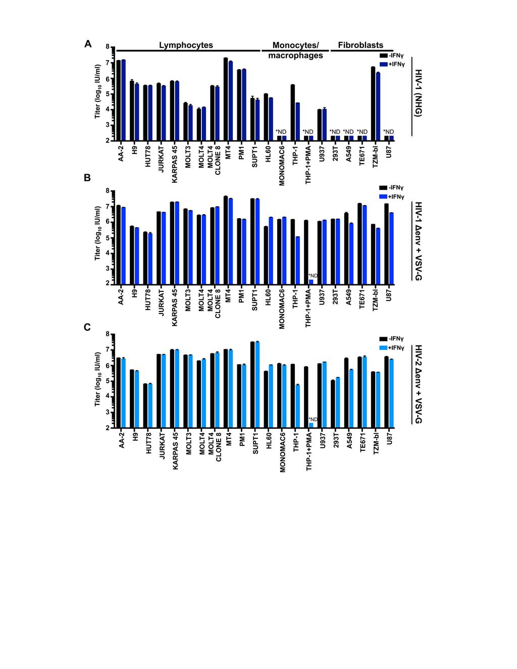
49
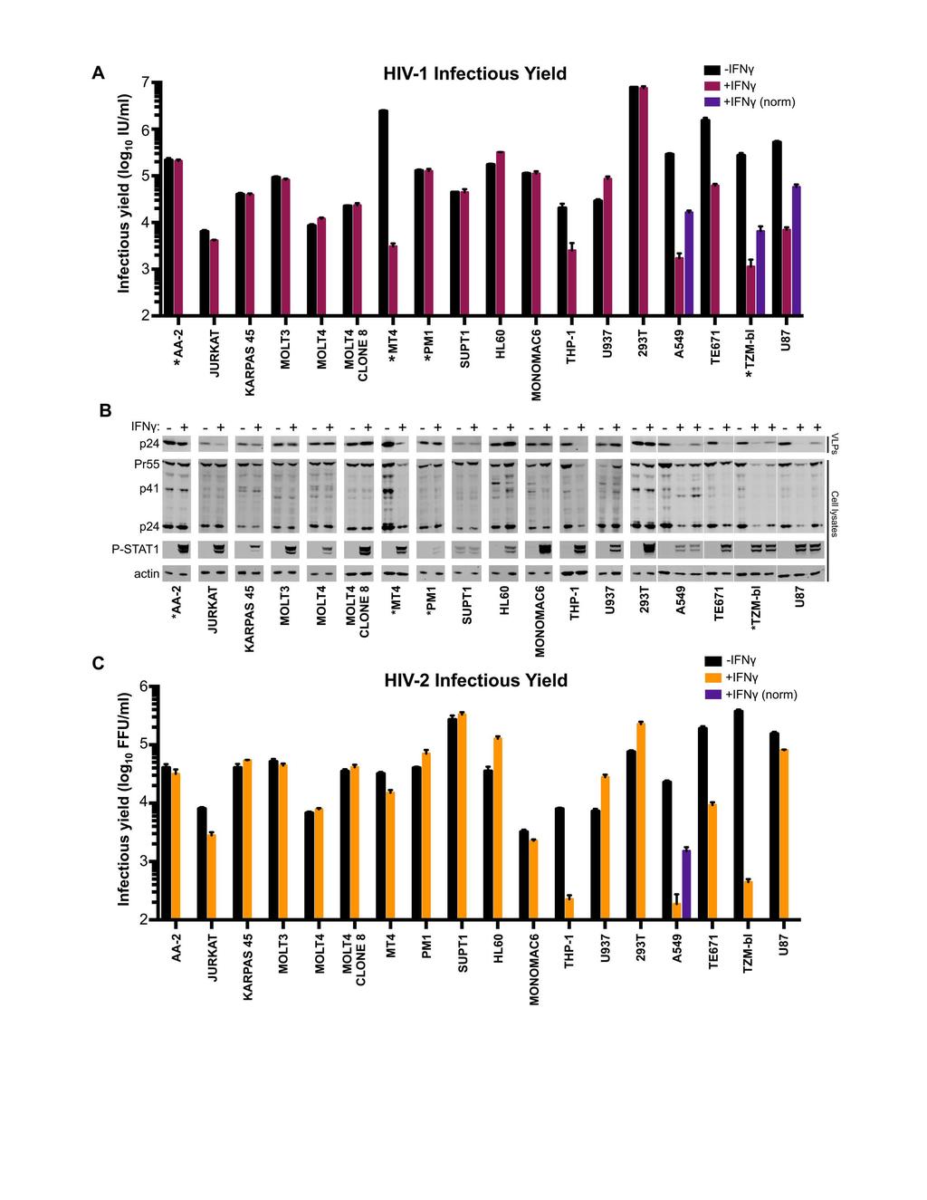
50
51
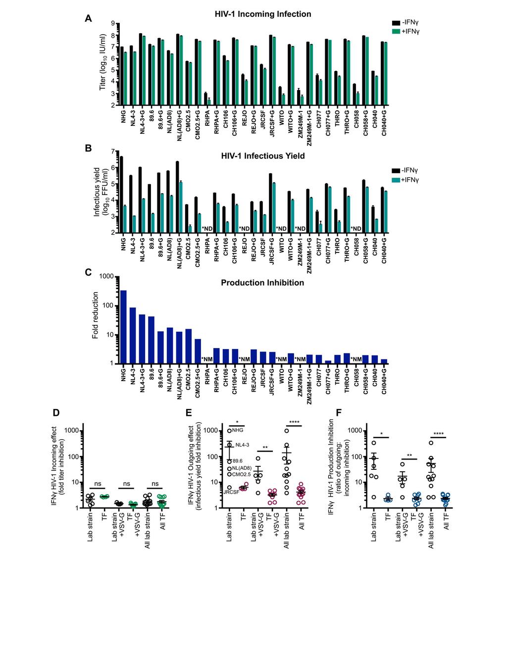
52
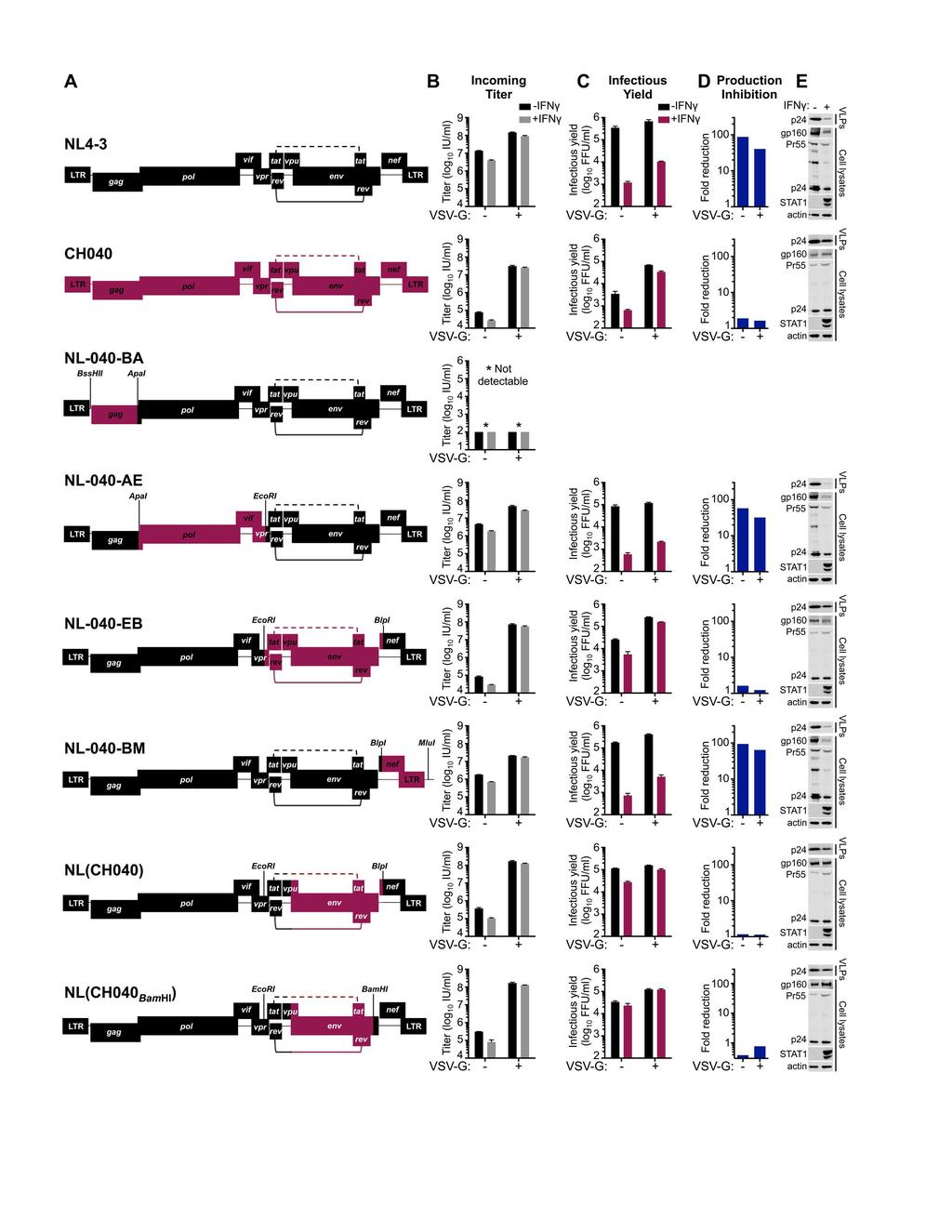
53
54

55
Supplementary Table 3. 3 UTR primer sequences. Primer sequences used to amplify and clone the 3 UTR of each indicated gene are listed.
 Supplemental Figure 1. DLKI-DIO3 mirna/mrna complementarity. Complementarity between the indicated DLK1-DIO3 cluster mirnas and the UTR of SOX2, SOX9, HIF1A, ZEB1, ZEB2, STAT3 and CDH1with mirsvr and PhastCons
Supplemental Data. Shin et al. Plant Cell. (2012) /tpc YFP N
 /tpc YFP N") MYC YFP N PIF5 YFP C N-TIC TIC Supplemental Data. Shin et al. Plant Cell. ()..5/tpc..95 Supplemental Figure. TIC interacts with MYC in the nucleus. Bimolecular fluorescence complementation assay using
MYC YFP N PIF5 YFP C N-TIC TIC Supplemental Data. Shin et al. Plant Cell. ()..5/tpc..95 Supplemental Figure. TIC interacts with MYC in the nucleus. Bimolecular fluorescence complementation assay using
c Tuj1(-) apoptotic live 1 DIV 2 DIV 1 DIV 2 DIV Tuj1(+) Tuj1/GFP/DAPI Tuj1 DAPI GFP
 apoptotic live 1 DIV 2 DIV 1 DIV 2 DIV Tuj1(+) Tuj1/GFP/DAPI Tuj1 DAPI GFP") Supplementary Figure 1 Establishment of the gain- and loss-of-function experiments and cell survival assays. a Relative expression of mature mir-484 30 20 10 0 **** **** NCP mir- 484P NCP mir- 484P b Relative
Supplementary Figure 1 Establishment of the gain- and loss-of-function experiments and cell survival assays. a Relative expression of mature mir-484 30 20 10 0 **** **** NCP mir- 484P NCP mir- 484P b Relative
Supplementary Document
 Supplementary Document 1. Supplementary Table legends 2. Supplementary Figure legends 3. Supplementary Tables 4. Supplementary Figures 5. Supplementary References 1. Supplementary Table legends Suppl.
Nature Structural & Molecular Biology: doi: /nsmb Supplementary Figure 1
 Supplementary Figure 1 U1 inhibition causes a shift of RNA-seq reads from exons to introns. (a) Evidence for the high purity of 4-shU-labeled RNAs used for RNA-seq. HeLa cells transfected with control
Supplementary Appendix
 Supplementary Appendix This appendix has been provided by the authors to give readers additional information about their work. Supplement to: Sherman SI, Wirth LJ, Droz J-P, et al. Motesanib diphosphate
Supplementary Figure 1. ROS induces rapid Sod1 nuclear localization in a dosagedependent manner. WT yeast cells (SZy1051) were treated with 4NQO at
 were treated with 4NQO at") Supplementary Figure 1. ROS induces rapid Sod1 nuclear localization in a dosagedependent manner. WT yeast cells (SZy1051) were treated with 4NQO at different concentrations for 30 min and analyzed for
Supplementary Figure 1. ROS induces rapid Sod1 nuclear localization in a dosagedependent manner. WT yeast cells (SZy1051) were treated with 4NQO at different concentrations for 30 min and analyzed for
CD31 5'-AGA GAC GGT CTT GTC GCA GT-3' 5 ' -TAC TGG GCT TCG AGA GCA GT-3'
 Table S1. The primer sets used for real-time RT-PCR analysis. Gene Forward Reverse VEGF PDGFB TGF-β MCP-1 5'-GTT GCA GCA TGA ATC TGA GG-3' 5'-GGA GAC TCT TCG AGG AGC ACT T-3' 5'-GAA TCA GGC ATC GAG AGA
Figure S1. Analysis of genomic and cdna sequences of the targeted regions in WT-KI and
 Figure S1. Analysis of genomic and sequences of the targeted regions in and indicated mutant KI cells, with WT and corresponding mutant sequences underlined. (A) cells; (B) K21E-KI cells; (C) D33A-KI cells;
Supplementary Figure 1 a
 Supplementary Figure a Normalized expression/tbp (A.U.).6... Trip-br transcripts Trans Trans Trans b..5. Trip-br Ctrl LPS Normalized expression/tbp (A.U.) c Trip-br transcripts. adipocytes.... Trans Trans
a) Primary cultures derived from the pancreas of an 11-week-old Pdx1-Cre; K-MADM-p53
 Primary cultures derived from the pancreas of an 11-week-old Pdx1-Cre; K-MADM-p53") 1 2 3 4 5 6 7 8 9 10 Supplementary Figure 1. Induction of p53 LOH by MADM. a) Primary cultures derived from the pancreas of an 11-week-old Pdx1-Cre; K-MADM-p53 mouse revealed increased p53 KO/KO (green,
1 2 3 4 5 6 7 8 9 10 Supplementary Figure 1. Induction of p53 LOH by MADM. a) Primary cultures derived from the pancreas of an 11-week-old Pdx1-Cre; K-MADM-p53 mouse revealed increased p53 KO/KO (green,
Supplementary Materials
 Supplementary Materials 1 Supplementary Table 1. List of primers used for quantitative PCR analysis. Gene name Gene symbol Accession IDs Sequence range Product Primer sequences size (bp) β-actin Actb gi
Abbreviations: P- paraffin-embedded section; C, cryosection; Bio-SA, biotin-streptavidin-conjugated fluorescein amplification.
 Supplementary Table 1. Sequence of primers for real time PCR. Gene Forward primer Reverse primer S25 5 -GTG GTC CAC ACT ACT CTC TGA GTT TC-3 5 - GAC TTT CCG GCA TCC TTC TTC-3 Mafa cds 5 -CTT CAG CAA GGA
Supplementary Figure 1 MicroRNA expression in human synovial fibroblasts from different locations. MicroRNA, which were identified by RNAseq as most
 Supplementary Figure 1 MicroRNA expression in human synovial fibroblasts from different locations. MicroRNA, which were identified by RNAseq as most differentially expressed between human synovial fibroblasts
Toluidin-Staining of mast cells Ear tissue was fixed with Carnoy (60% ethanol, 30% chloroform, 10% acetic acid) overnight at 4 C, afterwards
 overnight at 4 C, afterwards") Toluidin-Staining of mast cells Ear tissue was fixed with Carnoy (60% ethanol, 30% chloroform, 10% acetic acid) overnight at 4 C, afterwards incubated in 100 % ethanol overnight at 4 C and embedded in
Toluidin-Staining of mast cells Ear tissue was fixed with Carnoy (60% ethanol, 30% chloroform, 10% acetic acid) overnight at 4 C, afterwards incubated in 100 % ethanol overnight at 4 C and embedded in
Supplementary Table 2. Conserved regulatory elements in the promoters of CD36.
 Supplementary Table 1. RT-qPCR primers for CD3, PPARg and CEBP. Assay Forward Primer Reverse Primer 1A CAT TTG TGG CCT TGT GCT CTT TGA TGA GTC ACA GAA AGA ATC AAT TC 1B AGG AAA TGA ACT GAT GAG TCA CAG
Supplementary Figures
 Supplementary Figures Supplementary Figure 1. H3F3B expression in lung cancer. a. Comparison of H3F3B expression in relapsed and non-relapsed lung cancer patients. b. Prognosis of two groups of lung cancer
SUPPLEMENTARY INFORMATION
 doi: 10.1038/nature05883 SUPPLEMENTARY INFORMATION Supplemental Figure 1 Prostaglandin agonists and antagonists alter runx1/cmyb expression. a-e, Embryos were exposed to (b) PGE2 and (c) PGI2 (20μM) and
Table S1. Oligonucleotides used for the in-house RT-PCR assays targeting the M, H7 or N9. Assay (s) Target Name Sequence (5 3 ) Comments
 Target Name Sequence (5 3 ) Comments") SUPPLEMENTAL INFORMATION 2 3 Table S. Oligonucleotides used for the in-house RT-PCR assays targeting the M, H7 or N9 genes. Assay (s) Target Name Sequence (5 3 ) Comments CDC M InfA Forward (NS), CDC M
SUPPLEMENTAL INFORMATION 2 3 Table S. Oligonucleotides used for the in-house RT-PCR assays targeting the M, H7 or N9 genes. Assay (s) Target Name Sequence (5 3 ) Comments CDC M InfA Forward (NS), CDC M
Citation for published version (APA): Oosterveer, M. H. (2009). Control of metabolic flux by nutrient sensors Groningen: s.n.
: Oosterveer, M. H. (2009). Control of metabolic flux by nutrient sensors Groningen: s.n.") University of Groningen Control of metabolic flux by nutrient sensors Oosterveer, Maaike IMPORTANT NOTE: You are advised to consult the publisher's version (publisher's PDF) if you wish to cite from it.
University of Groningen Control of metabolic flux by nutrient sensors Oosterveer, Maaike IMPORTANT NOTE: You are advised to consult the publisher's version (publisher's PDF) if you wish to cite from it.
 www.lessonplansinc.com Topic: Protein Synthesis - Sentence Activity Summary: Students will simulate transcription and translation by building a sentence/polypeptide from words/amino acids. Goals & Objectives:
Supplementary Figure 1
 Supplementary Figure 1 Supplementary Figure 1: Cryopreservation alters CD62L expression by CD4 T cells. Freshly isolated (left) or cryopreserved PBMCs (right) were stained with the mix of antibodies described
SUPPLEMENTARY DATA. Supplementary Table 1. Primer sequences for qrt-pcr
 Supplementary Table 1. Primer sequences for qrt-pcr Gene PRDM16 UCP1 PGC1α Dio2 Elovl3 Cidea Cox8b PPARγ AP2 mttfam CyCs Nampt NRF1 16s-rRNA Hexokinase 2, intron 9 β-actin Primer Sequences 5'-CCA CCA GCG
BIOLOGY 621 Identification of the Snorks
 Name: Date: Block: BIOLOGY 621 Identification of the Snorks INTRODUCTION: In this simulation activity, you will examine the DNA sequence of a fictitious organism - the Snork. Snorks were discovered on
Journal of Cell Science Supplementary information. Arl8b +/- Arl8b -/- Inset B. electron density. genotype
 J. Cell Sci. : doi:.4/jcs.59: Supplementary information E9. A Arl8b /- Arl8b -/- Arl8b Arl8b non-specific band Gapdh Tbp E7.5 HE Inset B D Control al am hf C E Arl8b -/- al am hf E8.5 F low middle high
A smart acid nanosystem for ultrasensitive. live cell mrna imaging by the target-triggered intracellular self-assembly
 Electronic Supplementary Material (ESI) for Chemical Science. This journal is The Royal Society of Chemistry 2017 A smart ZnO@polydopamine-nucleic acid nanosystem for ultrasensitive live cell mrna imaging
Nature Immunology: doi: /ni.3836
 Supplementary Figure 1 Recombinant LIGHT-VTP induces pericyte contractility and endothelial cell activation. (a) Western blot showing purification steps for full length murine LIGHT-VTP (CGKRK) protein:
Plasmids Western blot analysis and immunostaining Flow Cytometry Cell surface biotinylation RNA isolation and cdna synthesis
 Plasmids psuper-retro-s100a10 shrna1 was constructed by cloning the dsdna oligo 5 -GAT CCC CGT GGG CTT CCA GAG CTT CTT TCA AGA GAA GAA GCT CTG GAA GCC CAC TTT TTA-3 and 5 -AGC TTA AAA AGT GGG CTT CCA GAG
Culture Density (OD600) 0.1. Culture Density (OD600) Culture Density (OD600) Culture Density (OD600) Culture Density (OD600)
 0.1. Culture Density (OD600) Culture Density (OD600) Culture Density (OD600) Culture Density (OD600)") A. B. C. D. E. PA JSRI JSRI 2 PA DSAM DSAM 2 DSAM 3 PA LNAP LNAP 2 LNAP 3 PAO Fcor Fcor 2 Fcor 3 PAO Wtho Wtho 2 Wtho 3 Wtho 4 DTSB Low Iron 2 4 6 8 2 4 6 8 2 22 DTSB Low Iron 2 4 6 8 2 4 6 8 2 22 DTSB
A. B. C. D. E. PA JSRI JSRI 2 PA DSAM DSAM 2 DSAM 3 PA LNAP LNAP 2 LNAP 3 PAO Fcor Fcor 2 Fcor 3 PAO Wtho Wtho 2 Wtho 3 Wtho 4 DTSB Low Iron 2 4 6 8 2 4 6 8 2 22 DTSB Low Iron 2 4 6 8 2 4 6 8 2 22 DTSB
Phylogenetic analysis of human and chicken importins. Only five of six importins were studied because
 Supplementary Figure S1 Phylogenetic analysis of human and chicken importins. Only five of six importins were studied because importin-α6 was shown to be testis-specific. Human and chicken importin protein
Supplemental Materials and Methods Plasmids and viruses Quantitative Reverse Transcription PCR Generation of molecular standard for quantitative PCR
 Supplemental Materials and Methods Plasmids and viruses To generate pseudotyped viruses, the previously described recombinant plasmids pnl4-3-δnef-gfp or pnl4-3-δ6-drgfp and a vector expressing HIV-1 X4
Lezione 10. Sommario. Bioinformatica. Lezione 10: Sintesi proteica Synthesis of proteins Central dogma: DNA makes RNA makes proteins Genetic code
 Lezione 10 Bioinformatica Mauro Ceccanti e Alberto Paoluzzi Lezione 10: Sintesi proteica Synthesis of proteins Dip. Informatica e Automazione Università Roma Tre Dip. Medicina Clinica Università La Sapienza
Nucleotide Sequence of the Australian Bluetongue Virus Serotype 1 RNA Segment 10
 J. gen. Virol. (1988), 69, 945-949. Printed in Great Britain 945 Key words: BTV/genome segment lo/nucleotide sequence Nucleotide Sequence of the Australian Bluetongue Virus Serotype 1 RNA Segment 10 By
Characterizing intra-host influenza virus populations to predict emergence
 Characterizing intra-host influenza virus populations to predict emergence June 12, 2012 Forum on Microbial Threats Washington, DC Elodie Ghedin Center for Vaccine Research Dept. Computational & Systems
*To whom correspondence should be addressed. This PDF file includes:
 www.sciencemag.org/cgi/content/full/science.1212182/dc1 Supporting Online Material for Partial Retraction to Detection of an Infectious Retrovirus, XMRV, in Blood Cells of Patients with Chronic Fatigue
Supplementary Materials and Methods
 DD2 suppresses tumorigenicity of ovarian cancer cells by limiting cancer stem cell population Chunhua Han et al. Supplementary Materials and Methods Analysis of publicly available datasets: To analyze
Detection of 549 new HLA alleles in potential stem cell donors from the United States, Poland and Germany
 HLA ISSN 2059-2302 BRIEF COMMUNICATION Detection of 549 new HLA alleles in potential stem cell donors from the United States, Poland and Germany C. J. Hernández-Frederick 1, N. Cereb 2,A.S.Giani 1, J.
Supplemental Figures: Supplemental Figure 1
 Supplemental Figures: Supplemental Figure 1 Suppl. Figure 1. BM-DC infection with H. pylori does not induce cytotoxicity and treatment of BM-DCs with H. pylori sonicate, but not heat-inactivated bacteria,
BHP 2-7 and Nthy-ori 3-1 cells were grown in RPMI1640 medium (Hyclone) supplemented with 10% fetal bovine serum (Gibco), 2mM L-glutamine, and 100 U/mL
 supplemented with 10% fetal bovine serum (Gibco), 2mM L-glutamine, and 100 U/mL") 1 2 3 4 Materials and Methods Cell culture BHP 2-7 and Nthy-ori 3-1 cells were grown in RPMI1640 medium (Hyclone) 5 supplemented with 10% fetal bovine serum (Gibco), 2mM L-glutamine, and 100 U/mL 6 penicillin-streptomycin.
1 2 3 4 Materials and Methods Cell culture BHP 2-7 and Nthy-ori 3-1 cells were grown in RPMI1640 medium (Hyclone) 5 supplemented with 10% fetal bovine serum (Gibco), 2mM L-glutamine, and 100 U/mL 6 penicillin-streptomycin.
Supplemental Information. Cancer-Associated Fibroblasts Neutralize. the Anti-tumor Effect of CSF1 Receptor Blockade
 Cancer Cell, Volume 32 Supplemental Information Cancer-Associated Fibroblasts Neutralize the Anti-tumor Effect of CSF1 Receptor Blockade by Inducing PMN-MDSC Infiltration of Tumors Vinit Kumar, Laxminarasimha
Supplementary Figure 1
 Supplementary Figure 1 3 3 3 1 1 Bregma -1.6mm 3 : Bregma Ref) Http://www.mbl.org/atlas165/atlas165_start.html Bregma -.18mm Supplementary Figure 1 Schematic representation of the utilized brain slice
Supplemental Information. Th17 Lymphocytes Induce Neuronal. Cell Death in a Human ipsc-based. Model of Parkinson's Disease
 Cell Stem Cell, Volume 23 Supplemental Information Th17 Lymphocytes Induce Neuronal Cell Death in a Human ipsc-based Model of Parkinson's Disease Annika Sommer, Franz Maxreiter, Florian Krach, Tanja Fadler,
Astaxanthin prevents and reverses diet-induced insulin resistance and. steatohepatitis in mice: A comparison with vitamin E
 Supplementary Information Astaxanthin prevents and reverses diet-induced insulin resistance and steatohepatitis in mice: A comparison with vitamin E Yinhua Ni, 1,2 Mayumi Nagashimada, 1 Fen Zhuge, 1 Lili
Supplementary Figure 1
 Metastatic melanoma Primary melanoma Healthy human skin Supplementary Figure 1 CD22 IgG4 Supplementary Figure 1: Immunohisochemical analysis of CD22+ (left) and IgG4 (right), cells (shown in red and indicated
Supplementary Figure 1
 Supplementary Figure 1 Supplementary Figure 1. Lats1/2 deleted ihbs and ihps showed decreased transcripts of hepatocyte related genes (a and b) Western blots (a) and recombination PCR (b) of control and
Supplementary Figure 1a
 Supplementary Figure 1a Hours: E-cadherin TGF-β On TGF-β Off 0 12 24 36 48 24 48 72 Vimentin βactin Fig. S1a. Treatment of AML12 cells with TGF-β induces EMT. Treatment of AML12 cells with TGF-β results
Cross-talk between mineralocorticoid and angiotensin II signaling for cardiac
 ONLINE SUPPLEMENT TO Crosstalk between mineralocorticoid and angiotensin II signaling for cardiac remodeling An Di ZHANG,,3, Aurelie NGUYEN DINH CAT*,,3, Christelle SOUKASEUM *,,3, Brigitte ESCOUBET, 4,
TetR repressor-based bioreporters for the detection of doxycycline using Escherichia
 Supplementary materials TetR repressor-based bioreporters for the detection of doxycycline using Escherichia coli and Acinetobacter oleivorans Hyerim Hong and Woojun Park * Department of Environmental
SUPPORTING INFORMATION
 SUPPORTING INFORMATION Biology is different in small volumes: endogenous signals shape phenotype of primary hepatocytes cultured in microfluidic channels Amranul Haque, Pantea Gheibi, Yandong Gao, Elena
Expression of Selected Inflammatory Cytokine Genes in Bladder Biopsies
 Borneo Journal of Resource Science and Technology (2013) 3(2): 15-20 Expression of Selected Inflammatory Cytokine Genes in Bladder Biopsies EDMUND UI-HANG SIM *1, NUR DIANA ANUAR 2, TENG-AIK ONG 3, GUAN-
Beta Thalassemia Case Study Introduction to Bioinformatics
 Beta Thalassemia Case Study Sami Khuri Department of Computer Science San José State University San José, California, USA sami.khuri@sjsu.edu www.cs.sjsu.edu/faculty/khuri Outline v Hemoglobin v Alpha
CIRCRESAHA/2004/098145/R1 - ONLINE 1. Validation by Semi-quantitative Real-Time Reverse Transcription PCR
 CIRCRESAHA/2004/098145/R1 - ONLINE 1 Expanded Materials and Methods Validation by Semi-quantitative Real-Time Reverse Transcription PCR Expression patterns of 13 genes (Online Table 2), selected with respect
HCV Persistence and Immune Evasion in the Absence of Memory T Cell Help.
 SOM Text HCV Persistence and Immune Evasion in the Absence of Memory T Cell Help. Arash Grakoui 1, Naglaa H. Shoukry 2, David J. Woollard 2, Jin-Hwan Han 1, Holly L. Hanson 1, John Ghrayeb 3, Krishna K.
A basic helix loop helix transcription factor controls cell growth
 A basic helix loop helix transcription factor controls cell growth and size in root hairs Keke Yi 1,2, Benoît Menand 1,3, Elizabeth Bell 1, Liam Dolan 1,4 Supplementary note Low soil phosphate availability
ice-cold 70% ethanol with gentle vortexing, incubated at -20 C for 4 hours, and washed with PBS.
 Cell cycle analysis For cell cycle analysis, single cell suspensions of E12.5 fetal liver cells were suspended in 4 ml ice-cold 7% ethanol with gentle vortexing, incubated at -2 C for 4 hours, and washed
Beta Thalassemia Sami Khuri Department of Computer Science San José State University Spring 2015
 Bioinformatics in Medical Product Development SMPD 287 Three Beta Thalassemia Sami Khuri Department of Computer Science San José State University Hemoglobin Outline Anatomy of a gene Hemoglobinopathies
Supporting Information
 Supporting Information Malapeira et al. 10.1073/pnas.1217022110 SI Materials and Methods Plant Material and Growth Conditions. A. thaliana seedlings were stratified at 4 C in the dark for 3 d on Murashige
Patterns of hemagglutinin evolution and the epidemiology of influenza
 2 8 US Annual Mortality Rate All causes Infectious Disease Patterns of hemagglutinin evolution and the epidemiology of influenza DIMACS Working Group on Genetics and Evolution of Pathogens, 25 Nov 3 Deaths
Supplementary Information. Bamboo shoot fiber prevents obesity in mice by. modulating the gut microbiota
 Supplementary Information Bamboo shoot fiber prevents obesity in mice by modulating the gut microbiota Xiufen Li 1,2, Juan Guo 1, Kailong Ji 1,2, and Ping Zhang 1,* 1 Key Laboratory of Tropical Plant Resources
SUPPLEMENTAL METHODS Cell culture RNA extraction and analysis Immunohistochemical analysis and laser capture microdissection (LCM)
") SUPPLEMENTAL METHODS Cell culture Human peripheral blood mononuclear cells were isolated from healthy donors by Ficoll density gradient centrifugation. Monocyte differentiation to resting macrophages ()
SUPPLEMENTAL METHODS Cell culture Human peripheral blood mononuclear cells were isolated from healthy donors by Ficoll density gradient centrifugation. Monocyte differentiation to resting macrophages ()
Single-Molecule Analysis of Gene Expression Using Two-Color RNA- Labeling in Live Yeast
 Supplemental Figures, Tables and Results Single-Molecule Analysis of Gene Expression Using Two-Color RNA- Labeling in Live Yeast Sami Hocine 1, Pascal Raymond 2, Daniel Zenklusen 2, Jeffrey A. Chao 1 &
L I F E S C I E N C E S
 1a L I F E S C I E N C E S 5 -UUA AUA UUC GAA AGC UGC AUC GAA AAC UGU GAA UCA-3 5 -TTA ATA TTC GAA AGC TGC ATC GAA AAC TGT GAA TCA-3 3 -AAT TAT AAG CTT TCG ACG TAG CTT TTG ACA CTT AGT-5 OCTOBER 31, 2006
Description of Supplementary Files. File Name: Supplementary Information Description: Supplementary Figures and Supplementary Tables
 Description of Supplementary Files File Name: Supplementary Information Description: Supplementary Figures and Supplementary Tables Supplementary Figure 1: (A), HCT116 IDH1-WT and IDH1-R132H cells were
Baseline clinical characteristics for the 81 CMML patients Routine diagnostic testing and statistical analyses... 3
 Next-Generation Sequencing Technology Reveals a Characteristic Pattern of Molecular Mutations in 72.8% of Chronic Myelomonocytic Leukemia (CMML) by Detecting Frequent Alterations in TET2, CBL, RAS, and
Finding protein sites where resistance has evolved
 Finding protein sites where resistance has evolved The amino acid (Ka) and synonymous (Ks) substitution rates Please sit in row K or forward The Berlin patient: first person cured of HIV Contracted HIV
Formylpeptide receptor2 contributes to colon epithelial homeostasis, inflammation, and tumorigenesis
 Supplementary Data Formylpeptide receptor2 contributes to colon epithelial homeostasis, inflammation, and tumorigenesis Keqiang Chen, Mingyong Liu, Ying Liu, Teizo Yoshimura, Wei Shen, Yingying Le, Scott
Supporting Information
 Supporting Information Hatziioannou et al. 10.1073/pnas.0812587106 SI Text Viral Constructs and Viral Stock Generation. To generate the HIV-1 constructs used throughout these studies, the env gene in an
Analysis of Human Cytomegalovirus orilyt Sequence Requirements in the Context of the Viral Genome
 JOURNAL OF VIROLOGY, Mar. 2005, p. 3615 3626 Vol. 79, No. 6 0022-538X/05/$08.00 0 doi:10.1128/jvi.79.6.3615 3626.2005 Copyright 2005, American Society for Microbiology. All Rights Reserved. Analysis of
Development of RT-qPCR-based molecular diagnostic assays for therapeutic target selection of breast cancer patients
 Development of RT-qPCR-based molecular diagnostic assays for therapeutic target selection of breast cancer patients Sangjung Park The Graduate School Yonsei University Department of Biomedical Laboratory
Advanced Subsidiary Unit 1: Lifestyle, Transport, Genes and Health
 Write your name here Surname Other names Edexcel GCE Centre Number Candidate Number Biology Advanced Subsidiary Unit 1: Lifestyle, Transport, Genes and Health Thursday 8 January 2009 Morning Time: 1 hour
Supporting Information
 Supporting Information Molecular Recognition Based DNA Nanoassemblies on the Surfaces of Nanosized Exosomes Shuo Wan,, Liqin Zhang,,, Sai Wang, Yuan Liu,, Cuichen Wu, Cheng Cui, Hao Sun, Muling Shi, Ying
Supplementary information. MARCH8 inhibits HIV-1 infection by reducing virion incorporation of envelope glycoproteins
 Supplementary information inhibits HIV-1 infection by reducing virion incorporation of envelope glycoproteins Takuya Tada, Yanzhao Zhang, Takayoshi Koyama, Minoru Tobiume, Yasuko Tsunetsugu-Yokota, Shoji
Oligo Sequence* bp %GC Tm Hair Hm Ht Position Size Ref. HIVrt-F 5 -CTA-gAA-CTT-TRA-ATg-CAT-ggg-TAA-AAg-TA
 Human immunodeficiency virus (HIV) detection & quantitation by qrt-pcr (Taqman). Created on: Oct 26, 2010; Last modified by: Jul 17, 2017; Version: 3.0 This protocol describes the qrt-pcr taqman based
Integration Solutions
 Integration Solutions (1) a) With no active glycosyltransferase of either type, an ii individual would not be able to add any sugars to the O form of the lipopolysaccharide. Thus, the only lipopolysaccharide
Supplementary Information
 Supplementary Information Remodeling of heterochromatin structure slows neuropathological progression and prolongs survival in an animal model of Huntington s disease Junghee Lee, Yu Jin Hwang, Yunha Kim,
What do you think of when you here the word genome?
 What do you think of when you here the word genome? What do you think of when you here the word genome? Personal Genomics Outline Review of pre-lab work Genomics and Medicine Case Overview & Assignment
The Effect of Epstein-Barr Virus Latent Membrane Protein 2 Expression on the Kinetics of Early B Cell Infection
 The Effect of Epstein-Barr Virus Latent Membrane Protein 2 Expression on the Kinetics of Early B Cell Infection Laura R. Wasil, Monica J. Tomaszewski, Aki Hoji, David T. Rowe* Department of Infectious
Isolation and Genetic Characterization of New Reassortant H3N1 Swine Influenza Virus from Pigs in the Midwestern United States
 JOURNAL OF VIROLOGY, May 2006, p. 5092 5096 Vol. 80, No. 10 0022-538X/06/$08.00 0 doi:10.1128/jvi.80.10.5092 5096.2006 Copyright 2006, American Society for Microbiology. All Rights Reserved. Isolation
Resistance to Tetracycline Antibiotics by Wangrong Yang, Ian F. Moore, Kalinka P. Koteva, Donald W. Hughes, David C. Bareich and Gerard D. Wright.
 Supplementary Data for TetX is a Flavin-Dependent Monooxygenase Conferring Resistance to Tetracycline Antibiotics by Wangrong Yang, Ian F. Moore, Kalinka P. Koteva, Donald W. Hughes, David C. Bareich and
TLR7/8 agonist induces a post entry SAMHD1 independent block to HIV 1 infection of monocytes
 DOI 10.1186/s12977-016-0316-3 Retrovirology RESEARCH TLR7/8 agonist induces a post entry SAMHD1 independent block to HIV 1 infection of monocytes Henning Hofmann 1,2, Bénédicte Vanwalscappel 1, Nicolin
Received 22 October 1997/Accepted 9 February 1998
 JOURNAL OF VIROLOGY, May 1998, p. 4308 4319 Vol. 72, No. 5 0022-538X/98/$04.00 0 Copyright 1998, American Society for Microbiology gag, vif, and nef Genes Contribute to the Homologous Viral Interference
Influenza viruses are classified as members of the family
 Rewiring the RNAs of influenza virus to prevent reassortment Qinshan Gao a and Peter Palese a,b,1 Departments of a Microbiology and b Medicine, Mount Sinai School of Medicine, New York, NY 10029 Contributed
Supplementary information
 Supplementary information Unique polypharmacology nuclear receptor modulator blocks inflammatory signaling pathways Mi Ra Chang 1, Anthony Ciesla 1, Timothy S. Strutzenberg 1, Scott J. Novick 1, Yuanjun
Supporting Information. Mutational analysis of a phenazine biosynthetic gene cluster in
 Supporting Information for Mutational analysis of a phenazine biosynthetic gene cluster in Streptomyces anulatus 9663 Orwah Saleh 1, Katrin Flinspach 1, Lucia Westrich 1, Andreas Kulik 2, Bertolt Gust
Relationship of the APOA5/A4/C3/A1 gene cluster and APOB gene polymorphisms with dyslipidemia
 elationship of the APOA5/A4/C3/A1 gene cluster and APOB gene polymorphisms with dyslipidemia H.J. Ou 1, G. Huang 2, W. Liu 3, X.L. Ma 2, Y. Wei 4, T. Zhou 5 and Z.M. Pan 3 1 Department of Neurology, The
Citation for published version (APA): Von Eije, K. J. (2009). RNAi based gene therapy for HIV-1, from bench to bedside
: Von Eije, K. J. (2009). RNAi based gene therapy for HIV-1, from bench to bedside") UvA-DARE (Digital Academic Repository) RNAi based gene therapy for HIV-1, from bench to bedside Von Eije, K.J. Link to publication Citation for published version (APA): Von Eije, K. J. (2009). RNAi based
UvA-DARE (Digital Academic Repository) RNAi based gene therapy for HIV-1, from bench to bedside Von Eije, K.J. Link to publication Citation for published version (APA): Von Eije, K. J. (2009). RNAi based
The Clinical Performance of Primary HPV Screening, Primary HPV Screening Plus Cytology Cotesting, and Cytology Alone at a Tertiary Care Hospital
 The Clinical Performance of Primary HPV Screening, Primary HPV Screening Plus Cytology Cotesting, and Cytology Alone at a Tertiary Care Hospital Jung-Woo Choi MD, PhD; Younghye Kim MD, PhD; Ju-Han Lee
Mutation Screening and Association Studies of the Human UCP 3 Gene in Normoglycemic and NIDDM Morbidly Obese Patients
 Mutation Screening and Association Studies of the Human UCP 3 Gene in Normoglycemic and NIDDM Morbidly Obese Patients Shuichi OTABE, Karine CLEMENT, Séverine DUBOIS, Frederic LEPRETRE, Veronique PELLOUX,
Loyer, et al. microrna-21 contributes to NASH Suppl 1/15
 Loyer, et al. microrna-21 contributes to NASH Suppl 1/15 SUPPLEMENTARY MATERIAL: Liver MicroRNA-21 is Overexpressed in Non Alcoholic Steatohepatitis and Contributes to the Disease in Experimental Models
without LOI phenotype by breeding female wild-type C57BL/6J and male H19 +/.
 Sakatani et al. 1 Supporting Online Material Materials and methods Mice and genotyping: H19 mutant mice with C57BL/6J background carrying a deletion in the structural H19 gene (3 kb) and 10 kb of 5 flanking
SUPPLEMENTARY RESULTS
 SUPPLEMENTARY RESULTS Supplementary Table 1. hfpr1- Flpln-CHO hfpr2-flpln-cho pec 50 E max (%) Log( /K A) Log( /K A) N pec 50 E max (%) Log( /K A) Log( /K A) n ERK1/2 phosphorylation fmlp 9.0±0.6 80±7
PATIENTS AND METHODS. Subjects
 PATIENTS AND METHODS Subjects Twenty-nine morbidly obese subjects involved in a gastric surgery program were enrolled in the study between October 25 and March 21. Bariatric surgery was performed in patients
A Rapid and Sensitive Chip-based Assay for Detection of rpob Gene Mutations Conferring Rifampicin Resistance in Mycobacterium tuberculosis (TB).
.") A Rapid and Sensitive Chip-based Assay for Detection of rpob Gene Mutations Conferring Rifampicin Resistance in Mycobacterium tuberculosis (TB). Wanyuan Ao, Steve Aldous, Evelyn Woodruff, Brian Hicke,
A Rapid and Sensitive Chip-based Assay for Detection of rpob Gene Mutations Conferring Rifampicin Resistance in Mycobacterium tuberculosis (TB). Wanyuan Ao, Steve Aldous, Evelyn Woodruff, Brian Hicke,
Protein sequence alignment using binary string
 Available online at www.scholarsresearchlibrary.com Scholars Research Library Der Pharmacia Lettre, 2015, 7 (5):220-225 (http://scholarsresearchlibrary.com/archive.html) ISSN 0975-5071 USA CODEN: DPLEB4
Figure S1. Schematic presentation of genomic replication of idsiv after transfection and infection. After transfection of idsiv plasmid DNA into 293T
 Figure S1. Schematic presentation of genomic replication of idsiv after transfection and infection. After transfection of idsiv plasmid DNA into 293T cells, the RNA genomes with all modifications are generated
Micropathology Ltd. University of Warwick Science Park, Venture Centre, Sir William Lyons Road, Coventry CV4 7EZ
 www.micropathology.com info@micropathology.com Micropathology Ltd Tel 24hrs: +44 (0) 24-76 323222 Fax / Ans: +44 (0) 24-76 - 323333 University of Warwick Science Park, Venture Centre, Sir William Lyons
Viral quantification and filogenetic analysis Chikungunya virus strains imported to Italy Antonino Di Caro
 Viral quantification a filogenetic analysis Chikungunya virus strains imported to Italy Antonino Di Caro National Institute for Infectious Diseases L. Spallanzani, Rome BACKGROUND 1 Since 2005, more than
McAlpine PERK-GSK3 regulates foam cell formation. Supplemental Material. Supplementary Table I. Sequences of real time PCR primers.
 Mclpine PERK-GSK3 regulates foam cell formation Supplemental Material Supplementary Table I. Sequences of real time PCR primers. Primer Name Primer Sequences (5-3 ) Product Size (bp) GRP78 (human) Fwd:
Viral hepatitis, which affects half a billion people
 GASTROENTEROLOGY 2006;130:435 452 BASIC LIVER, PANCREAS, AND BILIARY TRACT Natural Killer Cells Ameliorate Liver Fibrosis by Killing Activated Stellate Cells in NKG2D-Dependent and Tumor Necrosis Factor
Supplementary Figure 1. SC35M polymerase activity in the presence of Bat or SC35M NP encoded from the phw2000 rescue plasmid.
 1 2 3 4 5 6 7 8 9 10 11 12 13 14 15 16 17 18 19 20 21 22 23 24 25 26 27 Supplementary Figure 1. SC35M polymerase activity in the presence of Bat or SC35M NP encoded from the phw2000 rescue plasmid. HEK293T