X/01/$ DOI: /JVI Copyright 2001, American Society for Microbiology. All Rights Reserved.
|
|
|
- Harvey Curtis
- 6 years ago
- Views:
Transcription
1 JOURNAL OF VIROLOGY, June 2001, p Vol. 75, No X/01/$ DOI: /JVI Copyright 2001, American Society for Microbiology. All Rights Reserved. Spindle Cell Conversion by Kaposi s Sarcoma-Associated Herpesvirus: Formation of Colonies and Plaques with Mixed Lytic and Latent Gene Expression in Infected Primary Dermal Microvascular Endothelial Cell Cultures DOLORES M. CIUFO, JENNIFER S. CANNON, LYNN J. POOLE, FREDERICK Y. WU, PAUL MURRAY, RICHARD F. AMBINDER, AND GARY S. HAYWARD* Molecular Virology Program, Department of Oncology, The Johns Hopkins University School of Medicine Cancer Center, Baltimore, Maryland Received 20 November 2000/Accepted 20 March 2001 Angiogenic Kaposi s sarcoma (KS) skin lesions found in both AIDS and non-aids patients are universally associated with infection by the presumed causative agent, known as KS-associated herpesvirus (KSHV) or human herpesvirus 8. KSHV genomes expressing latent state virus-encoded mrnas and the LANA1 (latent nuclear antigen 1) protein are consistently present in spindle-like tumor cells that are thought to be of endothelial cell origin. Although the KSHV lytic cycle can be induced in rare latently infected primary effusion lymphoma (PEL) cell lines, the ability to transmit or assay infectious KSHV has so far eluded investigators. Here, we demonstrate that infection with supernatant virions derived from three different tetradecanoyl phorbol acetate-induced PEL cell lines can induce cultured primary human dermal microvascular endothelial cells (DMVEC) to form colonies of proliferating latently infected spindle-shaped cells, all of which express the KSHV-encoded LANA1 protein. Although their initial infectivity varied widely (JSC1 > > BC3 > BCP1), virions from all three cell lines produced distinctive spindle cell colonies and plaques without affecting the contact-inhibited cobblestone-like phenotype of adjacent uninfected DMVEC. Each infected culture could also be expanded into a completely spindloid persistently infected culture displaying aggregated swirls of spindle cells resembling those in KS lesions. Formation of new colonies and plaques was inhibited in the presence of phosphonoacetic acid or gangciclovir, but these antiherpesvirus agents had little effect on the propagation of already latently infected spindloid cultures. In persistently infected secondary cultures, patches of up to 10% of the spindloid cells constitutively expressed several early viral lytic cycle proteins, and 1 to 2% of the cells also formed typical herpesvirus DNA replication compartments, displayed cytopathic rounding effects, and expressed late viral antigens. We conclude that de novo KSHV infection induces a spindle cell conversion phenotype in primary DMVEC cultures that is directly associated with latent state expression of the LANA1 protein. However, these cultures also spontaneously reactivate to produce an unusual combination of both latent and productive but slow lytic cycle infection. The formation of spindle cell colonies and plaques in DMVEC cultures provides for the first time a quantitative assay for directly measuring the infectivity of KSHV virion preparations. Classic Kaposi s sarcoma (KS) is a rare angiogenic skin lesion found primarily in males in the Mediterranean area and in central Africa where KS-associated herpesvirus (KSHV) infection is common. However, in the setting of combined immunosuppression and KSHV infection in renal transplant patients or of both human immunodeficiency virus and KSHV infection in AIDS patients, the rates of KS increase 500- and 20,000- fold, respectively, compared to ethnically matched populations. The higher rate in AIDS patients is at least partly explained by much higher KSHV infection rates in male homosexual AIDS patients (up to 85%) compared to the average U.S. or U.K. population (0.5 to 2%). KS is now the most abundant tumor found in many parts of central and southern Africa and can occasionally become highly invasive and aggressive. KSHV DNA is invariably found in KS tumors (9, 16, 33), * Corresponding author. Mailing address: Molecular Virology Laboratories, Rm. 3M-08 Bunting-Blaustein Cancer Research Building, The Johns Hopkins University School of Medicine, 1650 Orleans St., Baltimore, MD Phone: (410) Fax: (410) ghayward@jhmi.edu. and viral mrnas for the latent state LANA1 (latent nuclear antigen 1), vcyc-d, and vflip genes, as well as the T0.7 transcripts, are found consistently in spindle cells in late-stage nodular or invasive tumors as well as in at least some cells of early-stage plaque and patch lesions (10, 29). Some reports also describe scattered single cells in some tumors that express presumed lytic cycle viral mrnas for viral interleukin-6 (vil6), viral macrophage inflammatory protein 1 (vmip1), T1.1, or ORF23 (28), but only LANA1 has been observed to be expressed consistently in most spindle cells at the protein level. KSHV genomes are also found consistently in multicentric Castleman s disease (27) and in rare AIDS-associated primary effusion lymphoma (PEL) tumors, several of which have been established as permanent cell lines that stably maintain multicopy extrachromosomal KSHV plasmids or episomes (2, 5, 7). The ability to induce lytic cycle KSHV infection including production of some mature virions after tetradecanoyl phorbal acetate (TPA) or sodium butyrate treatment of PEL cell lines has revolutionized study of the molecular genetics and molecular biology of KSHV (19, 24, 26, 30); however, the ability to 5614
2 VOL. 75, 2001 ENDOTHELIAL CELL PLAQUE ASSAY FOR KSHV 5615 transmit virus from these cells to other cultures or to assay infectious virus has been particularly elusive. Two early studies claimed some degree of success in passing very low levels of virus onto B lymphocytes and 293 cells (12), but no cell type tested was susceptible in other studies (23), and the evidence consisted only of DNA or reverse transcription-pcr analysis at a level that most observers considered dubious at best. Another initial study used PCR procedures to suggest that KSHV DNA persisted for several months after addition of supernatant virus from a PEL cell line culture to primary dermal microvascular endothelial cells (DMVEC) (21). Subsequently, Flore et al. (11) documented a long-term persistent infection of primary bone marrow-derived endothelial cells (BMEC) using PEL supernatant virus. This system showed enhanced longevity and acquisition of a spindle cell phenotype, although only 1% of the cells were thought to be infected, leading to a presumed paracrine effect on surrounding surviving cells in the culture. More recently, Moses et al. (17) used human papillomavirus E6/E7-immortalized human DMVEC to establish a similar but more completely infected system in which most cells became spindloid and expressed LANA1. Here we have used specific antisera directed against several KSHV-encoded nuclear and cytoplasmic lytic cycle proteins to demonstrate that filtered supernatant virus from three PEL cell lines can be used to infect contact-inhibited monolayers of primary human DMVEC, resulting in single isolated colonies and plaques that can be expanded into completely converted spindle cell cultures. These infections could also then be efficiently propagated and passaged when added to new DMVEC cultures in which all cells underwent spindle cell conversion. All spindle cells expressed KSHV-encoded LANA1, and under appropriate conditions clusters of up to 10% of the spindle cells were spontaneously reactivated to express specific viral early lytic cycle proteins, with a small subset of these progressing to develop cytopathic effects (CPE) and form distinctive viral DNA replication compartments typical of a full lytic cycle program. MATERIALS AND METHODS PEL cell lines. PEL cell lines BC3 (American Type Culture Collection) (2) and BCP1 (American Type Culture Collection) (5) were grown in RPMI medium (Gibco BRL, Gaithersburg, Md.) supplemented with 20% Fetalclone I serum (HyClone). The JSC1 PEL cell line established by Cannon et al. (6) was grown in RPMI medium supplemented with 10% Fetalclone I. At the time of use, these cell lines had spontaneous lytic reactivation levels of 2, 0.5, and 25%, respectively, as measured by indirect immunofluorescence array (IFA) for vil6 and of 0.2, 0.1, and 4%, respectively, as measured by IFA for the nuclear ORF59 DNA replication protein. These values increased 5- to 10-fold for BC3 and BCP1 and 3-fold for JSC1 after TPA induction for 48 h. DMVEC cultures. Primary adult or fetal DMVEC were obtained commercially (Clonetics Inc. HMVEC-d; catalog no. CC-2543) and grown in EGM2-MV Bullet kit medium (Clonetics catalog no. CC-3202) containing vascular endothelial growth factor (VEGF), basic fibroblast growth factor, and epithelial growth factor and maintained as instructed. These cells retained the ability to divide and form cobblestone-like contact-inhibited monolayers suitable for use in KSHV infection studies for up to six passages. Virus infection. PEL cell line cultures (total of to cells at 10 6 /ml) were induced with TPA at 20 ng/ml for 96 h. Clarified supernatant were passed through a m-pore-size filter (Millipore) to remove all cell debris, and the virions were pelleted by high-speed centrifugation for 2.5 h at 4 C at 20,000 g. The virus pellet was resuspended in 2 ml of phosphate-buffered saline and used immediately to infect DMVEC cultures that were approximately 80% confluent (100 l per 25-cm 2 flask). Fresh medium was added to the virus inoculum after a 2-h adsorption step, and the culture medium was replaced every 48 h. For long-term propagation of secondary KSHV-infected cultures, the spindle cells were trypsinized and passaged as needed by seeding at a 1:10 ratio with uninfected DMVEC. For plaque assay experiments, the culture dishes were washed and fixed after 22 to 35 days and stained with crystal violet. The amounts of DNase I-resistant KSHV DNA in the resuspended virus pellet preparations were measured by a quantitative PCR procedure using the original supernatant from the TPA-treated JSC1, BC3, and BCP1 PEL cell cultures (6). The herpesvirus DNA replication inhibitors phosphonoacetic acid (PAA; Sigma) at 400 g/ml and ganciclovir (GCV; Hoffman-LaRoche Inc. Nutley, N.J.) at 50 g/ml were used to block lytic cycle events. Antibodies. Rabbit polyclonal antibodies recognizing KSHV-encoded proteins were generated against selected synthetic peptides conjugated to keyhole limpet hemocyanin as described previously (31). The specific peptides used were as follows: vil6, N-192-(Y)DSIPDVTPDVHDKR(SC)-204-C; ZMP-A (zinc finger membrane protein A; ORF-K5), N-242-(Y)PTKKPVRKNHPKNNG(SC)-256-C; and ORF-K8, N-16-(Y)DNSEKDEAVIEED(SC)-28-C. Other specific KSHV antipeptide antibodies used included anti-lana, anti-zmp-b (ORF-K3), anti- SSB (ORF6), anti-viral G-protein-coupled receptor N terminus [vgcr(n)], anti-rta (ORF50), and anti-mta (ORF57). Mouse monoclonal antibodies (MAbs) directed against other KSHV-encoded proteins, including LANA (LN53) (13), ORF59 (primase-associated factor [PAF]) (8), and ORF-K8.1 (32) were kind gifts from Chris Boshoff (University College of London, London, United Kingdom) and Bala Chandran (University of Kansas, Lawrence), respectively. Mouse MAbs used included anti-e-cadherin and anti-von Willebrand factor (Santa Cruz Inc. Calif.). IFA. Infected or uninfected DMVEC were seeded into two-chamber Lab-Tek slides with changes of medium every 48 h, followed by fixation in methanol for IFA. Slides were stored at 20 C. For secondary culture lytic cycle IFA, KSHVconverted spindle cells were mixed with a 10:1 excess of uninfected DMVEC and grown for 7 to 10 days before fixation. Primary rabbit polyclonal antibodies (PAbs) were diluted 1:400 in 2% normal goat or donkey serum in phosphatebuffered saline and (Ca and Mg free), ORF59 and LANA1 MAbs obtained as cell culture supernatants were diluted 1:100, and conjugated secondary antibodies (affinity purified and absorbed for the appropriate dual-label studies; Chemicon) were diluted 1:100. Primary antibodies were incubated for 2 h at 37 C, and secondary antibodies were incubated for 30 min at 37 C. IHC. Immunohistochemical staining was also used to detect viral protein expression in DMVEC spindle cell cultures grown directly in slide chambers for 3 days and then fixed in 50% methanol 50% acetone ( 20 C, 10 min) for IHC. In one experiment for LANA1 IHC assays, PEL supernatant virus was added (25 l/well), and the primary infected cultures were incubated for various times up to 28 days (6). Rabbit anti-vil6, anti-orf-k8, anti-zmp-a, or anti-vgcr(n) PAb and mouse anti-orf K8.1 or rat anti-lana MAb were applied at a 1:1,000 dilution in blocking solution and incubated for 18 h. Immunodetection of rabbit PAbs was carried out using alkaline phosphatase conjugate followed by Vector red phosphate chromagen (Vector Laboratories, Burlingame, Calif.). Immunodetection of mouse MAbs was carried out using streptavidin-biotin with a horseradish peroxidase conjugate (DAKO) and diaminobenzidine (DAB) color development. For double-label IHC assays, the MAb peroxidase-dab reactions were carried out first, followed by the PAb alkaline phosphatase-vector red reactions. Hematoxylin counterstain was applied for all IHC assays. RESULTS Formation of spindle cell colonies and plaques in DMVEC cultures infected with supernatant virus from induced BC3 cells. Human primary DMVEC cultures grown in the presence of VEGF, basic fibroblast growth factor, and epithelial growth factor formed cobblestone-like monolayers of contact-inhibited cuboidal cells that could be stably maintained with frequent medium changes over a period of up to 6 weeks. To examine possible effects of KSHV infection in these cells, filtered supernatant virion preparations from three different PEL cell lines that had been treated with TPA for 96 h were pelleted and resuspended and then added to subconfluent DMVEC cultures in 25-cm 2 plaque dishes. One such DMVEC culture receiving 100 l of pelleted virus from TPA-treated BC3 cells produced four discrete colonies of spindle-shaped cells at 25 days, two of which reached 0.5 cm in size and had large cyto-
.")
3 5616 CIUFO ET AL. J. VIROL. pathic areas in the center (Fig. 1A). Two smaller colonies consisted of up to 1000 spindle cells each but lacked cytopathic areas. At higher power, highly bunched spindle cells can be seen piling up as a circular colony adjacent to the otherwise unaffected monolayer cells (Fig. 1B). A parallel DMVEC culture that received an equal volume of the same BC3 supernatant virus was subsequently passaged by trypsinization and mixing with a 10-fold excess of fresh uninfected DMVEC, yielding a secondary culture with more than 500 spindle cell colonies at 14 days. Infection of this culture then progressed further so that by 24 days all cells displayed a spindle cell phenotype. A similar 100 l sample of pelleted supernatant material from TPA-treated BCP1 cells failed to produce any detectable spindle colonies or spindle plaques at the first passage. Nevertheless, this latter culture gave 70 discrete plaques from a 1:10 dilution into fresh DMVEC at second passage, indicating the presence of at least one cryptic infectious center in the initial culture plate (not shown). In contrast, parallel second-passage cultures from uninfected DMVEC monolayers consistently failed to produce any evidence of a spindle cell phenotype. Complete spindle cell conversion of cultured endothelial cells by supernatant virus from the induced JSC1 PEL cell line. In contrast to the discrete colonies produced from BC3 cells, addition of filtered and pelleted supernatant virions from JSC1 PEL cells (6) led to dramatic changes in the morphology of all cells in the DMVEC culture within 14 to 20 days. Photomicrographs of cells stained with crystal violet at 24 days from two parallel dishes that received 100 l of resuspended pelleted material from either untreated or TPA-treated JSC1 supernatants are shown in Fig. 1C and D. Both the control uninfected culture and the cells receiving the untreated pellet (Fig. 1C) gave an unaltered flat uniform cobblestone appearance with occasional patches of syncytium formation. However, the sample receiving pelleted virions from TPA-treated JSC1 cells (Fig. 1D) produced an altered pattern, with all cells in the culture showing an elongated spindloid morphology with swirls of aligned cell clusters. A parallel culture receiving 500 l of pelleted supernatant JSC1 virions produced even more aggregated and piled-up swirls of proliferating spindle cells that stained heavily (not shown). Spindle cell-converted DMVEC cultures can be propagated continuously by addition to fresh uninfected DMVEC cultures. After 10 passages over 4 months, the JSC1-infected DMVEC spindle cell cultures deteriorated and could not be established FIG. 1. KSHV infection of contact-inhibited DMVEC cultures produces either colonies and plaques or total conversion to an elongated spindle cell phenotype. (A) Plaque dish assay for infectious KSHV in primary human DMVEC monolayer cultures showing four individual spindle cell colonies (circled) that developed 25 days after infection with a 100- l sample of pelleted filtered supernatant virions from TPA-treated BC3 PEL cells. The two largest colonies contain large central patches showing CPE ( plaques). (Crystal violet stain.) (B) Medium-power photomicrograph of the top edge of one of the two smaller BC3 colonies from panel A, containing several hundred spindle cells and showing the boundaries of the colony with adjacent cobblestone-like areas of uninfected DMVEC. (Crystal violet stain; 16 objective.) (C) Control cobblestone-like monolayer of cuboidal contact-inhibited DMVEC 20 days after addition of 100 l of resuspended pelleted material from the filtered supernatant of untreated JSC1 cells. (Crystal violet stain; 16 objective.) (D) Parallel culture of completely, spindle cell-converted DMVEC photographed at 20 days after receiving 100 l of resuspended pelleted KSHV virions from the filtered supernatant of TPA-treated JSCI cells. (Crystal violet stain; 16 objective.)

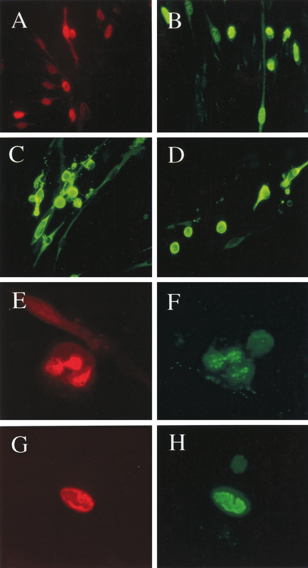
4 VOL. 75, 2001 ENDOTHELIAL CELL PLAQUE ASSAY FOR KSHV 5617 as a continuous cell line. However, when split and passaged 1:10 with fresh cultures of uninfected DMVEC, which then also all acquired a spindle cell morphology after 7 to 10 days (Fig. 2A), the infected cultures could be maintained and propagated indefinitely. Parallel uninfected DMVEC cultures remained contact inhibited but became difficult to passage and lost viability after 6 to 8 passages. The secondary BC3 spindle cell phenotype could also be passaged continuously as a completely spindle cell-converted culture by addition of fresh uninfected DMVEC. Evidence that all spindle cells in the DMVEC culture are latently infected. Flore et al. (11) suggested that in their BMEC system adjacent uninfected cells were converted to spindle cells by paracrine effects produced by factors secreted from the relatively small proportion (1%) of KSHV-infected cells present. They also showed that apparently virus-free supernatants from the infected culture could be used to convert human umbilical vein endothelial cell cultures to a spindle cell phenotype, although it was not clear how long these could be maintained in the absence of infection. In contrast, in our system, there did not appear to be any kind of soluble paracrine effects involved because of the ability to maintain normal cobblestone monolayers directly adjacent to spindle cell colonies and plaques for long periods of time (Fig. 1A and B). Therefore, we investigated the alternative possibility that all spindle cells in the DMVEC cultures may harbor a latent state infection. Completely converted JSC1 or BC3 spindle cell cultures were grown directly in microscope slide chambers and processed for IFA after 3 days. Two different antibodies directed against the KSHV latent nuclear antigen protein LANA1, which both gave nuclear IFA signals in the majority of the cells in PEL cell lines, were used, and the results revealed that essentially all converted spindle cells were LANA1 positive by IFA with anti-lana1 antibody (Fig. 2D). To more convincingly demonstrate that only cells with the spindle phenotype correlate with LANA1 expression, we also examined LANA1 staining by IHC at 17 days after primary infection of a DMVEC culture with just 25 l of pelleted JSC1 virions. In this experiment (Fig. 2B), virtually all cells in two typical adjacent spindle cell colonies of 30 to 80 cells each (one oval and FIG. 2. The LANA1 protein is expressed in all spindle cell-converted KSHV-infected DMVEC. (A) High-power phase-contrast photomicrographs of converted spindle cells in a secondary JSC1-infected DMVEC culture at 7 days after 1:10 dilution with fresh uninfected DMVEC. (Crystal violet stain; 40 objective.) (B) Expression of the LANA1 latency protein (brown nuclei) in all spindle cells within two distinct 30- to 80-cell colonies that had formed in a primary infected DMVEC culture at 17 days after addition of JSC1 supernatant virus. LANA1 was detected by IHC with rat anti-lana1 MAb and peroxidase-dab chromogen (40 objective.) (C) High-power phasecontrast photomicrograph of a partially spindle cell-converted secondpassage DMVEC culture at 24 days after addition of filtered supernatant from a fresh JSC1-infected DMVEC spindle cell culture showing a latently infected spindle cell colony containing a small patch or plaque of rounded cells with typical herpesvirus CPE and some remaining uninfected cuboidal cells. (Crystal violet stain; 40 objective.) (D) Double-label IHC detection of both latent and late lytic cycle KSHV-encoded proteins in a completely spindle cell-converted JSC1- infected DMVEC culture at 3 days after reseeding in a slide chamber dish. Brown chromogen, LANA1 nuclear staining in all latently infected spindle cells (rat anti-lana1 MAb; peroxidase and DAB). Red/pink chromogen, late lytic cytoplasmic gpk8.1 membrane protein in the subfraction of cells showing rounding and CPE (mouse MAb; phosphatase and Vector red; 40 objective.)
5 5618 CIUFO ET AL. J. VIROL. the other highly elongated) displayed LANA1-positive nuclei, whereas the adjacent cobblestone monolayer cells in the same culture were all negative for LANA1. In most spindle cells in both primary and secondary infected cultures, both rat MAb and rabbit PAb specific to LANA1 gave characteristic punctate nuclear patterns (visible at higher magnification by IFA and IHC in Fig. 3A and B), which probably represent the LANA1 protein bound to multiple copies of episomal viral genomes (4). Titration of KSHV PFU. In an attempt to assay the number of infectious virions present in the JSC1 PEL cell line supernatant, a parallel dish of DMVEC receiving 25 l of pelleted supernatant virus yielded 73 discrete spindle cell colonies after 17 days. Therefore, the titer of infectious KSHV from the original JSC1 cells in this experiment was measured at PFU/ml (Table 1, experiment 1). Again, a completely infected spindle cell-converted culture was obtained after the second passage as a 1:10 dilution with fresh DMVEC. A similar result was obtained in an experiment in which filtered and polyethylene glycol-precipitated virions from TPA-treated JSC1 supernatants were compared to a pelleted filtrate from washed and freeze-thawed JSC1 cells, which both gave titers measured in dilution series of to PFU/ml (Table 1, Experiment 2). During a second passage from one of these infected cultures, 100 l of unpelleted filtered DMVEC supernatant (without any TPA treatment) gave 400 colonies and plaques on a fresh monolayer of DMVEC at 17 days (Table 1, Experiment 2A), whereas the infected cells themselves gave a completely spindloid culture after a 1:10 split. Again, the second passage from an uninfected DMVEC monolayer failed to do so. Considering that all dishes received complete medium changes every 2 days, this result indicated that the spindle cell cultures were themselves producing new progeny infectious cell-free virions in the absence of TPA induction. Blocking of infection and spindle cell conversion with viral DNA synthesis-inhibitors. To assess whether new viral DNA synthesis was necessary for formation of colonies and plaques, pelleted virus from the filtered supernatant from TPA-treated JSC1 cells was added to a fresh culture of contact-inhibited DMVEC in the presence or absence of either PAA or GCV, and the cultures were fixed and stained at 21 days. In this case, undiluted virus from a TPA-treated culture again gave complete spindle cell conversion, and titers in diluted samples in the absence of inhibitors were measured at PFU/ml (Table 1, Experiment 3). However, in the presence of PAA, the spindle cell conversion was totally abolished, the DMVEC cells remained contact inhibited, and no plaques or colonies were obtained. In the presence of GCV, a moderate number of small spindloid plaques (200 PFU/ml) were obtained. In contrast, addition of PAA to an already completely converted spindle cell culture for a period of 17 days had no obvious effects on the spindle cell phenotype. Evidently, initiation of colony or plaque formation requires lytic cycle virus growth in the DMVEC, but continued proliferation of the spindle cell colonies once initiated probably does not require lytic cycle events. Expression of early lytic cycle proteins in KSHV-infected DMVEC spindle cell cultures. After several weeks in culture, both the completely converted spindle cell DMVEC and the individual larger spindle cell colonies frequently developed plaque-like patches or clusters of cells with a rounded cytopathic phenotype (Fig. 1A and 2C). Staining for DNA with 4,6-diamidino-2-phenylindole suggested that these latter cells did not have the fragmented nuclear bodies typical of cells undergoing apoptosis. These findings, together with the ability to passage the spindle cell phenotype with filtered DMVEC supernatants, suggested that some cells have been undergoing spontaneous virus lytic cycle replication. Therefore, we examined whether a variety of early and late viral lytic cycle proteins were being expressed. Initially, both IFA and IHC with rabbit PAb or mouse MAbs directed against several early nuclear or cytoplasmic KSHV-encoded proteins, including vil6, ORF- K8, SSB (ORF6), vgcr (ORF74), ZMP-A (ORF-K5), and ORF59 (PAF), were carried out directly at 3 days after replating undiluted spindle cultures in slide chambers. Typical results for both IFA and IHC detection are shown for nuclear ORF-K8 (Fig. 3E; Fig. 4C and D) and cytoplasmic ZMP-A (Fig. 3F; Fig. 4A and B), which are both among the earliest lytic cycle KSHV proteins expressed after TPA induction in PEL cell lines. These two proteins were both expressed in between 5 and 10% of all cells in the cultures. Most of the early lytic antigen-positive cells remained spindloid, indistinguishable in morphology from those expressing LANA1 only. Some spindle cells gave a punctate nuclear ORF-K8 pattern (not shown), which is typical of the very earliest stages of induction in PEL cells and represents colocalization with PML oncogenic bodies (31). Unlike in PEL cells (6, 15, 18), vil6 was relatively difficult to detect by IFA in these cultures, but some spindle cells (many fewer than for K8 or ZMP-A) were also strongly positive for the two viral delayed-early nuclear DNA replication proteins SSB and ORF59 (see below), as well as for the delayed-early vgcr membrane signaling protein (C.-J. Chiou et al., submitted for publication). Loss of LANA1 and von Willebrand factor signals in cells undergoing lytic cycle. Double-label IFA experiments were also carried out in infected DMVEC spindle cell cultures to compare the distribution of cells expressing the cytoplasmic early lytic protein ZMP-A with those expressing the latent state nuclear protein LANA1. The results revealed that although the majority of spindle cells still showed typical nuclear punctate LANA1 patterns, some of the same subset of spindle cells that had entered the lytic cycle as judged by expression of cytoplasmic ZMP-A were now negative for LANA1 (not shown). Therefore, cells that have entered the lytic cycle evidently tend to lose the punctate LANA1 IFA patterns that are characteristic of the latent state, and the few cells that were both LANA1 and ZMP-A positive probably represent just the very earliest stages of lytic cycle reactivation. To confirm that both the latent and lytic KSHV-infected cells were indeed endothelial cells and to begin to address whether any changes in endothelial cell markers were occurring in the infected spindle cells, we also carried out doublelabel IFA staining for von Willebrand factor and LANA1 in a partially converted spindle cell culture (Fig. 3C and D). Almost all uninfected DMVEC showed strong staining of Weibel- Palade bodies in the cytoplasm, as expected. However, although some spindle cells still retained significant von Willebrand factor, many LANA1-positive spindle cells displayed fewer Weibel-Palade bodies and much less overall positive

6 VOL. 75, 2001 ENDOTHELIAL CELL PLAQUE ASSAY FOR KSHV 5619 FIG. 3. Presence of punctate LANA1 patterns, loss of von Willebrand factor positivity, and spontaneous reactivation of early lytic viral protein expression in persistently infected DMVEC spindle cell cultures. (A and B) High-power photomicrographs of punctate nuclear LANA1 staining patterns in JSC1-infected DMVEC spindle cells. (A) IFA with rabbit anti-lana1 PAb green (fluorescein isothiocyanate [FITC] label; 63 objective) in a persistently infected secondary spindle cell culture; (B) IHC with rat anti-lana1 MAb and peroxidase-dab (brown; 100 objective) in a 16-day primary spindle cell colony. (C and D) Expression of von Willebrand factor and LANA1 detected by double-label IFA in partially JSC1-infected DMVEC cell culture. (C) Mouse anti-vwf MAb (FITC, green cytoplasm bodies); (D) rabbit anti-lana1 PAb (rhodamine label, red nuclei) in the same field (63 objective). (E) Expression of KSHV-encoded early lytic cycle ORF-K8 nuclear protein in sporadic spindle cells in a JSCS-infected DMVEC spindle cell culture detected by IFA with rabbit anti-k8 PAb (green FITC label; 40 objective). (F) Expression of the KSHV-encoded early lytic cycle cytoplasmic membrane protein ZMP-A(K5) in sporadic spindle cells in a JSC1-infected DMVEC spindle cell culture as detected by IFA with rabbit anti-zmp-a PAb (green FITC label; 40 objective).
7 5620 CIUFO ET AL. J. VIROL. TABLE 1. Titration of infectious KSHV from PEL cell line supernatants as measured by plaque assay in DMVEC Virus source Exptl condition Time (days) Titer (PFU/ml) JSC1 TPA Filtered pelleted supernatant (exp 1) 25 2,900 JSC1 TPA Filtered pelleted supernatant (exp 1) 25 2 JSC1 TPA Filtered polyethylene glycol-precipitated supernatant (exp 2) 17 5,000 JSC1 TPA Filtered polyethylene glycol-precipitated supernatant (exp 2) 17 2 JSC1 TPA Freeze-thawed cell pellet (exp 2) 17 4,000 Filtered pelleted supernatant (exp 3) 20 5,000 JSC1 TPA Filtered pelleted supernatant (exp 3) 20 5 JSC1 TPA Filtered pelleted supernatant PAA (exp 3) Filtered pelleted supernatant PAA GCV (exp 3) b DMVEC supernatant (exp 2A) 22 4,000 BC3 TPA Filtered, pelleted supernatant (exp 1) BC3 TPA Filtered, pelleted supernatant (exp 1) BC3 TPA 2nd passage of DMVEC a (exp 1A) 22 5,000 BC3 TPA 2nd passage of DMVEC a (exp 1A) BCP1 TPA Filtered, pelleted supernatant (exp 1) BCP1 TPA Filtered, pelleted supernatant (exp 1) BCP1 TPA 2nd passage of DMVEC a (exp 1A) BCP1 TPA 2nd passage of DMVEC a (exp 1A) a Cells split and replated 1:10. b Very small colonies only. signal for von Willebrand factor. Loss of von Willebrand factor staining was especially pronounced for a small subset of rounded cells expressing late lytic cycle antigens (not shown; see below). Uninfected DMVEC were also all positive for the E-cadherin endothelial cell marker, but its expression was not appreciably affected by KSHV infection or spindle cell phenotype (not shown). Development of CPE correlates with late lytic cycle protein expression and formation of viral DNA replication compartments. In other experiments, secondary infections were carried out by plating a mixture of completely spindloid KSHV latently infected DMVEC with a 10-fold excess of added fresh uninfected DMVEC in microscope slide chamber dishes. After 7 to 10 days of growth, these cultures became completely spindloid, but also developed small patches of between 4 and 20 adjacent cells that all expressed early K8 or ZMP-A protein. Interestingly, often a small subset of these clustered early antigenpositive cells had also rounded up, showing typical herpesviruslike CPE and forming miniplaques. Significantly, within these clusters all of the cytopathic cells that had lost their spindle phenotype still proved to be expressing KSHV lytic cycle antigens by IFA, including both the K8 and ORF59 nuclear proteins (Fig. 5A and B), as well as the early membrane protein ZMP-A (Fig. 5C and D). Furthermore, expression of the true late lytic virion component glycoprotein ORF-K8.1 was found to occur only in the few cells showing CPE. For example, Fig. 2D shows a completely converted spindle cell secondary culture containing cells with CPE after direct reseeding for 3 days in a slide chamber culture dish. Again, double-label IHC revealed expression of the ORF-K8.1 protein only in a dispersed subfraction of rounded lytic cells showing CPE (red cytoplasm), in contrast to positive LANA1 nuclear expression patterns in all spindle cells in the culture (brown nuclei). Double-label IFA experiments also revealed that in the 10-day secondary infected spindle cell cultures, both ORF59 and SSB were found predominantly within the subsets of ZMP-A-positive spindle cells, as well as in all rounded cells (not shown), whereas ORF-K8 and ZMP-A were also present in other adjacent cells that remained spindloid and did not yet express either ORF59 or SSB (Fig. 5A to C). Overall, although clusters of up to 10% of the cells were often positive for early lytic proteins, the fraction that showed CPE and true late antigen expression rarely totaled more than 1 to 2% of all cells in the culture. In all spindle cell-converted DMVEC cultures that contained a subset of cells displaying spontaneous CPE, we also noticed that up to 50% of the ORF59-positive cells and 20% of the ORF-K8-positive cells contained large intranuclear inclusion bodies (one to three per cell) that filled much of the nucleoplasm but spared the nucleoli (Fig. 5A, B, E, and G). ZMP-A and vgcr remained cytoplasmic in these same cells when examined by double-label IFA (not shown). Furthermore, all cells showing CPE displayed these large subnuclear structures, suggesting that they represented typical herpesvirus viral DNA replication compartments formed at late stages during the lytic cycle. Further investigation by double-label IFA confirmed (i) that the large ORF-K8-positive nuclear bodies all colocalized with the ORF59 replication compartments (Fig. 5E and F) and (ii) that most of them were actively synthesizing DNA, as judged by colocalization with pulse-labeled bromodeoxyuridine (BUdR) (Fig. 5G and H). DISCUSSION We have shown here that cultured primary human DMVEC cultures can be infected at different efficiencies by KSHV virions derived from several PEL cell lines. The infection causes a phenotype involving aligned and swirling bunches of elongated spindle-shaped cells that closely resembles that displayed by the pathogenic spindle cells of nodular KS lesions. Importantly, KSHV infection of the DMVEC cultures and the associated spindle cell conversion phenotype correlate almost completely with constitutive LANA1 protein expression in the same cells. Recent studies by Dupin et al. (10) have also con-
, implying that the viral genomes also establish")
8 VOL. 75, 2001 ENDOTHELIAL CELL PLAQUE ASSAY FOR KSHV 5621 firmed that almost all spindle cells in late nodular KS lesions express LANA1. The similarity to the latent state found in both PEL cell lines and KS lesions is emphasized by the punctate nuclear character of the LANA1 signal (4), implying that the viral genomes also establish a multicopy episomal state in infected spindloid DMVEC. However, although the KSHVinfected spindle cells have high mitotic indices and clearly proliferate (6), they could be propagated directly only for a few weeks and several passages longer than the parent uninfected DMVEC cells and did not appear to be truly immortalized or oncogenically transformed. In contrast, the infected spindloid cell phenotype could be passaged and maintained continuously by addition of either infected cells or cell-free supernatant to fresh uninfected DMVEC cultures, suggesting that in addition to proliferation of the latently infected spindle cells, low levels of progeny infectious virions are produced that can spread the spindle phenotype to adjacent uninfected cells. We have also demonstrated directly that patches of spontaneous lytic infection rather than just latent infection occur within the KSHV-infected DMVEC spindle cell cultures and have adapted the system to demonstrate for the first time that infectious KSHV virions can be titered by an infectious center assay involving mixed spindle cell colony and plaque formation. Extensive IFA and IHC evidence confirmed that spontaneous lytic cycle induction occurs in a significant subfraction (5 to 10%) of the spindle cells and that most of the same early and late lytic cycle viral proteins that can be induced by TPA or butyrate treatment of PEL cell lines are synthesized over a relatively long time course that includes a second morphological change to a rounded CPE phenotype at late stages. Under appropriate undisturbed culture conditions, including the 10- day 1:10-diluted secondary cultures, small clusters or plaques of cells with CPE expressing late viral proteins develop within otherwise completely converted spindle cell cultures. Several previous studies have provided preliminary evidence that supernatant KSHV derived from PEL cell lines or from fresh KS lesion biopsies may be able to persist at minimal levels in both infected 293 cells and primary DMVEC but not in numerous other cell types tested (12, 21, 23). However, that evidence consisted only of very low level viral DNA or RNA positivity by PCR. Flore et al. (11) provided provocative evidence that PEL supernatant KSHV could be used to produce a continuously passageable spindle phenotype in BMEC cultures. Evidently, only a small subfraction of these cells carried the virus, but this was associated with increased longevity of the whole culture, apparently involving paracrine effects on FIG. 4. Spontaneous lytic cycle reactivation demonstrated by IHC in persistently KSHV-infected DMVEC spindle cell cultures. (A and B) Low-power (16 objective) and high-power (40 objective) views of KSHV-encoded early cytoplasmic membrane protein ZMP-A expressed in a subset of the spindle cells in a completely converted JSC1-infected DMVEC spindle culture at 3 days after reseeding in slide chambers, as detected by IHC with rabbit anti-zmpa PAb (phosphatase and Vector red chromogen). (C and D) Low-power (16 objective) and high-power (40 objective) views of KSHV-encoded ORF-K8 early nuclear protein expressed in a subset of the spindle cells in a completely converted JSC1-infected DMVEC spindle cell culture at 3 days after reseeding in slide chambers, as detected by IHC with rabbit anti-orfk8 PAb (phosphatase and Vector red chromogen).
9 5622
10 VOL. 75, 2001 ENDOTHELIAL CELL PLAQUE ASSAY FOR KSHV 5623 adjacent cells. Obviously, our system does not show the same disseminated paracrine effects as described by Flore et al. (11) because the spindle phenotype occurs only in infected DMVEC, but the moderately extended life span of the latently infected DMVEC spindle cell cultures without becoming immortalized or transformed somewhat resembles the overall effect seen in their BMEC system. Like the BMEC system (11), only a small subfraction (0.1%) of the KSHV-infected spindloid DMVEC produced colonies in soft agar assays (not shown), and these were small (0.2- to 0.4-mm diameter) compared to agar colonies grown from the same DMVEC cultures after infection with an E6/E7/Neo r retrovirus. Moses et al. (17) described a system more similar to ours, except that PEL supernatant virus was used to infect human papillomavirus E6/E7-immortalized DMVEC cultures. In their experiments also, most cells appeared to become latently infected and LANA-positive cells produced a spindloid morphology, but assayable plaque and colony formation was not demonstrated. In both the Flore and Moses systems, some preliminary evidence for both lytic cycle induction by TPA treatment and passaging of virus was presented, but we have shown here that in the primary DMVEC system both K8 and ZMP-A viral lytic cycle protein expression occurs spontaneously after 2 to 3 weeks in the center of large spindle cell colonies as well as in small clusters (totaling 5 to 10% of all cells) within 7 to 10 days after confluency in fully spindloid cultures or in secondary 1:10-diluted cultures. Furthermore, a subfraction of these cells (totaling 1 to 2% of all cells) also show typical herpesvirus rounding and CPE and express the true late lytic ORF-K8.1 glycoprotein, as well as containing active viral DNA replication compartments that colocalize with the ORF59 (PAF) viral DNA polymerase-associated protein. Finally, we have shown elsewhere by transmission electron microscopy studies (20) that the rounded lytically infected DMVEC showing CPE also contain numerous herpesvirus-like filled and empty capsids in the nucleus, as well as enveloped virions in the cytoplasm and endoplasmic reticulum and in adjacent extracellular spaces. We have not been able to establish a permanent latently infected DMVEC line, perhaps because the cultures eventually succumb to spontaneous lytic cycle reactivation. Although the most efficient infection was established with supernatant virions derived from the JSC1 PEL cell line, which carries latent state Epstein-Barr virus (EBV) in addition to KSHV (6), similar mixed latent and lytically infected spindle cell cultures could also be established from KSHV virions derived from two PEL cell lines (BC3 and BCP1) that do not carry EBV genomes. The higher plaque titers and relative ease with which complete spindloid cultures could be obtained in our studies with virus from the JSC1 cell line is at least partially explained by the production of greater numbers of progeny supernatant virions (6), although there may be a qualitative effect as well. A number of observations support this conclusion. First, uninduced JSC1 cells consistently display higher spontaneous early lytic antigen expression levels of 20 to 30% vil6-positive cells, compared to 0.3, 2, 2, and 0.5%, respectively, for spontaneous vil6-positive cells in the BC1/HBL6, BC3, BCP1, and BCBL1 cell lines. Second, uninduced JSC1 cells also show much further progression of the lytic cycle compared to BC1/HBL6, BC3, BCP1, and BCBL1, cells, as assayed by the presence of vgcr (ORF74) protein-positive cells (3 to 5%) and by the numbers of cells that have ORF59- positive DNA replication compartments (1 to 3%); however, even uninduced JSC1 cells are still negative for the late ORF- K8.1 protein and evidently do not release infectious virions. Third, uninduced JSC1 cells contain considerably higher KSHV viral DNA copy numbers per microgram of total cell DNA compared to BC3, BCP1, and BCBL1 cells (as measured by Southern blot hybridization [not shown]). Fourth, although TPA treatment greatly increases all measures of lytic infection in all five cell lines, the others still produce relatively few fully lytic cells, as judged by ORF-K8.1 expression values of only 1 to 5%, compared to 20% for JSC1 cells. Direct measurements of virion-associated KSHV DNA levels by quantitative PCR after DNase I treatment and lysis (6) also revealed the presence of 2 orders of magnitude more DNase I-resistant KSHV DNA in our pelleted TPA-induced JSC1 cell supernatants ( genome equivalents/ml) compared to parallel TPA-treated BC3 or BCP1 supernatant virion preparations ( genome equivalents/ml). These were the same pelleted supernatant virion preparations that were used here for the DMVEC infectivity assays that showed 100- and 500-fold-higher titers of infectious centers for JSC1 compared to BC3 and BCP1, respectively (Table 1). These results allow calculations of particle to PFU ratios of approximately 500:1 for JSC1 and BC3 and 2,500:1 for BCP1. The quality in addition to quantity of viral genomes from JSC1 cells may also be different because in time course studies with low input doses of supernatant virus, the BC3 virus-infected DMVEC cultures took up to 22 days before any cells became LANA1 positive, whereas cultures receiving diluted JSC1 virus took only 4 days to initiate multiple LANA1-posi- FIG. 5. Development of clusters of infected DMVEC displaying late lytic CPE and containing active viral DNA replication compartments. (A) Expression of the KSHV-encoded ORF59 protein including typical late stage herpesvirus nuclear DNA replication compartments within a clustered miniplaque in a 10-day confluent secondary JSC1 spindle cell culture, as detected by IFA with mouse anti-orf59 MAb (red rhodamine label, 63 objective). (B) Expression of ORF-K8 including nuclear DNA replication compartment-associated forms within a clustered miniplaque in a 10-day confluent secondary JSC1 spindle cell culture, as detected by IFA with rabbit anti-k8 PAb (green fluorescein isothiocyanate [FITC] label; 63 objective). (C and D) Expression of the KSHV-encoded lytic membrane protein ZMP-A in small plaque-like patches of rounded late stage lytic cycle-infected DMVEC cells displaying CPE. Two different fields within a 10-day secondary JSC1 spindle cell culture detected by IFA with rabbit anti-zmp-a PAb (green FITC label; 63 objective) are shown. (E and F) Colocalization of ORF59 and ORF-K8 by double-label IFA in viral DNA replication compartments in a late lytic stage JSCI-infected DMVEC cell, showing rounding and CPE, as detected with mouse anti-orf59 MAb (E; red rhodamine label) and rabbit anti-orf8 PAb (F; green FITC label) in the same field (100 objective). (G and H) Colocalization of newly synthesized DNA (BUdR pulse-label) and the ORF-K8 nuclear protein in a mature viral DNA replication compartment in a late lytic stage rounded JSC1-infected DMVEC cell as detected by double-label IFA using mouse anti-budr MAb (G; red rhodamine label) and rabbit anti-k8 PAb (H; green FITC label) in the same field (100 objective).
11 5624 CIUFO ET AL. J. VIROL. tive colonies (6). Genotypically, the K1 and K15 (22) subtypes of the three KSHV strains in question here are C3/P, C3/P and A1 /P, respectively, which seems less likely to be important than other factors. For example, one could imagine a situation similar to that encountered with the P3HR-1 lymphoblastoid cell line, in which defective tandemly repeated EBV genomes containing all three lytic cycle transactivator genes (Rta, Zta, and Mta) under the control of the rearranged latent state W and C promoters are packaged in the presence of wild-type helper genomes and give rise to persistent lytic cycle gene expression both in the P3HR1 cells themselves and in newly infected EBV-positive Raji cells. However, preliminary Southern blot analysis of several regions of the JSC1 genome has not shown any evidence for P3HR1-like tandem repeat DNA molecules. Furthermore, one would not expect to be able to successfully passage rearranged lytic JSC1 defective viruses onto fresh naive DMVEC cultures from filtered supernatant as we were able to do here. Alternatively, some or all genomes in the extensively in vitro-passaged BC3 and BCP1 cell lines may be rearranged, deleted, or mutated in key lytic cycle genes, leading initially to reduced or cryptic reactivation or infectivity. For example, in BC1/HBL6 cells, the KSHV genome structure is known to be aberrant, with larger than usual 250-kb episomes consisting of 1.3 copies of the expected 180-kb viral genome and encompassing an extra set of the terminal tandem repeat sequences (25). Such genomes might not be packaged or infectious until after specific rearrangements occur. It is not known whether the BC3 or BCP1 episomal genome is also partially reiterated, but Southern blotting data with KSHV terminal tandem repeat unit DNA probes does not show any evidence for rearranged JSC1 episomal genomes (data not shown). Importantly, although between 100- and 500-fold less infectious virus was obtained initially from TPA-induced BC3 and BCP1 cell cultures than from the JSC1 cultures, both of the latter viruses could subsequently be maintained as easily passaged spindloid cultures, suggesting that once established in DMVECs all three viruses may produce equivalent levels of infectious centers or transmissible infectious particles. There is also the point that because the JSC1 cells (unlike BC3 and BCP1 cells) contain episomal EBV genomes in addition to the KSHV genomes, it is possible that EBV contributes (directly or indirectly) to the apparently higher infectivity of the JSC1 cell line-derived virions. However, extensive evaluation of KSHV and EBV expression in the JSC1 cell line (6) has shown that any direct effects are extremely unlikely. First, as for other dually infected PEL cell lines such as BC1/HBL6 (14), the level of latent EBV gene expression in JSC1 cells is highly restricted. Second, the level of spontaneous and TPAinduced lytic cycle EBV gene expression (as measured by Zta protein expression) in JSC1 cells is extremely low (less than 0.1% of the cells). Third, there was between 10- and 100-fold less DNase I-resistant EBV DNA present in the JSC1 pelleted supernatant used for the DMVEC infectivity assays than there was KSHV DNA, as measured by quantitative DNA PCR (6). Fourth, parallel assays for EBV Zta protein expression by IFA in the DMVEC cultures were negative, and no EBV DNA (less than 20 copies in 10 4 cells) could be detected by quantitative PCR within the passaged JSC1-infected DMVEC cell cultures. Whether or not the presence of EBV in the parent JSC1 PELs may have indirect positive effects on the yields or quality of the KSHV virions obtained is difficult to assess at present, but in the BC1/HBL6 cell line at least, the effect of EBV on inducibility of the KSHV genomes by TPA is evidently negative. As demonstrated by the LANA1 IFA, the majority of spindle cells in the KSHV-infected cultures appear to be equivalent to the latent state studied in uninduced PEL cell lines, including the presence of multicopy KSHV episomes associated with a punctate nuclear LANA1 distribution. Therefore, both the spindle phenotype and LANA1 expression are diagnostic of KSHV infection. However, in time course studies presented elsewhere (6), we found that small colonies of four to eight adjacent cuboidal DMVEC cells could be detected first by IHC for LANA1 protein expression within just 4 days after addition of JSC1 virus to DMVEC cultures. Although the cells in these earliest and smallest LANA1-positive colonies were not yet spindloid, by 6 and 9 days nearly all LANA1- positive cells in colonies of 10 to 30 cells in size were spindloid. Therefore, in establishing primary infection, LANA1 expression evidently precedes the spindle cell phenotype by several days. Considering that the initial establishment of proliferating LANA1-positive converted spindle cells has all the hallmarks of true latent infection, the ability of PAA to totally block colony formation was quite surprising (Table 1). Future studies at the single-cell level will need to address de novo DMVEC infection with initial time course studies to determine when LANA1 first becomes punctate and what the effects of PAA treatment on LANA1 expression patterns are, as well as whether PAA blocks the apparent early episomal genomic DNA amplification events. Most interestingly, both completely spindloid cultures and large spindle colonies often gave rise to occasional small patches of adjacent cells that showed a second morphological change, becoming rounded with typical herpesvirus CPE indicative of spontaneous lytic cycle reactivation. All of these cells proved to be undergoing KSHV DNA synthesis and late stage KSHV lytic viral antigen expression, as judged by BUdR incorporation and colocalization with the viral ORF59 replication fork protein in typical large subnuclear viral DNA replication compartments. These same 1 to 2% of the cells also showed positive expression of all lytic cycle KSHV-encoded viral proteins tested, including the true late ORF-K8.1 membrane glycoprotein (32), as well as the presence of abundant mature and immature herpesvirus particles (20). Furthermore, many additional spindle cells without CPE displayed spontaneous expression of just the earliest lytic proteins, including ZMP-A and K8. These often amounted to 5 to 10% of all spindle cells in 7- and 10-day secondary diluted cultures and often themselves formed additional small clusters or lay adjacent to cells with CPE. These latter cells may either represent a high fraction of abortive lytically infected cells that are unable to progress to later stages of the lytic cycle or, more likely, indicate that the lytic cycle is so slow that it takes many days overall to traverse from the early to late stages. The number of lytic cycle-positive cells was not increased by more than twofold after TPA treatment of the KSHV-infected DMVEC cultures for 48 or 72 h, but the proportion of cells displaying CPE, late ORF-K8.1 protein expression, and DNA replication compartments was increased four- to fivefold by
Use of the red fluorescent protein as a marker of Kaposi s sarcoma-associated herpesvirus lytic gene expression
 Virology 325 (2004) 225 240 www.elsevier.com/locate/yviro Use of the red fluorescent protein as a marker of Kaposi s sarcoma-associated herpesvirus lytic gene expression Jeffrey Vieira* and Patricia M.
Electron Microscope Studies of HeLa Cells Infected with Herpes Virus
 244 STOKER, M. G. P., SMITH, K. M. & Ross, R. W. (1958). J. gen. Microbiol. 19,244-249 Electron Microscope Studies of HeLa Cells Infected with Herpes Virus BY M: G. P. STOKER, K. M. SMITH AND R. W. ROSS
Quantitative Assay of Paravaccinia Virus Based
 APPrU MICROBIOLOGY, JUly 1972, p. 138-142 Copyright 1972 American Society for Microbiology Vol. 24, No. 1 Printed in U.S.A. Quantitative Assay of Paravaccinia Virus Based on Enumeration of Inclusion-Containing
SHORT COMMUNICATION. Human Papillomavirus Type 11 E1 Ú E4 and L1 Proteins Colocalize in the Mouse Xenograft System at Multiple Time Points
 VIROLOGY 214, 259 263 (1995) SHORT COMMUNICATION Human Papillomavirus Type 11 E1 Ú E4 and L1 Proteins Colocalize in the Mouse Xenograft System at Multiple Time Points DARRON R. BROWN,*,,1 JANINE T. BRYAN,
Determination of the temporal pattern and importance of BALF1 expression in Epstein-Barr viral infection
 Determination of the temporal pattern and importance of BALF1 expression in Epstein-Barr viral infection Melissa Mihelidakis May 6, 2004 7.340 Research Proposal Introduction Apoptosis, or programmed cell
Mechanism of Pock Formation by Shope Fibroma
 JOURNAL OF BACTERIOLOGY, Sept., 1966 Copyright ( 1966 American Society for Microbiology Vol. 92, No. 3 Printed in U.S.A. Mechanism of Pock Formation by Shope Fibroma Virus on Monolayers of Rabbit Cells
Egr-1 regulates RTA transcription through a cooperative involvement of transcriptional regulators
 /, 2017, Vol. 8, (No. 53), pp: 91425-91444 Egr-1 regulates RTA transcription through a cooperative involvement of transcriptional regulators Roni Sarkar 1 and Subhash C. Verma 1 1 Department of Microbiology
Chapter 6- An Introduction to Viruses*
 Chapter 6- An Introduction to Viruses* *Lecture notes are to be used as a study guide only and do not represent the comprehensive information you will need to know for the exams. 6.1 Overview of Viruses
Polyomaviridae. Spring
 Polyomaviridae Spring 2002 331 Antibody Prevalence for BK & JC Viruses Spring 2002 332 Polyoma Viruses General characteristics Papovaviridae: PA - papilloma; PO - polyoma; VA - vacuolating agent a. 45nm
Epstein-Barr Virus: Stimulation By 5 '-Iododeoxy uridine or 5 '-Brom odeoxy uridine in Human Lymphoblastoid Cells F ro m a Rhabdom yosarcom a*
 A n n a ls o f C l i n i c a l L a b o r a t o r y S c i e n c e, Vol. 3, No. 6 Copyright 1973, Institute for Clinical Science Epstein-Barr Virus: Stimulation By 5 '-Iododeoxy uridine or 5 '-Brom odeoxy
Overview of primary HHV-8 infection
 Overview of primary HHV-8 infection HHV-8, also known as Kaposi sarcoma-associated herpesvirus (KSHV), is a gamma herpesvirus primarily transmitted through saliva. The virus initially replicates in epithelial
The Infectious Cycle. Lecture 2 Biology W3310/4310 Virology Spring You know my methods, Watson --SIR ARTHUR CONAN DOYLE
 The Infectious Cycle Lecture 2 Biology W3310/4310 Virology Spring 2016 You know my methods, Watson --SIR ARTHUR CONAN DOYLE The Infectious Cycle Virologists divide the infectious cycle into steps to facilitate
Identification of Microbes Lecture: 12
 Diagnostic Microbiology Identification of Microbes Lecture: 12 Electron Microscopy 106 virus particles per ml required for visualization, 50,000-60,000 magnification normally used. Viruses may be detected
Viruses Tomasz Kordula, Ph.D.
 Viruses Tomasz Kordula, Ph.D. Resources: Alberts et al., Molecular Biology of the Cell, pp. 295, 1330, 1431 1433; Lehninger CD Movie A0002201. Learning Objectives: 1. Understand parasitic life cycle of
JOURNAL OF VIROLOGY, Dec. 1999, p Vol. 73, No. 12. Copyright 1999, American Society for Microbiology. All Rights Reserved.
 JOURNAL OF VIROLOGY, Dec. 1999, p. 10458 10471 Vol. 73, No. 12 0022-538X/99/$04.00 0 Copyright 1999, American Society for Microbiology. All Rights Reserved. The Human Cytomegalovirus IE2 and UL112-113
7.012 Quiz 3 Answers
 MIT Biology Department 7.012: Introductory Biology - Fall 2004 Instructors: Professor Eric Lander, Professor Robert A. Weinberg, Dr. Claudette Gardel Friday 11/12/04 7.012 Quiz 3 Answers A > 85 B 72-84
EBV infection B cells and lymphomagenesis. Sridhar Chaganti
 EBV infection B cells and lymphomagenesis Sridhar Chaganti How EBV infects B-cells How viral genes influence the infected B cell Differences and similarities between in vitro and in vivo infection How
Temperature-Sensitive Mutants Isolated from Hamster and
 JOURNAL OF VIROLOGY, Nov. 1975, p. 1332-1336 Copyright i 1975 American Society for Microbiology Vol. 16, No. 5 Printed in U.S.A. Temperature-Sensitive Mutants Isolated from Hamster and Canine Cell Lines
Hepatitis B Antiviral Drug Development Multi-Marker Screening Assay
 Hepatitis B Antiviral Drug Development Multi-Marker Screening Assay Background ImQuest BioSciences has developed and qualified a single-plate method to expedite the screening of antiviral agents against
7.013 Spring 2005 Problem Set 7
 MI Department of Biology 7.013: Introductory Biology - Spring 2005 Instructors: Professor Hazel Sive, Professor yler Jacks, Dr. Claudette Gardel 7.013 Spring 2005 Problem Set 7 FRIDAY May 6th, 2005 Question
Virology Introduction. Definitions. Introduction. Structure of virus. Virus transmission. Classification of virus. DNA Virus. RNA Virus. Treatment.
 DEVH Virology Introduction Definitions. Introduction. Structure of virus. Virus transmission. Classification of virus. DNA Virus. RNA Virus. Treatment. Definitions Virology: The science which study the
Harinivas H. Krishnan, 1 Pramod P. Naranatt, 1 Marilyn S. Smith, 1 Ling Zeng, 1 Clark Bloomer, 2 and Bala Chandran 1 *
 JOURNAL OF VIROLOGY, Apr. 2004, p. 3601 3620 Vol. 78, No. 7 0022-538X/04/$08.00 0 DOI: 10.1128/JVI.78.7.3601 3620.2004 Copyright 2004, American Society for Microbiology. All Rights Reserved. Concurrent
Nanoparticles and persistent virus infection a dangerous liaison for the development of chronic lung disease(s)? Tobias Stöger
? Tobias Stöger") Nanoparticles and persistent virus infection a dangerous liaison for the development of chronic lung disease(s)? Tobias Stöger Herpesviruses and lung disease Double-stranded DNA-viruses (a, b, g- herpesviruses)
Nanoparticles and persistent virus infection a dangerous liaison for the development of chronic lung disease(s)? Tobias Stöger Herpesviruses and lung disease Double-stranded DNA-viruses (a, b, g- herpesviruses)
Comparison of Herpes Simplex Virus Reactivation in Ganglia In Vivo and in Explants Demonstrates Quantitative and Qualitative Differences
 JOURNAL OF VIROLOGY, July 2004, p. 7784 7794 Vol. 78, No. 14 0022-538X/04/$08.00 0 DOI: 10.1128/JVI.78.14.7784 7794.2004 Copyright 2004, American Society for Microbiology. All Rights Reserved. Comparison
Inefficient establishment of KSHV latency suggests an additional role for continued lytic replication in Kaposi sarcoma pathogenesis
 Inefficient establishment of KSHV latency suggests an additional role for continued lytic replication in Kaposi sarcoma pathogenesis See the related Commentary beginning on page 21. Adam Grundhoff and
ISOLATION OF A SARCOMA VIRUS FROM A SPONTANEOUS CHICKEN TUMOR
 ISOLATION OF A SARCOMA VIRUS FROM A SPONTANEOUS CHICKEN TUMOR Shigeyoshi ITOHARA, Kouichi HIRATA, Makoto INOUE, Masanori Veterinary Pathology, Faculty of Agriculture, Yamaguchi University* HATSUOKA, and
Fayth K. Yoshimura, Ph.D. September 7, of 7 HIV - BASIC PROPERTIES
 1 of 7 I. Viral Origin. A. Retrovirus - animal lentiviruses. HIV - BASIC PROPERTIES 1. HIV is a member of the Retrovirus family and more specifically it is a member of the Lentivirus genus of this family.
Mike Reichelt,* Jennifer Brady, and Ann M. Arvin
 JOURNAL OF VIROLOGY, Apr. 2009, p. 3904 3918 Vol. 83, No. 8 0022-538X/09/$08.00 0 doi:10.1128/jvi.02137-08 Copyright 2009, American Society for Microbiology. All Rights Reserved. The Replication Cycle
Chapter13 Characterizing and Classifying Viruses, Viroids, and Prions
 Chapter13 Characterizing and Classifying Viruses, Viroids, and Prions 11/20/2017 MDufilho 1 Characteristics of Viruses Viruses Minuscule, acellular, infectious agent having either DNA or RNA Cause infections
Herpesviruses. Virion. Genome. Genes and proteins. Viruses and hosts. Diseases. Distinctive characteristics
 Herpesviruses Virion Genome Genes and proteins Viruses and hosts Diseases Distinctive characteristics Virion Enveloped icosahedral capsid (T=16), diameter 125 nm Diameter of enveloped virion 200 nm Capsid
Kaposi s sarcoma (KS) is an angioproliferative and highly vascularized
 is an angioproliferative and highly vascularized") Kaposi s Sarcoma-Associated Herpesvirus Latency-Associated Nuclear Antigen Interacts with Multifunctional Angiogenin To Utilize Its Antiapoptotic Functions Nitika Paudel, a Sathish Sadagopan, a Sayan Chakraborty,
Kaposi s Sarcoma-Associated Herpesvirus Latency-Associated Nuclear Antigen Interacts with Multifunctional Angiogenin To Utilize Its Antiapoptotic Functions Nitika Paudel, a Sathish Sadagopan, a Sayan Chakraborty,
Nucleic acid: singled stranded, double stranded, RNA, or DNA, linear or circular. Capsid: protein coat that is most of the mass of the virus.
 Viruses General Characteristics of Viruses 1. Depending on view may be regarded as exceptionally complex aggregates of nonliving chemicals or as exceptionally simple living microbes. 2. Contain a single
KSHV infects a subset of human tonsillar B cells, driving proliferation and plasmablast differentiation
 Research article KSHV infects a subset of human tonsillar B cells, driving proliferation and plasmablast differentiation Lynn M. Hassman, 1,2 Thomas J. Ellison, 1,2 and Dean H. Kedes 1,2,3 1 Myles H. Thaler
Animal hosts Natural host Laboratory animals Rabbits Mice Rats Hamsters Newborn or suckling rodents Animal models for viral pathogenesis 4 Growth of v
 Principles of Virology Department of Molecular Genetics & Microbiology Univ ersity of Florida, Gainesv ille, FL 1 Outline Virus cultivation Assay of viruses Virus genetics 2 Virus isolation Evidence of
Role of Interferon in the Propagation of MM Virus in L Cells
 APPLIED MICROBIOLOGY, Oct. 1969, p. 584-588 Copyright ( 1969 American Society for Microbiology Vol. 18, No. 4 Printed in U S A. Role of Interferon in the Propagation of MM Virus in L Cells DAVID J. GIRON
Altered Patterns of Cellular Gene Expression in Dermal Microvascular Endothelial Cells Infected with Kaposi s Sarcoma-Associated Herpesvirus
 JOURNAL OF VIROLOGY, Apr. 2002, p. 3395 3420 Vol. 76, No. 7 0022-538X/02/$04.00 0 DOI: 10.1128/JVI.76.7.3395 3420.2002 Copyright 2002, American Society for Microbiology. All Rights Reserved. Altered Patterns
EVALUATION OF THE EFFECTIVENESS OF A 7% ACCELERATED HYDROGEN PEROXIDE-BASED FORMULATION AGAINST CANINE PARVOVIRUS
 Final report submitted to Virox Technologies, Inc. EVALUATION OF THE EFFECTIVENESS OF A 7% ACCELERATED HYDROGEN PEROXIDE-BASED FORMULATION AGAINST CANINE PARVOVIRUS Syed A. Sattar, M.Sc., Dip. Bact., M.S.,
VIRUSES AND CANCER Michael Lea
 VIRUSES AND CANCER 2010 Michael Lea VIRAL ONCOLOGY - LECTURE OUTLINE 1. Historical Review 2. Viruses Associated with Cancer 3. RNA Tumor Viruses 4. DNA Tumor Viruses HISTORICAL REVIEW Historical Review
(A) PCR primers (arrows) designed to distinguish wild type (P1+P2), targeted (P1+P2) and excised (P1+P3)14-
 PCR primers (arrows) designed to distinguish wild type (P1+P2), targeted (P1+P2) and excised (P1+P3)14-") 1 Supplemental Figure Legends Figure S1. Mammary tumors of ErbB2 KI mice with 14-3-3σ ablation have elevated ErbB2 transcript levels and cell proliferation (A) PCR primers (arrows) designed to distinguish
1 Supplemental Figure Legends Figure S1. Mammary tumors of ErbB2 KI mice with 14-3-3σ ablation have elevated ErbB2 transcript levels and cell proliferation (A) PCR primers (arrows) designed to distinguish
BIT 120. Copy of Cancer/HIV Lecture
 BIT 120 Copy of Cancer/HIV Lecture Cancer DEFINITION Any abnormal growth of cells that has malignant potential i.e.. Leukemia Uncontrolled mitosis in WBC Genetic disease caused by an accumulation of mutations
MedChem 401~ Retroviridae. Retroviridae
 MedChem 401~ Retroviridae Retroviruses plus-sense RNA genome (!8-10 kb) protein capsid lipid envelop envelope glycoproteins reverse transcriptase enzyme integrase enzyme protease enzyme Retroviridae The
11/15/2011. Outline. Structural Features and Characteristics. The Good the Bad and the Ugly. Viral Genomes. Structural Features and Characteristics
 Chapter 19 - Viruses Outline I. Viruses A. Structure of viruses B. Common Characteristics of Viruses C. Viral replication D. HIV II. Prions The Good the Bad and the Ugly Viruses fit into the bad category
Chronic Viral Infections vs. Our Immune System: Revisiting our view of viruses as pathogens
 Chronic Viral Infections vs. Our Immune System: Revisiting our view of viruses as pathogens Tiffany A. Reese Assistant Professor Departments of Immunology and Microbiology Challenge your idea of classic
Adenovirus Manual 1. Table of Contents. Large Scale Prep 2. Quick MOI Test 4. Infection of MNT-1 Cells 8. Adenovirus Stocks 9
 Adenovirus Manual 1 Table of Contents Large Scale Prep 2 Quick MOI Test 4 TCID 50 Titration 5 Infection of MNT-1 Cells 8 Adenovirus Stocks 9 CAUTION: Always use filter tips and bleach everything!!! Adenovirus
Chapter 13 Viruses, Viroids, and Prions. Biology 1009 Microbiology Johnson-Summer 2003
 Chapter 13 Viruses, Viroids, and Prions Biology 1009 Microbiology Johnson-Summer 2003 Viruses Virology-study of viruses Characteristics: acellular obligate intracellular parasites no ribosomes or means
19/06/2013. Viruses are not organisms (do not belong to any kingdom). Viruses are not made of cells, have no cytoplasm, and no membranes.
. Viruses are not made of cells, have no cytoplasm, and no membranes.") VIRUSES Many diseases of plants and animals are caused by bacteria or viruses that invade the body. Bacteria and viruses are NOT similar kinds of micro-organisms. Bacteria are classified as living organisms,
VIRUSES Many diseases of plants and animals are caused by bacteria or viruses that invade the body. Bacteria and viruses are NOT similar kinds of micro-organisms. Bacteria are classified as living organisms,
Chair of Medical Biology, Microbiology, Virology, and Immunology STRUCTURE, CLASSIFICATION AND PHYSIOLOGY OF VIRUSES
 Chair of Medical Biology, Microbiology, Virology, and Immunology STRUCTURE, CLASSIFICATION AND PHYSIOLOGY OF VIRUSES Viruses are small obligate intracellular parasites, which by definition contain either
Chapter 19: Viruses. 1. Viral Structure & Reproduction. 2. Bacteriophages. 3. Animal Viruses. 4. Viroids & Prions
 Chapter 19: Viruses 1. Viral Structure & Reproduction 2. Bacteriophages 3. Animal Viruses 4. Viroids & Prions 1. Viral Structure & Reproduction Chapter Reading pp. 393-396 What exactly is a Virus? Viruses
Medical Virology. Herpesviruses, Orthomyxoviruses, and Retro virus. - Herpesviruses Structure & Composition: Herpesviruses
 Medical Virology Lecture 2 Asst. Prof. Dr. Dalya Basil Herpesviruses, Orthomyxoviruses, and Retro virus - Herpesviruses Structure & Composition: Herpesviruses Enveloped DNA viruses. All herpesviruses have
Lecture 2: Virology. I. Background
 Lecture 2: Virology I. Background A. Properties 1. Simple biological systems a. Aggregates of nucleic acids and protein 2. Non-living a. Cannot reproduce or carry out metabolic activities outside of a
Julianne Edwards. Retroviruses. Spring 2010
 Retroviruses Spring 2010 A retrovirus can simply be referred to as an infectious particle which replicates backwards even though there are many different types of retroviruses. More specifically, a retrovirus
Chapter 5. Virus isolation and identification of measles and rubella in cell culture
 Chapter 5. Virus isolation and identification of measles and rubella in cell culture In this chapter: 5.1 Recommended cell line for measles and rubella virus isolation 5.2 Propagation of Vero/hSLAM cells
Human Herpesvirus 8 Infects and Replicates in Primary Cultures of Activated B Lymphocytes through DC-SIGN
 JOURNAL OF VIROLOGY, May 2008, p. 4793 4806 Vol. 82, No. 10 0022-538X/08/$08.00 0 doi:10.1128/jvi.01587-07 Copyright 2008, American Society for Microbiology. All Rights Reserved. Human Herpesvirus 8 Infects
Replication Defective Enterovirus Infections: Implications for Type I Diabetes
 Replication Defective Enterovirus Infections: Implications for Type I Diabetes N. M. Chapman Department of Pathology & Microbiology University of Nebraska Medical Center Enterovirus Genome and 2 Capsid
19 Viruses BIOLOGY. Outline. Structural Features and Characteristics. The Good the Bad and the Ugly. Structural Features and Characteristics
 9 Viruses CAMPBELL BIOLOGY TENTH EDITION Reece Urry Cain Wasserman Minorsky Jackson Outline I. Viruses A. Structure of viruses B. Common Characteristics of Viruses C. Viral replication D. HIV Lecture Presentation
DEVELOPMENT OF AN INFECTIVITY ASSAY FOR HUMAN HERPESVIRUS-8. Sagar Vijay Nadgir. B.S. in Biology, Virginia Commonwealth University, 2010
 DEVELOPMENT OF AN INFECTIVITY ASSAY FOR HUMAN HERPESVIRUS-8 by Sagar Vijay Nadgir B.S. in Biology, Virginia Commonwealth University, 2010 Submitted to the Graduate Faculty of Infectious Diseases and Microbiology
Chapter 19: The Genetics of Viruses and Bacteria
 Chapter 19: The Genetics of Viruses and Bacteria What is Microbiology? Microbiology is the science that studies microorganisms = living things that are too small to be seen with the naked eye Microorganisms
Chapter 19: Viruses. 1. Viral Structure & Reproduction. What exactly is a Virus? 11/7/ Viral Structure & Reproduction. 2.
 Chapter 19: Viruses 1. Viral Structure & Reproduction 2. Bacteriophages 3. Animal Viruses 4. Viroids & Prions 1. Viral Structure & Reproduction Chapter Reading pp. 393-396 What exactly is a Virus? Viruses
LESSON 4.6 WORKBOOK. Designing an antiviral drug The challenge of HIV
 LESSON 4.6 WORKBOOK Designing an antiviral drug The challenge of HIV In the last two lessons we discussed the how the viral life cycle causes host cell damage. But is there anything we can do to prevent
7.012 Problem Set 6 Solutions
 Name Section 7.012 Problem Set 6 Solutions Question 1 The viral family Orthomyxoviridae contains the influenza A, B and C viruses. These viruses have a (-)ss RNA genome surrounded by a capsid composed
Immunodeficiency. (2 of 2)
") Immunodeficiency (2 of 2) Acquired (secondary) immunodeficiencies More common Many causes such as therapy, cancer, sarcoidosis, malnutrition, infection & renal disease The most common of which is therapy-related
Immunodeficiency (2 of 2) Acquired (secondary) immunodeficiencies More common Many causes such as therapy, cancer, sarcoidosis, malnutrition, infection & renal disease The most common of which is therapy-related
COURSE: Medical Microbiology, MBIM 650/720 - Fall TOPIC: Antigen Processing, MHC Restriction, & Role of Thymus Lecture 12
 COURSE: Medical Microbiology, MBIM 650/720 - Fall 2008 TOPIC: Antigen Processing, MHC Restriction, & Role of Thymus Lecture 12 FACULTY: Dr. Mayer Office: Bldg. #1, Rm B32 Phone: 733-3281 Email: MAYER@MED.SC.EDU
Early Activation of the Kaposi s Sarcoma-Associated Herpesvirus RTA, RAP, and MTA Promoters by the Tetradecanoyl Phorbol Acetate-Induced AP1 Pathway
 JOURNAL OF VIROLOGY, Apr. 2004, p. 4248 4267 Vol. 78, No. 8 0022-538X/04/$08.00 0 DOI: 10.1128/JVI.78.8.4248 4267.2004 Copyright 2004, American Society for Microbiology. All Rights Reserved. Early Activation
Epstein-Barr Virus Polypeptides: Identification of Early Proteins and Their Synthesis and Glycosylation
 JOURNAL OF VIROLOGY, Aug. 1981, p. 651-655 0022-538X/81/080651-05$02.00/0 Vol. 39, No. 2 Epstein-Barr Virus Polypeptides: Identification of Early Proteins and Their Synthesis and Glycosylation ROBERT J.
Chapter 5. Virus isolation and identification of measles and rubella in cell culture
 Chapter 5. Virus isolation and identification of measles and rubella in cell culture In this chapter: 5.1. Recommended cell line for measles and rubella virus isolation 5.2. Propagation of Vero/hSLAM cells
Gastric Carcinoma with Lymphoid Stroma: Association with Epstein Virus Genome demonstrated by PCR
 Gastric Carcinoma with Lymphoid Stroma: Association with Epstein Virus Genome demonstrated by PCR Pages with reference to book, From 305 To 307 Irshad N. Soomro,Samina Noorali,Syed Abdul Aziz,Suhail Muzaffar,Shahid
Transcription Program of Murine Gammaherpesvirus 68
 JOURNAL OF VIROLOGY, Oct. 2003, p. 10488 10503 Vol. 77, No. 19 0022-538X/03/$08.00 0 DOI: 10.1128/JVI.77.19.10488 10503.2003 Copyright 2003, American Society for Microbiology. All Rights Reserved. Transcription
Fayth K. Yoshimura, Ph.D. September 7, of 7 RETROVIRUSES. 2. HTLV-II causes hairy T-cell leukemia
 1 of 7 I. Diseases Caused by Retroviruses RETROVIRUSES A. Human retroviruses that cause cancers 1. HTLV-I causes adult T-cell leukemia and tropical spastic paraparesis 2. HTLV-II causes hairy T-cell leukemia
Practice Problems 8. a) What do we define as a beneficial or advantageous mutation to the virus? Why?
 What do we define as a beneficial or advantageous mutation to the virus? Why?") Life Sciences 1a Practice Problems 8 1. You have two strains of HIV one is a wild type strain of HIV and the second has acquired a mutation in the gene encoding the protease. This mutation has a dual effect
Life Sciences 1a Practice Problems 8 1. You have two strains of HIV one is a wild type strain of HIV and the second has acquired a mutation in the gene encoding the protease. This mutation has a dual effect
(From The Rockefeller Institute) Materials and Methods. Observations with the Electron Microscope
 Materials and Methods. Observations with the Electron Microscope") ELECTRON MICROSCOPE STUDY OF THE DEVELOPMENT OF THE PAPILLOMA VIRUS IN THE SKIN OF THE RABBIT* BY ROBERT S. STONE,~ M.D., RICHARD E. SHOPE, M.D., DAN H. MOORE, P,~.D. (From The Rockefeller Institute) PLATES
ELECTRON MICROSCOPE STUDY OF THE DEVELOPMENT OF THE PAPILLOMA VIRUS IN THE SKIN OF THE RABBIT* BY ROBERT S. STONE,~ M.D., RICHARD E. SHOPE, M.D., DAN H. MOORE, P,~.D. (From The Rockefeller Institute) PLATES
Recombinant Protein Expression Retroviral system
 Recombinant Protein Expression Retroviral system Viruses Contains genome DNA or RNA Genome encased in a protein coat or capsid. Some viruses have membrane covering protein coat enveloped virus Ø Essential
Large DNA viruses: Herpesviruses, Poxviruses, Baculoviruses and Giant viruses
 Large DNA viruses: Herpesviruses, Poxviruses, Baculoviruses and Giant viruses Viruses are the only obstacles to the domination of the Earth by mankind. -Joshua Lederberg Recommended reading: Field s Virology
Instructions for Use. APO-AB Annexin V-Biotin Apoptosis Detection Kit 100 tests
 3URGXFW,QIRUPDWLRQ Sigma TACS Annexin V Apoptosis Detection Kits Instructions for Use APO-AB Annexin V-Biotin Apoptosis Detection Kit 100 tests For Research Use Only. Not for use in diagnostic procedures.
Influenza or flu is a
 Clinical and Research Area Infectious Diseases Influenza Virus Types A and B Influenza or flu is a respiratory illness that is caused by influenza viruses. Influenza viruses type A and type B cause seasonal
VIRUSES. 1. Describe the structure of a virus by completing the following chart.
 AP BIOLOGY MOLECULAR GENETICS ACTIVITY #3 NAME DATE HOUR VIRUSES 1. Describe the structure of a virus by completing the following chart. Viral Part Description of Part 2. Some viruses have an envelope
Application of μmacs Streptavidin MicroBeads for the analysis of HIV-1 directly from patient plasma
 Excerpt from MACS&more Vol 8 1/2004 Application of μmacs Streptavidin MicroBeads for the analysis of HIV-1 directly from patient plasma L. Davis Lupo and Salvatore T. Butera HIV and Retrovirology Branch,
ANTIMICROBIAL AGENTS AND CHEMOTHERAPY, Aug. 1999, p Vol. 43, No. 8. Copyright 1999, American Society for Microbiology. All Rights Reserved.
 ANTIMICROBIAL AGENTS AND CHEMOTHERAPY, Aug. 1999, p. 2017 2026 Vol. 43, No. 8 0066-4804/99/$04.00 0 Copyright 1999, American Society for Microbiology. All Rights Reserved. Use of the Hepatitis B Virus
Human Immunodeficiency Virus. Acquired Immune Deficiency Syndrome AIDS
 Human Immunodeficiency Virus Acquired Immune Deficiency Syndrome AIDS Sudden outbreak in USA of opportunistic infections and cancers in young men in 1981 Pneumocystis carinii pneumonia (PCP), Kaposi s
Chapter 4 Cellular Oncogenes ~ 4.6 -
 Chapter 4 Cellular Oncogenes - 4.2 ~ 4.6 - Many retroviruses carrying oncogenes have been found in chickens and mice However, attempts undertaken during the 1970s to isolate viruses from most types of
Received 25 November 2003/Accepted 7 April 2004
 JOURNAL OF VIROLOGY, Aug. 2004, p. 8615 8629 Vol. 78, No. 16 0022-538X/04/$08.00 0 DOI: 10.1128/JVI.78.16.8615 8629.2004 Copyright 2004, American Society for Microbiology. All Rights Reserved. Kaposi s
Lab 3: Pathogenesis of Virus Infections & Pattern 450 MIC PRACTICAL PART SECTION (30397) MIC AMAL ALGHAMDI 1
 MIC AMAL ALGHAMDI 1") Lab 3: Pathogenesis of Virus Infections & Pattern 450 MIC PRACTICAL PART SECTION (30397) 2018 450 MIC AMAL ALGHAMDI 1 Learning Outcomes The pathogenesis of viral infection The viral disease pattern Specific
Lab 3: Pathogenesis of Virus Infections & Pattern 450 MIC PRACTICAL PART SECTION (30397) 2018 450 MIC AMAL ALGHAMDI 1 Learning Outcomes The pathogenesis of viral infection The viral disease pattern Specific
Structural vs. nonstructural proteins
 Why would you want to study proteins associated with viruses or virus infection? Receptors Mechanism of uncoating How is gene expression carried out, exclusively by viral enzymes? Gene expression phases?
CANCER. Inherited Cancer Syndromes. Affects 25% of US population. Kills 19% of US population (2nd largest killer after heart disease)
") CANCER Affects 25% of US population Kills 19% of US population (2nd largest killer after heart disease) NOT one disease but 200-300 different defects Etiologic Factors In Cancer: Relative contributions
CANCER Affects 25% of US population Kills 19% of US population (2nd largest killer after heart disease) NOT one disease but 200-300 different defects Etiologic Factors In Cancer: Relative contributions
number Done by Corrected by Doctor Ashraf
 number 4 Done by Nedaa Bani Ata Corrected by Rama Nada Doctor Ashraf Genome replication and gene expression Remember the steps of viral replication from the last lecture: Attachment, Adsorption, Penetration,
Superinfection with Vaccinia Virus
 JOURNAL OF VIROLOGY, Aug. 1975, p. 322-329 Copyright 1975 American Society for Microbiology Vol. 16, No. 2 Printed in U.S.A. Abortive Infection of a Rabbit Cornea Cell Line by Vesicular Stomatitis Virus:
L6 GLUT4myc Cell Growth Protocol
 L6 GLUT4myc Cell Growth Protocol Background: Parental L6 cells selected for high fusion (2, 3) were stably transfected with a rat GLUT4 cdna carrying a myc epitope (recognized by the commercially available
Overview of virus life cycle
 Overview of virus life cycle cell recognition and internalization release from cells progeny virus assembly membrane breaching nucleus capsid disassembly and genome release replication and translation
VIROLOGY. Engineering Viral Genomes: Retrovirus Vectors
 VIROLOGY Engineering Viral Genomes: Retrovirus Vectors Viral vectors Retrovirus replicative cycle Most mammalian retroviruses use trna PRO, trna Lys3, trna Lys1,2 The partially unfolded trna is annealed
ELECTRON MICROSCOPIC STUDIES ON EQUINE ENCEPHALOSIS VIRUS
 Onderstepoort]. vet. Res. 40 (2), 53-58 (1973) ELECTRON MICROSCOPIC STUDIES ON EQUINE ENCEPHALOSIS VIRUS G. LECATSAS, B. J. ERASMUS and H. J. ELS, Veterinary Research Institute, Onderstepoort ABSTRACT
HIV-1 p24 ELISA Pair Set Cat#: orb54951 (ELISA Manual)
") HIV-1 p24 ELISA Pair Set Cat#: orb54951 (ELISA Manual) BACKGROUND Human Immunodeficiency Virus ( HIV ) can be divided into two major types, HIV type 1 (HIV-1) and HIV type 2 (HIV-2). HIV-1 is related to
HIV-1 p24 ELISA Pair Set Cat#: orb54951 (ELISA Manual) BACKGROUND Human Immunodeficiency Virus ( HIV ) can be divided into two major types, HIV type 1 (HIV-1) and HIV type 2 (HIV-2). HIV-1 is related to
In the Name of God. Talat Mokhtari-Azad Director of National Influenza Center
 In the Name of God Overview of influenza laboratory diagnostic technology: advantages and disadvantages of each test available Talat Mokhtari-Azad Director of National Influenza Center Tehran- Iran 1 1)
Nature Medicine: doi: /nm.2109
 HIV 1 Infects Multipotent Progenitor Cells Causing Cell Death and Establishing Latent Cellular Reservoirs Christoph C. Carter, Adewunmi Onafuwa Nuga, Lucy A. M c Namara, James Riddell IV, Dale Bixby, Michael
Viral structure م.م رنا مشعل
 Viral structure م.م رنا مشعل Viruses must reproduce (replicate) within cells, because they cannot generate energy or synthesize proteins. Because they can reproduce only within cells, viruses are obligate
Virus Replication and Localization of Varicella-Zoster Virus Antigens in Human Embryonic Fibroblast Cells Infected with Cell-Free Virus
 INFECTION AND IMMUNITY, May 1980, p. 536-541 0019-9567/80/05-0536/06$02.00/0 Vol. 28, No. 2 Virus Replication and Localization of Varicella-Zoster Virus Antigens in Human Embryonic Fibroblast Cells Infected
Human Immunodeficiency Virus
 Human Immunodeficiency Virus Virion Genome Genes and proteins Viruses and hosts Diseases Distinctive characteristics Viruses and hosts Lentivirus from Latin lentis (slow), for slow progression of disease
Production of Exosomes in a Hollow Fiber Bioreactor
 Production of Exosomes in a Hollow Fiber Bioreactor John J S Cadwell, President and CEO, FiberCell Systems Inc INTRODUCTION Exosomes are small lipid membrane vesicles (80-120 nm) of endocytic origin generated
Supplementary materials
 Supplementary materials Chemical library from ChemBridge 50,240 structurally diverse small molecule compounds dissolved in DMSO Hits Controls: No virus added μ Primary screening at 20 g/ml of compounds
AP Biology. Viral diseases Polio. Chapter 18. Smallpox. Influenza: 1918 epidemic. Emerging viruses. A sense of size
 Hepatitis Viral diseases Polio Chapter 18. Measles Viral Genetics Influenza: 1918 epidemic 30-40 million deaths world-wide Chicken pox Smallpox Eradicated in 1976 vaccinations ceased in 1980 at risk population?