Transcription of the herpes simplex virus, type 1 genome during productive and quiescent. infection of neuronal and non-neuronal cells.
|
|
|
- Ariel Flowers
- 6 years ago
- Views:
Transcription
1 JVI Accepts, published online ahead of print on 9 April 2014 J. Virol. doi: /jvi Copyright 2014, American Society for Microbiology. All Rights Reserved Transcription of the herpes simplex virus, type 1 genome during productive and quiescent infection of neuronal and non-neuronal cells. Justine M. Harness, Muhamuda Kadar, and Neal A. DeLuca* Department of Microbiology and Molecular Genetics, University of Pittsburgh School of Medicine, Pittsburgh, Pennsylvania, USA 8 9 Running title: Transcription of HSV in neuronal and non-neuronal cells Key words: HSV, gene expression, RNAseq, quiescence, TG neurons * Corresponding author: Neal A. DeLuca Department of Microbiology and Molecular Genetics University of Pittsburgh School of Medicine 514 Bridgeside Point II 450 Technology Dr. Pittsburgh, PA ndeluca@pitt.edu Phone:(412) FAX: (421)
2 ABSTRACT Herpes simplex virus, type 1 (HSV-1) can undergo a productive infection in non-neuronal and neuronal cells such that the genes of the virus are transcribed in an ordered cascade. HSV-1 can also establish a more quiescent or latent infection in peripheral neurons, where gene expression is substantially reduced relative to productive infection. HSV mutants defective in multiple immediate early (IE) gene functions are highly defective for later gene expression and model some aspects of latency in vivo. We compared the expression of wild-type (wt) virus and IE gene mutants in non-neuronal cells (MRC5) and adult murine trigeminal ganglion (TG) neurons using the Illumina platform for RNA sequencing (RNAseq). RNAseq analysis of wild type virus revealed that expression of the genome mostly followed the previously established kinetics, validating the method, while highlighting variations in gene expression within individual kinetic classes. The accumulation of immediate early transcripts differed between MRC5 cells and neurons, with a greater abundance in neurons. Analysis of a mutant defective in all five IE genes (d109) showed dysregulated genome wide low-level transcription that was more highly attenuated in MRC5 cells than in TG neurons. Furthermore, a subset of genes in d109 was more abundantly expressed over time in neurons. While the majority of the viral genome became relatively quiescent, the latency-associated transcript was specifically upregulated. Unexpectedly, other genes within repeat regions of the genome, as well as the unique genes just adjacent the repeat regions also remained relatively active in neurons. The relative permissiveness of TG neurons to viral gene expression near the joint region is likely significant during the establishment and reactivation of latency
3 IMPORTANCE During productive infection the genes of HSV-1 are transcribed in an ordered cascade. HSV can also establish a more quiescent or latent infection in peripheral neurons. HSV mutants defective in multiple immediate early (IE) genes establish a quiescent infection that models aspects of latency in vivo. We simultaneously quantified the expression of all the HSV genes in non-neuronal and neuronal cells by RNAseq analysis. The results for productive infection shed further light on the nature of genes and promoters of different kinetic classes. In quiescent infection, there was greater transcription across the genome in neurons relative to non-neuronal cells. In particular, the transcription of the latency-associated transcript (LAT), IE genes, and genes in the unique regions adjacent to the repeats, persisted in neurons. The relative activity of this region of the genome in the absence of viral activators suggests a more dynamic state for quiescent genomes persisting in neurons
4 INTRODUCTION Herpes Simplex Virus 1 (HSV-1) productively replicates in non-neuronal cells at peripheral sites of individuals where it gains access to nerve endings of the peripheral nervous system. In sensory neurons, the virus can either replicate or it can establish life long latency, where the viral genome is relatively quiescent compared to productive infection. Therefore, the viral genome can be expressed to produce progeny virions in both non-neuronal and neuronal cells, but can also establish a reversible silent state in some neuron populations. As some studies suggest [1,2], there may be differences in how the genome is expressed in neuronal and nonneuronal cells, however some aspects of the former study may be explained by viral spread in vivo [3]. The transcription of HSV genes in non-neuronal cells by RNA pol II [4] is sequentially and coordinately regulated [5,6], generally being described as a cascade where three classes of genes are expressed, the Immediate Early (IE), Early (E), and Late (L) genes. IE gene transcription is activated in the absence of prior viral protein synthesis by VP16 present in the infecting virion [7-9]. The products of IE genes are required for the transcription of the E genes, which encode the DNA synthetic machinery. IE and E proteins along with viral DNA replication are required for efficient late (L) transcription, the protein products of which mostly comprise the virion structure or are required for its assembly. Promoters for the genes of the 3 kinetic classes are generally thought to share common features. IE gene promoters contain a TATA box [10,11], elements that are responsive to VP16 in the incoming virion, and upstream binding sites for cellular transcription factors [7,8,12]. Early gene promoters that have been characterized contain a TATA box, and upstream binding sites for cellular transcription factors, such as found in the thymidine kinase promoter [13,14]. 4
5 Late genes that are tightly dependent on viral DNA synthesis contain a TATA box and an initiator element [15] at the start sites of transcription [16]. Despite these similarities, genes of a particular class may be expressed quite differently. While viral cis- and trans-acting elements are a determinant of how individual viral genes are expressed, the transcriptional environment in different cell types likely influences the expression of the genome as well. When HSV enters sensory neurons, gene expression may be repressed and the virus can establish a latent infection. The genome of the latent virus persists in a state repressed by heterochromatin [17,18], where the predominant gene that is expressed is the latency associated transcript or LAT [19]. The genome can periodically reactivate to produce new virus by a poorly understood process that may involve the initial low-level dysregulated expression of the genome [20,21]. We and others have utilized a system that employs viral mutants, which are defective for the expression of IE genes to model some aspects of latency in vitro [22,23]. d109 is a mutant that does not express the ie proteins, is nontoxic to most cells in culture and its genome is relatively quiescent in infected Vero cells or diploid human fibroblasts [23]. d109 genomes persist in an endless form in cultured cells [24], as do latent virus in vivo [25]. Moreover, the expression of select viral genes from d109 genomes persisting in human fibroblasts is several orders of magnitude less than wild-type virus and the genomes are bound by heterochromatin [26,27] as they are in latency. While it is reasonable to pursue this model of latency given the similarities with latency in vivo, the expression of mutants such as d109 in cells where latency is naturally established has not been studied in detail. The intent of this study was to simultaneously quantify the expression of all the viral genes during productive infection with wt virus and during quiescent infection of non-neuronal 5
6 (diploid fibroblasts) and neuronal (adult trigeminal neurons) cells to provide insight into: i) what determines the kinetics of expression of individual viral genes, ii) neuronal specific differences in viral gene expression, and iii) the expression of the viral genome as it persists in TG neurons. Previous approaches to simultaneously quantify the expression of the viral genes have used hybridization to microarrays of viral sequences representing the viral genes [28]. In the present study we used RNA-sequencing (RNAseq) of cdna derived from infected MRC5 cells and neurons. This method proved to be sufficiently sensitive to measure the expression of all the viral genes in the absence of viral activators in MRC5 cells and TG neurons
7 MATERIALS AND METHODS Ethics statement: This study was carried out in strict accordance with the recommendations in the Guide for the Care and Use of Laboratory Animals of the National Institutes of Health. All animal procedures were performed according to a protocol approved by the Institutional Animal Care and Use Committee of the University of Pittsburgh (protocol number: B, most recently approved January 8, 2014). Appropriate sedatives, anesthetics and analgesics were used during handling and surgical manipulations to ensure minimal pain, suffering and distress to animals Virus and Cells. Experiments were performed using human embryonic lung cells (MRC5) and primary mouse trigeminal neurons. MRC5 cells were obtained from and propagated as recommended by the American Type Culture Collection. Neurons were isolated from the trigeminal ganglia of 6 week old CD1 mice, as described below. The viruses used in this study were d109 (ICP4- ICP0- ICP22- ICP27- ICP47-), d106 (ICP4- ICP22- ICP27- ICP47-), n12 (ICP4-), and the wild type virus KOS. d109, d106, n12, and KOS viruses were propagated on FO6F1, E11, E5, and Vero cells respectively [23,29,30] Generation of neuronal cultures. The trigeminal ganglia (TG) were dissected from six week old CD1 mice and neurons were isolated following a protocol similar to a previously described procedure [31]. Neurons from the TG of 10 mice were used to seed a 24 well plate. Following 7
8 establishment of cultures, the cells were maintained in Neurobasal-A media supplemented with 2% B27, 1% Penicillin-Streptavidin,.5mM L-glutamine, 50ng/mL nerve growth factor (NGF), glial cell-derived neurotrophic factor (GDNF), and neurturin. Media was changed once per week RNA Isolation and Reverse Transcription MRC-5 cells in 60-mm plates were infected at a MOI of 10 PFU/cell of d109, d106, n12, or KOS. For the infection of neurons, 10 6 PFU of d109 or KOS were added to each well. The infection was performed at room temperature for 1 hour with rocking every 10 minutes. Following infection, the inoculum was removed, the cells were washed, and fresh pre-warmed media was added. RNA was isolated with the Ambion RNaqueous-4PCR kit by following the included protocol. RNA was harvested at the indicated time points by removing the medium and adding 500 l lysis/binding buffer to MRC5 cell cultures and by adding 100 l lysis/binding buffer per well to neuronal cell culture. For the neuron cultures, 8 wells were pooled per sample. The following steps are the same for both neuron and MRC5 samples. Cells were collected and vortexed. An equal volume of 67% ethanol was added. The solution was applied to a filter spin column and centrifuged at 12,500 rpm for 1 min. The bound RNA was washed with wash buffers 1 and 2/3. The column was centrifuged dry to remove residual wash buffer from the column. RNA was eluted in two steps with 60 l and 20 l 75 C elution solution. The RNA was treated with DNase I at 37 C for 30 min to degrade remaining DNA. The DNAse was inactivated with the supplied reagent. To generate cdna, two micrograms of total RNA was reverse transcribed in a reaction volume of 20 l containing 0.5 l Riboguard RNase inhibitor (40 U/ul), 1 l 10pM OligodT Primer,.5 l MMLV High Performance Reverse Transcriptase (200U/ L), 2 l 100 mm 8
9 dithiothreitol (DTT), 4 l nucleotide mix (2.5mM concentration of each dntp) and 2 l 10x reaction buffer. The reaction tube was incubated at 65 C for 2 min to remove RNA secondary structure, and the RT reaction was carried out for 1 h at 37 C. Following completion of the RT reaction, the reaction tube was incubated at 85 C for 5 min to inactivate the reverse transcriptase Quantitative PCR. RNA was diluted to 1 g in 60 l and cdna was diluted 1:6 in ultrapure water. A master mix containing 0.3 l of each primer (stock concentration, 100 M), 5 l Applied Biosystems SYBR green mix with 1.0 M 6-carboxy-X-rhodamine and.4 l of water for a total of 6 l for each reaction was made. The primers used in this study were: for gc, gtgacgtttgcctggttcctgg, and gcacgactcctgggccgtaacg; for ICP27, gggccctttgacgccgagaccaga, and atggccttggcggtcgatgcg. The TK primers were the same primers used in previous studies [26]. A 96 well plate was prepared with 6 l master mix and 4 l of sample or standard. All samples were run in triplicate. Purified viral DNA was used to create a standard curve of 1:10 dilutions from to 40 copies, which covers the threshold cycle value range for the samples tested. qpcr was run on a StepOne Plus real-time PCR machine. The conditions for the run were 95 C for 10 min and 40 cycles of 95 C for 15 s and 60 C for 1 min. At the end of the run, a dissociation curve was completed to determine the purity of the amplified products. Results were analyzed using the StepOne v2.1 software from Applied Biosystems and compiled in Microsoft Excel RNA-Sequencing. RNA was harvested using the Ambion kit as described above and prepared for sequencing following the Illumina TruSeq RNA Sample Preparation v2 Guide and accompanying kit (Illumina, San Diego, CA). The libraries were analyzed for length and concentration using the Agilent Bioanalyzer. Samples for an experiment were mixed in 9
10 equimolar concentration and sent to the Tufts University Core Facilty, Boston, MA, for sequencing. Following sequencing, the Illumina reads were uploaded onto the Galaxy server for analysis (1,2,3). The reads were first groomed using the Fastq groomer function and trimmed from the 3 end to remove low quality score base reads. The reads were then mapped to the appropriate reference genome using the TopHat read mapper. For wt HSV, the strain KOS sequence was used (Genbank accession number: JQ780693). For alignments, the terminal repeats (TR L and TR S ) were removed since they are redundant with the internal repeats. For d109, the plasmids used to generate the virus were sequenced [23], and the changes were incorporated into the KOS sequence. The terminal repeats were also removed for the purpose of alignment. Cufflinks was used to quantify the mapped reads, outputting data in FPKM format. Since the number of reads and mapped reads varied between samples, the data was renormalized to the total number of reads
11 RESULTS RNAseq analysis of productive infection in human fibroblasts and trigeminal neuronal cultures. In order to determine the kinetics of expression of all the HSV genes in non-neuronal and neuronal cells, monolayers of MRC5 cells and cultures of adult TG neurons were infected with HSV-1, strain KOS. Total RNA was collected at different times post infection, and processed and subjected to Illumina sequencing as described in the Materials and Methods. Illumina reads were processed and aligned to a modified KOS sequence. Figure 1A shows the percentage of total reads that map to the viral genome. The percent of viral reads in the cell rapidly increases from less than 5% at 1hpi to over 60% by 6 hpi. This accumulation begins to plateau after 8 hpi, where approximately 80% of the total mrna in the cell is virally derived. Similar results were obtained in trigeminal neuron cultures, although the percent viral reads were somewhat reduced compared to MRC5 cells. The results in MRC5 cells are consistent with previous studies using metabolically labeled RNA from infected Hela cells and hybridization techniques [32], where it was found that 25-30% of newly synthesized nuclear polya+ RNA in the nucleus was HSV specific at 2-3 hpi, and this increased to 33% between 5-6 hpi. The same study found that most of the polysome associated polya+ RNA was virus specific at both times. While the previous methods differ considerably from ours, the similarity with our results helps validate the approach. To further validate our approach, we compared the amount of signal from representative IE, E and L genes as determined by RNAseq and quantitative reverse transcription PCR (RT- PCR). Figs. 1 B, C and D represent the results from ICP27 (IE), tk or UL23 (E), and gc or 11
12 UL44 (L), respectively. The accumulation of these three genes was similar when measured by RNAseq and RT-PCR, demonstrating that RNA-sequencing is sufficiently sensitive and accurate to detect changes in transcript abundance over time, with the obvious additional ability to quantitatively compare differences in the expression of many genes. The reads that mapped to the viral genome from the RNA-seq of wt-virus (KOS) infection were visualized using the Integrated Genomics Viewer [33]. Figure 2 shows the reads as a function of time post infection mapped to the KOS genome. Changes in the expression of individual genes over time can readily be seen and in many cases appear to reflect the established transcriptional cascade of viral genes. At the 1 and 2 hour time points, signals for the immediate early genes, ICP27 (UL54), ICP0 (RL2), ICP22 (US1) and ICP47 (US12) are evident. The accumulation of ICP4 is not readily seen at this level of sensitivity. The three peaks between kb map to the exons of the ICP0 locus, further validating the precision of RNA sequencing for HSV mrna profiling. At 4 hours post infection, the abundance of ICP27 and ICP22 mrna begins to decline. ICP0 abundance continues to increase throughout the infection, but constitutes a lower proportion of expressed genes. This is consistent with previous observations [34]. Concurrently, early genes begin to be expressed. Thymidine kinase (UL23), located between 37 and 39kb, peaks between 3-4 hours post infection. Expression of DNA replication machinery genes ICP8 (UL29) and polymerase (UL30), between 49 and 52kb and 53.5 and 57kb, respectively, also peak at a similar time to UL23. Multiple other early genes show similar expression patterns. Replication of viral DNA begins around 4 hours post infection, which enables or enhances the expression of late genes. Many late genes, particularly in the region between the genes for gc (UL44) and UL49, predominate at this time. The peak for the mrna encoding the 12
13 tegument protein VP16 (UL48) appears between 94.2 and 95.7kb. It is expressed as early as 2 hours post infection, but its abundance continues to increase following DNA replication. UL46 also shows a similar expression pattern. By contrast, UL44, a true late gene, is not expressed until 4 hpi, after which its expression increases significantly. UL10, another true late gene located between 14 and 15.4 kb, does not have a distinguishable peak until 4 hours post infection and becomes much more abundantly expressed later in infection. We next aligned the reads from the infections of trigeminal neurons to the virus genome. The aligned reads of cdna from mrna derived from TG neurons infected for 2, 4 and 8hpi are shown in Fig. 3. In addition to the reduced abundance of total viral transcripts in TG neurons, there were some notable differences in the accumulation of some individual transcripts over the 8h. VP16 (UL48) accumulation is more abundant early in infection of TG neurons relative to MRC5 cells. It has been suggested that VP16 possess promoter elements conferring neuron specific transcription [35]. Additionally, there was a larger proportion of the immediate early transcript region expressed, including ICP4, ICP22, and ICP47. The visual representations of the kinetics of mrna accumulation (Fig. 2 and 3) provide a picture of the simultaneous transcription of all the HSV genes on the genome, but they lack the sensitivity to accurately reflect the transcription of many of the HSV genes. For example, an abundant transcript, such as UL44, may give rise 2 x10 5 reads, and its peak is clearly evident, while poorly transcribed genes, such as UL28 or UL36, are barely visually evident (Fig. 2). However, there are approximately 2 x 10 7 total reads in each of the multiplexed samples in Fig. 2, providing sufficient sensitivity to quantify the poorly expressed genes such as UL28 and UL36. Therefore, the expression of a subset of immediate early, early and late genes was quantified in MRC5 cells and neurons using the Cufflinks software package on the galaxy server. 13
14 The accumulation of transcripts from the IE genes in MRC5 cells and neurons are shown in Figure 4A and 4B, respectively. The expression kinetics for ICP27 and ICP0 was quantitatively similar in both cells types. The kinetics of ICP4 expression was similar between the two cell types, however ICP4 was 2-5 fold more abundant in neurons than in MRC5 cells. There was a considerable difference in the expression profile of ICP22 (US1). In MRC5 cells, ICP22 peaked at 4hpi, however transcript abundance continues to increase in neurons. Genes specifying the proteins involved in viral DNA synthesis are considered early genes, as is the viral thymidine kinase. Figures 4C and 4D show the kinetics of expression of these genes in MRC5 cells and neurons, respectively. UL23, UL29, and UL30 followed the established expression profile for early genes, with an increase in mrna abundance early in infection followed by a decrease after 3-4 hpi. Expression of UL42 was the highest expressed DNA replication gene reaching maximal expression by 6 hpi and subsequently remaining high through the duration of the time course. The number of UL42 specific reads indicates that the abundance of this mrna comprised over 1% of the total polyadenylated RNA in the cell. The somewhat delayed peak in the accumulation of UL42 mrna is consistent with earlier studies [36]. Unlike immediate early genes, the expression patterns of these early genes in MRC5 cells and neurons was similar, although transcript abundance in neurons was less overall. The read abundance of UL5, 8, 52 and UL9 was considerably less than the others and their accumulation continued to increase following replication of DNA, reaching their maximal levels late after infection. Their accumulation appears to be more consistent with late gene expression kinetics. Late genes (γ) require DNA synthesis for maximum expression and continue to accumulate throughout infection. Some L genes, the γ1, also have characteristics of E genes and are some of the most abundantly expressed genes of the virus. Figure 4E and F shows the 14
15 accumulation of 4 γ1 genes, UL19 (major capsid protein), UL27 (gb), UL48 (VP16), and US6 (gd) in MRC5 cells and trigeminal neurons, respectively. The transcripts for these genes began to accumulate at 2 hpi and continued to accumulate to relatively high levels. VP16 and gb in particular began to accumulate very early in neurons each eventually comprising 0.4% (4x10 3 MR/MTR/Kb) of the total mrna in the cell. γ2 genes require DNA synthesis for their transcription. The accumulation of transcripts for 9 γ2 genes in MRC5 cells and TG neurons is shown in Fig 4G and H, respectively. Like the transcripts for early genes (Figs 4C and D), the expression patterns of these late genes were similar in neurons and MRC5 cells. However, there was a large difference in the level of accumulation of the different transcripts in this group of genes. The three most highly expressed late genes were UL44, UL45, and UL35. While displaying similar expression kinetics, the accumulation of UL10, 22, 34, 37, 38, and US8 were considerably less Immediate Early Gene Mutants Immediate early proteins of HSV are required for the expression of the remainder of the HSV genome [6]. Specifically, ICP4 is required for the expression of viral early and late genes [37-39]. Mutants deleted in the ICP4 gene are defective for transcription beyond the IE phase [29,40], such that the expression of a prototypic early gene, UL23 (tk) for example, is approximately two orders of magnitude less than that seen in wt virus infection [41]. ICP0 promotes virus growth and reactivation from latency [42-44]. The deletion of ICP0 from HSV already deleted for ICP4 results in the further restriction of viral gene expression [23,26,30]. Abrogating the expression of all IE genes results in a virus that is not toxic to cell, and persists for long periods of time [23] in a repressed heterochromatic state [26,27] resembling that seen 15
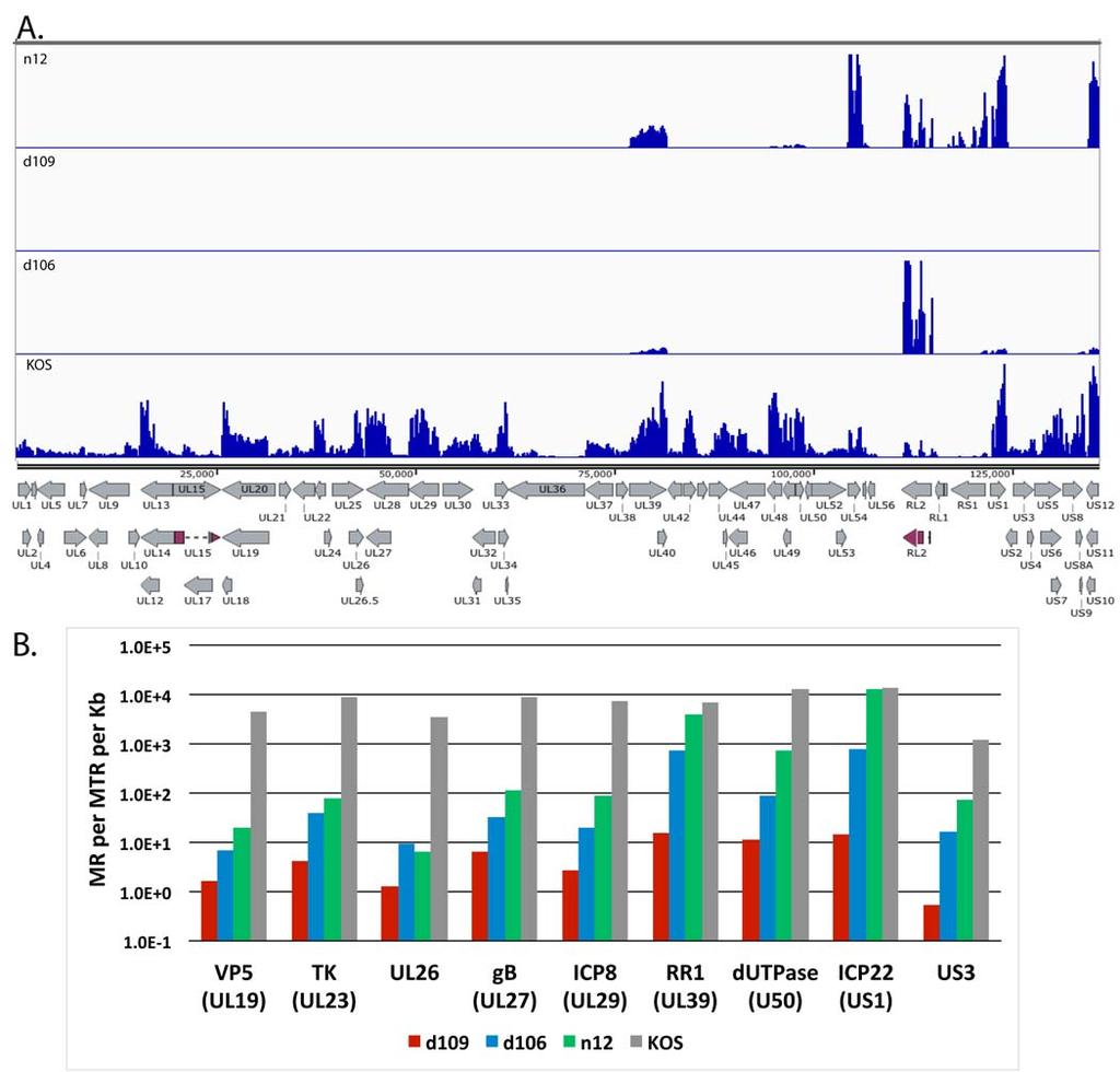
16 with wt virus in vivo [17,45]. To examine the expression across the entire genome of key IE gene mutants, MRC5 cells were infected with KOS, n12, d106, and d109 at an MOI of 10 PFU/cell for 4h and processed for RNAseq. n12 contains a nonsense mutation in the coding region of ICP4, rendering it non-functional [29]. d106 is mutated such that ICP0 is the only intact IE gene and d109 is defective for the expression of all the IE genes [23]. The mapped reads from each virus relative to the KOS genome are shown in the same scale in Figure 5A to illustrate the disparity in gene expression between the 4 different viruses. Transcription of numerous genes can be seen across the entire KOS genome, while transcription of the n12 genome appears to be restricted to the UL39, 40 loci, ICP27 (UL54), ICP0 (RL2), ICP4 (RS1), ICP22 (US1), and ICP47 (US12). The levels of ICP0, ICP27 and ICP4 transcripts are considerably higher in n12-infected cells compared to KOS, consistent with the previously defined phenotype of n12 [29]. The expression profiles in Fig 5A of d106 and d109 are also consistent with their published phenotypes [23]. However, this representation does not quantitatively describe the expression of the genome in the absence of the IE proteins. Considering there were ~20 million reads in each sample and the percent viral reads were 44, 13, 4.9 and.07, for KOS, n12, d106, and d109, respectively, then there are a considerable number of reads to accurately quantify the relatively low abundance transcription of across the genome in these mutant backgrounds. For example, while no signal is visible for d109 in figure 5A, there are still 1.4 x 10 4 viral reads in the d109 sample. Fig. 5B shows the number of reads mapped to a particular viral gene per million total reads per kilobase pair (MR per MTR per Kb) for selected viral genes in the different mutant backgrounds. The genes are for ICP22 (IE), tk (E), ICP8 (E), RR1 (E), dutpase (E), US3 (L), VP5 (L), UL26 (L) and gb (L). Several observations can be made from the results; i. IE genes (when present) are highly expressed in the absence of ICP4, 16
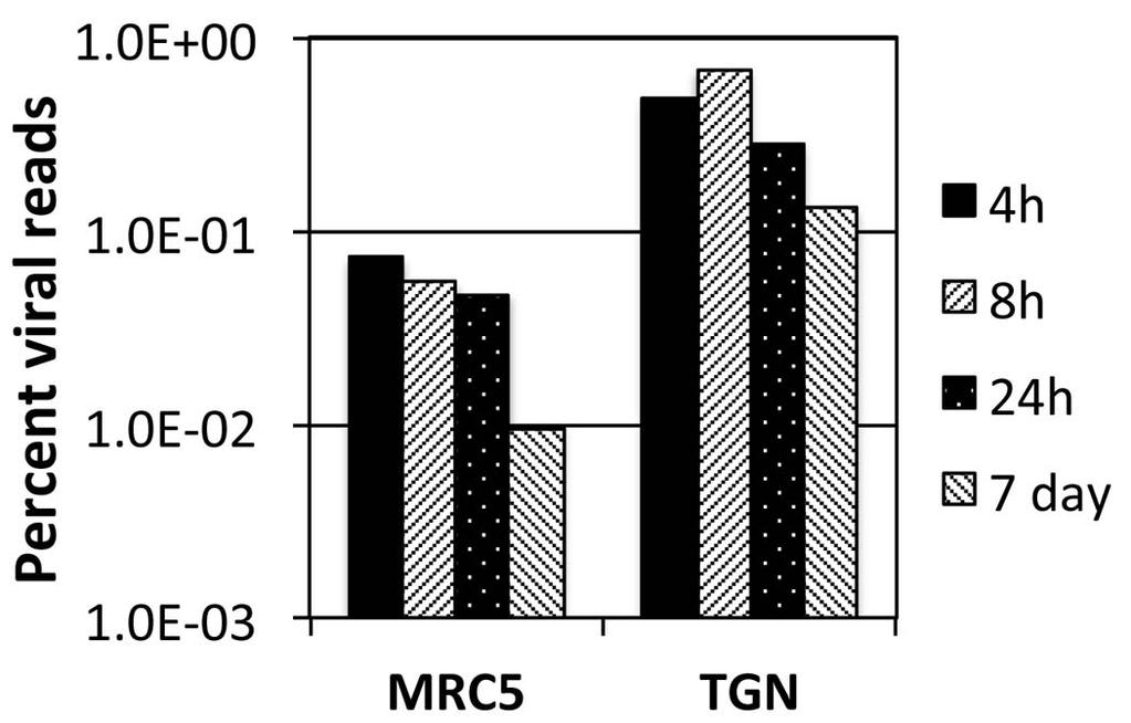
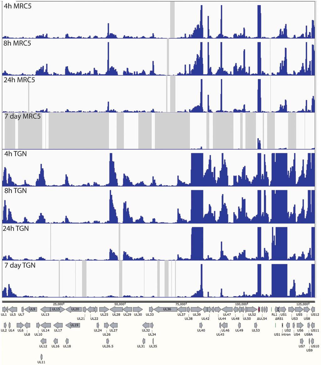
17 ii. In the absence of ICP4 (n12), early and late gene transcripts accumulate to 0.1% to 5.0% the level seen in wt virus infection. An exception is RR1 (UL39), the transcription of which is relatively independent of ICP4, iii. Early and late gene expression is further reduced 3-5 fold in the d106 background at this time post infection, and iv. In the absence of IE proteins early and late transcription is 3 to 4 orders of magnitude less than wt virus at 4 hpi in MRC5 cells. For example the total number of reads for the tk mrna is 3 x 10 5 and 1.5 x 10 2, for KOS and d109, respectively. Expression from the d109 genome in MRC5 cells is further reduced with time due to the ongoing formation of heterochromatin [26]. To examine how this repression affects expression across the viral genome, and to further compare expression in MRC5 cells to that in trigeminal neurons in the absence of IE proteins, MRC5 cells and TG neuronal cultures were infected with d109 at a MOI of approximately 10 PFU/cell. mrna was isolated at 4, 8 and 24 hours and 7 days post infection, and analyzed by RNAseq as before. The percent of viral reads from the MRC5 samples decreased from 0.07% at 4h to just under 0.01% at 7 days, while the percent viral reads in trigeminal neurons was 0.5 % at 4h and a little more than 0.1% at 7days (fig. 6). Therefore, the number of viral reads was about 10-fold higher in d109-infected neurons than in d109-infected MRC5 cells, suggesting a more transcriptionally active genome in trigeminal neurons. This is in contrast to what was seen in KOS infected cells, where the proportion of viral mrna in neurons was less than in MRC5 cells. The locations of the reads are shown in Figure 7 aligned to a modified d109 genome. Unlike Fig. 5A, the scale of the graphs was chosen to view the relatively low level expression across the entire genome. As a consequence the signals for UL39, 40, the internal repeats and GFP are off the scale. They are represented in Fig. 8. Regions where there were no reads are represented by gray areas. 17
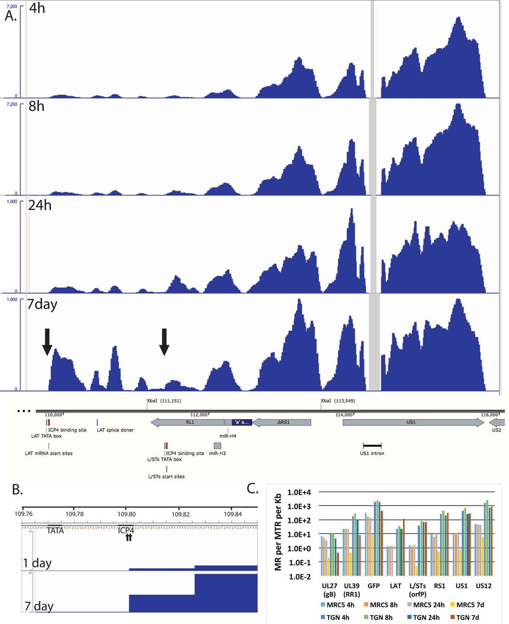
18 The accumulation of gene specific reads can readily be seen. Consistent with the higher level of transcription overall, there was a general trend toward higher levels of individual transcripts in neurons than in MRC5 cells. For example, the levels of glycoprotein B (UL27), ICP8 (UL29), UL30, UL41, UL42 were higher in neurons than in MRC5 cells, even at 7 days post infection. While transcription across the d109 genome was detected in neurons at 7 days post infection, no transcription was detected from many loci in MRC5 cells at this time as indicated by the gray shaded regions. This is not due to a lack of persisting d109 genomes, since it can be demonstrated that the provision of ICP0 can quantitatively reactivate the quiescent d109 genomes in MRC5 cells in this system [27]. One interesting observation is the pattern of expression in neurons of genes just outside the long and short repeat regions. In the short unique region of the genome, expression of the US3/4, US5/6/7, and US8/9 clusters is evident, and decreases over the course of 24h. However, the abundance of the US3/4 and US8/9 increase from 24h to 7days, as that of US5/6/7 decreases, as most of the gene do. Likewise UL1/2 and UL4/5 mrnas decrease over the course of 24 h, but increase at 7days. This pattern is also in contrast to that of all the other HSV genes in U L. In neurons, the pattern of expression of the genes adjacent to the long and short repeat regions is also seen within the repeats. Figure 8A shows the region of the d109 genome from the LAT promoter proximal region through to US2. Fig. 8A also shows the RNAseq reads in this region from d109 in neurons. These signals were off the scale in Figure 7. Note the only place in this region where there were no reads was in the US1 intron. The expression of this region overall was considerably greater than that of genes in the unique long and short regions (with the exception of UL39 and GFP). While the numbers of reads in this region generally decreased over 24h, they were sustained or increased from 24 h to 7 days. The signal just immediately 18
19 downstream from the previously defined LAT mrna start site that contains an ICP4 binding site [46,47] increased considerably from 1 to 7 days post infection. The LAT start site is indicated by the arrow to the left in Fig. 8A and by the 2 small arrows (previously defined by primer extension analysis [48]) in Fig. 6B. Fig. 8B also shows the ICP4 binding site and the TATA box for the LAT transcript along with the RNAseq reads for d109 in neurons at 1 and 7 days post infection. The elevated RNAseq signal begins precisely at the previously described LAT mrna start site, strongly indicating that it is transcribed from the LAT promoter. The abundance of transcripts from this region of d109 in MRC5 cells and neurons were quantified and plotted in Fig. 8C. The XbaI site at 111,151 (Fig. 8A) marks the locus of the ICP0 deletion in d109 [23], therefore the sequence from the LAT mrna start site to the XbaI site (111,151) is collinear with the 5 end of the LAT primary transcript [49]. The region quantified and designated LAT in Fig. 8C corresponds to the region from the LAT mrna start site to 1 kb downstream. The transcription of this region is quite complicated with the RL1 gene and the L/STs (orf P) transcripts antiparallel to each other. The L/STs TATA box, mrna start site and ICP4 binding sites are indicated (Fig. 8A) [50,51]. For the sake of clarity and because there is a distinct increase in reads on the L/STs start site (right arrow), the quantification in Fig. 8C represented as L/STs are the reads from the L/STs start site to 1 kb downstream. Also quantified in Fig. 6C are ΔRS1, the second exons of US1 and US12, GFP, UL39, and UL27. Fig. 8C shows several results; i. The abundance of all the transcripts in this region is higher in TG neurons than in MRC5 cells. ii. The abundance of all the HSV transcripts in MRC5 cells decrease with time such that the maximum abundance is less than 1 in 10 6 at seven days (US1) and LAT is undetectable (<1 in 2 x 10 7 ). iii. In neurons, the abundance of UL27, UL39 and GFP decrease from 1 to 7 days by ~10-fold, whereas the abundance of ΔRS1, US1 and US12, 19
20 and LAT all increase from 1 to 7 days post infection, with LAT increasing the most. iv. The numbers of reads for LAT, L/STs, ΔRS1, US1, and US12 reflect a transcript abundance for each that is between1 in 3x10 3 and 1 in 10 4 of all transcripts in the cell. This suggest that this region of the genome is considerable more transcriptionally active in TG neurons than in MRC5 cells, despite repression of the remainder of the viral genome, even in the absence of viral activators
21 DISCUSSION Next generation sequencing has emerged as a high throughput methodology to quantify changes in transcriptome wide gene expression. RNA-sequencing has been shown to be more sensitive for detection of low abundance transcripts and to have a wider range of detection than traditional microarrays [52]. The goal of this study was to determine how the presence and absence of immediate early proteins impacted viral gene expression in neuronal (TG neurons) and non-neuronal cells (MRC5). In particular, we were interested in the expression from a virus that establishes a persistent quiescent infection in trigeminal neurons as a model for what might be occurring during latency. From the RNAseq experiments performed, we have made the following findings; i) during productive infection, there are deviations from the established cascade paradigm for early genes, ii) the level of expression of late genes largely a function of the TATA box and INR, iii) in the absence of immediate early proteins, transcription from the viral genome occurs at low level independent of kinetic class, iv) neurons are less restrictive to viral transcription than MRC5 cells, and v) genes near the joint/repeat region of the genome, and the LAT locus in particular, are more transcriptionally active during persistence in neurons Productive Infection Gene Regulation and Promoter Architecture The promoters of HSV early genes are characterized by the presence of upstream elements for cellular transcription factors. Several early genes were quantified in figure 4C and D. ICP8 and TK were abundantly expressed. The promoter for tk gene contains three distinct upstream elements, two Sp1 binding sites and one CCAAT element [13]. ICP8 also contains two Sp1 binding sites. The DNA polymerase promoter contains a degenerate TATA box, and one Sp1 binding site [53]. Expression of this transcript is reduced approximately 4 fold relative to tk 21
22 and 2 fold relative to ICP8. UL42 was the most abundantly expressed gene, comprising approximately 1% of the total cellular RNA and peaking at 6 hours post infection, consistent with previous results [36]. The abundance of these 4 transcripts decreased late after infection. The mrna for the components of the helicase-primase complex UL5, UL8, and UL52 [54] and the origin binding protein UL9 [55] were expressed with similar kinetics and intensity during early time points prior to DNA replication. Expression of these four transcripts slowly accumulated throughout the time course to low levels. The UL8 and UL9 mrna start sites have been mapped [56]. UL8 and UL9 both have degenerate TATA boxes and UL8 has an Sp1 site 22 bp upstream of the TATA box. UL 9 has been reported to have an upstream Sp1 site, however none are evident in the KOS sequence [56]. The UL5 and UL52 mrna start sites have not been determined making it difficult to localize transcription signals, however the only potential TATA boxes within 400 bp of these genes consist of 4 A and T bp. Therefore, genes with promoters possessing good TATA boxes and multiple sites for upstream factors are abundantly expressed early after infection and their decline later in infection is pronounced. This represents the early gene paradigm. However, it is difficult to fit some genes whose products are clearly required for DNA synthesis into thisparadigm. Those with poor TATA boxes, lacking upstream elements, are expressed at a low-level, their decline later in infection is not as pronounced, and in some cases accumulation continues at a low level (UL52, UL9). This is reminiscent of some previous studies with mutant and chimeric promoters [48,57,58]. Late genes (γ2) require viral DNA synthesis for their expression and their promoters consist of at TATA box and a initiator element [59]. The promoter elements of the late genes represented in Fig. 4G and H are shown in Table 1. The expression of these genes varies 22
23 considerably. There are 4 transcripts with abundance above 10,000 mapped fragments per million reads, representing the most highly expressed late genes. The three most highly expressed genes, UL35, UL45 and UL44, have a TATA box that completely matches the consensus sequence and an initiator element with a perfect match to the first 4 bases of the consensus. UL10 is expressed to a lesser extent, and has a mismatch in the TATA box. Genes expressed to levels below 10,000 have multiple mismatches the TATA box, the first 4 nucleotides of the initiator element, or a combination of both. There was no apparent correlation to the downstream activation sequence, though it likely plays an important role on a gene by gene basis. Therefore, the expression of late genes varies considerably, and their levels of transcription correlate with the degree of match to both the TATA box and INR element consensus, suggesting that the ability of TFIID to bind to the promoter is the main determinant in late gene transcription. γ1 genes are transcribed early after infection and their synthesis increases after DNA replication. 4 γ1 genes are presented in Fig 4 E and F. This group of genes is more uniform in their expression relative to the other groups of genes and are represented by a minimum of 1% of the total reads (virus + cell). The early and sustained high level of expression is most likely due to the presence of a TATA box INR and binding sites for upstream factors in their promoters [60]. The binding sites for upstream factors enables expression early after infection, and the INR allows for the continued expression late after infection. We hypothesize that late after infection, cellular activator function and the activity of ICP4 on early promoters becomes less active. We have shown that Sp1 gets phosphorylated after DNA replication and is less active in vitro [61], and that TFIIA, which is required for the activation of tk by ICP4 in vitro, decreases late after infection [62,63]. In addition, upstream 23
24 activators require a form of the mediator complex for activation, and we have shown that as infection proceeds, ICP4 interacts with a form of the mediator complex that is often associated with repression of transcription [64]. Therefore, weaker promoters, such as UL8 or UL9, which not activated to the same extent by upstream factors such as Sp1, might not show as dramatic a decrease, or any at all, in activity late after infection. The sustained high-level activity of late genes is most likely a function of the ability of ICP4 to activate transcription through the INR sequence present at the start site of late genes [65,66]. Gene Expression during quiescent infection. HSV mutants engineered to not express viral activators can establish a quiescent infection in cells in culture such that their genomes are transcriptionally repressed and persist in cells [22,23,67]. Heterochromatin forms on d109 genomes persisting in MRC5 cells [26,27], reminiscent of the structure of latent viral genomes [17,68]. We have also shown that d109 genomes may not be as tightly repressed in TG neurons as in non-neuronal cells in culture based on GFP transgene expression and the ability to reactivate quiescent virus [69]. Therefore, we sought to compare the expression of the entire d109 genome in MRC5 cells, as a model for nonneuronal cells, and TG neurons. Trigeminal neurons were significantly more permissive for the expression of genes across the persisting d109 genome than MRC5 cells (Figs. 7 and 8). The MRC5 cells are presumably more homogeneous than these tg cultures, which are known to contain populations of neurons that differ in their permissiveness for LAT expression [31]. Therefore it is possible that some infected neurons are more permissive for HSV gene expression than others. Moreover, expression bore little relation to kinetic class. This dysregulated pattern of HSV gene expression has been seen in two different systems when reactivation stimuli were applied to 24
25 latent HSV [20,21]. In our experiments, the persisting genomes are void of genes that are thought to activate viral early and late genes, and since the infected neuronal cultures are maintained in NGF, known reactivation stimuli were not applied. The elevated level of transcription of persisting vial genomes in neurons relative to nonneuronal cells most likely is due to the different transcriptional environment in neurons. In mammalian cells, there are three different histone H3 variants, H3.1 H3.2 and H3.3. During differentiation of neurons, the abundance of variants H3.1 and H3.2 decrease while H3.3 accumulates [70]. H3.1 contains a combination of both activating and repressive modifications, H3.2 is enriched in inactive marks, and H3.3 is enriched in activating markers and is localized to transcriptionally active regions [71-73]. The association of H3.3 with the viral genome has been shown to facilitate lytic gene expression [74]. Perhaps H3.3 facilitates transcription from d109 genomes in TG neurons, while H3.1 and 3.2 restrict gene expression in MRC5 cells. This remains to be studied. While the d109 genome in general is more transcriptionally active in TG neurons than in MRC5 cells, a large majority of the transcripts in neurons at 7 days post infection map to loci around the joint regions and in the unique short region of the virus, Figure 7 and 8. There are several possible explanations for this. i) Neuronal specific promoters could be directing the expression of all theses genes. With the exception of LAT, which is generally thought to possess a neuronal specific promoter, it is unlikely that all of these genes (UL1,2,4,5, US1,3,4,8,9,12 RS1, and LAT) have neuron specific promoters. The underlying basis for their enhanced expression more likely has to do with a general structural property of the region. ii) Perhaps the high G+C content of the internal repeats relative to rest of the genome results in reduced binding of repressive chromatin in TG neurons. iii) It is also possible that chromatin insulators, DNA 25
26 elements that function to separate euchromatic and heterochromatic areas of the genome [75,76] are involved. Seven distinct regions containing CTCF binding sites have been identified in the herpes genome [77]. These are all located in the repeat region of the genome and 5 remain in d109. Two specific CTCF binding sites surrounding the LAT enhancer have been shown to function as enhancer-blocking elements and maintain the localization of euchromatin to the LAT locus [78]. The presence of these elements could explain the specific upregulation of LAT expression during persistence of d109 in neurons. Other insulator elements are located surrounding other immediate early genes near in the repeat regions [79]. The presence of these insulators could slow the accumulation of heterochromatin in the regions, enabling low-level transcription of IE genes in the absence of VP16. The physiological consequences of the elevated low-level expression of genes in the joint region are not known. However, an intriguing possibility involves the interplay between HSV gene products and micrornas encoded in this region [80,81]. During latent infection, the primary latency associated transcript serves as a precursor to several mirnas [81], which may target ICP4 and ICP0 [82]. The low-level expression of the IE genes and mirnas in this region could provide the basis for the ability of the virus to reactivate in a regulated way, independent of viral activators. 26
27 549 ACKNOWLEDGMENTS This work was supported by NIH grant R01AI and R01AI to NAD. J.M.H. was supported by the NIH training grant T32 AI
28 554 FIGURE LEGENDS Figure 1. Evaluation of RNAseq reads from infected MRC5 cells. Monolayers of MRC5 cells and cultures of adult trigeminal neurons were infected with HSV-1, strain KOS at an MOI of 10 PFU/cell. Total RNA was collected from MRC5 cells at 1, 2, 3, 4, 6, 8, 12, and 16 hours post infection, and from TG neurons at 2, 4, and 8 hours post infection. cdna was prepared and subject to Illumina sequencing as described in the Materials and Methods. Illumina reads were processed and aligned to a modified KOS sequence using the TopHat mapper in the Galaxy Cloud software package. A. The percent of sequencing reads mapping to the HSV genome relative to the total number of reads. B. Comparison of kinetics of ICP27 (UL54) accumulation by RNAseq and RT-PCR. C. Comparison of kinetics of tk (UL23) accumulation by RNAseq and RT-PCR. D. Comparison of kinetics of gc (UL44) accumulation by RNAseq and RT-PCR. The units for RT-PCR were cdna copies per ug RNA as determined by comparison to standards using CsCl-purified viral genomic DNA. The units for RNAseq are reads mapped to the viral genome per million total reads (viral+cell) per kilobasepair (MR per MTR per Kb) Figure 2. Locations of RNAseq reads across the HSV genome as a function of time post infection in MRC5 cells. The locations of reads for each of the time points is shown relative to a map of the mrna for each of the HSV genes. The scales of the graphs were set to normalize to the number of total reads to enable qualitative comparison of transcript abundance between different time points. The reads and the genome are represented without the long and short terminal repeats since their sequences are represented within the internal repeats. The exons of RL2 and UL15 are also shown by darker shading. 28
29 Figure 3. Locations of RNAseq reads across the HSV genome as a function of time post infection in cultured TG neurons. As described in the legend for Fig Figure 4. The accumulation of HSV transcripts in MRC5 cells and trigeminal neurons. Quantification of IE () (A and B), E () (C and D), L (1) (E and F), and L (2) (G and H) transcripts in MRC5 cells (A, C, E, and G) and neurons (B, D, F, and H). The units for RNAseq are reads mapped to the viral genome per million total reads (viral+cell) per kilobasepair (MR per MTR per Kb) Figure 5. Transcript accumulation in IE mutant-infected MRC5 cells. MRC5 cells were infected with KOS, n12, d106, and d109 at an moi of 10 PFU/cell for 4 hr, and processed for RNA seq as previously stated. A. Locations of mapped reads relative to the KOS genome. The Illumina reads were aligned to the modified KOS sequence as before. The maximum on the y- axis is 30,000 reads. B. Quantification of the numbers of reads for select HSV genes Figure 6. Percent viral reads in d109 infected MRC5 cells and neurons. The numbers of reads that mapped to the d109 genome were divided by the total number of reads and multiplied by Figure 7. HSV transcripts synthesized in d109-infected MRC5 cells and TG neurons. MRC cells and cultures of TG neurons were infected with d109 (MOI = 10 PFU/cell) for 4h, 8h, 24h and 7d. The RNA synthesized in the cells was analyzed by RNAseq as before, and the reads 29
30 were mapped to a modified d109 genome. The reads were aligned to the d109 genome. The GFP gene is shown in red in deleted ICP27 locus. The maximum on the y-axes of the graphs was set to 100 reads. The gray shaded areas indicate regions were there were no reads Figure 8. Transcription of the internal repeat region from persisting genomes. A. The RNAseq reads from d109-infected TG neurons mapping to the region encoding the LAT promoter to US2 are shown relative to features in this region of the genome. The Xba1 site at 111,151 marks the locus of the ICP0 deletion and the site 113,549 marks the locus of the deletion that removed the VP16 responsive elements from the region. The numbers shown are in bp from the 5 end of the modified d109 genome in the parental orientation. The maximum of the Y-axes for the 4 and 8h graphs was set at 7,200 reads, while those for the 24h and 7 day graphs were set at 1,800 reads. B. The RNAseq reads for the 1 and 7 day d109-infected TG neuron samples are shown in the region of the LAT TATA box, mrna start sites, and ICP4 binding site. The maximum on the Y-axis is 100 reads. The beginning of the signal corresponds to arrow to the left in the 7 d graph of panel A. C. The quantification of the reads from the samples in figure 5 for select viral genes are shown
31 617 Table 1 TATA Box INR DAS *Consensus TATAAA YYANWYY GAGNNYGAG UL10 TAAAA GCACAC none UL22 AATAAAA TCATAAA GAGTGCCAG UL34 TATAAA GCAGAC CACGGCGAG UL35 TATAAA CTAATCG none UL37 TATAACA TCGCAAC GAGGGCCGC UL38 TTTAAA CCAGTCG GAGGCGGTAG UL44 TATAAA CTACCGA GAGGCCGCA UL45 TATAAA TCATCCT GGCGTGGAG US8 ATTTAA CTTCTGG GAGCCCAAC 618 *bold lettering highlights mismatches from the consensus sequence
32 620 REFERENCES Kosz-Vnenchak M, Jacobson J, Coen DM, Knipe DM (1993) Evidence for a novel regulatory pathway for herpes simplex virus gene expression in trigeminal ganglion neurons. J Virol 67: Nichol PF, Chang JY, Johnson EM, Jr., Olivo PD (1996) Herpes simplex virus gene expression in neurons: viral DNA synthesis is a critical regulatory event in the branch point between the lytic and latent pathways. J Virol 70: Pesola JM, Zhu J, Knipe DM, Coen DM (2005) Herpes simplex virus 1 immediate-early and early gene expression during reactivation from latency under conditions that prevent infectious virus production. J Virol 79: Alwine JC, Steinhart WL, Hill CW (1974) Transcription of herpes simplex type 1 DNA in nuclei isolated from infected HEp-2 and KB cells. Virology 60: Honess RW, Roizman B (1974) Regulation of herpesvirus macromolecular synthesis. I. Cascade regulation of the synthesis of three groups of viral proteins. J Virol 14: Honess RW, Roizman B (1975) Regulation of herpesvirus macromolecular synthesis: sequential transition of polypeptide synthesis requires functional viral polypeptides. Proc Natl Acad Sci U S A 72: Batterson W, Roizman B (1983) Characterization of the herpes simplex virion-associated factor responsible for the induction of alpha genes. J Virol 46: Campbell ME, Palfreyman JW, Preston CM (1984) Identification of herpes simplex virus DNA sequences which encode a trans-acting polypeptide responsible for stimulation of immediate early transcription. J Mol Biol 180:
33 Post LE, Mackem S, Roizman B (1981) Regulation of alpha genes of herpes simplex virus: expression of chimeric genes produced by fusion of thymidine kinase with alpha gene promoters. Cell 24: Gannon F, O'Hare K, Perrin F, LePennec JP, Benoist C, et al. (1979) Organisation and sequences at the 5' end of a cloned complete ovalbumin gene. Nature 278: Wasylyk B, Derbyshire R, Guy A, Molko D, Roget A, et al. (1980) Specific in vitro transcription of conalbumin gene is drastically decreased by single-point mutation in T- A-T-A box homology sequence. Proc Natl Acad Sci U S A 77: Mackem S, Roizman B (1982) Structural features of the herpes simplex virus alpha gene 4, 0, and 27 promoter-regulatory sequences which confer alpha regulation on chimeric thymidine kinase genes. J Virol 44: Eisenberg SP, Coen DM, McKnight SL (1985) Promoter domains required for expression of plasmid-borne copies of the herpes simplex virus thymidine kinase gene in virus-infected mouse fibroblasts and microinjected frog oocytes. Mol Cell Biol 5: Coen DM, Weinheimer SP, McKnight SL (1986) A genetic approach to promoter recognition during trans induction of viral gene expression. Science 234: Smale ST, Baltimore D (1989) The "initiator" as a transcription control element. Cell 57: Guzowski JF, Wagner EK (1993) Mutational analysis of the herpes simplex virus type 1 strict late UL38 promoter/leader reveals two regions critical in transcriptional regulation. J Virol 67:
34 Kubat NJ, Tran RK, McAnany P, Bloom DC (2004) Specific histone tail modification and not DNA methylation is a determinant of herpes simplex virus type 1 latent gene expression. J Virol 78: Wang QY, Zhou C, Johnson KE, Colgrove RC, Coen DM, et al. (2005) Herpesviral latencyassociated transcript gene promotes assembly of heterochromatin on viral lytic-gene promoters in latent infection. Proc Natl Acad Sci U S A 102: Stevens JG, Wagner EK, Devi-Rao GB, Cook ML, Feldman LT (1987) RNA complementary to a herpesvirus alpha gene mrna is prominent in latently infected neurons. Science 235: Du T, Zhou G, Roizman B (2011) HSV-1 gene expression from reactivated ganglia is disordered and concurrent with suppression of latency-associated transcript and mirnas. Proc Natl Acad Sci U S A 108: Kim JY, Mandarino A, Chao MV, Mohr I, Wilson AC (2012) Transient reversal of episome silencing precedes VP16-dependent transcription during reactivation of latent HSV-1 in neurons. PLoS Pathog 8: e Preston CM, Nicholl MJ (1997) Repression of gene expression upon infection of cells with herpes simplex virus type 1 mutants impaired for immediate-early protein synthesis. J Virol 71: Samaniego LA, Neiderhiser L, DeLuca NA (1998) Persistence and expression of the herpes simplex virus genome in the absence of immediate-early proteins. J Virol 72: Jackson SA, DeLuca NA (2003) Relationship of herpes simplex virus genome configuration to productive and persistent infections. Proc Natl Acad Sci U S A 100:
35 Rock DL, Fraser NW (1983) Detection of HSV-1 genome in central nervous system of latently infected mice. Nature 302: Ferenczy MW, DeLuca NA (2009) Epigenetic modulation of gene expression from quiescent herpes simplex virus genomes. J Virol 83: Ferenczy MW, DeLuca NA (2011) Reversal of heterochromatic silencing of quiescent herpes simplex virus type 1 by ICP0. J Virol 85: Stingley SW, Ramirez JJ, Aguilar SA, Simmen K, Sandri-Goldin RM, et al. (2000) Global analysis of herpes simplex virus type 1 transcription using an oligonucleotide-based DNA microarray. J Virol 74: DeLuca NA, Schaffer PA (1988) Physical and functional domains of the herpes simplex virus transcriptional regulatory protein ICP4. J Virol 62: Samaniego LA, Wu N, DeLuca NA (1997) The herpes simplex virus immediate-early protein ICP0 affects transcription from the viral genome and infected-cell survival in the absence of ICP4 and ICP27. J Virol 71: Bertke AS, Swanson SM, Chen J, Imai Y, Kinchington PR, et al. (2011) A5-positive primary sensory neurons are nonpermissive for productive infection with herpes simplex virus 1 in vitro. J Virol 85: Stringer JR, Holland LE, Swanstrom RI, Pivo K, Wagner EK (1977) Quantitation of herpes simplex virus type 1 RNA in infected HeLa cells. J Virol 21: Thorvaldsdottir H, Robinson JT, Mesirov JP (2013) Integrative Genomics Viewer (IGV): high-performance genomics data visualization and exploration. Brief Bioinform 14:
36 Weinheimer SP, McKnight SL (1987) Transcriptional and post-transcriptional controls establish the cascade of herpes simplex virus protein synthesis. J Mol Biol 195: Thompson RL, Preston CM, Sawtell NM (2009) De novo synthesis of VP16 coordinates the exit from HSV latency in vivo. PLoS Pathog 5: e Goodrich LD, Rixon FJ, Parris DS (1989) Kinetics of expression of the gene encoding the 65-kilodalton DNA-binding protein of herpes simplex virus type 1. J Virol 63: Watson RJ, Clements JB (1980) A herpes simplex virus type 1 function continuously required for early and late virus RNA synthesis. Nature 285: Preston CM (1979) Control of herpes simplex virus type 1 mrna synthesis in cells infected with wild-type virus or the temperature-sensitive mutant tsk. J Virol 29: Dixon RA, Schaffer PA (1980) Fine-structure mapping and functional analysis of temperature-sensitive mutants in the gene encoding the herpes simplex virus type 1 immediate early protein VP175. J Virol 36: DeLuca NA, McCarthy AM, Schaffer PA (1985) Isolation and characterization of deletion mutants of herpes simplex virus type 1 in the gene encoding immediate-early regulatory protein ICP4. Journal of Virology 56: Imbalzano AN, Coen DM, DeLuca NA (1991) Herpes simplex virus transactivator ICP4 operationally substitutes for the cellular transcription factor Sp1 for efficient expression of the viral thymidine kinase gene. J Virol 65: Leib DA, Coen DM, Bogard CL, Hicks KA, Yager DR, et al. (1989) Immediate-early regulatory gene mutants define different stages in the establishment and reactivation of herpes simplex virus latency. J Virol 63:
37 Sacks WR, Schaffer PA (1987) Deletion mutants in the gene encoding the herpes simplex virus type 1 immediate-early protein ICP0 exhibit impaired growth in cell culture. J Virol 61: Stow ND, Stow EC (1986) Isolation and characterization of a herpes simplex virus type 1 mutant containing a deletion within the gene encoding the immediate early polypeptide Vmw110. J Gen Virol 67 ( Pt 12): Cliffe AR, Garber DA, Knipe DM (2009) Transcription of the herpes simplex virus latencyassociated transcript promotes the formation of facultative heterochromatin on lytic promoters. J Virol 83: Batchelor AH, O'Hare P (1990) Regulation and cell-type-specific activity of a promoter located upstream of the latency-associated transcript of herpes simplex virus type 1. J Virol 64: Farrell MJ, Margolis TP, Gomes WA, Feldman LT (1994) Effect of the transcription start region of the herpes simplex virus type 1 latency-associated transcript promoter on expression of productively infected neurons in vivo. J Virol 68: Rivera-Gonzalez R, Imbalzano AN, Gu B, Deluca NA (1994) The role of ICP4 repressor activity in temporal expression of the IE-3 and latency-associated transcript promoters during HSV-1 infection. Virology 202: Devi-Rao GB, Goodart SA, Hecht LM, Rochford R, Rice MK, et al. (1991) Relationship between polyadenylated and nonpolyadenylated herpes simplex virus type 1 latencyassociated transcripts. J Virol 65:
38 Bohenzky RA, Papavassiliou AG, Gelman IH, Silverstein S (1993) Identification of a promoter mapping within the reiterated sequences that flank the herpes simplex virus type 1 UL region. J Virol 67: Yeh L, Schaffer PA (1993) A novel class of transcripts expressed with late kinetics in the absence of ICP4 spans the junction between the long and short segments of the herpes simplex virus type 1 genome. J Virol 67: Zhao S, Fung-Leung WP, Bittner A, Ngo K, Liu X (2014) Comparison of RNA-Seq and Microarray in Transcriptome Profiling of Activated T Cells. PLoS One 9: e Yager DR, Coen DM (1988) Analysis of the transcript of the herpes simplex virus DNA polymerase gene provides evidence that polymerase expression is inefficient at the level of translation. J Virol 62: Crute JJ, Tsurumi T, Zhu LA, Weller SK, Olivo PD, et al. (1989) Herpes simplex virus 1 helicase-primase: a complex of three herpes-encoded gene products. Proc Natl Acad Sci U S A 86: Olivo PD, Nelson NJ, Challberg MD (1988) Herpes simplex virus DNA replication: the UL9 gene encodes an origin-binding protein. Proc Natl Acad Sci U S A 85: Baradaran K, Dabrowski CE, Schaffer PA (1994) Transcriptional analysis of the region of the herpes simplex virus type 1 genome containing the UL8, UL9, and UL10 genes and identification of a novel delayed-early gene product, OBPC. J Virol 68: Cook WJ, Gu B, DeLuca NA, Moynihan EB, Coen DM (1995) Induction of transcription by a viral regulatory protein depends on the relative strengths of functional TATA boxes. Mol Cell Biol 15:
39 Imbalzano AN, DeLuca NA (1992) Substitution of a TATA box from a herpes simplex virus late gene in the viral thymidine kinase promoter alters ICP4 inducibility but not temporal expression. J Virol 66: Smale ST, Jain A, Kaufmann J, Emami KH, Lo K, et al. (1998) The initiator element: a paradigm for core promoter heterogeneity within metazoan protein-coding genes. Cold Spring Harb Symp Quant Biol 63: Lieu PT, Wagner EK (2000) Two leaky-late HSV-1 promoters differ significantly in structural architecture. Virology 272: Kim DB, DeLuca NA (2002) Phosphorylation of transcription factor Sp1 during herpes simplex virus type 1 infection. J Virol 76: Zabierowski S, DeLuca NA (2004) Differential cellular requirements for activation of herpes simplex virus type 1 early (tk) and late (gc) promoters by ICP4. J Virol 78: Zabierowski SE, Deluca NA (2008) Stabilized binding of TBP to the TATA box of herpes simplex virus type 1 early (tk) and late (gc) promoters by TFIIA and ICP4. J Virol 82: Wagner LM, DeLuca NA (2013) Temporal association of herpes simplex virus ICP4 with cellular complexes functioning at multiple steps in PolII transcription. PLoS One 8: e Gu B, DeLuca N (1994) Requirements for activation of the herpes simplex virus glycoprotein C promoter in vitro by the viral regulatory protein ICP4. J Virol 68: Kim DB, Zabierowski S, DeLuca NA (2002) The initiator element in a herpes simplex virus type 1 late-gene promoter enhances activation by ICP4, resulting in abundant late-gene expression. J Virol 76:
40 Jamieson DR, Robinson LH, Daksis JI, Nicholl MJ, Preston CM (1995) Quiescent viral genomes in human fibroblasts after infection with herpes simplex virus type 1 Vmw65 mutants. J Gen Virol 76 ( Pt 6): Cliffe AR, Knipe DM (2008) Herpes simplex virus ICP0 promotes both histone removal and acetylation on viral DNA during lytic infection. J Virol 82: Terry-Allison T, Smith CA, DeLuca NA (2007) Relaxed repression of herpes simplex virus type 1 genomes in Murine trigeminal neurons. J Virol 81: Meshorer E (2007) Chromatin in embryonic stem cell neuronal differentiation. Histol Histopathol 22: Ahmad K, Henikoff S (2002) The histone variant H3.3 marks active chromatin by replication-independent nucleosome assembly. Mol Cell 9: Chow CM, Georgiou A, Szutorisz H, Maia e Silva A, Pombo A, et al. (2005) Variant histone H3.3 marks promoters of transcriptionally active genes during mammalian cell division. EMBO Rep 6: Hake SB, Garcia BA, Duncan EM, Kauer M, Dellaire G, et al. (2006) Expression patterns and post-translational modifications associated with mammalian histone H3 variants. J Biol Chem 281: Placek BJ, Huang J, Kent JR, Dorsey J, Rice L, et al. (2009) The histone variant H3.3 regulates gene expression during lytic infection with herpes simplex virus type 1. J Virol 83: Felsenfeld G, Burgess-Beusse B, Farrell C, Gaszner M, Ghirlando R, et al. (2004) Chromatin boundaries and chromatin domains. Cold Spring Harb Symp Quant Biol 69:
41 Zhao H, Dean A (2004) An insulator blocks spreading of histone acetylation and interferes with RNA polymerase II transfer between an enhancer and gene. Nucleic Acids Res 32: Bloom DC, Giordani NV, Kwiatkowski DL (2010) Epigenetic regulation of latent HSV-1 gene expression. Biochim Biophys Acta 1799: Chen Q, Lin L, Smith S, Huang J, Berger SL, et al. (2007) CTCF-dependent chromatin boundary element between the latency-associated transcript and ICP0 promoters in the herpes simplex virus type 1 genome. J Virol 81: Amelio AL, McAnany PK, Bloom DC (2006) A chromatin insulator-like element in the herpes simplex virus type 1 latency-associated transcript region binds CCCTC-binding factor and displays enhancer-blocking and silencing activities. J Virol 80: Jurak I, Kramer MF, Mellor JC, van Lint AL, Roth FP, et al. (2010) Numerous conserved and divergent micrornas expressed by herpes simplex viruses 1 and 2. J Virol 84: Umbach JL, Kramer MF, Jurak I, Karnowski HW, Coen DM, et al. (2008) MicroRNAs expressed by herpes simplex virus 1 during latent infection regulate viral mrnas. Nature 454: Flores O, Nakayama S, Whisnant AW, Javanbakht H, Cullen BR, et al. (2013) Mutational inactivation of herpes simplex virus 1 micrornas identifies viral mrna targets and reveals phenotypic effects in culture. J Virol 87:
42
43
44
45
46
47
48
49
Selective depletion of abundant RNAs to enable transcriptome analysis of lowinput and highly-degraded RNA from FFPE breast cancer samples
 DNA CLONING DNA AMPLIFICATION & PCR EPIGENETICS RNA ANALYSIS Selective depletion of abundant RNAs to enable transcriptome analysis of lowinput and highly-degraded RNA from FFPE breast cancer samples LIBRARY
Polyomaviridae. Spring
 Polyomaviridae Spring 2002 331 Antibody Prevalence for BK & JC Viruses Spring 2002 332 Polyoma Viruses General characteristics Papovaviridae: PA - papilloma; PO - polyoma; VA - vacuolating agent a. 45nm
Herpesviruses. Virion. Genome. Genes and proteins. Viruses and hosts. Diseases. Distinctive characteristics
 Herpesviruses Virion Genome Genes and proteins Viruses and hosts Diseases Distinctive characteristics Virion Enveloped icosahedral capsid (T=16), diameter 125 nm Diameter of enveloped virion 200 nm Capsid
Analysis of Massively Parallel Sequencing Data Application of Illumina Sequencing to the Genetics of Human Cancers
 Analysis of Massively Parallel Sequencing Data Application of Illumina Sequencing to the Genetics of Human Cancers Gordon Blackshields Senior Bioinformatician Source BioScience 1 To Cancer Genetics Studies
Restricted VZV transcription in human trigeminal ganglia
 JVI Accepts, published online ahead of print on 27 June 2012 J. Virol. doi:10.1128/jvi.01331-12 Copyright 2012, American Society for Microbiology. All Rights Reserved. 1 2 Restricted VZV transcription
genomics for systems biology / ISB2020 RNA sequencing (RNA-seq)
") RNA sequencing (RNA-seq) Module Outline MO 13-Mar-2017 RNA sequencing: Introduction 1 WE 15-Mar-2017 RNA sequencing: Introduction 2 MO 20-Mar-2017 Paper: PMID 25954002: Human genomics. The human transcriptome
RNA sequencing (RNA-seq) Module Outline MO 13-Mar-2017 RNA sequencing: Introduction 1 WE 15-Mar-2017 RNA sequencing: Introduction 2 MO 20-Mar-2017 Paper: PMID 25954002: Human genomics. The human transcriptome
Transcription Program of Murine Gammaherpesvirus 68
 JOURNAL OF VIROLOGY, Oct. 2003, p. 10488 10503 Vol. 77, No. 19 0022-538X/03/$08.00 0 DOI: 10.1128/JVI.77.19.10488 10503.2003 Copyright 2003, American Society for Microbiology. All Rights Reserved. Transcription
7.012 Quiz 3 Answers
 MIT Biology Department 7.012: Introductory Biology - Fall 2004 Instructors: Professor Eric Lander, Professor Robert A. Weinberg, Dr. Claudette Gardel Friday 11/12/04 7.012 Quiz 3 Answers A > 85 B 72-84
Nature Genetics: doi: /ng Supplementary Figure 1. Assessment of sample purity and quality.
 Supplementary Figure 1 Assessment of sample purity and quality. (a) Hematoxylin and eosin staining of formaldehyde-fixed, paraffin-embedded sections from a human testis biopsy collected concurrently with
RNA-seq Introduction
 RNA-seq Introduction DNA is the same in all cells but which RNAs that is present is different in all cells There is a wide variety of different functional RNAs Which RNAs (and sometimes then translated
Supplemental Figure S1. Expression of Cirbp mrna in mouse tissues and NIH3T3 cells.
 SUPPLEMENTAL FIGURE AND TABLE LEGENDS Supplemental Figure S1. Expression of Cirbp mrna in mouse tissues and NIH3T3 cells. A) Cirbp mrna expression levels in various mouse tissues collected around the clock
Phosphate buffered saline (PBS) for washing the cells TE buffer (nuclease-free) ph 7.5 for use with the PrimePCR Reverse Transcription Control Assay
 for washing the cells TE buffer (nuclease-free) ph 7.5 for use with the PrimePCR Reverse Transcription Control Assay") Catalog # Description 172-5080 SingleShot Cell Lysis Kit, 100 x 50 µl reactions 172-5081 SingleShot Cell Lysis Kit, 500 x 50 µl reactions For research purposes only. Introduction The SingleShot Cell Lysis
Catalog # Description 172-5080 SingleShot Cell Lysis Kit, 100 x 50 µl reactions 172-5081 SingleShot Cell Lysis Kit, 500 x 50 µl reactions For research purposes only. Introduction The SingleShot Cell Lysis
Mechanisms of alternative splicing regulation
 Mechanisms of alternative splicing regulation The number of mechanisms that are known to be involved in splicing regulation approximates the number of splicing decisions that have been analyzed in detail.
Supplemental Materials and Methods Plasmids and viruses Quantitative Reverse Transcription PCR Generation of molecular standard for quantitative PCR
 Supplemental Materials and Methods Plasmids and viruses To generate pseudotyped viruses, the previously described recombinant plasmids pnl4-3-δnef-gfp or pnl4-3-δ6-drgfp and a vector expressing HIV-1 X4
~Lentivirus production~
 ~Lentivirus production~ May 30, 2008 RNAi core R&D group member Lentivirus Production Session Lentivirus!!! Is it health threatening to lab technician? What s so good about this RNAi library? How to produce
Eukaryotic transcription (III)
") Eukaryotic transcription (III) 1. Chromosome and chromatin structure Chromatin, chromatid, and chromosome chromatin Genomes exist as chromatins before or after cell division (interphase) but as chromatids
Eukaryotic transcription (III) 1. Chromosome and chromatin structure Chromatin, chromatid, and chromosome chromatin Genomes exist as chromatins before or after cell division (interphase) but as chromatids
Last time we talked about the few steps in viral replication cycle and the un-coating stage:
 Zeina Al-Momani Last time we talked about the few steps in viral replication cycle and the un-coating stage: Un-coating: is a general term for the events which occur after penetration, we talked about
INFLUENCE OF HERPES SIMPLEX VIRUS TYPE-1 GLYCOPROTEIN B
 INFLUENCE OF HERPES SIMPLEX VIRUS TYPE-1 GLYCOPROTEIN B EXPRESSION ON VIRAL PATHOGENECITY AND THE CD8 + T CELL RESPONSE by Srividya Ramachandran BSc. Biotechnology, Monash University, 2003 Submitted to
Received 18 July 2005/Accepted 12 September 2005
 JOURNAL OF VIROLOGY, Dec. 2005, p. 14516 14525 Vol. 79, No. 23 0022-538X/05/$08.00 0 doi:10.1128/jvi.79.23.14516 14525.2005 Copyright 2005, American Society for Microbiology. All Rights Reserved. Herpes
Received 12 June 2002/Accepted 13 September 2002
 JOURNAL OF VIROLOGY, Dec. 2002, p. 12758 12774 Vol. 76, No. 24 0022-538X/02/$04.00 0 DOI: 10.1128/JVI.76.24.12758 12774.2002 Copyright 2002, American Society for Microbiology. All Rights Reserved. General
Transcriptome Analysis
 Transcriptome Analysis Data Preprocessing Sample Preparation Illumina Sequencing Demultiplexing Raw FastQ Reference Genome (fasta) Reference Annotation (GTF) Reference Genome Analysis Tophat Accepted hits
Supplementary Information Titles Journal: Nature Medicine
 Supplementary Information Titles Journal: Nature Medicine Article Title: Corresponding Author: Supplementary Item & Number Supplementary Fig.1 Fig.2 Fig.3 Fig.4 Fig.5 Fig.6 Fig.7 Fig.8 Fig.9 Fig. Fig.11
QIAsymphony DSP Circulating DNA Kit
 QIAsymphony DSP Circulating DNA Kit February 2017 Performance Characteristics 937556 Sample to Insight Contents Performance Characteristics... 4 Basic performance... 4 Run precision... 6 Equivalent performance
7SK ChIRP-seq is specifically RNA dependent and conserved between mice and humans.
 Supplementary Figure 1 7SK ChIRP-seq is specifically RNA dependent and conserved between mice and humans. Regions targeted by the Even and Odd ChIRP probes mapped to a secondary structure model 56 of the
Egr-1 regulates RTA transcription through a cooperative involvement of transcriptional regulators
 /, 2017, Vol. 8, (No. 53), pp: 91425-91444 Egr-1 regulates RTA transcription through a cooperative involvement of transcriptional regulators Roni Sarkar 1 and Subhash C. Verma 1 1 Department of Microbiology
Transcription and RNA processing
 Transcription and RNA processing Lecture 7 Biology 3310/4310 Virology Spring 2018 It is possible that Nature invented DNA for the purpose of achieving regulation at the transcriptional rather than at the
RASA: Robust Alternative Splicing Analysis for Human Transcriptome Arrays
 Supplementary Materials RASA: Robust Alternative Splicing Analysis for Human Transcriptome Arrays Junhee Seok 1*, Weihong Xu 2, Ronald W. Davis 2, Wenzhong Xiao 2,3* 1 School of Electrical Engineering,
Neurotrophic factor GDNF and camp suppress glucocorticoid-inducible PNMT expression in a mouse pheochromocytoma model.
 161 Neurotrophic factor GDNF and camp suppress glucocorticoid-inducible PNMT expression in a mouse pheochromocytoma model. Marian J. Evinger a, James F. Powers b and Arthur S. Tischler b a. Department
Ali Alabbadi. Bann. Bann. Dr. Belal
 31 Ali Alabbadi Bann Bann Dr. Belal Topics to be discussed in this sheet: Particles-to-PFU Single-step and multi-step growth cycles Multiplicity of infection (MOI) Physical measurements of virus particles
Hepatitis B Antiviral Drug Development Multi-Marker Screening Assay
 Hepatitis B Antiviral Drug Development Multi-Marker Screening Assay Background ImQuest BioSciences has developed and qualified a single-plate method to expedite the screening of antiviral agents against
Large DNA viruses: Herpesviruses, Poxviruses, Baculoviruses and Giant viruses
 Large DNA viruses: Herpesviruses, Poxviruses, Baculoviruses and Giant viruses Viruses are the only obstacles to the domination of the Earth by mankind. -Joshua Lederberg Recommended reading: Field s Virology
Computational Analysis of UHT Sequences Histone modifications, CAGE, RNA-Seq
 Computational Analysis of UHT Sequences Histone modifications, CAGE, RNA-Seq Philipp Bucher Wednesday January 21, 2009 SIB graduate school course EPFL, Lausanne ChIP-seq against histone variants: Biological
Section 6. Junaid Malek, M.D.
 Section 6 Junaid Malek, M.D. The Golgi and gp160 gp160 transported from ER to the Golgi in coated vesicles These coated vesicles fuse to the cis portion of the Golgi and deposit their cargo in the cisternae
Products for cfdna and mirna isolation. Subhead Circulating Cover nucleic acids from plasma
 MACHEREY-NAGEL Products for cfdna and mirna isolation Bioanalysis Subhead Circulating Cover nucleic acids from plasma n Flexible solutions for small and large blood plasma volumes n Highly efficient recovery
Problem Set 5 KEY
 2006 7.012 Problem Set 5 KEY ** Due before 5 PM on THURSDAY, November 9, 2006. ** Turn answers in to the box outside of 68-120. PLEASE WRITE YOUR ANSWERS ON THIS PRINTOUT. 1. You are studying the development
7.014 Problem Set 7 Solutions
 MIT Department of Biology 7.014 Introductory Biology, Spring 2005 7.014 Problem Set 7 Solutions Question 1 Part A Antigen binding site Antigen binding site Variable region Light chain Light chain Variable
EPIGENOMICS PROFILING SERVICES
 EPIGENOMICS PROFILING SERVICES Chromatin analysis DNA methylation analysis RNA-seq analysis Diagenode helps you uncover the mysteries of epigenetics PAGE 3 Integrative epigenomics analysis DNA methylation
RNA SEQUENCING AND DATA ANALYSIS
 RNA SEQUENCING AND DATA ANALYSIS Length of mrna transcripts in the human genome 5,000 5,000 4,000 3,000 2,000 4,000 1,000 0 0 200 400 600 800 3,000 2,000 1,000 0 0 2,000 4,000 6,000 8,000 10,000 Length
Introduction retroposon
 17.1 - Introduction A retrovirus is an RNA virus able to convert its sequence into DNA by reverse transcription A retroposon (retrotransposon) is a transposon that mobilizes via an RNA form; the DNA element
VIROLOGY. Engineering Viral Genomes: Retrovirus Vectors
 VIROLOGY Engineering Viral Genomes: Retrovirus Vectors Viral vectors Retrovirus replicative cycle Most mammalian retroviruses use trna PRO, trna Lys3, trna Lys1,2 The partially unfolded trna is annealed
Lentiviral Delivery of Combinatorial mirna Expression Constructs Provides Efficient Target Gene Repression.
 Supplementary Figure 1 Lentiviral Delivery of Combinatorial mirna Expression Constructs Provides Efficient Target Gene Repression. a, Design for lentiviral combinatorial mirna expression and sensor constructs.
Soft Agar Assay. For each cell pool, 100,000 cells were resuspended in 0.35% (w/v)
") SUPPLEMENTARY MATERIAL AND METHODS Soft Agar Assay. For each cell pool, 100,000 cells were resuspended in 0.35% (w/v) top agar (LONZA, SeaKem LE Agarose cat.5004) and plated onto 0.5% (w/v) basal agar.
SUPPLEMENTARY MATERIAL AND METHODS Soft Agar Assay. For each cell pool, 100,000 cells were resuspended in 0.35% (w/v) top agar (LONZA, SeaKem LE Agarose cat.5004) and plated onto 0.5% (w/v) basal agar.
The Biology and Genetics of Cells and Organisms The Biology of Cancer
 The Biology and Genetics of Cells and Organisms The Biology of Cancer Mendel and Genetics How many distinct genes are present in the genomes of mammals? - 21,000 for human. - Genetic information is carried
Regulation of Gene Expression in Eukaryotes
 Ch. 19 Regulation of Gene Expression in Eukaryotes BIOL 222 Differential Gene Expression in Eukaryotes Signal Cells in a multicellular eukaryotic organism genetically identical differential gene expression
Iso-Seq Method Updates and Target Enrichment Without Amplification for SMRT Sequencing
 Iso-Seq Method Updates and Target Enrichment Without Amplification for SMRT Sequencing PacBio Americas User Group Meeting Sample Prep Workshop June.27.2017 Tyson Clark, Ph.D. For Research Use Only. Not
ExoQuick Exosome Isolation and RNA Purification Kits
 ExoQuick Exosome Isolation and RNA Purification Kits Cat # EQ806A-1, EQ806TC-1, EQ808A-1 User Manual Storage: Please see individual components Version 2 8/14/2018 A limited-use label license covers this
Circular RNAs (circrnas) act a stable mirna sponges
 act a stable mirna sponges") Circular RNAs (circrnas) act a stable mirna sponges cernas compete for mirnas Ancestal mrna (+3 UTR) Pseudogene RNA (+3 UTR homolgy region) The model holds true for all RNAs that share a mirna binding
Circular RNAs (circrnas) act a stable mirna sponges cernas compete for mirnas Ancestal mrna (+3 UTR) Pseudogene RNA (+3 UTR homolgy region) The model holds true for all RNAs that share a mirna binding
Determination of the temporal pattern and importance of BALF1 expression in Epstein-Barr viral infection
 Determination of the temporal pattern and importance of BALF1 expression in Epstein-Barr viral infection Melissa Mihelidakis May 6, 2004 7.340 Research Proposal Introduction Apoptosis, or programmed cell
HEK293FT cells were transiently transfected with reporters, N3-ICD construct and
 Supplementary Information Luciferase reporter assay HEK293FT cells were transiently transfected with reporters, N3-ICD construct and increased amounts of wild type or kinase inactive EGFR. Transfections
Supporting Online Material for
 www.sciencemag.org/cgi/content/full/1171320/dc1 Supporting Online Material for A Frazzled/DCC-Dependent Transcriptional Switch Regulates Midline Axon Guidance Long Yang, David S. Garbe, Greg J. Bashaw*
Bi 8 Lecture 17. interference. Ellen Rothenberg 1 March 2016
 Bi 8 Lecture 17 REGulation by RNA interference Ellen Rothenberg 1 March 2016 Protein is not the only regulatory molecule affecting gene expression: RNA itself can be negative regulator RNA does not need
Page 32 AP Biology: 2013 Exam Review CONCEPT 6 REGULATION
 Page 32 AP Biology: 2013 Exam Review CONCEPT 6 REGULATION 1. Feedback a. Negative feedback mechanisms maintain dynamic homeostasis for a particular condition (variable) by regulating physiological processes,
Supplementary Figure 1. SC35M polymerase activity in the presence of Bat or SC35M NP encoded from the phw2000 rescue plasmid.
 1 2 3 4 5 6 7 8 9 10 11 12 13 14 15 16 17 18 19 20 21 22 23 24 25 26 27 Supplementary Figure 1. SC35M polymerase activity in the presence of Bat or SC35M NP encoded from the phw2000 rescue plasmid. HEK293T
Today. Genomic Imprinting & X-Inactivation
 Today 1. Quiz (~12 min) 2. Genomic imprinting in mammals 3. X-chromosome inactivation in mammals Note that readings on Dosage Compensation and Genomic Imprinting in Mammals are on our web site. Genomic
Not IN Our Genes - A Different Kind of Inheritance.! Christopher Phiel, Ph.D. University of Colorado Denver Mini-STEM School February 4, 2014
 Not IN Our Genes - A Different Kind of Inheritance! Christopher Phiel, Ph.D. University of Colorado Denver Mini-STEM School February 4, 2014 Epigenetics in Mainstream Media Epigenetics *Current definition:
Gammaherpesviral Gene Expression and Virion Composition Are Broadly Controlled by Accelerated mrna Degradation
 Gammaherpesviral Gene Expression and Virion Composition Are Broadly Controlled by Accelerated mrna Degradation Emma Abernathy 1, Karen Clyde 1, Rukhsana Yeasmin 2, Laurie T. Krug 3, Al Burlingame 4, Laurent
Transcription and RNA processing
 Transcription and RNA processing Lecture 7 Biology W3310/4310 Virology Spring 2016 It is possible that Nature invented DNA for the purpose of achieving regulation at the transcriptional rather than at
Herpes Simplex Virus Type 1 and Bovine Herpesvirus 1 Latency
 CLINICAL MICROBIOLOGY REVIEWS, Jan. 2003, p. 79 95 Vol. 16, No. 1 0893-8512/03/$08.00 0 DOI: 10.1128/CMR.16.1.79 95.2003 Copyright 2003, American Society for Microbiology. All Rights Reserved. Herpes Simplex
Product Contents. 1 Specifications 1 Product Description. 2 Buffer Preparation... 3 Protocol. 3 Ordering Information 4
 INSTRUCTION MANUAL Quick-RNA Midiprep Kit Catalog No. R1056 Highlights 10 minute method for isolating RNA (up to 1 mg) from a wide range of cell types and tissue samples. Clean-Spin column technology allows
Molecular Biology (BIOL 4320) Exam #2 May 3, 2004
 Exam #2 May 3, 2004") Molecular Biology (BIOL 4320) Exam #2 May 3, 2004 Name SS# This exam is worth a total of 100 points. The number of points each question is worth is shown in parentheses after the question number. Good
Molecular Biology (BIOL 4320) Exam #2 May 3, 2004 Name SS# This exam is worth a total of 100 points. The number of points each question is worth is shown in parentheses after the question number. Good
Comparison of Herpes Simplex Virus Reactivation in Ganglia In Vivo and in Explants Demonstrates Quantitative and Qualitative Differences
 JOURNAL OF VIROLOGY, July 2004, p. 7784 7794 Vol. 78, No. 14 0022-538X/04/$08.00 0 DOI: 10.1128/JVI.78.14.7784 7794.2004 Copyright 2004, American Society for Microbiology. All Rights Reserved. Comparison
Supplementary Figure 1 IL-27 IL
 Tim-3 Supplementary Figure 1 Tc0 49.5 0.6 Tc1 63.5 0.84 Un 49.8 0.16 35.5 0.16 10 4 61.2 5.53 10 3 64.5 5.66 10 2 10 1 10 0 31 2.22 10 0 10 1 10 2 10 3 10 4 IL-10 28.2 1.69 IL-27 Supplementary Figure 1.
7.012 Problem Set 6 Solutions
 Name Section 7.012 Problem Set 6 Solutions Question 1 The viral family Orthomyxoviridae contains the influenza A, B and C viruses. These viruses have a (-)ss RNA genome surrounded by a capsid composed
AP Biology Reading Guide. Concept 19.1 A virus consists of a nucleic acid surrounded by a protein coat
 AP Biology Reading Guide Name Chapter 19: Viruses Overview Experimental work with viruses has provided important evidence that genes are made of nucleic acids. Viruses were also important in working out
RNA interference induced hepatotoxicity results from loss of the first synthesized isoform of microrna-122 in mice
 SUPPLEMENTARY INFORMATION RNA interference induced hepatotoxicity results from loss of the first synthesized isoform of microrna-122 in mice Paul N Valdmanis, Shuo Gu, Kirk Chu, Lan Jin, Feijie Zhang,
Stability determinants of Murine Cytomegalovirus long non-coding RNA7.2
 JVI Accepts, published online ahead of print on 23 July 2014 J. Virol. doi:10.1128/jvi.01695-14 Copyright 2014, American Society for Microbiology. All Rights Reserved. 1 2 3 Stability determinants of Murine
In vitro DNase I foot printing. In vitro DNase I footprinting was performed as described
 Supplemental Methods In vitro DNase I foot printing. In vitro DNase I footprinting was performed as described previously 1 2 using 32P-labeled 211 bp fragment from 3 HS1. Footprinting reaction mixes contained
1. Identify and characterize interesting phenomena! 2. Characterization should stimulate some questions/models! 3. Combine biochemistry and genetics
 1. Identify and characterize interesting phenomena! 2. Characterization should stimulate some questions/models! 3. Combine biochemistry and genetics to gain mechanistic insight! 4. Return to step 2, as
Large Scale Infection for Pooled Screens of shrna libraries
 Last modified 01/11/09 Large Scale Infection for Pooled Screens of shrna libraries Biao Luo, Glenn Cowley, Michael Okamoto, Tanaz Sharifnia This protocol can be further optimized if cells being used are
For in vitro Veterinary Diagnostics only. Kylt Rotavirus A. Real-Time RT-PCR Detection.
 For in vitro Veterinary Diagnostics only. Kylt Rotavirus A Real-Time RT-PCR Detection www.kylt.eu DIRECTION FOR USE Kylt Rotavirus A Real-Time RT-PCR Detection A. General Kylt Rotavirus A products are
Norgen s HIV proviral DNA PCR Kit was developed and validated to be used with the following PCR instruments: Qiagen Rotor-Gene Q BioRad icycler
 3430 Schmon Parkway Thorold, ON, Canada L2V 4Y6 Phone: (905) 227-8848 Fax: (905) 227-1061 Email: techsupport@norgenbiotek.com HIV Proviral DNA PCR Kit Product # 33840 Product Insert Background Information
Results. Abstract. Introduc4on. Conclusions. Methods. Funding
 . expression that plays a role in many cellular processes affecting a variety of traits. In this study DNA methylation was assessed in neuronal tissue from three pigs (frontal lobe) and one great tit (whole
Nature Medicine: doi: /nm.4322
 1 2 3 4 5 6 7 8 9 10 11 Supplementary Figure 1. Predicted RNA structure of 3 UTR and sequence alignment of deleted nucleotides. (a) Predicted RNA secondary structure of ZIKV 3 UTR. The stem-loop structure
EpiQuik Circulating Acetyl Histone H3K18 ELISA Kit (Colorimetric)
") EpiQuik Circulating Acetyl Histone H3K18 ELISA Kit (Colorimetric) Base Catalog # PLEASE READ THIS ENTIRE USER GUIDE BEFORE USE Uses: The EpiQuik Circulating Acetyl Histone H3K18 ELISA Kit (Colorimetric)
EpiQuik Circulating Acetyl Histone H3K18 ELISA Kit (Colorimetric) Base Catalog # PLEASE READ THIS ENTIRE USER GUIDE BEFORE USE Uses: The EpiQuik Circulating Acetyl Histone H3K18 ELISA Kit (Colorimetric)
MODULE 4: SPLICING. Removal of introns from messenger RNA by splicing
 Last update: 05/10/2017 MODULE 4: SPLICING Lesson Plan: Title MEG LAAKSO Removal of introns from messenger RNA by splicing Objectives Identify splice donor and acceptor sites that are best supported by
WHO Prequalification of In Vitro Diagnostics PUBLIC REPORT. Product: Alere q HIV-1/2 Detect WHO reference number: PQDx
 WHO Prequalification of In Vitro Diagnostics PUBLIC REPORT Product: Alere q HIV-1/2 Detect WHO reference number: PQDx 0226-032-00 Alere q HIV-1/2 Detect with product codes 270110050, 270110010 and 270300001,
TRANSCRIPTION. DNA à mrna
 TRANSCRIPTION DNA à mrna Central Dogma Animation DNA: The Secret of Life (from PBS) http://www.youtube.com/watch? v=41_ne5ms2ls&list=pl2b2bd56e908da696&index=3 Transcription http://highered.mcgraw-hill.com/sites/0072507470/student_view0/
Effects of UBL5 knockdown on cell cycle distribution and sister chromatid cohesion
 Supplementary Figure S1. Effects of UBL5 knockdown on cell cycle distribution and sister chromatid cohesion A. Representative examples of flow cytometry profiles of HeLa cells transfected with indicated
Abstract. Optimization strategy of Copy Number Variant calling using Multiplicom solutions APPLICATION NOTE. Introduction
 Optimization strategy of Copy Number Variant calling using Multiplicom solutions Michael Vyverman, PhD; Laura Standaert, PhD and Wouter Bossuyt, PhD Abstract Copy number variations (CNVs) represent a significant
The dual role of gamma interferon during herpes simplex virus type 1 infection. Vilma Decman
 The dual role of gamma interferon during herpes simplex virus type 1 infection by Vilma Decman M.Sc., University of Zagreb, Faculty of Natural Sciences and Mathematics, Croatia 1999 Submitted to the Graduate
Human Rotavirus A. genesig Standard Kit. Non structural protein 5 (NSP5) 150 tests. Primerdesign Ltd. For general laboratory and research use only
 150 tests. Primerdesign Ltd. For general laboratory and research use only") TM Primerdesign Ltd Human Rotavirus A Non structural protein 5 (NSP5) genesig Standard Kit 150 tests For general laboratory and research use only 1 Introduction to Human Rotavirus A Rotavirus is a genus
TM Primerdesign Ltd Human Rotavirus A Non structural protein 5 (NSP5) genesig Standard Kit 150 tests For general laboratory and research use only 1 Introduction to Human Rotavirus A Rotavirus is a genus
Supplementary Fig. 1. Delivery of mirnas via Red Fluorescent Protein.
 prfp-vector RFP Exon1 Intron RFP Exon2 prfp-mir-124 mir-93/124 RFP Exon1 Intron RFP Exon2 Untransfected prfp-vector prfp-mir-93 prfp-mir-124 Supplementary Fig. 1. Delivery of mirnas via Red Fluorescent
Product Manual. Omni-Array Sense Strand mrna Amplification Kit, 2 ng to 100 ng Version Catalog No.: Reactions
 Genetic Tools and Reagents Universal mrna amplification, sense strand amplification, antisense amplification, cdna synthesis, micro arrays, gene expression, human, mouse, rat, guinea pig, cloning Omni-Array
Oncolytic virus strategy
 Oncolytic viruses Oncolytic virus strategy normal tumor NO replication replication survival lysis Oncolytic virus strategy Mechanisms of tumor selectivity of several, some of them naturally, oncolytic
Genetics and Genomics in Medicine Chapter 6 Questions
 Genetics and Genomics in Medicine Chapter 6 Questions Multiple Choice Questions Question 6.1 With respect to the interconversion between open and condensed chromatin shown below: Which of the directions
Breast cancer. Risk factors you cannot change include: Treatment Plan Selection. Inferring Transcriptional Module from Breast Cancer Profile Data
 Breast cancer Inferring Transcriptional Module from Breast Cancer Profile Data Breast Cancer and Targeted Therapy Microarray Profile Data Inferring Transcriptional Module Methods CSC 177 Data Warehousing
Human Rotavirus A. genesig Advanced Kit. Non structural protein 5 (NSP5) 150 tests. Primerdesign Ltd. For general laboratory and research use only
 150 tests. Primerdesign Ltd. For general laboratory and research use only") TM Primerdesign Ltd Human Rotavirus A Non structural protein 5 (NSP5) genesig Advanced Kit 150 tests For general laboratory and research use only 1 Introduction to Human Rotavirus A Rotavirus is a genus
TM Primerdesign Ltd Human Rotavirus A Non structural protein 5 (NSP5) genesig Advanced Kit 150 tests For general laboratory and research use only 1 Introduction to Human Rotavirus A Rotavirus is a genus
VIRAL TITER COUNTS. The best methods of measuring infectious lentiviral titer
 VIRAL TITER COUNTS The best methods of measuring infectious lentiviral titer FLUORESCENCE CYCLES qpcr of Viral RNA SUMMARY Viral vectors are now routinely used for gene transduction in a wide variety of
Ambient temperature regulated flowering time
 Ambient temperature regulated flowering time Applications of RNAseq RNA- seq course: The power of RNA-seq June 7 th, 2013; Richard Immink Overview Introduction: Biological research question/hypothesis
RNA-Seq profiling of circular RNAs in human colorectal Cancer liver metastasis and the potential biomarkers
 Xu et al. Molecular Cancer (2019) 18:8 https://doi.org/10.1186/s12943-018-0932-8 LETTER TO THE EDITOR RNA-Seq profiling of circular RNAs in human colorectal Cancer liver metastasis and the potential biomarkers
Raymond Auerbach PhD Candidate, Yale University Gerstein and Snyder Labs August 30, 2012
 Elucidating Transcriptional Regulation at Multiple Scales Using High-Throughput Sequencing, Data Integration, and Computational Methods Raymond Auerbach PhD Candidate, Yale University Gerstein and Snyder
A complete next-generation sequencing workfl ow for circulating cell-free DNA isolation and analysis
 APPLICATION NOTE Cell-Free DNA Isolation Kit A complete next-generation sequencing workfl ow for circulating cell-free DNA isolation and analysis Abstract Circulating cell-free DNA (cfdna) has been shown
Supplementary Appendix
 Supplementary Appendix This appendix has been provided by the authors to give readers additional information about their work. Supplement to: Choi YL, Soda M, Yamashita Y, et al. EML4-ALK mutations in
Norgen s HIV Proviral DNA PCR Kit was developed and validated to be used with the following PCR instruments: Qiagen Rotor-Gene Q BioRad T1000 Cycler
 3430 Schmon Parkway Thorold, ON, Canada L2V 4Y6 Phone: 866-667-4362 (905) 227-8848 Fax: (905) 227-1061 Email: techsupport@norgenbiotek.com HIV Proviral DNA PCR Kit Product# 33840 Product Insert Intended
EBV infection B cells and lymphomagenesis. Sridhar Chaganti
 EBV infection B cells and lymphomagenesis Sridhar Chaganti How EBV infects B-cells How viral genes influence the infected B cell Differences and similarities between in vitro and in vivo infection How
Supplementary information
 Supplementary information Human Cytomegalovirus MicroRNA mir-us4-1 Inhibits CD8 + T Cell Response by Targeting ERAP1 Sungchul Kim, Sanghyun Lee, Jinwook Shin, Youngkyun Kim, Irini Evnouchidou, Donghyun
TITLE: Selective Oncolytic Therapy for Hypoxic Breast Cancer Cells. CONTRACTING ORGANIZATION: Ordway Research Institute Albany, New York 12208
 AD Award Number: W81XWH-06-1-0586 TITLE: Selective Oncolytic Therapy for Hypoxic Breast Cancer Cells PRINCIPAL INVESTIGATOR: Michael T. Fasullo, Ph.D. CONTRACTING ORGANIZATION: Ordway Research Institute
(DNA) Real-time PCR. Exicycler 96 Rotor-Gene Q/6000 PCR
 Real-time PCR. Exicycler 96 Rotor-Gene Q/6000 PCR") Real-Time (DNA) Real-time Exicycler 96 Rotor-Gene Q/6000 IU Mix1 Mix2 IU/μl IU/μl IU/μl IU/μl IU/μl IPC NTC C 1 Lot# 2 Freeze & thawing 1 MSDS: Material Safety Data Sheets Real- (TaqMan time ' FAM ' BHQ1
Real-Time (DNA) Real-time Exicycler 96 Rotor-Gene Q/6000 IU Mix1 Mix2 IU/μl IU/μl IU/μl IU/μl IU/μl IPC NTC C 1 Lot# 2 Freeze & thawing 1 MSDS: Material Safety Data Sheets Real- (TaqMan time ' FAM ' BHQ1
WHO Prequalification of Diagnostics Programme PUBLIC REPORT. Product: VERSANT HIV-1 RNA 1.0 Assay (kpcr) Number: PQDx
 Number: PQDx") WHO Prequalification of Diagnostics Programme PUBLIC REPORT Product: VERSANT HIV-1 RNA 1.0 Assay (kpcr) Number: PQDx 0115-041-00 Abstract The VERSANT HIV-1 RNA 1.0 Assay (kpcr) with product codes 10375763,
WHO Prequalification of Diagnostics Programme PUBLIC REPORT Product: VERSANT HIV-1 RNA 1.0 Assay (kpcr) Number: PQDx 0115-041-00 Abstract The VERSANT HIV-1 RNA 1.0 Assay (kpcr) with product codes 10375763,