Regulation and Programming of Antibody Effector Function through IgG Glycosylation
|
|
|
- Johnathan Barker
- 6 years ago
- Views:
Transcription
1 Regulation and Programming of Antibody Effector Function through IgG Glycosylation The Harvard community has made this article openly available. Please share how this access benefits you. Your story matters. Citation Accessed Citable Link Terms of Use Mahan, Alison Emilia Regulation and Programming of Antibody Effector Function through IgG Glycosylation. Doctoral dissertation, Harvard University. January 11, :38:29 PM EST This article was downloaded from Harvard University's DASH repository, and is made available under the terms and conditions applicable to Other Posted Material, as set forth at (Article begins on next page)
2 Alison Emilia Mahan All rights reserved.
3 Dissertation advisor: Professor Galit Alter Alison Emilia Mahan Regulation and Programming of Antibody Effector Function through IgG Glycosylation Abstract Antibodies are the defining characteristic of the humoral immune response. Their functions are diverse, including direct neutralization of pathogens and recruitment of other immune molecules or cells. While most successful vaccines induce protective neutralizing antibody responses, effective vaccine-elicited neutralizing antibodies against some pathogens, including HIV, HCV, malaria, and TB, remain elusive. Thus, researchers have begun to focus on how vaccines can elicit strong non-neutralizing antibody functions, including recruitment of innate immune factors for antibody-dependent cellular cytotoxicity, complement deposition, and antibody-dependent phagocytosis. The antibody s constant region (Fc) mediates most effector functions through isotype and subclass selection or alteration of the structure of the Fc-attached N-glycan, which controls function with exquisite specificity. Glycan modifications are naturally induced during inflammatory conditions such as autoimmune disease and natural infection however, the specific signals that regulate Fc-glycosylation remain unknown. This dissertation sought to understand how antibody glycosylation is regulated and how it can be programmed through vaccination. To do this, we first developed a technique to analyze antibody glycan structures both of bulk Fc and antigenspecific antibodies. Using this technique, we observed significant modulation of antibody glycans during viral infection as well as in vaccine-elicited antibodies. To iii
4 Dissertation advisor: Professor Galit Alter Alison Emilia Mahan identify specific signals important for altering the antibody glycan, we transcriptionally profiled stimulated B cells and identified a set of innate and adaptive stimuli that regulate the genes responsible for antibody glycosylation. The results described in this dissertation begin to define the specific mechanism(s) by which infection and vaccination modulate antibody glycosylation to elicit functional antibodies that can ultimately provide effective and sustained protection from infection. iv
5 Contents 1 Introduction The immune system The innate response The adaptive response The humoral immune response B cell development Antigen-specific activation of B cells Structure and diversity of antibodies Functions of IgG Fc-mediated antibody functions Protective IgG in vaccination Regulation of Fc-mediated antibody functions N-linked glycosylation Nomenclature and structure N-glycosylation in the ER and Golgi IgG glycosylation Glycosylation of therapeutic monoclonal antibodies Natural modulation of IgG glycosylation Regulation of N-glycosylation Natural regulation of N-glycosylation Artificial modulation of IgG N-glycosylation v
6 1.5.3 Mechanisms of N-glycosylation regulation Dissertation objectives High throughput analysis of IgG Fc glycosylation by capillary electrophoresis Background Methods Results Analysis of glycans by capillary electrophoresis is highly sensitive and reproducible Capillary electrophoresis is comparable to HPLC Fc and Fab are reliably separated by IdeS digestion IgG Fc and Fab have different glycosylation profiles IgG glycan profiles differ among humans, rhesus and mice Discussion Glycosylation of IgG is modified in HIV infection Background Methods Results Bulk IgG Fc glycosylation is modified in HIV infection Bulk Fc glycosylation changes over the course of HIV infection Glycosylation changes are associated with inflammatory cytokines in HIV infection Bulk IgG glycosylation is associated with specific antibody effector functions Antigen-specific antibodies have distinct glycosylation profiles Discussion vi
7 4 Antigen-specific IgG glycosylation is directed by HIV Vaccination Background Methods Results Bulk IgG Fc-glycosylation is highly variable in different regions Vaccination elicits glycosylation patterns independent of bulk glycosylation Vaccine-induced antibody glycosylation is independent of regional bulk glycosylation differences Vaccines elicit distinct glycosylation profiles Discussion Additional data HIV risk factor is a potential driver of IgG Fc-glycosylation Glycosyltransferase expression is altered by innate and adaptive signals Background Methods Results Glycosyltransferases associated with inflammatory IgG glycosylation are regulated in a coordinated manner Glycosyltransferases associated with effector functionality are resistant to change after innate stimulation BCR engagement down-regulates expression of glycosyltransferases T-cell help is a modulator of innate immune signals Discussion Conclusions 134 vii
8 6.1 Dissertation summary Inflammatory signals are associated with changes in bulk IgG glycosylation during natural infection Glycosylation is regulated at an antigen-specific level Polyclonal antigen-specific glycosylation is associated with particular antibody functions Innate and adaptive signals can alter glycosyltransferase expression in B cells A hypothetical model of IgG glycosylation Future directions References 149 Colophon 177 viii
9 Listing of figures 1.1 Immunoglobulin structure and diversity Symbolic representations of glycan subunits N-Glycosylation steps in the endoplasmic reticulum N-glycan trimming to produce complex structures N-Glycan structure of IgG Sensitivity of capillary electrophoresis Reproducibility of capillary electrophoresis Comparison of capillary electrophoresis and HPLC Digestion and fractionation of IgG Glycosylation of Fab, Fc, and intact IgG Species based variation in IgG glycosylation Fc Glycosylation in HIV infection Bulk Fc glycosylation in acute versus chronic HIV infection Association of bulk Fc glycan with age, sex, and viral load Association of Fc glycan with plasma cytokines Association of glycosylation with antibody function Glycosylation of antigen-specific antibodies Association of antigen-specific IgG glycosylation and function Summary of bulk Fc-glycan structures in HIV infection Geographical differences in bulk IgG glycosylation ix
10 4.2 Glycosylation of adenovirus vectored vaccine-elicited IgGs Vaccine specific IgG glycosylation Bulk Fc glycosylation in RV144 and VAX003 participants RV144- and VAX003-specific IgG glycosylation Transcription of glycosyltransferases in TLR-stimulated B cells Transcriptional analysis of B cells with BCR crosslinking Transcriptional analysis of B cells with CD40 stimulation Hypothetical model of IgG glycosylation mirna expression in stimulated B cells Gene expression in antigen-specific B cells CpG islands in glycosyltransferase genes x
11 Dedicated to the most important people in my life, you know who you are. xi
12 Acknowledgments First and foremost I want to thank my advisor Galit Alter for making the decision to take me on as her first PhD student. I appreciate all her contributions of time, ideas, and funding to make my graduate school experience productive and stimulating. I am also thankful for the excellent example she has provided as a successful female scientist. Thanks to the members of my dissertation advisory committee: Dan Barouch, Shiv Pillai, and Ron Desrosiers, for your advice and support. Additional thanks to Margie Ackerman for being a critical eye and supportive mentor from afar. Many thanks to the Gates Foundation for supporting me financially and recognizing my research efforts over the years. I would like to thank all the members of the Alter lab who helped me through the last five years. Particular thanks to Anne-Sophie Dugast, Bonnie Gunn and Cormac Cosgrove who were essential for this final piece of the process. Thank you to all my friends at the Ragon Institute, especially Eileen Scully for being a cheerleader and helping me decide to finish. Invaluable technical assistance was provided by Kendall Dionne and Jacquelynne Tedesco. I could not have accomplished so much without their help; nor could I have had so much fun. Additional technical assistance and lab support was provided by Justin Eusebio, Anastassia Mikhailova, Claire Ruberman, Marcela Trocha and all the members of the Alter lab over the years. xii
13 Thanks also to my family who supported my decision to go back for another half decade of schooling. Most importantly, my greatest thanks to my emotional supporters. To Damien Soghoian, a better best friend does not exist, thank you for being the one to help me through the tough times. Also thanks to the other Damien, Dr. Rochford, who saw me through the bulk of this project; I m glad we both made it to the other side. Finally to Eric O Connor, who has been a tireless supporter, advisor, and partner; I have tested your patience and tried your nerves and you have been unwaveringly amazing. xiii
14 1 Introduction The immune system is a complex and effective mechanism to protect a host from pathogen. When it is in perfect working order, it can eliminate dangerous foreign entities while ignoring host cells and proteins. However, when it fails, as in the case of auto-reactivity or chronic infection, it is important to understand the factors that cause this failure so that it can be prevented or corrected. One of the most significant examples of immune failure is the rampant and uncontrolled infection of millions of people with pathogens against which there are no effective vaccines, such as human immunodeficiency virus (HIV), tuberculosis (TB), and malaria. In order to develop better strategies for vaccine development, we must
15 understand how the immune system works naturally and how we can direct it to elicit protective responses after vaccination. 1.1 The immune system The innate response The mammalian immune system has two major mechanisms of action, the fast acting, but non-specific innate system, and the slow to activate, but highly specific adaptive response. Cells of the innate system are first to respond to invading pathogens, and they can recognize infection through non-specific signals of danger, such as common bacterial proteins or carbohydrates and the presence of foreign nucleic acids [222]. These signals are recognized through pattern recognition receptors, of which the best understood are the toll-like receptors (TLRs), a family of receptors expressed on a variety of cells, including cells of the innate and adaptive immune system [215]. Each of the ten human TLRs recognizes a specific molecular pattern commonly found on bacterial or viral pathogens, but not expressed by mammalian cells (Table 1.1) [10]. Once these receptors recognize their target, they activate cells to recruit additional cells of the innate immune system [9]. Once cells of the innate immune system recognize the presence of pathogens, a cascade of signaling molecules called cytokines are produced to recruit additional innate cells to the site of infection [134]. The innate response is rapid because it is mediated by potent cytokine signals, which initiate inflammation, however 2
16 Table 1.1: Toll-like receptors Human toll-like receptors, their targets and cellular localization [1]. Receptor Target Subcellular Location TLR1/2 Bacterial lipopeptides Plasma membrane TLR2 Bacterial peptidoglycan and Plasma membrane lipoprotein. Viral hemagglutinin TLR3 Viral double stranded RNA Endosomal membrane TLR4 Gram negative bacterial Plasma membrane lipopolysaccharide (LPS) TLR5 Bacterial flagellin Plasma membrane TLR2/6 Bacterial lipopeptides Plasma membrane TLR7 Viral single stranded RNA Endosomal membrane TLR8 Viral single stranded RNA Endosomal membrane TLR9 Viral and bacterial unmethylated Endosomal membrane CpG DNA 3
17 this response is also non-specific and can cause general inflammation and even allergy, since the danger signals detected by innate cells may be present on nonpathogenic organisms [204]. Additionally, some pathogens have evolved mechanisms to evade the innate response through mimicry of self or down-regulation of innate signals [83]. Thus, while the innate system in essential in the early stages of infection, the adaptive response is often required to eliminate infectious agents The adaptive response During the fast response of the innate system, innate cells ingest pathogens and bring them to specialized immune organs to activate the adaptive response. In the lymph nodes, innate cells present digested pathogens to cells of the adaptive immune system, which are activated in a pathogen-specific manner [1]. Where the innate system expresses a set of receptors that have a limited ability to bind to a broad set of molecules generally associated with pathogens, the adaptive cells have exceptionally specific receptors. The two major players of the adaptive response, B and T cells, create this specificity through their highly variable receptors: B and T cell receptors (BCR and TCR, respectively). Adaptive lymphocyte receptors are unique to each clonal population of cells; a product of genetic rearrangement of the receptor genes [154]. Within each adaptive cell, random assortment of the antigen-receptor alleles generates a unique receptor binding site [98], which in B cells, serves as a precursor to secreted antibody, whose binding specificity will mature during activation of the humoral immune response [148]. 4
18 1.2 The humoral immune response While the immune system is broadly classified into the innate and adaptive responses, the humoral response can bridge these categories by linking the pathogen specificity of the adaptive response to the speed and force of the innate response. The hallmark of the humoral response is the immunoglobulin molecule, or antibody, which is produced by B cells that have been stimulated in the presence of their antigen-specific pathogen B cell development B, T and natural killer (NK) cells all develop from the common lymphoid progenitor (CLP), which matures from the pluripotent hematopoietic stem cells of the bone marrow and fetal liver [1]. The CLP can mature into a pro-b cell, which is an immature lymphocyte with no antigen-specific cellular receptors [79]. These cells develop into pre-b cells and begin to express the non-specific pre-antigen receptor, which is required for continued development [187]. Once the pre-b cell completes its development, it proliferates to produce a clonal population of cells, which mature to express complete antigen receptors [135]. The production of antigenspecific receptors occurs through genetic rearrangement of the heavy chain of the immunoglobulin molecule [131]. At this stage, genetic rearrangement of the immunoglobulin (Ig) locus allows for the generation of a diverse set of unique antigen receptors within the expanding pre-b cell population [85]. Upon selection of an immunoglobulin light chain allele, a complete B cell receptor (BCR) is generated 5
19 [148]. The resulting immature B cell is subject to selection, whereby strong affinity of the BCR for self-generated antigens results in the apoptotic deletion of the cell [187]. Weak BCR affinity, however, drives the cell to continue development into a mature B cell [1]. At this point, the mature B cell exits the bone marrow into the lymphatic circulation to transit to lymphoid organs including the spleen and lymph nodes [148] Antigen-specific activation of B cells Once a B cell is mature, it remains naïve until it is stimulated through recognition of its cognate antigen [1]. Recognition of antigen by the B cell can occur in a T- cell dependent or -independent manner. In T-dependent activation, a B cell recognizes its antigen through its BCR and pulls attached protein or pathogen inside itself for degradation and presentation on surface receptors called major histocompatibility complexes (MHC) [245]. Mature, antigen-specific T cells recognize MHC embedded antigen and activate the B cell through cytokine production and activation of CD40 with its T cell expressed ligand (CD40L) [1]. This combination of signals activates the B cell to proliferate and mature its BCR. Maturation of the BCR involves further genetic mutation of the Ig locus within each cell to create a repertoire of unique BCRs within the expanding B cell population [85]. Each of these receptors will be tested against the pathogen and only those cells expressing BCRs with strong affinities will be maintained and expanded [187]. This system allows for the generation of an enormous population of unique antigen-specific cells, each with a strong affinity for specific antigens expressed by the pathogen. 6
20 T-cell independent activation occurs less frequently and requires a highly repetitive, or polyvalent, antigen, which allows for multiple molecules of the BCR to bind the antigen and link together at the surface of the B cell. This linking allows the receptors to interact with each other and initiate an activating signal strong enough to overcome the lack of co-stimulatory signals from T cells [1]. Both T dependent and independent activation of B cells results in the generation of soluble, secreted immunoglobulin (Ig), or antibody. Antibody is generated by the removal of the transmembrane domain of the BCR to generate a soluble antigen-specific molecule that is highly specific to their antigen and are an important factor in effective protection from pathogens [148]. Once an antibody producing cell is generated, it can mature into a long-lived plasma cell, which will return to the bone marrow and may live as long as its host [1]. Additionally, memory B cells are generated during the proliferative stages of B cell activation and these remain in the circulation or reside in lymphoid organs in order to initiate a rapid secondary response in the event of re-exposure to its cognate antigen, for instance during new infection with a previously encountered pathogen [1]. As part of their ability to bridge innate and adaptive immunity, B cells express a variety of the innate TLR receptors [25]. While the particular role of TLR signaling in B cells is not completely known, studies in humans and mice reveal a general increase in cellular TLR expression and activation as B cells develop from naïve to mature [167]. In particular, B cell stimulation by TLR ligation induces proliferation, antibody production and inhibition of apoptosis, as well as a driving activated cells to differentiate into plasma cells [35, 71, 96]. This effect of TLR signaling on 7
21 the adaptive response has led to significant interest in eliciting TLR activation during vaccination through the use of TLR agonists as vaccine adjuvants [175] Structure and diversity of antibodies Secreted immunoglobulins contain two identical units of both heavy and light chain proteins and can be divided into functional units of the Fab (antigen binding) and Fc (crystallizable) fragments (Figure 1.1A). The Fab fragment contains the highly variable, genetically mutated regions of the heavy and light chains of immunoglobulin, which interact with antigen and define the binding specificity of the antibody [197]. In contrast, the Fc fragment, which contains half of the heavy chain, is often referred to as constant since it has a set sequence and structure depending on the isotype and subclass of the Ig [67]. Depending on the signals present during T-dependent activation of a B cell, different classes, or isotypes, of immunoglobulin molecules are produced [40]. Immunoglobulin isotype is a critical determinant of antibody function since each class mediates different interactions with innate cells and immune molecules [160]. Naïve B cells produce IgM, which is a low affinity, high-avidity antibody produced before maturation of the BCR [1]. The low affinity of IgM is compensated for by its ability to multimerize into pentamers or hexamers, which are linked together with the J chain linker, as shown in Figure 1.1B [1]. IgM molecules function primarily through opsonization, whereby they coat pathogens and signal immune cells to destroy them [1]. The presence of specific cytokine signals during B cell maturation can drive im- 8

22 A Antigen-binding Fragment (Fab) N-Glycan Crystallizable Fragment (Fc) B V L C L IgM C μ 1 V H C μ 2 C μ 3 C μ 4 J Chain V H C α 1 V L C L IgE IgA C C α 2 α 3 C α 4 J Chain IgG V L C L V H C ε 1 C ε 2 V L C L C γ 1 V H C γ 2 C ε 3 C γ 3 C ε 4
23 munoglobulin class switching to produce IgA, IgG or IgE isotypes [1]. The IgA can be produced as single units or as a dimerized form, connected through the J chain linker (Figure 1.1B) and is important for protection of mucosal barriers such as the gut, nose, and reproductive tract because it is easily transported across epithelial borders into mucosal surfaces [1]. IgE is generated in the presence of specific cytokines that during B cell maturation and is important for clearance of helminth infection, though it is most often associated with allergic responses [1]. Generation of the IgG isotype is the most common response to B cell activation and occurs most often through T cell-dependent activation, though it is sometimes elicited after T-independent activation [211]. Circulating IgG concentrations can be as high as 10 mg/ml in healthy human blood serum and this molecule has a variety of important immunological roles [1]. Once the IgG isotype is selected, further subclass selection can greatly affect the activity of the antibody. In humans, there are four IgG subclasses: IgG1, IgG2, IgG3, and IgG4, each of which has a different function and is produced at different concentrations during an immune response [102]. Since IgG makes up the vast majority of circulating, high-affinity immunoglobulin, it is an important component of a protective adaptive immune response [37, 88] Functions of IgG The functions of IgG can be broadly classified into Fab- and Fc-mediated. The best recognized Fab-mediated function of antibodies is that of neutralization. This function is regulated primarily by the variability of the antigen-binding domain 10
24 and is dependent on the binding specificity of the Fab. Neutralization of a pathogen requires highly specific blocking of the viral or bacterial entry machinery to eliminate interaction with and/or entry into host cells [132]. Additionally, antibodies can bind directly to pathogen-generated toxins to neutralize pathogenic effects through specific binding [148]. While most existing vaccine strategies elicit protective neutralizing antibodies, vaccine-elicited neutralizing antibodies have never been generated against some pathogens, including HIV, TB and malaria [171]. Interestingly, although HIV neutralizing antibodies are protective in animal models of passive antibody transfer [142], they do not appear to protect the 10-30% of humans who develop them naturally, likely because they are generated only after years of infection [88, 196]. Independent of the antigen-binding domain, function and potency are mediated by variation of the IgG constant region or crystallizable fraction (Fc), which controls recruitment of innate immune cells [160], inflammatory signals [150], and complement deposition [105], and as well as altering the antibody s half-life in the circulation [20]. These functions do not necessarily rely on the specific recognition of neutralizing pathogen epitope, but they do require sufficient specificity to bind the pathogen. Given the difficulty in eliciting neutralizing antibodies against HIV, elicitation of non-neutralizing antibodies with strong Fc-mediated effector functions is an appealing goal for vaccine strategies [2, 61, 212]. 11
25 1.2.5 Fc-mediated antibody functions The best-characterized Fc-mediated functions of IgG are controlled by interaction with the membrane-bound Fc gamma receptors (FcγRs), which bind to the Fc portion of IgG to activate or inhibit cells [159]. Within the class of FcγRs, there are five subtypes, each of which have different cellular expression patterns and functions in the immune response [157]. In particular, FcγRI has high affinity for IgG1 and IgG3, and can mediate activation of the phagocytic cells that express it [1]. FcγRIIIA activates NK cells [228], while FcγRIIA binding initiates an inhibitory signal to B cells and innate immune cells [206]. The remaining receptors have a role primarily in antibody-dependent cellular phagocytosis [157, 158, 206]. Antibod-dependent cellular cytotoxicity Natural killer (NK) cells are important for eliminating bacteria- or virus-infected cells to prevent the replication and release of new infectious agents. NK cells recognize infected cells through two main mechanisms [148]. The first mechanism is through recognition of stress markers that are expressed on the surface on infected cells by NK cell receptors [51]. The second mechanism involves recruitment and activation of NK cells by IgG-coated target cells through a process known as antibody-dependent cellular cytotoxicity (ADCC) [46]. ADCC is initiated when antigen-specific antibodies bind to the surface of an infected cell that is expressing pathogen-specific proteins and epitopes on its surface [148]. The cell-bound antibody recruits NK cells through interaction with NK-expressed FcγRIIIA and induces NK cells to release cytotoxic molecules and destroy the antibody-coated tar- 12
26 get cell [1, 148]. This antibody function is important in HIV infection, given the its association with slower progression to AIDS [66] and protection from infection by simian immunodeficiency virus (SIV) or the humanized form of SIV, SHIV, after vaccination or passive transfer [12, 75, 91]. Especially interesting is the observation that ADCC was a secondary correlate of protection in the only protective HIV vaccine trial to date, RV144 [30, 80, 104, 182]. Antibody-dependent cellular phagocytosis Similar to ADCC in its mechanism of activation, the process of antibody-dependent cellular phagocytosis (ADCP) links innate immune cell to infected cells, or directly to pathogen, via antibody. While ADCP is less clearly regulated than ADCC, it is at least partially controlled by interactions between the Fc region of the cell- or pathogen-bound antibody and the FcRs of the phagocytic cell, including FcγRI, IIA, and IIIB [1]. Phagocytic cells, which include macrophages, monocytes, neutrophils, and eosinophils, express FcRs on their surface to recognize antibodycoated cells or pathogens and initiate, phagocytosis, or cellular ingestion, of the antibody-coated target [147]. Once inside the phagocyte, the pathogen or infected cell is degraded in acidic compartments called phagosomes [5]. Complement-dependent cytotoxicity The complement system is a component of the innate immune system that consists of over thirty cell-associated and soluble factors [233, 234]. When activated, the 13
27 complement system can lead to direct lysis of pathogens or infected cells. Complement activation can occur through three mechanisms: the classical, alternative, and lectin-dependent pathways [1, 148]. The classical pathway is activated by IgG or IgM mediated recruitment of the initiator molecule, C1q [77, 233]. Antigenbound IgG or IgM forms immune complexes, which are recognized by C1q to generate the C1 molecular complex and initiate a cascade of complement molecule deposition leading to lysis or phagocytosis of the infected cell or pathogen [77]. The importance of complement-dependent cytotoxicity has been well established in cancer therapeutics, where complement-recruiting activity is specifically engineered into in vitro produced monoclonal antibodies directed against malignant cells [151, 152] Protective IgG in vaccination Neutralization is the primary correlates of protection in most successful vaccines to date [170], however, neutralizing antibodies have yet to be elicited in an HIV vaccine. So while elicitation of a strong, and sustained, neutralizing antibody response remains the primary focus of the HIV vaccine field, strategies to elicit nonneutralizing antibodies with strong effector functions are gaining wider attention [2, 61, 212, 220]. In particular, the only mildly protective HIV vaccine trial to date, RV144, failed to induce broadly neutralizing antibodies in vaccinees [103, 182], however, secondary correlates analysis revealed an association between protection and ADCC activity in vaccine recipients [30, 80]. 14
28 1.2.7 Regulation of Fc-mediated antibody functions The quality of Fc effector functions is controlled through a variety of factors. Antibody titer is an important determinant of functional activity since antibodies must present in high enough concentration to initiate binding to pathogens and generate immune complexes to opsonize pathogens [82]. Isotype and subclass are crucial for determining the strength and type of antibody functions [92], however, they are rigid and difficult to alter quickly since the determination of isotype and subclass are the result of irreversible genetic modifications that occur during B cell activation [211]. In order to have finer control over antibody effector functionality, the immune system can modulate glycosylation of the Fc to quickly and specifically tune the Fc-mediated functions of the IgG molecule [95, 126, 138, 197]. 1.3 N-linked glycosylation Glycosylation is the post-, or often co-, translational modification of proteins by the addition of carbohydrate, accounting for the majority of all post-translation modifications [189]. The addition of linear or highly branched chains of carbohydrates, or glycans, to polypeptides allows for an exponential increase in protein structural diversity and heterogeneity since the variation and complexity of potential glycan structures are not limited by genomic sequence as amino acids are [26]. N-linked glycosylation refers to the specific addition of carbohydrate, or glycan, structures to an asparagine residue in an [Asn-X-Ser/Thr], or less commonly, an 15
29 [Asn-X-Cys], motif where X cannot be proline [210]. N-glycosylation is the most common modification of secreted and membrane-bound proteins and is often required for proper protein folding and function [144]. The pathway of N-glycan synthesis and protein-attachment is an evolutionarily conserved process that is shared among animals, plants and yeast [230]. However, some structural variations in complex N-glycans do exist among species, suggesting that selection pressure has driven the evolution and modification of glycosylation in different species [229] Nomenclature and structure The building blocks of human N-linked glycans are individual sugar subunits including N-Acetylglucosamine (GlcNAc), mannose (Man), glucose (Glc), galactose (Gal), N-Acetylneuraminic acid (Neu5Ac), a type of sialic acid, and fucose (Fuc) (See standard symbolic representations in Figure 1.2) [230]. These sugars can be linked to each other in highly specific ways to create the glycan structures commonly found on glycoproteins [210]. Linkages of the sugars can occur in two major types, α and β, depending on whether the linked subunits have the same or opposite stereochemistry (R or S), respectively [26]. Linkages are made between specific carbons of each sugar, and the specific linkage type is often indicated when referring to structures, for example, Manβ1-4GlcNAc indicates a β linkage of carbon 1 of mannose with carbon 4 of GlcNAc. Proteins called glycosyltransferases (GTs) are highly specific enzymes that catalyze the addition of sugar nucleotides to acceptor substrates, of- 16
30 N-Acetylglucosamine (GlcNAc) Mannose (Man) Glucose (Glc) Galactose (Gal) N-Acetylneuraminic acid (Neu5Ac) Sialic Acid Fucose (Fuc)
31 ten an existing carbohydrate chain, in a sugar donor/acceptor and linkage specific manner [184]. For instance the enzyme β-1,4-mannosyl-glycoprotein 4-beta-Nacetylglycosaminyltransferase, encoded by the MGAT3 gene, catalyzes the Manβ1-4GlcNAc linkage but cannot create a Manα1-6GlcNac linkage between the same sugars N-glycosylation in the ER and Golgi N-glycosylation is a highly regulated and systematic process that begins while a protein is being translated in the endoplasmic reticulum (ER), as shown in Figure 1.3 [231]. On the cytoplasmic face of the ER, a short carbohydrate chain is synthesized starting with the transfer of a GlcNAc-phosphate (GlcNAc-P), from nucleotide UDP-GlcNAc to the membrane-embedded dolichol phosphate (Dol- P,.1 in Figure 1.3) to generate Dol-P-P-GlcNAc [210]. Glycosyltransferases catalyze the sequential addition of one more GlcNAc and five mannose subunits (from GDP-Man) to create a Man 5 GlcNAc 2 -P-P-Dol (.2 in Figure 1.3) structure, which is flipped into the lumen of the ER by the flippase enzyme [210]. In the ER lumen, glycosyltransferases add an additional four mannose subunits, followed by three glucose units (from UDP-Glc, transferred to the lumen as Glc-P-Dol) to form Glc 3 Man 9 GlcNAc 2 -P-P-Dol (.3 in Figure 1.3) [97]. This final structure is transferred onto the glycoprotein by oligosaccharyltransferase (OST) through a GlcNAc-β-Asn glycosidic bond [208]. Next, a series of trimming steps occur as the protein transits through the ER and the Golgi, before further additions to gen- 18
32 UDP UDP ALG5 GDP GDP DPM1 3 P P P P P P P P P P P P P P P P P P P P P P P P P P P P P P P P P P P P Asn-X-Ser/Thr OST ALG10 ALG8 ALG6 ALG9 ALG12 ALG9 ALG3 1 ER Lumen 2 Flippase P ALG7 UDP UMP P P ALG13 ALG14 UDP UDP Cytoplasm P P ALG1 GDP GDP P P ALG2 GDP GDP P P ALG2 GDP GDP P P ALG11 GDP GDP P P ALG11 GDP GDP P P
33 erate a complex N-glycan structure [231]. To generate glycoproteins with complex N-glycan structures, the Glc 3 Man 9 Glc- NAc 2 -P-P-Dol (.1 in Figure 1.4) is attached to the protein as it is being translated into the ER [231]. Once translation is complete, the newly glycosylated protein undergoes glycan trimming in the ER, losing all glucose residues and one mannose to become Man 8 GlcNAc 2 (.2 in Figure 1.4), which transits into the Cis-Golgi [210]. These trimming steps are essential to induce proper folding of the protein, and recognition of incorrectly trimmed glycans will shunt the glycoprotein to the proteasome for degradation [8, 144]. Addition and subsequent removal of sugar subunits occurs before the removal of one mannose and the addition of Glc- NAc in the Medial-Golgi, creating a hybrid structure that has one arm containing multiple mannose subunits and one arm with a complex structure (.3 in Figure 1.4) [210]. To generate a fully complex structure, enzymes trim the two remaining mannose units and add a second GlcNAc, resulting in a biantennary structure, GlcNAc 2 Man 3 GlcNAc 2 [210]. Once it is in a complex structure form, the N-glycan can acquire additional sugar subunits as it moves through the Medial-Golgi and encounters the GTs responsible for modifying complex N-glycans [194]. In the Medial-Golgi, the fucosyltransferase encoded by FUT8 may add a core fucose (.4 in Figure 1.4) to the first GlcNAc subunit, then as the glycoprotein transits into the Trans-Golgi, the galactosyltransferases encoded by B4GALT1, 2, or 3, can add galactoses to either arm of the structure (. 5 in Figure 1.4) [231]. Once galactoses have been added, the sialyltransferases encoded by ST6GAL1 or 2 can add Neu5Ac (sialic acids) (. 6 20
34 1 P P OST Asn-X-Ser/Thr P P ER Asn MOGS GANAB GANAB Asn MAN1C1 MAN1A1 MAN1B1 MAN1A2 2 Asn 3 Asn Cis-Golgi MGAT1 UDP Asn MAN1C1 MAN1A1 MAN1B1 MAN1A2 Asn Asn UDP Medial-Golgi l MAN2A1 MAN2A2 MAN2A1 MAN2A2 Asn UDP UDP MGAT2 Asn UDP UDP FUT8 Asn 4 UDP 7 Asn MGAT3 UDP Asn 6 ST6GAL1 ST6GAL2 CMP Trans-Golgi Asn 5 B4GALT1 B4GALT2 B4GALT3 UDP Asn CMP UDP Secretory vesicle Asn Cytoplasm
35 in Figure 1.4) to either or both galactose subunits [210] and the MGAT3 encoded enzyme can add a bisecting GlcNAc. 7 [230]. While all of these additions are possible, regulation of the described GTs results in the generation of a staggering variety of different glycan structures, which are produced in a cell- and proteinspecific manner [208]. 1.4 IgG glycosylation IgGs are glycosylated very specifically with an N-linked glycan at a single point in their Fc region, which is essential for proper folding, and thermodynamic stability of the antibody (shown in yellow in Figure 1.1A) [31, 137]. While other proteins can be glycosylated with a diverse array of different sugar subunits, antibody glycosylation relies on a mere four sugar subunits: fucose, galactose, N- Acetylglucosamine (GlcNac), and N-Acetylneuraminic acid, a type of sialic acid [90] The presence or absence of these sugar subunits on the IgG N-glycan (Figure 1.5) allows for the generation of 32 possible glycoforms [230]. Each of these structures imparts slightly different inflammatory and effector function profiles to the attached antibody, making glycosylation an exquisitely powerful and sensitive modification for controlling the humoral immune response [20, 176] Glycosylation of therapeutic monoclonal antibodies Modification of IgG glycosylation is highly relevant for the generation of therapeutic monoclonal antibodies where techniques to modify and tune antibody effector functions have focused on genetic engineering of the glycosylation machin- 22
36 B4GALT1 ST6GAL1 Asn β FUT8 α1-6 β1-4 β1-4 α1-6 α1-3 β1-2 β1-4 MGAT3 β1-4 B4GALT1 α1-6 ST6GAL1 β1-2 β1-4 α1-6
37 ery [93, 238]. In particular tuning of IgG ADCC or CDC activity through modification of glycan structures is widely used in antibody therapeutics designed to attack abnormal tumor cells, such as CD20 expressing B cells in non-hodgkin s lymphoma [128] or HER2 positive cells in breast carcinoma [99, 213]. The best characterized IgG glycan modification is that of the core fucose, the absence of which increases the ADCC activity of an antibody by as much as 50-fold compared to a fucosylated antibody [153, 168, 201, 203]. This change in function is mediated by an increased affinity of FcγRIIIA for afucosylated Fc glycans [201] due to steric hindrance of a fucose within the FcR s Fc binding domain [62, 63, 141]. Thus, fucosyltransferase (FUT8) knockout cell lines are widely used for the production of therapeutic monoclonal antibodies with optimized ADCC activity against tumor cells [129]. Bisecting GlcNac has also been observed to affect ADCC activity in a fucose independent manner in vitro [122, 225], also mediated by affinity for FcγRIII [55]. Additionally, a shorter, agalactosylated antibody has been associated with better ADCC activity in vitro [178]. In addition to ADCC recruitment, therapeutic antibodies have been designed to effectively recruit complement deposition and subsequent complement-dependent cytotoxicity (CDC) [130, 181]. These effects are mostly elicited through the removal of galactose from the glycan, creating an agalactosylated antibody with a strong affinity for the mannose-binding lectin of the complement pathway [127]. These complement-recruiting effects are mediated in an FcR independent manner, showing the diversity of Fc N-glycan binding targets. 24
38 1.4.2 Natural modulation of IgG glycosylation Studies of IgG glycosylation have focused primarily on engineering optimally effective therapeutic IgGs, however, glycosylation of IgGs in vivo has been well characterized in autoimmune diseases, especially in rheumatoid arthritis (RA) [165], systemic lupus erythematosus [219], and Sjögren s syndrome [28]. Each IgG glycan has two possible sites for galactosylation (Figure 1.5), and the presence of one (G1), two (G2), or no (G0) galactose units influences a variety of factors. Antibodies without galactose, IgG-G0, make up about 30% of a healthy individual s IgGs [172, 174]. However, during active RA, the proportion of IgG-G0 antibodies increases with disease severity, with agalactosylated antibodies reaching up to 50% of the total IgG proportion [165, 166]. Interestingly, during disease remission, either after treatment or during pregnancy, a normal proportion of IgG-G0 is restored [226, 227]. In contrast, the variability of IgG galactosylation over time in a healthy adult is as low as 5% [76], suggesting that the changes observed during disease are due to active changes in glycan processing. In opposition to the inflammatory agalactosylated IgG, fully galactosylated antibodies, IgG-G2, which make up about 20% of healthy IgGs [172], can mediate compensatory inflammatory responses [195]. The presence of one or two terminal sialic acids on the IgG glycan induces an active anti-inflammatory response through their interaction with DC-SIGN [17]. This effect of sialylated antibodies make them potent initiators of anti-inflammatory cytokine cascades, which is likely an important factor in the use of passively transferred intravenous immunoglobulin (IVIg) as treatment for autoimmune disorders [17, 27, 100] including 25
39 chronic inflammatory demyelinating polyneuropathy [214] and Guillain-Barre syndrome [65] as well as in the anti-rhesus D IgG treatment given to Rhesus factor negative pregnant women [236]. Given the potent in vitro and in vivo effects of low fucose and high b-glcnac, it interesting to note that around 90% of a healthy individual s Fc glycans are fucosylated and bisecting GlcNAc occurs in a small proportion of antibodies, as low as 10-20% in healthy adults [172], which leaves a significant margin in which to increase effector functionality naturally by decreasing fucose or increasing b-glcnac content. Since most antibodies are fucosylated or lack a b-glcnac, most circulating antibodies have low effector functionality, which may help prevent overactivation of immune responses. 1.5 Regulation of N-glycosylation Controlling N-glycosylation is of critical importance for glycoprotein production and function [230]. Many points in the production of complex N-glycans can be regulated, and there are natural examples for each [210]. Limitation or expansion of the pool of nucleotide sugars that act as sugar donors can severely affect the rate of addition of particular sugar subunits [118, 192, 194]. Additionally, transcriptional regulation of GTs is known to alter the structure of produced glycans, for instance down-regulation of the transcription of the galactosyltransferase gene B4GALT1 correlates with decreased production of galactosylated proteins in people with active rheumatoid arthritis [22, 107]. Perturbations to cellular metabolism also induce significant changes in glycan patterns, in particular 26
40 hypoxia-induced oxidative stress severely changes the structures found on IgGs produced in vitro, which may be due altered kinetics of protein transit through the ER and Golgi network [114] Natural regulation of N-glycosylation Examples of natural changes in systemic N-glycosylation show how labile N-glycosylation is during specific disease states as well as in natural aging. Studies of plasma protein glycosylation have observed modification of protein glycosylation in a variety of cancers including gastric [111], ovarian [140, 191], and thyroid [42]. Additionally, inflammatory diseases are associated with increased agalactosylated glycoproteins, for example, during Alzheimer s disease [124], after systemic inflammation due to surgical intervention [162] and in people with high body mass indexes [110, 155]. Interesting associations between sex and age have also been described, with women decreasing their plasma protein fucosylation with age while men s increase [24, 110]. Additionally, enrichment of specific plasma glycoprotein N-glycans has been associated with familial longevity in a study of children of long-lived parents [54, 173, 190]. Taken together, these studies suggest that specific immunological signals that are induced during inflammatory disease states must be capable of altering glycan structures naturally Artificial modulation of IgG N-glycosylation Investigation into the artificial immune regulation of IgG has focused on vaccination or in vitro stimulation of antibody-producing B cells. Studies in mice show 27
41 that vaccination can alter IgG glycan structures, in particular, IgG fucosylation is increased in mice immunized multiple times against OVA [78]. In humans, vaccination against flu or tetanus induces changes in vaccine-specific Fc glycosylation including increased sialylation and decreased bisecting GlcNac [200]. In vitro studies of the regulation of IgG glycosylation show that specific immune signals can alter IgG glycosylation in B cells, particularly galactosylation and sialylation. [235], showing that immune signals are capable of making specific changes to the overall IgG glycan Mechanisms of N-glycosylation regulation While it is clear that IgG glycosylation is altered during inflammatory states, the specific mechanisms that control glycosylation are highly diverse. Genetic polymorphisms Functional genetic mutations in N-glycosylation machinery are usually lethal, but those that are not often result in significant developmental retardation and immune defects [68, 69], which is evidence of the importance of normal N-glycosylation in development and immune function. The least severe congenital disorders of glycosylation (CDG) can occur on a systemic level or a cell-type specific manner and have been observed to cause a variety of clinical symptoms including neurological defects and immune defects caused by non-functional complement systems [38]. 28
42 Transcriptional The suggestion of epigenetic regulation of glycosylation has been postulated as a mechanism for controlling the incredibly complex and highly ordered pathways of glycosylation [86, 119, 133], which could explain the substantial regulation of this un-templated system of protein modification. In particular, fucosylation has been shown to be epigenetically regulated in the case of the glycosylation of the cytokine TRAIL, in which methylation of the FUT4 gene shuts down fucose addition in some cancer cell lines [146]. A link between GT transcription and actual changes in N-glycosylation is important for any study that seeks to use glycosyltransferase transcription as a surrogate marker of changes in glycosylation. Fortunately, many studies have reported a link between GT transcription and glycan changes in a variety of systems including in studies of tissue-specific glycosylation [123, 149], alteration of IgG glycosylation in rheumatoid arthritis [22, 108], and in cell culture systems [62, 169]. Perturbation of metabolism While transcriptional regulation of GTs is a powerful mechanism to modify glycan structures, the N-glycosylation pathway is a complex network, and many factors can be modified to alter the resulting glycan structure. In particular, perturbations to the nucleotide sugar pools severely limits sugar subunit additions if donor sugars are not being metabolized in sufficient quantity [230]. Additionally, localization of glycosylation enzymes, including transferases and the catabolizing glycoside hydrolases, is very important for the production of specific glycan structures [184], 29
43 thus changes to the subcellular localization of any of the enzymes of the pathway could result in alterations to final glycan structure [231]. Alternations to cellular metabolism can also alter functional glycosylation, possibly as a function of overall cellular changes or as a specifically regulated mechanism to responds to stress. For instance, in monoclonal antibody production systems, alteration of oxygen concentration and induction of hypoxia strongly affects IgG glycosylation [114]. Additionally, studies of HIV viral envelope production have observed that the env glycans are rarely the complex N-glycan structures produced during transit through the Medial-Golgi, but instead they are almost exclusively high mannose (Man 5-9 GlcNAc 2 ) structures [58]. The cause of this differential glycosylation is not well understood, but may be caused by overproduction of viral proteins, which stress the protein production and glycosylation machinery causing proteins to skip over the glycosylation steps that occur in the later Golgi compartments [231]. Interestingly, chemical intervention to reduce or increase complex glycans on HIV env does not negatively impact envelope protein function or virus infectivity, showing that this change is not advantageous to the virus and suggesting that high mannose glycosylation is not a virus-regulated process, but a general consequence of altered protein production and translational modification in infected cell [60]. 1.6 Dissertation objectives Taken together, it is clear that IgG is a crucial component of an effective immune response, and in particular, IgGs that have strong and specific effector functions are 30
44 important for disease protection. Literature highlighting a role for non-neutralizing antibodies in protection from HIV in vaccination [80], passive transfer [81], and natural viral control [66], supports the development of a vaccine strategy to preferentially induce non-neutralizing antibodies with strong effector functionality. The role of IgG N-glycosylation in mediating effective functional responses is clear, so understanding how IgG glycosylation is regulated is a crucial step toward harnessing this system in vaccination. The purpose of this study was to understand how specific glycosylation is tuned naturally and how it can be altered using external stimuli. To do this, we developed a novel Fc-specific IgG glycosylation analysis technique to investigate IgG glycosylation changes during natural infection and in response to HIV vaccination. We observed that bulk IgG glycosylation is altered during HIV infection and the modification to IgG glycan is associated with the production of inflammatory cytokines, including IP10 and C-reactive protein, suggesting a role for systemic inflammatory signaling in driving modification of IgG glycosylation. Additionally, we observed that glycosylation of antigen-specific antibodies differs among different antigens, suggesting that glycosylation is also regulated at the antigen-specific cell level. Recognizing that glycosylation is regulated by immune signals, we characterized some of the specific signals that alter GT expression in primary B cells ex vivo and found that particular toll-like receptor agonists can alter the GT expression profiles and putative glycan profiles of stimulated cells. The results of this study show that not only is glycosylation regulated, it can be specifically elicited through vaccination using specific immune signals. 31
45 Based on a submitted manuscript. Authors: Alison E Mahan, Jacquelynne Tedesco, Kendall Dionne, Kavitha Baruah, Hao D. Cheng, Philip L. De Jager, Dan Barouch, Todd Suscovich, Margaret Ackerman, Max Crispin, Galit Alter. 2 High throughput analysis of IgG Fc glycosylation by capillary electrophoresis The N-glycan of the IgG constant region (Fc) plays a central role in tuning and directing multiple antibody functions in vivo, including antibody-dependent cellular cytotoxicity, complement deposition, and the regulation of inflammation. However, traditional methods of N-glycan analysis, including HPLC and mass spectrometry are technically challenging and ill-suited to handle large numbers
46 of low concentration samples needed for the analysis of the N-glycans on polyclonal IgG derived from clinical or animal sources. Here we describe a capillary electrophoresis-based technique to analyze plasma-derived polyclonal IgG-glycosylation quickly and accurately in a cost-effective, sensitive manner well suited for high-throughput analyses. Additionally, because a significant fraction of polyclonal IgG is glycosylated on both Fc and Fab domains, we describe domain-specific glycosylation analysis of polyclonal human, rhesus and mouse IgGs. This approach allows for the rapid, accurate, and sensitive analysis of Fc-specific IgG glycosylation, critical for population-level studies of how antibody glycosylation varies in response to vaccination or infection, and across disease states ranging from autoimmunity to cancer in both clinical and animal studies. 2.1 Background Beyond their ability to neutralize pathogens, antibodies can mediate an array of effector functions through their interaction with Fc-receptors, and recruitment of complement molecules and mammalian lectin-like molecules [102]. While the neutralizing activity of an antibody is mediated primarily by its variable domain (Fab, antigen-binding fragment), its ability to perform extra-neutralizing functions is determined by the constant domain (Fc, crystallizable fragment) [197]. Though the Fc is referred to as constant, it is, in fact, variable in two major aspects: a) protein sequence varies through subclass or isotype selection [160] and b) posttranslational modification of N-glycosylation that can occur rapidly during an immunologic response [241]. Together, these alterations in the antibody Fc signifi- 33
47 cantly modify the effector function of antibodies, such as antibody-dependent cellular cytotoxicity (ADCC) [55, 201, 203, 205] and complement-dependent cytotoxicity (CDC) [105]. While high-throughput methods are available to profile the isotype/subclass selection profile of polyclonal antibody pools [33], comparable methods for efficient analysis of glycan profiles are not available. Studies of therapeutic monoclonal antibodies have clearly demonstrated the critical nature of the antibody glycan: monoclonal therapeutic antibodies that lack fucose [201, 203, 205] or contain a bisecting N-Acetylglucosamine (GlcNAc) [55] have significantly higher ADCC activity. In addition to their role in determining effector function, inflammatory responses are dramatically modulated by Fc glycosylation. In particular, the addition of terminal sialic acids to the Fc glycan results in the induction of a potent anti-inflammatory response [16, 27]. Moreover, population level studies have shown that IgG glycosylation varies significantly with age, pregnancy, and during autoimmune-disease flares [42, 106, 164, 227]. More recent studies identify antigen-specific antibody glycan alterations, suggesting that IgG-glycosylation is determined at the level of individual B cells [4, 49, 200, 242]. Given the significant impact of N-glycosylation on programming antibody function, high-resolution analyses of polyclonal humoral immune responses will depend on the ability to profile the N-linked glycan in addition to antibody subclass and isotype. However, studies of IgG-glycosylation in vivo have been historically limited by the low-throughput nature of existing analytical techniques, which additionally tend to require prohibitively expensive instrumentation and large quantities of sample, limiting the scope of research into natural regulation 34
48 of IgG-glycosylation. However, the known importance of this post-translational modification motivates the development of new tools to enable investigation of IgG-glycans elicited through vaccination and in natural infection, both on the level of total plasma IgG and among antigen-specific antibodies. Traditional approaches to analyzing IgG N-glycosylation have relied primarily on high-performance liquid chromatography (HPLC) or mass spectrometry (MS), both of which require relatively large quantities of antibody for accurate analysis as well as significant time and expertise to acquire and analyze data [90]. While MS offers remarkable structural resolution of N-glycans, it is poorly quantitative, on the other hand, while HPLC is highly quantitative, it is expensive, and both methods are distinctly low throughput. As studies of IgG glycosylation begin to focus on in vivo modifications, both in human populations and animal models, the sample quantity available decreases as the number of samples increases. Thus, a clear need exists for the development of a simple technique that combines sensitive quantitation with high-throughput capacity. Capillary electrophoresis (CE) offers a unique high-throughput, quantitative analytical tool for the analysis of antibody glycosylation. Specifically, the use of conventional DNA sequencing equipment to perform glycan structure analysis by capillary electrophoresis is an excellent alternative to the established methods, with advantages in simplicity, ease, throughput, structural resolution, and sensitivity [39, 89, 116, 183]. Previously described CE techniques for antibody glycan analysis have focused on the analysis of whole IgG, as the large majority of monoclonal antibodies lack Fab glycan-sites [185]. However, as many as 30% 35
49 of serum-derived Fab fragments contain an N-glycosylation motif, and Fab glycans differ significantly from those typically found on the Fc-domain, in particular, Fab N-glycans contain increased sialic acid and decreased fucose content [18, 84, 136]. Thus, studies interrogating polyclonal antibody glycosylation aimed at understanding the functional significance of these regulated post-translational modifications will depend on the ability to resolve Fc and Fab glycans separately. Here we describe a high-throughput, inexpensive, sensitive, and accurate approach for IgG N-glycan analysis of polyclonal antibodies. This methodology allows for separate analysis of the N-glycans from whole IgG, Fc, or Fab domains using capillary electrophoresis performed on a DNA-sequencer, providing an accurate, quantitative, and relatively inexpensive and simple tool to probe IgG glycosylation, even when sample quantities are limited. This technique will be useful for the analysis of antibody glycan variation following vaccination, in natural infection, as well as in non-infectious pathological conditions both in humans and animal models, facilitating our understanding of the immunological impact of the ability of a B cell to tune antibody activity via variant glycosylation. 2.2 Methods Samples Optimization of digestion and separation conditions was performed on commercially available, pooled IgG from healthy donors (Sigma Aldrich, human IgG and mouse IgG) or a pool of IgGs purified from healthy rhesus monkeys (Non-Human 36
50 Primate Reagent Resource). Healthy human subjects were recruited through Brigham and Women s Hospital PhenoGenetic Project. The Institutional Review Board of Partners Healthcare approved the study, and each subject gave written informed consent. Rhesus macaque plasma was obtained from healthy, non-immunized animals, provided by D. Barouch. All studies involving rhesus monkeys were approved by the Harvard Medical School Institutional Animal Care and Use Committee (IACUC). Plasma from C57Bl/6 mice was purchased from the Jackson Laboratory (Bar Harbor, Maine). Isolation of IgG Human and rhesus plasma was collected from fresh blood drawn in ACD tubes by centrifugation and frozen at -80 C. IgGs from human and rhesus were isolated using Melon Gel IgG purification resin (Thermo Fisher) according to the manufacturer s instructions. Mouse IgG was isolated using protein A/G columns (Thermo Fisher) and eluted in 0.1 M citrate buffer ph 2.9, and subsequently neutralized in potassium 0.1 M phosphate buffer ph 8.9. All IgGs were purified and stored in buffers without primary amines, to avoid problems with downstream glycan labeling. IgG concentrations were determined by measuring A 280 on a Nanodrop spectrophotometer. Removal of N-glycan from protein Once IgG was purified, glycans were released from protein using enzymatic digestion with Peptide-N-Glycosidase F (PNGaseF, New England Biolabs). Pro- 37
51 tein was denatured using 2 μl of the provided denaturation buffer and incubated at 95 C for 10 minutes. Samples were cooled on ice before the addition of 4 μl of G7 digestion buffer provided by the manufacturer, 4 μl of 10% NP-40, 375 Units of PNGaseF and water to a total volume of 40 μl. Reaction mixture was incubated at 37 C for 30 minutes. Two hundred microliters of ice-cold ethanol was added to each well to precipitate protein and separate release glycans. Plates were incubated for ten minutes at -20 C and precipitated protein was pelleted by centrifugation at 2700 x g for ten minutes. Glycan containing supernatants were transferred to fresh plates and dried completely in a Labconco centrivap. Dried glycans can be stored at -20 C indefinitely. Labeling and cleanup of free glycan Thoroughly dried glycans were labeled by reductive amination using 2 μl of a 1:1 mixture of 25 mm 8-aminopyrene-1,3,6-trisulfonic acid, APTS (Life Technologies) in 1.2 M citric acid and 1 M sodium-cyanoborohydride in THF (Sigma Aldrich). Plates were sealed tightly, and the reduction reaction proceeded protected from light at 55 C for 3 hours. Unreacted dye was removed using fresh size-exclusion columns, prepared by pipetting 250 μl of 50% P2 gel slurry (Bio-Rad) to the wells of a 96 well empty filter plate (Harvard Apparatus), packed by centrifugation at 750 x g for 1 minute and washed with 100 μl of ultrapure water. Fluorescently labeled glycans were resuspended in 60 μ of ultrapure water and applied directly to the top of the packed size-exclusion columns in the filter plate. These were spun at 750 x g for 2 minutes, releasing labeled glycans in the flow-through. Glycans were 38
52 stored at 4 C until analysis on a DNA sequencer. Electrophoretic separation and analysis of glycan Samples were transferred into a PCR plate, diluted 1:10 in ultrapure water, and loaded onto a 3130XL ABI DNA sequencer. Parameters for run are as described [116] using POP7 polymer in a 36 cm capillary. Acquisition software was set to export data as.fsa files. Data files were exported from acquisition software as.fsa, which were converted to.xml using conversion software provided by Applied Biosystems. Converted files were analyzed using MATLAB (The MathWorks, Inc) to align peaks and calculate the area under the curve for each peak using a custom designed script. Glycan peak identification Individual fluorescently labeled glycans pass through the acrylamide matrix at a rate dependent on both the size and charge of the glycan. The APTS dye provides charge to all the molecules to allow for electrophoretic migration. Sialylated species contain additional charge depending on the number of sialic acids and thus travel fastest through the matrix. To identify peaks, including high mannose structures, known glycan standards (Prozyme) were labeled with APTS as described for IgG glycans and spiked into human IgG-derived samples. Prozyme standards and IgG-derived glycans were digested with exoglycosidases (NEB) to determine additional structures, see Table 2.1. For clarity in reporting results, the 21 individual structures that we observe by 39
53
54
55
56
57
58
59 CE are categorized into six major groups: agalactosylated (G0), monogalactosylated (G1), digalactosylated (G2), fucosylated (F), bisected (B), and sialylated (S), as described in Table 2.2. HPLC analysis of IgG glycan IgG samples were separated by SDS PAGE on 4-12% Bis-Tris gels (Life Technologies, UK) and stained with Coomassie blue stain for 30 minutes. Gel bands were excised and alternately washed with 100% Acetonitrile and ultrapure water until the gel bands were sufficiently dehydrated. Dried gel bands were incubated overnight at 37 C with 100 U/ml of PNgaseF (New England Biolabs, UK) in 30 μl distilled water. The following day, free glycans were eluted by serial washing of gel slices with distilled water. Eluted glycans were dried down in a centrifugal evaporator (Vacufuge plus, Eppendorf, Germany) and labeled with 2-aminobenzoic acid (2-AA) using the Ludger 2-AA labeling kit and clean up cartridges (Ludger, Culham, UK). Normal phase high-performance liquid chromatography was used to separate and analyze the 2-AA labeled N-glycans. Samples were injected into a Ludger amide HPLC column (150 mm length and 4.6 mm internal diameter) on an Alliance Waters 2695 separation module fitted with a Waters 2475 multi-wavelength fluorescence detector (λ ex 330nm and λ em 420nm). The solvent gradient started with 35% Ammonium Formate (50 mm, ph 4.4) and 65% Acetonitrile at 1ml/min and was gradually increased to 100% Ammonium Formate over 22.5 minutes and then subsequently gradually decreased back to 35% Ammonium Formate over the 46
60 Table 2.2: Major categories of glycan structures. Individual glycan structures are categorized based on their galactose, fucose, bisecting GlcNAc, or sialic acid content as described. Numbers indicate peak assignment as identified in Table 2.1 and Figure 2.1. Category Specific Peaks (Peak number from Table 1) Agalactosylated (G0) G0 (8b), G0B (9), G0F (10), G0FB (12) Monogalactosylated (G1) G1 (11 & 11.5), G1F (13 & 14), G1B (12.5), G1BF (15), G1S1 (4.5), G1S1F (5), G1S1B (ND), G1S1FB (ND) Digalactosylated (G2) G2 (15), G2F (16), G2B (15.5), G2FB (17), G2S1(6), G2S1F (8a), G2S1B (7), G2S1FB (ND), G2S2 (1), G2S2F (3), G2S2B (2), G2S2FB (4) Fucosylated G0F (10), G0FB (12), G1F (13 & 14), G1FB (15), G1S1F (5), G1S1FB (ND), G2F (16), G2FB (17), G2S1F (8a), G2S1FB (ND), G2S2F (3), G2S2FB (4) Bisected G0B (9), G0FB (12), G1B (12.5), G1FB (15), G2B (15.5), G2FB (17), G1S1B (ND), G1S1FB (ND), G2S1B (7), G2S1FB (ND), G2S2B (2), G2S2FB (4) Sialic Acid G1S1 (4.5), G1S1F (5), G1S1B (ND), G1S1FB (ND), G2S1 (6), G2S1F (8a), G2S1B (7), G2S1FB (ND), G2S2 (1), G2S2F (3), G2S2B (2), G2S2FB (4) 47
61 next 7.5 minutes. Peak assignments were confirmed by comparison with standards (Ludger 2AA labeled IgG Glycan library) and also with previous mass spectrometric profiles of IgG glycans. Digestion of IgG into Fab and Fc fragments Purified human and rhesus IgG were digested into Fc and F(ab ) 2 fragments using IdeS, Immunoglobulin G degrading enzyme of S. pyogenes (Genovis, Sweden). Twenty micrograms of purified human IgG, at a concentration of 1 mg/ml in PBS, was digested with two-fold serial dilutions of between and 40 Units of enzyme for 1 hour at 37 C. Rhesus IgG required 18 hours incubation at 37 C to fully digest the same amount of IgG. Because mouse IgG is resistant to IdeS cleavage, the streptococcal cysteine protease SpeB (Genovis, Sweden) was used to degrade the sample into Fd (digested fragment containing the variable region of the heavy chain) and Fc fragments. Twenty micrograms of murine IgG was digested with two-fold dilutions of SpeB between and 10 Units in 1 Analysis of IgG fragmentation and separation was performed using precast 4-12% Bis-Tris gradient gels (Life Technologies). Protein was loaded at 5 μg per lane alongside a prestained ladder and bands were visualized using Coomassie. Band intensity was quantified using GelQuant.NET software provided by biochemlabsolutions.com. 48
62 Separation of Fc and Fab fragments To separate the digested fragments, protein A or G coated magnetic beads (Millipore) were used to bind and enrich the Fc portions. In a 96 well PCR plate, 1, 5, 10, 15, 20, or 30 μl of protein A or G beads were added to each well. Beads were washed twice with 200 μl PBS, using a 96 well magnetic plate to immobilize beads (SPRIPlate Beckman Coulter). Digested IgG was diluted in 80 μl of PBS, and the entire volume was transferred to the plate containing washed beads. Plates were sealed tightly and incubated at room temperature with vortexing for 1 hour. Plates were spun down and set into the magnet for five minutes to separate beads. F(ab ) 2 and Fab fragments, which bind poorly to protein A and G, remained in the supernatant, along with IdeS or SpeB, which contain no N-glycosylation sites and thus does not interfere with glycan analysis. The supernatant fraction was removed and dried to approximately 18 μl in a Labconco Centivap to concentrate Fab protein. The bead-bound fraction contained Fc and any incompletely digested IgG. The beads were washed twice in 200 μl of PBS and brought up in 18 μl of PBS or water. Neither protein A/G nor the magnetic beads contain any N-linked glycans, so glycan release can occur without removing beads. 49
63 2.3 Results Analysis of glycans by capillary electrophoresis is highly sensitive and reproducible To test our glycan analysis technique, IgG glycans were enzymatically released, purified, fluorescently labeled and electrophoretically separated on the DNA sequencer as described. Individual peaks on the CE spectrum, as shown in Figure 2.1A, were identified using standards and exoglycosidase digestion as described in Table 2.1. Individual glycan structures were broadly classified into categories as described in Table 2.2. These categories give a broad description of the inflammatory profile (galactosylation and sialylation) and the effector functionality (fucosylation and bisected-glcnac-containing) profile of the polyclonal IgG population, providing a simplified and more easily interpreted snapshot of the glyco-profile of each sample. Next, we sought to establish the level of sensitivity of this analytic platform. Using the CE technique described, starting at 50 μg two-fold serial dilutions of total human IgG were analyzed as whole IgG, Fc, and Fab. We observed that reliable signals could be collected for several categories of glycan structures using as little as μg of starting whole IgG (Figure 2.1B), a quantity present in approximately 1 μl of plasma. Moreover, analysis of neutral (asialylated) glycans was more sensitive, with robust signals over background detected using as little as 0.73 μg of IgG (Figure 2.1C), equivalent to less than 0.1 μl of plasma. The lower sensitivity for sialylated glycan detection likely results from the position of sialylated species 50
64 A % of total B Total Glycans 50ug 25ug 12.5ug 6.25ug 3.125ug Neutral Glycans 50ug 25ug 12.5ug 6.25ug 3.125ug 1.53ug 0.73ug G0 G1 G2 Fucose 0 Bisecting Sialic Acid 0 G0 G1 G2 Fucose Bisecting
65 elution, which is near the elution point of unreacted dye, thus increasing the noise to signal ratio. To assess the variation and reproducibility and usefulness of this technique, two independent experiments were performed: a) one sample was prepared and run twenty independent times to measure reproducibility, and b) twenty purified immunoglobulin samples from different healthy human donors were run at once to quantify the level of variation within a population sample set (Figure 2.2A). Critically, the coefficient of variation [(Standard deviation of all runs / Average of all runs) x 100%] calculated for a) was below 8% for each structure (Figure 2.2B in black), demonstrating robust reproducibility. In contrast, the average variation from the mean among samples from different subjects ranged from 30 and 70%, with some structures being highly variable, including bisecting GlcNAc-containing, agalactosylated and sialylated structures (Figure 2.2B in gray). This result demonstrates the feasibility of accurately detecting variation within populations as well as changes within an individual over time Capillary electrophoresis is comparable to HPLC Traditionally, glycan analysis is performed using MS or HPLC. These techniques benefit from decades of standardization and are considered gold-standards for glycan analysis. While ultra-performance liquid chromatography can use very small volumes of sample, each run takes approximately two hours. Thus, the advantages of capillary electrophoresis for antibody glycan analysis include lower cost, ease of use, increased sensitivity, and most critically, increased speed of data output. 52
66 A 100 Run to run variation 80 % of total B 0 G0 G1 G2 Fucose Bisecting Sialic Acid Variation of a single sample Variation among different samples % Variation in 20 runs G0 G1 G2 Fucose Bisecting Total Sialic Acid G0 G1 G2 Fucose Bisecting Total Sialic Acid Figure 2.2: Capillary electrophoresis is highly reproducible. A: Total proportions of glycan structure categories, as described in Table 2.2, are represented for one sample prepared and analyzed 20 times, black lines, and 20 different samples analyzed together, gray lines. B: Percent variation of the repeated analyzes is expressed for the single sample, in black, and the multiple samples, in gray. 53
67 Therefore, we sought to determine the accuracy of CE compared to HPLC. When we compared the samples run by HPLC versus CE (Figure 2.3), we observed excellent concordance between the techniques for every structure category except b-glcnac. Importantly, nearly all structures were significantly correlated though some discrepancy was observed in the absolute numbers of the structures calculated, likely due to the differences in sample preparation and labeling chemistry. Differences in b-glcnac in particular may come from differences in the ability of each technique to resolve specific structures. Overall, these results clearly illustrate that changes in glycosylation that are measurable by HPLC can be reliably captured by CE Fc and Fab are reliably separated by IdeS digestion Between ten and thirty percent of serum polyclonal antibodies exhibit Fab glycosylation, which differ significantly from that of Fc, though only Fc domain glycosylation is known to directly impact effector function [178]. Thus, reliable separation of the Fc and Fab fragments of an antibody is critical for the characterization of the Fc-glycan modifications that determine antibody functionality. Therefore, to limit the contribution of Fab-associated glycans to the overall glycan signal, we developed a high-throughput, 96 well-plate-based method to separate IgG Fc from Fab domains. For human samples, digestion with 5 Units of IdeS is sufficient to separate 90% of the IgG into Fc and F(ab ) 2 (Figure 2.4A). In contrast, 40 Units of IdeS were required for the digestion of rhesus IgG to achieve 99% digestion (Figure 2.41B). Finally, to digest murine IgG, 10 Units of SpeB enzyme was used to digest 54
68 60 40 Agalactosylated r = p < Monogalactosylated Digalactosylated % of IgG glycan by CE Sialylated 10 r = p = Fucosylated 20 r = p < ns Bisected 20 r = p = r = p = % of IgG glycan by HPLC
69 A B C % of total IgG protein Human IdeS Digestion Fc & F(ab) 2 Whole Units of IdeS % of total IgG protein Rhesus IdeS Digestion Fc & F(ab) 2 Whol e Units of IdeS % of total heavy chain protein Mouse SpeB Digestion Fc Whole Units of SpeB D Human E Rhesus F Mouse % Recovered Bead Bound Fraction Unbound Fraction 100 Fab Fc ul of Protein G Beads Fab Fc ul of Protein G Beads Whole Fc ul of Protein A Beads
70 92% of the IgG into Fc, Fd (digested fragment of the heavy chain containing the variable region) and light chain fragments (Figure 2.4C). Next, protein A or G was used to isolate the Fc fractions from the cleaved Fab portion of the antibody. For human IgG, 5 μl of magnetic protein G beads, which bind to all four subclasses of IgG, was sufficient to recover the maximum of 80% of total Fc while 90% of the F(ab) 2 remained in the unbound fraction (Figure 2.4D). For rhesus, protein G exhibited lower binding of IgG, 60% of the Fc was recovered on 20 μl of beads, which contained less than 10% of contaminating F(ab) 2 (Figure 2.4E). Finally, for murine IgG, protein A beads were used to maximize efficiency, 30 μl of beads was suitable to isolate 60% of total Fc with no contaminating Fab or whole IgG (Figure 2.4F) IgG Fc and Fab have different glycosylation profiles Unlike monoclonal antibodies, as many as 20-30% of total Fab fragments are glycosylated in a naturally produced, polyclonal mixture of antibodies [185]. To date, modulation of Fab glycosylation in vivo has been largely overlooked, likely due to the cumbersome nature of the existing analysis techniques, so the role of Fab glycosylation in antibody function is poorly understood. However, it is known that Fab domain-attached glycans have different structures, in particular they are consistently more sialylated and less fucosylated than those on the Fc domain [18]. Thus, studies to profile glycan-mediated changes known to impact Fc-function must focus specifically on the Fc-glycan, rather than that of the intact antibody, as can be achieved by separating the Fc and Fab portions so that they can be easily analyzed 57
71 in parallel. Analysis of each domain confirmed the significant disparity in sialylation and fucosylation of the glycan structures attached to each portion of the antibody (Figure 2.5A). Additionally, we observed differences in all structure categories except bisecting GlcNAc-containing structures, which were not significantly different between Fc and Fab fragments (Figure 2.5A). Given the importance of fucose content in driving ADCC activity [161, 163], the decreased proportion of fucosylation in the Fab fragments has the potential to significantly skew the interpretation of the functional capacity of a given antibody population if intact IgG glycans are analyzed. As would be expected, whole antibody consistently exhibited a glycosylation profile that was intermediate between the Fc and Fab profiles, showing that analysis of intact IgG likely obscures variation of both Fc and Fab glycosylation. While many MS and HPLC techniques are performed on glycans released from the heavy chain of the antibody to eliminate the light chain variable regionderived glycans, the inclusion of heavy chain variable domain-derived Fab glycans remains a limitation to specific assessment of the nature of Fc-glycan modulation. Thus, proteolytic-based separation and analysis of the IgG fragments is a more reliable technique for analyzing fragment specific glycosylation. To determine the sensitivity of detection for the separate IgG fragments, we performed two-fold serial dilutions of intact IgG, starting at 50 μg, then fragmented and fractionated antibody before glycan release, separation, and analysis. By separating Fc from Fab, we observed a decrease in sialylation allowing for sensitivity of as little as 0.73 μg of starting IgG (Figure 2.5B), Conversely, Fab analysis was 58
72 A Agalactosylated **** Monogalactosylated **** Digalactosylated 100 **** % of IgG Glycan Fc Intact Fab Sialylated **** 20 0 Fucosylated 100 **** Fc Intact Fab Fc Intact Fab Bisected B % of total Fc Intact Fab Fc Glycosylation 0 Fc Intact Fab 50ug 25ug 12.5ug 6.25ug 3.125ug Fc Intact Fab Fab Glycosylation 6.25ug 25ug 12.5ug 50ug 0 G0 G1 G2 Fucose Bisecting G0 G1 G2 Fucose 0 Bisecting Sialic Acid
73 less sensitive, requiring at least 6.25 μg of IgG (Figure 2.5C), likely due to the high proportion of sialic acid containing glycans IgG glycan profiles differ among humans, rhesus and mice Experimental animal models, including rodents and non-human primates, play a critical role in the study and development of antibody and immunomodulatory therapeutics and vaccines against many diseases and infectious pathogens. Specifically, the non-human primate model of HIV, simian immunodeficiency (SIV), has played a central role in advancing our understanding of the correlates of protection from infection following vaccination, as well as understanding the protective role of non-neutralizing antibody activities in vivo [21, 75, 81]. Thus, the application of CE-based Fc-glycan profiling to samples from non-human animal models may accelerate our understanding of the role and regulation of Fc-effector functions mediated by glycosylation. Accordingly, we modified or methods for human glycan analysis for evaluation of rhesus IgG by using 40 Units of IdeS in an overnight digestion, followed by Fc separation using 30 μl of protein G beads. To isolate murine Fc, we used 10 Units of SpeB enzyme, as described in the methods section, and 30 μl of Protein A beads to separate the Fc from Fd and light chain fragments. These adaptations allowed us to analyze whole, Fc and Fab portions of human, rhesus and mouse IgGs. While all three species have an attached complex biantennary glycan structure, the proportions of each particular structure vary significantly [6, 179]. Interestingly, while rhesus and mice attach a sialic acid molecule known as N-Glyconeuraminic 60
74 acid (NGNA), humans attach N-Acetylneuraminic acid (NANA) due to a genetic deletion of the NGNA producing enzyme over one million years ago [13, 44, 72]. However, this molecular difference does not impact our analysis since the two sialic acids behave similarly in their impact on glycan migration. We observed specific differences in the glycans from all three species. In particular, mice have significantly greater sialylation and lower agalactosylation than humans and rhesus (Figure 2.6). Additionally, mice appear to have lower fucosylation than primates. Finally, rhesus IgGs have more bisected Fc glycans, driven by an overall greater variance among animals [6, 179]. Observation of these differences among mammals suggests a strong evolutionary pressure to diversify IgG glycosylation, perhaps in response to pressure from infectious agents. Use of this technique in experimental animal models may allow for both understanding of the impact of variant IgG glycosylation on immunity, but identification of mechanisms whereby antibody Fc-glycosylation may be tuned and ultimately rationally modified to drive production of functionally optimized antibodies. Collectively, the ability to apply Fc-targeted CE-based glycan analysis to diverse animal species reflects the versatility of this technique and may aid in the clinical translation of studies performed in animal models. 2.4 Discussion With growing appreciation of the role of antibody glycosylation in tuning antibody effector function, an analytical technique capable of analyzing the glycosylation profiles of large numbers of serum-derived, polyclonal antibodies is needed. Ul- 61
75 Agalactosylated ** Monogalactosylated ** * Digalactosylated ** ** % of IgG Glycan Fc Intact Fab Fc Intact Fab Fc Intact Fab Human Rhesus Mouse Sialylated *** * Fc Intact Fab Fc Fucosylated ** Intact Fab * Fc Intact Fab Human Rhesus Mouse Fc Intact Fab Fc Intact Fab Bisected ** * Fc Intact Fab Human Rhesus Mouse 0 Fc Intact Fab Fc Intact Fab Fc Intact Fab Human Rhesus Mouse 0 Fc Intact Fab Fc Intact Fab Fc Intact Fab Human Rhesus Mouse 0 Fc Intact Fab Fc Intact Fab Fc Intact Fab Human Rhesus Mouse
76 timately, an analytical tool aimed at characterizing antibody glycosylation in vivo must (a) have a high-throughput capacity, (b) exhibit robust reliability and sensitivity for Fc-specific glycan analysis in multiple species, (c) be cost-effective, and (d) be easy to implement in laboratories globally, requiring minimal specialized analytical staff. Here we present such a high-throughput, low-cost technique that additionally allows for rapid analysis and demonstrate its application to intact IgG, Fc, and Fab across multiple species. In comparison to HPLC, which takes as long as an hour to run a single sample, an ABI 3500 can analyze 24 samples simultaneously in the span of approximately 15 minutes, with the ability to load two 96 well plates simultaneously, thus enabling the processing of as many as one thousand samples in a day. In addition to its remarkable throughput, this approach demonstrates robust sensitivity, permitting full glycan analysis with small quantities of high-value samples, requiring as little as 1 μl of plasma. This high-throughput, sample-sparing technology is of particular importance in the clinical evaluation of vaccine trials that seek to profile the functional potential of vaccine-induced humoral immune responses in large populations. Importantly, the cost of this assay is significantly lower than HPLC or MS. This cost-effectiveness is linked to a reduction in equipment and labor costs. Unlike HPLC or MS, which require specialized equipment and specially trained staff, any lab with access to a DNA sequencer can perform this assay. Core facility equipment is sufficient, as the protocol does not require permanent modifications to the sequencer and glycan analysis can be run alongside DNA samples with ease. More- 63
77 over, data analysis is user-friendly and can be done using any peak-area calculating software, including ImageJ or customized MATLAB scripts, allowing widespread adoption. At a cost of less than 5USD for labeling and running each sample, and with the possibility for further cost-reductions with process automation, this technique allows for significant scale up. Altogether, the technique can be used to analyze thousands of samples in longitudinal or cross-sectional studies, increasing the power to find meaningful results in large studies. Since most studies of infection and vaccination begin in non-human animal studies, it is critical that a tool to assess antibody characteristics is applicable across species. Some important relationships between glycan structure and effector function are well conserved among mammals [198] and IgG glycovariation may prove to be an important endpoint measure for vaccine efficacy. The technique described is easily adapted to either mouse or rhesus IgG samples and thus provides the ability to study humoral immune responses in both animal models and the clinic. The existing study was performed on bulk IgG, which includes various subclasses depending on the species, IgG1-4 in human and rhesus, IgG1, 2a/b and 3 in mouse. Given the high sensitivity of the technique, future studies could be performed on purified IgG subclasses to characterize the differences in subclass glycosylation across cohorts. Additionally, the sensitivity allows for characterization of antigen-specific antibodies, which will contribute to our understanding of the pathogen-specific humoral response. In order to support the evolving studies of antibody glycan variation, and its importance in tuning antibody functionality, we describe a high-throughput ap- 64
78 proach for the analysis of this immunological marker that is well suited to studies in large cohorts and animal models. Ultimately, due to the simplicity of this method, and wide distribution of DNA analyzers, we anticipate that this technique will expand the analysis of IgG glycosylation across a wide variety of cohorts including those collected for studies of aging, infectious disease, vaccination, autoimmunity, and more. 65
79 Based on a manuscript in preparation. Technical assistance provided by Kendall Dionne, Jacquelynne Tedesco, and Anastassia Mikhailova. 3 Glycosylation of IgG is modified in HIV infection Changes in the structure of the IgG Fc-attached N-linked glycan can potently modify antibody inflammatory profile as well as its functionality, including antibody-dependent cellular cytotoxicity (ADCC) and complement deposition. Thus, IgG glycosylation is an important regulator of the humoral immune response. While antibody glycosylation is altered in several immune diseases, in-
80 cluding rheumatoid arthritis, some cancers, and HIV infection, the particular signals that drive glycosylation changes are not well understood. Given that HIV infection induces significant changes in bulk Fc glycosylation, we sought to identify the factors associated with these changes. To do this, we measured plasma cytokine concentrations in a cohort of HIV-positive individuals and observed that specific inflammatory cytokines are associated with the production of inflammatory glycan structures, suggesting a role for general inflammation in driving IgG glycosylation. Additionally, we sought to determine whether the bulk glycosylation changes that we observed in HIV-infected individuals are relevant to function by performing antibody functional assays and we observed a correlation between specific Fc-glycosylation patterns and strong effector functionality, including ADCC, antibody-dependent phagocytosis, natural killer cell activation, and complement deposition. Finally, we characterized the glycosylation profiles of antigen-specific anti-hiv and flu antibodies and observed differences suggesting that glycosylation regulation occurs at the antigen-specific level. Altogether, this study is the first to link specific immune factors to the abnormal glycosylation observed in HIV infection. Additionally, we have identified an important relationship between polyclonal IgG glycosylation and specific antibody functions within an HIV-positive cohort and discovered that glycosylation is likely differentially regulated within antigen-specific cells. Ultimately, understanding which immune signals drive IgG glycosylation, and subsequent antibody function, will be important for designing both vaccine and treatment strategies for infectious and autoimmune diseases. 67
81 3.1 Background Antibody function and inflammatory profile is controlled primarily by the constant region or crystallizable fraction (Fc) of IgG, which can act to recruit innate immune cells [126], inflammatory signaling molecules [180], complement deposition [218], as well as alter the antibody serum half-life [102, 197]. While isotype and subclass play an important role in determining much of the antibody s functionality [92, 160], it is glycosylation of the Fc that most specifically and potently tunes many of functions of the Ig molecule, including antibody-dependent cytotoxicity and complement deposition [126, 197]. The IgG Fc contains a single N-linked glycosylation site and variation of the structure of the attached glycan induces significant changes in antibody function [176]. A mere four sugar subunits, fucose, galactose, N-Acetylglucosamine (GlcNAc), and N-Acetylneuraminic acid, a type of sialic acid, create a repertoire of over 30 possible glycan structures [230], each with a slightly different inflammatory and effector function profiles [20]. Thus, glycosylation is an exquisitely sensitive and powerful modifier of the humoral immune response. Modification of the inflammatory profile of an antibody is primarily controlled by the addition of galactose and sialic acid [32]. Each Fc-bound glycan has two possible sites for galactosylation, and the existence of one (G1), two (G2), or no (G0) galactose units contributes to its inflammatory profile [70]. Antibodies without galactose make up about 30% of a healthy individual s IgGs [174], but the proportion of agalactosylated antibodies increases dramatically in inflammatory dis- 68
82 eases such as rheumatoid arthritis [127, 165] and HIV infection [3]. Fully galactosylated antibodies (G2) make up about 20% of healthy IgGs [174] and can have one or two terminal sialic acids, which induce an active anti-inflammatory response [209]. Experiments in mice suggest that terminal sialic acids interact directly with DC-SIGN to mediate anti-inflammatory cytokine cascades [16], which likely drives the therapeutic effect of intravenous immunoglobulin treatment in inflammatory autoimmune diseases [14, 17, 27, 100, 198]. In addition to their role determining an antibody s inflammatory profile, the presence of galactose and sialic acid can impact its effector function. Specifically, agalactosylated glycans activate complement deposition by recognition of the exposed Glc- NAc sugars by the mannose-binding lectin [127], while the presence of terminal sialic acids decreases ADCC activity [193]. Modification of the remaining sugar subunits, fucose and the bisecting GlcNAc (b-glcnac), primarily influence antibody effector functions through interaction with Fc receptors (FcR) [177]. These modifications have been studied extensively in the therapeutic antibody field, where techniques to modify and tune the antibody effector functions in vitro have focused on genetic engineering of the glycosylation machinery in order to make potent ADCC or complement recruiting IgGs [73, 92, 213, 232]). The best characterized IgG sugar addition is that of the core fucose. Around 90% of the Fc glycans in a healthy individual are fucosylated [172, 174], but the presence of fucose dampens an antibody s ADCC activity by as much as 50-fold [168, 203]. This effect is mediated by an increased affinity of FcγRIIIA for afucosylated Fcs [63, 201] due to steric hindrance of the fucose in the 69
83 FcR binding domain [141]. The potent effect of fucosylation on antibody effector functionality has led to the wide use of fucosyltransferase (FUT8) knockout cells for the production of monoclonal antibodies against tumors, in order to optimize the ADCC activity of therapeutic antibodies [129]. The addition of a b-glcnac increases ADCC activity of an attached antibody in a fucose independent manner [122, 225], also mediated by alteration of Fc affinity for FcγRIIIA [55]. The presence of this sugar occurs in a small proportion of antibodies in healthy adults, as low as 10-20% [172, 174]. Studies of IgG glycosylation have focused primarily on methods to engineer optimally effective monoclonal therapeutic IgGs through genetic modification of cell culture lines [62, 180]. However, in vivo studies have observed that changes in polyclonal glycosylation is associated with a variety of inflammatory conditions, including multiple autoimmune diseases [23, 105, 219], cancer [19, 41, 101], obesity [110, 155], aging [110, 164]and infection [3, 7, 188]. Interestingly, the resolution of inflammatory symptoms of rheumatoid arthritis during pregnancy or treatment is associated with a return to normal IgG galactosylation [217, 227], while in HIV infection, treatment or natural control of virus are associated with maintenance of high proportions of G0 glycans [3]. This apparent modulation of galactosylation during inflammation strongly suggests that immune signals can tune the inflammatory profile of antibodies. While studies have linked high plasma concentrations of the inflammatory cytokine IL6 with increased proportions of G0 glycans during rheumatoid arthritis [223], the signals responsible for the regulation of IgG glycosylation during infection have not been explored. Altogether, these 70
84 studies suggest that specific immune signals are capable of making specific changes to the overall IgG glycan; however the particular immune signals important for altering glycosylation remain to be identified, especially in infectious disease. Our study sought to determine how HIV infection affects bulk Fc-glycosylation and determine which types of signals are associated with aberrant glycosylation during HIV infection. Previous work in our lab has shown that HIV infection creates significant changes in IgG glycosylation [3, 145], so we have expanded our study to determine which immune factors are associated with changes in glycosylation in HIV-positive individuals. To do this, we investigated the relationship between glycosylation and age, sex, viral load, CD4 count and concentration of specific cytokines. Additionally, we sought to determine whether bulk Fcglycosylation is a relevant factor in determining antibody function by measuring the functional capacity of HIV-positive IgGs and comparing this with their glycosylation profiles. Finally, we characterized the glycosylation profiles of IgGs specific for HIV or influenza and linked gp120-specific antibody glycosylation to its function in vitro. Ultimately, by understanding the natural mechanisms of glycosylation regulation, we will be able to use specific signals to tune glycosylation to elicit better humoral immune responses through vaccination and treatment for autoimmune or infectious diseases. 71
85 3.2 Methods Samples HIV-positive individuals were recruited through the Ragon Institute at Massachusetts General Hospital. All donors provided written informed consent and the Partners Healthcare Institutional Review Board approved this study. Our HIVpositive cohort included chronically infected individuals naïve to treatment and acutely infected individuals within six months of their presumed date of infection. Additionally, plasma was collected from a longitudinal cohort of acute patients over approximately 1.5 years post infection. A larger cohort of HIV-infected individuals included natural controllers, who maintain low viral loads (<1000) for at least one year without treatment, as well as chronic progressors on antiretroviral treatment. Healthy human subjects with no known active viral infection were recruited through Brigham and Women s Hospital PhenoGenetic Project. The Institutional Review Board of Partners Healthcare approved the study, and each subject gave written informed consent. Fc glycan isolation Plasma was collected from whole blood and stored at -80 C until use. IgG was isolated using Melon Gel IgG purification resin (Thermo Fisher) according to the manufacturer s instructions. Fc and Fab fragments were separated through enzymatic digestion with 40 Units of IdeS (Genovis) and Fc purified using protein 72
86 G magnetic beads (Millipore). N-glycans were released from Fc fragments with on-bead enzymatic digestion with 100 Units of PNGaseF (NEB). Released glycans were purified by precipitating deglycosylated protein with ice-cold ethanol and supernatants were dried in a centrifugal evaporator. Dried glycans were fluorescently labeled with a 1:1 ratio of 25 mm 8-aminopyrene-1,3,6-trisulfonic acid, APTS (Life Technologies) in 1.2 M citric acid and 1 M sodium-cyanoborohydride in tetrahydrofuran (Sigma Aldrich) at 55 C for 3 hours. Unreacted dye was removed using freshly made 250 μl P2-resin (Bio-Rad) size exclusion columns. Capillary electrophoresis of Fc glycan Cleaned, labeled glycans were diluted 1:10 in ultrapure water and loaded onto a 3130 XL ABI DNA sequencer (Life Technologies). Glycans were run through 36 cm capillaries of POP7 acrylamide matrix using the parameters described in [116]. Peaks were compared to those of known glycan standards (Prozyme) and peak area was calculated for each structure using a custom designed MATLAB script. The relative proportion of 21 individual structures was calculated for each sample and for simple, straightforward presentation of data, glycan structures were summed into categories based on galactose content (G0, G1, G2), sialylation, fucosylation and the presence of bisecting-glcnac. Purification of antigen-specific IgGs Recombinant HIV proteins gp120 (YU2) and p24 (HXBc2) (ImmuneTech) were biotinylated on primary amines with a long chain biotin reagent (Thermo Fisher). 73
87 Influenza HA proteins were selected to contain a pool of endemic seasonal and vaccine influenza strains, including H1N1 (A/Brisbane/59/2007, A/New Caledonia/20/99 and A/Solomon Islands/3/2006), H3N2 (A/Brisbane/10/2007 and A/Wisconsin/67/X ) and subtype B (B/Florida/4/2006, B/Malaysia/- 2506/2004) all from ImmuneTech. Biotinylated proteins were immobilized in micro-volume streptavidin resin columns (Agilent). IgG was concentrated to 1-5 mg/ml and passed over the columns by centrifugation, washed with PBS and eluted in 0.1 M citrate buffer, ph 2.9. Plasma cytokine measurements The concentration of specific cytokines in plasma samples isolated from chronically HIV-infected individuals were measured using a multiplexed bead-based assay (Millipore) according to manufacturer s instructions. Complement deposition To measure the capacity of IgGs to recruit the complement component C3b to the surface of target cells, primary CD4+ cells from healthy HIV-negative donors were coated with recombinant gp120 and incubated with IgGs isolated from HIVpositive donors, based on the assay described in [216]. Pooled human serum containing active complement factors was added to the antibody-coated cells and incubated for 20 minutes. Complement deposition was measured by staining complement-coated cells using a rabbit anti-human C3b antibody conjugated to FITC (MP Biomedical) for flow cytometry. A complement response score was calcu- 74
88 lated as the percent of C3b positive cells above that of an IgG negative control. Antibody-dependent cellular phagocytosis To determine the capacity of an individual s IgGs to induce antibody-dependent phagocytosis, we coated streptavidin-coated fluorescent beads (Life Technologies) with biotinylated recombinant gp120 and incubated gp120-coated beads with IgG from HIV-positive donors. Antibody-coated beads were added to wells containing THP-1 cells (phagocytic monocytes, American Type Culture Collection). Cellular uptake of beads was measured using flow cytometry, and phagocytic score was calculated by multiplying the percentage of FITC positive cells by their mean fluorescence intensity (MFI) of FITC. An IgG negative condition was used to determine antibody-independent phagocytic activity, which was subtracted from the experimental phagocytic score, as described in [5]. Antibody-dependent cellular viral inhibition Viral inhibition through antibody-dependent effects was measured, as described in [66], by infecting primary CD4+ cells from HIV-negative patients with the lab adapted HIV strain JRCSF. Infected cells were incubated with IgGs derived from HIV-positive individuals and natural killer (NK) cells from the autologous CD4+ cell donor were added. Viral inhibition was measured by determining the amount of infectious virus in the cell supernatant at four and seven days after infection. To measure infectious virus, cell supernatants were added to TZM-bl cells, a genetically modified cell line that produces luciferase when infected with HIV, and 75
89 luciferase production was measured. The viral inhibition assay measures a variety of potential factors at once, including direct killing of infected cells by activated NKs, chemokine mediated viral inhibition, and cytokine-induced apoptosis, among others, however each of these effects cannot be measured in isolation with this assay. NK Degranulation We measured the capacity of IgGs to activate NK cells by coating 96-well plates with recombinant gp120 and adding IgGs from HIV-positive donors. NK cells from healthy HIV-negative donors were added to antibody-coated wells for six hours, and NK activation was measured by flow cytometry, using intracellular staining for MIP1β and IFNγ, as well as internalization of CD107a, as described in [47]. Rapid fluorometric antibody-dependent cellular cytotoxicity The ability of IgGs to initiate direct antibody-dependent killing by NK cells was measured by creating NK targets from CEM cells, a CD4 expressing cell line, by coating them with recombinant gp120. These cells were dual-labeled with an intracellular dye, CFSE, and a membrane dye, PKH, before incubation with IgG from HIV-positive donors. Labeled cells were incubated with NK cells from healthy HIV-negative donors for four hours and analyzed by flow cytometry. NK-killed cells have permeable membranes, so they lose cytoplasmic dye while retaining their membrane, thus target cell killing was calculated by measuring the percentage of CFSE negative, PKH positive cells as a total of double positive cells, based 76
90 on [74]. Statistics GraphPad Prism 6 was used to graphically plot data and perform statistical analyses as described in each figure. 3.3 Results Bulk IgG Fc glycosylation is modified in HIV infection In order to characterize IgG glycosylation changes during HIV infection, we analyzed the bulk Fc glycosylation of IgGs from HIV-positive and negative individuals and observed that HIV induces significant differences in Fc glycosylation, including an overall increase in agalactosylated structures (Figure 3.1), as has been described [3]. Interestingly, we observed that HIV-positive individuals have a significantly greater proportion of sialylated antibodies, which are known to have potent anti-inflammatory effects [16]. Additionally, HIV infection is associated with bulk Fc-glycans that are less fucosylated and more bisected, which suggests that IgGs in HIV-positive individuals are associated with good effector functionality, given the independent roles of low fucose and high bisection in increasing ADCC activity [55, 201]. 77
91 % of Fc Glycan Galactosylation * **** **** Uninfected HIV+ G2 G1 G0 Sialylation **** Uninfected HIV+ Fucosylation 100 **** Uninfected HIV+ Bisection 40 **** Uninfected HIV+
92 3.3.2 Bulk Fc glycosylation changes over the course of HIV infection To determine when during HIV infection glycosylation is altered, we separated our cohorts into acute and chronic infection, according to clinical definitions. HIV infection is considered acute before viral set point, usually 3-6 months after infection [52, 64] while chronic infection is marked by maintenance of a viral load set point. After separating our cohort by acute and chronic phase, we observed that Fc glycosylation is significantly different between the early and late stage of infection (Figure 3.2A). In particular, the inflammatory profile of the bulk Fc glycans is increased as patients move from acute to chronic infection, as indicated by higher proportions of G0 and lower proportions of G2. Interestingly, the sialylation of Fcs from HIV-positive individuals is most aberrant, as compared to glycosylation of IgGs from uninfected donors (gray lines in Figure 3.2A), during the acute phase of infection. We observed that the proportion of sialylated structures declines during the transition to chronicity, thus further increasing the inflammatory profile of the chronically infected individuals Fc glycans. Additionally, the proportion of bisecting-glcnac, which is highest in acute patients, decreases significantly as the infection progresses to the chronic phase, though never to the level of uninfected individuals. Overall, the observation that HIV infection is associated with distinct glycosylation patterns during the acute and chronic phases suggests that active changes in glycosylation occur during the course of infection. To confirm the changes that we observed in bulk Fc glycosylation between acute 79
93 and chronically infected HIV-positive individuals, we used samples from a separate longitudinal cohort of individuals collected during the acute phase of HIV infection and tracked for approximately one and a half years after approximate time of infection. Using at least five time points for each patient, we analyzed the Fc glycosylation over time. Since each patient has a different baseline glycosylation profile, we calculated percent change since first time point for each category of glycan to better characterize change over time. Interestingly, we observed a significant correlation between time post-infection and the proportion of agalactosylated, digalactosylated, and sialylated Fc glycans (Figure 3.2B). These correlations support the observation of a transition from highly sialylated and galactosylated structures during acute infection toward less sialylated and more agalactosylated glycans in the chronic phase, as observed in our cross-sectional acute and chronic cohorts. Since acute infection is associated with high viral load before the lower viral set point of chronic infection, these data suggest that the move to more inflammatory glycan profiles is not a direct effect of high viremia, but that it is a long-term effect of immune dysregulation contemporaneous with the transition to chronic HIV infection. In both of our analyses, we observed no change in fucosylation between acute and chronic infection, and yet the proportion of fucosylation is significantly lower in HIV infection compared to uninfected individuals. This result is suggestive of an early change in the regulation of fucose addition during HIV infection, given that it is already abnormally low during the acute phase of infection. Finally, while we observed a significant difference in b-glcnac in our initial analysis of acute versus chronic HIV infection, there was no significant change in b-glcnac 80
94 A B 200 Galactosylation **** *** **** Acute HIV Chronic HIV Agalactosylation G2 G1 G0 Sialylation **** Acute HIV Chronic HIV Monogalactosylation Fucosylation Acute HIV Chronic HIV 200 Bisection 40 **** Acute HIV Chronic HIV Digalactosylation % of earliest time point r = p = Sialylation r = p = r = p = Fucosylation r = p = Bisection r = p = r = p = Weeks post enrollment Figure 3.2: Acute and chronic HIV infection are associated with different bulk Fc glycosylation patterns. A: Bulk Fc glycans from HIV-positive individuals were analyzed and data were separated depending on the phase of infection: acute (hatched bars, n=39) or chronic (solid bars, n=20). Gray lines indicate median values for uninfected individuals. Statistical significance was determined using the t-test. B: Bulk Fc glycosylation was measured in a longitudinal cohort of HIV-infected individuals. Changes were normalized to percent of change since the first time point, which is approximate to suspected date of infection and time of enrollment. Spearman correlation r- and p- values are expressed for each category of glycan structure. Statistically significant values are in black, others in gray (n = 10 individuals with 5 time points each). Gray dotted line indicates 100% of first time point. 81
95 proportion in our longitudinal cohort. This may be due to changes that occur at a later time point in infection than the 1.5 years of our longitudinal cohort, or the change may have a much lower slope than we have statistical power to detect in our cohort Glycosylation changes are associated with inflammatory cytokines in HIV infection Given that HIV infection is associated with significantly altered Fc glycosylation, we sought to investigate the association between markers of infection and glycosylation changes. To do this, we assembled a cohort of two hundred HIV-infected individuals, including the chronic untreated patients described, together with chronic patients on treatment as well as individuals who control virus at low levels without treatment. First, we determined whether previously described factors, such as age and sex, were a confounder in our analysis of Fc glycosylation. Age is known to correlate with changes in glycosylation, in particular galactose is known to increase with age [54, 110, 164]. However, in our cohort, we did not observe any significant change in glycosylation in relation to donor age (Figure 3.3). This is not surprising, given that the age range in our cohort only spans two and a half decades from 20 to 45, which is the range of ages in which the fewest changes in glycosylation occur [164]. Previous studies have also described differences in IgG glycosylation based on sex [42], so we separated our cohort by sex and determined that there were no significant differences between males and females in bulk Fc glycosylation. These data indicate that observed differences in Fc glycosylation in our cohort are not 82
96 A 100 Age % of IgG Fc Glycan Agalactosylated Sialylated Fucosylated Bisected B Agalactosylation Age (years) Sialylation 60 Fucosylation Bisection % of IgG Fc Glycan C Female Male 0 Viral Load Female Male D 50 Female 100 Male 0 Female Male CD4 Count % of IgG Fc Glycan Viral Load CD4 Count
97 being driven by these confounding factors. Next, we investigated the relationship between clinical markers of HIV infection and glycosylation in our cohort. First, we hypothesized that viremia drives the inflammatory IgG glycosylation of chronically infected HIV-positive individuals. However, we observed that there was no significant relationship between viral load and glycosylation profiles, even if we exclude controllers and treated individuals, both of whom have low viral loads, but high inflammatory glycan signatures [3]. Another important marker of immune dysregulation in HIV is CD4 count, however, we similarly saw no significant associations between CD4 count and glycosylation profile. These results suggest that the factor(s) that drive glycosylation differences in HIV infection are not directly associated with viremia or disease progression, as measured by CD4 count. Given our results showing that common measures of disease progression do not correlate with changes in IgG glycosylation, we sought to determine whether inflammatory cytokine signatures are associated with abnormal IgG glycosylation. In order to identify specific markers of inflammation in our HIV cohort, we measured the plasma concentration of a variety of cytokines, including some specifically associated with systemic inflammation in HIV infection that remain elevated even during treatment or natural control [43, 121]. We observed that IgG glycosylation profiles are correlated with the plasma concentration of certain cytokines (Figure 3.4). In particular IP10 is most strongly associated with inflammatory glycan structures, including low levels of digalactosylated and sialylated glycans as well as an increased proportion agalactosylated and bisected glycans. Given that 84

98 Figure 3.4: Glycan changes are associated with inflammatory cytokines. Plasma concentrations were measured using a multiplexed bead-based assay and the relationship between specific cytokines and glycan structures was determined using Spearman correlations. Spearman r-values are represented on the heat map, with positive associations in yellow and negative in blue. Correlation p-values for significant associations are shown in each box (* = p < 0.05, ** = p <0.01, **** = p < ). 85
99 IP10, or CXCL10, is produced in response to IFNγ production [125], this suggests a link between general systemic inflammatory signals and specific glycosylation changes, in particular toward inflammatory glycan profiles. We also observed that C-reactive protein (CRP) is positively associated with agalactosylated glycans, as has been described in rheumatoid arthritis [223]. This relationship is unsurprising given that CRP levels rise during the acute phase of autoimmune and infectious phases of inflammation, and plasma CRP levels are widely used as a clinical marker of inflammation [48]. We observed a positive correlation between RANTES, a lymphocyte attracting chemokine produced primarily by T cells [207], and fucosylation, which is the first observation of an association between cytokine concentration and the level of fucosylation in vivo, suggesting a role for T cell signaling in guiding B cell glycosylation of IgG. Finally, we observed a negative correlation between agalactosylation and both IL4, a potent activator of B and T cells [243] and the closely related cytokine, IL13 [57, 246], both of which are produced during Th2 type responses and are essential for B cell antibody class switching [34, 240]. These cytokines are generally considered anti-inflammatory [56, 139], despite their B cell activating effects, suggesting that they may be able to decrease IgG inflammatory profiles by inducing anti-inflammatory IgG glycosylation. Interestingly, we did not observe an association between IL6 and increased agalactosylated IgG glycans, as has been described in rheumatoid arthritis [223]. This lack of association may be due to the difference in inflammatory profiles elicited during autoimmune versus infectious disease. 86
100 3.3.4 Bulk IgG glycosylation is associated with specific antibody effector functions Ultimately, the relevance of glycosylation in vivo depends on the link between glycosylation and antibody function. While the functional effect of specific glycans is well-characterized in monoclonal antibodies [62], the effect of sialylation in intravenous immunoglobulin therapy is the only well characterized glycosylation effect in polyclonal IgGs [244]. Thus, we sought to determine whether the glycosylation of a polyclonal population of antibodies is relevant to antibody effector function, including the ability to induce complement deposition, phagocytosis, antibody-dependent viral inhibition, NK cell activation or ADCC. To do this, we measured the functional activity of antibodies from a cohort of 200 HIV-positive individuals and compared the IgG functional activities to their glycosylation profiles. Spearman correlations were calculated and r-values were plotted in a heat map to visualize relevant relationships (Figure 3.5). We observed associations between specific IgG glycan structures and effector functions previously identified in studies of monoclonal therapeutics. In particular, a high proportion of agalactosylated structures (G0) is associated with better complement deposition and ADCC activity [178, 242] and decreased fucosylation or sialylation are correlated with stronger ADCC in the RF-ADCC assay [161, 193]. Interestingly, we also identified previously undescribed relationships, such as a positive correlation between highly agalactosylated structures with phagocytic activity as well as a positive association between b-glcnac content and both complement deposition and general NK activation. The association of b-glcnac with these functions highlights two 87
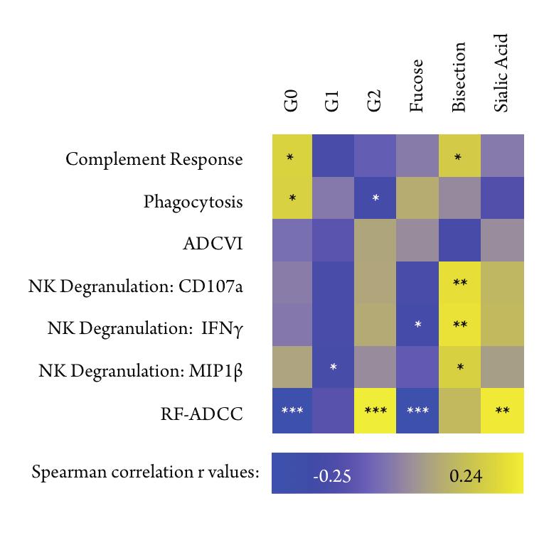
101
102 previously unknown effects of this modification on antibody function. Overall, the significant associations between glycan modifications and antibody functionality suggest that the effect of glycosylation is not limited to monoclonal therapeutics, but may also be relevant in polyclonal populations. While the results of these assays are driven by the activity of antigen-specific, in particular antigp120, antibodies, we have observed that the functionality of the tiny fraction of antigen-specific IgGs can be partially predicted by the glycosylation patterns of the bulk Fc. Of note, we did not observe strong correlations between any glycan structures and ADCVI, but because this assay measures a complicated interaction of a variety of cellular and viral factors, so may be that glycosylation alone cannot predict viral inhibition in this assay very accurately Antigen-specific antibodies have distinct glycosylation profiles Given that the functional assays we performed are driven by antigen-specific antibodies, particularly against gp120, we sought to characterize the glycosylation profiles of these HIV-specific antibodies. In comparison of the anti-hiv response, we also analyzed the glycosylation of antibodies directed against influenza virus in the same cohort of HIV-positive donors. To do this, we isolated gp120-, p24-, and influenza HA-specific antibodies and analyzed the attached glycans by capillary electrophoresis. Since the volume of isolated antigen-specific antibodies is small, we analyzed the glycan of the intact IgG, which includes Fab domain-attached glycans in addition to the functionally relevant Fc glycosylation, so we compared antigen- 89
103 % of IgG Fc Glycan Galactosylation **** * **** **** **** **** **** Bulk gp120-specific p24-specific HA-specific G2 G1 G Sialylation **** **** **** **** Bulk gp120-specific p24-specific HA-specific Fucosylation **** **** **** Bulk gp120-specific p24-specific HA-specific Bisection **** * *** **** Bulk gp120-specific p24-specific HA-specific Figure 3.6: Glycosylation is regulated on an antigen specific level. Antigen-specific antibodies against gp120 (n = 103), p24 (n = 47), and influenza HA (n = 40) were isolated from a cohort of HIV-positive individuals and the attached glycans of these antibodies were compared to bulk IgG glycosylation (n =193). Mean galactosylation is expressed as stacked bars, mean proportions of sialylation, fucosylation, and bisection (with GlcNAc) are represented as mean with standard deviation. Statistical significance between means was determined using Kruskal-Wallis test with Dunn s multiple comparison test (* = p < 0.05, *** = p <0.001, **** = p < ). 90
104 specific glycans to those of intact bulk IgGs from the same HIV-positive donors. In comparing the glycosylation profile for each antigen-specific antibody, we observed that each group of specific antibodies has a significantly different glycosylation profile (Figure 3.6). Specifically, HIV-specific antibodies against gp120 and p24 have significantly more inflamed structures with higher proportions of agalactosylated and lower proportions of sialylated glycans. The HIV-specific antibody glycans also have higher fucosylation compared to bulk antibodies, suggestive of lower ADCC activity. Interestingly, bisection is higher in gp120-specific antibodies, even in comparison to p24 antibodies, which, based on the role of b-glcnac in ADCC [55], is suggestive of increased effector function. The antigen-specific, but non-hiv, anti-ha antibodies have a profile more like that of bulk IgG glycans in every category, except in b-glcnac, for which the HA-specific antibodies have a lower proportion, suggestive of less ADCC activity. Interestingly, the intact HIV-specific antibodies have an Fc-like glycosylation profile, with less sialylation and more fucose, while the bulk and flu-specific antibodies have profiles that suggest the presence of Fab glycosylation [84, 136]. This effect may be due to a decreased tolerance for Fab glycosylation in antibodies directed against HIV antigen or a difference in the regulation of glycosylation in HIVspecific B cells. Ultimately, these data strongly suggest that glycosylation is regulated at the level of the antigen-producing B cell, not only in response to systemic inflammatory signals. Given our analysis of the glycosylation of gp120 specific antibodies, we can more accurately link the glycan profile with the measured gp120-specific antibody 91
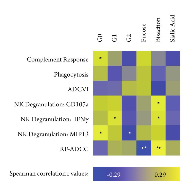
105
106 function. We compared the same functional activity data used in Figure 3.5, to the glycosylation profiles of gp120-specific IgGs and observed many of the same relationships (Figure 3.7), including associations between complement and G0, ADCC and low fucose, and NK activation and bisection. While there is less power in the antigen-specific analysis (n=103 versus 200), we observed most of the same associations. Given these similarities, it is unsurprising that the bulk Fc glycan profile can predict the functionality of the relatively small proportion of gp120- specific antibodies. 3.4 Discussion This study is the first to characterize associations between particular cytokines and changes in the proportion of specific bulk Fc glycans in HIV-infected individuals and to show that bulk Fc-glycosylation is a relevant measure of overall effector functionality in HIV infection. By investigating the effect of HIV infection on bulk glycosylation, we observed that the Fc glycans of IgGs from HIV-positive individuals are significantly altered, during both acute and chronic infection, as summarized in Figure 3.8. While uninfected patients have mostly G1 glycans, acute HIV infection is associated with a significant rise in G2 and sialylated glycans, which swings dramatically to a high proportion of G0, asialylated structures upon transition to chronic infection. This finding suggests that during the acute phase, patients can overcome the inflammatory signals induced by the virus through the generation of compensatory anti- 93
107
108 inflammatory structures. Ultimately, however, the immune system may be overwhelmed by HIV-induced immune dysregulation, resulting in an increase in production of inflamed G0 structures as the generation of anti-inflammatory sialylated structures decreases. Another change in glycosylation during the transition from acute to chronic HIV infection occurs in the proportion of b-glcnac, which is highest during acute infection, but decreases during chronic infection, though not to the level observed in uninfected individuals. The kinetics of this change suggest that the signals that drive production of bisected Fc glycans are present early in infection, but wane as individuals progress to chronic infection. Given that HIV infection induces changes in bulk glycosylation, we sought to identify some of the signals associated with these changes. We observed that IP10 and CRP are both associated with higher proportions of inflammatory glycans, suggesting that systemic inflammation is a major determinant of the IgG glycosylation profile. A link between inflammatory cytokines and agalactosylated IgGs has been observed in autoimmune disease, including an association with both IL6 and IL2 concentration [29], however, these relationships were not observed in our cohort, suggesting that regulation of glycosylation during autoimmune disease and infectious disease may be different. Further characterization of the factors driving glycosylation change in different infections will further elucidate the types of signals important for this process. Furthermore, it is apparent that the glycosylation of gp120- and p24- specific antibodies are more similar to each other than that of antibodies against influenza. This finding shows that despite the inflammatory signals associated with changes 95
109 in the bulk Fc glycosylation profile, within an individual, glycosylation is regulated at the level of the antigen-producing cell. Importantly, this suggests that glycosylation is being regulated within individual B cells and that signals specific to the antigen or the environment at the time of exposure to antigen can influence the ultimate glycosylation pattern on the produced IgG. Importantly, we observed that the modification of glycosylation in a polyclonal population is relevant for determining the functionality of antibodies from HIVpositive donors. We found links between glycan structures and Fc-mediated antibody functions including complement deposition, NK activation, phagocytosis and ADCC. This shows for the first time that glycosylation of polyclonal IgGs is relevant for determining effector function in vitro. In investigating the effect of HIV infection on bulk Fc glycosylation, this study identified a set of cytokine signals that are associated with glycan changes in HIV infection and we observed a link between the Fc glycosylation of HIV-specific IgGs and their functional activity. Most interestingly, we observed that glycosylation is also regulated at the level of the antigen-specific B cell. These results point to the importance of doing further work to determine which specific signals and stimuli drive the regulation of IgG glycosylation, which will ultimately be a potent tool for programming antibody effector function and inflammatory profile through vaccination. 96
110 Based on a manuscript to be submitted. Authors: Alison E Mahan, Kendall Dionne, Jacquelynne Tedesco, Amy Chung, Punnee Pitisuttithum, Donald Francis, Lindsey Baden, Dan Barouch, Galit Alter. 4 Antigen-specific IgG glycosylation is directed by HIV Vaccination In human studies of HIV and non-human primate models of SIV and SHIV infection, non-neutralizing antibodies are effective at preventing or controlling viral infection. Thus, determining how non-neutralizing antibodies are induced will provide insights into the design of a potentially protective vaccine strategy. Nonneutralizing antibody functions, including antibody-dependent cellular cytotoxi-
111 city, complement-dependent cytotoxicity and antibody-dependent phagocytosis, are modulated by the immune response in two major ways: through changes in antibody subclass and alterations of the N-linked glycan of the Fc domain. However, little is known about how glycosylation is tuned and programmed in vivo. To better understand IgG glycosylation in HIV vaccine recipients, we used capillary electrophoresis to characterize bulk and vaccine-specific glycosylation in HIV vaccine recipients. Using samples collected from a vaccine trial using an adenoviral vector (IPCAVD-004) administered in three geographical areas, we observed significant difference in bulk IgG-glycan patterns, associated with geographical location. However, vaccine-specific antibodies have glycosylation patterns distinct from that of bulk Fc, suggesting that vaccine specific signals can overcome systemic differences in bulk antibody glycosylation. In contrast, comparison of two different vaccines, (IPCAVD-004 and VAX003, a recombinant protein/alum vaccine) induced significantly different glycosylation patterns on vaccine-specific antibodies, suggesting that different signals during vaccination can tune the elicited IgG-glycan. These results show for the first time that vaccines elicit specific and distinct glycosylation patterns on antigen-specific IgGs that are distinct from bulk glycosylation patterns. Given the importance of IgG-glycosylation in mediating effector functions, which are important in non-neutralizing protection from HIV infection, this study highlights the importance of including antigen-specific glycan profiling in future immune monitoring and correlates analyses of vaccine trials. 98
112 4.1 Background To date, the only known correlate of decreased risk of infection in an HIV vaccine trial is strong non-iga titers against the V1 and V2 loops of IgG elicited in the Thai RV144 trial [80, 182]. Secondary correlates analysis revealed an additional association between protection and strong antibody-dependent cellular cytotoxicity (ADCC) activity [30, 80, 182]. However, the RV144 trial only temporarily elicited these antibodies, with protection waning one year after the final vaccine boost [103, 109, 186]. Thus, it remains unknown how effective and sustained effector antibodies can be elicited against HIV through vaccination. Understanding all the correlates of protection for an effective and sustained vaccine-elicited response is an important goal for moving the field forward. However, it is still unknown what factors may be important for or correlated with a long-lasting, protective response against HIV. Thus, characterizing a range of biophysical and functional parameters of the vaccine-elicited immune response, including factors that have not yet been associated with protection, is of vital importance. These parameters should include factors associated with non-neutralizing functional activity, such as antibody subclass, titer, breadth of binding, and N-glycosylation. While most non-hiv vaccines elicit protective, neutralizing antibodies, an HIV vaccine has yet to successfully elicit strong neutralizing responses. Thus, the induction of non-neutralizing antibodies with potent effector functionality represents a potentially powerful approach for creating a successful vaccine [212, 237]. It is well established that antibodies with potent effector functionality, especially 99
113 ADCC, and possibly antibody-dependent cellular phagocytosis (ADCP) and complement-dependent cytotoxicity (CDC), can inhibit viral replication in vitro [224], is associated with slower progression of AIDS-related disease [66, 115], and is protective in animal studies of passive antibody transfer and vaccination [21, 75, 143]. Thus, these types of antibody responses, if long-lived, could be the key to an effective vaccine strategy. Effector functionality in antibodies is regulated strongly by IgG subclass selection, which is known to affect IgG affinity for Fc receptors [92, 156, 160]. Interestingly, the RV144 vaccine elicited higher proportions of the IgG3 subclass than VAX003 [45], resulting in more polyfunctional antibodies in RV144. However, IgG3 antibodies usually wane quickly after an initial spike in production, which may explain the lack of sustained protection in RV144. Beyond subclass, antibody glycosylation is a rapid and potent regulator of inflammatory profile and effector function. The IgG glycan, positioned in the CH2 domain of the constant region (Fc), is an asparagine-linked biantennary structure which varies based on the presence or absence of four sugar subunits: galactose, sialic acid, fucose and an N- Acetyl-glucosamine (GlcNAc) that bisects the arms of the structure. Galactosylation allows for the attachment of terminal sialic acids, both of which are potent regulators of the inflammatory profile of the IgG. Agalactosylated IgG structures are associated with active inflammatory disease, including rheumatoid arthritis [166, 227] and chronic HIV infection [3], while sialic is actively anti-inflammatory, in particular during therapeutic administration of pooled healthy immunoglobulin to treat autoimmune disease [15, 27, 49, 244]. In addition to the modulation of 100
114 antibody inflammatory profiles, changes to the fucose and bisecting-glcnac (b- GlcNAc) content can potently affect effector function. A lack of fucose increases ADCC activity of monoclonal antibodies by fold [201, 203], while the addition of b-glcnac can increase ADCC activity [55]. Lack of sialic acid may play a role in increasing ADCC activity [193] while conflicting data exist on the effect of galactose on ADCC [32, 113]. Recruitment of complement is aided by agalactosylated antibodies [127] and additional relationships between effector function and glycosylation likely exist, but remain uncharacterized. Altogether, the presence or absence of these four sugar subunits allows for a theoretical potential of 36 different glycan structures, each with the capacity to differentially drive effector functionality and inflammation. This level of specificity makes Fc glycosylation a potentially potent mechanism for harnessing specific effects of the humoral immune response. Given that glycosylation is a powerful regulator of non-neutralizing antibody functions and is modified during autoimmune disease and natural infection, this study sought to determine whether glycosylation can be specifically altered through vaccination. To do this, we characterized both bulk Fc and vaccine-induced antibody glycosylation elicited in a vaccine trial that used the same adjuvant and immunogen in three geographically distinct populations. Additionally, we investigated glycosylation in a trial that used an alternative adjuvant and immunogen combination. We observed that while bulk Fc glycosylation is highly dependent on geography, the vaccine-elicited antibody glycosylation is not affected by this, but is driven by particular signals within the vaccine preparation. 101
115 4.2 Methods Vaccine Cohorts The IPCAVD-004 vaccine trial (clinicaltrials.gov ID: NCT ) was a safety and immunogenicity trial of a combination of adenovirus vectors expressing gp120 ENVA. Both Ad26 and Ad35 vectors were used in combination or alone in three test sites: the United States, East Africa, and South Africa. Four arms were tested in each location with three or six months between prime and boost using either Ad26 followed by Ad35, vice versa, or two doses of the same vector. The trial recruited low-risk, HIV-negative adults to receive 5x10¹⁰ viral particles per dose intramuscularly. Individuals in the placebo group received a dose of 10 mm Tris/HCl buffer. During the course of our study, we were blinded and do not know which experimental group each sample belongs to, only which geographical region they were collected in. Samples used in this study were collected at peak immunogenicity, two weeks after final immunization. The VAX003 trial was a phase 3 efficacy trial administered in Thailand, but in a high-risk population of intravenous drug users (IVDUs) (clinicaltrials.gov ID NCT ). This trial used seven doses of the same AIDSVAX B/E, a recombinant gp120 clade B/E with alum as the adjuvant. All samples used in this study were collected at peak immunogenicity, two weeks after the final vaccination. 102
116 Fc glycan preparation Plasma was collected by vaccine trial staff and stored at -80 C until use. IgG was isolated using Melon Gel IgG purification resin (Thermo Fisher) according to the manufacturer s instructions. Fc and Fab fragments were separated through enzymatic digestion with IdeS (Genovis) and Fc purified using protein G magnetic beads (Millipore). N-glycan was released from Fc fragments with on-bead treatment with PNGaseF (NEB). Released glycans were purified by precipitating deglycosylated protein with ice-cold ethanol and dried in a centrifugal evaporator. Dried glycans were fluorescently labeled with a 1:1 ratio of 25 mm 8-aminopyrene- 1,3,6-trisulfonic acid, APTS (Life Technologies), in 1.2 M citric acid and 1 M sodium-cyanoborohydride in THF (Sigma Aldrich) at 55 C for 3 hours. Unreacted dye was removed using freshly made 250 μl P2-resin (Bio-Rad) size exclusion columns. Antigen specific antibody purification Vaccine-specific gp120 antigens (EnvA for IPCAVD and a 1:1 mix of A244 and MN for RV144 and VAX003, ImmuneTech) were biotinylated on primary amines using a long chain biotin reagent (Thermo) and free biotin was removed using dialysis. Biotinylated antigen was immobilized on micro-volume streptavidin resin columns (Agilent) and purified IgG, concentrated to 3-5 mg/ml was run over the columns using centrifugation. After washes with PBS, antigen-bound antibodies were eluted with 0.1 M citrate buffer, ph 2.9 and IgGs were immediately treated with PNGaseF to release attached N-glycans for analysis. 103
117 Capillary electrophoresis Cleaned, labeled glycans were diluted 1:10 in ultrapure water and loaded onto a 3130XL ABI DNA sequencer (Life Technologies). Glycans were run through 36cm capillaries of POP7 acrylamide matrix using parameters described[116]. Glycan peaks were compared to those of known glycan standards (Prozyme) and peak area was calculated for each structure using a custom designed MATLAB script. The relative proportion of 21 individual structures was calculated for each sample, and for simple, straightforward presentation of data, glycan structures were summed into categories based on galactose content (G0, G1, G2), sialylation, fucosylation and the presence of bisecting-glcnac. Statistics Univariate data were analyzed in GraphPad Prism Version 6.0e for Mac (Graph- Pad Software, San Diego, California) for statistical significance and graphical representation. Statistical tests used are indicated for each figure. Multivariate analyses were performed using MATLAB and Statistics Toolbox Release 2013b (the MathWorks Inc., Natick, Massachusetts). Multivariate analysis Using the proportions of all the individual glycan structures, of which 21 were identified by our capillary electrophoresis technique, we can use multivariate analysis to analyze the power of the observed variables to separate the samples collected at each test site in an unbiased manner. Using principal component analy- 104
118 sis (PCA), a descriptive function is generated for each experimentally measured variable using weighted values of all the other input variables. These functions are ranked by their ability to describe the variability of the whole data set and we can visualize the separation of the data sets by plotting the results of the best function (x-axis) against the second best function (y-axis). This scores plot represents the output of the two best descriptive functions for each sample analyzed. PCA is unbiased and attempts to group the data based solely on the variation within the complete data set, so after the analysis is complete, we can color the sample dots according to which group they belong to and assess the separation of each group of samples. Finally, to assess which variables are most important in separating our data set, we can plot the weights of each variable in the respective analysis in a loadings plot. The loadings plot contains all the variables measured and the longer their vector, the more power they contributed to the descriptive function. 4.3 Results Bulk IgG Fc-glycosylation is highly variable in different regions To determine whether geography impacts bulk IgG glycosylation, we analyzed samples from an HIV vaccine trial in which the same adjuvant and immunogen was administered in three distinct geographical regions. IPCAVD-004 used a combination of Ad26 and Ad35 vectors expressing EnvA, and was administered to healthy adults in the United States, Kenya/Rwanda (East Africa), and South Africa. Com- 105
119 paring the vaccine recipients in these three trials, we observed that there are significant differences in the bulk IgG Fc-galactosylation and sialylation in the enrolled individuals, which is independent of the vaccine-specific response since they all received the same vaccine (Figure 4.1A). In particular, individuals from both African regions have significantly higher proportions of agalactosylated (G0) Fcs. While both African groups have lower sialylation than US vaccinees, East Africans have the lowest bulk antibody sialylation. Given the role of low galactose and sialylation in determining the inflammatory activity of antibodies [126], these data suggest that at baseline, bulk antibody glycosylation in Africa is associated with inflammatory glycosylation, with East Africans having the most inflammatory profile. In addition to changes in galactose and sialic acid, fucose and bisecting-glcnac alter antibody function [55, 201]. Interestingly, in our South African cohort, we observed higher fucosylation of bulk Fc compared to either group (Figure 4.1A). Additionally, South Africans have lower bisecting-glcnac compared to both groups. A high fucose, low b-glcnac glycan is predicted to have low functional activity. These results highlight that even within a single continent, significant differences may arise in antibody glycosylation of bulk antibodies. To gain a more complete understanding of the differences in bulk IgG glycosylation, we next used principle component analysis to compare the antibody glycan profiles among our groups, including all 21 glycan structures that we detect by capillary electrophoresis. In multivariate space, the three arms of the vaccine trial are very well resolved (Figure 4.1B), suggesting that fundamentally different glycosylation profiles exist within each region. This separation is largely driven 106
120 A % of Fc Glycan B 2nd Principal Component Galactosylation **** **** **** **** *** **** USA Kenya/Rwanda South Africa G2 G1 G Sialylation ** **** **** United States Kenya/Rwanda South Africa st Principal Component Component Fucosylation 100 * ** United States Kenya/Rwanda South Africa 0.1 G1F G1F 0 Total G0 Total G1 0.1 G0F G2F G0B G1S1F Bisection * **** United States Kenya/Rwanda South Africa G2S1F Total G2B Monosialylated Total Fucosylation G1S1 G2S1B G1B G0FB G1 Total Sialylated Total G2 G2FB G2S2 G1FB Total Bisection G2S2B G2S2F G2S1 G2S2FB Total Disialylated Component 1
121 inflammatory glycan structures, as shown by the length of the vectors on the loadings plot, which illustrates the power of each variable to contribute to resolving the separation among the cohorts. Interestingly, the East African vaccinees separate completely from the other groups, while the South African and US recipients overlap slightly in their bulk Fc glycan profiles, suggesting that while variations in bulk Fc glycosylation exist among all groups, there are greater differences between the two African cohorts Vaccination elicits glycosylation patterns independent of bulk glycosylation Given the significant differences in Fc glycosylation patterns based on geographical location, we sought to determine whether vaccine-elicited antibody glycosylation is similar or distinct from the bulk Fc glycosylation profile. To investigate this, vaccine-elicited antibodies were purified from bulk IgG using vaccine-matched gp120 antigens in a subset of vaccinees from each region. These antibodies were then analyzed using capillary electrophoresis to characterize the attached glycans. Strikingly, the vaccine-elicited antibodies are significantly different from the bulk glycans (Figure 4.2A). In multivariate PCA, there is no overlap of the antibody glycosylation profiles of antigen-specific (maroon) and bulk antibodies (blue) (Figure 4.2A, right). As illustrated in the loadings plot (Figure 4.2A, right), this separation is driven primarily by differences in fucosylation and sialylation. This analysis shows that vaccine-specific antibody glycosylation is fundamentally different than bulk antibody glycosylation, suggesting that vaccine signals actively tune distinct 108
122 A B 2nd Prinicpal Component st Principal Component Galactosylation 100 Component 2 Sialylation Total Fucosylation G2S1F G2F Total Monosialylated G1F G1F G2S2F Total G2 Total G1 Total Bisection G0FB G0F Total G0 G1S1 G2S2B Total Sialylation G2S2 Total Disialylated Fucosylation Component 1 40 Bisection % of gp120-specific IgG glycans 50 G2 G1 G USA Kenya/Rwanda South Africa 0 United States Kenya/Rwanda South Africa 0 United States Kenya/Rwanda South Africa 0 United States Kenya/Rwanda South Africa
123 changes in antigen-specific glycosylation Vaccine-induced antibody glycosylation is independent of regional bulk glycosylation differences Because the vaccine induced glycan profile is distinct from bulk IgG glycosylation, we next aimed to determine whether regional differences in bulk Fc glycosylation influence the vaccine-elicited glycan profiles by comparing the glycan profile of gp120-specific antibodies across the three geographical regions. Interestingly, antigen-specific antibody glycosylation in vaccinated individuals does not differ among the geographical locations tested (Figure 4.2B). While there is a trend toward a smaller proportion of bisecting-glcnac in the Kenyan/Rwandan population, this is the opposite of the profile observed in the bulk Fc glycosylation (Figure 4.1A), demonstrating a disconnect between vaccine-specific and bulk antibody glycosylation. These data strongly suggest that vaccine-induced glycosylation changes are not directly influenced by the bulk Fc-glycosylation differences we observed among geographical regions Vaccines elicit distinct glycosylation profiles Given that IPCAVD-004 is an adenovirus-vectored vaccine and this vaccine regimen elicited similar glycosylation in all recipients, regardless of geographical region, we sought to determine whether different vaccine preparations induce distinct glycosylation patterns. To investigate this, we compared the antigen-specific glycosylation of antibodies elicited by the IPCAVD vaccine compared to the gly- 110
124 can profiles of gp120-specific antibodies in recipients of VAX003 an alum adjuvanted recombinant gp120 vaccine administered in Thailand. Interestingly, we observed that the IPCAVD vaccine-induced more anti-inflammatory glycan structures, with higher proportions of G2 and sialic acid compared to VAX003 (Figure 4.3). IPCAVD also induced an increased proportion of bisected structures, suggesting that these antibodies may possess increased functionality, based on the role of bisecting-glcnac in increasing ADCC activity [55]. Taken together, these data demonstrate that distinct vaccines can specifically tune the glycosylation of antigen-specific antibodies, independent of bulk Fc glycosylation. Given the importance of glycosylation in modulating antibody effector functions, and our data demonstrating the capacity for vaccines to induce specific glycosylation profiles, this study points to the need to include antigen-specific glycosylation profiling as a critical qualitative measure of non-neutralizing vaccine-induced humoral immunity. 4.4 Discussion This study is the first to analyze antibody glycosylation in the context of vaccination against HIV with the goal of understanding vaccine-elicited antibody glycosylation. In this study, we show that while geography has a significant impact on bulk Fc glycosylation patterns, a particular vaccine can overcome systemic differences to elicit similar glycan patterns on vaccine-induced antibodies independent of bulk Fc glycosylation. In contrast, we observed that distinct vaccines elicit different antigen-specific antibody glycosylation profiles. 111
125 Galactosylation 100 Sialylation 100 ** Fucosylation 80 Bisection 40 **** % of gp120-specific IgG glycans 50 0 IPCAVD *** ** * VAX003 G2 G1 G IPCAVD VAX IPCAVD VAX IPCAVD VAX003
126 The observed population level differences in bulk Fc glycosylation are possibly due to factors such as endemic infections, especially given the effect of some natural infections on bulk glycosylation [3, 7]. The impact of nutrition may also be important in determining bulk Fc glycosylation based on established associations between body mass index and production of inflammatory IgG glycosylation [110, 155]. The role of genetics in determining Fc glycosylation in this study cannot be excluded, though other studies of genetically distinct populations have not observed such strong differences among groups [172]. Additionally, the finding that vaccines elicit the same glycosylation patterns in people with different bulk glycosylation patterns suggests that genetic differences are not a major factor. While we did not analyze the glycan profile of the different IgG subclasses, another important factor in producing different glycosylation patterns may be the baseline proportions of particular IgG subclasses within each group as each IgG may have different glycosylation patterns [106, 239]. Despite the differences in Fc glycosylation driven by geographical region, we observed no difference in the glycan patterns of antigen-specific IgGs elicited by a given vaccine preparation. The ability of an adenovirus vectored vaccine to elicit the same glycosylation pattern in antigen-specific antibodies, independent of baseline bulk Fc glycosylation, is an encouraging finding that suggests that vaccinespecific signaling can tune the antibody glycan irrespective of existing humoral responses. This will be very important for HIV vaccination since vaccine recipients are likely to be highly heterogeneous in their pre-exposure to factors that alter their existing IgG glycosylation profiles. 113
127 Importantly, while the adenoviral vector elicited a specific glycosylation profile on antigen-specific antibodies, IgGs elicited by alum adjuvanted recombinant protein vaccine VAX003, were significantly different. The differences illustrate that vaccine-specific signals, either through adjuvant, immunogen, or a combination of the two, can actively program the glycosylation of IgG. Interestingly, given the effect of b-glcnac on effector function [55], IPCAVD may have produced highly effective antibodies with a high proportion of bisecting-glcnac containing glycans. Additionally, in comparison to VAX003, the IPCAVD vaccine-induced less inflammatory glycan structures, with higher proportions of sialylated glycans and fewer agalactosylated glycans, which may be similarly advantageous in producing an effective vaccine response without inducing an overly inflammatory response. However, since the IPCAVD-004 study was a safety and immunogenicity trial, not an efficacy trial, protection data is unavailable, so the relationship between vaccine-specific antibody glycosylation and protection cannot be established at this time. This study is the first to show that bulk Fc glycosylation does not directly determine vaccine-induced antibody glycosylation, rather vaccine-specific signals drive the generation of antigen-specific glycan profiles. Ultimately, these data suggest that a combination of specific adjuvants and/or immunogens can elicit a specific glycan pattern with a protective balance of inflammatory and functional characteristics. 114
128 4.5 Additional data RV144 was a phase 2B vaccine trial administered in Thailand to low-risk, HIVnegative adults (clinicaltrials.gov ID NCT ). The trial consisted of six immunizations with a canary pox viral vector, ALVAC HIV (vcp1521), which contains HIV env, gag, and pol genes, and two immunizations of alum-adjuvanted recombinant gp120 (AIDSVAX B/E), administered concurrent with the last two ALVAC doses. The trial lasted 24 months and all samples used in this study were collected at peak immunogenicity at two weeks after final vaccination HIV risk factor is a potential driver of IgG Fc-glycosylation Two major vaccine trials in Thailand recruited volunteers from groups with different risk factors. RV144 was administered in year-old, TB-negative men and women with low risk of HIV acquisition and VAX003 recruited intravenous drug users (IVDUs) years-old. Given that geographical factors can influence bulk Fc glycosylation, we hypothesized that IV drug use may also influence IgG glycosylation. In these trials, we benefited from having a number of samples from placebo recipients, so we could compare bulk glycosylation with and without vaccine administration. First, to determine whether either vaccine preparation altered bulk Fc-glycosylation, vaccine (solid bars Figure 4.4A) and placebo (hatched bars) recipient IgG glycans were compared, and we found that no significant differences exist between the groups in either trial, showing that neither vaccine preparation was able to sig- 115
129 2nd Principal Component A % of Fc glycan B Galactosylation RV144 **** **** VAX003 G2 G1 G st Principal Component Sialylation *** RV144 VAX003 Component G0FB Total Fucosylation G1F Total G1 G2FB G1FG1FB G2B G0FG1B Total G0 Fucosylation RV144 VAX003 G0B G2S1B G1S1 G2F G2S1F Total Monosialylated G1S1F G2S2F G2S1 G2S2 Total Sialylated G2S2FB G2S2B Total Bisection Component 1 RV144 Bisection * VAX003 Total G2 Total Disialylated
130 nificantly alter bulk Fc glycosylation. When comparing the two vaccine recipient groups, however, we observed that IVDUs enrolled in the VAX003 (maroon, Figure 4.4A) trial, have significantly more inflamed glycan structures than the low-risk RV144 trial participants (blue, Figure 4.4A), as indicated by higher proportions of agalactosylated and lower proportions of sialylated Fc glycans. Additionally, VAX003 recipients have a slightly increased proportion of bisected glycan structures (Figure 4.4A), indicative of a more functional antibody glycan profiles. Interestingly, when we analyzed all the individual glycan structures using multivariate analysis by PCA, the populations do not separate as well as in the geographically distinct populations (Figure 4.4B). This suggests that risk factor, in particular IV drug use, is not as influential on Fc glycosylation as other, as yet unidentified, environmental factors. While the glycan structures associated with inflammation, galactose and sialic acid, were again on the periphery of the loadings plots, these variables did not resolve the two populations very well. Finally, given that RV144 and VAX003 recipients have such different bulk Fc glycosylation profiles, we sought to determine whether the vaccine-elicited antibody glycosylation was also different. The comparison of these two trials is especially important since RV144 proved to be a mildly protective vaccine, while VAX003 was not. In comparing RV144 to VAX003, however, it is important to note that while these trials both used AIDSVAX B/E, an alum adjuvanted recombinant gp120, the RV144 trial also contained six doses of ALVAC, a canary pox vector expressing HIV proteins. Despite these relatively different preparations, 117
131 RV144 and VAX003 trials are often compared because they are similar both in their use of AIDSVAX B/E and in geographical location. Since RV144 was protective, we expected to observe some differences between the glycosylation of vaccineelicited antibodies in each trial, however we observed no differences in any category of glycan structure (Figure 4.5). Additional Discussion In further investigation of IgG glycosylation after HIV vaccination, we compared two similar, but distinct trials. We saw that VAX003 placebo recipients and vaccinees have significantly more inflamed glycan structures in their bulk Fcs than RV144 participants. This effect is likely driven by the inflammatory effect of injected drugs, which are associated with general inflammation and infections that are not commonly observed in healthy populations [36, 199, 221], though this relationship has not been directly established. Additionally, we observed an increased proportion of bisecting-glcnac in the VAX003 cohort, suggesting that the effects of general inflammation or infection may influence b-glcnac addition along with galactose and sialic acids, however little is know about natural regulation of IgG glycan bisection. While VAX003 and RV144 were different vaccine preparations, they are often compared to each other to attempt to identify potential correlates of protection in RV144. Surprisingly, we observed that the use of ALVAC in the RV144 vaccine strategy did not elicit differences in vaccine-specific antibody glycans, when compared to the AIDSVAX only preparation. The similarity between the glycans 118
132 Galactosylation 100 Sialylation 100 Fucosylation Bisection % of gp120-specific IgG glycans 50 G2 G1 G RV144 VAX RV144 VAX003 0 RV144 VAX003 0 RV144 VAX003
133 of these two trials suggests that the role of the AIDSVAX portion of the vaccine drove the selection of IgG glycosylation patterns more than the canary pox vector. Since the combination of AIDSVAX and ALVAC elicited the same glycan profile as AIDSVAX alone, it is apparent that glycosylation was not a dominant factor in the efficacy of RV144. However, this does not discount the possibility that using a specific combination of adjuvants and immunogens will drive the production of antigen-specific antibodies with optimal glycosylation patterns for protection from infection. 120
134 Technical assistance provided by Justin Eusebio, Kendall Dionne, and Jacquelynne Tedesco. 5 Glycosyltransferase expression is altered by innate and adaptive signals IgG glycosylation is modified during viral infection, autoimmune disease, and in response to vaccination, which indicates that immune signals can regulate antibody glycan structure. However, it is still unknown which specific signals are important for eliciting particular structures and whether glycosylation changes are transient or programmed within antibody-producing cells. Identifying relevant
135 glycosylation regulatory signals is an important step in understanding how particular IgG glycan structures can be elicited through vaccination, so, to begin to understand this, we investigated the effect of three types of stimuli on the transcriptional profile of glycosylation machinery in human B cells: a) innate, including toll-like receptor (TLR) signaling, and adaptive including b) B cell receptor (BCR) stimulation and c) T cell help. Ultimately, these results are important for understanding how changes to the IgG glycan are influenced by specific immune signals, which will allow for the elicitation of specific and potent antibody effector functions for long-lasting, protective humoral immune responses. 5.1 Background Modulation of antibody effector function is important for effective monoclonal antibody therapeutics against specific types of cancer [130, 213] and protection from natural infections, including HIV [61]. In the case of therapeutic antibodies, extensive efforts to characterize and tailor the effector function of antibodies produced in vitro have focused on modulating functionality through alteration of the glycan structure of the IgG Fc [94]. For instance, the interaction of IgG1 with FcγRIIIA is 50 fold better when the IgG glycan does not contain a core fucose [201]. Additional alterations of the glycan structure of in vitro-produced antibodies include changes to increase the proportion of bisecting (GlcNac) structures, which can further increase binding to the ADCC activating receptor, FcγRIIIA [55]. IgG sialylation is inversely correlated with induction of inflammatory signals [16, 27], while low galactose content is associated with inflammation in au- 122
136 toimmune and infectious disease [145, 165]. While the effect of IgG glycosylation is well-characterized, the specific mechanisms of regulation of these glycosylation changes in vivo it is still poorly understood. Alteration of IgG glycosylation occurs during the course of immunological and inflammatory diseases, in particular in cancer [41, 101, 111], autoimmune disease [219, 226], and specific infections, [145, 188]. These examples highlight that glycosylation is a naturally regulated part of the immune response and that it is likely controlled by immunological signals. Our data, presented in Chapter 3, show that inflammatory cytokines are associated with changes in IgG glycosylation during HIV infection, while signals delivered during vaccination can specifically tune the antigen-specific antibody glycosylation, as shown in Chapter 4. Attachment of asparagine-linked N-glycans to IgGs occurs in the ER and early Golgi compartments, where highly specific glycosylation enzymes sequentially add sugar subunits to the glycan as the protein transits through the Golgi apparatus [231]. Regulation of these enzymes, either transcriptionally or post-translationaly, is relevant to changes in glycan structure [149], however the mechanisms and signals responsible for its regulation are not well understood, especially in the context of immune activation. Thus, we sought to understand how glycosylation machinery in B cells is regulated by identifying specific stimuli that alter the transcription of glycosyltransferases (GTs). Pattern recognition receptors, in particular toll-like receptors (TLRs), are a class of innate immune receptors that can detect commonly produced viral or bacterial molecules [112]. Their primary function is to create a rapid, non-specific response 123
137 while the adaptive response is initiated to create a highly specific response [222]. Their role in adaptive immune cells, however, may be a more complex one allowing cells of the innate or adaptive response to gain information about the pathogen in question, including whether it is bacterial or viral, intra- or extracellular [167]. Thus, we hypothesized that the activation of TLRs, many of which are expressed in B cells of humans [53], may play a role in tuning the antibody response by programming IgG glycosylation changes, and subsequently altering the effector function of antibodies. This study was designed to identify specific immune signals, in particular agonists of TLRs, that alter the glycosylation profiles of IgGs using a transcriptional profiling approach. Ultimately, given the importance of IgG glycosylation in determining antibody function, understanding which signals regulate and program IgG glycosylation will be crucial for developing vaccine strategies that elicit potent, long-lasting, protective antibody responses. 5.2 Methods Isolation of B cells. Leukaphoeresis of HIV negative donors were obtained from the MGH blood bank. B cells were purified to >99% CD19+ by using B cell Rosette Sep reagent (Stem Cell, Canada) followed by a magnetic bead-based B cell Negative-Selection Easy Sep (Stem Cell, Canada), according to the manufacturer s directions. Cells were cultured in RPMI supplemented with 10% fetal bovine serum at 1-2x10⁶ cells/ml. 124
138 Stimulation of ex vivo cells. Toll like receptor (TLR) agonists were diluted in endotoxin free water and added to B cell cultures at indicated final concentrations: TLR 1/2-Pam3CSK4 (200 ng/ml), TLR 3- Poly(I:C) (10 μg/ml), TLR 4- Ultra pure LPS (1 μg/ml), TLR 5- Ultrapure Flagellin (100 ng/ml), TLR 2/6- Pam2CSK4 (100 ng/ml), TLR 7- Imiquimod (5 μg/ml), TLR 7/8- CL097 (5 μg/ml), and TLR 9- CpG ODN2006 (5 μg/ml) all from Invivogen (San Diego, CA). Anti-BCR stimulations were performed using donkey anti-human IgG F(ab) 2 ( Jackson Immuno Research) at 10 μg/ml. Soluble Mega-CD40L (Enzo Life Sciences) was used at 100 ng/ml. After 16 hours of stimulation, media was removed and cells were lysed in RLT buffer. Transcriptional Profiling of Glycosylation Enzymes RNA was extracted using an RNeasy kit (Qiagen), cdna was synthesized using MuLV reverse transcriptase (NEB) and used in TaqMan FAM hydrolysis probebased qpcr (Life Technologies). Specific glycosyltranferase genes measured included B4GALT1, ST6GAL1, FUT8 and MGAT3; GAPDH was used as a housekeeping gene. Statistical analysis Data were plotted, and statistics were calculated using GraphPad Prism. Statistical tests used are indicated for each figure. 125
139 5.3 Results Glycosyltransferases associated with inflammatory IgG glycosylation are regulated in a coordinated manner To explore the role of TLRs in IgG glycosylation, we stimulated B cells with TLR agonists to determine whether these signals could alter the transcription of genes responsible for IgG glycosylation. We found that B cells stimulated with agonists of TLR 4, 7/8 or 9 significantly decrease expression of the genes that regulate galactosylation and sialylation B4GALT1 and ST6GAL1, respectively (Figure 5.1A). Given the role of galactose and sialic acid in determining the inflammatory profile of IgG [150], the sensitivity of these genes to TLR stimulation suggests that there is a coordinated response to inflammatory signals. The coordinated expression of B4GALT1 and ST6GAL1 is predicted given their closely related functions in inducing inflammatory profiles. Of note, human B cells are not thought to express TLR 8 [59], so the observed effect of CL097 may be due to a stronger interaction of this agonist with TLR 7 than the TLR 7-only agonist Imiquimod. Additionally, though B cells do not express TLR 4, the potent nature of LPS may have activated the small number of non-b cells in our cultures (<2%), which may have stimulated B cells indirectly through cytokine production. 126
140 A Relative expression (to GAPDH) B Relative expression (to GAPDH) Galactosyltransferase Expression nil TLR2 * ** *** TLR3 TLR4 TLR5 TLR7 TLR7/8 TLR9 Fucosyltransferase Expression nil TLR2 TLR3 TLR4 TLR5 TLR7 TLR7/8 TLR nil nil Sialyltransferase Expression * *** *** *** TLR2 TLR3 TLR4 TLR5 TLR7 TLR7/8 TLR9 GlcNac Transferase Expression ** TLR2 TLR3 TLR4 TLR5 TLR7 TLR7/8 TLR9 Figure 5.1: Transcription of glycosyltransferases associated with inflammation is altered by TLR stimulation. A: Analysis of B4GALT1 and ST6GAL1, galactosyltransferase and sialyltransferase, respectively, expression in B cells stimulated with TLR agonists for 18 hours. B: Expression of FUT8, fucosyltransferase and MGAT3, GlcNAc-transferase, in B cells stimulated with TLR agonists for 18 hours. Statistical analysis was performed using Wilcoxon paired t-test to the unstimulated condition (n=13) * = p < 0.05, ** = p < 0.01, *** = p <
141 5.3.2 Glycosyltransferases associated with effector functionality are resistant to change after innate stimulation In contrast to the coordinated expression of the GTs associated with inflammatory glycosylation profiles, we observed distinct expression profiles for the GTs that add effector function-modulating sugars, fucose or bisecting-glcnac (Figure 5.1B). We observed that none of the tested TLR stimulations altered expression of the fucosyltransferase FUT8. Given the effect of fucose, which can increase ADCC activity by 50-fold [168], this resistance to expression changes may be a consequence of the importance and potency of fucosylation. In contrast, though similarly important for effector function [55], MGAT3 expression is increased in response to the activation of TLR 5 by flagellin BCR engagement down-regulates expression of glycosyltransferases In addition to innate signals, B cells are influenced by activation of their BCR and interaction with other cells, both adaptive and innate. As part of the adaptive immune system, B cells receive signals through recognition of antigen through the B cell receptor (BCR), which activates the humoral response to produce antibody [202]. Additional activation signals come from T-cells, which express CD40L and interact with B cell CD40 to induce expansion and antibody production within the antigen-specific B cells [131]. To explore the role of these adaptive signals on IgG glycosylation, we used an anti-igg reagent, in a F(ab) 2 format to prevent ligation 128
142 of Fcγ receptors on the B cells, and a soluble CD40L reagent to recapitulate T cell help. We observed that activation of memory B cells using anti-igg to crosslink BCRs induced significant down-regulation of all measured GT expression (Figure 5.2A). This is not surprising given the potency of the BCR signaling pathway. This strong and non-specific decrease in expression may be a result of a change in the metabolic pathway after a strongly activating signal, perhaps due to proliferation in response to simulated antigen-recognition. By combining innate stimuli with BCR engagement, we observed that downregulation of ST6GAL1 expression is additive since TLR 7 and 7/8 agonists induce a further decrease in ST6GAL1 beyond the α-igg-alone condition (Figure 5.2B). Additionally, TLR 9 stimulation significantly decreases expression of both B4GALT1 and ST6GAL1. Interestingly, the effect of BCR ligation with TLR 9 activation induces lower expression of MGAT3 and FUT8. This example of specific down-regulation of these genes is suggestive of a synergistic effect between the adaptive BCR activation signal and the innate TLR 9 activation pathways T-cell help is a modulator of innate immune signals. To recapitulate T cell signaling, soluble CD40L was used to stimulate ex vivo B cells. We observed that CD40L alone does not significantly alter the expression of most of the GTs except B4GALT1, which is down-regulated (Figure 5.3A). For the most part, the expression of GTs with scd40l stimulation has the same profile of that with TLR stimulation alone (Figure 5.3B). However, the addition of innate 129
143 A Relative expression (to GAPDH) B Relative expression (to GAPDH) B4GALT1 ** nil α-bcr ST6GAL1 ** nil α-bcr Galactosyltransferase Expression α-bcr TLR2 TLR3 TLR4 TLR5 + α-bcr * TLR7 TLR7/8 TLR nil FUT8 *** α-bcr MGAT3 *** nil α-bcr Sialyltransferase Expression α-bcr TLR2 * TLR3 TLR4 TLR5 + α-bcr ** * * TLR7 TLR7/8 TLR9 Relative expression (to GAPDH) Fucosyltransferase Expression 0.04 * α-bcr TLR2 TLR3 TLR4 TLR5 + α-bcr TLR7 TLR7/8 TLR9 GlcNac Transferase Expression α-bcr TLR2 TLR3 TLR4 TLR5 + α-bcr * TLR7 TLR7/8 TLR9 Figure 5.2: Transcriptional analysis of B cells with or without BCR crosslinking. A: B cells were stimulated with α-igg for 18 hours and quantitative RT-PCR was used to measure GT expression. Paired t-test was performed to determine statistically significant differences (n=12). B: B cells were stimulated with α-igg in addition to TLR agonists as described previously. Statistical analysis was performed using Wilcoxon paired t-test to α-igg-alone condition (n=12) * = p < 0.05, ** = p < 0.01, *** = p <
144 A Relative expression (to GAPDH) B Relative expression (to GAPDH) B4GALT1 ** nil CD40L ST6GAL1 nil CD40L Galactosyltransferase Expression CD40L TLR2 TLR3 TLR4 TLR5 + CD40L * TLR7 TLR7/8 TLR nil FUT8 CD40L MGAT3 nil CD40L Sialyltransferase Expression CD40L TLR2 TLR3 TLR4 TLR5 + CD40L ** * ** TLR7 TLR7/8 TLR9 Fucosyltransferase Expression 0.06 GlcNac Transferase Expression 0.8 Relative expression (to GAPDH) CD40L TLR2 TLR3 TLR4 TLR5 + CD40L TLR7 TLR7/8 TLR CD40L TLR2 TLR3 TLR4 TLR5 + CD40L * TLR7 TLR7/8 TLR9 ** Figure 5.3: Soluble CD40L induces synergistic GT expression changes with innate immune signals. A: B cells were stimulated with soluble CD40L for 18 hours and quantitative RT-PCR was used to measure GT expression. Paired t-test was performed to assess statistically significant differences (n=8). B: B cells were stimulated with soluble CD40L in addition to TLR agonists as described previously. Statistical analysis was performed using Wilcoxon paired t-test to CD40L alone condition (n=8) * = p < 0.05, ** = p < 0.01, *** = p <
145 stimulation, through TLRs, induced changes in the expression of MGAT3, which is more sensitive to TLR 7 or TLR 9 ligation in the presence of scd40l, suggesting that these pathways are synergistic in their regulation of this GT. 5.4 Discussion Given the alteration of IgG glycosylation during inflammatory autoimmune disease and infections [3, 165, 188], the antibody glycan is clearly regulated by immunological signals. In this study, we sought to identify some of the particular signals that drive specific changes in IgG glycosylation so that these signals can be used to elicit more effective humoral responses through vaccination. This is the first study to investigate the effect of a variety of innate and adaptive immune signals and determine which signals can alter GT expression in human B cells. In human B cells, we observed that galactosylation and sialylation are the most susceptible to changes in expression since B4GALT1 and ST6GAL1 are downregulated in response to a variety of signals, including agonists of the intracellular nucleic acid sensors, TLR 7, 8, and 9, as well as adaptive B cell receptor ligation. The rapid and strong action of decreased galactosylation and sialylation is supported by the fact that changes in galactose content is the most common modification to antibody glycosylation during diseases, including rheumatoid arthritis [11], cancer [41], and HIV infection [117, 145]. The coordinated modification of galactose and sialic acid glycosyltransferases highlights how important changes in the IgG inflammatory profile are for fast and potent responses to pathogens. 132
146 In contrast to the observed inflammatory response, the effector function altering sugars, fucose and bisecting-glcnac, are more resistant to change, which may be a consequence of the importance of these sugars in determining the activity of the humoral response and their significant potential to increase antibody effector functions, including ADCC and complement. This potent activity likely require careful regulation to prevent strong auto-reactive responses. We observed that FUT8 expression is especially resistant to change, except in the presence of BCR activation, which strongly shuts down expression. It is interesting to note that the adaptive signal of BCR activation is the only stimuli tested that has a strong effect on FUT8 expression, suggesting a role for changes in fucosylation during the initial stages of B cell activation when a cell recognizes its cognate antigen. We observed that the GlcNAc transferase gene MGAT3 is the only GT gene whose expression increases in response to the stimulation conditions tested, which is interesting given that its presence increases functionality in ADCC [55], and possibly complement and NK cell activation (Chapter 3). While the results of ex vivo stimulations highlight the types of innate and adaptive stimuli that are important for tuning glycosylation of IgG, it is important to note that these ex vivo stimulations may not directly link to effects observed in vivo, so animal studies will be required to clarify the role of adjuvants on determining antibody glycosylation. Ultimately, the results of this study show that the expression of genes important for IgG glycosylation can be altered by innate and adaptive signals, which will be useful for designing new vaccination strategies to elicit specific glycan structures on antigen-specific IgGs. 133
147 6 Conclusions The ability to regulate and tune IgG glycosylation will prove to be a powerful tool in eliciting protective non-neutralizing antibody responses through vaccination. This dissertation sought to identify some of the signals important for regulating IgG glycosylation and test whether specific immune signals can program
148 IgG glycosylation during immunization. As described, we observed that bulk Fc glycosylation is altered during the course HIV infection and that the generation of particular Fc glycan structures is associated with the concentration of specific inflammatory cytokines. We showed that in infection and after vaccination, antigenspecific antibodies can have glycan patterns that are independent of the bulk IgG glycan profile and that different vaccines can drive the production of specific glycan patterns. Furthermore, we observed that particular IgG glycosylation profiles, both in bulk and antigen-specific antibodies, are associated with IgG functionality in a polyclonal population. Finally, by stimulating human B cells ex vivo we observed that innate and adaptive immune signals can induce significant changes in the expression of glycosyltransferases in B cells. Altogether, this presents us with a hypothetical model of IgG glycosylation regulation, discussed below, which provides a host of new research questions to explore. 6.1 Dissertation summary Inflammatory signals are associated with changes in bulk IgG glycosylation during natural infection Using a novel approach to analyzing IgG glycans, as described in Chapter 2, we profiled bulk Fc glycosylation in HIV infection and found that during acute infection, bulk Fc glycans are mostly sialylated, a modification associated with decreased inflammation. During the transition to chronic infection, the IgG glycans become more inflamed, with an increase in agalactosylated structures. We ob- 135
149 served that this aberrant glycosylation in chronic HIV infection is associated with the concentration of inflammatory cytokines, in particular IP10 and C-reactive protein (Figure 3.4). This led us to hypothesize that systemic inflammatory signals can drive B cells to produce more inflamed glycan structures on their antibodies. While this is an important observation, the ultimate goal of vaccination is to elicit a highly specific immune response that is independent of the bulk response, in order to safely and effectively prevent infection Glycosylation is regulated at an antigen-specific level To investigate glycosylation on the antigen-specific level, we again used the glycan analysis technique we developed, but on the tiny fraction of the total antibody population that is directed against specific antigens. In the context of HIV infection, we isolated antibodies against gp120 and p24, as well as anti-influenza antibodies against hemagglutinin (HA), as described in Chapter 3. Additionally, we characterized the glycosylation of the vaccine-elicited anti-gp120 antibodies after HIV vaccination in Chapter 4. In both cases, we observed that antigen-specific antibodies have glycosylation profiles that are independent of the bulk IgG glycosylation. In HIV infection, we showed that glycans of anti-gp120 and -p24 antibodies are significantly different than those of bulk IgGs, and each other, while anti- HA antibodies are more similar to bulk, though still significantly different (Figure 3.6). After vaccination, we observed that despite significant differences in the bulk Fc glycan profile of vaccinees from different geographical regions, the IgG glycan elicited by the same vaccine preparation was the same among all groups (Figure 136
150 4.2B). Interestingly, however, different vaccine preparations elicited different glycosylation profiles on the vaccine-specific antibodies (Figure 4.3). Taken together, these data provide evidence that regulation of IgG glycosylation occurs at the level of the antigen-specific B cell. In combination with our previous findings supporting a role for systemic signals in regulating IgG glycosylation, the antigen-specific glycosylation data suggest that more specific signals, such as those provided by virus or vaccine adjuvants, can override systemic signals to produce specific glycan patterns. These data highlight that particular glycosylation patterns can be elicited by a specific set of signals, even in populations of vaccinees with heterogeneous baseline IgG glycosylation profiles Polyclonal antigen-specific glycosylation is associated with particular antibody functions Given that the ultimate goal of altering antibody glycosylation through vaccination is to modulate antibody effector functions, we sought to determine whether these polyclonal glycosylation changes are relevant for antibody function. To do this, we profiled both the bulk Fc glycosylation and gp120-specific antibody glycans and compared them to the results of a variety of antibody effector function assays (Figures 3.5 & 3.7). Interestingly, we observed significant correlations between multiple functions and particular antibody structures, including previously described relationships between low galactose and complement activity and low fucose and ADCC activity. We also observed relationships that have not been described previously, in particular between bisecting GlcNAc and both NK cell 137
151 activation and complement-recruiting activity. While these associations are not conclusive evidence for the role of the IgG glycan in determining particular in vivo functions, they strongly suggest that polyclonal IgG glycosylation is relevant for determining antibody function Innate and adaptive signals can alter glycosyltransferase expression in B cells Given that IgG glycosylation appears to be regulated at both the systemic, bulk level and the more focused, antigen-specific level, we were interested in identifying some of the signals that drive IgG glycosylation changes. While the association with bulk glycosylation and inflammatory cytokines is interesting in the context of autoimmunity and inflammatory disease, we are most particularly interested in programming protective responses against specific pathogens. Thus, we are interested in what types of signals act directly on B cells and could be used in a vaccine. This led us to investigate the role of toll-like receptor (TLR) agonists in altering the transcription of IgG glycosylation machinery. As described in Chapter 5, we found that the activation of these innate immune receptors, in particular the intracellular nucleic acid sensors, strongly increases the potential to produce inflammatory glycans structure, with lowered galactosyltransferase and sialyltransferases expression. We also observed that innate stimuli require additional adaptive signals, either CD40L or BCR cross-linking, to alter the expression of the glycosyltransferases (GTs) that add sugars associated with effector function, fucose and bisecting-glcnac. This suggests that the glycan changes associated with in- 138
152 flammation are highly sensitive to innate stimulation, while the effector functionaltering GTs are much more stable in their expression. The observed stability of fucose and b-glcnac altering enzymes highlights the importance and functional power of these sugars and may be an important mechanism to reduce the risk of developing non-specific or self-directed antibodies with strong effector functions. 6.2 A hypothetical model of IgG glycosylation Given the results described in this dissertation, we can begin to develop a hypothetical model of the regulation of IgG glycosylation within B cells, shown in Figure 6.1. We hypothesize that B cells that have not been exposed to an antigen have a baseline, or default, glycosylation program. This program likely produces a glycan that has one galactose, no b-glcnac and a core fucose, based on the bulk IgG glycosylation structures that we have observed in healthy adults. Once this B cell is exposed to antigen (A in Figure 6.1), a variety of signals can act to tune the glycan program within the B cell. These signals may include those provided by T cell help, as well as direct BCR-antigen interactions. Additional information may come from the innate immune receptors that sense the pathogen as well as the cytokine milieu that the B cell experiences as it interacts with its antigen. The combination of these signals may help the B cell determine whether the pathogen is bacterial or viral, intra- or extra-cellular, and subsequently drive the production of antibodies with the appropriate effector functions. For instance, intracellular pathogens may require good ADCC activity to kill infected cells, while, on the other hand, extracellular pathogens are unaffected by antibodies with good ADCC activity, but are 139


ZQPUIFUJDBM NPEFM PG *H(")
153 A. Initial antigen binding in the presence of : -T cell help -Innate signals -Cytokine milieu B. Systemic inflammatory environment: -Cytokines -Innate signals Baseline glycosylation program inflammatory state: -Cytokines -Transcription factors -mirna -Post-translational modification Epigenetic programming Naïve B C. Return to normal -Transcription factors -Post-translational modification Memory B Produce IgG Sets fucose/ b-glcnac Decrease galactose/ sialic acid Reset galactose/ sialic acid 'JHVSF Ț ȕ )ZQPUIFUJDBM NPEFM PG *H( HMZDPTZMBUJPO 0VS IZQPUIFUJDBM NPEFM PG SFHVMBUFE *H( HMZDPTZMBUJPO ljnjLj
154 especially susceptible to antibodies that recruit strong phagocytic activity or complement deposition. Thus, we hypothesize that B cells can sense and respond to information about a pathogen by producing the most appropriate antibody glycosylation pattern. We have observed that changes to the effector function modifying sugars, fucose and b-glcnac are especially difficult to elicit through signaling, possibly because they are already programmed within the memory B cells that we have tested. This led us to hypothesize that these GTs may be epigenetically controlled, which is supported by evidence of epigenetic programming of some glycosyltransferases [87, 146, 191]. Epigenetic programming would allow for permanent, in the case of CpG methylation, or long-lived, in the case of histone modification, programming of GT expression, thus setting a particular program of glycan structure within the cell. Given evidence of systemic changes in IgG glycosylation, in particular during inflammatory states of disease, we hypothesize that the regulation of galactosylation and sialylation is more labile than that of fucosylation. Given our data showing that galactose and sialic acid are often modified in infection and autoimmunity, and that the galactose and sialic acid-adding GTs are quickly down-regulated in response to a variety of stimulation conditions, we believe that these changes are elicited quickly and are potentially transient. Thus, we hypothesize that memory B cells, when exposed to a new inflammatory state (B in Figure 6.1), as signaled through innate immune or cytokine receptors, can modify the inflammatory IgG glycosylation program within the B cell. To produce the types of changes we ob- 141
155 serve ex vivo, we hypothesize that these GT genes are sensitive to a variety of factors that may include specific transcription factors and micrornas. The ability to change the inflammatory profile of IgG using these mechanisms allows for the rapid, temporary modification of IgG galactosylation and sialylation, which can be restored to the original program given a return to a normal inflammatory state (C in Figure 6.1). We hypothesize that removal of innate immune or cytokine signals will remove the transient signals that altered the glycosylation program, and reset the IgG glycan to the program elicited during the initial recognition of antigen (A in Figure 6.1). 6.3 Future directions This hypothetical model and the results of this dissertation project point to many additional research questions that need to be answered to further refine and describe the regulation of IgG glycosylation. First, animals studies of vaccination will be required to confirm the results of ex vivo stimulations and determine whether changes in GT expression result in changes to IgG glycosylation. Our hypothetical model of IgG glycosylation includes particular mechanisms of regulation that we have begun to explore, but require further study. First, to determine whether micrornas may play a role in shutting down expression of particular GTs, we analyzed mrna expression in stimulated B cells over time and found that expression decreases within hours after addition of certain stimuli. We used a micro RNA array platform to identify potential mirnas that are expressed after stimulation and may target the GTs involved in IgG glycosylation and we identified 142
156 a few mirnas as potential regulators of GTs based on their expression in B cells after stimulation and their sequence-predicted targets (Figure 6.2). These three mirnas are excellent targets for further study to determine definitively whether they are important for regulation of IgG glycosylation. An additional mechanism of GT regulation may be the expression and activation of transcription factors that act directly to alter GT expression. In a pilot study to identify potential transcription factors, we isolated antigen-specific B cells from HIV-infected donors, including anti-gp120, -p24, and HA antibodies and compared the single cell transcriptional profiles of these cells to those of bulk B cells. Using an array of 96 genes, we identified a handful of transcription factors and activation markers that correlate with the expression the GTs responsible for IgG glycosylation. Interestingly, we also observed that the transcription of some genes differs between HIV-specific and flu-specific or bulk B cells, including the IgG glycosylation GTs (Figure 6.3A). Additional analysis reveals a set of transcription factors and B cell activating molecules whose expression correlate with GT expression (Figure 6.3B). These results highlight the potential role for transcriptional regulation of GTs by specific transcription factors in determining antigen-specific differences in antibody glycosylation. Further work to establish the effect of these particular factors in GT expression remains to be done. Finally, a look into the potential epigenetic regulation of GT expression may identify a further mechanism for controlling IgG glycosylation. A role for epigenetic regulation of some glycosyltransferase has already been described [86, 120, 146], and the genome sequences of IgG glycosylation GTs contain large CpG is- 143
157 Fold Change over Unstimulated TLR4 mir155-5p 8 * TLR7/8 TLR TLR4 mir720 *** *** TLR7/8 TLR TLR4 mir4454 * TLR7/8 TLR9 Figure 6.2: MicroRNAs are up-regulated in response to B cell stimulation. Three mirna targets, selected after a screen of 800 mirnas are up-regulated in response to the same stimulations that down-regulate B4GALT1 and ST6GAL1. Analysis of potential mirna targets by sequence specificity indicate a possible role for regulation of GT expression. 144

 RUNX2 IKZF1(HELIOS) IKZF2(IKAROS) EOMES FOXP3 BCL3 Sialyltransferase Expression 0.07 0.06 0.05 0.04 IKZF1(HELIOS) 0.03 0.040 0.045 0.")
158 A IRF4 BACH2 CCR7 MAPK6 CD274 (PD1) MGAT3 BIRC3 CD86 IKZF1 (HELIOS) ST6GAL1 FUT8 IKBKE CD84 IGHG1 CD72 FCRL3 TNFSF13B B4GALT1 FOXP3 SLAMF7 IKZF2 (IKAROS) PDCDL1G2 (PDL2) BLK IGHD FAS CD27 LRMP RUNX2 CD80 CXCR5 FCRL1 FCRL2 CIITA CD22 CD19 CD40 CD70 MTA3 IFNAR2 IL2RA SELL IRF8 IL21R MYC BCL3 NEU1 CD86 SELPLG FUCA1 MKI67 EOMES PTPRC CD59 CD38 HSPA5 RUNX1 TNFRSF17 B4GALT3 ICAM2 FUCA2 RUNX3 CCND3 BCL6 DUSP1 EBF1 PRDM1 IL4R CR2 bulk HA p24 gp140 B Normalized Gene Expression (1/Ct) Galactosyltransferase Expression Normalized B4GALT1 Expression (1/Ct) RUNX2 IKZF1(HELIOS) IKZF2(IKAROS) EOMES FOXP3 BCL3 Sialyltransferase Expression IKZF1(HELIOS) Normalized ST6GAL1 Expression (1/Ct) Fucosyltransferase expression 0.10 SLAMF Normalized FUT8 Expression (1/Ct) GlcNAc-transferase Expression CD274(PD1) Normalized MGAT3 Expression (1/Ct)
159 lands (Figure 6.4), which could be targeted by DNA methylases to permanently shut down gene transcription. Additional mechanisms of epigenetic regulation, such as histone modifications, are also potential regulators of GT transcription. Both of these types of modifications can be characterized through pull down assays that isolate segments of chromatin to assess the level of DNA or histone modifications associated with particular genes. In addition to the regulatory mechanisms of IgG glycosylation, we have interesting results that highlight novel potential roles for specific glycan structures in determining antibody functionality. In particular, we have observed an association between b-glcnac and complement recruitment and NK activation, which can be further explored by producing antibodies in cells that are engineered to add b-glcnac to their IgG glycans. Using genetic engineering, IgGs containing a variety of glycan structures can be created and tested for functional activity first in our existing in vitro functional assays, and ultimately in passive transfer studies, including SIV and SHIV protection studies. These experiments will be especially important given that the particular antibody effector functions that are most protective against HIV or other diseases remain to be determined. Further assessment of the protective effects of antibody effector functions in animal studies of SIV and SHIV infection, as well as studies of human disease, will help to determine which effector functions are most important and will provide a goal for new vaccine strategies. Once these functions are identified, the appropriately functional IgG glycan can be selected and elicited using a specific set of signals delivered during vaccination. The results of this work show that glycosylation is naturally regulated, possibly 146
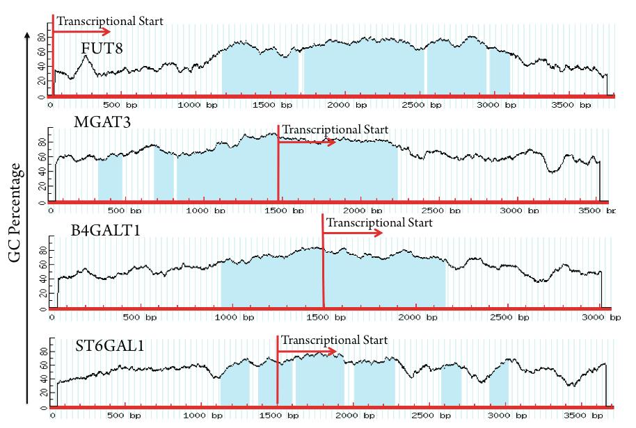
160
Adaptive Immunity: Humoral Immune Responses
 MICR2209 Adaptive Immunity: Humoral Immune Responses Dr Allison Imrie 1 Synopsis: In this lecture we will review the different mechanisms which constitute the humoral immune response, and examine the antibody
The Adaptive Immune Response. B-cells
 The Adaptive Immune Response B-cells The innate immune system provides immediate protection. The adaptive response takes time to develop and is antigen specific. Activation of B and T lymphocytes Naive
Principles of Adaptive Immunity
 Principles of Adaptive Immunity Chapter 3 Parham Hans de Haard 17 th of May 2010 Agenda Recognition molecules of adaptive immune system Features adaptive immune system Immunoglobulins and T-cell receptors
Third line of Defense
 Chapter 15 Specific Immunity and Immunization Topics -3 rd of Defense - B cells - T cells - Specific Immunities Third line of Defense Specific immunity is a complex interaction of immune cells (leukocytes)
Third line of Defense. Topic 8 Specific Immunity (adaptive) (18) 3 rd Line = Prophylaxis via Immunization!
 (18) 3 rd Line = Prophylaxis via Immunization!") Topic 8 Specific Immunity (adaptive) (18) Topics - 3 rd Line of Defense - B cells - T cells - Specific Immunities 1 3 rd Line = Prophylaxis via Immunization! (a) A painting of Edward Jenner depicts a cow
Topic 8 Specific Immunity (adaptive) (18) Topics - 3 rd Line of Defense - B cells - T cells - Specific Immunities 1 3 rd Line = Prophylaxis via Immunization! (a) A painting of Edward Jenner depicts a cow
All animals have innate immunity, a defense active immediately upon infection Vertebrates also have adaptive immunity
 1 2 3 4 5 6 7 8 9 The Immune System All animals have innate immunity, a defense active immediately upon infection Vertebrates also have adaptive immunity Figure 43.2 In innate immunity, recognition and
Chapter 23 Immunity Exam Study Questions
 Chapter 23 Immunity Exam Study Questions 1. Define 1) Immunity 2) Neutrophils 3) Macrophage 4) Epitopes 5) Interferon 6) Complement system 7) Histamine 8) Mast cells 9) Antigen 10) Antigens receptors 11)
2014 Pearson Education, Inc. Exposure to pathogens naturally activates the immune system. Takes days to be effective Pearson Education, Inc.
 The innate immune interact with the adaptive immune system 1. Damage to skin causes bleeding = bradykinin activated, resulting in inflammation 2. Dendritic phagocytose pathogens Adaptive immunity 4. Dendritic
Medical Virology Immunology. Dr. Sameer Naji, MB, BCh, PhD (UK) Head of Basic Medical Sciences Dept. Faculty of Medicine The Hashemite University
 Head of Basic Medical Sciences Dept. Faculty of Medicine The Hashemite University") Medical Virology Immunology Dr. Sameer Naji, MB, BCh, PhD (UK) Head of Basic Medical Sciences Dept. Faculty of Medicine The Hashemite University Human blood cells Phases of immune responses Microbe Naïve
Medical Virology Immunology Dr. Sameer Naji, MB, BCh, PhD (UK) Head of Basic Medical Sciences Dept. Faculty of Medicine The Hashemite University Human blood cells Phases of immune responses Microbe Naïve
Adaptive Immunity: Specific Defenses of the Host
 17 Adaptive Immunity: Specific Defenses of the Host SLOs Differentiate between innate and adaptive immunity, and humoral and cellular immunity. Define antigen, epitope, and hapten. Explain the function
The Immune System All animals have innate immunity, a defense active immediately
 The Immune System All animals have innate immunity, a defense active immediately upon infection Vertebrates also have adaptive immunity Figure 43.2 INNATE IMMUNITY (all animals) Recognition of traits shared
CELL BIOLOGY - CLUTCH CH THE IMMUNE SYSTEM.
 !! www.clutchprep.com CONCEPT: OVERVIEW OF HOST DEFENSES The human body contains three lines of against infectious agents (pathogens) 1. Mechanical and chemical boundaries (part of the innate immune system)
Structure and Function of Antigen Recognition Molecules
 MICR2209 Structure and Function of Antigen Recognition Molecules Dr Allison Imrie allison.imrie@uwa.edu.au 1 Synopsis: In this lecture we will examine the major receptors used by cells of the innate and
Macrophage Activation & Cytokine Release. Dendritic Cells & Antigen Presentation. Neutrophils & Innate Defense
 Macrophage Activation & Cytokine Release Dendritic Cells & Antigen Presentation Neutrophils & Innate Defense Neutrophils Polymorphonuclear cells (PMNs) are recruited to the site of infection where they
COURSE: Medical Microbiology, MBIM 650/720 - Fall TOPIC: Antigen Processing, MHC Restriction, & Role of Thymus Lecture 12
 COURSE: Medical Microbiology, MBIM 650/720 - Fall 2008 TOPIC: Antigen Processing, MHC Restriction, & Role of Thymus Lecture 12 FACULTY: Dr. Mayer Office: Bldg. #1, Rm B32 Phone: 733-3281 Email: MAYER@MED.SC.EDU
The Adaptive Immune Responses
 The Adaptive Immune Responses The two arms of the immune responses are; 1) the cell mediated, and 2) the humoral responses. In this chapter we will discuss the two responses in detail and we will start
Blood and Immune system Acquired Immunity
 Blood and Immune system Acquired Immunity Immunity Acquired (Adaptive) Immunity Defensive mechanisms include : 1) Innate immunity (Natural or Non specific) 2) Acquired immunity (Adaptive or Specific) Cell-mediated
all of the above the ability to impart long term memory adaptive immunity all of the above bone marrow none of the above
 1. (3 points) Immediately after a pathogen enters the body, it faces the cells and soluble proteins of the innate immune system. Which of the following are characteristics of innate immunity? a. inflammation
Physiology Unit 3. ADAPTIVE IMMUNITY The Specific Immune Response
 Physiology Unit 3 ADAPTIVE IMMUNITY The Specific Immune Response In Physiology Today The Adaptive Arm of the Immune System Specific Immune Response Internal defense against a specific pathogen Acquired
White Blood Cells (WBCs)
") YOUR ACTIVE IMMUNE DEFENSES 1 ADAPTIVE IMMUNE RESPONSE 2! Innate Immunity - invariant (generalized) - early, limited specificity - the first line of defense 1. Barriers - skin, tears 2. Phagocytes - neutrophils,
YOUR ACTIVE IMMUNE DEFENSES 1 ADAPTIVE IMMUNE RESPONSE 2! Innate Immunity - invariant (generalized) - early, limited specificity - the first line of defense 1. Barriers - skin, tears 2. Phagocytes - neutrophils,
Immune System AP SBI4UP
 Immune System AP SBI4UP TYPES OF IMMUNITY INNATE IMMUNITY ACQUIRED IMMUNITY EXTERNAL DEFENCES INTERNAL DEFENCES HUMORAL RESPONSE Skin Phagocytic Cells CELL- MEDIATED RESPONSE Mucus layer Antimicrobial
Chapter 35 Active Reading Guide The Immune System
 Name: AP Biology Mr. Croft Chapter 35 Active Reading Guide The Immune System Section 1 Phagocytosis plays an important role in the immune systems of both invertebrates and vertebrates. Review the process
Overview: The immune responses of animals can be divided into innate immunity and acquired immunity.
 GUIDED READING - Ch. 43 - THE IMMUNE SYSTEM NAME: Please print out these pages and HANDWRITE the answers directly on the printouts. Typed work or answers on separate sheets of paper will not be accepted.
WHY IS THIS IMPORTANT?
 CHAPTER 16 THE ADAPTIVE IMMUNE RESPONSE WHY IS THIS IMPORTANT? The adaptive immune system protects us from many infections The adaptive immune system has memory so we are not infected by the same pathogen
chapter 17: specific/adaptable defenses of the host: the immune response
 chapter 17: specific/adaptable defenses of the host: the immune response defense against infection & illness body defenses innate/ non-specific adaptable/ specific epithelium, fever, inflammation, complement,
The Immune System: Innate and Adaptive Body Defenses Outline PART 1: INNATE DEFENSES 21.1 Surface barriers act as the first line of defense to keep
 The Immune System: Innate and Adaptive Body Defenses Outline PART 1: INNATE DEFENSES 21.1 Surface barriers act as the first line of defense to keep invaders out of the body (pp. 772 773; Fig. 21.1; Table
Immunobiology 7. The Humoral Immune Response
 Janeway Murphy Travers Walport Immunobiology 7 Chapter 9 The Humoral Immune Response Copyright Garland Science 2008 Tim Worbs Institute of Immunology Hannover Medical School 1 The course of a typical antibody
VMC-221: Veterinary Immunology and Serology (1+1) Question Bank
 Question Bank") VMC-221: Veterinary Immunology and Serology (1+1) Objective type Questions Question Bank Q. No. 1 - Fill up the blanks with correct words 1. The British physician, who developed the first vaccine against
VMC-221: Veterinary Immunology and Serology (1+1) Objective type Questions Question Bank Q. No. 1 - Fill up the blanks with correct words 1. The British physician, who developed the first vaccine against
Chapter 1. Chapter 1 Concepts. MCMP422 Immunology and Biologics Immunology is important personally and professionally!
 MCMP422 Immunology and Biologics Immunology is important personally and professionally! Learn the language - use the glossary and index RNR - Reading, Note taking, Reviewing All materials in Chapters 1-3
Defensive mechanisms include :
 Acquired Immunity Defensive mechanisms include : 1) Innate immunity (Natural or Non specific) 2) Acquired immunity (Adaptive or Specific) Cell-mediated immunity Humoral immunity Two mechanisms 1) Humoral
The Immune System. These are classified as the Innate and Adaptive Immune Responses. Innate Immunity
 The Immune System Biological mechanisms that defend an organism must be 1. triggered by a stimulus upon injury or pathogen attack 2. able to counteract the injury or invasion 3. able to recognise foreign
Chapter 17B: Adaptive Immunity Part II
 Chapter 17B: Adaptive Immunity Part II 1. Cell-Mediated Immune Response 2. Humoral Immune Response 3. Antibodies 1. The Cell-Mediated Immune Response Basic Steps of Cell-Mediated IR 1 2a CD4 + MHC cl.
1. Overview of Adaptive Immunity
 Chapter 17A: Adaptive Immunity Part I 1. Overview of Adaptive Immunity 2. T and B Cell Production 3. Antigens & Antigen Presentation 4. Helper T cells 1. Overview of Adaptive Immunity The Nature of Adaptive
Topics. Humoral Immune Response Part II Accessory cells Fc Receptors Opsonization and killing mechanisms of phagocytes NK, mast, eosynophils
 Topics Humoral Immune Response Part II Accessory cells Fc Receptors Opsonization and killing mechanisms of phagocytes NK, mast, eosynophils Immune regulation Idiotypic network 2/15/2005 MICR 415 / 515
Adaptive Immunity. PowerPoint Lecture Presentations prepared by Mindy Miller-Kittrell, North Carolina State University C H A P T E R
 PowerPoint Lecture Presentations prepared by Mindy Miller-Kittrell, North Carolina State University C H A P T E R 16 Adaptive Immunity The Body s Third Line of Defense Adaptive Immunity Adaptive immunity
محاضرة مناعت مدرس المادة :ا.م. هدى عبدالهادي علي النصراوي Immunity to Infectious Diseases
 محاضرة مناعت مدرس المادة :ا.م. هدى عبدالهادي علي النصراوي Immunity to Infectious Diseases Immunity to infection depends on a combination of innate mechanisms (phagocytosis, complement, etc.) and antigen
General Biology. A summary of innate and acquired immunity. 11. The Immune System. Repetition. The Lymphatic System. Course No: BNG2003 Credits: 3.
 A summary of innate and acquired immunity General iology INNATE IMMUNITY Rapid responses to a broad range of microbes Course No: NG00 Credits:.00 External defenses Invading microbes (pathogens). The Immune
The Immune System is the Third Line of Defense Against Infection. Components of Human Immune System
 Chapter 17: Specific Host Defenses: The Immune Response The Immune Response Immunity: Free from burden. Ability of an organism to recognize and defend itself against specific pathogens or antigens. Immune
What sort of Science is Glycoscience? (Introductory lecture)
") Glycosciences: Glycobiology & Glycochemistry e-learning course What sort of Science is Glycoscience? (Introductory lecture) Paula Videira Faculdade de Ciências Médicas Nova University, Lisbon Portugal
Glycosciences: Glycobiology & Glycochemistry e-learning course What sort of Science is Glycoscience? (Introductory lecture) Paula Videira Faculdade de Ciências Médicas Nova University, Lisbon Portugal
T-cell activation T cells migrate to secondary lymphoid tissues where they interact with antigen, antigen-presenting cells, and other lymphocytes:
 Interactions between innate immunity & adaptive immunity What happens to T cells after they leave the thymus? Naïve T cells exit the thymus and enter the bloodstream. If they remain in the bloodstream,
Antigen Receptor Structures October 14, Ram Savan
 Antigen Receptor Structures October 14, 2016 Ram Savan savanram@uw.edu 441 Lecture #8 Slide 1 of 28 Three lectures on antigen receptors Part 1 (Today): Structural features of the BCR and TCR Janeway Chapter
Immune system. Aims. Immune system. Lymphatic organs. Inflammation. Natural immune system. Adaptive immune system
 Aims Immune system Lymphatic organs Inflammation Natural immune system Adaptive immune system Major histocompatibility complex (MHC) Disorders of the immune system 1 2 Immune system Lymphoid organs Immune
Immunology - Lecture 2 Adaptive Immune System 1
 Immunology - Lecture 2 Adaptive Immune System 1 Book chapters: Molecules of the Adaptive Immunity 6 Adaptive Cells and Organs 7 Generation of Immune Diversity Lymphocyte Antigen Receptors - 8 CD markers
Chapter 13 Lymphatic and Immune Systems
 The Chapter 13 Lymphatic and Immune Systems 1 The Lymphatic Vessels Lymphoid Organs Three functions contribute to homeostasis 1. Return excess tissue fluid to the bloodstream 2. Help defend the body against
The Lymphatic System and Body Defenses
 PowerPoint Lecture Slide Presentation by Patty Bostwick-Taylor, Florence-Darlington Technical College The Lymphatic System and Body Defenses 12PART B Adaptive Defense System: Third Line of Defense Immune
LESSON 2: THE ADAPTIVE IMMUNITY
 Introduction to immunology. LESSON 2: THE ADAPTIVE IMMUNITY Today we will get to know: The adaptive immunity T- and B-cells Antigens and their recognition How T-cells work 1 The adaptive immunity Unlike
Adaptive Immunity. PowerPoint Lecture Presentations prepared by Mindy Miller-Kittrell, North Carolina State University C H A P T E R
 CSLO7. Describe functions of host defenses and the immune system in combating infectious diseases and explain how immunizations protect against specific diseases. PowerPoint Lecture Presentations prepared
Immunology for the Rheumatologist
 Immunology for the Rheumatologist Rheumatologists frequently deal with the immune system gone awry, rarely studying normal immunology. This program is an overview and discussion of the function of the
Foundations in Microbiology
 Foundations in Microbiology Fifth Edition Talaro Chapter 15 The Acquisition of Specific Immunity and Its Applications Chapter 15 2 Chapter Overview 1. Development of the Dual Lymphocyte System 2. Entrance
Chapter 11. B cell generation, Activation, and Differentiation. Pro-B cells. - B cells mature in the bone marrow.
 Chapter B cell generation, Activation, and Differentiation - B cells mature in the bone marrow. - B cells proceed through a number of distinct maturational stages: ) Pro-B cell ) Pre-B cell ) Immature
How the Innate Immune System Profiles Pathogens
 How the Innate Immune System Profiles Pathogens Receptors on macrophages, neutrophils, dendritic cells for bacteria and viruses Broad specificity - Two main groups of bacteria: gram positive, gram-negative
I. Critical Vocabulary
 I. Critical Vocabulary A. Immune System: a set of glands, tissues, cells, and dissolved proteins that combine to defend against non-self entities B. Antigen: any non-self chemical that triggers an immune
The Immune System. Chapter 43. Biology Eighth Edition Neil Campbell and Jane Reece. PowerPoint Lecture Presentations for
 Chapter 43 The Immune System PowerPoint Lecture Presentations for Biology Eighth Edition Neil Campbell and Jane Reece Lectures by Chris Romero, updated by Erin Barley with contributions from Joan Sharp
HLA and antigen presentation. Department of Immunology Charles University, 2nd Medical School University Hospital Motol
 HLA and antigen presentation Department of Immunology Charles University, 2nd Medical School University Hospital Motol MHC in adaptive immunity Characteristics Specificity Innate For structures shared
Adaptive Immune System
 Short Course on Immunology Adaptive Immune System Bhargavi Duvvuri Ph.D IIIrd Year (Immunology) bhargavi@yorku.ca Supervisor Dr.Gillian E Wu Professor, School of Kinesiology and Health Sciences York University,
The Innate Immune Response
 The Innate Immune Response FUNCTIONS OF THE IMMUNE SYSTEM: Recognize, destroy and clear a diversity of pathogens. Initiate tissue and wound healing processes. Recognize and clear damaged self components.
For questions 1-5, match the following with their correct descriptions. (24-39) A. Class I B. Class II C. Class III D. TH1 E. TH2
 A. Class I B. Class II C. Class III D. TH1 E. TH2") Questions Made by SI ATTENDEES!! :) Page 1 of 6 Student-Made Practice Exam Activity All questions, answers, and slide numbers are based off of Monday s SI activity, where students/attendees created possible
Questions Made by SI ATTENDEES!! :) Page 1 of 6 Student-Made Practice Exam Activity All questions, answers, and slide numbers are based off of Monday s SI activity, where students/attendees created possible
Unit 5 The Human Immune Response to Infection
 Unit 5 The Human Immune Response to Infection Unit 5-page 1 FOM Chapter 21 Resistance and the Immune System: Innate Immunity Preview: In Chapter 21, we will learn about the branch of the immune system
Fluid movement in capillaries. Not all fluid is reclaimed at the venous end of the capillaries; that is the job of the lymphatic system
 Capillary exchange Fluid movement in capillaries Not all fluid is reclaimed at the venous end of the capillaries; that is the job of the lymphatic system Lymphatic vessels Lymphatic capillaries permeate
CHAPTER-VII IMMUNOLOGY R.KAVITHA, M.PHARM, LECTURER, DEPARTMENT OF PHARMACEUTICS, SRM COLLEGE OF PHARMACY, SRM UNIVERSITY, KATTANKULATHUR.
 CHAPTER-VII IMMUNOLOGY R.KAVITHA, M.PHARM, LECTURER, DEPARTMENT OF PHARMACEUTICS, SRM COLLEGE OF PHARMACY, SRM UNIVERSITY, KATTANKULATHUR. The Immune Response Immunity: Free from burden. Ability of an
General Overview of Immunology. Kimberly S. Schluns, Ph.D. Associate Professor Department of Immunology UT MD Anderson Cancer Center
 General Overview of Immunology Kimberly S. Schluns, Ph.D. Associate Professor Department of Immunology UT MD Anderson Cancer Center Objectives Describe differences between innate and adaptive immune responses
11/25/2017. THE IMMUNE SYSTEM Chapter 43 IMMUNITY INNATE IMMUNITY EXAMPLE IN INSECTS BARRIER DEFENSES INNATE IMMUNITY OF VERTEBRATES
 THE IMMUNE SYSTEM Chapter 43 IMMUNITY INNATE IMMUNITY EXAMPLE IN INSECTS Exoskeleton made of chitin forms the first barrier to pathogens Digestive system is protected by a chitin-based barrier and lysozyme,
The T cell receptor for MHC-associated peptide antigens
 1 The T cell receptor for MHC-associated peptide antigens T lymphocytes have a dual specificity: they recognize polymporphic residues of self MHC molecules, and they also recognize residues of peptide
1. The scavenger receptor, CD36, functions as a coreceptor for which TLR? a. TLR ½ b. TLR 3 c. TLR 4 d. TLR 2/6
 Allergy and Immunology Review Corner: Cellular and Molecular Immunology, 8th Edition By Abul K. Abbas, MBBS, Andrew H. H. Lichtman, MD, PhD and Shiv Pillai, MBBS, PhD. Chapter 4 (pages 62-74): Innate Immunity
Chapter 11. B cell generation, Activation, and Differentiation. Pro-B cells. - B cells mature in the bone marrow.
 Chapter B cell generation, Activation, and Differentiation - B cells mature in the bone marrow. - B cells proceed through a number of distinct maturational stages: ) Pro-B cell ) Pre-B cell ) Immature
Mon, Wed, Fri 11:00 AM-12:00 PM. Owen, Judy, Jenni Punt, and Sharon Stranford Kuby-Immunology, 7th. Edition. W.H. Freeman and Co., New York.
 Course Title: Course Number: Immunology Biol-341/541 Semester: Fall 2013 Location: HS 268 Time: Instructor: 8:00-9:30 AM Tue/Thur Dr. Colleen M. McDermott Office: Nursing Ed 101 (424-1217) E-mail*: mcdermot@uwosh.edu
Defense mechanism against pathogens
 Defense mechanism against pathogens Immune System What is immune system? Cells and organs within an animal s body that contribute to immune defenses against pathogens ( ) Bacteria -Major entry points ;open
Body Defense Mechanisms
 BIOLOGY OF HUMANS Concepts, Applications, and Issues Fifth Edition Judith Goodenough Betty McGuire 13 Body Defense Mechanisms Lecture Presentation Anne Gasc Hawaii Pacific University and University of
TCR, MHC and coreceptors
 Cooperation In Immune Responses Antigen processing how peptides get into MHC Antigen processing involves the intracellular proteolytic generation of MHC binding proteins Protein antigens may be processed
The Adaptive Immune Response: T lymphocytes and Their Functional Types *
 OpenStax-CNX module: m46560 1 The Adaptive Immune Response: T lymphocytes and Their Functional Types * OpenStax This work is produced by OpenStax-CNX and licensed under the Creative Commons Attribution
SINGLE CHOICE. 5. The gamma invariant chain binds to this molecule during its intracytoplasmic transport. A TCR B BCR C MHC II D MHC I E FcγR
 A Name: Group: SINGLE CHOICE 1. Which is the most important ligand of TLR5? A endospore B flagellin C polysaccharide capsule D DNA E pilus 2. The antibody-binding site is formed primarily by... A the constant
Significance and Functions of Carbohydrates. Bacterial Cell Walls
 Biochemistry 462a - Carbohydrate Function Reading - Chapter 9 Practice problems - Chapter 9: 2, 4a, 4b, 6, 9, 10, 13, 14, 15, 16a, 17; Carbohydrate extra problems Significance and Functions of Carbohydrates
There are 2 major lines of defense: Non-specific (Innate Immunity) and. Specific. (Adaptive Immunity) Photo of macrophage cell
 and. Specific. (Adaptive Immunity) Photo of macrophage cell") There are 2 major lines of defense: Non-specific (Innate Immunity) and Specific (Adaptive Immunity) Photo of macrophage cell Development of the Immune System ery pl neu mφ nk CD8 + CTL CD4 + thy TH1 mye
There are 2 major lines of defense: Non-specific (Innate Immunity) and Specific (Adaptive Immunity) Photo of macrophage cell Development of the Immune System ery pl neu mφ nk CD8 + CTL CD4 + thy TH1 mye
LYMPHOCYTES & IMMUNOGLOBULINS. Dr Mere Kende, Lecturer SMHS
 LYMPHOCYTES & IMMUNOGLOBULINS Dr Mere Kende, Lecturer SMHS Immunity Immune- protection against dangers of non-self/invader eg organism 3 components of immune system 1 st line: skin/mucosa/cilia/hair/saliva/fatty
IMMUNITY AND DISEASE II
 IMMUNITY AND DISEASE II A. Evolution of the immune system. 1. Figure 1--57.25, p. 1167 from Raven and Johnson Biology 6 th ed. shows how the immune system evolved. Figure 1. How the immune system evolved.
生命科学基础 (21)- 动物的免疫器官. The Immune System. KE, Yuehai 柯越海. Zhejiang University, School of Basic Medical Sciences (BMS-ZJU) 浙江大学基础医学院
- 动物的免疫器官. The Immune System. KE, Yuehai 柯越海. Zhejiang University, School of Basic Medical Sciences (BMS-ZJU) 浙江大学基础医学院") 生命科学基础 (21)- 动物的免疫器官 The Immune System KE, Yuehai 柯越海 Zhejiang University, School of Basic Medical Sciences (BMS-ZJU) 浙江大学基础医学院 Outlines The Immune System 1. Innate immunity 2. Adaptive immunity 3. Immune
生命科学基础 (21)- 动物的免疫器官 The Immune System KE, Yuehai 柯越海 Zhejiang University, School of Basic Medical Sciences (BMS-ZJU) 浙江大学基础医学院 Outlines The Immune System 1. Innate immunity 2. Adaptive immunity 3. Immune
Chapter 24 The Immune System
 Chapter 24 The Immune System The Immune System Layered defense system The skin and chemical barriers The innate and adaptive immune systems Immunity The body s ability to recognize and destroy specific
Topics in Parasitology BLY Vertebrate Immune System
 Topics in Parasitology BLY 533-2008 Vertebrate Immune System V. Vertebrate Immune System A. Non-specific defenses against pathogens 1. Skin - physical barrier a. Tough armor protein KERATIN b. Surface
The Adaptive Immune Response. T-cells
 The Adaptive Immune Response T-cells T Lymphocytes T lymphocytes develop from precursors in the thymus. Mature T cells are found in the blood, where they constitute 60% to 70% of lymphocytes, and in T-cell
Two categories of immune response. immune response. infection. (adaptive) Later immune response. immune response
 Later immune response. immune response") Ivana FELLNEROVÁ E-mail: fellneri@hotmail.com, mob. 732154801 Basic immunogenetic terminology innate and adaptive immunity specificity and polymorphism immunoglobuline gene superfamily immunogenetics MHC
Ivana FELLNEROVÁ E-mail: fellneri@hotmail.com, mob. 732154801 Basic immunogenetic terminology innate and adaptive immunity specificity and polymorphism immunoglobuline gene superfamily immunogenetics MHC
Helminth worm, Schistosomiasis Trypanosomes, sleeping sickness Pneumocystis carinii. Ringworm fungus HIV Influenza
 Helminth worm, Schistosomiasis Trypanosomes, sleeping sickness Pneumocystis carinii Ringworm fungus HIV Influenza Candida Staph aureus Mycobacterium tuberculosis Listeria Salmonella Streptococcus Levels
Clinical Basis of the Immune Response and the Complement Cascade
 Clinical Basis of the Immune Response and the Complement Cascade Bryan L. Martin, DO, MMAS, FACAAI, FAAAAI, FACOI, FACP Emeritus Professor of Medicine and Pediatrics President, American College of Allergy,
PLAN. Réponses B thymodépendantes et thymoindépendantes. B cell development and classification. B cell activation. Thymodependent B(2) cell response
 cell response") Réponses B thymodépendantes et thymoindépendantes PLAN B cell development and classification B cell activation Thymodependent B(2) cell response BMC 423 (IF) - 2007 Antonino Nicoletti Thymo-independent
Réponses B thymodépendantes et thymoindépendantes PLAN B cell development and classification B cell activation Thymodependent B(2) cell response BMC 423 (IF) - 2007 Antonino Nicoletti Thymo-independent
LECTURE: 21. Title IMMUNOGLOBULINS FUNCTIONS & THEIR RECEPTORS LEARNING OBJECTIVES:
 LECTURE: 21 Title IMMUNOGLOBULINS FUNCTIONS & THEIR RECEPTORS LEARNING OBJECTIVES: The student should be able to: Determine predominant immunoglobulin isotypes in serum. Determine the predominant immunoglobulin
LECTURE PRESENTATIONS
 LECTURE PRESENTATIONS For CAMPBELL BIOLOGY, NINTH EDITION Jane B. Reece, Lisa A. Urry, Michael L. Cain, Steven A. Wasserman, Peter V. Minorsky, Robert B. Jackson Chapter 43 The Immune System Lectures by
Overview. Barriers help animals defend against many dangerous pathogens they encounter.
 Immunity Overview Barriers help animals defend against many dangerous pathogens they encounter. The immune system recognizes foreign bodies and responds with the production of immune cells and proteins.
HLA and antigen presentation. Department of Immunology Charles University, 2nd Medical School University Hospital Motol
 HLA and antigen presentation Department of Immunology Charles University, 2nd Medical School University Hospital Motol MHC in adaptive immunity Characteristics Specificity Innate For structures shared
Adaptive Immunity. PowerPoint Lecture Presentations prepared by Mindy Miller-Kittrell, North Carolina State University C H A P T E R
 PowerPoint Lecture Presentations prepared by Mindy Miller-Kittrell, North Carolina State University C H A P T E R 16 Adaptive Immunity The Body s First Line of Defense Innate Immunity The Role of Skin
Immunity. Acquired immunity differs from innate immunity in specificity & memory from 1 st exposure
 Immunity (1) Non specific (innate) immunity (2) Specific (acquired) immunity Characters: (1) Non specific: does not need special recognition of the foreign cell. (2) Innate: does not need previous exposure.
The Major Histocompatibility Complex (MHC)
") The Major Histocompatibility Complex (MHC) An introduction to adaptive immune system before we discuss MHC B cells The main cells of adaptive immune system are: -B cells -T cells B cells: Recognize antigens
The Major Histocompatibility Complex (MHC) An introduction to adaptive immune system before we discuss MHC B cells The main cells of adaptive immune system are: -B cells -T cells B cells: Recognize antigens
3/28/2012. Immune System. Activation of Innate Immunity. Innate (non-specific) Immunity
 Immunity") Chapter 5 Outline Defense Mechansims Functions of B Lymphocytes Functions of T Lymphocytes Active and Passive Immunity Tumor Immunology Diseases Caused By Immune System Immune System Anatomy - Lymphoid
Chapter 5 Outline Defense Mechansims Functions of B Lymphocytes Functions of T Lymphocytes Active and Passive Immunity Tumor Immunology Diseases Caused By Immune System Immune System Anatomy - Lymphoid
MICROBIO320 EXAM 1-Spring 2011 Name True/False (1 point each) T 2. T cell receptors are composed of constant and variable regions.
 T 2. T cell receptors are composed of constant and variable regions.") True/False (1 point each) T 1. Mature T cells (lymphocytes) bind only to processed antigen. T 2. T cell receptors are composed of constant and variable regions. F 3. Natural Killer cells do not proliferate
True/False (1 point each) T 1. Mature T cells (lymphocytes) bind only to processed antigen. T 2. T cell receptors are composed of constant and variable regions. F 3. Natural Killer cells do not proliferate
I. Lines of Defense Pathogen: Table 1: Types of Immune Mechanisms. Table 2: Innate Immunity: First Lines of Defense
 I. Lines of Defense Pathogen: Table 1: Types of Immune Mechanisms Table 2: Innate Immunity: First Lines of Defense Innate Immunity involves nonspecific physical & chemical barriers that are adapted for
Skin. Mucin-containing mucous membranes. Desmosome (attaches keratincontaining
 Immunity 1 Fig. 43.2 2 Skin Mucin-containing mucous membranes Desmosome (attaches keratincontaining skin cells together) http://www.mhhe.com/biosci/ap/histology_mh/pseudos2l.jpg http://training.seer.cancer.gov/module_anatomy/images/
Acquired Immunity Cells are initially and require before they can work Responds to individual microbes
 1 of 10 THE IMMUNE SYSTEM CHAPTER 43; PAGES 898 921 WHY DO WE NEED AN IMMUNE SYSTEM? It s a dirty, dirty world out there and we are vastly outnumbered Bacteria and parasites are everywhere The body has
General information. Cell mediated immunity. 455 LSA, Tuesday 11 to noon. Anytime after class.
 General information Cell mediated immunity 455 LSA, Tuesday 11 to noon Anytime after class T-cell precursors Thymus Naive T-cells (CD8 or CD4) email: lcoscoy@berkeley.edu edu Use MCB150 as subject line
Prof. Ibtesam Kamel Afifi Professor of Medical Microbiology & Immunology
 By Prof. Ibtesam Kamel Afifi Professor of Medical Microbiology & Immunology Lecture objectives: At the end of the lecture you should be able to: Enumerate features that characterize acquired immune response
Antigen-Independent B-Cell Development Bone Marrow
 Antigen-Independent B-Cell Development Bone Marrow 1. DNA rearrangements establish the primary repertoire, creating diversity 2. Allelic exclusion ensures that each clone expresses a single antibody on
Antigen Presentation and T Lymphocyte Activation. Abul K. Abbas UCSF. FOCiS
 1 Antigen Presentation and T Lymphocyte Activation Abul K. Abbas UCSF FOCiS 2 Lecture outline Dendritic cells and antigen presentation The role of the MHC T cell activation Costimulation, the B7:CD28 family