HIV-1 SUBTYPE C MOTHER-TO-CHILD TRANSMISSION: GENETIC AND IMMUNOLOGIC CORRELATES. Elizabeth Susan Russell
|
|
|
- Hillary Cannon
- 6 years ago
- Views:
Transcription
1 HIV-1 SUBTYPE C MOTHER-TO-CHILD TRANSMISSION: GENETIC AND IMMUNOLOGIC CORRELATES Elizabeth Susan Russell A dissertation submitted to the faculty of the University of North Carolina at Chapel Hill in partial fulfillment of the requirements for the degree of Doctor of Philosophy in the School of Medicine (Microbiology and Immunology) Chapel Hill 2009 Approved by: Dr. Ronald Swanstrom Dr. Steven Meshnick Dr. Susan Fiscus Dr. Nancy Davis Dr. Joseph Eron
2 ABSTRACT Mother-to-child transmission (MTCT) of human immunodeficiency virus-1 (HIV- 1) infects over 300,000 infants each year. This transmission can occur in utero (IU), intrapartum (IP), or post-partum through breastfeeding (PP). One feature of transmission from mother to child is a reduction (or bottleneck ) in viral genetic diversity, particularly within the envelope (env) gene. A heteroduplex tracking assay was used to examine env diversity in women whose infants remained uninfected at least through 6 weeks, and in mother-infant IU and IP transmission pairs. Maternal diversity was similar regardless of transmission status. We confirmed a bottleneck in subtype C IU and IP transmission. We further found that infants infected IU had fewer variants than those infected IP, and that these variants transmitted IU were major variants in the maternal populations more often than variants transmitted IP. Also, minor maternal variants were transmitted with a frequency that demonstrates neither IU nor IP transmission is stochastic. Shorter env sequences and fewer glycosylation sites, ie more compact viruses, have been associated with greater neutralization sensitivity, and compact subtype C viruses are often transmitted through horizontal infection. env genes from a subset of IU and IP transmission pairs were sequenced and showed that compact maternal variants were transmitted IP, but not IU. env sequences from 3 mother-infant pairs where transmission occurred through breastfeeding were also analyzed and we found reductions in genetic diversity, sequence length, and glycosylation. These results demonstrate selection occurs ii
3 in MTCT and mechanisms may vary with the timing of transmission. High titers of neutralizing antibodies (NAB) have been correlated with lower rates of horizontal and vertical transmission in animal models, and in some small studies of human transmission. Because we identified selection had occurred in these transmission pairs, we next tested sera from non-transmitting, IU-, and IP-transmitting women for neutralizing activity against virus pseudotyped with heterologous subtype B and C Env proteins. Though non-transmitting women more often had NAB titers against multiple Envs, NAB titer to any one Env did not correlate to transmission status. Thus, we cannot attribute vertical transmission or a lack of transmission to different levels of neutralizing antibodies in the context of subtype C HIV-1 transmission events. iii
4 Acknowledgements I would like to thank my advisor, Ron Swanstrom, for his guidance through the years to help me become a scientist, and also his generous willingness to let me explore public health aspects of my project and research interests. I also owe a huge thanks to Jesse Kwiek for 3 years of a fruitful and entertaining collaboration. His energy, research experience, and curiosity carried me through the inevitable rough patches! I am very grateful to all of the people I have worked with in lab over the years. It was an amazing, friendly work environment. Finally, I would like to thank my family. Their emphasis on education brought me to graduate school in the first place, and their support and encouragement has made all the difference through the years. iv
5 Table of Contents List of Figures... vii List of Tables... viii List of Abbreviations... ix Chapter HIV Overview Clinical Course of HIV Infection HIV Virology Viral Mechanisms for Diversity Host Mechanisms that Drive Evolution Virologic Characteristics of Transmission Conclusions Chapter Abstract Introduction Materials and Methods Results Discussion Acknowledgements Chapter v
6 3.1 Introduction Materials and Methods Results Discussion Chapter Introduction Materials and Methods Results Discussion Future Directions Chapter Introduction Materials and Methods Results Discussion Chapter References vi
7 List of Figures Figure 2.1 Heteroduplex Tracking Assay..46 Figure 2.2 Phylogenetic Tree.47 Figure 2.3 HIV-1 envelope V1/V2 Variant Transmission.48 Figure 2.4 Umbilical Cord Plasma.49 Figure 3.1 Representative maximum likelihood phylogenetic trees..69 Figure 3.2 Highlighter plot of 312 transmission pair.70 Figure 4.1 Lowest titer of maternal serum with >50% inhibition of infection...79 Figure 5.1 Pairwise comparison of maternal and infant sequence populations..90 Figure 5.2 Maximum likelihood phylogenetic trees of gp120 sequences...91 Figure 5.3 Maternal and infant sequence populations by nucleotide differences.. 92 Figure 5.4 Length and glycosylation site differences.93 vii
8 List of Tables Table 2.1 Patient Characteristics..45 Table 3.1 Patient Characteristics...67 Table 3.2 Mean sequence length and putative N-linked glycosylation site differences between mother-infant pairs viii
9 List of Abbreviations Ab AIDS CTL HIV IP IU MTCT NAB NT PP Antibody Acquired Immune Deficiency Syndrome Cytotoxic T-Lymphocyte Human Immunodeficiency Syndrome Intrapartum In utero Mother-to-Child Transmission Neutralizing Antibody Non-transmitter Post-partum ix
10 Chapter 1 Introduction 1.0 HIV Overview In ,000 children under the age of 15 were infected with human immunodeficiency virus-1 (HIV-1) (149), nearly all of whom were infected in infancy through mother-to-child transmission (MTCT). Subtype C is the most prevalent subtype of HIV-1 in Sub-Saharan Africa, where 90% of infected children live. Sixty percent of infections in Sub-Saharan Africa in 2007 were in women (149), and 15-40% of HIV+ pregnant women will transmit the virus to their offspring (92), suggesting infants will continue to be born to HIV+ women for the foreseeable future. HIV positive infants are born in countries with limited resources to manage either the infant s or their mother s infections. Interventions that prevent MTCT most effectively, to <2%, involve months of expensive drug regimens for both the mother and the child, an elective cesarean section, and formula feeding from birth for the infant. Short-course drug regimens alone for mother and infant significantly reduce MTCT to ~10% (92), yet providing safe and acceptable alternatives to breastfeeding in many areas leaves infants at risk for water-borne infections. New interventions to prevent MTCT are needed, yet little is known about the mechanisms of transmission. The work described herein investigates characteristics of subtype C MTCT, and aims to provide a base for
11 future work to identify new targets for simpler and more cost-effective interventions to prevent transmission. In the first chapter I will introduce HIV infection from a clinical, virologic, and host-pathogen point of view, highlighting differences between infant and adult infections. I will then review what has been studied about viral genetics and antibody responses during both horizontal and vertical HIV transmission. 1.1 Clinical Course of HIV Infection HIV is transmitted through bodily fluids such as blood, semen, and vaginal secretions. The most common routes of transmission are vaginal sex, anal sex, needles (all types of horizontal transmission), and from mother-to-child (vertical transmission). Vertical transmission can occur during pregnancy (in utero, IU), during labor and delivery (intrapartum, IP), and through breast milk (post-partum, PP). The typical clinical course of disease in adults begins with acute infection. There is rapid viral replication in the first 3 weeks after infection and a drop in CD4+ cell counts. During acute infection CD8+ T cells become activated and as the number of CD8+ T cells rises, the viral load falls to a set point, and CD4+ T cell counts increase slightly. The viral load will remain relatively constant at this set point during the next phase of disease, clinical latency. Latency can last anywhere from weeks to decades during which there is a slow decline in CD4+ T cells. Longer latency periods are associated with lower viral set points and strong CD8+ T cell responses (109). Latency is followed by the disease stage, acquired immune deficiency syndrome (AIDS). AIDS is defined by several factors, including CD4+ T cell count and infection with certain 2
12 illnesses. The average life span after infection with HIV-1, without treatment, is 10 years (52). Infants follow a significantly more rapid disease progression. On average, viral set points are higher and immune responses are delayed and less robust in infants (4). HIV-1 replication in infants is localized to the thymus, which often results in severe atrophy of this organ. Failure to control viral replication and aberrant immune responses likely contribute to the diminished life span of infected infants; some studies have shown death rates as high as 52% by 2 years of age (104). 1.2 HIV Virology HIV-1 is part of the Retroviridae family. This family of enveloped viruses is characterized by a plus-strand RNA genome that is reverse transcribed into a linear DNA intermediate that is inserted into the host genome, where it is expressed by the host transcription machinery. HIV is a complex retrovirus (meaning it has accessory viral proteins) in the Lentivirus genus. Lentiviruses are named for their long infection times (lenti-, Latin for slow), and, unlike other retroviruses, they do not induce tumors despite integration (69) Life Cycle HIV-1 infection of a new cell (reviewed in (69)) begins with binding of the envelope protein (Env) gp120 to CD4. This binding initiates a conformational change in gp120, which reveals a protein surface for coreceptor binding. The CCR5 coreceptor is used almost exclusively in early infection (R5 virus), with a switch to both CCR5 and 3
13 CXCR4 (R5/X4), or only CXCR4 use (X4) later in infection in some patients. A conformational change in the transmembrane Env gp41 then mediates fusion of the HIV- 1 virion with the plasma membrane of the target cell. After fusion, the core of the HIV-1 particle enters the cell and uncoats to reveal the genome for transcription. The RNA genome is reverse transcribed into doublestranded DNA, transported into the nucleus of the cell, and integrated into a host cell chromosome. With cellular factors and HIV regulatory proteins Tat and Rev, the viral genome is transcribed, mrna templates are exported to the cytoplasm, and viral proteins are translated from spliced and unspliced transcripts. Viral proteins, along with 2 copies of genomic RNA, are then assembled at the plasma membrane, and new virus buds from the cell. For this new virus to become infectious maturation must occur, which is visible as a structural change in the viral core that occurs with the cleavage of the Gag polyprotein Viral Proteins Each protein produced by HIV has a unique role in the life cycle (reviewed in (69)). All are necessary for viral replication and optimal infectivity in vivo. The Gag coding region contains four structural proteins matrix (MA), capsid (CA), nucleocapsid (NC), and p6 that form the core of the virus and provide attachment to the viral envelope. These proteins are translated as a polyprotein (Gag), and remain as a polyprotein until budding when the precursor is cleaved by the viral protease. NC binds the genomic RNA in the viral core and helps chaperone nucleic acids during the life cycle. CA is important for assembly and forms the capsid shell of the viral core, while 4
14 MA facilitates the targeting and binding of the Gag polyprotein to the cell membrane. The small p6 protein has a late domain that aids in virus release, and p6 also brings the accessory protein vpr into the virion. The other structural proteins are translated from the env ORF. The translation of the polyprotein gp160 occurs on the rough ER. This polyprotein is transferred to the golgi and cleaved by cellular enzymes to make two proteins, the transmembrane gp41, and the noncovalently associated surface protein gp120. Env protein gp120 is highly glycosylated and facilitates attachment of the virus with the target cell. HIV-1 has 2 regulatory proteins, Tat and Rev. Tat helps to direct transcription by binding to the TAR RNA structure downstream of the transcription start site to encourage RNA polymerase II complex processivity. Rev binds to a separate RNA structure, the Rev Responsive Element (RRE). Rev brings unspliced and partially spliced RNA transcripts that contain this region out of the nucleus through nuclear export machinery. Without Rev neither genomic nor partially spliced RNA transcripts would make it out of the nucleus for translation or assembly. HIV-1 also has 4 accessory proteins with important functions in vivo (69). Vif directs the degradation of the cellular antiviral protein APOBEC3G (discussed further in Diversity). Vpr has several functions related to transcription: it stimulates LTR driven gene expression, promotes nuclear transport of the HIV DNA complex, and can modulate RT mutation rates. Vpu is an integral membrane protein that is not found in virions. One function of Vpu is to downregulate CD4 in the ER, allowing envelope protein bound to CD4 to continue to the cell surface. Vpu also enhances virus release by interacting with the cellular protein Tetherin, which retain virus to the cell surface (102). The final 5
15 accessory protein is Nef. Nef is membrane-associated and is synthesized at high levels very early in infection. It downregulates several cell-surface markers, including CD4 and MHC class I and II (discussed below) Env gp120 is the main viral surface protein seen by the immune system on circulating virus, and is therefore a frequent target of the humoral immune response. During the course of disease the env gene becomes highly diverse within a virus population in a single person (51, 141). This diversity is largely generated by random mutations that occur during reverse transcription followed by selection. Mutations that result in immune evasion confer a selective advantage in subsequent viral replication (16). The advantage, however, must be greater than any deleterious fitness effects the mutation might have on subsequent viral replication (111, 130). These mutations build over the course of infection through continuous targeting by the immune system, which will be discussed further below. Diversity in the coding region of gp120 is not uniform. There are 5 regions of high variability in gp120 coding region, called variable regions 1-5 (91). These regions correlate to flexible loops without secondary structure, most of which are on the surface of the protein and are likely to come into contact with antibodies. The amino acid sequence flexibility of variable loops allows the virus to tolerate changes that evade selective immune pressure. Changes in length and glycosylation patterns within variable regions 1, 2, and 4 are predicted to affect CD4 and coreceptor binding of the virus. V1 and V2 lie near the CD4 binding site on gp120, and changes in V4 alter the orientation 6
16 and packing of shielding glycans near the CD4 binding site (35). Without these loops the virus can be more easily targeted by antibodies (discussed in 1.5.1). 1.3 Viral Mechanisms for Diversity As discussed above, reverse transcription creates a large amount of viral genetic diversity that accumulates during HIV infection, resulting in a viral quasispecies within each chronically infected patient. Using this diversity HIV can expand its host range and evade immune pressures. It is also the biggest obstacle to stopping the pandemic, as mutations result in drug resistance and a moving vaccine target. HIV genetic diversity is a direct result of several factors. These factors include the viral mutation and recombination rates, the viral replication rate, the size of the viral population, and selective forces (including immune selection as well as competition between viruses). In this section I will discuss viral mutation and recombination rates, followed by host selective forces and how these impact the disease course Reverse Transcription Reverse transcriptase (RT) is a heterodimer with RNA-dependent and DNAdependent polymerase, and RNase H activity. RT is incorporated into the core of the virus making it ready to begin reverse transcription without new protein synthesis in the target cell. Reverse transcription (69) occurs in the cytoplasm of the host cell after entry of the virus into the cell and uncoating of the viral core. The process of genome replication requires the binding of a cellular trna to the primer binding site (PBS) at the 5 end of 7
17 the positive-sense RNA that makes up the HIV genome. Minus-strand DNA synthesis is initiated using this trna primer and continues through the repeat region R at the 5 end of the genome to create the minus-strand strong-stop DNA (-ssdna). As this minus strand is copied, the RNase H domain of the reverse transcriptase heterodimer follows behind the polymerase activity to degrade the template RNA. The ssdna is transferred to the 3 end of the RNA genome facilitated by binding to the 3 repeat region R that is identical to the 5 repeat. Minus-strand synthesis continues to the end of the RNA genome, which due to the RNase H activity is now at the PBS. RNase H degrades all of the genome except two small pieces of RNA, one immediately upstream of U3 called the PPT. The PPT RNA remains bound to the newly formed minus-strand DNA, and serves as the primer and thus initiation site for positive-strand DNA synthesis. Positive-strand synthesis continues to the 5 end of the minus-strand template. This new positive-strand DNA is called plus strong stop DNA (+ssdna). At this point a second strand-transfer event occurs. The 3 end of the +ssdna binds to the 3 end of the minus-strand DNA using the PBS and the complementary trna sequence copied at the end of the +ssdna. Reverse transcription continues for both strands, using each other as template. The resulting double-stranded DNA makes the linear intermediate that is transported to the nucleus for integration. Because reverse transcription relies on 2 template switches, it is hypothesized that HIV RT has evolved to have low template affinity and processivity (69). These traits then lead to high error rates, including substitutions, insertions, deletions, and recombination. 8
18 1.3.2 Substitutions, insertions, and deletions Mutations are thought to principally depend on the fidelity of 4 steps (115, 146): minus-strand reverse transcription, plus-strand reverse transcription, cellular RNApolymerase II, and modification of templates. RT has no exonucleolytic proofreading activity, and errors can occur in either minus or plus-strand synthesis. The rate of these errors depends on the type of error and the sequence context of the error, including secondary structure. If mutations occur in the minus strand, they will be copied into the plus strand. Mutations that occur in the plus strand will only be in one strand, though these mismatches can be repaired by cellular machinery (either back to wt or to the mutation). Substitutions are the most frequent type of error, but RT commonly makes other mutations. Frameshift mutations occur in stretches of identical nucleotides, and the longer the stretch the more likely the frameshift. Deletions and insertions are the result of template switching, a third kind of RT error. Cellular RNA polymerase II transcribes proviral HIV DNA to make new genomes. However, considering retroviruses have mutation rates approximately 1 million times greater than eukaryotic cells, it is likely any mutations made by RNApolymerase II are vastly overshadowed by RT errors (115). The final mechanism for mutation is the modification of templates. Members of the APOBEC (apolipoprotein B mrna-editing enzyme) family exert cytosine deaminase activity on the single-stranded minus-strand DNA during reverse transcription (3, 142). These C to T changes in the minus strand register as G to A transitions in the plus-strand sequence. The previous round of replication determines the ability of APOBEC to cause mutations in the subsequent round of replication. The viral protein Vif can facilitate the 9
19 degradation of APOBEC in producer cells, thus excluding it from new virions and preventing its activity in the subsequent round of reverse transcription. Newer studies have also found evidence that APOBEC blocks infectivity of HIV even without cytosine deaminase activity (12, 105). APOBEC may also interfere with elongation of DNA synthesis and strand transfer during reverse transcription Recombination Recombination is a significant mechanism of HIV diversity, as evidenced by the large number and distribution of circulating recombinant forms of HIV (106). Recombination requires that the 2 different viral genomes be present in a single virion, which can occur in virus produced from a dually infected cell. Work by Zhang et al. (165) has shown that at least 98% of recombination events occur during minus strand synthesis. There are 2 leading hypotheses of recombination during minus strand synthesis (57, 165). The first is forced-copy-choice. In this model, when the RT complex encounters a break in the genomic RNA during HIV minus-strand synthesis, it switches to the second intact RNA template and continues reverse transcription. The second hypothesis, the minus-strand replacement model, also occurs during minus-strand synthesis. In this model, the tail of the newly forming strand of DNA is single-stranded after RNase H activity, this tail then forms a hybrid duplex with the second template, and eventually displace the RT complex to this new template. Both of these models could occur to produce the large amount of recombination seen in HIV infection. 10
20 1.4 Host Mechanisms that Drive Evolution The wide variety of random mutations during reverse transcription creates the mutants that can evade immune system pressure of the host. Replication of viruses without these adaptive mutations is prevented by the immune system, and the mutants that can grow become a significant component of the viral population. In fact, several studies have correlated lower rates of non-synonymous mutations with disease progression, considering this a marker for a weaker immune response (122, 141, 157). Cytotoxic T-lymphocytes and neutralizing antibodies drive the selective outgrowth of these mutants Cytotoxic T-Lymphocytes Cytotoxic T-lymphocytes (CTLs) have a significant role in driving HIV evolution and restricting viral replication (38, 58, 90, 96). CTLs are part of the T-lymphocyte (Tcell) lineage. These cells recognize foreign peptides generated from proteins inside infected cells (59). There are 2 main lineages of T-lymphocytes, CD4+ T-cells and CD8+ T cells. The main function of CD4+ T cells is to activate either macrophages or B- cell, serving a helper function. CD8+ T cells recognize cells infected with pathogens and directly kill these target cells, and are therefore named cytotoxic. Because HIV-1 infects cells, it becomes a major target of CTLs during infection. CTLs recognize foreign peptides presented as part of the human leukocyte antigen (HLA) complex class I. HLA class I are cell surface proteins found on nearly all nucleated cells that display linear peptides from the cytoplasm of the cell in a groove on the surface of the HLA. Therefore, when foreign peptides are presented in HLA class I it 11
21 indicates the cell itself is infected with a pathogen, and CTLs subsequently kill the infected cell. Each CTL recognizes a specific peptide/hla complex determined by genetic recombination of the T-cell receptor genes during T-cell development. HLA class I binds peptides of 8-10 amino acids in length which are held in the HLA groove by interactions at either end of the peptide. Other anchor amino acids within a peptide also stabilize this interaction. Each human genome has 3 HLA loci (thus up to 6 alleles) and each different allele encodes a protein that will bind peptides of certain characteristics depending on the composition of the HLA groove. Thus, when HIV infects a cell, some peptides from viral proteins being translated in the cytoplasm will fit in these grooves and are presented on the surface of the cell. The CTL specific for this peptide will bind and become activated. If this is a new infection, the CTL will be naïve and require 2-7 days to differentiate and proliferate into an army of effector cells. If prior recognition has occurred, memory CTLs will recognize the peptide and can activate much more rapidly. HIV is able to survive in the face of this immune pressure in part because of the large number of random mutations created by RT. Once a CTL recognizes a particular peptide, infected cells are killed and replication of that variant is controlled (17). If a mutation occurs at an amino acid important for a target peptide binding in the HLA groove, this mutant will avoid recognition and continue to replicate. The ability of CTLs to control viral replication overall, however, depends on several factors, including HLA haplotype. Certain alleles, and therefore certain epitope specificities, have been found to be associated with rapid (24) or slow (64) progression of disease, and homozygosity at the HLA class I loci has been associated with a poor disease prognosis (24). Fewer HLA 12
22 alleles are hypothesized to result in a CTL response of less breadth. In general, a dynamic interplay exists between the CTL response and viral diversity over time Cytotoxic T-Lymphocytes in Infants CTL responses in infants are slower to develop, more narrow, and weaker than those of adults (25, 87). HIV-specific CTL are infrequently seen prior to six months of age, even though all lineages of immune system cells develop during gestation, and can be found in the fetus after 12 weeks gestation (60). The naïve-cell bias and activation unfriendly characteristics of the immature immune system likely have a role in the reduced effectiveness of the infant immune system, as compared to adults, in responding to HIV. Because HIV infects immune system cells that help to regulate the immune system as a whole, targeting these cells could have a distinct effect in the naïve, activation-refractory immune environment of the infant (139). Even though the infant immune response is deficient through 3 years of age (22, 89, 131), several studies have seen sequence evidence for selection early in infant infection that includes CTL epitopes (77, 87) Neutralizing Antibodies in Adults A second method of immune pressure on HIV by the host are antibodies (59). Antibodies are made by B-lymphocytes (B-cells) that, upon activation, differentiate into the antibody-producing plasma cells. B-cell receptors (BCR), which are also created through genetic recombination during B-cell development, bind their specific antigen and migrate to lymph nodes where they receive additional maturation signals from CD4+ 13
23 cells and cytokines. From these signals they become activated and differentiate into plasma cells. Antibodies serve dual functions of neutralization and opsonization to stop pathogens in the extracellular environment. Antibodies are shaped like a Y and are made up of 2 heavy and 2 light chains. Variable regions in each chain, along with 1 constant region from each chain, form the arms of the Y, and are called the Fab (Fragment Antigen Binding) region of the antibody. The Fab region is unique for each B-cell, in both the surface-expressed BCR and secreted antibodies. The 2 constant regions of each heavy chain that form the base of the Y are called the Fc region. The Fc region determines the effector function of the Ig. There are 5 classes of Fc regions, each with different functions. IgG is the principle Ab of the blood and extracellular fluid in tissues. It efficiently promotes opsonization of target antigen by phagocytes and activation of complement. IgG antibodies are the most abundant isotype and make up a large proportion of the anti-hiv antibody pool. Neutralizing antibodies to autologous HIV-1 typically appear within 1-2 months of infection (32, 94, 156) and increase in breadth and number during the course of infection (47, 81, 120). Antibodies recognize extracellular antigens, not processed peptides, and thus can directly neutralize the spread of infections. Neutralizing antibodies, or antibodies that interfere with HIV receptor binding or fusion with the target cell, often correlate with changes in env (47, 120). Viruses with mutations in envelope that reduce the affinity of a neutralizing antibody for its epitope can selectively replicate over wild-type virus, thus changing the viral population over time with successive responses. Also, as with CTLs, the breadth and number of antibody responses to certain HIV epitopes has been correlated to disease progression (6, 42). Overall, neutralizing 14
24 antibodies play a significant role in shaping the evolution of HIV throughout the course of infection Neutralizing Antibodies in Infants Antibody responses are delayed and less potent in the first months of life. B cells, like T cells, are also mostly naïve in the neonatal immune system. Because B cells rely on helper T cells and lymph node organization (which is not fully developed at birth (60)) for activation, antibody production is also deficient in the first months of life. Infant antibodies to HIV proteins of narrow breadth may be seen as early as 2 months (113), and this response increases through the first year of life (138). Infant IgG production does not reach full capacity until 1 year, and other isotypes until 7 years, of age (60). Infants gain some protection from HIV infection in the face of this deficiency through the fortification of their immune system with maternal IgG antibodies. Maternal antibodies can enter the infant circulation through both the placenta and breast milk (60). Neutralizing antibody titers found in infants are proportional to those in their mothers (144, 163). The relationship between maternal antibodies and infant HIV infection will be discussed in detail below. 1.5 Virologic Characteristics of Transmission A significant bottleneck occurs during both vertical and horizontal transmission. Despite the high viral diversity within a chronically infected person, often only a single variant is transmitted to a new host. This phenomenon has recently been extensively studied in horizontal transmission, with fewer, and smaller, vertical transmission studies. 15
25 Data are conflicting on the mechanisms driving these transmissions, and 3 hypotheses have been suggested (168). The first hypothesis is that a limited inoculum seeds new infections, thus the bottleneck is a stochastic event. Selective amplification is the second hypothesis, and argues that multiple variants are transmitted, but that biological characteristics determine which is able to dominate the population. Selective transmission is the third hypothesis, this postulates that certain viral characteristics allow particular viruses to be transmitted from the donor more easily. To aid in the development of both new interventions to prevent transmission and effective vaccine strategies, the driving mechanism(s) must be identified, and detailed transmission mechanisms elucidated. This work should include distinctions between horizontal and vertical transmission, as well as distinctions between types of transmission within these groups (injecting drug use versus heterosexual sex, in utero versus intrapartum, etc) and subtype. This would determine if future prevention strategies that are developed can be universal or will be limited by transmission method or subtype Horizontal Transmission Horizontal transmission is the main method of HIV-1 transmission worldwide. The most common routes of horizontal transmission include vaginal and anal sex as well as parenteral exposures (149). High viral RNA load increases rates of transmission for all routes, while the presence of ulcers on HIV-exposed tissues increases sexual transmission rates. The most effective interventions to prevent transmission either reduce viral load (through antiretroviral drugs), or provide a barrier between the donor and possible recipient (condoms, etc). Breaks in the mucosal epithelium caused by ulcers 16
26 likely increase the virus access to target cells, and increased viral load means more virus and an increased chance of the low-probability event (114). The mechanisms used by viruses to infect, however, remain murky. The data discussed below aim to determine whether the transmission bottleneck is driven by selection or random chance. Bottleneck during Transmission. There is significantly less genetic diversity in cohorts of acutely infected subjects compared to cohorts of chronically infected subjects. Because env is the most variable gene in the HIV genome, it is often studied as a marker of viral population diversity. Previous studies have shown that chronically infected subjects have high env diversity, while subjects with acute infection have relatively homogeneous viral populations (78, 84, 121, 126, 158, 168). These results were consistent across multiple subtypes and modes of horizontal transmission. Where donor viral populations were available, the transmitted virus was often a minor variant in the donor. These data often looked at small regions of env in only a small number of subjects, but did reliably show a strong genetic bottleneck during horizontal transmission. Advances in sequencing technology have paved the way for larger regions of env to be studied, and more quantitative analyses to be completed. Keele and colleagues (67) recently published a paper using single genome amplification to examine the viral RNA population in 102 individuals recently infected with subtype B HIV. Their methods reduce artificial mutations and recombination that occur during extra rounds of PCR used in traditional methods of cloning and sequencing. Seventy-six percent of the subjects were infected with a single variant from the donor, while the other 24% were infected with between 2 and 5 viruses. A second study by Abrahams and colleagues (2) showed similar findings for 69 subjects recently infected with subtype C. They found 78% of 17
27 subjects were infected with a single variant, and 22% with multiple variants. As part of their analysis, Abrahams reported that combining the 102 subjects published in the Keele study with the 69 subjects in her study and looking at the distribution of multiple variant transmission events, they do not follow the Poisson distribution, and thus do not appear as independent events of low probability. Additional mechanisms could therefore drive transmission of multiple variants. These data include a large number of subjects and strongly indicate that while single variants are most often transmitted, transmission of multiple variants is not rare and occurs at a rate of approximately 1 in 4 horizontal transmissions. Characteristics of Transmitted Virus. Viral variants comprising new infections have been shown to have a more compact gp120, with shorter V1/2 regions and fewer N- linked glycosylation sites, than virus in chronic infection (27, 35, 82), though not all have confirmed this finding (44, 83). Much of the work involved less than 10 transmission pairs, and there are also several important differences between the studies that could explain the conflicting results. First, they were done on 3 different subtypes. The 2 studies of subtype C infected pairs both show shorter variable loop lengths and fewer glycosylation sites in recently transmitted virus populations (27, 35). In subtype B transmission pairs, 2 studies showed no differences (27, 44), and one showed only a trend that was not significant after correcting for multiple comparisons (83). An additional study of subtype B infection did find, however, that both loop length and glycosylation sites increased from the time of acute infection, suggesting a decrease occurred previously (82). Another confounding factor comparing viral characteristics within pairs is the status of the donor. A donor who was recently infected would already have 18
28 relatively homogeneous compact virus, and there would be no room for selection. This was considered by Liu and colleagues and they re-analyzed a previous study of subtype B transmission pairs (83) along with their new data, only including transmission pairs where the diversity in the donor was more than 1% greater than the diversity in the recipient, and still found no significant differences. This is an imperfect measure of recent infection, however, because recently infected individuals may have high overall diversity within their viral population if they were infected with multiple variants. This analysis could be re-done to include only pairs where the donor had high diversity, indicative of chronic infection using phylogenetic tree analysis. Larger studies must be done to resolve these conflicting data in subtype B and confirm the result in subtype C. Currently published studies, however, provide reasonable evidence to suggest that newly transmitted viruses have shorter variable loops and fewer N-linked glycosylation sites than virus from chronically infected subjects infected with subtypes A or C. Neutralizing Antibodies and Transmission. One hypothesis for compact virus transmission is that the recipient has no neutralizing antibodies to HIV-1, and because compact viruses are more fit for replication they take over the viral population. There is an abundance of evidence to show that virus with shorter V1, V2, and V4 variable loops and fewer glycosylation sites are more neutralization sensitive (40, 62, 63, 70, 112, 162). Other studies have also shown transmitted virus in the recipient to be more sensitive to neutralizing antibodies from the donor than the donor viral population (35, 47). Virus within a chronically infected person evades immune pressure over time through changes in glycosylation sites and variable loops (107). Before an immune response is mounted in a newly infected recipient, however, glycosylation sites and variable loops are not 19
29 under selection and viral characteristics will drive the composition of the viral population. More compact viruses may be transmitted because they are more fit, with greater ease of receptor/coreceptor binding and more efficient replication (111). While more data are needed to confirm these hypotheses and examine subtype differences, the current model suggests that within a transmission pair, compact viruses may be transmitted more often, possibly because they are more fit in the donor Vertical Transmission Transmission from mother-to-child (vertical transmission) correlates with increased maternal viral RNA load, decreased CD4 count, prolonged membrane rupture prior to delivery, and presence of coinfections (133). As with factors associated with horizontal transmission, it is easy to create probable hypotheses as to how these factors would increase transmission rates. What is less clear, however, is the underlying mechanism(s) driving transmission overall. Though additional factors complicate these studies, a benefit to vertical transmission studies is that they more often include transmission pairs than horizontal transmission studies. These pairs provide unique and valuable information about the donor virus population compared with the recipient population. While this information may aid in a better understanding of vertical transmission, differences between vertical and horizontal transmission may limit the ability to generalize findings. Infants share HLA alleles with their mothers, which has been shown to give some CTL escape variants an advantage in the infant (88). Also, maternal antibodies are passed into the infant through the placenta and breast milk, thus the virus inoculum encounters a partial 20
30 immune response already primed for that particular variant. Despite the challenges of studying vertical transmission, the continued high rates of vertical transmission and scientific opportunity provided by transmission pairs, make discovering the mechanisms of HIV transmission from mother-to-child an urgent need. In the next section I will discuss what is known about vertical transmission with regard to the three leading hypotheses: limited inoculum, selective amplification, and selective transmission (168). Transmission that occurs at different times (in utero, intrapartum, post-partum) could have distinct driving mechanisms. While the ability to study MTCT is limited for ethical reasons, there are leading hypotheses for each transmission mode. In utero transmission likely occurs through the placenta. Studies have shown that HIV can at least pass through layers of the trophoblast cells that line the placental barrier in vitro, and that chroioamnionitis increases transmission rates. It should be noted that levels of maternal IgG increase in the infant during pregnancy, reach a peak at the time of delivery, then drop slowly after birth at a rate dependent on breastfeeding (60). Intrapartum transmission likely occurs primarily through the placenta, though some studies suggest a role for vaginal secretions in natural births (61). Prolonged placental membrane rupture during labor also increases transmission rates (74), likely due to mixing of the maternal and infant circulations. Post-partum transmission occurs through breastfeeding, which is increased with mastitis. It is important to consider how each proposed mechanism of transmission might differ with timing. Bottleneck during Transmission. A viral genetic bottleneck also occurs during vertical HIV-1 transmission (18, 158, 159, 166). Many early studies of vertical transmission amplified small regions of env to demonstrate the bottleneck and determine 21
31 qualitative selection characteristics. Dickover et al. (37) used the heteroduplex mobility assay (HMA) to measure viral diversity among mother-infant pairs (MIPs) during MTCT of subtype B HIV-1. They examined V3-V5 in 36 non-transmitting women, and 14 in utero (IU) and 9 intrapartum (IP) transmitting MIPs. Their data showed that women whose infants were infected IU had lower viral diversity, lower CD4+ T-cell counts, and a higher viral RNA load than women who did not transmit the virus. They also observed that infants infected IU were more likely to be infected with maternal variants that were detected in the mothers ( major maternal variants, 8/14 versus 1/9), while infants infected IP were more likely to be infected with a single variant that was not detected in the mother ( minor maternal variants, 6/9 versus 2/14). The remaining infants were infected with multiple variants. They concluded that, compared to both the IP- and nontransmitting women, the women who transmitted IU had poor immunologic control of their infections, and that significant differences exist in selection mechanisms of IU transmission compared to IP and NT. In another study, Renjifo et al. (118) reported that 42/53 infants infected IU harbored a single variant at transmission, and 11/53 received multiple variants. They did not amplify maternal samples to determine if major or minor variants were transmitted, but they did not find an association between maternal viral load and multiple variants being transmitted. Other studies including small numbers of pairs with no transmission timing data have demonstrated transmission of both minor (5), major, and multiple (110, 135) maternal variants. More recent studies examining larger regions of env have confirmed this bottleneck (129, 154). While all studies identified relatively homogeneous infant populations, only the study by Dickover et al. had the power and methodology to address selection in relation to transmitted maternal variant 22
32 abundance. Thus, genetic diversity data within env suggest a different driving mechanisms between IU and IP transmission, and that the genetic bottleneck during transmission is not random. Characteristics of Transmitted Virus. Sequences of larger regions of env have also been analyzed to determine if preferential transmission of compact viruses occurs in mother-to-child transmission pairs. Samleerat et al. (129) sequenced V1-V5 in 6 IU and 11 IP transmission pairs infected with subtype CRF01_AE. They found no evidence for shorter variable loops or number of glycosylation sites in IU or IP pairs, or for all pairs. They did, however, find evidence of selection for specific glycosylation sites during transmission. This suggests that while glycosylation overall may not alter transmission rates, particular glycans may aid in either transmission or amplification once in the host. A second study of 12 IP pairs (160) found that while there was not a difference in V1-V5 sequence length, there were fewer glycosylation sites in infant sequences compared to matched maternal sequences. This study included subjects infected with subtypes A, C, and D. This study also found shorter V1-V5 length to correlate with greater neutralization sensitivity, though not number of glycosylation sites. Because these are the first studies able to examine these characteristics, and for only one subtype was there a sizeable number of transmission pairs, larger datasets should be analyzed to confirm results and clarify existing results. Thus, current data show differing evidence, possibly dependent on subtype, for whether or not compact viruses are preferentially transmitted during motherto-child transmission. Neutralizing Antibodies and Transmission. Neutralizing antibodies (NAB) are able to prevent or modulate HIV infection in animal models, and efforts are underway to 23
33 harness the power of neutralizing antibodies as part of HIV prevention and treatment strategies. In a model macaque system, combinations of neutralizing antibodies given intravenously protected infant macaques from oral challenge with SHIV (7, 56, 125). In another study of macaques immunized at birth, upon a subsequent challenge with SIV, the immunized monkeys were infected, but had higher titers of broadly reactive antibody responses and progressed more slowly than unvaccinated infected monkeys (152). Another study demonstrated the value of broadly NAB to show that monkeys infected with SIVsm that progressed rapidly to disease either lacked an antibody response or it was narrow in breadth (43). This same study also reported results from cases of both horizontal and vertical transmissions in humans. They detected NAB in only 50% of infected subjects, but the subjects with these antibodies had lower rates of transmission. The level and breadth of antibody responses needed to prevent transmission or slow disease, as well as how to elicit these responses, remains unknown. In addition, responses to subtype B have been characterized in significantly more detail than responses to the prevalent subtypes worldwide, subtypes A and C. The study of broadly reactive NAB responses that occur naturally could provide clues as to how to develop and use antibodies for treatment and prevention purposes. Viral resistance to autologous neutralizing antibodies is often transmitted vertically. One study of subtype B transmission events found that mothers of infants infected IU had lower autologous NAB titers than mothers of infants infected IP or NT (36). Wu et al. (160) found that even for a subset of women enriched for those whose virus was sensitive to autologous sera, the virus from their IP infected infants was 24
34 resistant to maternal sera. These women were infected with either subtypes A, C, or D. Therefore, a lack of NAB response may increase the risk of IU transmission. Broadly Neutralizing Antibodies. Additional studies have correlated the neutralizing antibody titer of maternal sera against heterologous virus with MTCT. Two studies testing neutralizing antibody titer against the tissue culture lab-adapted strain MN found opposite results. One study by Guevara et al found mothers infected with subtype C HIV had increased neutralizing antibody titer in transmitters (49), while another by Bongertz et al found women with presumed subtype B infections (based on location) had decreased NAB titer in transmitters (14). Using a heterologous subtype C primary isolate, however, Guevara et al found no difference in NAB titer with transmission. In another study of NAB titer to virus pseudotyped with the envelopes of heterologous virus of the same subtype, in 3 of 4 envelopes there was no difference in NAB titer with transmission (9). With the 4 th envelope, increased titer was seen in non-transmitters compared to transmitters, and when stratified by timing there was a significantly lower NAB titer among IP transmitting mothers as compared to NT. A very similar study did not find a correlation between binding or neutralizing antibodies to primary cultured virus and MTCT (85), but did not consider timing of transmission. The 2 studies looked at women infected with different HIV-1 subtypes, yet otherwise had very similar methods. These studies again suggest timing should be an important consideration when analyzing studies of NAB and transmission. Results also seem to vary considerably depending on the virus used in the neutralization assays, particularly whether the subtype of the virus used in the assay is the same as the subtype of the infected serum donor. 25
35 Subtype and neutralizing antibody activity. Horizontal transmission studies have demonstrated neutralizing antibodies can have different effects depending on mode and/or the infecting HIV-1 subtype. Viral envelopes isolated from people acutely infected with subtype C are sensitive to neutralization by antibodies from the chronically infected transmission partner, while envelopes from people acutely infected with subtype B are not (35, 44). These studies also found antibodies from people early in infection with subtype C were more potent, yet more restricted in the recognition of heterologous envelopes, than antibodies from people infected with subtype B. In addition, several antibodies with broad neutralizing activity against subtype B HIV-1 have been identified, yet these antibodies do not have the same activity against subtype C, nor have similarly broadly reactive neutralizing antibodies been found against subtype C (11, 80). Whether this is because these antibodies do not exist, or because subtype C has not been as widely studied as subtype B, is unknown. This limitation only further indicates the need to perform studies with all prevalent subtypes before any generalizations about HIV-1 mother-to-child transmission mechanisms can be made. Sequence Evolution in Early Infection. After T cell activation in the infant, HLA alleles common to both the mother and infant could continue certain CTL pressure, while new infant alleles could select new mutations. Infants lack memory immune responses, and both B and T cells are slow to activate in the infant. If IgG in the mother were the main driver of the glycosylation pattern and loop length in her viral population, then one might predict there would be no significant changes in these characteristics after transmission to the infant. If immune responses other than maternal IgG contributed a 26
36 significant portion of selection against compact virus from the mother, then differences may be detected in comparing the viral populations in the MIPs in future studies. 1.6 Conclusions The following work aims to build upon the data described above to further clarify viral characteristics during in utero, intrapartum, and post-partum transmission of subtype C HIV-1. It will study broadly reactive neutralizing antibody titers in subtype C infected mothers who did and did not transmit to their infants. 27
37 Chapter 2 The Molecular Epidemiology of HIV-1 Envelope Diversity During HIV-1 Subtype C Vertical Transmission in Malawian Mother-Infant Pairs The following material was published as a manuscript in the journal AIDS under the following citation: Jesse J. Kwiek 1, Elizabeth S. Russell 1, Kristen K. Dang, Christina L. Burch, Victor Mwapasa, Steven R. Meshnick, and Ronald Swanstrom The molecular epidemiology of HIV-1 envelope diversity during HIV-1 subtype C vertical transmission in Malawian mother-infant pairs. AIDS. 22: These authors contributed equally to this work. This material is copyrighted by and reproduced with the permission of Wolters Kluwer. 2.1 Abstract Objective: To study the relationship between HIV-1 subtype C genetic diversity and mother-to-child transmission and to determine if transmission of HIV-1C V1/V2 env variants occurs stochastically. Design: Case-case-control study of Malawian mother-infant pairs consisting of 32 nontransmitting women, 25 intrauterine (IU) transmitters and 23 intrapartum (IP) transmitters in Blantyre, Malawi. Methods: A heteroduplex tracking assay against the highly variable HIV env V1/V2 region was used to characterize the relationship between HIV diversity and HIV-1
38 MTCT. The relative abundance of the maternal env variants was quantified, and based on the env variants detected in the infant plasma, categorized as transmitted or untransmitted. The V1/V2 region was sequenced from two mother-infant pairs and a phylogenetic tree was built. Results: No relationship was found between transmission and overall maternal env diversity. Infants had less diverse HIV-1 populations than their mothers, and IU-infected infants had fewer V1/V2 variants and were more likely to harbor a homogeneous V1/V2 population than infants infected IP. V1/V2 sequences cloned from two mother-infant transmission pairs support multiple env variant transmission when multiple variants are detected, rather than single variant transmission followed by diversification. Almost 50% of the HIV-infected infants contained V1/V2 env variants that were not detected in maternal plasma samples, and, transmission of env variants was not related to their abundance in maternal blood. Conclusions: These data suggest that the predominant mechanism(s) of HIV-1 subtype C MTCT differs by the timing of transmission and is unlikely to be explained by a simple stochastic model. 29
39 2.2 Introduction HIV-1 subtype C is the most prevalent subtype worldwide, and it is the dominant subtype in Sub-Saharan Africa, where one-half of all infected women and children live (46). Approximately 30% of infants born to untreated HIV-positive women will become infected with the virus, of whom approximately 20% will become infected in utero, 50% intrapartum, and the remaining 30% through breast milk (31). Little is known about the mechanism of transmission among these distinct groups, but several maternal characteristics are associated with increased rates of MTCT, including high viral RNA load, advanced disease status(159), and low CD4+ T-cell count (133). One consistent feature of vertical HIV-1 transmission is a viral genetic bottleneck from mother to infant (18, 158, 159, 166), whereby the genetic diversity of HIV-1 in the maternal viral population is greater than that in their infected infants. The bottleneck has been attributed to various factors involving selection, including some that are virusspecific while others the result of donor/recipient immune responses (36, 160). In contrast to selective mechanisms, vertical transmission could also be a stochastic event, dependent solely on the donor s viral burden with chance favoring the most abundant maternal variant for transmission, as suggested by at least one study (154). Most previous studies of HIV-1 MTCT have involved small numbers of motherinfant pairs; in this report, we used a heteroduplex tracking assay (HTA) and phylogenetic analyses to study the viral diversity of the HIV-1 subtype C in 25 IU mother-infant pairs (MIPs), 23 IP MIPs, and 32 nontransmitting HIV-1-positive mothers. In addition, we used a mathematical simulation in an attempt to fit our data to a stochastic model of transmission. 30
40 2.3 Materials and Methods Study Participants The HIV-1 seropositive pregnant women and their infants included in this study were participants in the Malaria and HIV-1 in Pregnancy (MHP) prospective cohort (74, 98, 99). This study was approved by both the Malawi College of Medicine Research Committee and the UNC IRB. Informed consent was obtained from the participants. Plasma was isolated from maternal blood collected at labor-ward admission, from the umbilical cord at delivery, and from infant heel-sticks at three time-points: within 48 hours of birth, 6-weeks, and 12-weeks of age. Women and their newborn infants received single-dose nevirapine according to the HIVNET 012 protocol (48). Infants who were HIV-1 DNA negative by real-time PCR (86) at 0 and 6 weeks had their mothers defined as non-transmitters (NT); infants who were HIV-1 DNA positive at birth were defined as in utero (IU) infections; and infants who were HIV-1 DNA negative at birth but DNA positive at 6 weeks were defined as intrapartum infections (IP) (19). However, four infants who were HIV-1 DNA negative but HIV-1 RNA positive by reverse-transcriptase polymerase chain reaction (RT-PCR) at birth were classified as IU. As reported elsewhere, there were 65 infants infected IU, 89 infants infected IP or post-partum, and 418 infants HIV-free at 12 weeks. Samples were chosen from these participants, based on availability, and after RT-PCR, a total of 32/418 NT, 25/65 IU, and 23/89 IP samples were included in the final dataset. Samples were excluded from this study if there was insufficient maternal/infant plasma, the RT-PCR was negative where 31
41 the DNA was positive, or the HTA patterns were not reproducible due to poor sampling of low abundance variants. Compared to the included samples, the median log 10 RNA copies/ml was significantly lower in the excluded samples Laboratory Tests Genomic DNA was analyzed for the presence of HIV-1 DNA by real-time PCR (86). Plasma HIV-1 RNA was quantified using Amplicor HIV-1 Monitor v1.5 (Roche Diagnostics). CD4+ T-cells were quantified by FACScan (Becton Dickinson) Viral RNA Isolation Viral RNA was isolated from peripheral blood plasma using the QIAmp viral RNA kit (Qiagen). Plasma from 6 women whose RT-PCR reaction was negative was concentrated by centrifugation and amplicons were obtained from RT-PCR The Titan One-Tube RT-PCR system (Roche) or the Stratagene Accuscript RT- PCR system was used to amplify the HIV-1 subtype C V1/V2 region of the env gene as previously described (68). All samples were RT-PCR amplified in two independent reactions to allow assessment of the quality of sampling Heteroduplex Tracking Assay (HTA) The HTA (33, 34, 68) was used to document viral diversity as previously described (53) using a subtype C V1/V2 env probe derived from the DU151 clone (20, 68). 32
42 2.3.6 Data Analysis ImageQuant TL software (Molecular Dynamics/GE Healthcare) was used to quantify the intensity of each heteroduplex band and calculate the percent abundance. An HTA band was included as an env variant if: 1) it was not present in the probe alone lane; 2) on average it comprised greater than 2 % of the total viral population; and 3) it was present in both PCR replicates. Reproducibility in sampling of the population of HIV-1 variants was determined using the percent change between duplicates, as previously described (103). The maternal replicates had a median 7 % (IQR:3,10) difference; the reproducibility of the replicates among the first positive infant samples was ~1%, perhaps due to presumed high RNA viral loads and observed low viral complexity among the infants. Validation of proper sampling is done to limit the appearance of population differences where none exists (53). Infant V1/V2 env variant bands with a corresponding band in the maternal sample (determined by migration in the gel) were defined as detected. If an infant V1/V2 env variant had no corresponding band in the maternal sample (or the band was below the level of detection, as described above), the band was defined as undetected Sequencing of V1/V2 RT-PCR products were amplified and cloned into a plasmid vector (121). V1/V2 sequences from individual clones were manually edited and aligned with MAFFT version 5.8, using the L-INS-i method (66). A maximum likelihood phylogenetic tree was constructed using Tree-puzzle (version 5.2) with a gamma time-reversible (GTR) 33
43 evolutionary model (137). Phylogenetic trees were subjected to 1000 puzzling steps, with reliability values greater than 0.70 considered significant Mathematical Modeling Transmission was modeled as a multinomial experiment, where variants were selected from the mother, with replacement, at probabilities equal to their observed frequency in the maternal viral population. For each mother-infant pair, the number of viruses sampled was equal to the number of variants observed in the infant. We simulated 10,000 multinomial transmission events for each pair and recorded the probability of each event depending on whether the infant received the mother s most frequent variant. The probability of the infant not receiving the most abundant maternal variant is the binomial probability of 0 successes: (1) P(y = 0) = n p 0 (1 p) n 0 and the probability of receiving it is the probability of 1 or more success: (2) P(y 1) =1 n p 0 (1 p) n 0 where n = the number of variants in the infant and p = the proportion of the mother s most abundant variant. We calculated the joint probability for all IP (or IU) transmissions as the sum of the log probabilities, since transmission events for each pair are independent. The significance of the observed probability value is equal to the fraction of the random simulations that generated a probability equal to or less than the probability of the observed data. A low 34
44 value rejects the hypothesis of stochastic transmission for the observed data. All modeling was conducted in R (1), and scripts are available on request Statistical Methods Parametric, continuous variables were compared using a two-sided t-test. Nonparametric, continuous variables were compared using the Mann-Whitney or Kruskal-Wallis statistic. Paired non-parametric continuous variables were compared with the Wilcoxon matched-pairs signed-ranks test. A chi-squared or two-sided Fisher s exact statistic was used to compare proportions. All calculations were done using STATA v Results Participant Characteristics In this study we analyzed plasma from 32 non-transmitting mothers (NT), 25 transmitting mother-infant pairs (MIPs) whose infants were infected with HIV-1 in utero (IU), and 23 transmitting MIPs whose infants were infected intrapartum (IP) (74). Baseline characteristics of the subset of mothers selected for the three groups (NT, IU, and IP) are outlined in Table I V1/V2 env Diversity in Mothers The number of unique HIV-1 variants in each subject was determined using a heteroduplex tracking assay (HTA) querying the HIV-1 env variable regions 1 and 2 (V1/V2). Representative HTA autoradiographs are shown in Figure 1. Among the 80 pregnant women characterized, we detected a median of 3 V1/V2 env variants per subject (interquartile range [IQR]: 2, 4.5). There was a weak positive correlation between the number of maternal V1/V2 35
45 env variants and log 10 HIV-1 RNA copies (correlation coefficient = 0.23, p=0.05). CD4 T-cell counts below 200 cells/ml were associated with a greater number of maternal V1/V2 env variants (p=0.01). Table I shows that the transmission groups had a similar number of maternal V1/V2 env variants. This suggests that differences in maternal V1/V2 env diversity are not significantly associated with vertical HIV-1 transmission V1/V2 env Diversity in Infected Infants In order to characterize the transmission of HIV-1 variants, we compared the V1/V2 env variants present in the maternal plasma at enrollment with the variants detected in the infant s first HIV-1-positive plasma sample: at birth for the children infected IU and at 6 weeks for the children infected IP (Table I). Fewer V1/V2 env variants were detected in the IU- and IPinfected infants than in their mothers (IU p=0.0006, IP p=0.005). Thus, during vertical HIV-1 transmission a restricted number of variants are transmitted from mother to child, representing a genetic bottleneck. We observed a contrast between the infant V1/V2 env diversity patterns during IU and IP transmission, suggesting a qualitative difference in HIV-1 transmission: IUinfected infants tend to be infected with single variants that are more often detected in the maternal plasma, while IP-infected infants tend to be infected with multiple V1/V2 env variants typically composed of a mixture of detected and undetected maternal variants (Table I). Overall, there was no association between the number of variants transmitted and maternal CD4+T cell count less than 200 cells/ml (p=0.2). To confirm whether the multiple HTA bands in the infant correspond to the transmission of multiple maternal variants, as opposed to the rapid diversification of a 36
46 single transmitted variant, we created a phylogenetic tree of V1/V2 env region sequences from two mother-infant pairs whose infant samples harbored multiple variants (Fig. 2). If multiple maternal variants were transmitted we would expect multiple branches of intermingled maternal and infant sequences, while if transmission of a single maternal variant were followed by outgrowth and diversification in the infant then the infant samples would cluster together on the same branch. In the MHP-2017 transmission pair, the HTA indicated that the infant was infected with one detected and one undetected maternal variant that composed 86% and 14% of the infant viral population, respectively. In the tree, a majority of the maternal and infant sequences cluster together, likely representing the variant with high abundance, while a separate branch at the top of the tree likely represents the low abundance variant. In MHP-3765, the HTA indicated that the infant was infected with two env variants, composing 84% and 16% of the infant viral population, both detected in the maternal plasma. The phylogenetic tree for this pair shows that maternal and infant sequences are commingled on multiple branches, suggesting transmission of multiple maternal variants. Therefore, in the two motherinfant pairs that were sequenced, the phylogenetic trees are consistent with the HTA data and support the transmission of multiple variants Modeling the Genetic Bottleneck at Vertical Transmission We determined the relative abundance of each maternal V1/V2 env variants within the sample population, and used that information to determine if our data were consistent with a stochastic mechanism of transmission. Transmitted variants that were undetected in the maternal peripheral plasma viral population were assigned an 37
47 abundance of 1%. As seen in Fig. 3A & B, both high and low abundance maternal variants were detected in the first positive infant sample; this suggests variant abundance was not strongly associated with either IU or IP transmission (IU, p=0.6; IP, p=0.6). The probability of IU or IP transmission of the observed variants, according to their abundance in maternal plasma, was compared to a set of 10,000 simulated transmissions where the maternal variants were sampled based on abundance (Fig. 3C & D). When the observed data are compared to the simulated data sets, they do not support the bottleneck being generated by random sampling of plasma-associated maternal variants based on abundance; in other words, the observed data correspond to an uncommon outcome (IU p=0.003, IP p=0.007). In order to exclude the possibility that our observed transmission pattern was skewed by the inclusion of the undetected maternal variants, we repeated the simulation using only the detected maternal variants. Similar to the previous simulation, the observed transmission pattern remained an uncommon outcome (IU p=0.02, IP p=0.006), providing further support for a non-stochastic bottleneck mechanism Umbilical Cord Plasma Finally, we used the V1/V2 env HTA to determine if HIV-1 isolated from umbilical cord plasma more closely resembles the infant or the maternal viral population. Umbilical cord plasma samples from the six NT women examined were V1/V2 env RT- PCR negative (data not shown). Similarly, for three infants infected IP, the cord blood V1/V2 env RT-PCR reaction was negative (Fig. 4). In contrast, in four infants infected IU the cord blood sample had a viral population that was indistinguishable from the infant birth sample but distinct from the mother s sample. These results show that cord 38
48 blood plasma represents the HIV-1 population present in the infant and suggests that cord blood plasma could be a readily accessible source of the HIV-1 population present at birth in IU-infected infants. 2.5 Discussion In this report, we describe the relationship between genetic diversity in HIV-1 env V1/V2 region and subtype C HIV-1 MTCT in NT mothers, and IU- and IP-transmitting mother-infant pairs. We found no relationship between the amount of maternal env diversity and the rate of MTCT, but we did observe a significant genetic bottleneck between the matched maternal and infant infections. The pattern of transmitted V1/V2 variants differed by the timing of HIV-1 transmission: infants infected IU frequently harbored single variants which were more often detected in the maternal plasma, and infants infected IP frequently harbored multiple variants that were more often a mixture of detected and undetected maternal variants. Finally, modeling of our data showed that on average MTCT did not favor transmission of the most abundant env variants present in maternal plasma, arguing against a stochastic model of vertical transmission. These conclusions are based on data generated with a HTA against the env V1/V2 region, which could have several limitations. First, although the HTA cannot reliably sample genomic variants composing less than 1% of the viral population, sampling of these low abundance variants with DNA sequencing would require a minimum of 300 cloned env genes per sample. Second, it could be argued that a measure of HIV-1 diversity should sample larger regions than the approximately 400 base-pairs sampled with our assay. However, the HTA is most sensitive to sequence and size changes on 39
49 genomic regions of this size, and this region is one of the most heterogeneous region in the HIV-1 genome (145). These limitations must be balanced against the resources required to generate similar data via DNA sequencing, and owing to this constraint, we have chosen to sample a larger number of mother-infant pairs, in a hypervariable region of the env gene, rather than report diversity of longer regions of env in fewer motherinfant pairs. Finally, any misclassification of population diversity derived from using the V1/V2 region as a surrogate for actual diversity is likely to be non-differential, and is unlikely to bias our comparisons. The observation of similarity in HIV-1 env diversity in women in this study is different from the findings of Dickover and colleagues (37), who examined HIV-1 subtype B env diversity (using a heteroduplex mobility assay approach). Dickover et al. observed that women who transmitted IU had lower V3/V4 diversity (and lower CD4+ T cell counts), suggesting that women who transmit IU have poor immunologic control of their HIV-1. In our study women in all groups had similar diversity. There are, however, many differences between that US-based cohort and our Malawi-based cohort, such as coinfections, that could account for this difference. Other differences between the studies that could have caused this discrepancy are the region of the env gene examined, the sensitivity of the HTA as compared to the HMA, and the presence of subtype B versus C HIV-1 in the two different cohorts (161). The bottleneck of population diversity during vertical HIV-1 transmission seen here has been previously reported (18, 158, 159, 166), though few have had a large enough sample size to reach significance or detect other characteristics of transmission. In addition to the reduction in viral diversity in infants, we found that the pattern of 40
50 transmitted V1/V2 variants differed by the timing of HIV-1 transmission, with IU transmission more often representing a single variant, and IP transmission more often involving multiple variants. A confounder of this result could be that the first positive sample from infants infected IU was collected within 48 hours of single-dose nevirapine treatment, which may have lowered viral RNA load and created an artificial bottleneck. However, we do not believe this to be significant considering the turnover rate of HIVinfected cells, the slow decline of viral RNA in the presence of a single dose of nevirapine relative to the timing of sampling (97), and the relative ease of HIV amplification in these samples from small volumes of infant plasma, which suggests high viral RNA loads. Among the 48 transmission events examined in this study, nearly 50% included the transmission of variants we were unable to detect in the mother s blood plasma. While the origin of these undetected variants is unknown, there are several possibilities, including the following: a compartmentalized HIV-1 population that was not in equilibrium with the sampled peripheral blood; low-abundance maternal variants; or variants that arose in the infant de novo, as the virus evolved in response to the single dose nevirapine exposure or its new environment. If infants are being infected with compartmentalized viruses, it remains possible that the transmitted viruses were the most abundant variants in those compartments. Regardless, on average, in our data set, the most abundant maternal variant observed in the blood plasma was not the most frequently transmitted variant in the infant by either time window of transmission (IU or IP). Given that the data presented herein fail to support a simple stochastic MTCT model, the most plausible mechanisms for the bottleneck are either transmission of many 41
51 variants followed by selective amplification of the detected variants, or selective transmission (168). In the selective amplification model, viruses representing the maternal repertoire are transmitted, but only a subpopulation grows out in the new host. Maternal antibodies, antiretroviral drugs, or the infant immune response could all restrict outgrowth of some variants in the infant or be involved in selection. Distinguishing between these mechanisms is difficult as variants in the infant need to undergo amplification before they can be detected. Despite the strong bottleneck, more than one variant was often seen in infant viral populations. Multiple mechanisms could account for the presence of multiple variants in infant infections, including: 1) multiple transmissions of a single variant, 2) a single transmission of multiple variants, 3) a single transmission event with a multiplyinfected cell, or 4) a single transmission event with rapid diversification, or evolution, between the transmission event and the time of population sampling. We examined the potential for early evolution after transmission by comparing the viral population in the first positive infant sample with the subsequent samples collected at 6-week intervals (data not shown). Using changes in env diversity as a measure of viral evolution, we observed diversification in many of the IU- and IP-infected children following transmission. To begin to address the question of early evolution generating apparent diversity, we selected two mother-infant pairs, whose infant s viral populations were comprised of two variants, and subjected the viral populations to sequence analysis. Our results showed that in these two cases the magnitude of viral diversity measured in the infant was comparable to that present in the mother. Although we cannot exclude rapid diversification in the infant after transmission, this observation is most consistent with 42
52 transmission of multiple variants from the mother. In thirteen samples of cord blood, we observed that env diversity in cord blood always reflected the infant viral population and not the maternal population. This observation has practical implications as, in general, the study of early pediatric HIV-1 is limited by the amount of blood available from infant heel-stick or blood spot preparations. For children infected in utero, our finding mitigates this limitation, as milliliter amounts of cord blood are easily collected after delivery. In summary, we observed that in a majority of the infants infected IU we detected a single HIV-1 V1/V2 variant, while in a majority of the IP-infected infants we detected multiple variants in a study of the largest cohort of MTCT pairs published to date. There was also a trend for IP-infected infants to harbor more variants that were not detected in the maternal sample than IU-infected infants, resulting in undetected maternal variants in approximately 50% of all infected infants. Building on these observations we exploited the quantitative nature of the HTA, coupled with mathematical models, and found these data do not support a stochastic or abundance-based model of subtype C HIV-1 MTCT. Therefore, these findings argue for a mother-to-child transmission model involving selection or selective outgrowth. These results are similar to the subtype B data published by Dickover and colleagues (37), thus extending the MTCT paradigm to subtype C. 2.6 Acknowledgements We thank the Malawian mothers and infants for their participation; the MHP Nursing Staff and technicians for excellent logistical and technical support; Milloni Patel 43
53 and Janera Harris for help with sample extraction. This work was presented, in part, at the 13 th Conference on Retroviruses and Opportunistic Infections. This research was supported by NIH awards to JJK (K99-HD056586), SRM (R01-AI49084; R21- AI065369), RS (R37-AI44667), and by the UNC CFAR (P30-AI50410). 44
54 Table 2.1 Participant Characteristics Nontransmitters (N=32) Maternal Age median, (range) Maternal CD4 T-cells 200/µL Duration of membrane rupture (hrs.) median(iqr) IU MTCT (N = 25) IP MTCT (N = 23) 23.5 (16,31) 24 (18,32) 25 (16,37) 0.2 * 9 (29%) 4 (16%) 13 (57%) (0.17, 4) 1.0 (0.33, 13) 1.8 (0.1, 2.5) >0.5 * Gestation (weeks) median(iqr) 40 (38, 40) 38 (37, 39) 38 (36, 40) 0.01 Maternal HIV-1 RNA load (copies/ml) median, (IQR) 4.9 (4.5, 5.3) 4.8 (4.4, 5.3) 5.3 (4.9, 5.5) 0.1 Maternal NVP dose taken 30 (94%) 24 (96%) 22 (96%) >0.5 Infant NVP dose taken 32 (100%) 25 (100%) 23 (100%) >0.5 Primigravidae 9 (28%) 7 (28%) 2 (9%) 0.2 Spontaneous vertex delivery 20(63%) 17 (68%) 18 (78%) 0.4 Maternal V1/V2 variants 3 (2,4) 3 (2,4) 4 (2,6) >0.5 * Infant V1/V2 variants n/a 1 (1,2) 2 (1,4) 0.04 * P Infants initially infected with a single V1/V2 variant Transmitted virus Only detected variants Any undetected variant n/a 14 (56%) 6 (26%) 0.05 * n/a 16 (64%) 9 (36%) 9 (39%) 14 (61%) 0.15 Data are n (%) unless listed otherwise. 1 NT sample was missing CD4 T-cell data * Kruskal-Wallis test for equality of populations Fisher s exact statistic One-way ANOVA 4 IU and 6 IP participants did not have maternal viral load data Any detected includes pure populations of undetected maternal variants as well as mixtures of detected and undetected maternal variants 45
, an undetected maternal variant (MHP:1485), a detected maternal variant (MHP:345, 414), and a mixture of")
55 Figure 2.1 Heteroduplex Tracking Assays. Autoradiographs of V1/V2 heteroduplex tracking assays against plasma associated HIV-1 isolated from mother-infant pairs (MIPS). Panel A shows examples of IU MIPs who transmitted a single variant (MHP: 345, 1040, 414), an undetected maternal variant (MHP:1485), a detected maternal variant (MHP:345, 414), and a mixture of detected/undetected maternal variants (MHP: 896). Panel B shows examples of IP MIPS who transmitted multiple variants (MHP:158,312,2261,3663,3765), detected variants (MHP:312,377,3765) and mixtures of detected/undetected (MHP:158, 3663). M=maternal plasma at delivery, N= infant plasma at birth, Fu1= infant plasma 6 weeks post-partum, Fu2= infant plasma 12 weeks postpartum,*= single stranded probe. 46
56 Figure 2.2 Phylogenetic Tree. V1/V2 sequences from two mother-infant pairs whose infants were infected intrapartum. A total of V1/V2 env clones from each mother and infant were sequenced, aligned using MAFFT, and used to build a maximum likelihood phylogenetic tree. Maternal samples are represented by squares and infant samples are represented by circles (MHP 2017 = open shapes; MHP 3765=filled shapes). Parenthesis represent the number of clones with identical sequences. 47
57 Figure 2.3 HIV-1 envelope V1/V2 Variant Transmission. The proportional abundance of each maternal V1/V2 variant was determined by the heteroduplex tracking assay and quantified by phoshorimaging. Graphed are the abundances of the transmitted versus untransmitted variants in the A) IU mother-infant pairs, and B) IP mother-infant pairs. Transmitted variants that were undetected in the maternal viral population were assigned a relative abundance of The solid horizontal line represents the median abundance per group. Histogram of the probability of transmission obtained from random sampling, for both C) IP and D) IU transmissions, we sampled from the variant distributions described by the mothers HTAs according to their abundance in order to mimic a stochastic transmission process. Shown are the summed log probabilities (see Methods) for 10,000 such samples. The probability of the observed data is indicated by a vertical line. The fraction of the random samples attaining a probability equal to or less than the observed data is indicated. 48
 in utero, B) intrapartum.")
58 Figure 2.4: Umbilical Cord Plasma. Autoradiographs of V1/V2 heteroduplex tracking assays against plasma-associated HIV-1 isolated from mother-infant pairs whose infants became HIV-infected: A) in utero, B) intrapartum. PA= probe alone, Mat=maternal plasma at delivery, N= infant plasma at birth, Fu1= infant plasma 6 weeks post-partum, cord= venous umbilical cord plasma, * = single stranded probe. 49
59 Chapter 3 HIV-1 Subtype C env Diversity, Env Length and Glycosylation in in utero and intrapartum Mother-to-Child Transmission The following material will be submitted under the following authors: Russell ES, Kwiek JJ, Keys J, Barton K, Mwapasa V, Meshnick S, and Swanstrom R Table 3.1, 3.3 and Figure 3.1, 3.2 Russell ES with help from Kwiek JK and Barton K Table 3.2 Keys J 3.1 Introduction The mechanisms of HIV-1 mother-to-child transmission (MTCT) remain unclear. Without intervention, 15-35% of infants born to HIV+ women will become infected either in utero (IU), intrapartum (IP), or through breastfeeding. Short-course antiretroviral treatment can reduce this incidence to 10-15%, and the combination of elective cesarean sections, highly active antiretroviral therapy (HAART) and formula feeding can further reduce transmission events to <2% (92). Factors that correlate with increased transmission include higher maternal viral load, low CD4+ count, and some coinfections (133). One part of understanding transmission is defining characteristics of
60 viruses that initiate new infections, which could give insight as to how transmission occurs and which steps are the most vulnerable targets for a new intervention. The HIV-1 Env protein gp120 is on the surface of the virus, and is likely to participate in mechanisms of transmission. env gene diversity increases throughout infection (115), with mutations selected in response to immune system pressure (54, 140, 156). Despite the viral heterogeneity present within a chronically infected pregnant woman, a strong genetic bottleneck occurs during transmission, and a homogeneous virus population is often seen in vertically infected infants (5, 135, 154, 159). Multiple variants are also transmitted, though in a minority of infections (129, 154, 164). Viral characteristics have been identified that transmit more often in some transmission studies. Differences in glycosylation sites and variable loop lengths are common within a single quasispecies. Sugars protect the envelope protein from antibody recognition, and can become targets themselves (21, 141), while variable loops shield epitopes critical for viral replication from neutralizing antibodies (40, 112, 127). To date, horizontal transmission has been more extensively studied than vertical transmission. Shorter variable loops and fewer putative N-linked glycosylation sites (PNGS) in env were seen in acutely infected subjects compared to subjects with chronic infection in studies of subtypes A and C, though not in subtype B (27, 35, 81, 83, 129). Less work has been done in vertical transmission. In one study of subtype CRF_AE no difference in sequence length or glycosylation sites was seen, while another study found fewer glycosylation sites in transmitted viruses of multiple subtypes (129, 160). Though a genetic bottleneck occurs during both horizontal and vertical transmission, critical differences in the biological circumstances for these transmissions preclude the ability to 51
61 extend findings from one type of transmission to another. Maternal antibodies against the infecting quasispecies are present in the infant, unlike in horizontal transmission, and may play a role in selection (13). Also, even within vertical transmission, differences have been seen in viral populations transmitted IU or IP (9, 37, 75), and also in transmission rates by subtype (119, 161). Therefore, viral characteristics of transmission must be confirmed independently for different modes of transmission and subtypes. In a previous study we used the heteroduplex tracking assay to assess viral diversity in 32 non-transmitting women, and 25 IU and 23 IP transmitting mother-infant pairs (75). We found that IU infected infants were more likely to harbor a homogeneous viral population than infants infected IP, who also had more evidence of multiple variant transmission. Based on mathematical modeling of the data, we found that the most abundant maternal plasma-associated variant was not detected in the infant most often, indicating selection occurred at some point in the event. In this study we report env sequence data for 10 IU and 9 IP MTCT pairs infected with subtype C HIV-1. These data were obtained using single-genome amplification to avoid artifactual recombination during the PCR amplification, thus allowing analysis of sequence linkage as it existed in vivo. Homogeneous viral populations were seen in 7 IU-infected infants and 5 IP-infected infants, while the remaining infants had evidence of multiple variant transmissions. We found that virus from infants infected IP, but not from infants infected IU, have shorter variable loops and fewer putative N-linked glycosylation sites than matched maternal virus over the V1-V5 region. The differences were small, however, leaving the biological relevance unclear. We also saw evidence of selection and likely recombination occurring in the infant viral population over longitudinal samples, 52
62 again with possible differences depending on transmission timing. Our data suggest different prevailing mechanisms could drive MTCT depending on when the virus is transmitted, with implications for vaccine design. 3.2 Materials and Methods Study Participants Plasma samples were collected as part of the Malaria and HIV-1 in Pregnancy (MHP) prospective cohort (74, 75, 98, 99). The MHP study was approved by both the Malawi College of Medicine Research Committee and the Institutional Review Board at the University of North Carolina at Chapel Hill. Informed consent was obtained from all participants. Plasma was isolated from blood collected at labor-ward admission from the women, from the umbilical cord, and from infant heel-sticks at three time-points: within 48 hours of birth, at 6-weeks, and at 12-weeks of age. Women and their newborn infants received single-dose nevirapine according to the HIVNET 012 protocol (48). HIV transmission from mother-to-infant was categorized by timing according to Bryson et al. (19) as follows: infants who were HIV-1 DNA negative by real-time PCR (86) at 0 and 6 weeks were defined as non-transmitters (NT); infants who were HIV-1 DNA positive at birth were defined as in utero (IU) infections; and infants who were HIV-1 DNA negative at birth and DNA positive at 6 weeks were defined as intrapartum infections (IP). Given these definitions it is impossible to distinguish between late in utero, intrapartum and early breastfeeding transmissions. Overall, 10/35 in utero and 9/39 intrapartum transmission pairs were included in this study based on availability of the plasma, ability 53
63 to obtain PCR products, and the confirmation of a phylogenetic linkage of maternal and infant sequences Single-genome amplification Viral RNA was isolated from plasma samples using the QIAmp Viral RNA Mini Kit (Qiagen, Germantown, MD, USA). The single-genome amplification (SGA) method for the env gene was used for RT-PCR (128). Briefly, cdna was generated using Superscript III Reverse Transcriptase and Oligo(dT) Primer, followed by RNaseH treatment (Invitrogen Corp, Carlsbad, CA). The env gene was amplified by nested PCR from the dilution of cdna that resulted in approximately 30% positive PCR reactions. These conditions ensure that the large majority of amplifications are initiated with a single template, and eliminates artifactual recombination during PCR between multiple template sequences Phylogenetic Analysis V1-V5 sequences were generated then manually edited and aligned using MAFFT version 5.8, using the L-INS-i method (66). The alignment was converted to PHYLIP format and a maximum likelihood phylogenetic tree was constructed using PHYML (50) with an evolutionary model determined by FindModel ( Trees were resampled 100 times and bootstrap values greater than 70 were considered significant. A neighbor-joining tree including sequences from all pairs and consensus sequences from each subtype was constructed to assess quality control. Matched maternal and infant 54
64 sequences formed monophyletic groups separate from the other pairs, all pairs clustered with subtype C sequences Statistical Methods Sequence and glycosylation differences between pairs were calculated and hypotheses were tested using a linear generalized estimating equation (STATA Software), with standard errors calculated using robust variance estimators. The Bonferroni correction for multiple comparisons was used to determine p-value significance. To compare IU and IP maternal sequences to a dataset of subtype C chronic sequences we pulled 10 sequences from the dataset of >1000 sequences with replacement, calculated a mean for each population and variance, and then calculated a difference in means between each population and corresponding variance. This was repeated 10,000 times for each parameter and we obtained a bootstraped standard error around the difference in means between chronics and mothers. The confidence intervals were calculated using the bootstrapped SE to get bootstrapped CI s around the difference in means. Anything that crossed zero indicated that mean length was not statistically different between the two populations. 3.3 Results Subject Characteristics We amplified the entire env gene (~2600 bp) and sequenced the V1-V5 region (~1kb) generated from the blood for 19 mother-to-child transmission (MTCT) pairs. Ten 55
65 infants were HIV+ at birth and classified as being infected in utero (IU), nine infants were HIV- at birth but positive at 6 weeks and classified as being infected intrapartum (IP). We amplified a median of 15 sequences (range 4-24) per subject. Characteristics of these pairs can be found in Table What Is the Complexity of the Transmitted Virus In Vertical Transmission? We and others have observed a severe genetic bottleneck during vertical transmission (37, 75, 159). Here we confirm this observation in subtype C HIV-1 vertical transmission with sequence data. Using phylogenetic analysis, we found the maternal viral population was more diverse than the matched infant population in all transmission pairs (Fig. 3.1). We observed a homogenous sequence population in 70% of infants infected IU, and for 56% in infants infected IP. All infant sequences formed a single viral lineage for eight IU, and five IP transmission pairs (eg. pair 1100, Fig. 3.1a), though for one IU-infected infant, 3321, there is branching within that single lineage from a putative recombination event (data not shown). Recent maternal infection is suspected in pair 1585 suggested by few supported branches, and short branch lengths with intermingled mother and infant sequences (data not shown). Thus, our data indicate that a single variant was transmitted in more than half of IU and IP transmission pairs. The remaining 7 infants (3 IU and 4 IP) had evidence of infection with multiple maternal variants. In 5 cases we interpret 2 variants seeding the infection (3 IU- 1851, 2570, 3321; 2 IP- 2038, 2684), and in 2 cases we interpret 3 variants (IP- 312, 819). Sequences did not cluster on a single branch in these infants. Infants 1851 and 2570 had identical infant sequence tree topologies, with all but 1 or 2 variants homogeneous on a 56
66 single node, resembling single variant transmission (eg. pair 2570, Fig. 3.1b), while the remaining variants were found with maternal sequences in a separate lineage. As mentioned above, 3321 has branching within a single infant lineage. We examined these sequences using the Highlighter tool ( and found evidence that the 5 end of the amplicon is the product of recombination with a second variant. Though the second variant was not amplified from the maternal population, in that subject our sampling gave a 95% chance to detect sequences that made up 33% or more of the viral population. We conclude recombination occurred due to the identical pattern in 2 sequences, and the low probability that this new variant could have developed in the short time of infection. From infant 2038 we amplified 2 distinct homogeneous populations, 1 population comprised 11/17 sequences, and the second comprised 6 sequences (Fig. 3.1c). This demonstrates our only case of multiple homogeneous variants making up a significant portion of the viral population. In the other 3 IP pairs, 312, 2684 and 819, there was significant branching within the infant sequences, often mingled with maternal sequences and having low bootstrap values for nodes with infant sequences. We visually inspected these sequences using the Highlighter tool and found evidence of recombination (Fig. 3.2), though again, we did not amplify all parental variants. This recombination could have occurred in the mother and the multiple recombined sequences were transmitted to the infant, or, more likely, there could have been transmission of multiple variants to the infant and early recombination events followed by selective amplification. Regardless of when the recombination occurred, these data indicate that a minimum of 2 variants were transmitted. In cases where multiple variants are transmitted, their phylogenetic history can be quickly complicated by recombination and 57
67 potential selective outgrowth. Overall, there is a significant reduction in viral genetic diversity after transmission from mother to child for both IU and IP pairs Features of the Transmitted Env Sequence Infants infected IP had HIV-1 sequences that were shorter than the matched maternal sequences and had fewer PNG sites over the V1-V5 region of env; however this was not seen in the HIV-1 sequences of IU-infected infants. Maternal and infant sequence lengths were grouped according to transmission timing and compared using a linear generalized estimating equation with an exchangeable correlation matrix. Infant sequences were significantly shorter for IP (p<0.001), but not IU (p=0.409), transmission pairs (Table 3.2). Maternal sequences from IP mothers were longer than those from IU mothers raising the possibility that the IP mothers happened to represent a biased samples. But, based on bootstrapped SE (10,000 replications), neither the IU nor IP maternal mean sequence length were statistically different between our sample of maternal sequences and a large database of sequences from individuals with established subtype C infection (p=0.6 and 0.4 for IU and IP respectively). Therefore, we see statistical differences in mother and infant V1-V5 sequence length in IP, but not IU, transmission pairs. Given the small size of the differences, the biological significance is unknown. We determined the number of PNG sites in each sequence using the N-Glycosite program (167). Over the entire V1-V5 region, there were significantly fewer PNG sites comparing sequences from IP mother-infant pairs (Table 3.2, p<0.001). This significance is driven by differences in V2 and V4. While there were no glycosylation site changes in 58
68 IU transmission pairs overall, when analyzed by variable loop there were fewer sites in the V1 and V2 regions. This was countered, however, by a significant increase in glycosylation sites in C3. Because of the high variability within V1/V2, including insertions and deletions, we were unable to identify common sites that were lost, though it could be possible that the overall number of sites in V1-V5 is more important than the location of the lost sites. Thus, our data find that subtype C viruses with fewer N-linked glycosylation sites and shorter variable loops are transmitted IP though not IU, but whether the overall number is biologically important requires further testing Early env Gene Sequence Evolution In the Infant Several mechanisms could cause env gene diversity within the infant, including recombination, neutral mutations, and selected mutations. We examined our env sequences for evidence of each. As discussed above, we inferred that recombination occurred prior to the first positive time point in 3/9 IP and 3/10 IU-infected infants. Several identical sequences were amplified in each infant population. Recombination was inferred when one or more sequences had regions with identity to other infant sequences and regions with many differences. We were unable to confirm recombination by statistical tests due to the limited diversity between many of the sequences and because of limited template sampling, and also the absence of some parental sequences. An example of this inferred recombination is in sequences from infant 312 (Fig. 3.2). Of the 18 sequences amplified from the 6-week infant plasma sample, 12 are part of a homogeneous lineage (Inf6-a-l). The 6 other sequences have conserved mutations in the 3 third of the V1-V5 region that 59
69 are too numerous to be attributed to random mutations over 6-weeks (Inf6-m-r). We attribute these changes to a second variant that was transmitted and recombined with the major variant in this population. The argument for recombination is supported by the detection of a second variant at the 12-week plasma sample that contains nearly all of this 3 motif, and a distinct 5 sequence (Inf12-l,m). We cannot rule out, of course, an additional transmission event through breastfeeding of a maternal variant that matched the exact 3 sequence by chance, though this seems unlikely. Performing this analysis in all of the infant sequence populations, we infer recombination in 3 IU and 3 IP firstpositive infant samples. The random incorporation of neutral mutations could also contribute to infant diversity. We tested sequences from first positive time points for fit with the neutral mutation model of horizontal transmission developed by Keele and colleagues (67). We adjusted the model for sequence length, and tested it only against monophyletic lineages within each infant. This model uses the fraction of identical sequences obtained at a time point to predict the length of infection up to 50 days. This model does not incorporate selection because horizontally infected adults should not have pre-existing immune responses to HIV. Yet, infants have maternal antibodies that likely place the Env protein under selective pressure from the moment of transmission, thus the significance of the predictions is interpreted with caution. At 6-weeks, 3 of 9 IP-infected infants were modeled to be infected between days prior, 3 predicted significantly fewer days of infection (6-18 days prior), and 3 significantly greater (>50 days- outside the model prediction range). The model could be said to fit the data for 6 of the infants classified as infected IP, including the possibility that one or more of the 3 infants with shorter 60
70 predicted infection times were infected at a later time through breastfeeding. In infants infected IU, the model predicts that 6 of 10 infants were infected within the last 50 days of pregnancy, and 3 greater than 50 days prior to birth. Thus, based on the model developed by Keele and colleagues, and considering the possibility of early breastfeeding transmission events in IP-infected infants, neutral mutations can account for some early infant evolution. We calculated the synonymous to nonsynonymous substitution ratio (ds/dn) for all infant populations using SNAP (71). Synonymous nucleotide changes do not result in an amino change, while non-synonymous changes do. Where 2 variants were detected, a separate ds/dn ratio was calculated for each lineage (Table 3.3). No ds/dn ratio could be calculated for the sequence populations in the first times points of 3 IU and 3 IP pairs because there were only non-synonymous mutations. The ds/dn ratios were >1 for all calculated populations except the 12 week sample in infant 819. Values <1 would indicate that evolution in the population is being driven by selection. Therefore, even though only non-synonymous substitutions occurred in 6 viral populations analyzed, ds/dn calculations do not support selection as driving diversity in the V1-V5 region of infant sequences up to 12 weeks of age. While protein selection may not drive diversity over the entire V1-V5 region, many fixed and clustered mutations were detected in the infant populations. At the second time point 5 IP infants had fixed mutations and all 4 IU-infected infants had fixed mutations. We defined fixed mutations as changes from the infant consensus seen in multiple sequences. Further evidence of selection is seen in 1 IU and 5 IP infant sequence populations with different non-synonymous mutations at a single amino acid 61
71 and/or glycosylation site. In infant 312 there are 3 sequence changes and 1 deletion (some fixed) in both 6- and 12-week sequences that all remove the same glycosylation site in C3. These fixed and clustered mutations are likely due to selection by the high levels of maternal antibodies during the first 12 weeks, and possibly CD8+ T cell activity in some infants (87) Longitudinal Infant Evolution Plasma was available from a second time point for 8 of 9 IP-infected infants and 4 of 10 IU-infected infants. All but one of these samples were taken at 12 weeks, therefore 6 weeks from the previous positive sample for the IP-infected infants, and 12 weeks after birth for IU-infected infants. In 1 IU-infected infant, 2444, the 12-week sample was not available, and sequences were amplified from the 6-week time point. We observed 4 phenomena in comparing the infant viral populations in longitudinal samples. Populations were either similar, had evidence of recombination, had a new variant emerge, or had a significant shift in variant abundance. Using phylogenetic trees, we found that 3 of 8 IP and 3 of 4 IU infants had viral populations with little evolution between time points. Recombination was seen in 2 IP infants between 2 variants sequenced at time one for infants 819 and 2684, and recombination was seen with variants not previously sequenced in one additional IP infant, 312. A second maternal variant not detected in the first time point was amplified at 12 weeks in one IU-infected infant, 1551, and one IP-infected infant, Because our sensitivity was ~25%, these variants that were detected at the second time point could have been present as minor variants at the first time points, or they could be the result of second 62
72 transmission events through breastfeeding. In 2038 two variants were amplified at the first time point, with 11 and 6 sequences per variant. Both variants were again amplified at 12 weeks, but only 1 of 22 sequences clustered with the second population (Fig. 3.1d). This population shift was significant (38% to 4%; p < Fisher Exact Test). Thus, significant changes occurred over 6-12 weeks in more than half of the infant viral populations, with recombination being likely in 3/8 IP but not observed in four IUinfected infants. 3.4 Discussion We analyzed subtype C HIV-1 env sequences from the plasma of 10 IU and 9 IP mother-infant transmission pairs. A severe genetic bottleneck during transmission was confirmed in all pairs, and there were no significant differences in genetic diversity between IU or IP transmitting mothers or infected infants, though there is a trend in for increased IP infant diversity. These data agree with previous studies of mother-to-child transmission (5, 37, 129, 135, 154, 159). In our previous study using samples from this cohort, we examined the diversity of V1/V2 using the heteroduplex tracking assay (HTA) in 25 IU and 23 IP mother-infant pairs, as well as 32 women whose infants remained uninfected for 12 weeks. We concluded that fewer variants are transmitted to infants infected IU than IP (75). We see a similar trend in this study that includes fewer pairs, even considering the different sensitivities of the methods used. HTA can detect variants as low as 2% in abundance, while, with the average of 15 sequences per patient, this sequencing will detect 95% of variants with greater than 20% abundance. In the 3 infants (819, 2038, 2684) reported 63
73 with 2 variants with >20% abundance by HTA (24%, 34%, and 20%, respectively), the sequencing data showed a heterogeneous infant population. For all pairs where we performed both the HTA and sequencing analysis, the number of V1/V2 variants is similar. The sequence information in this study provides a significant amount of additional data, including specific mutations, glycosylation sites, and inferences about recombination and evolution. Thus, these data build upon our earlier study. We found evidence of heterogeneous viral populations in the first HIV+ sample for 3 of 10 IU-infected infants and 4 of 9 IP-infected infants, 8/19 overall. Previous studies using methods of similar sensitivities have been done looking at a similar env fragment. These studies looked at transmission of subtype A and CRF01_AE and found a heterogeneous infant viral population in 7/13 (4/6 IU, 3/7 IP) and 3/17 pairs (unknown transmission timing), respectively (129, 154). There is no significant difference in rates of multiple variant transmissions between our current study and these 2 previous studies. Thus, our data agree with previous studies of other subtypes on how often multiple variants are transmitted in MTCT. The reduction in sequence length and number of glycosylation site reported in horizontal transmission have yet to be examined in subtype C vertical transmission. Fewer glycosylation sites and shorter variable loops encoded in env were found in subjects recently infected through horizontal transmission with subtypes C and A (27, 35), though not with subtype B (67). It is hypothesized that variants with fewer glycosylation sites and shorter sequences are often found in acute infection because the recipient has not yet produced an immune response, and these characteristics are associated with sensitivity to neutralization (156). Because maternal antibodies are 64
74 transmitted to the infant, this rationale would not be supported in vertical transmission if antibodies are indeed a driving force in this selection. In a study by Samleerat and colleagues of MTCT of subtype CRF01_AE, no reduction in overall sequence length or number of glycosylation sites was seen between 17 maternal and infant pairs overall, or when stratified by transmission timing (6 IU, 11 IP) (129). Our report here, with subtype C MTCT pairs, demonstrates differences in sequence length and number of glycosylation sites between mother-infant pairs, and between transmission timing groups. Though statistically significant, the biological relevance has yet to be determined. A closer examination of the p-values slopes of the statistical models lends support to the idea that viral populations from infants infected IP with subtype C are different than IU, and transmission events in other subtypes. For glycosylation sites, Samleerat et al. reported p=0.40 for IU, and p=0.31 for IP pairs. Our subtype C IU pairs look similar, p=0.331, but not the IP pairs, p< Samleerat et al. did, however, identify glycosylation sites N301 and N 384 at the beginning of V3 and the end of C3, respectively, that appeared to be enriched in the infant populations. In our study infants infected both IU and IP had fewer glycosylation sites in V1/V2, but in V4 only IU infants had fewer sites. These data provide the preliminary data for a larger study to examine the overall difference in glycosylation sites and the specific sites that are gained or lost between mother-infant transmission pairs infected with different subtypes and at different times. Significant changes occurred in infant viral populations in the first 12 weeks of HIV-1 subtype C infection. These changes include neutral evolution, recombination, shifts in abundance of variants, and possibly super-infection through breast milk. All of these changes must be considered when devising new interventions to prevent 65
75 transmission. If a single variant is seeding the viral population, but several additional variants are in the infants at low levels (which could be the source of new recombinants/variants at 12 weeks), then the infant viral population may be more complex than it appears. The complexity and characteristics of the transmitted population could provide clues to mechanisms of transmission, and alter the approach to prevention, including a vaccine or new drug development. Studies of large cohorts with a deep analysis of the sequence population at early time points are needed. We also saw examples of clustered mutations occurring in the infants, possibly in response to CTL or neutralizing antibody responses. This was unexpected because infant immune responses are known to be very weak through this time (25, 87, 113, 138). Additional studies with longitudinal sampling in pairs with known HLA haplotypes and antibody specificities would lend insight into the source of these early selection events. We found that single and multiple variants of HIV-1 subtype C are transmitted from mother-to-infant at approximately the same rate as horizontal transmission. We also identified viruses with shorter sequence lengths and fewer glycosylation sites in V1- V5 compared to the matched maternal sequences that were transmitted to infants infected IP, but not IU. Neutral mutations drive evolution in the infant populations over the entire V1-V5 sequence, though selection does occur over small regions. 66
76 Table 3.1 Patient Characteristics Pair MHP Study ID Maternal Viral Load (log 10 ) Maternal CD4 Count Transmission No. Maternal Sequences No. 1st Positive Infant Sequences No. 12 Week Infant Sequences IU IU IU 9 14 ND IU ND 1851 ND 761 IU ND IU IU * 2570 ND 65 IU ND 2797 ND 399 IU ND 3321 ND 220 IU 9 11 ND IP IP IP IP ND IP IP IP IP IP * Six-week sample ND = no data 67
77 68
78 Figure 3.1 Representative maximum likelihood phylogenetic trees. Circles represent maternal V1-V5 sequences, squares represent first positive infant sequences, and triangles represent second positive infant sequences. a. Pair 1100, b. Pair 2570, c. Pair
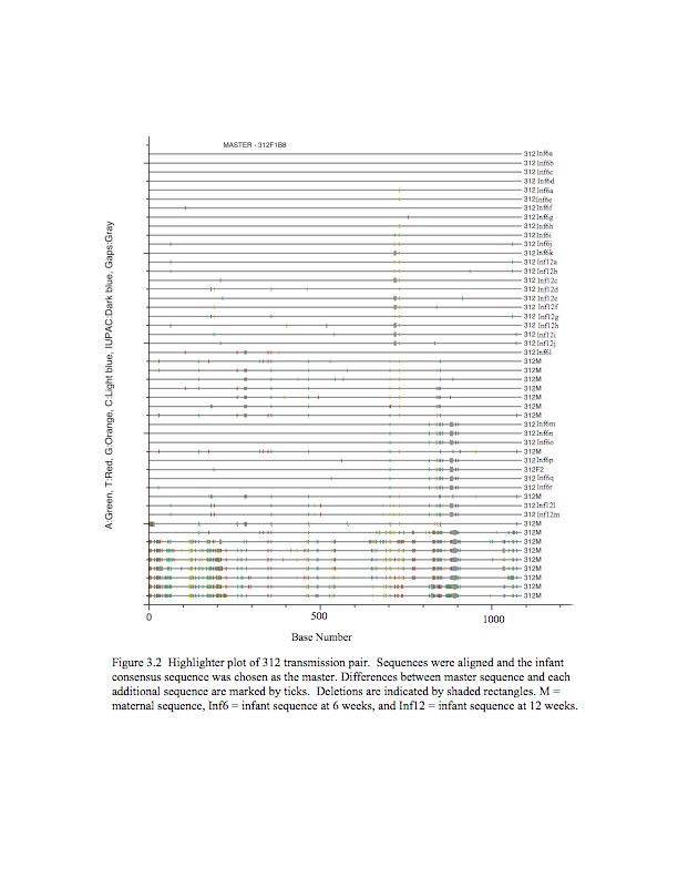
79 70
80 Chapter 4 Broadly Neutralizing Maternal Antibodies in Subtype C MTCT 4.1 Introduction Preventing mother-to-child transmission (MTCT) relies on long, expensive drug treatments, cesarean section, and formula feeding of the infant (72). These prevention methods are not feasible in most resource-poor areas of the world where MTCT continues to infect 15-40% of infants born to HIV+ women (92). Less expensive shortcourse drug therapies can reduce in utero and intrapartum transmission by 60-70%, yet coverage and cost remain barriers, and these therapies do not prevent post-partum transmission through breastfeeding. Breast milk infects 15% of infants born to HIV+ women (39). Even if formula were acceptable, feasible, and affordable, formula feeding cannot provide the protection from infectious diseases that breast milk can (29, 30, 147), making breast milk or formula about equal in terms of infant morbidity and mortality in resource-poor settings with high HIV-1 prevalence (28). Thus, interventions that could protect the infant from HIV infection, yet still allow the mother to breastfeed would be most desirable. While highly active antiretroviral therapy to the mother could fill this role, there are also many unresolved issues with long-term drug administration, including the high expense, limited access, possible toxicity to the infant (148), and the evolution of
81 resistance (95). An effective vaccine given to pregnant women and infants immediately after birth could protect the infant from MTCT, and possibly HIV transmission for life. Before a vaccine can be developed, however, it is necessary to determine mechanisms of transmission, including how transmission is naturally prevented in the majority of infants. If this mechanism were known, future interventions could elicit that protection in all pregnant women and their infants, or bolster it in those for whom it is weak. Neutralizing antibodies from the mother play a significant role in protecting infants from pathogens (26), and are a likely candidate for a natural protection mechanism against HIV infection (73, 100). Animal studies have demonstrated that neutralizing antibodies elicited by a vaccine can protect infant macaques against infection or at least slow disease progression (152), while direct administration of the appropriate antibodies can block transmission (45, 116). Studies of mother-to-child transmission have come to conflicting conclusions about the role of neutralizing antibodies (8, 9, 15, 49, 76, 134), though possibly for identifiable reasons. The breadth of the neutralizing antibody response has been shown to depend on subtype (13), and differences in neutralizing antibody levels can correlate with the timing of transmission (9). The fact that many previous MTCT studies did not analyze considering these factors could explain at least part of the discrepancies. Broadly reactive neutralizing antibodies that have been studied have the lowest neutralizing activity against subtype C virus (11), though this data is likely to be biased because most antibodies were originally isolated from subtype B infected subjects. A previous study found that sera from women infected with subtype CFR01_AE whose infants were infected intrapartum had higher neutralizing antibody titers against a viral 72
82 isolate of the same subtype than women whose infants were infected in utero or who remained uninfected (9). There were no differences in titer against 3 viral isolates of different subtypes. Comprehensive studies are needed to resolve these data and identify common neutralization sensitive epitopes among all prevalent subtypes. We correlated heterologous neutralizing antibody titers in sera from pregnant women infected with HIV-1 subtype C with the infection status of their infant. Women were classified as non-transmitters and women who transmitted in utero or intrapartum. Titers were measured against virus pseudotyped with two neutralization sensitive subtype B Env proteins and two neutralization sensitive subtype C Env proteins, one subtype C Env was generated from an isolate from the same country in which the sera were collected. There was no correlation between heterologous neutralizing antibody titer and transmission status. These data demonstrate the difficulty in identifying subtype C antibodies with broad neutralizing activity even within the same subtype. 4.2 Materials and Methods Patient Sera Serum samples were collected as part of the Malaria and HIV in Pregnancy Study (74, 75, 98, 99). Blood was collected from pregnant women upon admission to the labor ward. Infants were tested for HIV-1 infection at birth, then again at 6 and 12 weeks. All available sera from women who transmitted HIV in this study were used in the analysis of neutralizing antibodies, and sera from 48 non-transmitting women were randomly chosen from the study population. 73
83 4.2.2 Envelope Clones Envelope clones were chosen based on their known neutralization sensitivity and subtype. SF162 is highly sensitive to neutralization. JRCSF was isolated from a subject infected with subtype B and is known to be moderately sensitive to neutralization. TV-1 is a moderately sensitive subtype C Env. MW965 was isolated from a Malawian subject infected with subtype C and was chosen for its relative sensitivity to neutralization (David Montefiori, personal communication). JRCSF and SF612 were obtained from the AIDS Reference Reagent Repository, MW965 and TV-1 were generous gifts from David Montefiori Pseudotyped Virus Pseudotyped virus was made according to previously published protocols (93). Briefly, 293T cells were plated on day -1. The env expression plasmid and env pnlch backbone plus luciferase (a gift from Jerry Jeffrey) were cotransfected into the cells on day 0 using FuGENE 6 transfection reagent (Roche, Indianapolis, IN). On day 2 supernatants were harvested, centrifuged, and frozen at -80C in 1ml aliquots Neutralizing Antibody Assay Neutralizing antibody assays were performed as described (93). Virus was added to serum dilutions (1:20 and higher) and incubated at 37 C. After 1 hour TZM-bl cells were added. Two days later the cells were washed with PBS and lysed using Luciferase Reagent (Promega Corporation, Madison,WI). Plates were read on a luminometer. 74
84 Titers are reported as the highest dilution with greater than 50% inhibition of infectivity as compared to a no-serum infection within the same experiment Statistics Titers were compared with the Kruskal-Wallis statistic using Prism 4 (Graphpad Software, Inc.). 4.3 Results Subjects Serum was obtained from HIV+ pregnant women in labor in Blantyre, Malawi. Women were considered non-transmitting (NT) if their infants were uninfected at birth and 6 weeks. Women were classified to have transmitted in utero (IU) if their infants were HIV+ at birth, and transmitted intrapartum (IP) if their infants were HIV- at birth and positive at 6 weeks. Sera from 48 NT, 21 IU, and 20 IP women were available for this study Heterologous Neutralizing Antibody Titers Each serum was tested for neutralizing antibody activity against virus pseudotyped with 4 heterologous envelopes, 2 subtype C and 2 subtype B. TV-1 is a moderately sensitive subtype C Env. Median neutralizing antibody titers were similar for all transmission modes against this Env, NT, IP = 80, IU = 40 (p=0.30, data not shown). MW965 is a neutralization-sensitive subtype C envelope isolated from a Malawian subject. Women in this cohort are all likely infected with subtype C (unpublished data), 75
85 making MW965 a heterologous virus of the same subtype. Median neutralizing antibody titers were the same for women of all 3 transmission groups, NT, IU, and IP = 180 (p = 0.7) (Figure 4.1a). SF162 is a subtype C Env and is highly sensitive to neutralizing antibodies, and median titers were not different against this virus, NT, IU, and IP = 10 (p=0.71, data not shown). JRCSF is a moderately neutralization sensitive subtype B envelope. Median neutralizing antibody titers were also similar for JRCSF (NT=60, IU=60, IP=120; p = 0.4) (Figure 4.1b). Thus, neutralizing antibody titers against a heterologous virus of the same, or different, subtypes did not correlate with transmission or transmission timing of subtype C HIV-1 from mother-to-child. 4.4 Discussion Limited data are currently available to give insight into the role of neutralizing antibodies in mother-to-child transmission for relevant subtypes due to a lack of appropriate samples. In this study we tested for a correlation between mother-to-child transmission of subtype C HIV-1 and heterologous neutralizing antibody titer in sera from 48 non-transmitting (NT), 21 in utero transmitting (IU), and 20 intrapartum (IP) transmitting women. We found no differences in titer against a virus pseudotyped with a heterologous subtype C envelope, or of a subtype B envelope. Conflicting results have been found in studies correlating mother-to-child transmission and heterologous antibody responses (9, 15, 23, 49, 136). Many studies have been small, from women infected with a variety of subtypes, and have not separated transmission events by timing. A comprehensive study by Barin et al. (9) tested sera 76
86 from 62 NT, 11 IU, and 17 IP women, 81 of whom were infected with HIV-1 subtype CRF01_AE, against 4 heterologous viruses. The 4 viruses were of subtypes CRF01_AE, B, F, and CRF02_AG. They found that women whose infants were infected intrapartum had significantly lower neutralizing antibody titers to the CRF01_AE pseudovirus than NT women or women whose infants were infected IU. They also found that there was no correlation between neutralization and transmission status against the other 3 viruses of different subtypes. Thus, in their study, lower titers of heterologous antibodies against virus of the same subtype correlated with IP transmission, while there were no correlations with the titer of antibodies against viruses of different subtypes. In the study reported here, however, we found no association between heterologous NAB titer and transmission for either the subtype C or subtype B pseudotyped viruses we assayed. These results could be related to a reported lack of NAB breadth in subtype C infections (81). It could also be the case that a correlation would be seen against an Env protein from a different isolate. Regardless, unlike a similar study of women infected with subtype CRF01_AE, we found no difference between neutralizing antibody titers of sera from NT, IU, and IP women infected with subtype C HIV against heterologous virus of the same subtype. In this study we explored the relationship between heterologous neutralizing antibodies and mother-to-child transmission of subtype C HIV-1. We found no association between heterologous NAB titer and transmission, unlike has been seen with other subtypes (9). These results further highlight the importance of research on the prevalent subtypes of HIV-1. Subtype C currently causes more infections than any other 77
87 subtype (149), yet the least is known about how to neutralize it (11). Further work should identify epitopes common among subtype C viruses that are susceptible to neutralization. 4.4 Future Directions Autologous neutralizing antibodies have been shown to protect infants from MTCT, particularly for IP transmission. In one study by Wu et al (160) the transmitted viral variants were more resistant to maternal NAB than untransmitted maternal variants in infants infected IP with subtypes A, C, or D. This demonstrates that infants were protected from neutralization-sensitive variants. Another study found that low autologous NAB titer in women infected with subtype B correlated with IU transmission when compared to NT and IP women (36). Thus, neutralizing antibodies are able to protect infants from infection with viral variants that are sensitive to these antibodies. In the future we will examine the ability of autologous NAB to protect infants from infection in a subset of pairs from the MHP study. We are currently cloning env genes from viral RNA isolated from IU (n=10) and IP (n=6) transmission pairs. These clones will be used to make pseudotyped virus and will be tested for sensitivity to heterologous sera and monoclonal antibodies. These assays will determine whether transmitted variants are inherently more resistant to neutralization than matched maternal populations. The pseudoviruses will also be tested against autologous maternal sera. We hypothesize that in this relatively large number of subtype C MTCT pairs we will find that IP-infected infants are protected from infection with viruses that are sensitive to autologous neutralizing antibodies. 78


88 a. 0 NT In Utero Intrapartum b. 0 NT In Utero Intrapartum Figure 4.1. Lowest titer of maternal serum with > 50% inhibition of infectivity. NT = non-transmitting women. Median titer denoted by line. a. Titer against virus pseudotyped with subtype C Env MW965. b. Titer against virus pseudotyped with subtype B Env JRCSF. 79
89 Chapter 5 Diversity in env during HIV-1 Subtype C Mother-to-Child Transmission 5.1 Introduction HIV mother-to-child transmission (MTCT) through breastfeeding is responsible for one third to one half of infections in infants in Sub-Saharan Africa, where 90% of HIV infected children live (149). Short-course drug regimens, which can reduce in utero (IU) and intrapartum (IP) MTCT from 15-25% to approximately 10% (92), are currently recommended for HIV+ pregnant women in regions where HAART, elective cesarean section, and formula feeding are not feasible or safe. These regimens, however, do little to reduce transmission through breastfeeding, which infects approximately 15% of infants born to HIV+ women (39). New interventions are needed that can be applied in developing countries, yet this is difficult because little is known about the mechanism(s) of transmission. The oral cavity and gastrointestinal tract of breastfed infants come into frequent contact with viral particles each day from both cell-free and cell-associated HIV-1 (79, 101, 150). Higher concentrations of HIV in breast milk correlate to higher rates of transmission (123, 124). Common hypotheses for transmission routes include breaches in mucosal surfaces,
90 transport across M cells, or indirect infection of epithelial cells (108). The fact that 85% of infants remain uninfected despite ingesting large amounts of HIV-infected breast milk indicates the inefficiency of the transmission process and highlights the need to define the mechanism of transmission and the usually protective mechanisms that preclude transmission (55, 151, 153). This work so far has pointed to several protective innate immune factors against HIV-1 infection in vitro and in vivo,, many being natural ligands for the HIV coreceptors CCR5 and CXCR4, while other factors may increase infection (41). The acidic ph of the stomach (132), and the presence of anti-hiv factors in saliva (143) may also play protective roles. Studies must now begin to determine whether transmission is a result of a random virus breaking through these protections by chance, if there are specific viral characteristics that evade protection mechanisms, or a combination of these or other factors. There are limited data on the characteristics of virus transmitted through breastfeeding, and much of what is known has focused on small regions of env and/or the characterization of only a few sequences from each subject (117, 155), though a genetic bottleneck has been observed. Studies of in utero and intrapartum transmission have shown differences in the characteristics of transmitted virus if the infant was infected in utero or intrapartum (37, 75). In addition, studies of horizontal transmission have demonstrated differences in viral characteristics of transmitted viruses depending on the infecting subtype (27, 35). In subtypes C and A viruses with fewer glycosylation sites and shorter lengths (i.e. more compact viruses) are transmitted, though not in subtype B transmission events. Viral characteristics during breast milk transmission of subtype C 81
91 should also be determined in order to understand whether interventions for prevention of transmission would likely be effective for all modes of MTCT and all subtypes. In this study we amplified env from 3 HIV+ mother-infant pairs where the infant was infected through breast milk. We sequenced gp120 and found heterogeneous viral populations in the mothers and relatively homogenous populations in the infants. In two infants we found evidence of single variant transmission, while multiple variants were transmitted to one infant. Infant sequences had fewer N-linked glycosylation sites and shorter sequences than maternal sequences. Though the study is small, these results are consistent with selection for virus with shorter variable loops and fewer glycosylation sites during transmission of HIV-1 subtype C from mother-to-child through breast milk. 5.2 Materials and Methods Study Participants Plasma samples were collected as part of the Malaria and HIV-1 in Pregnancy (MHP) prospective cohort (74, 75, 98, 99). The MHP study was approved by both the Malawi College of Medicine Research Committee and the Institutional Review Board at the University of North Carolina at Chapel Hill. Informed consent was obtained for all participants. Plasma was isolated from blood collected at labor-ward admission from the women, from the umbilical cord, and from infant heel-sticks at three time-points: within 48 hours of birth, at 6-weeks, and at 12-weeks of age. Women and their newborn infants received single-dose nevirapine according to the HIVNET 012 protocol (48). HIV transmission from mother-to-infant was categorized by timing where infants who were 82
92 HIV-1 DNA negative by real-time PCR (86) at 0 and 6 weeks, then positive at 12 weeks were classified as infected post-partum through breastfeeding (BF) Single-genome amplification Viral RNA was isolated from plasma samples using the QIAmp Viral RNA Mini Kit (Qiagen, Germantown, MD, USA). The single-genome amplification (SGA) method for the env gene was used for RT-PCR (128). Briefly, cdna was generated using Superscript III Reverse Transcriptase and Oligo(dT) Primer, followed by RNaseH treatment (Invitrogen Corp, Carlsbad, CA). The env gene was amplified by nested PCR from the dilution of cdna that resulted in approximately 30% positive PCR reactions. These conditions ensure that the large majority of amplifications are initiated with a single template, and eliminates artifactual recombination during PCR between multiple template sequences Phylogenetic Analysis Sequences were generated then manually edited and aligned using MAFFT version 5.8, with the L-INS-i method (66). The alignment was converted to PHYLIP format and a maximum likelihood phylogenetic tree was constructed using PHYML with a HKY85 evolutionary model (50). Trees were resampled 100 times and bootstrap values greater than 70 were considered significant. A neighbor-joining tree including sequences from each pair was constructed to assess quality control. Matched maternal and infant sequences formed monophyletic groups distinct from the other pairs. Pairwise 83
93 comparison diversity was calculated within lineages with Molecular Evolutionary Genetics Analysis software (MEGA 4.0) using the Kimura-2 parameter Statistical Methods Values were compared using the Wilcoxon matched-pairs signed rank test. 5.3 Results Subjects Plasma samples were obtained from 3 mother-infant pairs where infants were HIV negative at birth and 6 weeks, and HIV positive at 12 weeks. Transmission was classified as having occurred post-partum through breastfeeding (BF). Single-genome amplification was used to obtain 103 env gene amplicons from mothers and infants. Phylogenetic linkage of the viral sequences from the mother and infant pairs was confirmed using a neighbor-joining tree (data not shown) Genetic bottleneck A genetic bottleneck was observed between maternal and infant env populations. Pairwise diversity was calculated for each viral population. All maternal populations were more heterogeneous than the paired infant populations by pairwise comparison, consistent with a bottleneck (Fig. 5.1). Sequences were next analyzed using maximum likelihood phylogenetic trees and the Highlighter tool ( Infant sequences formed a single lineage with no intermingled maternal sequences in pairs 1266 and 1677, likely representing transmission of a minor maternal plasma variant (Fig. 5.2b-c). 84
94 Because we sequenced an average of 17 maternal env genes, we have 95% confidence we sampled variants that comprised >18% of the maternal population. These minor variants detected in the infants could be maternal variants comprising <18%, or they could be viral variants compartmentalized in the breast milk. In pair 1266 all infant sequences form a single lineage, but one maternal sequence has continuous sequence identity with the infant sequence over 50% of the length of env, likely indicating recombination occurred between the similar maternal sequence and an additional unamplified variant. This is further evidence that this variant exists in the maternal plasma population, albeit at a low frequency. Thus in 2 of 3 BF mother-infant pairs we found that a single variant was transmitted to the infant, and this variant did not represent a majority of the maternal plasma viral population. In the remaining mother-infant pair there is evidence for the transmission of at least 2 variants. In pair 942, all infant sequences were found in a single lineage that also had 2/19 maternal sequences intermingled (Fig. 5.2a). Examining these sequences with Highlighter, 14/17 infant sequences were nearly identical and were distinct from amplified maternal sequences (Fig. 5.3a). The remaining 2 maternal sequences and 1 infant sequence were more similar to the majority of the infant sequences than the other maternal sequences, but had numerous common differences from the infant consensus. The final infant sequence appears to be a recombinant of the infant consensus and minor infant variants. Because we used single-genome amplification, this recombination event could not have occurred during PCR, and we propose recombination within the infant as the likely source between 2 different sequences. Both sequences could have transmitted to the infant during the same transmission event, or through 2 separate events. Thus, our 85
95 data indicate in 1 of 3 BF transmission pairs, 2 closely related variants were transmitted, one of which was also detected in the maternal population Viral Genetic Characteristics We compared the number of glycosylation sites and sequence length between mother and infant viral populations. Previous work has shown more compact viruses with fewer glycosylation sites and shorter variable loops are transmitted during horizontal transmission of subtype C (35), and during IP vertical transmission (our unpublished results). These differences were seen over the entire env gene in all three pairs (Fig. 5.4), though not for each variable loop between sequences from each pair (data not shown). In this data set we found fewer glycosylation sites and shorter variable loops in BF transmitted subtype C variants. The small sample size of this initial study precludes a test for statistical significance Selective Pressure in Infant Keele et al. (67) recently created a neutral model of HIV-1 sequence evolution through the first 50 days of infection. This model is based on early sequence data from subjects infected through horizontal transmission. We tested this model against our sequence data from infants who had tested HIV-1 negative 42 days prior to the positive sample (i.e. 6 weeks after birth). According to the model, with an infection of 42 days or less, >60% of sequences should be identical. Infants in this study had 33, 7, and 20% identical sequences (942-major lineage, 1266, and 1677, respectively). According to this model, the infants have more sequence heterogeneity than would be accounted for by 86
96 random neutral mutations. If we extend the model beyond 50 days it would predict infection times of 99, 238, and 136 days in these infants, respectively. These infection times are extremely unlikely given the additional HIV negative results at birth, and therefore suggest selection occurred in these infants. Because maternal antibodies are transmitted to infants through breast milk, this could alter the pattern of evolution in a vertically infected infant compared to that of horizontally infected subjects. However, the mutations in the infant sequences do not cluster within variable loops, and are in fact often in gp41 (Fig. 5.3a-c). This region is not targeted by antibodies as often as variable loops, thus this could demonstrate cytotoxic T-lymphocyte selection. Sites of multiple mutations can be located in areas of high CTL selection. We are unable to confirm specific epitopes because the HLA type of these women and infants is unknown. Therefore, unlike subjects infected through horizontal transmission, env sequences from infants infected through breast milk show evidence of selection within the first 6 weeks of infection, and this selection may be more consistent with cytotoxic T-lymphocytes rather than maternal antibodies. 5.4 Discussion In this study we analyzed env sequences from 3 mother-infant pairs where HIV-1 subtype C was transmitted through breastfeeding (BF). Similar to previous studies of in utero and intrapartum transmission from mother-to-child (129, 154), and one study with limited subtype A sequence data by Rainwater et al. (117), we saw a strong viral genetic bottleneck in all three pairs. For the pairs described herein, pairwise diversity was considerably less for the infant, as compared to maternal, sequences for all 3 pairs. 87
97 Recently, large studies have determined the likelihood of transmission of multiple variants in horizontal transmission (2, 67), along with smaller studies in vertical transmission (37, 129, 154). These studies all have similar results, where the majority of recently infected subjects have a viral population containing a single variant, while approximately 20% have multiple variants. Even with our small dataset we mirror these results, two variants were detected in 1/3 infants, while only one variant was detected in 2 infants. The presence of multiple variants in the infant could represent separate transmission events, or multiple variants transmitting as part of the same event. In the infant described the two variants are similar yet transmitted from a more diverse maternal viral population, therefore multiple transmission events would indicate a strong selection for similar characteristics (either during transmission or through selective amplification in the infant), or a dramatically different viral population in breast milk compared to plasma. No studies have definitively compared viral populations in plasma and breast milk, and the small studies that have been published are conflicting (10, 53, 65). More data are needed to determine the relationship between maternal plasma and breast milk viral populations. Sequences from infants had fewer N-linked glycosylation sites and shorter variable loops than matched maternal sequences. Other studies of subtype C and A horizontal (27, 35), and subtype C IP (our unpublished results) transmission have identified this selection for shorter variable loops and fewer glycosylation sites. Here we extend this finding with a trend in 3 subtype C BF transmission pairs. No published studies have examined loop length and number of glycosylation sites in BF transmission. 88
98 Research has recently begun to focus in more detail on BF transmission, and additional studies with large numbers of pairs covering all relevant subtypes will hopefully be completed to fully characterize the BF transmitted virus. A better understanding of what mechanisms drive BF transmission and shared traits of viruses seeding acute infections would help in developing new interventions. Early sequence changes in the infant viral population may also provide clues to what immune pressures are present and/or absent. New interventions could bolster immune responses naturally present, and perhaps activate deficient responses. In horizontal transmission of HIV-1 the virus recipient immune system is naïve to HIV, but has a functional, developed immune system. Infants receive maternal antibodies through the placenta and breast milk, yet new infant responses are slower to activate (25, 87). We analyzed infant sequences using a neutral evolution model developed from subjects horizontally infected with HIV-1. Sequences from these infants infected through BF had more heterogeneity than could be accounted for by random neutral evolution. Thus, we conclude that selection is occurring in these infants within the first 42 days of infection. If this selection was due to maternal antibody selection, mutations would likely cluster in the variable regions of env. Mutations instead often clustered in gp41 near known CTL epitopes. Not knowing the HLA type of these subjects we cannot confirm CTL escape, but it raises an interesting question for future study. Additional longitudinal studies of larger numbers of subjects could be done to characterize early infant responses and sequence evolution to determine the source of this diversity. In this study we analyzed env sequence data from 3 HIV-1 subtype C breastfeeding transmission pairs. A strong genetic bottleneck took place during 89
99 transmission, even when multiple variants were transmitted. Transmitted variants had fewer glycosylation sites and shorter sequences than the average of the maternal population. We also saw evidence for selective evolution in these infants, perhaps as a result of cytotoxic T-lymphocytes. Thus, we present data for selection during both transmission and early evolution in these 3 infants. Figure 5.1 Pairwise comparison of maternal and infant sequence populations. Error bars indicate standard deviation. 90
100 91
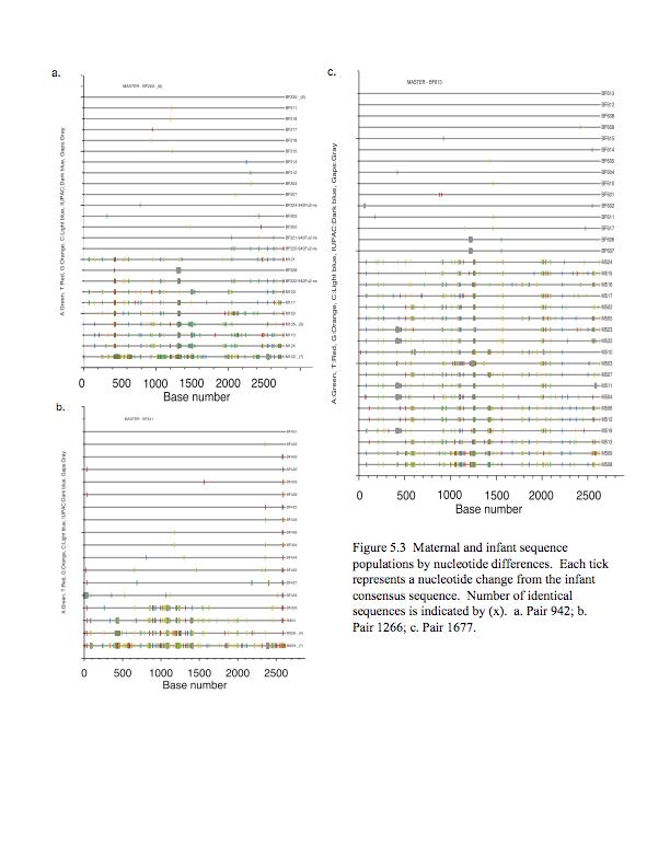
101 92
Fayth K. Yoshimura, Ph.D. September 7, of 7 HIV - BASIC PROPERTIES
 1 of 7 I. Viral Origin. A. Retrovirus - animal lentiviruses. HIV - BASIC PROPERTIES 1. HIV is a member of the Retrovirus family and more specifically it is a member of the Lentivirus genus of this family.
Human Immunodeficiency Virus
 Human Immunodeficiency Virus Virion Genome Genes and proteins Viruses and hosts Diseases Distinctive characteristics Viruses and hosts Lentivirus from Latin lentis (slow), for slow progression of disease
MedChem 401~ Retroviridae. Retroviridae
 MedChem 401~ Retroviridae Retroviruses plus-sense RNA genome (!8-10 kb) protein capsid lipid envelop envelope glycoproteins reverse transcriptase enzyme integrase enzyme protease enzyme Retroviridae The
HIV Immunopathogenesis. Modeling the Immune System May 2, 2007
 HIV Immunopathogenesis Modeling the Immune System May 2, 2007 Question 1 : Explain how HIV infects the host Zafer Iscan Yuanjian Wang Zufferey Abhishek Garg How does HIV infect the host? HIV infection
HIV INFECTION: An Overview
 HIV INFECTION: An Overview UNIVERSITY OF PAPUA NEW GUINEA SCHOOL OF MEDICINE AND HEALTH SCIENCES DIVISION OF BASIC MEDICAL SCIENCES DISCIPLINE OF BIOCHEMISTRY & MOLECULAR BIOLOGY PBL MBBS II SEMINAR VJ
HIV & AIDS: Overview
 HIV & AIDS: Overview UNIVERSITY OF PAPUA NEW GUINEA SCHOOL OF MEDICINE AND HEALTH SCIENCES DIVISION OF BASIC MEDICAL SCIENCES DISCIPLINE OF BIOCHEMISTRY & MOLECULAR BIOLOGY PBL SEMINAR VJ TEMPLE 1 What
Lecture 2: Virology. I. Background
 Lecture 2: Virology I. Background A. Properties 1. Simple biological systems a. Aggregates of nucleic acids and protein 2. Non-living a. Cannot reproduce or carry out metabolic activities outside of a
How HIV Causes Disease Prof. Bruce D. Walker
 How HIV Causes Disease Howard Hughes Medical Institute Massachusetts General Hospital Harvard Medical School 1 The global AIDS crisis 60 million infections 20 million deaths 2 3 The screen versions of
HIV/AIDS. Biology of HIV. Research Feature. Related Links. See Also
 6/1/2011 Biology of HIV Biology of HIV HIV belongs to a class of viruses known as retroviruses. Retroviruses are viruses that contain RNA (ribonucleic acid) as their genetic material. After infecting a
RAISON D ETRE OF THE IMMUNE SYSTEM:
 RAISON D ETRE OF THE IMMUNE SYSTEM: To Distinguish Self from Non-Self Thereby Protecting Us From Our Hostile Environment. Innate Immunity Acquired Immunity Innate immunity: (Antigen nonspecific) defense
Principles of Adaptive Immunity
 Principles of Adaptive Immunity Chapter 3 Parham Hans de Haard 17 th of May 2010 Agenda Recognition molecules of adaptive immune system Features adaptive immune system Immunoglobulins and T-cell receptors
Immunodeficiency. (2 of 2)
") Immunodeficiency (2 of 2) Acquired (secondary) immunodeficiencies More common Many causes such as therapy, cancer, sarcoidosis, malnutrition, infection & renal disease The most common of which is therapy-related
Immunodeficiency (2 of 2) Acquired (secondary) immunodeficiencies More common Many causes such as therapy, cancer, sarcoidosis, malnutrition, infection & renal disease The most common of which is therapy-related
LESSON 4.6 WORKBOOK. Designing an antiviral drug The challenge of HIV
 LESSON 4.6 WORKBOOK Designing an antiviral drug The challenge of HIV In the last two lessons we discussed the how the viral life cycle causes host cell damage. But is there anything we can do to prevent
 CDC site UNAIDS Aids Knowledge Base http://www.cdc.gov/hiv/dhap.htm http://hivinsite.ucsf.edu/insite.jsp?page=kb National Institute of Allergy and Infectious Diseases http://www.niaid.nih.gov/default.htm
I. Bacteria II. Viruses including HIV. Domain Bacteria Characteristics. 5. Cell wall present in many species. 6. Reproduction by binary fission
 Disease Diseases I. Bacteria II. Viruses including are disease-causing organisms Biol 105 Lecture 17 Chapter 13a Domain Bacteria Characteristics 1. Domain Bacteria are prokaryotic 2. Lack a membrane-bound
Fig. 1: Schematic diagram of basic structure of HIV
 UNIVERSITY OF PAPUA NEW GUINEA SCHOOL OF MEDICINE AND HEALTH SCIENCES DIVISION OF BASIC MEDICAL SCIENCES DISCIPLINE OF BIOCHEMISTRY & MOLECULAR BIOLOGY PBL SEMINAR HIV & AIDS: An Overview What is HIV?
8/13/2009. Diseases. Disease. Pathogens. Domain Bacteria Characteristics. Bacteria Shapes. Domain Bacteria Characteristics
 Disease Diseases I. Bacteria II. Viruses including Biol 105 Lecture 17 Chapter 13a are disease-causing organisms Domain Bacteria Characteristics 1. Domain Bacteria are prokaryotic 2. Lack a membrane-bound
Retroviruses. ---The name retrovirus comes from the enzyme, reverse transcriptase.
 Retroviruses ---The name retrovirus comes from the enzyme, reverse transcriptase. ---Reverse transcriptase (RT) converts the RNA genome present in the virus particle into DNA. ---RT discovered in 1970.
, virus identified as the causative agent and ELISA test produced which showed the extent of the epidemic
 1 Two attributes make AIDS unique among infectious diseases: it is uniformly fatal, and most of its devastating symptoms are not due to the causative agent Male to Male sex is the highest risk group in
RAISON D ETRE OF THE IMMUNE SYSTEM:
 RAISON D ETRE OF THE IMMUNE SYSTEM: To Distinguish Self from Non-Self Thereby Protecting Us From Our Hostile Environment. Innate Immunity Adaptive Immunity Innate immunity: (Antigen - nonspecific) defense
Fayth K. Yoshimura, Ph.D. September 7, of 7 RETROVIRUSES. 2. HTLV-II causes hairy T-cell leukemia
 1 of 7 I. Diseases Caused by Retroviruses RETROVIRUSES A. Human retroviruses that cause cancers 1. HTLV-I causes adult T-cell leukemia and tropical spastic paraparesis 2. HTLV-II causes hairy T-cell leukemia
Medical Virology Immunology. Dr. Sameer Naji, MB, BCh, PhD (UK) Head of Basic Medical Sciences Dept. Faculty of Medicine The Hashemite University
 Head of Basic Medical Sciences Dept. Faculty of Medicine The Hashemite University") Medical Virology Immunology Dr. Sameer Naji, MB, BCh, PhD (UK) Head of Basic Medical Sciences Dept. Faculty of Medicine The Hashemite University Human blood cells Phases of immune responses Microbe Naïve
Medical Virology Immunology Dr. Sameer Naji, MB, BCh, PhD (UK) Head of Basic Medical Sciences Dept. Faculty of Medicine The Hashemite University Human blood cells Phases of immune responses Microbe Naïve
Virology Introduction. Definitions. Introduction. Structure of virus. Virus transmission. Classification of virus. DNA Virus. RNA Virus. Treatment.
 DEVH Virology Introduction Definitions. Introduction. Structure of virus. Virus transmission. Classification of virus. DNA Virus. RNA Virus. Treatment. Definitions Virology: The science which study the
11/15/2011. Outline. Structural Features and Characteristics. The Good the Bad and the Ugly. Viral Genomes. Structural Features and Characteristics
 Chapter 19 - Viruses Outline I. Viruses A. Structure of viruses B. Common Characteristics of Viruses C. Viral replication D. HIV II. Prions The Good the Bad and the Ugly Viruses fit into the bad category
Viral Genetics. BIT 220 Chapter 16
 Viral Genetics BIT 220 Chapter 16 Details of the Virus Classified According to a. DNA or RNA b. Enveloped or Non-Enveloped c. Single-stranded or double-stranded Viruses contain only a few genes Reverse
7.014 Problem Set 7 Solutions
 MIT Department of Biology 7.014 Introductory Biology, Spring 2005 7.014 Problem Set 7 Solutions Question 1 Part A Antigen binding site Antigen binding site Variable region Light chain Light chain Variable
Lentiviruses: HIV-1 Pathogenesis
 Lentiviruses: HIV-1 Pathogenesis Human Immunodeficiency Virus, HIV, computer graphic by Russell Kightley Tsafi Pe ery, Ph.D. Departments of Medicine and Biochemistry & Molecular Biology NJMS, UMDNJ. e-mail:
Third line of Defense
 Chapter 15 Specific Immunity and Immunization Topics -3 rd of Defense - B cells - T cells - Specific Immunities Third line of Defense Specific immunity is a complex interaction of immune cells (leukocytes)
Adaptive Immunity: Humoral Immune Responses
 MICR2209 Adaptive Immunity: Humoral Immune Responses Dr Allison Imrie 1 Synopsis: In this lecture we will review the different mechanisms which constitute the humoral immune response, and examine the antibody
Section 6. Junaid Malek, M.D.
 Section 6 Junaid Malek, M.D. The Golgi and gp160 gp160 transported from ER to the Golgi in coated vesicles These coated vesicles fuse to the cis portion of the Golgi and deposit their cargo in the cisternae
CELL BIOLOGY - CLUTCH CH THE IMMUNE SYSTEM.
 !! www.clutchprep.com CONCEPT: OVERVIEW OF HOST DEFENSES The human body contains three lines of against infectious agents (pathogens) 1. Mechanical and chemical boundaries (part of the innate immune system)
5. Over the last ten years, the proportion of HIV-infected persons who are women has: a. Increased b. Decreased c. Remained about the same 1
 Epidemiology 227 April 24, 2009 MID-TERM EXAMINATION Select the best answer for the multiple choice questions. There are 60 questions and 9 pages on the examination. Each question will count one point.
Acquired immune deficiency syndrome.
 Acquired immune deficiency syndrome 491 Acquired immune deficiency syndrome. The first cases of acquired immune deficiency syndrome (AIDS) were reported in 1981 but it is now clear that cases of the disease
Hepatitis virus immunity. Mar 9, 2005 Rehermann and Nascimbeni review Crispe review
 Hepatitis virus immunity Mar 9, 2005 Rehermann and Nascimbeni review Crispe review HBV & HCV infection outcomes Both viruses cause immune-mediated active and chronic hepatitis HBV Vertical transmission
Foundations in Microbiology
 Foundations in Microbiology Fifth Edition Talaro Chapter 15 The Acquisition of Specific Immunity and Its Applications Chapter 15 2 Chapter Overview 1. Development of the Dual Lymphocyte System 2. Entrance
Antigen Receptor Structures October 14, Ram Savan
 Antigen Receptor Structures October 14, 2016 Ram Savan savanram@uw.edu 441 Lecture #8 Slide 1 of 28 Three lectures on antigen receptors Part 1 (Today): Structural features of the BCR and TCR Janeway Chapter
Human Immunodeficiency Virus. Acquired Immune Deficiency Syndrome AIDS
 Human Immunodeficiency Virus Acquired Immune Deficiency Syndrome AIDS Sudden outbreak in USA of opportunistic infections and cancers in young men in 1981 Pneumocystis carinii pneumonia (PCP), Kaposi s
19 Viruses BIOLOGY. Outline. Structural Features and Characteristics. The Good the Bad and the Ugly. Structural Features and Characteristics
 9 Viruses CAMPBELL BIOLOGY TENTH EDITION Reece Urry Cain Wasserman Minorsky Jackson Outline I. Viruses A. Structure of viruses B. Common Characteristics of Viruses C. Viral replication D. HIV Lecture Presentation
HIV 101: Fundamentals of HIV Infection
 HIV 101: Fundamentals of HIV Infection David H. Spach, MD Professor of Medicine University of Washington Seattle, Washington Learning Objectives After attending this presentation, learners will be able
Citation for published version (APA): Von Eije, K. J. (2009). RNAi based gene therapy for HIV-1, from bench to bedside
: Von Eije, K. J. (2009). RNAi based gene therapy for HIV-1, from bench to bedside") UvA-DARE (Digital Academic Repository) RNAi based gene therapy for HIV-1, from bench to bedside Von Eije, K.J. Link to publication Citation for published version (APA): Von Eije, K. J. (2009). RNAi based
UvA-DARE (Digital Academic Repository) RNAi based gene therapy for HIV-1, from bench to bedside Von Eije, K.J. Link to publication Citation for published version (APA): Von Eije, K. J. (2009). RNAi based
LESSON 4.4 WORKBOOK. How viruses make us sick: Viral Replication
 DEFINITIONS OF TERMS Eukaryotic: Non-bacterial cell type (bacteria are prokaryotes).. LESSON 4.4 WORKBOOK How viruses make us sick: Viral Replication This lesson extends the principles we learned in Unit
Introduction retroposon
 17.1 - Introduction A retrovirus is an RNA virus able to convert its sequence into DNA by reverse transcription A retroposon (retrotransposon) is a transposon that mobilizes via an RNA form; the DNA element
number Done by Corrected by Doctor Ashraf
 number 4 Done by Nedaa Bani Ata Corrected by Rama Nada Doctor Ashraf Genome replication and gene expression Remember the steps of viral replication from the last lecture: Attachment, Adsorption, Penetration,
numbe r Done by Corrected by Doctor
 numbe r 5 Done by Mustafa Khader Corrected by Mahdi Sharawi Doctor Ashraf Khasawneh Viral Replication Mechanisms: (Protein Synthesis) 1. Monocistronic Method: All human cells practice the monocistronic
Third line of Defense. Topic 8 Specific Immunity (adaptive) (18) 3 rd Line = Prophylaxis via Immunization!
 (18) 3 rd Line = Prophylaxis via Immunization!") Topic 8 Specific Immunity (adaptive) (18) Topics - 3 rd Line of Defense - B cells - T cells - Specific Immunities 1 3 rd Line = Prophylaxis via Immunization! (a) A painting of Edward Jenner depicts a cow
Topic 8 Specific Immunity (adaptive) (18) Topics - 3 rd Line of Defense - B cells - T cells - Specific Immunities 1 3 rd Line = Prophylaxis via Immunization! (a) A painting of Edward Jenner depicts a cow
Lecture 11. Immunology and disease: parasite antigenic diversity
 Lecture 11 Immunology and disease: parasite antigenic diversity RNAi interference video and tutorial (you are responsible for this material, so check it out.) http://www.pbs.org/wgbh/nova/sciencenow/3210/02.html
Julianne Edwards. Retroviruses. Spring 2010
 Retroviruses Spring 2010 A retrovirus can simply be referred to as an infectious particle which replicates backwards even though there are many different types of retroviruses. More specifically, a retrovirus
Immunology - Lecture 2 Adaptive Immune System 1
 Immunology - Lecture 2 Adaptive Immune System 1 Book chapters: Molecules of the Adaptive Immunity 6 Adaptive Cells and Organs 7 Generation of Immune Diversity Lymphocyte Antigen Receptors - 8 CD markers
All animals have innate immunity, a defense active immediately upon infection Vertebrates also have adaptive immunity
 1 2 3 4 5 6 7 8 9 The Immune System All animals have innate immunity, a defense active immediately upon infection Vertebrates also have adaptive immunity Figure 43.2 In innate immunity, recognition and
Micropathology Ltd. University of Warwick Science Park, Venture Centre, Sir William Lyons Road, Coventry CV4 7EZ
 www.micropathology.com info@micropathology.com Micropathology Ltd Tel 24hrs: +44 (0) 24-76 323222 Fax / Ans: +44 (0) 24-76 - 323333 University of Warwick Science Park, Venture Centre, Sir William Lyons
October 26, Lecture Readings. Vesicular Trafficking, Secretory Pathway, HIV Assembly and Exit from Cell
 October 26, 2006 Vesicular Trafficking, Secretory Pathway, HIV Assembly and Exit from Cell 1. Secretory pathway a. Formation of coated vesicles b. SNAREs and vesicle targeting 2. Membrane fusion a. SNAREs
Centers for Disease Control August 9, 2004
 HIV CDC site UNAIDS Aids Knowledge Base http://www.cdc.gov/hiv/dhap.htm http://hivinsite.ucsf.edu/insite.jsp?page=kb National Institute of Allergy and Infectious Diseases http://www.niaid.nih.gov/default.htm
Identification of Mutation(s) in. Associated with Neutralization Resistance. Miah Blomquist
 in. Associated with Neutralization Resistance. Miah Blomquist") Identification of Mutation(s) in the HIV 1 gp41 Subunit Associated with Neutralization Resistance Miah Blomquist What is HIV 1? HIV-1 is an epidemic that affects over 34 million people worldwide. HIV-1
Identification of Mutation(s) in the HIV 1 gp41 Subunit Associated with Neutralization Resistance Miah Blomquist What is HIV 1? HIV-1 is an epidemic that affects over 34 million people worldwide. HIV-1
Immunobiology 7. The Humoral Immune Response
 Janeway Murphy Travers Walport Immunobiology 7 Chapter 9 The Humoral Immune Response Copyright Garland Science 2008 Tim Worbs Institute of Immunology Hannover Medical School 1 The course of a typical antibody
7.012 Quiz 3 Answers
 MIT Biology Department 7.012: Introductory Biology - Fall 2004 Instructors: Professor Eric Lander, Professor Robert A. Weinberg, Dr. Claudette Gardel Friday 11/12/04 7.012 Quiz 3 Answers A > 85 B 72-84
An Evolutionary Story about HIV
 An Evolutionary Story about HIV Charles Goodnight University of Vermont Based on Freeman and Herron Evolutionary Analysis The Aids Epidemic HIV has infected 60 million people. 1/3 have died so far Worst
Last time we talked about the few steps in viral replication cycle and the un-coating stage:
 Zeina Al-Momani Last time we talked about the few steps in viral replication cycle and the un-coating stage: Un-coating: is a general term for the events which occur after penetration, we talked about
Overview: Chapter 19 Viruses: A Borrowed Life
 Overview: Chapter 19 Viruses: A Borrowed Life Viruses called bacteriophages can infect and set in motion a genetic takeover of bacteria, such as Escherichia coli Viruses lead a kind of borrowed life between
Campbell's Biology: Concepts and Connections, 7e (Reece et al.) Chapter 24 The Immune System Multiple-Choice Questions
 Chapter 24 The Immune System Multiple-Choice Questions") Campbell's Biology: Concepts and Connections, 7e (Reece et al.) Chapter 24 The Immune System 24.1 Multiple-Choice Questions 1) The body's innate defenses against infection include A) several nonspecific
Campbell's Biology: Concepts and Connections, 7e (Reece et al.) Chapter 24 The Immune System 24.1 Multiple-Choice Questions 1) The body's innate defenses against infection include A) several nonspecific
Influenza viruses. Virion. Genome. Genes and proteins. Viruses and hosts. Diseases. Distinctive characteristics
 Influenza viruses Virion Genome Genes and proteins Viruses and hosts Diseases Distinctive characteristics Virion Enveloped particles, quasi-spherical or filamentous Diameter 80-120 nm Envelope is derived
COURSE: Medical Microbiology, MBIM 650/720 - Fall TOPIC: Antigen Processing, MHC Restriction, & Role of Thymus Lecture 12
 COURSE: Medical Microbiology, MBIM 650/720 - Fall 2008 TOPIC: Antigen Processing, MHC Restriction, & Role of Thymus Lecture 12 FACULTY: Dr. Mayer Office: Bldg. #1, Rm B32 Phone: 733-3281 Email: MAYER@MED.SC.EDU
A PROJECT ON HIV INTRODUCED BY. Abdul Wahab Ali Gabeen Mahmoud Kamal Singer
 A PROJECT ON HIV INTRODUCED BY Abdul Wahab Ali Gabeen Mahmoud Kamal Singer Introduction: Three groups of nations have been identified in which the epidemiology of HIV(Human Immunodeficiency Virus) varies:
Under the Radar Screen: How Bugs Trick Our Immune Defenses
 Under the Radar Screen: How Bugs Trick Our Immune Defenses Session 7: Cytokines Marie-Eve Paquet and Gijsbert Grotenbreg Whitehead Institute for Biomedical Research HHV-8 Discovered in the 1980 s at the
VIRUSES. Biology Applications Control. David R. Harper. Garland Science Taylor & Francis Group NEW YORK AND LONDON
 VIRUSES Biology Applications Control David R. Harper GS Garland Science Taylor & Francis Group NEW YORK AND LONDON vii Chapter 1 Virus Structure and 2.2 VIRUS MORPHOLOGY 26 Infection 1 2.3 VIRAL CLASSIFICATION
Ch 18 Infectious Diseases Affecting Cardiovascular and Lymphatic Systems
 Ch 18 Infectious Diseases Affecting Cardiovascular and Lymphatic Systems Highlight Disease: Malaria World s dominant protozoal disease. Four species of Plasmodium: P. falciparum (malignant), P. vivax (begnin),
The Adaptive Immune Responses
 The Adaptive Immune Responses The two arms of the immune responses are; 1) the cell mediated, and 2) the humoral responses. In this chapter we will discuss the two responses in detail and we will start
AP Biology. Viral diseases Polio. Chapter 18. Smallpox. Influenza: 1918 epidemic. Emerging viruses. A sense of size
 Hepatitis Viral diseases Polio Chapter 18. Measles Viral Genetics Influenza: 1918 epidemic 30-40 million deaths world-wide Chicken pox Smallpox Eradicated in 1976 vaccinations ceased in 1980 at risk population?
MID-TERM EXAMINATION
 Epidemiology 227 May 2, 2007 MID-TERM EXAMINATION Select the best answer for the multiple choice questions. There are 75 questions and 11 pages on the examination. Each question will count one point. Notify
Some living things are made of ONE cell, and are called. Other organisms are composed of many cells, and are called. (SEE PAGE 6)
") Section: 1.1 Question of the Day: Name: Review of Old Information: N/A New Information: We tend to only think of animals as living. However, there is a great diversity of organisms that we consider living
Section: 1.1 Question of the Day: Name: Review of Old Information: N/A New Information: We tend to only think of animals as living. However, there is a great diversity of organisms that we consider living
CURRENT DEVELOMENTS AND FUTURE PROSPECTS FOR HIV GENE THERAPY USING INTERFERING RNA-BASED STRATEGIES
 [Frontiers in Bioscience 5, d527-555, May 1, 2000] CURRENT DEVELOMENTS AND FUTURE PROSPECTS FOR HIV GENE THERAPY USING INTERFERING RNA-BASED STRATEGIES Betty Lamothe, Sadhna Joshi Department of Medical
Viral Vectors In The Research Laboratory: Just How Safe Are They? Dawn P. Wooley, Ph.D., SM(NRM), RBP, CBSP
, RBP, CBSP") Viral Vectors In The Research Laboratory: Just How Safe Are They? Dawn P. Wooley, Ph.D., SM(NRM), RBP, CBSP 1 Learning Objectives Recognize hazards associated with viral vectors in research and animal
Viral Vectors In The Research Laboratory: Just How Safe Are They? Dawn P. Wooley, Ph.D., SM(NRM), RBP, CBSP 1 Learning Objectives Recognize hazards associated with viral vectors in research and animal
Structure of HIV. Virion contains a membrane envelope with a single viral protein= Env protein. Capsid made up of Gag protein
 Structure of HIV Virion contains a membrane envelope with a single viral protein= Env protein Important in receptor recognition Capsid made up of Gag protein (group-specific antigen) Icosahedral Interior
The Adaptive Immune Response: T lymphocytes and Their Functional Types *
 OpenStax-CNX module: m46560 1 The Adaptive Immune Response: T lymphocytes and Their Functional Types * OpenStax This work is produced by OpenStax-CNX and licensed under the Creative Commons Attribution
Rama Nada. - Malik
 - 2 - Rama Nada - - Malik 1 P a g e We talked about HAV in the previous lecture, now we ll continue the remaining types.. Hepatitis E It s similar to virus that infect swine, so its most likely infect
NK mediated Antibody Dependent Cellular Cytotoxicity in HIV infections
 NK mediated Antibody Dependent Cellular Cytotoxicity in HIV infections Amy Chung Dr. Ivan Stratov Prof. Stephen Kent ADCC process consists of Target cell QuickTime and a TIFF (Uncompressed) FcγR decompressor
ACQUIRED IMMUNODEFICIENCY SYNDROME AND ITS OCULAR COMPLICATIONS
 ACQUIRED IMMUNODEFICIENCY SYNDROME AND ITS OCULAR COMPLICATIONS Acquired immunodeficiency syndrome (AIDS ) is an infectious disease caused by a retrovirus, the human immunodeficiency virus(hiv). AIDS is
White Blood Cells (WBCs)
") YOUR ACTIVE IMMUNE DEFENSES 1 ADAPTIVE IMMUNE RESPONSE 2! Innate Immunity - invariant (generalized) - early, limited specificity - the first line of defense 1. Barriers - skin, tears 2. Phagocytes - neutrophils,
YOUR ACTIVE IMMUNE DEFENSES 1 ADAPTIVE IMMUNE RESPONSE 2! Innate Immunity - invariant (generalized) - early, limited specificity - the first line of defense 1. Barriers - skin, tears 2. Phagocytes - neutrophils,
Irina Maljkovic Berry
 Thesis for doctoral degree (Ph.D.) 2008 Thesis for doctoral degree (Ph.D.) 2008 Genetic aspects of HIV-1 evolution and transmission Irina Maljkovic Berry Genetic aspects of HIV-1 evolution and transmission
Patricia Fitzgerald-Bocarsly
 FLU Patricia Fitzgerald-Bocarsly October 23, 2008 Orthomyxoviruses Orthomyxo virus (ortho = true or correct ) Negative-sense RNA virus (complementary to mrna) Five different genera Influenza A, B, C Thogotovirus
Chapter 13B: Animal Viruses
 Chapter 13B: Animal Viruses 1. Overview of Animal Viruses 2. DNA Viruses 3. RNA Viruses 4. Prions 1. Overview of Animal Viruses Life Cycle of Animal Viruses The basic life cycle stages of animal viruses
Chapter 13 Viruses, Viroids, and Prions. Biology 1009 Microbiology Johnson-Summer 2003
 Chapter 13 Viruses, Viroids, and Prions Biology 1009 Microbiology Johnson-Summer 2003 Viruses Virology-study of viruses Characteristics: acellular obligate intracellular parasites no ribosomes or means
Running Head: AN UNDERSTANDING OF HIV- 1, SYMPTOMS, AND TREATMENTS. An Understanding of HIV- 1, Symptoms, and Treatments.
 Running Head: AN UNDERSTANDING OF HIV- 1, SYMPTOMS, AND TREATMENTS An Understanding of HIV- 1, Symptoms, and Treatments Benjamin Mills Abstract HIV- 1 is a virus that has had major impacts worldwide. Numerous
General information. Cell mediated immunity. 455 LSA, Tuesday 11 to noon. Anytime after class.
 General information Cell mediated immunity 455 LSA, Tuesday 11 to noon Anytime after class T-cell precursors Thymus Naive T-cells (CD8 or CD4) email: lcoscoy@berkeley.edu edu Use MCB150 as subject line
Epidemiology 227 Mid-term Examination May 1, 2013
 Epidemiology 227 Mid-term Examination May 1, 2013 Select the best answer from the multiple choice questions. There are 78 questions and 12 pages on the examination. Notify your instructor if your examination
The Major Histocompatibility Complex (MHC)
") The Major Histocompatibility Complex (MHC) An introduction to adaptive immune system before we discuss MHC B cells The main cells of adaptive immune system are: -B cells -T cells B cells: Recognize antigens
The Major Histocompatibility Complex (MHC) An introduction to adaptive immune system before we discuss MHC B cells The main cells of adaptive immune system are: -B cells -T cells B cells: Recognize antigens
Lecture Readings. Vesicular Trafficking, Secretory Pathway, HIV Assembly and Exit from Cell
 October 26, 2006 1 Vesicular Trafficking, Secretory Pathway, HIV Assembly and Exit from Cell 1. Secretory pathway a. Formation of coated vesicles b. SNAREs and vesicle targeting 2. Membrane fusion a. SNAREs
Chapter 08 Lecture Outline
 Chapter 08 Lecture Outline See separate PowerPoint slides for all figures and tables preinserted into PowerPoint without notes. Copyright 2016 McGraw-Hill Education. Permission required for reproduction
Chapter 22: The Lymphatic System and Immunity
 Bio40C schedule Lecture Immune system Lab Quiz 2 this week; bring a scantron! Study guide on my website (see lab assignments) Extra credit Critical thinking questions at end of chapters 5 pts/chapter Due
Generation of antibody diversity October 18, Ram Savan
 Generation of antibody diversity October 18, 2016 Ram Savan savanram@uw.edu 441 Lecture #10 Slide 1 of 30 Three lectures on antigen receptors Part 1 : Structural features of the BCR and TCR Janeway Chapter
5/6/17. Diseases. Disease. Pathogens. Domain Bacteria Characteristics. Bacteria Viruses (including HIV) Pathogens are disease-causing organisms
 Pathogens are disease-causing organisms") 5/6/17 Disease Diseases I. II. Bacteria Viruses (including HIV) Biol 105 Chapter 13a Pathogens Pathogens are disease-causing organisms Domain Bacteria Characteristics 1. Domain Bacteria are prokaryotic.
5/6/17 Disease Diseases I. II. Bacteria Viruses (including HIV) Biol 105 Chapter 13a Pathogens Pathogens are disease-causing organisms Domain Bacteria Characteristics 1. Domain Bacteria are prokaryotic.
2014 Pearson Education, Inc. Exposure to pathogens naturally activates the immune system. Takes days to be effective Pearson Education, Inc.
 The innate immune interact with the adaptive immune system 1. Damage to skin causes bleeding = bradykinin activated, resulting in inflammation 2. Dendritic phagocytose pathogens Adaptive immunity 4. Dendritic
Prevention of infection 2 : immunisation. How infection influences the host : viruses. Peter
 Prevention of infection 2 : immunisation How infection influences the host : viruses Peter Balfe, p.balfe@bham.ac.uk @pbalfeuk Let s have some LO s just for fun 1. Define the Immune response to viruses,
Antiviral Drugs Lecture 5
 Antiviral Drugs Lecture 5 Antimicrobial Chemotherapy (MLAB 366) 1 Dr. Mohamed A. El-Sakhawy 2 Introduction Viruses are microscopic organisms that can infect all living cells. They are parasitic and multiply
Coronaviruses. Virion. Genome. Genes and proteins. Viruses and hosts. Diseases. Distinctive characteristics
 Coronaviruses Virion Genome Genes and proteins Viruses and hosts Diseases Distinctive characteristics Virion Spherical enveloped particles studded with clubbed spikes Diameter 120-160 nm Coiled helical
Chapter 6- An Introduction to Viruses*
 Chapter 6- An Introduction to Viruses* *Lecture notes are to be used as a study guide only and do not represent the comprehensive information you will need to know for the exams. 6.1 Overview of Viruses
Chapter 1. Chapter 1 Concepts. MCMP422 Immunology and Biologics Immunology is important personally and professionally!
 MCMP422 Immunology and Biologics Immunology is important personally and professionally! Learn the language - use the glossary and index RNR - Reading, Note taking, Reviewing All materials in Chapters 1-3
Chapter 19: Viruses. 1. Viral Structure & Reproduction. 2. Bacteriophages. 3. Animal Viruses. 4. Viroids & Prions
 Chapter 19: Viruses 1. Viral Structure & Reproduction 2. Bacteriophages 3. Animal Viruses 4. Viroids & Prions 1. Viral Structure & Reproduction Chapter Reading pp. 393-396 What exactly is a Virus? Viruses
A VACCINE FOR HIV BIOE 301 LECTURE 10 MITALI BANERJEE HAART
 BIOE 301 LECTURE 10 MITALI BANERJEE A VACCINE FOR HIV HIV HAART Visit wikipedia.org and learn the mechanism of action of the five classes of antiretroviral drugs. (1) Reverse transcriptase inhibitors (RTIs)
AP Biology Reading Guide. Concept 19.1 A virus consists of a nucleic acid surrounded by a protein coat
 AP Biology Reading Guide Name Chapter 19: Viruses Overview Experimental work with viruses has provided important evidence that genes are made of nucleic acids. Viruses were also important in working out