PATHOLOGY OF AIDS. Version 16. Edward C. Klatt, MD. Florida State University College of Medicine July 27, 2005
|
|
|
- Caroline Rich
- 6 years ago
- Views:
Transcription
1 PATHOLOGY OF AIDS Version 16 by Edward C. Klatt, MD Florida State University College of Medicine July 27, 2005 Copyright by Edward C. Klatt, MD All rights reserved worldwide
2 Page 2 TABLE OF CONTENTS CHAPTER 1 - HUMAN IMMUNODEFICIENCY VIRUS 5 INTRODUCTION 5 BIOLOGY OF HUMAN IMMUNODEFICIENCY VIRUS 7 OTHER HUMAN RETROVIRUSES 16 EPIDEMIOLOGY OF AIDS 18 PATTERNS FOR HUMAN IMMUNODEFICIENCY VIRUS INFECTION 23 RISK GROUPS FOR HUMAN IMMUNODEFICIENCY VIRUS INFECTION 23 NATURAL HISTORY OF HIV INFECTION 24 PROGRESSION OF HIV INFECTION 27 IDIOPATHIC CD4+ T-LYMPHOCYTOPENIA 30 PREVENTION OF HIV TRANSMISSION 31 TREATMENT FOR AIDS 33 FUTURE THERAPIES FOR AIDS 39 CHAPTER 2 - DIAGNOSIS OF AIDS 40 DIAGNOSTIC TESTS FOR HUMAN IMMUNODEFICIENCY VIRUS 40 DIAGNOSTIC CRITERIA FOR AIDS IN ADOLESCENTS AND ADULTS 48 DEFINITIVE DIAGNOSTIC METHODS FOR DISEASES INDICATIVE OF AIDS 53 DISEASES INDICATIVE OF AIDS DIAGNOSED PRESUMPTIVELY 53 PEDIATRIC HIV INFECTION AND AIDS 55 CRITERIA FOR PERSISTANT GENERALIZED LYMPHADENOPATHY 61 CRITERIA FOR AIDS-RELATED COMPLEX 61 OTHER CAUSES OF IMMUNOSUPPRESSION 62 ALTERNATIVE STAGING SYSTEMS FOR HIV INFECTION 63 CHAPTER 3 - OPPORTUNISTIC INFECTIONS IN AIDS 65 PNEUMOCYSTIS JIROVECII (CARINII) INFECTIONS 65
3 Page 3 CYTOMEGALOVIRUS INFECTIONS 68 MYCOBACTERIAL INFECTIONS 70 CRYPTOCOCCUS NEOFORMANS INFECTIONS 75 HERPESVIRUS INFECTIONS 77 CANDIDA INFECTIONS 80 TOXOPLASMA GONDII INFECTIONS 82 HISTOPLASMA CAPSULATUM INFECTIONS 84 COCCIDIOIDES IMMITIS INFECTIONS 86 GASTROINTESTINAL PROTOZOAL INFECTIONS 87 BACTERIAL INFECTIONS SEEN WITH AIDS 89 OTHER INFECTIONS 91 CHAPTER 4 - NEOPLASMS ASSOCIATED WITH AIDS 95 KAPOSI S SARCOMA 95 MALIGNANT LYMPHOMAS 98 OTHER NEOPLASMS 102 CHAPTER 5 - ORGAN SYSTEM PATHOLOGY IN AIDS 104 RESPIRATORY TRACT PATHOLOGY IN AIDS 104 GASTROINTESTINAL TRACT PATHOLOGY IN AIDS 118 CENTRAL NERVOUS SYSTEM PATHOLOGY IN AIDS 129 PERIPHERAL NERVE AND MUSCLE PATHOLOGY IN AIDS 144 OPHTHALMIC PATHOLOGY IN AIDS 149 LYMPH NODE PATHOLOGY IN AIDS 151 SPLEEN IN AIDS 155 BONE MARROW AND PERIPHERAL BLOOD IN AIDS 157 THYMUS IN AIDS 160 ENDOCRINE ORGAN PATHOLOGY IN AIDS 161 HEPATOBILIARY SYSTEM PATHOLOGY IN AIDS 163
4 Page 4 CARDIOVASCULAR PATHOLOGY IN AIDS 167 DERMATOPATHOLOGY IN AIDS 175 PANCREAS IN AIDS 182 PREGNANCY AND THE PLACENTA IN AIDS 184 HEAD AND NECK PATHOLOGY IN AIDS 185 BONE AND JOINT PATHOLOGY IN AIDS 187 CYTOPATHOLOGY IN AIDS 189 PEDIATRIC AIDS 190 CHAPTER 6 - SAFETY PROCEDURES WITH AIDS 194 EDUCATIONAL GOALS 194 UNIVERSAL PRECAUTIONS 195 OSHA REGULATIONS 196 DISINFECTION PROCEDURES 199 ACCIDENTAL EXPOSURES 200 INVASIVE AND SURGICAL PROCEDURES 202 THE SURGICAL PATHOLOGY LABORATORY 204 THE AIDS AUTOPSY 205 MORTUARY AND FORENSIC LABORATORY PROCEDURES 206 ATHLETICS AND HIV INFECTION 208 CHAPTER 7 - MEDICOLEGAL ISSUES AND AIDS 211 DEATH INVESTIGATION AND CERTIFICATION IN AIDS 211 DETERMINATION OF CAUSE AND MODE OF DEATH WITH HIV INFECTION 211 ETHICAL ISSUES ARISING FROM THE AIDS EPIDEMIC 213 HIV TESTING AND COUNSELING 214 BLOOD AND TISSUE BANKING AND AIDS 216 TABLES
5 Page 5 CHAPTER 1 - HUMAN IMMUNODEFICIENCY VIRUS INTRODUCTION The human immunodeficiency virus (HIV) was unknown until the early 1980's but since that time has infected millions of persons in a worldwide pandemic. The result of HIV infection is relentless destruction of the immune system leading to onset of the acquired immunodeficiency syndrome (AIDS). The AIDS epidemic has already resulted in the deaths of over half its victims. All HIV-infected persons are at risk for illness and death from opportunistic infectious and neoplastic complications as a consequence of the inevitable manifestations of AIDS.[1] The AIDS pandemic has evolved over time, with four main phases of evolution. In the initial phase, HIV emerged from endemic rural areas to spread among urban populations at an accelerating rate. In the second phase dissemination occurred and involved definable risk groups. Behaviors in these risk groups, including sexual promiscuity and injection drug use, led to the third phase of escalation which occurred through the 1980 s. A fourth phase of stabilization has occurred in some regions such as western Europe, North America, and Australia, where control measures appear to be having a positive effect. However, some regions such as central Africa and Asia continued to experience escalation of the pandemic through the 1990's.[2,3] Although the HIV infection rate in the United States increased rapidly in the 1980's, peaked in the early 1990 s, and has declined since, the reservoir of HIV-infected persons developing AIDS and requiring therapy continued to increase through the 1990's and into the 21 st century.[4,5] At the end of the 20 th century, over 21 million persons worldwide had died from AIDS, over 34 million were living with HIV infection, and over 95% of HIV infected persons resided in developing nations.[6,7] The scope of the AIDS pandemic has already led to serious consequences, not only for health care systems of countries unable to cope with many AIDS victims, but also for the national economies of those countries because of the loss of young to middle aged who are economically most productive. Worldwide, about half the victims of AIDS are women, and a consequence of this is perinatal infection resulting in a significant number of children born with HIV infection.[2] Costs for detection, diagnosis, and treatment are considerable when effective therapies for persons with complications of HIV infection are instituted to prolong survival. In the 1990 s in the U.S., the average cost for medical care of an HIV-infected patient was double the average income for half of all such patients.[8] Though the pharmacologic therapies exist for prolonging the lives of persons infected with HIV, such therapies are expensive and out-of-reach for most persons worldwide. The years of useful life lost by the predominantly younger population infected by HIV has a serious economic impact.[9] According to the United Nations Development Program, when the prevalence of AIDS reaches 1% of the adult population, the epidemic will become difficult to constrain or reverse unless drastic and effective measures are taken.[10] In Eastern Europe, Asia, and Africa governmental responses to the spread of HIV have often been delayed and haphazard. The notable exception has been Thailand, which mounted a country-wide campaign to educate and screen its population. When less than 5% of adult men visit commercial sex workers, or barrier precaution use is high, and rates of injection drug use remain low, then the spread of HIV remains low. Treatment programs for those with AIDS are expensive and difficult to administer. Some pharmaceutical manufacturers have agreed to subsidize the costs, or allowed generic production of antiretroviral agents, lessening therapy to about 1$ U.S. per day, but the numbers of infected persons make treatment an expensive option for many countries. Lack of resources for health care have limited budgets to deal with HIV when other health problems loomed large.[11] Considerable effort has been placed into education of persons potentially at risk for acquiring HIV.[12] A proper understanding of AIDS issues, including the nature of HIV and its means of spread, should precede decisions regarding allocation of health care resources and control measures.[13] Prevention strategies for HIV will require ongoing education, despite a general
6 Page 6 public perception, particularly among young persons, that AIDS is a peripheral threat that does not call for changes in lifestyle. The battle against AIDS will require political alliances that allow prevention strategies to be implemented across national borders. The reservoir of infected persons is so large, global human interaction so broad, and costs of AIDS so high that everyone on earth is affected in some way by the AIDS pandemic.[14,15] Prevention strategies can include the following:[16] Make HIV testing a routine part of medical care. Implement new models for diagnosing HIV infections outside medical settings. Prevent new infections by working with persons diagnosed with HIV and their partners. Further decrease perinatal HIV transmission.
7 Page 7 BIOLOGY OF HUMAN IMMUNODEFICIENCY VIRUS Human immunodeficiency virus (HIV) and its subtypes are retroviruses, and they are the etiologic agents of AIDS. Human retroviruses were unknown until the 1980's, though animal retroviruses such as feline leukemia virus had been detected previously. HIV belongs to a large family of ribonucleic acid (RNA) lentiviruses that are characterized by association with diseases of immunosuppression or central nervous system involvement and with long incubation periods following infection before manifestations of illness become apparent.[17,18] Lentiviruses similar to HIV have been found in a variety of primate species, and some of these are associated with a disease process called simian AIDS. Unlike other retroviruses, the primate lentiviruses are not transmitted through the germ line, and no endogenous copies of the virus exist in the genome of susceptible species.[19] Molecular epidemiologic data suggest that HIV type 1, the most common subtype of HIV that infects humans, has been derived from the simian immunodeficiency virus, called SIVcpz, of the Pan troglodytes troglodytes subspecies of chimpanzee. The lentivirus strain SIVcpz is highly homologous with HIV-1, and another form of simian immunodeficiency virus found in sooty mangabeys (SIVsm) has similarities as well. There is molecular epidemiologic evidence for at least seven cross-species transmissions of SIV to humans occurring in the first half of the 20 th century, probably through exposures to primate blood.[20,21] Zoonotic infection of humans may have occurred long in the past, but only in the late 20 th century did demographic and social conditions change significantly to permit more rapid spread of the virus. Zoonotic infection of man with retroviruses is possible, as documented by infection of primate handlers with simian foamy retroviruses.[22] Retrospective studies performed on frozen sera have shown evidence for HIV in patients in Africa prior to 1960.[23] Reports in the early 1980's referred to the agent causing AIDS as either human T-lymphocytotropic virus, type III (HTLV-III) or as lymphadenopathy associated virus (LAV). This originally discovered virus is known as HIV-1, with one additional major subtype discovered, called HIV-2, which has more similarity to simian immunodeficiency virus (SIV) than to HIV-1.[24] The mature virus consists of a bar-shaped electron dense core containing the viral genome-- two short strands of ribonucleic acid (RNA) about 9200 nucleotide bases long--along with the enzymes reverse transcriptase, protease, ribonuclease, and integrase, all encased in an outer lipid envelope derived from a host cell. This envelope has 72 surface projections containing an antigen, gp120, that aids in the binding of the virus to the target cells with CD4 receptors. A second glycoprotein, gp41, binds gp120 to the lipid envelope. By electron microscopy, the plasma membrane of an infected CD4+ lymphocyte exhibits budding virus particles approximately 90 to 100 nanometers in diameter.[18,25,26] The genome of HIV, similar to retroviruses in general, contains three major genes--gag, pol, and env. These genes code for the major structural and functional components of HIV, including envelope proteins and reverse transcriptase. The major structural components coded by env include the envelope glycoproteins, including the outer envelope glycoprotein gp120 and transmembrane glycoprotein gp41 derived from glycoprotein precursor gp160. Major components coded by the gag gene include core nucleocapsid proteins p55, p40, p24 (capsid, or core" antigen), p17 (matrix), and p7 (nucleocapsid); the important proteins coded by pol are the enzyme proteins p66 and p51 (reverse transcriptase), p11 (protease), and p32 (integrase). [18,25,26] Although most of the major HIV viral proteins, which include p24 (core antigen) and gp41 (envelope antigen), are highly immunogenic, the antibody responses vary according to the virus load and the immune competence of the host. The antigenicity of these various components provides a means for detection of antibody, the basis for most HIV testing.[27] A diagrammatic representation of HIV is shown below:

8 Page 8 Accessory genes carried by HIV include tat, rev, nef, vif, vpr, and vpu (for HIV-1) or vpx (for HIV-2). The functions of some of these genes is known. The tat gene produces a regulatory protein that speeds up transcription of the HIV provirus. The rev gene encodes for a regulatory protein which switches the processing of viral RNA transcripts to a pattern that predominates with established infection, leading to production of viral structural and enzymatic proteins. The nef gene produces a regulatory protein that modifies the infected cell to make it more suitable for producing HIV virions. The vif, vpr, and vpu genes encode proteins that appear to play a role in generating infectivity and pathologic effects.[18,25,26] The viral genome for HIV-1 is shown below: Retroviruses are unable to replicate outside of living host cells and do not contain deoxyribonucleic acid (DNA). The pathogenesis of HIV infection is a function of the virus life cycle, host cellular environment, and quantity of viruses in the infected individual. After entering the body, the viral particle is attracted to a cell with the appropriate CD4 receptor molecules where it attaches by fusion to a susceptible cell membrane or by endocytosis and then enters the cell. The probability of infection is a function of both the number of infective HIV virions in the body fluid which contacts the host as well as the number of cells available at the site of contact that have appropriate CD4 receptors.[26]
9 Page 9 HIV infection can occur through oropharyngeal, cervical, vaginal, and gastrointestinal mucosal surfaces, even in the absence of mucosal disruption. Routes of HIV entry into mucosal lamina propria include M cells, dendritic cells, and epithelial cells. Dendritic cells can bind to gp120 through a C type lectin, suggesting that dendritic cells that squeeze between tight epithelium may capture HIV-1 and deliver it to underlying T cells, resulting in dissemination to lymphoid organs. HIV can cross a tight epithelial barrier by transcytosis during contact between HIV-infected cells and the apical surface of an epithelial cell.[28] HIV primarily infects cells that have CD4 cell-surface receptor molecules, using the surface receptors to gain entry. Many cell types share common epitopes with this protein, though CD4 lymphocytes play a crucial role. Cells with CD4 receptors susceptible to HIV infection may include cells of the mononuclear phagocyte system, principally blood monocytes and tissue macrophages, T lymphocytes, natural killer (NK) lymphocytes, dendritic cells (Langerhans cells of epithelia and follicular dendritic cells in lymph nodes), hematopoietic stromal cells, and microglial cells in brain. The galactosylceramide receptors on cells within the brain and bowel may play a role as well.[18,29] In addition to the CD4 receptor, a coreceptor known as a chemokine is required for HIV to infect cells. Chemokines are cell surface membrane-bound fusion-mediating molecules found on many cells. A diagrammatic representation of the relationship of the chemokine receptor to the CD4 receptor is shown below. HIV entry into a host cell begins with gp120 binding to CD4 receptor, which induces a conformational change in gp120, exposing coreceptor binding sites. The V3 loop region of gp120 determines whether the host cell CCR5 or CXCR4 chemokine coreceptor will be engaged. After the chemokine coreceptor is engaged, the gp41 on the HIV surface undergoes a conformational cchange. The gp41 transmembrane coreceptor consists of HR1 and HR2 helical regions along with a fusion peptide. Conformational change in gp41 through HR1 and HR2 interaction leads to formation of a stable structure that allows fusion of HIV and host cell membranes, with a fusion pore through which the viral core enters the host cell. These cores can utilize host cell microtubules to move toward the cell nucleus.[30] The chemokine coreceptors include the CXC family (CXCR1 to CXCR5) and the CC family (CCR1 to CCR9). Their presence on cells can aid binding of the HIV envelope glycoprotein gp120, promoting infection. Initial binding of HIV to the CD4 receptor is mediated by conformational changes in the gp120 subunit, but such conformational changes are not sufficient for fusion. The chemokine receptors produce a conformational change in the gp41 subunit of HIV which allows fusion of HIV.[31] The differences in chemokine coreceptors that are present on a cell also explains how different strains of HIV may infect cells selectively. There are strains of HIV known as T-tropic strains which selectively interact with the CXCR4 chemokine coreceptor to infect lymphocytes. The M-tropic strains of HIV interact with the CCR5 chemokine coreceptor, and also CCR2 and CCR3, to infect macrophages and dendritic cells. CCR8 has been identified as a cofactor to permit infection by either T-cell tropic or by M-tropic strains of HIV. Dual tropic HIV strains have been identified that can use more than one chemokine coreceptor.[31] Over time, mutations in HIV may
10 Page 10 increase the ability of the virus to infect cells via these routes. [32] Infection with cytomegalovirus may serve to enhance HIV infection via this mechanism, because CMV encodes a chemokine receptor similar to human chemokine receptors.[33] The presence of chemokine coreceptor mutations may explain the phenomenon of resistance to HIV infection in some persons. Four mutational chemokine variants, including CCR5-delta32, CCR2-64I, CCR5-P1, and a primary ligand of CXCR4 known as SDF-1-3 A, have been discovered. These variants may impart resistance to HIV-1 infection and explain differences in infectivity within and among populations.[34] Cellular localization of chemokine receptors may help explain how HIV infection can occur. Macrophages and monocytes, as well as subpopulations of lymphocytes, can express the CCR5 receptor. Neurons, astrocytes, and microglia in the central nervous system also express this chemokine receptor. In other tissues, CCR5 is expressed on epithelium, endothelium, vascular smooth muscle, and fibroblasts. Areas of inflammation contain increased numbers of mononuclear cells with CCR5, and this may facilitate transmission of HIV at those sites.[35] Once within the cell, the viral particle uncoats from its spherical envelope to release its RNA. The enzyme product of the pol gene, a reverse transcriptase that is bound to the HIV RNA, provides for reverse transcription of HIV RNA to host cellular proviral DNA. It is this HIV proviral DNA which is then inserted into the host cell genomic DNA by the integrase enzyme of the HIV. Proviral DNA is activated and transcribed under direction of HIV tat and rev genes. The viral components are assembled at the inner part of the host cell membrane and begin to bud off. During the budding process, HIV protease cleaves viral proteins into their functional forms.[27,36] Cells with CD4 receptors at the site of HIV entry become infected and viral replication begins within them. The infected cells can then release virions by surface budding, or infected cells can undergo lysis with release of new HIV virions which can then infect additional cells. Some of the HIV virions are carried via the lymphatics to regional lymph nodes.[27,36,37] Though most macrophages become infected via HIV binding to gp120 and chemokine coreceptor with cell membrane fusion, macropinocytosis without cell surface binding can introduce HIV into macrophages. Most of the HIV is taken up into cytoplasmic macropinosomes and destroyed, but some HIV becomes localized to intracellular vesicles, escaping destruction and causing infection.[38] Within the lymph nodes, HIV virions are trapped in the processes of follicular dendritic cells, where they may infect CD4 lymphocytes that are percolating through the node. Langerhans cells in the epithelia perform similarly. The dendritic cells themselves become infected, but are not destroyed. Dendritic cells have a surface protein called DC-SIGN which can capture HIV by binding to the HIV envelope. DC-SIGN-bound HIV is more infectious and has a longer half-life than free HIV.[38] Dendritic cells can migrate in lymph and blood to carry HIV throughout the body.[39] The presence of gp120 of HIV appears to reduce the capacity of dendritic cells to produce interleukin-12, suppressing cell-mediated immune responses.[40] Within the cytoplasm of an infected cell, HIV reverse transcription begins in a reverse transcription complex (RTC). The RTC complex migrates to the cell nucleus. Proviral DNA is then transcribed. Proviral DNA is detectable within hours in infected CD4 lymphocytes, but may require 36 to 48 hours to appear within macrophages. Integration of HIV into host cellular DNA can occur without mitosis.[38] Release of HIV from the host cell occurs in several steps. The p55 protein of HIV directs formation of a capsid (CA) protein that surrounds the RNA of HIV, a nucleocapsid (NC) protein that interacts with the RNA within the capsid, and matrix (MA) protein that surrounds the capsid and lies just beneath the viral envelope. A protease enzyme encoded by the pol gene of HIV cleaves the large precursor proteins to produce the MA, CA, and NC proteins. Budding virions utilize host cell membrane to help form the outer virion envelope of the budding virion necessary for production of infectious particles. The process of viral budding relies on cellular endosomal sorting complexes required for transport (ESCRT) that sort proteins and form multvesicular bodies (MVBs) that are intermediates in the formation of secretory lysosomes.[30] After initial entry of HIV into host cells and the establishment of infection, HIV virions released from infected cells may then enter the systemic circulation and be carried to widespread
11 Page 11 sites within the body. Cells of the mononuclear phagocyte system, including those in lymph nodes, spleen, liver, and bone marrow can then become infected with HIV. Besides lymph nodes, the gut associated lymphoid tissue in gastrointestinal submucosa provides a substantial reservoir for HIV. Primary HIV infection is followed by a burst of viremia in which virus is easily detected in peripheral blood in mononuclear cells and plasma. In the period of clinical latency of HIV infection, there is little detectable virus in peripheral blood, but viral replication actively continues in lymphoid tissues.[37] Infection of the central nervous system by HIV requires that HIV-infected peripheral blood mononuclear cells cross the blood-brain barrier. Then infection of macrophages and microglial cells can occur. The immune activation leads to release of neurotoxic factors that further stimulate microglial activation along with neuronal apoptosis.[38] Once the HIV proviral DNA is within the infected cell's genome, it cannot be eliminated or destroyed except by destroying the cell itself. The HIV proviral DNA then directs its replication by infected host cells. This replication may at first occur within inflammatory cells at the site of infection or within peripheral blood mononuclear cells (CD4 lymphocytes and monocytes) but then the major site of replication quickly shifts to lymphoid tissues of the body (lymph nodes and gastrointestinal tract). The initial burst of viral replication that follows infection is followed by replication at a lower level, which accounts for the clinically apparent latency of infection. However, viral replication is stimulated by a variety of cytokines such as interleukins and tumor necrosis factor which activate CD4 lymphocytes and make them more susceptible to HIV infection.[27,36] Activation of viral synthesis leads to release of new infective particles from the host cell surface by budding. Replication may also cause cell lysis with release of additional infective viral particles. Host cell death may be mediated via several diverse mechanisms: direct viral cytopathic effects, fusion to multinucleated giant cells (syncytia formation), cytotoxic immune response by other lymphocytes (CD8+ cytotoxic T-lymphocytes), autoimmune mechanisms, disruptive interaction of HIV envelope proteins with the cell membrane, immune clearance from alteration of antigenicity of the host cell, activation of apoptosis (programmed cell death), or toxic accumulation of viral DNA, RNA, or proteins.[17,18,27,36] Apoptosis plays a key role in the decline in T cell numbers during HIV infection. Mechanisms that contribute to HIV-associated lymphocyte apoptosis include chronic immunologic activation via gp120/160 of the CD4 receptor, enhanced production of cytotoxic ligands or viral proteins by monocytes, macrophages, B cells, and CD8 cells, and direct infection of target cells by HIV resulting in apoptosis. Apoptosis of lymphocytes is increased with progression of HIV disease and diminished with effective antiretroviral therapy.[41] Subsets of the CD4+ lymphocyte population are important in determining the host response to infection. The subset known as TH1 (T helper 1) is responsible for directing a cytotoxic CD8+ T-lymphocyte response, but the TH2 (T helper 2) subset of CD4+ and CD8+ T- lymphocytes diminishes the cytotoxic lymphocyte response while increasing antibody production. Persons infected with HIV who have a dominant TH1 response tend to survive longer. CD8+ lymphocytes can inhibit HIV infection though both HLA-restricted cytolysis as well as suppressive activity mediated through release of multiple suppressive factors collectively termed CD8 antiviral factor (CAF).[38] The switch from a TH1 to a TH2 response has been suggested as a factor in the development of AIDS. Production of interleukin-5 (IL-5) and interferon-gamma (IFN-gamma) by CD4+ and CD8+ T-lymphocytes expressing CD30 is associated with promotion of B-lymphocyte immunoglobulin production.[42,43] The imbalance in the TH response to a predominantly TH2 response is mediated by HIV proteins gp120 and Tat, which trigger the release of cytokines necessary for a TH2 response. These HIV proteins stimulate mast cells and basophils. The Tat protein upregulates chemokine receptor CCR3 on mast cells and basophils, rendering them susceptible to infection by CCR3 tropic HIV. Increased serum IgE levels suggest that a TH2 response has occurred and predict a poorer prognosis.[44] Macrophages and dendritic (Langerhans) cells in epithelial tissues of the body, such as the genital tract, are also important both as reservoirs and vectors for spread of HIV in the body. Macrophages originate from blood monocytes and give rise to the body's mononuclear phagocyte
12 Page 12 system. Langerhans cells (a subset of blood dendritic cells) originate in bone marrow and migrate to peripheral epithelial locations in skin and mucus membranes, acting as antigen presenting cells for lymphocytes. Dendritic cells can cross endothelium and circulate freely into both lymphoid and mucosal tissues. HIV can be replicated within dendritic cells for up to 45 days.[45] Both macrophages and Langerhans cells can be HIV-infected but are not destroyed. Dendritic cells can capture HIV in their processes, providing a focus for infection of other cells. HIV can be carried elsewhere in the body, particularly to regional lymph nodes, by antigenpresenting cells such as macrophages or dendritic cells which act as a "Trojan horses".[46] Macrophages proliferating in response to other infections, such as mycobacterial infections, may increase this reservoir capacity and promote progression of HIV infection.[47] Langerhans cells can become infected with HIV, even at sites distant from initial infection and during primary infection.[48] In the host, HIV continues to replicate, mainly within lymphoid tissues. Germinal centers of lymph nodes contain many follicular dendritic cells (FDCs). Such FDCs not only have CD4 receptors on surface membranes, but also a surface protein, CD-SIGN, to which HIV envelope protein can bind. The FDCs can accumulate high numbers of HIV virions, acting as virion "warehouses". Any CD4 lymphocytes percolating through the germinal centers of lymph nodes may become infected through contact with FDCs harboring HIV virions on their surfaces. The virions can become trapped in the interdendritic spaces of FDCs, or they may even undergo receptor-mediated endocytosis to become localized within the FDCs, and may escape to reside freely within the FDC cytoplasm, providing a significant reservoir of HIV infection. The FDCs also proliferate in response to early HIV infection, leading to lymphadenopathy.[36,37,39,49] The magnitude of HIV-1 production in infected persons is enormous. The numbers of "productively infected cells" (those cells with 20 or more copies of HIV-1 RNA) are quite high. When primary HIV-1 infection occurs, most of the productively infected cells are CD4 lymphocytes, accounting for about 80% of all infected cells at the site(s) of mucosal inoculation and 90% of infected cells in lymphoid tissues. However, follicular dendritic cells (FDCs) within the lymphoid tissues provide the greatest reservoir in well-established HIV-1 infections, particularly throughout the clinically latent period before the onset of AIDS, harboring an estimated copies of HIV-1 RNA. The pool of 10 7 to 10 8 productively infected CD4 cells within the body, averaging copies per cell, gradually diminishes over time, eventually leading to immune failure and the onset of AIDS. The total virion production per day in an infected person averages greater than 10 9 to copies. Additional reservoirs of HIV-infected cells may be present in the central nervous system, lung, and liver.[50] Since the HIV provirus becomes part of the infected host's cellular DNA, the host's cells may be infectious even in the absence of a demonstrable HIV serum viremia or detectable HIV antibodies.[36] However, antibodies formed against HIV are not protective, and a viremic state can persist despite the presence of even high antibody titers. HIV has the additional ability to mutate easily, in large part due to the error rate in production of the reverse transcriptase enzyme, which introduces a mutation approximately once per 2000 incorporated nucleotides. This high mutation rate leads to the emergence of HIV variants within the infected person's cells that can then resist immune attack, exhibit greater cytotoxicity, generate syncytia more readily, or can impart drug resistance. Over time, various tissues of the infected host s body may harbor differing HIV variants.[17,18,25,51] Moreover, the primary target of HIV is the immune system itself, which is gradually destroyed. Viral replication actively continues following initial HIV infection, and the rate of CD4 lymphocyte destruction is progressive. Clinically, HIV infection may appear "latent" for years during this period of ongoing immune system destruction. During this time, enough of the immune system remains intact to provide immune surveillance and prevent most infections. Eventually, when a significant number of CD4 lymphocytes have been destroyed and when production of new CD4 cells cannot match destruction, then failure of the immune system leads to the appearance of clinical AIDS.[18,25] HIV infection is sustained through continuous viral replication with reinfection of additional host cells. Both HIV in host plasma and HIV-infected host cells appears to have a short lifespan,
13 Page 13 and late in the course of AIDS the half-life of plasma HIV is only about 2 days. Thus, the persistent viremia requires continuous reinfection of new CD4 lymphocytes followed by viral replication and cell turnover. This rapid turnover of HIV and CD4 lymphocytes promotes the origin of new strains of HIV as a consequence of the continuing mutation of HIV. Presence or emergence of different HIV subtypes may also account for the appearance of antiretroviral drug resistance as well as the variability in pathologic lesions as different cell types are targeted or different cytopathic effects are elicited during the course of infection.[18,52,53] Active replication of HIV occurs at all stages of the infection. However, soon after initial infection an equilibrium is established between HIV replication and control of HIV by the body s immune system. In general, clearance rates of HIV are similar among persons, but the rate of HIV production determines the viral load in the steady state. This marks the clinically latent phase of HIV infection. The presence of viremia, as detected by serum HIV-1 RNA, suggests that the immune system is less able to contain the virus. Increasing levels of serum HIV-1 RNA suggest a loss of the equilibrium and emergence from latency to a more rapid progression to AIDS. The absence of a detectable serum HIV-1 RNA suggests a slower progression to clinical AIDS. Greater HIV-1 RNA levels in patients with symptomatic acute HIV infection suggest that such persons may progress more rapidly to AIDS.[54] As the number of CD4 cells diminishes in the late stages of AIDS, macrophages still serve as key sites for continuing viral replication.[38] Cytokine activation of CD4 lymphocytes can increase the production of HIV by infected cells. Cytokines that stimulate virion production include tumor necrosis factor alpha (TNF-α), interleukin 2 (IL-2), and interleukin 6 (IL-6). These cytokines help to promote production of a cellular protein kinase called TAK that mediates the stimulation of HIV proviral transcription by the HIV Tat protein.[55] Genetic variability in HIV also leads to differences in biological phenotypic characteristics of viral pathogenetic effects. HIV can be divided into three groups: (1) non-syncytium-inducing (NSI) variants that have a low replicative capacity; (2) non-syncytium-inducing variants with a high replicative capacity; and (3) syncytium-inducing (SI) variants. From 30 to 60% of HIV-infected persons may eventually develop such variants. The SI variants appear to evolve from NSI variants, with a change in surface gp120, during the course of HIV infection, usually at a time marked by a peripheral blood CD4 lymphocyte count between 400 and 500/µL. The appearance of SI variants is associated with CD4+ cell tropism, rapid CD4+ cell decline, higher HIV-1 RNA plasma levels, symptomatic HIV disease, male sex, and rapid progression of HIV infection. However, only about half of patients with AIDS have the SI variants, and NSI variants can also be seen with disease progression.[54,56] Phylogenetic studies can identify genetic clusters of HIV-1 env genes which are known as subtypes, or clades, that have arisen along with progression of the AIDS epidemic worldwide. The V3 loop amino acid sequences of these genetic variants influence HIV phenotype and immune response.[57] Thus, the biologic properties of HIV can vary with the subtype. This is possible even within an individual HIV-infected person, where variants of HIV may arise over time that are "neurotropic" or "lymphocytotropic" for example.[18,51] Variability in transmission may occur, as with clade E, which is associated with greater heterosexual transmission, aided by its propensity to infect dendritic cells that can be found in mucosal epithelium.[58] However, the role of HIV-1 subtypes in transmission and pathogenesis of HIV remains, for the most part, unclear.[59] Different subtypes of HIV-1 that have arisen and will continue to arise in the course of the AIDS epidemic have been identified with certain geographic distributions, though movement of individuals among populations creates more variability over time.[60] Variability of HIV subtypes may also confound testing strategies, because diagnostic sensitivity and specificity of laboratory tests may not be the same across all subtypes.[61] At the beginning of the 21 st century, over 90% of new HIV infections are emerging in Asia, Africa, and Eastern Europe. The HIV-1 subtypes A, C, and E more prevalent in these regions appear to be transmitted more efficiently than the subtype B which more common to developed nations of Europe and North America. The more virulent subtype C accounts for half of all new infections. The evolutionary changes in HIV accounting for differences in subtype transmission
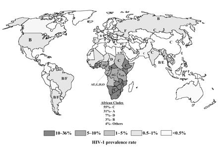
14 Page 14 have included env gene mediated receptor affinity and LTR and tat gene mediated transcriptional activation.[62] The detection of mosaic HIV-1 sequences suggests that persons can become coinfected with differing HIV-1 subtypes that can then undergo recombination to new strains which may have different biologic characteristics from the original strains. This has happened in Southeast Asia and Africa, where the recombinants A/E and A/G comprise the major circulating forms of HIV-1.[20] The major subtypes of HIV-1 are listed below. Subtypes A through H belong to the major group M, while group O is distinctly different and genetically more closely related to simian immunodeficiency virus (SIV) and HIV-2.[61,63] Group N appears to have arisen from interaction between a group M and a group O virus.[64] The major subgroups of HIV-1 are given below, with epidemiologic correlates for probable locations in the first decade of the AIDS epidemic. Recombinant forms (e.g., A/E or A/G or B/F are appearing more frequently as the epidemic progresses.[60] Subgroups of HIV-1 Group M Subtype A West and Central Africa Subtype B South America (including Brazil and Argentina), United States, Europe, Thailand, Russia Subtype C India, Sudan, Southern and Eastern Africa Subtype D East and Central Africa Subtype E Thailand, Philippines, China, Central Africa Subtype F Brazil, Argentina, Eastern Europe, Central Africa Subtype G Western and Eastern Africa, Central Europe Subtype H Central Africa Subtype J Central America Subtype K Democratic Republic of Congo, Cameroon Group N Cameroon Group O West Africa
15 Page 15
16 Page 16 OTHER HUMAN RETROVIRUSES HIV-2:-- The numerous strains of HIV-1 isolated from various geographic regions of the world are all immunologically similar and differ only slightly in their DNA sequences. A second retrovirus designated HIV-2 has been isolated from a number of patients with AIDS, first in West African countries and subsequently in Western Europe, the United States, and elsewhere. Most cases have appeared in West Africa and have appeared only sporadically in other parts of the world.[65] HIV-2 is believed to have been present in Africa as early as the 1940 s. HIV-2, which has greater homology to simian immunodeficiency virus (SIV) than to HIV-1, appears to have become established in human populations as a zoonotic infection from the primate reservoir of sooty mangabeys (Cercocebus atys).[20,29] HIV-2 is spread in a manner similar to HIV-1, though the high-risk groups are commercial sex workers and persons with other sexually transmitted diseases.[66] The peak age of persons infected with HIV-2 appears to be higher than that of HIV-1, but there appears to be no sex difference in rates of infection. HIV-2 appears to utilize the same cellular mechanisms for infection as HIV-1, including the use of CD4 receptors and chemokine coreceptors. Persons infected with HIV-2 live longer and have a course of disease much more variable than persons with HIV-1 infection.[31,67] Just as HIV-1 has distinct subtypes, so does HIV-2. The subtypes of HIV-2 have been designated from A through F. There is up to a 25% difference in genetic homology among these subtypes. All five subtypes can be detected by enzyme immunoassay (EIA) and Western blot assays for HIV-2 similar to those for HIV-1. The reverse transcriptase enzyme is similar in structure and function in both HIV-1 and HIV-2. Infection with HIV-2 eventually leads to AIDS. Persons can be coinfected with HIV-1 and HIV-2.[65,6668] The genetic sequences of HIV-1 and HIV-2 are only partially homologous. HIV-2, or other as yet uncharacterized members of the HIV-group of viruses, will not necessarily be detected by using the various laboratory tests for HIV-1 antibody, including enzyme immunoassay (EIA) and Western blot (WB) tests, in general use for HIV-1. Instead, separate EIA and WB assays are employed for diagnosis of HIV-2. HIV-2 is genetically more closely related to simian immunodeficiency virus (SIV) than HIV-1.[69] This potential problem of genetic variation with HIV was illustrated in 1994 with the detection of a strain of HIV-1 (designated MVP-5180, or subtype O), a new HIV variant originating in the region of West-Central Africa, which showed only slightly more homology with other HIV-1 strains than with HIV-2. This variant was still detectable with many testing methods for HIV-1, but false negative results may occur. This subtype O of HIV-1 demonstrates higher heterogeneity in env sequences than the more prevalent HIV-1 subtypes such as B.[63,70] The appearance of additional HIV subtypes requires more complex testing schemes in locations where HIV-2, or other possible HIV virus subtypes, are prevalent. The transmission of HIV-2 is similar to that for HIV-1, though perinatal transmission is much less frequent. The natural history of HIV-2 infection is characterized by a longer latent period before the appearance of AIDS, a less aggressive course of AIDS, and a lower viral load with higher CD4 lymphocyte counts than HIV-1 infection until late in the course of the disease, when clinical AIDS is apparent. Thus, the pathogenicity of HIV-2 appears to be lower than that of HIV-1. This may explain the more limited spread of HIV-2, compared to HIV-1, both in West African countries and elsewhere, due to less efficient transmission, particularly via heterosexual and perinatal modes.[65] The mortality rate from HIV-2 infection is only two-thirds that for HIV-1.[71] HTLV:-- Another group of human retroviruses different from HIV are the human T- lymphotrophic viruses, types 1 and 2 (HTLV-1 and HTLV-2). Along with simian T-cell lymphoma virus type 1, they constitute a group of retroviruses known as the primate T-cell leukemia/lymphoma viruses. HTLV's can be transmitted in the same manner as HIV, though even less efficiently. Persons can be coinfected with HIV and HTLV. The CD4 lymphocytes are the
17 Page 17 cells primarily infected by HTLV. Laboratory testing methodology for HTLV's is similar to that for HIV. The enzyme immunoassay test for HTLV-1 will also detect HTLV-2. Confirmatory Western blot testing, in combination with testing for the presence of envelope peptide p21env-r helps to distinguish HTLV-1 from HTLV-2.[72,73] HTLV-1 infection is widespread in tropical and subtropical regions, with the main endemic foci in the Caribbean, southern Japan, central Africa, South Africa, and South America, particularly Brazil. Other endemic foci are found in southern India, northern Iran, aboriginal populations of northern Australia, and islands in the tropics. In Europe and North America, HTLV-1 infection is primarily associated with injection drug users and with immigrants from endemic areas. In endemic areas, the seroprevalence varies widely, even in communities located close together, and ranges from 0.1 to 30%. The seroprevalence is higher in costal communities.[74] HTLV-1 is associated with adult T-cell leukemia/lymphoma (ATLL), with a form of chronic progressive neurologic disease known as HTLV-associated myelopathy/tropical spastic paraparesis (HAM/TSP).[75] HTLV-1 is also associated with inflammatory conditions including polymyositis, arthropathy, infective dermatitis, and uveitis. The time from exposure to onset of HTLV-1 related disease is long--from 2 to 3 decades on average. However, the lifetime risk for ATLL in infected persons is only about 5% for persons infected before the age of 20. There is an additional 5% risk for the less serious complications of infectious dermatitis, uveitis, polymyositis, and arthropathy. The lifetime risk for HAM/TSP is about 1-2%. ATLL is uniformly fatal, while HAM/TSP is not. Men are more likely to develop ATLL, while HAM/TSP is more common in women.[74] Persons infected with HTLV-1 infection tend to have higher creatine kinase and lactate dehydrogenase levels in serum than seronegative persons.[76] HTLV-2 has been identified as an endemic infection in two distinct populations: native peoples of the New World and pygmy tribes of Africa. In other populations, the spread of HTLV- 2 occurs primarily as a result of injection drug use, particularly in metropolitan areas.[77] There is no clear association between HTLV-2 and HTLL, but chronic neurologic disease, particularly spinocerebellar syndrome, may be linked to HTLV-2 infection.[78]
18 Page 18 EPIDEMIOLOGY OF AIDS Considerable epidemiologic and clinical work has been performed to understand the transmission of HIV from one person to another. As in past epidemics, the spread of AIDS is facilitated by human travel. Syphilis in the 16th century, bubonic plague in the 17th century, and influenza early in the 20th century also arose from endemic foci to become widespread. Modern means of travel by jet aircraft readily available to many people provide an easy route for the spread of AIDS from one location or population to another.[79] However, unlike most infections in past epidemics, AIDS is distinguished by a very long latent period before the development of any visible signs of infection in affected persons. The average HIV-infected person may have an initial acute self-limited illness, may take up to several weeks to become seropositive, and then may live up to 8 or 10 years, on average, before development of the clinical signs and symptoms of AIDS. In virtually all past infectious disease epidemics, infected persons were soon easily recognized so that measures could be taken to prevent the spread of disease. But persons infected with HIV cannot be recognized by appearance alone, are not prompted to seek medical attention, and are often unaware that they may be spreading the infection.[25,36,80] The transmission of HIV is a function both of where the virus appears in the body and how it is shed. HIV can be present in a variety of body fluids and secretions, as shown in Table 1. The presence of HIV in genital secretions and in blood, and to a lesser extent breast milk, is significant for spread of HIV. However, the appearance of HIV in saliva, urine, tears, and sweat is of no major clinical or social importance, as transmission of HIV through these fluids does not routinely occur, primarily because of the low concentration of HIV in these fluids.[81] Though infectious particles of HIV are frequent in cerebrospinal fluid, contact with this fluid in daily life is extremely rare.[18,82] The most important feature of HIV is the means of spread (Table 2). Unlike most epidemics of infectious diseases wherein much of a population is at risk, HIV infects definable population subgroups ("risk groups"). This happens because HIV is primarily a sexually transmissible disease. Homosexual, bisexual, and heterosexual transmission all can occur. Although sexual intercourse between males has remained the greatest risk for transmission in developed nations of Western Europe and the United States, heterosexual transmission is increasing in those regions but still remains less common than in Africa, Asia, or parts of the Caribbean.[80,83,84] Transmission of HIV can occur from male to male, male to female, and female to male. Female to female transmission remains extremely rare, though women with same-sex contact are also often bisexual and have additional risk factors for HIV infection.[85,86] Even a partial modification of sexual behavior practices may help retard the rate and extent of HIV transmission. Amongst males having sex with males in the U.S. in the 1990's, the prevalence of HIV infection remained high at 7.2%, and the prevalence of unprotected anal intercourse over a prior 6 month period was 41%.[87] Educational efforts in AIDS prevention must be ongoing and must specifically target not only persons belonging to identifiable risk groups for HIV infection but also teenagers beginning sexual intercourse (and who often lack a sense of their own mortality), as well as young adults. The Centers for Disease Control in the U.S. has a strategic plan to reduce HIV infection through a three-part plan that includes: (1) intensifying efforts to help all infected persons learn their HIV status; (2) establishing new prevention programs to help HIV-infected persons establish and maintain safer behaviors, combined with improved linkages to treatment and care; and (3) expanding highly targeted prevention programs to reach all HIV-negative persons at greatest risk.[88] Worldwide, heterosexual transmission accounts for the majority of cases of HIV infection. The important factors that promote heterosexual transmission include:[89]
19 Page 19 More sexual partners Frequent change of sexual partners Unprotected sexual intercourse (lack of barrier precautions) Presence of additional sexually transmitted diseases Lack of male circumcision Social vulnerability of women and young persons Economic and political instability of the community The lack of economic and political stability makes it difficult to institute programs to change behavior, to promote condom use, to treat sexually transmitted diseases, to test for HIV infection, and to treat HIV infection with antiretroviral therapies that reduce viral load and the risk of transmission.[89] Practicing "safe" sex will diminish the prevalence of HIV infection in populations where HIV has become well established. Though transmission of HIV can be reduced, it cannot be completely eliminated once it is established in a population.[12,13,90] Risk reduction interventions, including education on abstinence and safer sex, are beneficial. Abstinence intervention has a short term effect over months, while safer sex interventions have a longer lasting effect, particularly amongst adolescents who have previously had sexual intercourse. These interventions appear to reduce the frequency of sexual intercourse.[91] Promotion of the use of condoms as a barrier precaution has also been shown to reduce the rate of HIV infection, and is a mainstay of prevention efforts.[92] The availability of condoms has a significant effect upon condom use and does not appear to increase rates of sexual activity.[93] There are three major variables that explain the sexual transmission of HIV: (1) transmission efficiency, (2) number of sexual partners, and (3) seroprevalence (numbers of infected individuals in a population). HIV transmission through sexual exchange of semen or vaginal fluids is much less efficient than transmission of either gonorrhea or hepatitis B virus. Usually, multiple sexual exposures are necessary to increase the likelihood for transmission of HIV from infected persons. It is estimated that gonorrhea may be transmitted in 22 to 25% of sexual encounters involving an infected individual, hepatitis B virus in 20 to 30% of encounters, and hepatitis C in 2% of sexual encounters, while HIV transmission occurs much less often--approximately 0.3% per sexual contact with an HIV-infected person. However, some persons have become HIV-infected after a single sexual contact, while other persons have remained uninfected after hundreds of contacts.[94,95] The rate of sexual transmission of HIV may depend upon the number of viral particles in genital secretions. The number of CD4 cells per µl of seminal fluid ranges from 10 2 to 10 3, while the number of virions can range from undetectable to over The numbers of virions in the female genital tract is generally lower. Transmission can occur both cell-to-cell as well as from cellfree fluid.[96,97] Thus, the transmission rate is two to three times higher from infected males to females than from infected females to males, without other cofactors.[98] The location of HIV in cells of the genital tract of infected persons varies between men and women. In men, both the cells within seminal fluid, as well as the seminal fluid, harbor virions of HIV, but spermatozoa are not a major source for HIV. In women, the greatest number of virions are present at the squamocolumnar junction of the cervix, with far less HIV in vaginal epithelium. Langerhans cells and macrophages in the lamina propria capable of harboring HIV can be found in a variety of epithelia.[99] Since most of the cell-free HIV in the semen of men arises distal to the vas deferens, a vasectomy may have minimal impact on the infectivity of seropositive males to sexual partners.[100] For persons who have regular intercourse with a single HIV infected (index) partner, risk of transmission of HIV-1 depends upon the stage of HIV-1 infection. The risk is highest, /coital act, within 2.5 months of seroconversion of the index partner. The risk drops to /coital act within 6 to 15 months after index partner seroconversion and remains low throughout the stage of clinical latency of HIV-1 infection. The risk rises again in the late stage of clinical AIDS, /coital act, within 6 to 25 months of death of the index partner.[101]
20 Page 20 The rate of HIV sexual transmission may also be due to the low infectivity of an individual strain of virus, propensity for only selected individuals to transmit infective virus in secretions, or presence of individual susceptibility factors.[68] Some HIV-1 subtypes may be more easily transmitted heterosexually, particularly subtype E which is more prevalent in Asia and sub-saharan Africa. There is a greater tropism of the E subtype for Langerhans cells than subtype B which is more prevalent in the U.S. and Europe.[60,99] Sexual contact with persons whose HIV viral load is greater increases the transmission risk. The risk for HIV transmission from an HIV-infected person increases as that person's immune status diminishes, as measured by a decrease in CD4 lymphocytes or an increase in HIV-1 RNA in plasma, so that infectivity is greater in the later stages of AIDS; likewise, a greater risk for transmission exists with the pronounced HIV viremia during primary HIV infection. Transmission rarely occurs when the HIV-1 RNA level in serum is less than 1500 copies/ml.[102] Persons with HIV infection undergoing antiretroviral therapy that measurably lowers the viral burden in blood will have a reduction in viral particles in genital fluids of men and women that will render them less infective to others.[103,104] However, even with aggressive antiretroviral therapy, HIV may be detectable at low levels in blood and genital fluid.[105] The presence of specific chemokine receptors plays a role in HIV transmission. Chemokine receptors provide a pathway, separate from CD4 receptors, for entry of HIV into cells. Mutations in the chemokine receptor genes appear to afford increased resistance to HIV infection or progression of disease for hosts homozygous for this genetic trait. Approximately 11% of Caucasians and 2% of Blacks are homozygous for the CCR5-delta32 mutation.[99] The presence of cervical ectopia, oral contraceptive use, or pregnancy or menstruation in women, intact foreskin in men, and genital ulcer disease in either sex increases the risk for HIV infection. Thus, male circumcision affords some degree of protection, perhaps due to the large numbers of Langerhans cells in foreskin, so that the incidence of infection is reduced 8-fold over uncircumcised men. Cervical ectopy, with replacement of squamous by columnar epithelium, may increase the risk of HIV infection for women 5-fold.[94,99,106] The greatest determinant of HIV in cervical and vaginal secretions is the plasma level of HIV-1 RNA.[107] Increased detection of HIV can occur in women with vitamin A deficiency and in women receiving high dose oral contraceptives or depot contraceptives.[108] There are a variety of mechanisms by which the coexistence of other sexually transmissible diseases (STDs) may increase the infectivity of HIV. Both Chlamydia trachomatis and Treponema pallidum infection appear to increase HIV-1 replication, while Haemophilus ducreyi infection increases CCR5 chemokine coreceptor expression by macrophages. In men, urethritis with infection by Neisseria gonorrhoeae and Trichomonas has been shown to increase the amount of HIV-1 in semen. Likewise, in women cervicovaginal fluids contain more HIV-1, as well as CD4 cells when additional STDs are present. Chlamydia trachomatis, Neisseria gonorrhoeae, and Trichomonas, or diseases producing genital ulcers such as herpes simplex virus, chancroid (Haemophilus ducreyi) or syphilis (Treponema pallidum), all enhance infectivity by HIV. For example, HIV-1 virions can consistently be detected in genital ulcers caused by herpes simplex virus-2. Treatment of these STDs can help to reduce the number of new HIV-1 cases.[99,109,110] The cofactor effect of genital ulcer disease is approximately five times higher for female-to-male than for male-to-female transmission. A higher prevalence of STD s in the population will equalize HIV transmission between the sexes.[98] The use of crack cocaine can increase the transmission rate for HIV with oral sex. This increase in infectivity can be due either to the greater numbers of oral sores with inflammatory cells containing HIV in the infected person or to the increased numbers of inflammatory cells with CD4 receptors in the contact person waiting to become infected, from the loss of an intact epithelial barrier.[111] Genital ulcers with inflammation also provide a more direct route to lymphatics draining to lymph nodes containing many CD4 lymphocytes, macrophages, and follicular dendritic cells.[112] Tissue trauma during intercourse does not appear to play a role in HIV transmission.[94] HIV-1 can be demonstrated in semen even in the first few weeks following infection.[113] The
21 Page 21 transmission of HIV can occur with the act of sexual intercourse in any style or position, though a greater relative risk exists with anal receptive intercourse.[94] Once HIV is introduced into a promiscuous population, seroprevalence increases with time. Increasing the number of sexual partners increases the likelihood of contacting a seropositive individual.[114,115] If the number of infected individuals in a population is high, then even one sexual encounter carries a significant probability of contacting an infected individual. This was demonstrated in one high risk group over a three year period ( ) early in the AIDS epidemic in which the HIV infection rate was 44%.[116] Overall, the most important factor for both the spread and the risk of infection from HIV is the degree of sexual activity with multiple sexual partners.[95] HIV has another important secondary means of spread through blood or blood products (Table 2). Parenteral exposure to blood and blood products is the most highly efficient method of HIV transmission--close to 90%.[117] There are many more peripheral blood mononuclear cells capable of either harboring or becoming infected by HIV in blood than are present in other body fluids or secretions. The number of infectious HIV particles free in peripheral blood can range from undetectable to well over a million per ml.[102] The primary risk group for HIV transmission via blood is intravenous drug users sharing infected needles. If needles are not shared, then this form of transmission will not occur. Less common practices of blood commingling, or use of instruments such as tattoo needles not properly disinfected, also carries a potential risk (Table 2). Health care workers with percutaneous exposures to HIV-containing blood, however, are infected fewer than 1 in 300 times.[118,119] Before laboratory tests were developed to detect HIV, persons who received blood or blood products by transfusion were also at risk. Now when rigorous testing of donor blood is routinely done, this form of infection is extremely rare-- with a risk for occurrence of 1 case for single donor units of screened blood for persons receiving transfusions of blood products in the U.S.[120] However, in developing nations where economic and political problems interfere with screening programs for blood products, 5 to 10% of HIV infections may occur from exposure to infected blood products.[121] Even though HIV has been found in a variety of body fluids such as saliva, urine, feces, and tears, non-sexual transmission of HIV by these body fluids is improbable.[118,122,123] There is no evidence for HIV transmission by the aerosol route.[119] The lack of transmission is related in part to the paucity of HIV-infected cells in such secretions. Oral transmission of HIV is further impeded by the hypotonic inactivation of HIV-transmitting inflammatory cells by saliva. Oral transmission of HIV by seminal fluid, milk, and colostrum may be due to their isotonicity, which overcomes hypotonic salivary inactivation. Even though the amount of virus is small in body secretions and presents a very small risk with routine household contact, prolonged contact or contact in sexually intimate situations with such fluids should be avoided.[124] Routine transmission of HIV occurs only through semen, vaginal fluid, blood or blood components, and breast milk.[18,125] In a liquid environment at room temperature, the virus can survive for at least 15 days, but despite HIV presence and survival in such an environment, infection through casual household and institutional contacts is rare, even when hepatitis is transmitted in the same setting.[122,126,127] Significantly, HIV transmission by insect vectors such as mosquitos appears highly improbable.[123] HIV infection can also be acquired as a congenital infection perinatally or in infancy (Table 2). Mothers with HIV infection can pass the virus to their babies transplacentally, at the time of delivery through the birth canal, or through breast milk. In the absence of breast-feeding, intrauterine transmission accounts for 25 to 40% of infections, while 60 to 75% occur during labor and delivery.[128] The probability of breast-milk transmission of HIV-1 is calculated to be per liter ingested and per day of breast-feeding. Breast-milk infectivity is significantly higher for mothers with more advanced disease with higher prenatal HIV-1 RNA plasma levels and CD4 cell counts. The probability of HIV-1 infection per liter of breast milk ingested by an infant is similar in magnitude to the probability of heterosexual transmission of HIV-1 per unprotected sex act in adults.[129]
22 Page 22 Vertical transmission of HIV-1 from mother to child from breastfeeding has been estimated to occur in 14% to 16% of women who breast-feed with established maternal HIV-1 infection and in 29% with acute maternal HIV-1 infection. The risk for HIV-1 transmission from an infected mother to an infant through breast feeding is increased with the duration of breast feeding and with increased maternal viral load.[130] The risk is also increased with mastitis or breast abscess.[131] Most cases of transmission occur early during breastfeeding. HIV-1 can be detected in over half of breast milk samples from infected mothers.[125,128,132]. Replication of HIV-1 within mammary epithelial cells has been demonstrated, and is increased by hormonal stimulation in pregnancy.[133] Perinatal transmission with congenital AIDS occurs, on average, in about one fourth of babies born to HIV-1 infected mothers, with actual rates of transmission varying from 7 to 71%, depending upon the presence of risk factors for transmission during the course of HIV infection and pregnancy. The most significant maternal risk factor for perinatal transmission is the HIV-1 DNA load, followed by the HIV-1 RNA load.[134] Additional maternal factors cited for congenital HIV-1 transmission are: a low CD4 lymphocyte count, p24 antigenemia, prematurity, and placental chorioamnionitis or funisitis. Parity, race, mode of HIV acquisition, and sex of the baby do not appear to be factors in the vertical transmission of HIV.[135,136] The likelihood of vertical HIV-1 transmission is reduced by half with delivery by elective cesarean section, as compared with other modes of delivery.[137] Features of HIV-1 that appear to correlate with perinatal transmission include: rapid or high-titered replication in maternal human peripheral blood mononuclear cells, T-cell tropism, and resistance to neutralization or a sensitivity to enhancement of infection by maternal serum.[136] Also, an amniocentesis procedure, premature rupture of membranes, preterm labor, genital warts, and the presence of sexually transmitted diseases during pregnancy also increases the risk for transmission. Transmission most likely occurs in late third trimester and intrapartum.[138,139] Measurement of maternal HIV-1 RNA can predict perinatal transmission risk. High levels of HIV-1 RNA late in gestation and/or during labor and delivery increase the risk for perinatal transmission.[140] The frequency of perinatal HIV-1 transmission in the first and second trimesters is low.[141] Though HIV-1 transmission from mother to fetus may still occur over a wide range of plasma HIV-1 RNA levels and of CD4 lymphocyte counts, antiretroviral therapy that reduces the HIV-1 RNA level to below 500 copies/ml appears to minimize the risk of perinatal transmission as well as improve the health of the mother. Thus, the maternal HIV-1 RNA level can predict the risk, but not the timing, of HIV transmission to their infants.[142,143] To date, most reported perinatal HIV-1 cases in the United States have been a consequence of intravenous drug use by mothers, but an increasing proportion of cases is appearing from heterosexually acquired HIV by mothers.[135] Congenital AIDS is most common in populations where heterosexual HIV transmission and the frequency of women infected with HIV is higher. In contrast, perinatal transmission of HIV-2 occurs far less frequently, with a rate of only 1 to 2%.[144]
23 Page 23 PATTERNS FOR HUMAN IMMUNODEFICIENCY VIRUS INFECTION Worldwide, three patterns of HIV infection have been identified. In pattern 1, affecting primarily urban areas of the Americas and western Europe, the majority of HIV infections occur in males having sexual intercourse with other males (homosexual and bisexual males), followed by infections in intravenous drug users. Fewer cases are observed among heterosexuals. Pattern 2 occurs in those areas in which HIV has been present longer and the number of HIV-infected persons in the population is greater. Men and women are affected equally, and heterosexual intercourse is the major means of transmission for HIV. These areas include sub-saharan Africa and parts of the Caribbean where HIV infection occurs throughout the heterosexual population, and congenital AIDS is a significant problem. Pattern 3 occurs in areas of the world in which HIV has been introduced only recently, defined risk groups have not emerged, and only sporadic cases are reported.[145] Once HIV is well-established in a population, the median age at infection declines over time.[146] RISK GROUPS FOR HUMAN IMMUNODEFICIENCY VIRUS INFECTION Risk groups for HIV infection based upon behavior patterns that put persons at risk are detailed in Table 2. In Pattern 1 countries such as the United States, through the first decade of the AIDS pandemic, about half of AIDS cases have been reported in homosexual or bisexual. The second largest risk group is comprised of intravenous drug users, accounting for 20% to 25% of reported AIDS cases in the United States.[80,147] The percentage of HIV infections seen in heterosexual adults is increasing in developed nations. Pediatric AIDS in the United States is largely a function of maternal risk factors, particularly from intravenous drug use. In Pattern 2 countries, particularly in Africa, HIV infection is spread more widely in the population through heterosexually active urban adults.[115] The demography of the spread of HIV depends upon the population subgroups into which HIV has been introduced and the contact that other segments of the population have with them. Thus, prostitution and intravenous drug use may both be an important means for spread of HIV through the heterosexual population. AIDS among heterosexual adults in the United States is increasing more than any other risk group, and over half of all heterosexually acquired HIV infections occur in women. This represents a significant risk to the promiscuous or intravenous drug using heterosexual person. Screening of blood products for HIV has almost completely eliminated the risk from transfusion or blood product therapy in locations where such screening is routinely performed.[148] On average, about 5 to 10% of persons who develop AIDS will report no identifiable risk factor for HIV infection. Over time, many of them will be found to have a defined risk factor when historical data becomes available. The number of cases of HIV infection with no identifiable risk factor has not increased significantly over time, confirming the observation that HIV infection is not acquired through casual contact.[80,127]
24 Page 24 NATURAL HISTORY OF HIV INFECTION On average, there is a period of 8 to 10 years from initial infection to clinical AIDS in adults, though AIDS may be manifested in less than two years or be delayed in onset beyond 10 years.[149] About 10% of persons will rapidly progress to AIDS in 2 to 3 years following HIV infection, while about 10% have not progressed to AIDS even after 10 years.[150] It is clear that the longer an individual is infected, the more likely the development of illness and subsequent death will be. Thus, HIV infection does not follow the pattern of more traditional viral diseases in which the risk of serious illness or death decreases with time. There has been no study to date that shows a failure of HIV-infected persons to evolve to clinical AIDS over time, though the speed at which this evolution occurs may vary, and a small number of HIV-infected persons will not progress to AIDS for many years.[80] Primary HIV infection, also known as acute retroviral syndrome, may produce a mild and self-limited disease in 50 to 90% of persons infected with HIV, regardless of the mode of transmission. The time from mucosal infection to viremia is about 4 to 11 days. The time from exposure to development of symptoms averages 2 to 6 weeks. The symptoms may persist for 1 to 2 weeks, after which symptoms subside over 1 to 2 months. Prospective studies of acute HIV infections show that fever, fatigue, arthralgia or myalgia, lymphadenopathy, pharyngitis, diffuse erythematous macular or mixed maculopapular rash (often involving the trunk), diarrhea, nausea or vomiting, weight loss, night sweats, mucocutaneous ulcerations, and headache are the most common symptoms seen with acute HIV infection. An acute meningoencephalitis may be seen in some recent infections and appear as an aseptic meningitis. The symptoms of acute HIV infection resemble a flu-like or an infectious mononucleosis-like syndrome. Primary HIV infection is not life-threatening.[151,152] Primary HIV infection in children is usually accompanied by one or more of the following: mononucleosis-like syndrome, dermatitis, or generalized lymphadenopathy.[153] In acute HIV infection, the peripheral blood may demonstrate lymphopenia and/or thrombocytopenia. However, atypical lymphocytes are absent. Although the CD4 cells are decreasing, the levels may initially remain in the normal range, but depletion continues. Simultaneously, there is an increase in cytotoxic CD8 lymphocytes that continues as symptoms subside and viremia decreases.[152] During this acute phase of HIV infection, there is active viral replication, particularly in CD4 lymphocytes, and a marked HIV viremia. This peripheral blood viremia is at least as high as 50,000 copies/ml and often in the range of 1,000,000 to 10,000,000 copies/ml of HIV-1 RNA. High titers of cytopathic HIV are detectable in the blood so that the p24 antigen test is usually (but not always) positive, while HIV antibody tests (such as enzyme immunoassay) are often negative in the first three weeks. The viremia is greater in persons whose primary HIV infection is symptomatic.[17,54,151,152] During this viremic phase, HIV disseminates throughout the body to lymphoid tissues and other organs such as brain. There are alterations in peripheral blood mononuclear cells marked by a decline in CD4+ lymphocytes. Persons acutely infected with HIV are highly infectious as a consequence of the high levels of HIV, both in blood as well as in genital secretions. Over half of all HIV infections may be transmitted during this period.[151] Generally, within 3 weeks to 3 months following initial infection with HIV, the immune response is accompanied by a simultaneous decline in HIV viremia. Both humoral and cell mediated immune responses play a role. The CD4 lymphocytes rebound in number after primary HIV infection, but not to pre-infection levels. Seroconversion with detectable HIV antibody by laboratory testing such as enzyme immunoassay accompanies this immune response, sometimes in as little as a week, but more often in two to four weeks.[17,18,37] Prolonged HIV-1 infection without evidence for seroconversion, however, is an extremely rare event.[154] Persons infected with HIV who develop an acute retroviral illness and who have a shorter time to seroconversion tend to progress to AIDS faster than persons with longer seroconversion times.[155]
25 Page 25 The HIV infection then becomes clinically "latent." During this phase, there is little or no viral replication detectable in peripheral blood mononuclear cells and little or no culturable virus in peripheral blood. The CD4 lymphocyte count remains moderately decreased. However, the immune response to HIV is insufficient to prevent continued viral replication within lymphoid tissues. Though lymph nodes may not become enlarged and their architecture is maintained, active viral replication continues.[29,156] Tests for HIV antibody will remain positive during this time but p24 antigen tests are usually negative. There is no evidence to suggest that seroreversion, or loss of antibody, occurs in HIV-infected persons.[157] Though the time to development of AIDS is statistically similar in men and women, the viral load of women tends to be lower. Women with half the viral load of men have a similar time to development of AIDS as men. Women with the same viral load as men have a 1.6-fold higher risk of AIDS. The biologic basis for this difference is unclear.[158] In many viral infections, an immune response consisting of virus-specific CD4 lymphocytes helps to contain the infection. However, such a response is typically lacking in HIV-infected persons. A minority of HIV-infected persons, however, do mount a persistent polyclonal CD4 lymphocyte proliferation directed against HIV which controls viremia. This response results in a cytokine response with elaboration of interferon gamma and beta chemokines. Such a response may also occur with antiretroviral therapy.[159] As FDC s are diminished over time with HIV infection, the capacity for stimulation of CD4 lymphocytes is also diminished, and CD4 memory cells decline as well. However, remaining FDC s continue to promote ongoing production of antibody to HIV. CD4 memory cells may also be lost by formation of syncytia with infected FDC s. Finally, when the stage of AIDS is reached, development of FDC s from stem cells is diminished.[58] Though no clinical signs and symptoms are apparent, the immune system, primarily through depletion of CD4 lymphocytes, deteriorates. The virus continues to replicate in lymphoid organs, despite a low level or lack of viremia.[37] HIV can be found trapped extracellularly in the follicular dendritic cell network of germinal centers in lymphoid tissues or intracellularly as either latent or replicating virus in mononuclear cells. The period of clinical latency with HIV infection, when infected persons appear in good health, can be variable--from as short as 18 months to over 15 years. This latent period lasts, on average, from 8 to 10 years.[17,37] This interval tends to be longer with earlier age at time of initial infection with HIV.[146] The hallmarks of emergence of HIV infection from clinical latency are a marked decline in the CD4 lymphocyte count and an increase in viremia. Replication of HIV increases as the infection progresses. There is loss of normal lymph node architecture as the immune system fails. Before serologic and immunologic markers for HIV infection became available, clinical criteria established emergence from latency by development of generalized lymphadenopathy. This condition, described by the term persistent generalized lymphadenopathy (PGL), is not lifethreatening.[18] Another phase of HIV infection described clinically but no longer commonly diagnosed in practice, is the condition known as AIDS-related complex (ARC), which is not necessarily preceded by PGL. ARC lacks only the opportunistic infections and neoplasms which define AIDS. ARC patients usually show symptoms of fatigue, weight loss, and night sweats, along with superficial fungal infections of the mouth (oral thrush) and fingernails and toenails (onychomycosis). It is uncommon for HIV-infected persons to die at the stage of ARC. The staging of HIV disease progression through the use of CD4 lymphocyte counts and plasma HIV-1 RNA levels has made use of the terms PGL and ARC obsolete.[18] The stage of clinical AIDS that is reached years after initial infection is marked by the appearance of one or more of the typical opportunistic infections or neoplasms diagnostic of AIDS by definitional criteria. The progression to clinical AIDS is also marked by the appearance of syncytia-forming (SI) variants of HIV in about half of HIV-infected patients. These SI viral variants, derived from non-syncytia-forming (NSI) variants, have greater CD4+ cell tropism and are associated with more rapid CD4+ cell decline. The SI variants typically arise in association with a peripheral blood CD4 lymphocyte count between 400 and 500/µL, prior to the onset of clinical
26 Page 26 AIDS. However, appearance of the SI phenotype of HIV is a marker for progression to AIDS that is independent of CD4+ cell counts.[55]
27 Page 27 PROGRESSION OF HIV INFECTION The development of signs and symptoms of AIDS typically parallels laboratory testing for CD4 lymphocytes. A decrease in the total CD4 lymphocyte count below 500/µL presages the development of clinical AIDS, and a drop below 200/µL not only defines AIDS, but also indicates a high probability for the development of AIDS-related opportunistic infections and/or neoplasms. The risk for death from HIV infection above the 200/µL CD4 level is low.[160,161,162] Other laboratory findings which indicate progression to AIDS include HIV p24 antigen positivity, increased serum beta 2 -microglobulin (B2-M), elevated serum IgA, or increased neopterin levels in serum, cerebrospinal fluid, or urine. The p24 antigen is a highly specific predictor of progression, but only about 60% of HIV-infected persons develop p24 antigenemia prior to onset of clinical AIDS. B2-M is a polypeptide that forms the light chain of the class I major histocompatibility complex found on the surface membrane of most cells, including lymphocytes. It is increased with lymphocyte activation or destruction associated with HIV disease progression, but B2-M can also be elevated with viral infections such as cytomegalovirus and with malignant lymphomas. Neopterin, a product of macrophages, is also a measure of immune system activation and can predict HIV disease progression. The information provided by these tests is similar, so no advantage accrues from performing all of them simultaneously.[163] The best laboratory measure for determination of the progression of AIDS is the level of HIV-1 RNA in peripheral blood. The predictive value of HIV-1 RNA levels is independent of the CD4 lymphocyte count and of age in adults. During the acute phase of HIV infection prior to any immune response, plasma levels of HIV-1 RNA typically exceed 10,000 copies/µl. There is a sex difference. The initial viral load following HIV infection is 50,766 copies/ml in males and 15,103 copies/ml in females.[158] These levels of HIV-1 RNA generally drop, but fluctuate for a period of 6 to 9 months. After this time, a set point is reached for the level of HIV-1 RNA that remains relatively constant during the latent phase of HIV infection. Factors influencing this set point include the strain of HIV-1, host anti-hiv response, and the number of cells, including CD4 lymphocytes and macrophages, available for infection. The initial viremia may be higher, and the set point may not be reached until after infancy in cases of congenital HIV infection.[164] The set point levels of HIV-1 RNA correlate with the time to development of AIDS. The set point can range from <50 to 1,000,000 copies/ml. Persons with a higher set point tend to lose CD4 cells more rapidly and progress to AIDS more quickly. Levels of HIV-1 RNA can remain at a steady state for months to years, but usually fall with time. Levels in any individual person may vary with time and even change rapidly, though a variation of <0.7 log 10 copies/ml is typical, but an upward progression is an ominous sign of probable progression to AIDS. Less than half of persons with low levels (<4500 copies/ml) of HIV-1 RNA have progressed to AIDS at 10 years following seroconversion, and those with levels <200 copies/ml do not appear to progress at all. Conversely, persons with >100,000 copies/ml are 10 times more likely to progress to AIDS in 5 years. For persons in the top quartile (>36,270 copies/ml) the median time to development of AIDS is 3.5 years.[54,164] The presence of opportunistic infections and neoplasms increases the risk for progression to death from HIV infection.[165] In spite of the initial viral load difference between men and women, the rates of progression to AIDS are similar.[158] Persons with HIV infection can be categorized as typical progressors, rapid progressors, and nonprogressors toward AIDS. The typical progressors average 8 to 10 years of latent HIV infection before the appearance of clinical AIDS. These persons typically have a fall in HIV viremia following acute infection. They maintain nonsyncytium-inducing HIV variants that replicate slowly over time, until more rapidly replicating variants develop during progression to AIDS. About 10% of HIV-infected persons rapidly progress to AIDS in only 2 to 3 years following initial infection. These persons have a high viral load during acute HIV infection that does not fall to the levels seen with typical progressors. They may be infected with more virulent
28 Page 28 strains of HIV.[150] More rapid progression to AIDS is also seen in Caucasians who have the major histocompatibility complex (MHC) class I type HLA-B*35 allele.[166] Though most HIV infections follow a standard progression, the course can be variable, and previously asymptomatic persons may suddenly die from an overwhelming opportunistic infection, while persons with clinically defined AIDS may survive for years. Progression to AIDS in persons with HIV infection does not appear to be modified by gender, by race, or by pregnancy, and the course of HIV infection is not modified by risk factor for infection. Progression to AIDS does appear to be accelerated in persons who are older or who smoke.[167,168,169,170] Older age at seroconversion is associated with faster progression to AIDS.[171] Persons who manifest symptomatic acute HIV infection have a faster progression to clinical AIDS.[151] Co-infection with the flavivirus GB virus C (GBV-C), also known as hepatitis G virus, is associated with a slower rate of progression of HIV infection with a reduced mortality rate. Hepatitis G virus itself is not known to be associated with any specific disease process. However, the presence of GBV-C may inhibit HIV replication, as shown in vitro with inhibition of HIV with coinfection of peripheral blood mononuclear cells by GBV-C. The prevalence of GBV-C is 1.8% in blood donors, though its presence does not preclude blood donation. The prevalence of GBV-C in HIV infected persons is about 40%.[172] Dietary supplementation with the trace element selenium has been demonstrated to partially suppress induction of HIV-1 replication in chronically infected mononuclear cells exposed to tumor necrosis factor (TNF). Selenium supplementation also appears to increase cellular antioxidant activity. These findings may account for the observation that selenium deficiency is associated with increased mortality from HIV-1 infection.[173,174] About 10% of persons infected with HIV-1 are nonprogressors, or "long survivors," who do not demonstrate a significant and progressive decline in immune function over more than 10 years. They do not appear to progress to AIDS in a manner similar to the majority of HIV-infected persons. Findings in these "long survivors" include: a stable CD4 lymphocyte count, negative plasma cultures for HIV-1, fewer HIV-infected cells, and a strong virus-inhibitory CD8+ T- lymphocyte response. Differences in viral load do not appear to be associated with viral subtype, viral growth kinetics, or with the presence of neutralizing antibodies.[175] In addition, by microscopic examination the lymph node architecture of "long survivors" with HIV infection is maintained without either the hyperplasia or the lymphocyte depletion that is common to progression to AIDS. Though peripheral blood mononuclear cells contain detectable HIV-1 and viral replication continues in long survivors, their viral burden remains low.[176] A strong host virus-specific CD4 lymphocyte response in these persons may also aid in controlling HIV viremia.[159] There is evidence that genetic polymorphisms in the chemokine receptors present on cells susceptible to HIV infection may play a role in progression of AIDS. At least in some persons infected with HIV, the presence of chemokine receptor variants, including the CCR5 delta32 deletion or the CCR2B-64I mutation, have a favorable effect in slowing the progression of disease. The CCR5 gene encodes the coreceptor for macrophage-tropic HIV-1, so reduced expression of CCR5 leads to reduced HIV-1 replication in macrophages.[177] A lower density of CCR5 molecules on peripheral blood mononuclear cells has been shown to correlate with lower plasma HIV-1 RNA levels and reduced loss of CD4 cells over time.[178] Patients homozygous for the chemokine receptor CX3CR1 progress to AIDS more rapidly than those with other genotypes.[179] Genetic variations in major histocompatibility (HLA) genes may determine HIV disease progression. An HIV-1 envelope glycoprotein fragment mimics both HLA class 1C molecules and an immune regulatory epitope in the HLA DR beta chain which furnishes peptides predicted to bind optimally to HLA class 1B alleles. The HLA class I genes A29 and B22 are significantly associated with rapid progression, while the alleles B14 and C8 are significantly associated with non progression of AIDS.[180,181] For perinatally acquired HIV infection, the time period from birth or neonatal life to the development of clinical AIDS is variable and may be shorter than in adults. Clinical signs associated with HIV infection appear in over 80% of seropositive infants by the age of 5 months.
29 Page 29 Infants in whom such signs appear at 3 months or less tend to have decreased overall survival. On average, about half of children with perinatally acquired HIV infection are alive at 9 years, though use of antiretroviral therapy decreased the death rate significantly.[182,183] The level of HIV-1 RNA rises rapidly in the first one to two months of life but remains high, and declines only slowly during the first two years of life. This suggests that the neonatal and infant immune system is not able to effectively contain HIV replication. Those babies whose HIV- 1 RNA levels are very high, not only in the first few months of life, but also in the first two years, tend to progress to AIDS more rapidly than those with lower levels.[184] Progression of disease appears to be faster in children whose strains of HIV-1 show tropism for monocyte-derived macrophages and whose viral strains are rapidly replicating.[185] Skin test anergy as demonstrated by the loss of delayed-type hypersensitivity to standard antigens such as Candida and Trichophyton also correlates with HIV disease progression.[186] Adolescents with HIV infection may have variable courses. Those who acquired HIV infection during adolescence, generally via sexual intercourse or via injection drug use, tend to have progression of their infection similar to adults. Those with congenital AIDS or who acquired their infection from blood products as young children will have a course different from long-term surviving adults.[164] In summary, there are a variety of factors that can influence the time course over which persons infected with HIV will progress to AIDS. In general the prognosis for infected persons is worse from probable accelerated progression when: Less favorable chemokine receptor variants are present Syncytia-forming (SI) variants of HIV are present Acute HIV infection is symptomatic HIV infection occurs with a drug-resistant strain A higher set point of HIV-1 RNA follows initial viremia after infection There is an older age at seroconversion The infected person is a smoker An opportunistic infection or neoplasm is present In congenital cases there are signs of infection at <3 months of age In addition, progression to AIDS from the period of clinical latency in persons with HIV infection is suggested by: CD4 lymphocyte counts <500/µL Failure to maintain normal lymph node function p24 antigenemia appears in peripheral blood Increasing HIV-1 RNA levels
30 Page 30 IDIOPATHIC CD4+ T-LYMPHOCYTOPENIA Increased laboratory testing in patients with immunodeficiency states has led to the recognition that CD4 lymphocyte counts in some cases can be markedly decreased in the absence of laboratory evidence for HIV infection. These uncommon, sporadically reported cases are unlikely to represent infection by new HIV subtypes that are not detectable by current laboratory testing methods. Criteria for diagnosis of ICL include: The absolute CD4 lymphocyte count is <300/µL or more in adults and children >2 years (<1000/µL in children <2 years) on more than one determination, or in children a T-lymphocyte count that is <20% of total lymphocytes; and There is no serologic evidence for HIV infection (even if in a child and the mother is HIV seropositive); and There is no defined immunodeficiency or therapy associated with T-cell depletion Though some patients with ICL may have a risk factor for HIV infection or even an opportunistic infection, the CD4 lymphocyte count does not progressively decrease over time as with AIDS. Almost all patients with ICL have normal serum immunoglobulin levels. The stable CD4 lymphocyte counts may be accompanied by reductions in the levels of other lymphocyte subsets, including CD8+ T-lymphocytes, natural killer cells, and B-lymphocytes. This disorder appears to be rare and is generally associated with transient illness. The presence of ICL does not constitute evidence for a new transmissible infectious agent.[187,188] A Behçet-like syndrome has been reported in conjunction with ICL.[189] Criteria for diagnosis of ICL include:
31 Page 31 PREVENTION OF HIV TRANSMISSION The transmission of HIV in definable risk groups allows for control measures to be taken that prevent the spread of AIDS. Since HIV is primarily spread as a sexually transmissible disease, then educational efforts must be aimed at sexually active persons and must be explicit regarding the behaviors that lead to the spread of HIV. A significant number of both boys and girls become sexually active as teenagers and must be included in prevention strategies. Given that the level of promiscuity will often be difficult to modify within a population, then educational campaigns are best focused upon the use of barrier precautions, particularly condom use. Sexual activity does not appear to increase with condom use.[93] All sexually active persons with more than one sexual partner, or whose partner is a member of a risk group for AIDS, should use condoms. Persons who know that they are infected with HIV should inform their sexual partners.[190,191,192] The spread of HIV by injection drug use creates a major reservoir for HIV infection that can then be transmitted to other segments of the population, particularly heterosexual adults, including the sexual partners of injection drug users. Drug users must be educated about the risks of needle sharing. Cleaning of needles with undiluted bleach appears effective in preventing HIV transmission.[193] They can be provided with clean needles to prevent the spread of HIV, and can be advised to use condoms.[194] Congenital AIDS can be prevented by efforts to educate women of childbearing age about the hazards to the fetus if they are HIV-infected. Potential mothers can be provided with means of contraception. Antiretroviral therapy for mothers can reduce perinatal HIV transmission. Confidential HIV testing should be made available along with counseling services to persons in all risk groups to encourage voluntary testing and prevent unknowing transmission of HIV.[195] Transmission of HIV through blood product therapy has become vanishingly rare when screening and testing of donors is applied. Such screening is costly. Since HIV infection is not spread by casual contact in public places, households, or in the workplace, no modifications of routine activities of daily living or work practices is necessary. HIV is not spread by insect vectors, and insect control programs will have no effect upon HIV transmission in a population. A summary of AIDS prevention strategies are given below: Methods to Reduce Rates of HIV Transmission Treat HIV infection as an illness, not as a social stigma Reduce levels of poverty in society that lead to increased risks through drug abuse and promiscuity Provide HIV testing and counseling to identify infected persons who can reduce their risk to others Provide educational programs for children and adults which describe how to avoid sexually transmitted diseases Promote sexual barrier precautions among high risk commercial sex workers and clients Provide clean needles for injection drug users Create health care programs providing antiretroviral therapy to extend life and reduce HIV transmission rates Give HIV-infected pregnant women antiretroviral therapy to reduce perinatal HIV transmission
32 Page 32 AIDS prevention programs have successfully produced long-term behavior change with reduction in incidence of HIV infection.[196] In order for such programs to be effective, several principles must be applied: sustained interventions are more likely to lead to sustained behavior change; more intense interventions are more likely to result in greater risk reduction; accessibility to devices (such as clean needles and condoms) that are necessary to safer practices reduces the risk for HIV infection; modification of community norms appears to enhance behavior change; and explicit HIV prevention programs must be provided prior to the time that adolescents become sexually active.[12]
33 Page 33 TREATMENT FOR AIDS A variety of therapies have been developed since 1984 for persons infected with HIV. Bone marrow transplantation, lymphocyte transfusions, thymic transplantation, and therapeutic apheresis to remove virus-bearing cells were tried without significant success against HIV infection and are no longer employed.[197] Antiretroviral therapies are aimed at diminishing HIV replication and destruction of the immune system with progression to AIDS. A variety of pharmacologic agents have been developed to treat HIV infection. None of them will completely eliminate HIV from infected persons. Moreover, experimental in vitro anti-viral effects do not always occur in vivo.[198] Table 3 lists drugs available to treat HIV infection. The first pharmacologic agent that was developed that had significant effectiveness for treatment of HIV infection was the antiretroviral drug zidovudine (ZDV), a nucleoside analog. Some earlier literature refers to ZDV as azidothymidine (3'-azido-3'-deoxythymidine), or AZT. Zidovudine is a thymidine analogue which is phosphorylated by cellular enzymes to an active triphosphate form that, as an analog, interferes with viral reverse transcriptase. Zidovudine proved useful in prolonging the lives of treated patients by decreasing the frequency and severity of opportunistic infections, by partially suppressing HIV replication, and by transiently increasing CD4 lymphocyte counts.[199] In the 1990 s, additional nucleoside analog drugs with clinically useful antiretroviral effect against HIV were developed, including didanosine (ddi), zalcitabine (ddc), stavudine (d4t), lamivudine (3TC), and abacavir. These drugs, known as nucleoside reverse transcriptase inhibitors (NTRIs), have potential effectiveness in persons who cannot tolerate zidovudine or in whom such therapy is unsuccessful, as evidenced by laboratory markers such as decreasing CD4 lymphocyte counts and increasing HIV plasma viremia. All of the NRTI s require phosphorylation to an active triphosphate metabolite. Didanosine is converted to dideoxyadenosine and then phosphorylated to an active triphosphate within cells. Zalcitabine is metabolized within cells to dideoxycytidine (ddc) in an active triphosphate form. Stavudine is also phosphorylated intracellularly to the active form of the drug stavudine-5 -triphosphate. Lamivudine undergoes intracellular phosphorylation to lamivudine triphosphate. Abacavir, unlike the other NRTI s, is a guanine analogue that, when converted to the active form carbovir triphosphate, competes with the natural substrate dgtp.[200, 201] The acyclic nucleoside phosphonates include adefovir, tenofovir, and cidofovir and have antiretroviral activity but do not require phosphorylation. This feature helps to avoid the potential rate-limiting phosphorylation step that may limit activity in some infected cells. These drugs also have limited cross-resistance to the nucleoside NRTI drugs. Toxicity is similar to the NRTI s, but may also include nephrotoxicity from toxic acute tubular necrosis.[201,202] Another nucleotide reverse transcriptase inhibitor (NtRTI) is tenofovir (tenofovir disoproxil fumarate) that is an acyclic phosphonate analogue and which may be useful for treatment in cases where HIV mutations have rendered nucleoside analogue drugs ineffective.[203] Drug intolerance and drug toxicity are significant problems for all drugs used to treat HIV infection. Many of these adverse effects appear to be mediated via mitochondrial toxicity, such as liver toxicity with steatosis and lactic acidosis seen with NRTI therapy, manifested by abdominal pain, nausea, or vomiting, and with a mortality rate near 50%.[204] Patients must be monitored carefully for signs and symptoms of these complications. Zidovudine can cause gastrointestinal symptoms of nausea and vomiting, like other NRTI s, as well as headache, but more importantly it can occasionally lead to severe bone marrow suppression with anemia, usually in the first few months of administration. Myopathy may also occur with long term zidovudine therapy.[199] Major toxicities associated with didanosine therapy that limit its use include pancreatitis, peripheral neuropathy, and gastrointestinal problems such as diarrhea.[205] Zalcitabine therapy is most often complicated by peripheral neuropathy (which often limits its use), pancreatitis, maculovesicular cutaneous eruptions, and aphthous oral ulcers (stomatitis).[206] Stavudine s major side effect is peripheral neuropathy, though anemia and pancreatitis may also occur.[207] There are infrequent
34 Page 34 major adverse reactions with lamivudine therapy, the most common being gastrointestinal upset.[208] Abacavir therapy can be complicated by a hypersensitivity reaction with flu-like symptoms, abdominal cramping, diarrhea, and skin rash in up to 5% of cases.[201] Stevens- Johnson syndrome and/or toxic epidermal necrolysis has been reported to complicate NRTI, NNRTI, and PI therapy.[209] In addition to the NRTIs, non-nucleoside reverse transcriptase inhibitors (NNRTIs) have been developed to treat HIV infection. These drugs act via non-competitive binding to a hydrophobic pocket close to the active site of the reverse transcriptase enzyme of HIV. These drugs include nevirapine, delavirdine, and efavirenz. They are most useful when either is used in combination with other antiretroviral agents. The major complication with use of these NNRTIs is skin rash, though this rash is milder with efavirenz.[201,210,211] Hepatotoxicity with hepatic enzyme elevation has been reported with NNRTIs.[212] Mutations render HIV-1 group O and HIV-2 strains either resistant or less effective at non-toxic dosages to all drugs within the entire NNRTI class, due to a single amino acid, Leu-188.[66] Protease inhibitors have been developed as anti-hiv drugs. The processing of large HIV precursor proteins, such as p55 and p40 encoded by the gag and gag-pol genes of HIV, into structural proteins p17, p24, and p7 of the viral core is performed via proteolytic cleavage by an HIV-encoded aspartic protease and is necessary for maturation of immature viral particles into infectious virions. These drugs are synthetic analogues of the HIV protein and block the action of HIV-protease to interfere with viral replication. Protease inhibitors may also function by decreasing CD4 lymphocyte apoptosis through decreased CD4 interleukin-1ß-converting enzyme (ICE, or caspase 1) expression.[213,214] Tipranavir is a non-peptidic protease inhibitor.[215] Problems in the development of this class of drugs have included finding an effective, specific inhibitor of HIV protease that does not also interfere with normal cellular proteases, as well as HIV viral resistance. Such drugs include saquinavir (saquinavir mesylate), ritonavir, indinavir (indinavir sulfate), nelfinavir, and amprenavir, all of which are tolerated well and show efficacy by reduction in plasma HIV-1 RNA along with increases in CD4 lymphocyte counts. In addition, protease inhibitors show efficacy in combination with reverse transcriptase inhibitors.[213,216] Nevertheless, HIV resistance to protease inhibitors does occur and limits their effectiveness.[217] Protease inhibitors are often most effective at high dosages, but adverse reactions to these toxic agents may limit their use as well. All of them may be associated with gastrointestinal symptoms including nausea, vomiting, and diarrhea. Their use may be accompanied by a peculiar adipose tissue redistribution known as protease inhibitor-associated lipodystrophy (PIAL), though this phenomenon may occur in persons with AIDS not taking protease inhibitors. This syndrome is associated with loss of facial fat, dorsocervical tissue accumulation, increased internal abdominal fat accumulation, hyperlipidemia (often exceeding 1000 mg/dl), peripheral insulin resistance and impaired glucose tolerance, but there is a wide variation in the severity and clinical presentation of these metabolic side effects. The dyslipidemia is most pronounced with ritonavir.[218] The agent atazanavir appears to affect glucose metabolism less than other protease inhibitors.[219] All protease inhibitors, and ritonavir in particular, appear to be associated with hepatic transaminase elevations.[220] Nephrolithiasis may complicate indinavir therapy when patients do no receive adequate hydration. In addition, hyperbilirubinemia may occur with indinavir therapy. Paresthesias may complicate ritonavir therapy. Amprenavir is associated with skin rashes. Dosing regimens for these and other medications can be complex and difficult to follow for patients, but must be followed carefully in order to have maximum effectiveness and prevent development of HIV resistance.[201,213,216] Since protease inhibitors are metabolized by the cytochrome CYP P450 enzymes in the liver and small intestine, there is a potential for drug interactions via this metabolic pathway.[221] Another form of antiretroviral therapy is based upon blocking fusion of HIV with the target cell surface. A synthetic peptide that corresponds to 36 amino acids within the C-terminal heptad repeat region (HR2) of HIV-1 gp41 subunit of the viral envelope (Env) protein. This prevents conformational changes to form a stable complex required for membrane fusion to target cells. This drug, enfuvirtide, must be delivered by subcutaneous injection.[222]
35 Page 35 The significant mutation rate that occurs during reverse transcription of HIV to proviral DNA within host cells (approximately once per 2000 incorporated nucleotides) enhances the development of antiretroviral drug resistance. Resistance to antiretroviral agents increases with the length of therapy, as multiple amino acid changes accumulate over time to yield virus variants. The monitoring of patients with HIV-1 RNA and CD4 lymphocyte counts is the primary means for determination of potential resistance, with possible use of HIV-1 genotypic and phenotypic resistance assays, for subsequent changes in antiretroviral therapy.[223] Non-compliance by patients with dosing regimens may compound this problem.[224] Such patients are then at risk for transmission of resistant viral strains to other persons.[225] Loss of clones of CD4 lymphocytes through HIV-mediated destruction may be slowed by antiretroviral therapy, but such clones will not be replaced, leaving patients at continued risk for opportunistic infections.[216] The use of highly active antiretroviral therapy (HAART) with combinations of antiretroviral agents forms the basis for therapy of HIV infection. This form of therapy for HIV infection bears similarities to cancer chemotherapy. The use of HAART can reduce the total health care cost for persons with AIDS.[226] Therapy for persons with HIV infection is based upon CD4 cell count and HIV-1 RNA plasma levels as follows:[200,227] HIV-1 Therapy Based on Viral Load Testing Plasma HIV-1 RNA Level (copies/ml) CD4 cells < ,000 >30,000 (per µl) <350 Recommend Recommend Recommend Therapy Therapy Therapy Consider Recommend Recommend Therapy Therapy Therapy >500 Defer Consider Recommend Therapy Therapy Therapy The HAART regimens may include combinations of one or more of a nucleoside reverse transcriptase inhibitor (NRTI), non-nucleoside reverse transcriptase inhibitor (NNRTI), and protease inhibitor (PI). Various combinations are possible, based upon clinical trials that determine efficacy, adverse effects, interactions, and ease of use. The standard recommended regimens are as follows[227,228]: Standard HAART Regimens NRTI + NRTI + PI NRTI + NRTI + NNRTI NRTI + NRTI + PI + PI With HAART, the CD4 count typically increases.[229] Despite HAART, the reconstitution of the immune system may be partial or incomplete, with considerable variability in the magnitude of the response. CD4 counts may remain below normal. Persons with higher baseline HIV-1 RNA levels and more acute pre-therapy CD4 cell decreases have greater CD4 cell increases following institution of HAART. However, some patient continue to have good suppression of HIV replication in spite of no or only a modest increase in the CD4 cell count. Half of persons starting HAART may not suppress the HIV-1 RNA level below 400 copies/ml
36 Page 36 In persons with significant immune reconstitution, there may be a paradoxical increase in symptoms of opportunistic infections because of an increased inflammatory response to smoldering, subclinical infections or to ongoing infections. This is known as immune restoration disease (IRD). IRD is characterized by an atypical presentation of an opportunistic infection or tumor in an HIV-infected persons responding to HAART, along with a decrease in plasma HIV-1 RNA.[230,231] IRD is more likely to occur when HAART is initiated after the CD4 cell count has fallen below 100/microliter.[232] Additional features of IRD include a polyclonal hypergammaglobulinemia and autoimmune phenomena. Features can include high levels of CD8 lymphocytes, high levels of IL-6 and soluble IL-6 receptor, high levels of CD30 and CD26 activity, increased interferon gamma-producing inflammatory cells such as macrophages with delayed type hypersensitivity reactions, increased chemokine expression on inflammatory cells. Infectious diseases which may manifest with immune restoration include Mycobacterium tuberculosis, Mycobacterium avium-complex, Cryptococcus neoformans, viral hepatitis, CMV retinitis, herpes zoster dermatitis, herpes simplex virus, and JC papovavirus (progressive multifocal leukoencephalopathy) infections.[230,231] Antiretroviral therapy is recommended for all persons who are in the advanced stage of HIV infection. Problems associated with therapy at this stage include drug interactions with agents used to treat opportunistic infections, as well as problems with toxicity less tolerated by persons who are very ill. Such persons may not respond as well to initial therapy or may require more frequent changes in therapy.[164] Persons diagnosed with acute HIV infection may derive benefit from antiretroviral therapy. Such therapy may suppress the initial burst of viremia, potentially lower the set point of viremia that determines the rate of disease progression, and may reduce the rate of viral mutation.[152] Therapy should include a combination of two NRTI s and one protease inhibitor. However, if the patient is infected with a drug resistant HIV strain, or if viremia is not suppressed significantly, then there is the risk for increasing drug resistance that limits the effectiveness of future therapy. After a year of therapy, assessment of HIV-1 RNA levels and CD4 lymphocyte counts will determine whether continued therapy during asymptomatic HIV infection is warranted.[164] A dramatic reduction in the numbers of infected CD4 lymphocytes is demonstrated following potent antiretroviral therapy.[156] Pediatric patients may also benefit from antiretroviral therapy. In infants and children with HIV infection, combination therapy begun early, particularly in those infants whose HIV-1 viral load is high, has shown effectiveness. Adverse drug reactions are not significant in most cases.[233] Adolescents with HIV infection who acquired their infection during adolescence will typically have a clinical course of infection similar to adults. The use of highly active antiretroviral therapy (HAART) has been shown to markedly reduce mortality in children and adolescents infected with HIV.[234] Treatment of HIV-infected pregnant women with antiretroviral therapy is a complex issue. For women already on a treatment regimen when pregnancy is diagnosed, treatment may be discontinued because of a potential risk for teratogenicity during the first trimester, but the inevitable rise in HIV-1 RNA levels following discontinuation of therapy may place the fetus at greater risk for transmission of HIV later in pregnancy. HIV-infected women not on therapy who are diagnosed as pregnant may wish to delay instigation of antiretroviral therapy until after the first trimester. The goal in pregnancy should be to bring the viral load to levels that are undetectable. All pregnant women should receive antiretroviral therapy. In the vast majority of circumstances that therapy should be a highly active regimen that includes zidovudine. Stavudine and didanosine in combination should not be used unless other regimens are not available.[235] Even abbreviated regimens of zidovudine therapy have been demonstrated to reduce the rate of perinatal transmission of HIV.[236] A reduction in likelihood of perinatal transmission <2% is achieved with prenatal, intrapartum, and neonatal use of antiretroviral prophylaxis in combination with elective cesarean section and avoidance of breast feeding.[137] The rate is <1% in women with non detectable plasma HIV-1 RNA.[128] Response to antiretroviral therapy must be monitored by HIV-1 RNA and/or CD4 lymphocyte counts. The HIV-1 RNA level provides a better indicator of clinical benefit than does
37 Page 37 the CD4 count. Before initiation of therapy, baseline values must be established by obtaining at least two measurements of these parameters. Following institution of therapy, response may be monitored aggressively with HIV-1 RNA and/or CD4 lymphocyte assays every 1 to 3 months. More conservative monitoring may occur at 6 month intervals. The goal of aggressive therapy is a complete suppression to a measurable level <50 copies/ml of HIV-1 RNA in plasma.[227] In general, within two weeks of the start of aggressive antiretroviral therapy, plasma HIV-1 RNA will fall to about 1% of their initial value. A minimum 1.5 to 2.0-log decline should occur by 4 weeks, and an early response by 4 to 8 weeks suggests continued HIV suppression. Persons starting therapy with high plasma levels of HIV (>100,000 copies/ml) may take longer to suppress, but failure to suppress viremia <50 copies/ml by 16 to 24 weeks of therapy suggests poor adherence, inadequate drug absorption, or drug resistance.[228] Suppression of viremia will reduce the level of HIV in genital secretions and reduce transmissibility of HIV from infected persons.[96,237] In general suppression of viremia in serum to a level below 1500 copies/ml is associated with a low rate of transmission of HIV.[102] Despite suppression of viremia, even to undetectable levels, persons with HIV infection must still be considered infectious and should continue to avoid behaviors that could transmit infection to others. Even persons with undetectable levels of HIV-1 RNA in plasma may still have virus detectable in genital secretions.[105] Failure of treatment may not necessarily relate to the appearance of drug resistance. The problems of patient adherence to the drug regimen and the drug potency contribute to treatment failure. Though dosing regimens for highly active antiretroviral therapy (HAART) are complex, it is essential that patients adhere to the regimen for adequate and continued suppression of viremia.[238] An adherence rate of 95% is required for optimal suppression of viremia.[228] Despite antiretroviral therapy, proliferating CD4 lymphocytes and follicular dendritic cells within lymphoid tissues, and macrophages throughout the body, particularly in the central nervous system and gastrointestinal tract, remain as reservoirs of infection.[239,240] Though the turnover of peripheral CD4 cells is rapid, the half-life of FDCs averages two weeks to one month, while some long-lived CD4 memory cells have a half-life of 7 months, Thus, clearance of HIV requires months of antiretroviral therapy.[54] Regeneration of the immune system can occur to some degree even in late stages of HIV infection, but will be slow, variable, and partial.[50] An important goal of aggressive antiretroviral therapy is suppression of HIV replication to reduce the emergence of antiretroviral drug-resistance strains which are the rate-limiting factor to continued drug effectiveness and survival. At the end of the 20 th century, HAART therapy was unable to suppress HIV-1 RNA to less than 400 copies/ml in 10 to 40% of patients starting their first treatment regimen, and 20 to 60% of patients on a second or third antiretroviral regimen demonstrated treatment failure.[241,242] Suppression of viremia is best accomplished with simultaneous initiation of combination antiretroviral therapy, using drugs not previously given and drugs not known to be subject to cross-resistance.[164] If suppression of viremia is not adequate, then drug resistant HIV-1 variants arise that are capable of being transmitted to others and may impact the spread of HIV-1 through inability to suppress HIV-1 in infected persons.[243,244] In one study of newly infected persons, 16% had been infected with HIV-1 variants with known resistance to antiretroviral agents.[245] A change in the treatment regimen for HIV infection may be instituted for a variety of reasons. Such a change may be prompted by increasing drug resistance, as indicated by detectable HIV-1 RNA reappearing in plasma after complete suppression, or increasing HIV-1 RNA levels in plasma. The HIV-1 RNA in plasma gives a good indication of the level of a therapeutic response.[299] The toxic effects of the medications and intolerance may require that an alternative regimen be considered. In addition, failure of patient compliance may force a change. If the patient were on a suboptimal regimen, such as a single antiretroviral agent, then a change would be indicated.[200] A minimum of two CD4 cell counts and two HIV-1 RNA assays are recommended prior to initiating or changing antiretroviral therapy.[228] Strategies for dealing with drug resistance have been developed. Among these is the use of multidrug rescue therapy (MDRT), also called mega-haart therapy, which may combine up to 9 antiretroviral drugs. The use of MDRT is complex and complicated by expense, toxicity, and
38 Page 38 difficulty in patient compliance. The use of a drug holiday or planned interruption of treatment, may transiently lead to re-emergence of a wild type HIV that responds better to reinstitution of therapy, but reemergence of resistant HIV is inevitable. Despite failure of marked suppression of HIV, patients may continue to maintain some degree of immune competence and remain clinically stable, with standard HAART regimens that are continued without modification.[246] Testing for genotypic antiretroviral drug resistance (GART) can be performed in order to assess the potential for drug efficacy. In primary HIV infection, such testing may detect the transmission of a drug-resistant strain of HIV.[247] In established HIV infection, resistance testing may detect drug resistant HIV for selection of therapeutic regimens. In HIV infected pregnant women, resistance testing can help to optimize maternal treatment and prophylaxis for the neonate. GART is recommended for persons with acute or recent HIV infection, for certain persons who have been infected as long as 2 years or more prior to initiating antiretroviral therapy, in cases where initial or multiple antiretroviral treatment regimen failure has occurred, and during pregnancy.[248] Another potential complication of HAART that included a protease inhibitor is accelerated bone mineral loss. The incidence of osteopenia and osteoporosis is increased in HIV infected males on such therapy. The relative risk is This complication appears to occur independently of another complication of protease inhibitor therapy lipodystrophy.[249] Prophylaxis for Pneumocystis carinii (jirovecii) pneumonia in adults is indicated with CD4 lymphocyte counts below 200/µL and for patients with a history or oropharyngeal candidiasis.[250] Prophylaxis for PCP is indicated in infants at 4 to 6 weeks of life.[251] In patients receiving highly active antiretroviral therapy in whom the CD4 count has increased above 200/µL for more than 3 months, PCP prophylaxis can be safely discontinued.[252] The use of zidovudine and other antiretroviral agents, as well as increased effectiveness of treatments for opportunistic infections--and the use of prophylactic trimethoprim-sulfamethoxazole, dapsone, or aerosolized pentamidine (pentamidine isethionate) against Pneumocystis carinii pneumonia in particular--has significantly prolonged survival in persons with AIDS. Access to prompt medical care for ongoing care, prophylactic therapies, and life-threatening complications of AIDS is also important for survival, as is maintenance of good nutrition and also psychosocial support.[162,168,253] It is clear that use of combination therapies, particularly with inclusion of protease inhibitors, is quite effective in reducing both the morbidity and the mortality from HIV infection. The use of prophylactic therapies for prevention of Pneumocystis carinii pneumonia, cytomegalovirus, and Mycobacterium avium complex (MAC) infections are most effective in reducing the prevalence of these infections when aggressive antiretroviral therapy is applied. The declines in morbidity and mortality occur for all risk groups, ages, races, and sexes.[254] There has been considerable pressure to expedite investigational drug testing and approval, given the uniformly fatal outcome of AIDS. One problem for clinical research trials has been the propensity of AIDS patients to obtain drugs not on experimental protocols, thus confounding results of those trials. Many homeopathic, naturistic medicinal compounds, or other substances such as dinitrochlorobenzene or ozone have also been employed by patients who are understandably desperate to try anything that offers potential hope. As in other chronic diseases, the use of complementary and alternative medicine (CAM) therapies to standard antiretroviral and antimicrobial therapies is widespread in persons with AIDS, with over half using such alternative therapies. However, less than half of these patients report CAM use to their physician. The most commonly reported alternative therapies include exercise, lifestyle changes, dietary supplements, counseling, herbal medications, megavitamins, and prayer. Of the persons who used such therapies, 70% reported a quality of life improvement.[255]
39 Page 39 FUTURE THERAPIES FOR AIDS A large component of AIDS research has been aimed at development of an effective vaccine. Though a universally efficacious vaccine would help stop the spread of AIDS, such a vaccine would be of little help to the millions of currently HIV-infected persons worldwide. Vaccine development has encountered several obstacles: HIV epitope variability, HIV avoidance of immune response through cell to cell transmission, lack of an effective antibody response, and induction of adverse immune reactions through HIV homology to endogenous human proteins. Several vaccine strategies have been proposed, including induction of cell mediated and/or humoral immunity.[256] The most advanced vaccine research has centered on the use of the HIV gp120 or gp160 envelope proteins to induce a humoral response. Most neutralizing antibodies formed in persons infected with HIV are aimed at gp160. However most research studies have focused on use of gp120 because it was simpler to manufacture and did not have any major disadvantages compared to gp160. The immunogenic response may be enhanced by removal of carbohydrate moieties from the heavily glycosylated gp120.[257] Alternative approaches include the use of pox viruses such as vaccinia as recombinant vectors for vaccination with HIV envelope proteins, and this has the advantage of inducing mucosal immunity that could block infection through the portal of entry in mucosal surfaces. Development of an attenuated virus vaccine has the potential for induction of the most effective and long-lasting immunity, but the long latency of HIV infection makes assessment of non-pathogenicity of such a vaccine difficult to ascertain. Also, the vaccine must be effective against the various subtypes of HIV that have arisen or will arise. Through the mid-1990's, no effective HIV vaccine was produced.[28,256,258] Another approach to control of HIV infection is the use of gene therapy. One approach is immunization by direct injection of plasmid DNA encoding genes for specific HIV protein antigens.[256] Another approach is based upon introduction into susceptible cells, such as CD4 lymphocytes, of a gene that interferes with HIV viral replication. Such a foreign gene, if introduced into bone marrow stem cells, would offer lasting protection against infection. The vector for such gene transfer into the host cells would itself be a retrovirus, but replication-defective and incapable of reproduction to cause infection. The foreign genes introduced into human cells could either interfere with HIV replication within those cells or lead to death of infected cells before replication could occur.[259] Blocking HIV entry into host cells is another strategy that holds promise. HIV entry is a complex process involving several key steps. The initial step of HIV attachment via the CD4 receptor could be blocked by soluble preparations of CD4 to bind viral gp120 and prevent attachment to cellular CD4. This approach could potentially work in the period immediately following HIV exposure. Trials of a multivalent form of CD4 have been shown to block transmission of HIV, but weekly infusions would be required to maintain plasma levels adequate for sustained efficacy.[260] An inhibitor of the HIV integrase enzyme, which catalyzes the steps involved in the insertion of HIV proviral DNA into the host cell genome, would have potential use in therapy. The 1,3- diketo acids are potent inhibitors of integrase. Use of such compounds could potentially provide an additional means for blocking HIV replication.[260] The step involving HIV coreceptor binding (to either CCR5 or CXCR4 coreceptors) can be inhibited by drugs that target this interaction. The step of fusion of HIV with the host cell membrane is another step that could be blocked by peptides that inhibit conformational changes of viral gp41 necessary for fusion. These compounds require concomitant use of antiretroviral drugs.[261] Use of immunologic therapy to boost the body's immune response with an immunogen has been tried and found unsuccessful. The immunogen was a whole inactivated HIV isolate stripped of envelope proteins and conjugated with incomplete Freund adjuvant.[262]
40 Page 40 CHAPTER 2 - DIAGNOSIS OF AIDS DIAGNOSTIC TESTS FOR HUMAN IMMUNODEFICIENCY VIRUS There are several testing methods available. Most are serologic methods based upon detection of antibody to HIV in blood or body fluids, while the p24 assay detects HIV antigen. The polymerase chain reaction and in situ hybridization techniques are used primarily with fresh and fixed tissue samples, but can also be applied to blood samples. HIV viral culture can be performed on both fluids and tissues. Immunologic alterations detected through lymphocyte subset quantification in blood are used clinically to detect and follow the effect of HIV infection on the immune system. The serologic tests for HIV antibody make use of the human immunologic response to HIV infection in which antibodies, primarily directed against HIV proteins and glycoproteins such as gp120 and gp160, appear in acute HIV infection. Such antibodies typically appear within 3 to 12 weeks of infection and remain throughout the life of the infected person. The HIV core antigen p24 is useful primarily for detection of perinatal infection because of passively acquired maternal HIV antibody. ENZYME IMMUNOASSAY.-- The initial most commonly used method for detection of HIV infection is the enzyme-linked immunoassay (EIA) to detect both HIV and HIV-2 antibody. It is a fairly simple test to perform for clinical laboratories with trained technicians and, therefore, is the "gold standard" for testing used extensively in blood banking and patient screening in developed nations.[263] The sensitivity and specificity of EIA testing by standard methods using serum exceeds 99%. Use of rapid serum EIA methods, defined as any test that yields results in less than 30 minutes, provides accuracy nearly as good as routine EIAs. Rapid assays are simpler to perform, by persons without technical expertise, require no instrumentation, and can provide point-of-care testing. These rapid EIAs provide results to patients without a waiting period which loses some patients to follow up and counseling. A rapid EIA can allow post-exposure prophylaxis to begin within 2 hours for persons with occupational exposure to HIV. However, errors in performance and interpretation are more likely with rapid than with routine laboratory EIAs.[264,265] Use of EIA testing can also be applied to body fluid samples other than blood. Oral mucosal transudate (OMT) is a fluid derived from serum that enters the saliva from the gingival crevice and across oral mucosal surfaces. The OMT contains immunoglobulins that can be concentrated via collection devices such as pads held next to gums and oral mucosa. Testing via EIA of OMT yields results comparable to serum EIA s.[266] Saliva can also be utilized for rapid EIA testing, and has the advantage of simplified collection and processing. However, the results with rapid HIV tests using saliva are slightly less sensitive and specific than for serum EIA s.[267] The sensitivity can be increased by use of an assay that employs an IgG antibody-capture ELISA methodology (GACELISA).[268] An enzyme-linked fluorescent assay (ELFA) methodology has been employed on saliva and urine.[269] Urine testing has the added advantage of safer handling for laboratory personnel (HIV is not transmitted via urine), but urine EIA testing may not match serum EIA in specificity.[270] The use of rapid testing of urine and saliva is a cost-effective strategy in locations with high risk populations where resources limit standard serum EIA testing.[271,272] The EIA tests for HIV utilize either a whole virus lysate, recombinant proteins, or synthetic peptides for the solid phase antigen, and tests based on the latter two antigens are more sensitive and specific.[263] Kits are available that combine testing for both HIV-1 and HIV-2. These assays are very reliable. When antibodies to one or more antigenic components of HIV including reverse transcriptase (RT), p17, p24, p31, gp41, and gp120/160 are present, as in most cases, sensitivity and specificity are over 99%. Highly sensitive immune complex transfer enzyme immunoassay methods can also be employed.[273]
41 Page 41 Many EIA assays detect HIV-2. The emergence of subtypes of HIV-1 complicates testing, as evidenced by subtype O, which is not detected by all routine methods for HIV-1 testing.[70] In addition, the EIA method can be used for screening for HTLV. Tests employing synthetic peptide antigens can distinguish HTLV types I and II.[263] The standard protocol for EIA testing is initial determination of reactivity. Reactive tests are repeated in duplicate. If both repeat tests are reactive, the sample is considered positive and a confirmatory test is performed, If one of the repeat tests is negative, then there is a high probability of error in testing and another blood specimen should be obtained for testing. The level of reactivity gives an indication of predictive value of an EIA test the more reactive the test, the more likely the test result is a true positive.[274] Tests for confirmation of a reactive EIA include Western blot (WB), line immunoassay (LIA), indirect immunofluorescence assay (IFA), and additional EIA testing. The typical confirmatory test is WB. When funding for routine laboratory confirmatory tests is available, WB, LIA, or IFA can be done. When rapid and simple testing is required, then algorithms for use of EIAs include: (1) a standard EIA followed by a rapid EIA, (2) two standard EIAs, or (3) two rapid EIAs. EIA tests can be utilized which have reactivity to different HIV antigens from different sources or using different methodologies.[265] In a population with a low prevalence for HIV-1 (no risk factors), about 6 or 7 positive EIA test results per million tests performed will be false positives. False positive results may also occur in persons with hematologic malignancies, acute DNA viral infections, serum autoantibodies, autoimmune diseases, alcoholic hepatitis, renal failure, cystic fibrosis, multiple pregnancies or transfusions, hemodialysis, anti-hla-dr4 antibodies, and vaccinations for hepatitis B, rabies, or influenza. Positive specimens should be repeatedly positive, with confirmation by an additional laboratory test, before reporting them as such. Positive EIA tests are confirmed by the more specific, but expensive and difficult to perform, Western blot test.[274,275] False negative results can occur, and the EIA method will also miss recently infected persons in the "window" of time prior to seroconversion, which can be as little as a week or up to 3 weeks on average. EIA is of no value to detect infected infants of HIV-1 positive mothers since transplacentally acquired maternal antibody may persist up to 15 months postpartum. Though a very rare occurrence, not all HIV-1 infected persons have detectable antibody during all or part of their course because of delayed seroconversion.[154] Explanations for seronegativity include: marked hypogammaglobulinemia, B cell functional defects, chemotherapy, a non-detectable subgroup of HIV, or a laboratory error. In those patients with persistently decreased CD4 counts, the possibility of idiopathic CD4+ T-lymphocytopenia (ICL) may be considered. When there is evidence suggesting HIV infection but a negative EIA, then tests for p24 antigen, HIV-1 RNA, and/or viral culture can be considered.[276] There is no evidence for seroreversion, or loss of detectable antibody to HIV-1 once true seroconversion occurs.[277] In the U.S., home testing kits utilizing EIA methodology have been marketed. Blood specimens are collected via fingerstick. When properly collected, the accuracy is similar to that of standard serum EIA testing collected by health care workers. However, a sixth of specimens may not be properly collected. When combined with pre- and post-test telephone counseling, the use of home testing for HIV can be an effective alternative to standard testing offered in the health care setting.[278,279] WESTERN BLOT.-- The Western blot (WB) test is often used to confirm EIA positives because of its high sensitivity and specificity. The method utilizes a substrate made by fractionating purified HIV-1 by molecular weight, using polyacrylamide gel electrophoresis, into discrete bands that are then transferred by electrophoretic blotting to a nitrocellulose membrane that is then cut into strips. A patient serum, urine, or saliva specimen is placed on the strip and any HIV-1 antibodies present will bind to the viral antigens. The bands are visualized by immunohistochemical methods.[280] The Western blot technique can be utilized to distinguish HIV-1, HIV-2, HTLV-I, and HTLV-II infections.[263] Western blot testing requires high-quality reagents, careful interpretation of the band patterns, and rigorous quality control. Thus, WB testing should be done by or referred to qualified

42 Page 42 laboratories according to established criteria. Test strips showing no bands are negative. Positive findings are interpreted by a number of "standard" criteria that require the presence of two or more bands that represent specific denatured HIV-1 proteins including core (p17, p24, and p55), polymerase (p31, p51, p66), and envelope (gp41, gp120, gp160) proteins depending on the particular kit or method.[280] A Western blot is positive if reactivity is detected with either: gp41 and gp120/160 bands or either the gp41 or gp120/160 bands AND the p24 band In the example above, the patient sample in lane B is positive while the result for patient C is negative. The presence of any bands that do not meet the criteria for a positive result is considered an indeterminate result, as shown in lane A above. Errors in interpretation occur with sample misidentification, cross-contamination of negative samples by adjacent positive samples, HLA antibodies in the viral lysate used in the preparation, or misinterpretation of band patterns. A WB should not be used as an initial HIV screening test because it has a much higher false positive rate than EIA. Likewise, plasma HIV-1 RNA testing should not be used for screening because of the false positive rate up to 3% (suggested by an assay yielding a low plasma viral load).[274] The "indeterminate" WB assays may result from repeatedly reactive true positive EIA assays on sera that are negative to WB in individuals at the early stage of HIV infection. Up to 10 to 20% of repeatedly reactive EIA assays can have an indeterminate WB assay.[264] Indeterminate WB assays may also result from cross-reaction from infection with HIV-2, loss of core antibodies late in HIV-1 infection with loss of immunologic competence, and nonspecific antibody reactions with conditions similar to those for EIA. Indeterminate WB assays in persons who are truly HIV negative may occur from contaminating proteins in the viral lysate or from reactivity due to prior exposure to similar proteins or other viruses.[274] Approximately 3% of persons with indeterminate WB tests will subsequently be shown to be infected with HIV, and most of these persons will have identifiable risk factors for infection.
Fayth K. Yoshimura, Ph.D. September 7, of 7 HIV - BASIC PROPERTIES
 1 of 7 I. Viral Origin. A. Retrovirus - animal lentiviruses. HIV - BASIC PROPERTIES 1. HIV is a member of the Retrovirus family and more specifically it is a member of the Lentivirus genus of this family.
MedChem 401~ Retroviridae. Retroviridae
 MedChem 401~ Retroviridae Retroviruses plus-sense RNA genome (!8-10 kb) protein capsid lipid envelop envelope glycoproteins reverse transcriptase enzyme integrase enzyme protease enzyme Retroviridae The
5. Over the last ten years, the proportion of HIV-infected persons who are women has: a. Increased b. Decreased c. Remained about the same 1
 Epidemiology 227 April 24, 2009 MID-TERM EXAMINATION Select the best answer for the multiple choice questions. There are 60 questions and 9 pages on the examination. Each question will count one point.
Human Immunodeficiency Virus
 Human Immunodeficiency Virus Virion Genome Genes and proteins Viruses and hosts Diseases Distinctive characteristics Viruses and hosts Lentivirus from Latin lentis (slow), for slow progression of disease
Human Immunodeficiency Virus. Acquired Immune Deficiency Syndrome AIDS
 Human Immunodeficiency Virus Acquired Immune Deficiency Syndrome AIDS Sudden outbreak in USA of opportunistic infections and cancers in young men in 1981 Pneumocystis carinii pneumonia (PCP), Kaposi s
HIV INFECTION: An Overview
 HIV INFECTION: An Overview UNIVERSITY OF PAPUA NEW GUINEA SCHOOL OF MEDICINE AND HEALTH SCIENCES DIVISION OF BASIC MEDICAL SCIENCES DISCIPLINE OF BIOCHEMISTRY & MOLECULAR BIOLOGY PBL MBBS II SEMINAR VJ
MID-TERM EXAMINATION
 Epidemiology 227 May 2, 2007 MID-TERM EXAMINATION Select the best answer for the multiple choice questions. There are 75 questions and 11 pages on the examination. Each question will count one point. Notify
Immunodeficiency. (2 of 2)
") Immunodeficiency (2 of 2) Acquired (secondary) immunodeficiencies More common Many causes such as therapy, cancer, sarcoidosis, malnutrition, infection & renal disease The most common of which is therapy-related
Immunodeficiency (2 of 2) Acquired (secondary) immunodeficiencies More common Many causes such as therapy, cancer, sarcoidosis, malnutrition, infection & renal disease The most common of which is therapy-related
HIV/AIDS. Biology of HIV. Research Feature. Related Links. See Also
 6/1/2011 Biology of HIV Biology of HIV HIV belongs to a class of viruses known as retroviruses. Retroviruses are viruses that contain RNA (ribonucleic acid) as their genetic material. After infecting a
A PROJECT ON HIV INTRODUCED BY. Abdul Wahab Ali Gabeen Mahmoud Kamal Singer
 A PROJECT ON HIV INTRODUCED BY Abdul Wahab Ali Gabeen Mahmoud Kamal Singer Introduction: Three groups of nations have been identified in which the epidemiology of HIV(Human Immunodeficiency Virus) varies:
HIV & AIDS: Overview
 HIV & AIDS: Overview UNIVERSITY OF PAPUA NEW GUINEA SCHOOL OF MEDICINE AND HEALTH SCIENCES DIVISION OF BASIC MEDICAL SCIENCES DISCIPLINE OF BIOCHEMISTRY & MOLECULAR BIOLOGY PBL SEMINAR VJ TEMPLE 1 What
Medical Virology Immunology. Dr. Sameer Naji, MB, BCh, PhD (UK) Head of Basic Medical Sciences Dept. Faculty of Medicine The Hashemite University
 Head of Basic Medical Sciences Dept. Faculty of Medicine The Hashemite University") Medical Virology Immunology Dr. Sameer Naji, MB, BCh, PhD (UK) Head of Basic Medical Sciences Dept. Faculty of Medicine The Hashemite University Human blood cells Phases of immune responses Microbe Naïve
Medical Virology Immunology Dr. Sameer Naji, MB, BCh, PhD (UK) Head of Basic Medical Sciences Dept. Faculty of Medicine The Hashemite University Human blood cells Phases of immune responses Microbe Naïve
 CDC site UNAIDS Aids Knowledge Base http://www.cdc.gov/hiv/dhap.htm http://hivinsite.ucsf.edu/insite.jsp?page=kb National Institute of Allergy and Infectious Diseases http://www.niaid.nih.gov/default.htm
ACQUIRED IMMUNODEFICIENCY SYNDROME AND ITS OCULAR COMPLICATIONS
 ACQUIRED IMMUNODEFICIENCY SYNDROME AND ITS OCULAR COMPLICATIONS Acquired immunodeficiency syndrome (AIDS ) is an infectious disease caused by a retrovirus, the human immunodeficiency virus(hiv). AIDS is
LESSON 4.6 WORKBOOK. Designing an antiviral drug The challenge of HIV
 LESSON 4.6 WORKBOOK Designing an antiviral drug The challenge of HIV In the last two lessons we discussed the how the viral life cycle causes host cell damage. But is there anything we can do to prevent
Lecture 2: Virology. I. Background
 Lecture 2: Virology I. Background A. Properties 1. Simple biological systems a. Aggregates of nucleic acids and protein 2. Non-living a. Cannot reproduce or carry out metabolic activities outside of a
Fayth K. Yoshimura, Ph.D. September 7, of 7 RETROVIRUSES. 2. HTLV-II causes hairy T-cell leukemia
 1 of 7 I. Diseases Caused by Retroviruses RETROVIRUSES A. Human retroviruses that cause cancers 1. HTLV-I causes adult T-cell leukemia and tropical spastic paraparesis 2. HTLV-II causes hairy T-cell leukemia
Acquired immune deficiency syndrome.
 Acquired immune deficiency syndrome 491 Acquired immune deficiency syndrome. The first cases of acquired immune deficiency syndrome (AIDS) were reported in 1981 but it is now clear that cases of the disease
HIV Immunopathogenesis. Modeling the Immune System May 2, 2007
 HIV Immunopathogenesis Modeling the Immune System May 2, 2007 Question 1 : Explain how HIV infects the host Zafer Iscan Yuanjian Wang Zufferey Abhishek Garg How does HIV infect the host? HIV infection
Ch 18 Infectious Diseases Affecting Cardiovascular and Lymphatic Systems
 Ch 18 Infectious Diseases Affecting Cardiovascular and Lymphatic Systems Highlight Disease: Malaria World s dominant protozoal disease. Four species of Plasmodium: P. falciparum (malignant), P. vivax (begnin),
Retroviruses. ---The name retrovirus comes from the enzyme, reverse transcriptase.
 Retroviruses ---The name retrovirus comes from the enzyme, reverse transcriptase. ---Reverse transcriptase (RT) converts the RNA genome present in the virus particle into DNA. ---RT discovered in 1970.
Lentiviruses: HIV-1 Pathogenesis
 Lentiviruses: HIV-1 Pathogenesis Human Immunodeficiency Virus, HIV, computer graphic by Russell Kightley Tsafi Pe ery, Ph.D. Departments of Medicine and Biochemistry & Molecular Biology NJMS, UMDNJ. e-mail:
227 28, 2010 MIDTERM EXAMINATION KEY
 Epidemiology 227 April 28, 2010 MIDTERM EXAMINATION KEY Select the best answer for the multiple choice questions. There are 64 questions and 9 pages on the examination. Each question will count one point.
The Struggle with Infectious Disease. Lecture 6
 The Struggle with Infectious Disease Lecture 6 HIV/AIDS It is generally believed that: Human Immunodeficiency Virus --------- causes ------------- Acquired Immunodeficiency Syndrome History of HIV HIV
Centers for Disease Control August 9, 2004
 HIV CDC site UNAIDS Aids Knowledge Base http://www.cdc.gov/hiv/dhap.htm http://hivinsite.ucsf.edu/insite.jsp?page=kb National Institute of Allergy and Infectious Diseases http://www.niaid.nih.gov/default.htm
Immunodeficiencies HIV/AIDS
 Immunodeficiencies HIV/AIDS Immunodeficiencies Due to impaired function of one or more components of the immune or inflammatory responses. Problem may be with: B cells T cells phagocytes or complement
Adaptive Immunity: Humoral Immune Responses
 MICR2209 Adaptive Immunity: Humoral Immune Responses Dr Allison Imrie 1 Synopsis: In this lecture we will review the different mechanisms which constitute the humoral immune response, and examine the antibody
Fig. 1: Schematic diagram of basic structure of HIV
 UNIVERSITY OF PAPUA NEW GUINEA SCHOOL OF MEDICINE AND HEALTH SCIENCES DIVISION OF BASIC MEDICAL SCIENCES DISCIPLINE OF BIOCHEMISTRY & MOLECULAR BIOLOGY PBL SEMINAR HIV & AIDS: An Overview What is HIV?
, virus identified as the causative agent and ELISA test produced which showed the extent of the epidemic
 1 Two attributes make AIDS unique among infectious diseases: it is uniformly fatal, and most of its devastating symptoms are not due to the causative agent Male to Male sex is the highest risk group in
The Immune System. These are classified as the Innate and Adaptive Immune Responses. Innate Immunity
 The Immune System Biological mechanisms that defend an organism must be 1. triggered by a stimulus upon injury or pathogen attack 2. able to counteract the injury or invasion 3. able to recognise foreign
Virology Introduction. Definitions. Introduction. Structure of virus. Virus transmission. Classification of virus. DNA Virus. RNA Virus. Treatment.
 DEVH Virology Introduction Definitions. Introduction. Structure of virus. Virus transmission. Classification of virus. DNA Virus. RNA Virus. Treatment. Definitions Virology: The science which study the
General HIV/AIDS Information
 General HIV/AIDS Information The History of HIV In the summer of 1981, physicians in San Francisco observed that young, previously healthy homosexual men were developing an unusual type of pneumonia which
Acquired Immune Deficiency Syndrome (AIDS)
") Acquired Immune Deficiency Syndrome (AIDS) By Jennifer Osita Disease The disease I am studying is AIDS (Acquired Immune Deficiency Syndrome) which is when the immune system is too weak to fight off many
Acquired Immune Deficiency Syndrome (AIDS) By Jennifer Osita Disease The disease I am studying is AIDS (Acquired Immune Deficiency Syndrome) which is when the immune system is too weak to fight off many
Micropathology Ltd. University of Warwick Science Park, Venture Centre, Sir William Lyons Road, Coventry CV4 7EZ
 www.micropathology.com info@micropathology.com Micropathology Ltd Tel 24hrs: +44 (0) 24-76 323222 Fax / Ans: +44 (0) 24-76 - 323333 University of Warwick Science Park, Venture Centre, Sir William Lyons
RAISON D ETRE OF THE IMMUNE SYSTEM:
 RAISON D ETRE OF THE IMMUNE SYSTEM: To Distinguish Self from Non-Self Thereby Protecting Us From Our Hostile Environment. Innate Immunity Acquired Immunity Innate immunity: (Antigen nonspecific) defense
Micro 301 HIV/AIDS. Since its discovery 31 years ago 12/3/ Acquired Immunodeficiency Syndrome (AIDS) has killed >32 million people
 has killed >32 million people") Micro 301 HIV/AIDS Shiu-Lok Hu hus@uw.edu December 3, 2012 Since its discovery 31 years ago Acquired Immunodeficiency Syndrome (AIDS) has killed >32 million people In 2011 34.0 million [31.4 35.9 million]
Micro 301 HIV/AIDS Shiu-Lok Hu hus@uw.edu December 3, 2012 Since its discovery 31 years ago Acquired Immunodeficiency Syndrome (AIDS) has killed >32 million people In 2011 34.0 million [31.4 35.9 million]
MID 36. Cell. HIV Life Cycle. HIV Diagnosis and Pathogenesis. HIV-1 Virion HIV Entry. Life Cycle of HIV HIV Entry. Scott M. Hammer, M.D.
 Life Cycle Diagnosis and Pathogenesis Scott M. Hammer, M.D. -1 Virion Entry Life Cycle of Entry -1 virion -1 Virus virion envelope Cell membrane receptor RELEASE OF PROGENY VIRUS REVERSE Co- TRANSCRIPTION
An Evolutionary Story about HIV
 An Evolutionary Story about HIV Charles Goodnight University of Vermont Based on Freeman and Herron Evolutionary Analysis The Aids Epidemic HIV has infected 60 million people. 1/3 have died so far Worst
Under the Radar Screen: How Bugs Trick Our Immune Defenses
 Under the Radar Screen: How Bugs Trick Our Immune Defenses Session 7: Cytokines Marie-Eve Paquet and Gijsbert Grotenbreg Whitehead Institute for Biomedical Research HHV-8 Discovered in the 1980 s at the
Medical Virology. Herpesviruses, Orthomyxoviruses, and Retro virus. - Herpesviruses Structure & Composition: Herpesviruses
 Medical Virology Lecture 2 Asst. Prof. Dr. Dalya Basil Herpesviruses, Orthomyxoviruses, and Retro virus - Herpesviruses Structure & Composition: Herpesviruses Enveloped DNA viruses. All herpesviruses have
Julianne Edwards. Retroviruses. Spring 2010
 Retroviruses Spring 2010 A retrovirus can simply be referred to as an infectious particle which replicates backwards even though there are many different types of retroviruses. More specifically, a retrovirus
I. Bacteria II. Viruses including HIV. Domain Bacteria Characteristics. 5. Cell wall present in many species. 6. Reproduction by binary fission
 Disease Diseases I. Bacteria II. Viruses including are disease-causing organisms Biol 105 Lecture 17 Chapter 13a Domain Bacteria Characteristics 1. Domain Bacteria are prokaryotic 2. Lack a membrane-bound
Mayo Clinic HIV ecurriculum Series Essentials of HIV Medicine Module 2 HIV Virology
 Mayo Clinic HIV ecurriculum Series Essentials of HIV Medicine Module 2 HIV Virology Eric M. Poeschla, MD Professor of Medicine College of Medicine Consultant, Division of Infectious Diseases Mayo Clinic
Chapter 24 The Immune System
 Chapter 24 The Immune System The Immune System Layered defense system The skin and chemical barriers The innate and adaptive immune systems Immunity The body s ability to recognize and destroy specific
11/15/2011. Outline. Structural Features and Characteristics. The Good the Bad and the Ugly. Viral Genomes. Structural Features and Characteristics
 Chapter 19 - Viruses Outline I. Viruses A. Structure of viruses B. Common Characteristics of Viruses C. Viral replication D. HIV II. Prions The Good the Bad and the Ugly Viruses fit into the bad category
Antiviral Drugs Lecture 5
 Antiviral Drugs Lecture 5 Antimicrobial Chemotherapy (MLAB 366) 1 Dr. Mohamed A. El-Sakhawy 2 Introduction Viruses are microscopic organisms that can infect all living cells. They are parasitic and multiply
LESSON 1.4 WORKBOOK. Viral sizes and structures. Workbook Lesson 1.4
 Eukaryotes organisms that contain a membrane bound nucleus and organelles. Prokaryotes organisms that lack a nucleus or other membrane-bound organelles. Viruses small, non-cellular (lacking a cell), infectious
Chapter 13 Lymphatic and Immune Systems
 The Chapter 13 Lymphatic and Immune Systems 1 The Lymphatic Vessels Lymphoid Organs Three functions contribute to homeostasis 1. Return excess tissue fluid to the bloodstream 2. Help defend the body against
Epidemiology 227 Mid-term Examination May 1, 2013
 Epidemiology 227 Mid-term Examination May 1, 2013 Select the best answer from the multiple choice questions. There are 78 questions and 12 pages on the examination. Notify your instructor if your examination
Disorders Associated with the Immune System
 PowerPoint Lecture Presentations prepared by Bradley W. Christian, McLennan Community College C H A P T E R 19 Disorders Associated with the Immune System Disorders of the Immune System Disorders of the
Structure of HIV. Virion contains a membrane envelope with a single viral protein= Env protein. Capsid made up of Gag protein
 Structure of HIV Virion contains a membrane envelope with a single viral protein= Env protein Important in receptor recognition Capsid made up of Gag protein (group-specific antigen) Icosahedral Interior
RAISON D ETRE OF THE IMMUNE SYSTEM:
 RAISON D ETRE OF THE IMMUNE SYSTEM: To Distinguish Self from Non-Self Thereby Protecting Us From Our Hostile Environment. Innate Immunity Adaptive Immunity Innate immunity: (Antigen - nonspecific) defense
Discovery of HIV and Early Findings
 Discovery of HIV and Early Findings Jay A. Levy, M.D. Professor of Medicine Director, Laboratory for Tumor and AIDS Virus Research University of California, San Francisco, CA Major Epidemics of the World
Question 1. Kupffer cells, microglial cells and osteoclasts are all examples of what type of immune system cell?
 Abbas Chapter 2: Sarah Spriet February 8, 2015 Question 1. Kupffer cells, microglial cells and osteoclasts are all examples of what type of immune system cell? a. Dendritic cells b. Macrophages c. Monocytes
HIV/AIDS & Immune Evasion Strategies. The Year First Encounter: Dr. Michael Gottleib. Micro 320: Infectious Disease & Defense
 Micro 320: Infectious Disease & Defense HIV/AIDS & Immune Evasion Strategies Wilmore Webley Dept. of Microbiology The Year 1981 Reported by MS Gottlieb, MD, HM Schanker, MD, PT Fan, MD, A Saxon, MD, JD
Influenza viruses. Virion. Genome. Genes and proteins. Viruses and hosts. Diseases. Distinctive characteristics
 Influenza viruses Virion Genome Genes and proteins Viruses and hosts Diseases Distinctive characteristics Virion Enveloped particles, quasi-spherical or filamentous Diameter 80-120 nm Envelope is derived
cure research HIV & AIDS
 Glossary of terms HIV & AIDS cure research Antiretroviral Therapy (ART) ART involves the use of several (usually a cocktail of three or more) antiretroviral drugs to halt HIV replication. ART drugs may
8/13/2009. Diseases. Disease. Pathogens. Domain Bacteria Characteristics. Bacteria Shapes. Domain Bacteria Characteristics
 Disease Diseases I. Bacteria II. Viruses including Biol 105 Lecture 17 Chapter 13a are disease-causing organisms Domain Bacteria Characteristics 1. Domain Bacteria are prokaryotic 2. Lack a membrane-bound
Human Immunodeficiency Virus
 Human Immunodeficiency Virus 1. Identification of the AIDS Virus a) opportunistic infections observed in homosexual men (all had T4 helper cell depletion) -> termed Acquired Immune Deficiency Syndrome;
HIV 101: Fundamentals of HIV Infection
 HIV 101: Fundamentals of HIV Infection David H. Spach, MD Professor of Medicine University of Washington Seattle, Washington Learning Objectives After attending this presentation, learners will be able
Prokaryotic Biology. VIRAL STDs, HIV-1 AND AIDS
 Prokaryotic Biology VIRAL STDs, HIV-1 AND AIDS Prokaryotic Biology FROM THE CDC VIRAL STDs, HIV-1 AND AIDS VIRAL STDs & CONTACT VIRAL DISEASES A. GENITAL HERPES & COLD SORES 1. HERPES SIMPLEX VIRUS-2 (HHV-2)
Immunity and Infection. Chapter 17
 Immunity and Infection Chapter 17 The Chain of Infection Transmitted through a chain of infection (six links) Pathogen: Disease causing microorganism Reservoir: Natural environment of the pathogen Portal
Herpesviruses. Virion. Genome. Genes and proteins. Viruses and hosts. Diseases. Distinctive characteristics
 Herpesviruses Virion Genome Genes and proteins Viruses and hosts Diseases Distinctive characteristics Virion Enveloped icosahedral capsid (T=16), diameter 125 nm Diameter of enveloped virion 200 nm Capsid
Chapter 38- Immune System
 Chapter 38- Immune System First Line of Defense: Barriers Nonspecific defenses, such as the skin and mucous membranes, are barriers to potential pathogens. In addition to being a physical barrier to pathogens,
 CDC site UNAIDS Aids Knowledge Base http://www.cdc.gov/hiv/dhap.htm http://hivinsite.ucsf.edu/insite.jsp?page=kb National Institute of Allergy and Infectious Diseases http://www.niaid.nih.gov/default.htm
19/06/2013. Viruses are not organisms (do not belong to any kingdom). Viruses are not made of cells, have no cytoplasm, and no membranes.
. Viruses are not made of cells, have no cytoplasm, and no membranes.") VIRUSES Many diseases of plants and animals are caused by bacteria or viruses that invade the body. Bacteria and viruses are NOT similar kinds of micro-organisms. Bacteria are classified as living organisms,
VIRUSES Many diseases of plants and animals are caused by bacteria or viruses that invade the body. Bacteria and viruses are NOT similar kinds of micro-organisms. Bacteria are classified as living organisms,
19 Viruses BIOLOGY. Outline. Structural Features and Characteristics. The Good the Bad and the Ugly. Structural Features and Characteristics
 9 Viruses CAMPBELL BIOLOGY TENTH EDITION Reece Urry Cain Wasserman Minorsky Jackson Outline I. Viruses A. Structure of viruses B. Common Characteristics of Viruses C. Viral replication D. HIV Lecture Presentation
Campbell's Biology: Concepts and Connections, 7e (Reece et al.) Chapter 24 The Immune System Multiple-Choice Questions
 Chapter 24 The Immune System Multiple-Choice Questions") Campbell's Biology: Concepts and Connections, 7e (Reece et al.) Chapter 24 The Immune System 24.1 Multiple-Choice Questions 1) The body's innate defenses against infection include A) several nonspecific
Campbell's Biology: Concepts and Connections, 7e (Reece et al.) Chapter 24 The Immune System 24.1 Multiple-Choice Questions 1) The body's innate defenses against infection include A) several nonspecific
Immunobiology 7. The Humoral Immune Response
 Janeway Murphy Travers Walport Immunobiology 7 Chapter 9 The Humoral Immune Response Copyright Garland Science 2008 Tim Worbs Institute of Immunology Hannover Medical School 1 The course of a typical antibody
CELL BIOLOGY - CLUTCH CH THE IMMUNE SYSTEM.
 !! www.clutchprep.com CONCEPT: OVERVIEW OF HOST DEFENSES The human body contains three lines of against infectious agents (pathogens) 1. Mechanical and chemical boundaries (part of the innate immune system)
How HIV Causes Disease Prof. Bruce D. Walker
 How HIV Causes Disease Howard Hughes Medical Institute Massachusetts General Hospital Harvard Medical School 1 The global AIDS crisis 60 million infections 20 million deaths 2 3 The screen versions of
ACTIVATION OF T LYMPHOCYTES AND CELL MEDIATED IMMUNITY
 ACTIVATION OF T LYMPHOCYTES AND CELL MEDIATED IMMUNITY The recognition of specific antigen by naïve T cell induces its own activation and effector phases. T helper cells recognize peptide antigens through
A VACCINE FOR HIV BIOE 301 LECTURE 10 MITALI BANERJEE HAART
 BIOE 301 LECTURE 10 MITALI BANERJEE A VACCINE FOR HIV HIV HAART Visit wikipedia.org and learn the mechanism of action of the five classes of antiretroviral drugs. (1) Reverse transcriptase inhibitors (RTIs)
BIT 120. Copy of Cancer/HIV Lecture
 BIT 120 Copy of Cancer/HIV Lecture Cancer DEFINITION Any abnormal growth of cells that has malignant potential i.e.. Leukemia Uncontrolled mitosis in WBC Genetic disease caused by an accumulation of mutations
HIV in Obstetrics and Gynecology
 FAST FACTS HIV in Obstetrics and Gynecology Indispensable Guides to Clinical by J Richard Smith, Naomi Low-Beer and Bruce A Barron Practice HIV infection 7 Managing infected women 13 Preconceptual care
Physiology Unit 3. ADAPTIVE IMMUNITY The Specific Immune Response
 Physiology Unit 3 ADAPTIVE IMMUNITY The Specific Immune Response In Physiology Today The Adaptive Arm of the Immune System Specific Immune Response Internal defense against a specific pathogen Acquired
HIV-1 Subtypes: An Overview. Anna Maria Geretti Royal Free Hospital
 HIV-1 Subtypes: An Overview Anna Maria Geretti Royal Free Hospital Group M Subtypes A (1, 2, 3) B C D F (1, 2) G H J K Mechanisms of HIV-1 genetic diversification Point mutations RT error rate: ~1 per
Chapter 22: The Lymphatic System and Immunity
 Bio40C schedule Lecture Immune system Lab Quiz 2 this week; bring a scantron! Study guide on my website (see lab assignments) Extra credit Critical thinking questions at end of chapters 5 pts/chapter Due
Chapter 13 Viruses, Viroids, and Prions. Biology 1009 Microbiology Johnson-Summer 2003
 Chapter 13 Viruses, Viroids, and Prions Biology 1009 Microbiology Johnson-Summer 2003 Viruses Virology-study of viruses Characteristics: acellular obligate intracellular parasites no ribosomes or means
COURSE: Medical Microbiology, PAMB 650/720 - Fall 2008 Lecture 16
 COURSE: Medical Microbiology, PAMB 650/720 - Fall 2008 Lecture 16 Tumor Immunology M. Nagarkatti Teaching Objectives: Introduction to Cancer Immunology Know the antigens expressed by cancer cells Understand
All animals have innate immunity, a defense active immediately upon infection Vertebrates also have adaptive immunity
 1 2 3 4 5 6 7 8 9 The Immune System All animals have innate immunity, a defense active immediately upon infection Vertebrates also have adaptive immunity Figure 43.2 In innate immunity, recognition and
Third line of Defense
 Chapter 15 Specific Immunity and Immunization Topics -3 rd of Defense - B cells - T cells - Specific Immunities Third line of Defense Specific immunity is a complex interaction of immune cells (leukocytes)
I. Lines of Defense Pathogen: Table 1: Types of Immune Mechanisms. Table 2: Innate Immunity: First Lines of Defense
 I. Lines of Defense Pathogen: Table 1: Types of Immune Mechanisms Table 2: Innate Immunity: First Lines of Defense Innate Immunity involves nonspecific physical & chemical barriers that are adapted for
T-cell activation T cells migrate to secondary lymphoid tissues where they interact with antigen, antigen-presenting cells, and other lymphocytes:
 Interactions between innate immunity & adaptive immunity What happens to T cells after they leave the thymus? Naïve T cells exit the thymus and enter the bloodstream. If they remain in the bloodstream,
LESSON 1.4 WORKBOOK. Viral structures. Just how small are viruses? Workbook Lesson 1.4 1
 Eukaryotes- organisms that contain a membrane bound nucleus and organelles Prokaryotes- organisms that lack a nucleus or other membrane-bound organelles Viruses-small acellular (lacking a cell) infectious
محاضرة مناعت مدرس المادة :ا.م. هدى عبدالهادي علي النصراوي Immunity to Infectious Diseases
 محاضرة مناعت مدرس المادة :ا.م. هدى عبدالهادي علي النصراوي Immunity to Infectious Diseases Immunity to infection depends on a combination of innate mechanisms (phagocytosis, complement, etc.) and antigen
Dr. Ahmed K. Ali Attachment and entry of viruses into cells
 Lec. 6 Dr. Ahmed K. Ali Attachment and entry of viruses into cells The aim of a virus is to replicate itself, and in order to achieve this aim it needs to enter a host cell, make copies of itself and
Coronaviruses. Virion. Genome. Genes and proteins. Viruses and hosts. Diseases. Distinctive characteristics
 Coronaviruses Virion Genome Genes and proteins Viruses and hosts Diseases Distinctive characteristics Virion Spherical enveloped particles studded with clubbed spikes Diameter 120-160 nm Coiled helical
HIV Lecture. Anucha Apisarnthanarak, MD Division of Infectious Diseases Thammasart University Hospital
 HIV Lecture Anucha Apisarnthanarak, MD Division of Infectious Diseases Thammasart University Hospital End-2001 global estimates for children and adults People living with HIV/AIDS New HIV infections in
GOVX-B11: A Clade B HIV Vaccine for the Developed World
 GeoVax Labs, Inc. 19 Lake Park Drive Suite 3 Atlanta, GA 3 (678) 384-72 GOVX-B11: A Clade B HIV Vaccine for the Developed World Executive summary: GOVX-B11 is a Clade B HIV vaccine targeted for use in
Chapter 19: The Genetics of Viruses and Bacteria
 Chapter 19: The Genetics of Viruses and Bacteria What is Microbiology? Microbiology is the science that studies microorganisms = living things that are too small to be seen with the naked eye Microorganisms
Immune System AP SBI4UP
 Immune System AP SBI4UP TYPES OF IMMUNITY INNATE IMMUNITY ACQUIRED IMMUNITY EXTERNAL DEFENCES INTERNAL DEFENCES HUMORAL RESPONSE Skin Phagocytic Cells CELL- MEDIATED RESPONSE Mucus layer Antimicrobial
Viral Genetics. BIT 220 Chapter 16
 Viral Genetics BIT 220 Chapter 16 Details of the Virus Classified According to a. DNA or RNA b. Enveloped or Non-Enveloped c. Single-stranded or double-stranded Viruses contain only a few genes Reverse
VIRUSES. 1. Describe the structure of a virus by completing the following chart.
 AP BIOLOGY MOLECULAR GENETICS ACTIVITY #3 NAME DATE HOUR VIRUSES 1. Describe the structure of a virus by completing the following chart. Viral Part Description of Part 2. Some viruses have an envelope
Antigen Presentation and T Lymphocyte Activation. Abul K. Abbas UCSF. FOCiS
 1 Antigen Presentation and T Lymphocyte Activation Abul K. Abbas UCSF FOCiS 2 Lecture outline Dendritic cells and antigen presentation The role of the MHC T cell activation Costimulation, the B7:CD28 family
Citation for published version (APA): Von Eije, K. J. (2009). RNAi based gene therapy for HIV-1, from bench to bedside
: Von Eije, K. J. (2009). RNAi based gene therapy for HIV-1, from bench to bedside") UvA-DARE (Digital Academic Repository) RNAi based gene therapy for HIV-1, from bench to bedside Von Eije, K.J. Link to publication Citation for published version (APA): Von Eije, K. J. (2009). RNAi based
UvA-DARE (Digital Academic Repository) RNAi based gene therapy for HIV-1, from bench to bedside Von Eije, K.J. Link to publication Citation for published version (APA): Von Eije, K. J. (2009). RNAi based
1. Overview of Adaptive Immunity
 Chapter 17A: Adaptive Immunity Part I 1. Overview of Adaptive Immunity 2. T and B Cell Production 3. Antigens & Antigen Presentation 4. Helper T cells 1. Overview of Adaptive Immunity The Nature of Adaptive
NOTES: CH 43, part 2 Immunity; Immune Disruptions ( )
") NOTES: CH 43, part 2 Immunity; Immune Disruptions (43.3-43.4) Activated B & T Lymphocytes produce: CELL-MEDIATED IMMUNE RESPONSE: involves specialized T cells destroying infected host cells HUMORAL IMMUNE
NOTES: CH 43, part 2 Immunity; Immune Disruptions (43.3-43.4) Activated B & T Lymphocytes produce: CELL-MEDIATED IMMUNE RESPONSE: involves specialized T cells destroying infected host cells HUMORAL IMMUNE