Optimizing Intracellular Flow Cytometry
|
|
|
- Roland Greene
- 6 years ago
- Views:
Transcription

1 Optimizing Intracellular Flow Cytometry Detection of Cytokines, Transcription Factors, and Phosphoprotein by Flow Cytometry Presented by Erika O Donnell, PhD, BD Biosciences
2 Outline Basic principles of intracellular flow cytometry Detection of cytokines Detection of transcription factors Detection of phosphoprotein Combining techniques
3 Applications of Intracellular Flow Cytometry Identification/phenotyping of cell populations Study of cellular signaling, function, and differentiation Simultaneous analysis of multiple proteins Analysis of frequency and magnitude of responses within heterogeneous samples

4 Applications of Intracellular Flow Cytometry Human whole blood was stimulated with staphylococcal enterotoxin B or cytomegalovirus pp65 for 6 hours in the presence of Brefeldin A. Cells were fixed, permeabilized, and stained using the BD FastImmune 3-color CD4 intracellular cytokine detection kit. Cells were analyzed on a BD FACSVerse flow cytometer.
5 Overview of Intracellular Staining Treat with protein transport inhibitor (for cytokine staining only) Fix and permeabilize cells Stain cells Flow cytometry analysis
 are accessible using gentle conditions.")
6 Optimal Conditions for Intracellular Staining Depend on Epitope Accessibility To access intracellular antigens, cells must be fixed and permeabilized. Different permeabilization conditions favor the detection of different types of epitopes. Cytokines (once trapped inside the cell) are accessible using gentle conditions. Transcription factors and phosphoproteins often require stronger permeabilization buffers. Cellular fixation and permeabilization conditions can have adverse effects on surface antigens or fluorochromes.
 Perm/Wash Buffer I Methanol: Low Conc. Perm Buffer II Methanol: High Conc.")
 or PMA (Stat1 ps727).")
7 Optimal Conditions for Intracellular Staining Depend on Epitope Accessibility Stat1 (py701) PE Stat1 (ps727) PE Mild Detergent (Saponin) Perm/Wash Buffer I Methanol: Low Conc. Perm Buffer II Methanol: High Conc. Perm Buffer III Harsh Detergent Perm Buffer IV (1X) Human PBMCs were left untreated ( ) or were activated (+) with human IFN- (Stat1 py701) or PMA (Stat1 ps727). Cells were fixed using BD Cytofix fixation buffer and permeabilized using BD Phosflow perm buffer I, II, III, or IV prior to staining.

8 Detection of Cytokines by Flow Cytometry Treat with protein transport inhibitor (for cytokine staining only) Fix and permeabilize cells Stain cells Flow cytometry analysis Because cytokines are secreted proteins, they must be trapped inside the cell using a protein transport inhibitor. BD Cytofix/Cytoperm buffer is recommended for detection of cytokines by flow cytometry. Surface markers are usually stained prior to fixation and permeabilization.
9 Protein Transport Inhibitors for Cytokine Detection by Flow Cytometry Monensin (BD GolgiStop ) and Brefeldin A (BD GolgiPlug ) inhibitors are commonly used to trap cytokines inside the cell for analysis. Work by slightly different mechanisms Monensin prevents protein secretion by interacting with the Golgi transmembrane Na ++ /H + transport. Brefeldin A redistributes intracellularly produced proteins from the cis/medial Golgi complex to the endoplasmic reticulum. Species Human Human Cytokines IL-1, IL-6, IL-8, TNF- IFN-, IL-2, IL-10, IL-12, MCP-1, MCP-3, MIG, MIP-1, RANTES Transport Inhibitor Monensin Either monensin or brefeldin A Mouse IL-6, IL-12, TNF- Brefeldin A Mouse Mouse GM-CSF, IL-3, IL-4, IL-5, IL-10 IFN-, IL-2 Monensin Either monensin or brefeldin A Different inhibitors may work better for detection of different cytokines.


10 CD8 Brilliant Violet 421-A CD8 Brilliant Violet 421-A Example 1: IFN- and IL-2 Production in CD8 + Cells IFN- PE-A IL-2 APC-A Human PBMCs were stimulated with staphylococcal enterotoxin B for 6 hours in the presence of Brefeldin A. Cells were fixed and permeabilized using the BD Cytofix/Cytoperm buffer system. Cells were stained with CD3 FITC, CD4 PerCP-Cy 5.5, CD8 BD Horizon Brilliant Violet 421, IFN- PE, and IL-2 APC. Cells were analyzed on a BD FACSVerse flow cytometer.

11 Detection of Transcription Factors by Flow Cytometry Transcription factors are proteins that bind to specific DNA sequences and regulate gene expression. BD Pharmingen transcription factor buffer is the recommended starting buffer. Compatible with staining of most surface markers (stained before or after cellular permeabilization) and cytokines Fix and permeabilize cells Stain cells Flow cytometry analysis

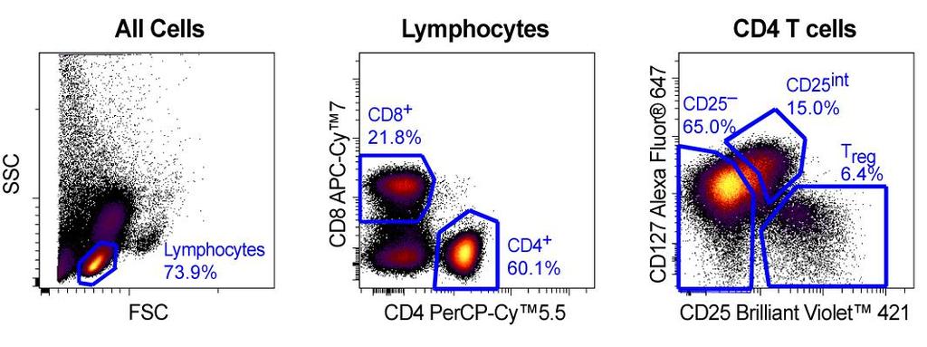
12 Example 2: Detection of FoxP3 Regulatory T Cells (Tregs) Count CD127 Alexa Fluor CD25 Brilliant Violet 421 Tregs are a subset of T cells that regulate the immune response by suppressing the activity of other T cells. Human PBMCs were stained for surface markers CD4 FITC, CD25 Brilliant Violet 421, and CD127 Alexa Fluor 647. After washing, cells were fixed and permeabilized with the BD Pharmingen transcription factor buffer set and stained with FoxP3 PE-CF594. Data was acquired on a BD FACSVerse flow cytometer FoxP3 PE-CF594
13 Detection of Phosphoprotein by Flow Cytometry (BD Phosflow) Proteins are phosphorylated in response to many types of stimuli including cytokines and small molecules. Protein phosphorylation is transient; cells must be fixed quickly to maintain phosphoepitopes. Perm buffer III is the recommended starting buffer for most BD Phosflow applications. Perm buffer III is a harsh denaturing buffer. Other perm buffers are available.
14 Considerations for Phosphospecific Flow Cytometry Stimulation kinetics: most phosphorylation events occur very rapidly Controls: Unlike isotype controls, unstimulated cells take into account basal phosphorylation and the unique background characteristics of each antibody Expression level of signaling protein of interest Perm buffer III can impact surface marker staining performed before or after fixation and permeabilization The BD FACSelect buffer compatibility resource lists buffer compatibility for many popular markers. (
. Do they respond differently to treatment with IL-2?")
15 Example 3: Enhanced IL-2 Sensitivity of Tregs Stimulation by IL-2 leads to Stat5 (py694) phosphorylation in most human T cells. -74 CD127 Alexa Fluor CD4 T cells CD8 T cells Tregs express large amounts of the IL-2 receptor alpha chain (CD25). Do they respond differently to treatment with IL-2? No Stim IL CD25 Brilliant Violet 421 Stat5 (py694) Alexa Fluor 647 Human whole blood was stimulated with 1, 10, or 100 ng/ml of IL-2 for 15 min prior to fixation, permeabilization, and staining with the BD Phosflow T-cell activation kit.

16 Determination of Buffer Compatibility T-cell subsets were identified using CD4 PerCP-Cy5.5, CD8 APC-Cy 7, CD25 Brilliant Violet 421, and CD127 Alexa Fluor 647. To determine compatibility and recommended staining conditions for perm buffer III, the BD FACSelect buffer compatibility resource was used.


17 Determination of Buffer Compatibility (continued) CD4 is compatible with perm buffer III and other buffers. CD127 is not compatible with post-permeabilization staining. Use an alternative protocol with CD127.
18 Example 3: Enhanced IL-2 Sensitivity of Tregs (continued) Human PBMCs were stained with CD127 Alexa Fluor 647 during a 15-minute stimulation with 0-, 0.01-, 0.1-, 1-, 10-, or 100-ng/mL doses of recombinant IL-2. Cells were fixed using BD Cytofix fixation buffer and permeabilized using perm buffer III. Cells were then stained with Stat5 (py694) Alexa Fluor 488, CD4 PerCP-Cy5.5, CD8 APC-Cy7, and CD25 Brilliant Violet 421. Samples were acquired using a BD LSRFortessa flow cytometer and analyzed using Cytobank software.

19 Example 3: Results
20 Considerations when Combining Different Intracellular Techniques Timing of signaling responses Signaling responses such as protein phosphorylation may have ended before others such as cytokine expression begin. Buffer selection Need to select markers and fluorochromes compatible with the permeabilization method needed. May need to try multiple buffers. Staining protocols Staining surface markers prior to cell permeabilization may be necessary.

21 Example 4: IL-2 Response in Th1-Like and Non-Th1 Effector Memory CD4 + T Cells In this experiment, T-bet was used to identify Th1-like cells. T-bet is a transcription factor that controls the expression of IFN-. The T-bet antibody is compatible with perm buffer III.
22 Example 4, continued Human whole blood was stimulated with various concentrations of IL-2 ( ng/ml) for 15 min. Cells were fixed with BD Phosflow lyse/fix buffer and permeabilized with perm buffer III. Cells were stained with CD3 Alexa Fluor 488, CD4 PE-Cy7, CD45RA V450, T-bet PE, and Stat5 (py694) Alexa Fluor 647. Samples were acquired using a BD LSR II flow cytometer and analyzed with Cytobank software.


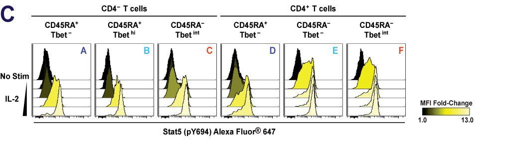
23 Example 4: Results A B C
24 Example 5: Phenotypic Analysis of Th17 Cells from Mouse Spleen and Thymus ROR T is important for the secretion of IL-17 and the maintenance of CD4 + CD8 + thymocytes. Cells isolated from BALB/c thymus and spleen were surface stained with fluorescently labeled antibodies to surface markers CD44, CD62L, CD196, and appropriate isotype controls. Cells were fixed and permeabilized with the BD Pharmingen transcription factor buffer set. Cells were then stained with antibodies to transcription factors ROR T and Foxp3 as well as cytokines IL-17A and IFN-.

25 Example 5: Results
26 Summary and Conclusions Intracellular flow cytometry is a powerful technique for the study of cellular signaling, function, and differentiation within subpopulations of cells. Different buffers work best for particular applications due to the biochemistry and cellular localization of the antigen. BD Cytofix/Cytoperm (Cat. No ) for cytokines BD Pharmingen transcription factor buffer set (Cat. No ) for transcription factors as well as transcription factors combined with cytokines BD Phosflow perm buffer III (Cat. No ) for phosphoprotein detection
27 If you have further questions: Contact your US Reagent Sales Rep or For Research Use Only. Not for use in diagnostic or therapeutic procedures. Alexa Fluor is a registered trademark of Life Technologies Corporation. CF is a trademark of Biotium, Inc. Cy is a trademark of Amersham Biosciences Corp.
Optimizing Intracellular Flow Cytometry:
 Optimizing Intracellular Flow Cytometry: Simultaneous Detection of Cytokines and Transcription Factors An encore presentation by Jurg Rohrer, PhD, BD Biosciences 10.26.10 Outline Introduction Cytokines
Optimizing Intracellular Flow Cytometry:
 Optimizing Intracellular Flow Cytometry: Simultaneous Detection of Cytokines and Transcription Factors Presented by Jurg Rohrer, PhD, BD Biosciences 23-10780-00 Outline Introduction Cytokines Transcription
Simultaneous correlation of cytokine production with Treg and Th17 cell proliferation
 Simultaneous correlation of cytokine production with Treg and Th17 cell proliferation Jurg Rohrer, PhD Director, R&D BD Biosciences 23-11773-00 For Research Use Only. Not for use in diagnostic or therapeutic
Bead Based Assays for Cytokine Detection
 Bead Based Assays for Cytokine Detection September 27, 2014 6 th EFIS-EJI South East European Immunology School SEEIS 2014 Timisoara, Romania The Cells of the Immune System The Immune Reaction (Th2) (Th1)
A step-by-step approach to build and analyze a multicolor panel
 Analyze A step-by-step approach to build and analyze a multicolor panel For Research Use Only. Not for use in diagnostic or therapeutic procedures. Alexa Fluor is a registered trademark of Life Technologies
T-Cell Research. Novel multicolor flow cytometry tools for the study of CD4 + T-cell differentiation and plasticity
 T-Cell Research Novel multicolor flow cytometry tools for the study of CD4 + T-cell differentiation and plasticity A Solid Commitment to Research: Flexible Ways to Study CD4 T-Cell Differentiation and
Recommended Protocols for Phospho Protein Detection in Human Cells
 Recommended Protocols for Phospho Protein Detection in Human Cells Depending on subcellular localization of the phospho protein of interest, as well as epitope susceptibility to cell fixing and permeabilizing
BD Pharmingen. Human Th1/Th2/Th17 Phenotyping Kit. Technical Data Sheet. Product Information. Description Components:
 Technical Data Sheet Human Th1/Th2/Th17 Phenotyping Kit Product Information Material Number: Size: Description Components: 560751 50 Tests BD Pharmingen 51-9006615 Human Th1/Th2/Th17 Phenotyping Cocktail
20 ml (1 ea) 40 ml (1 ea) 25 ml (1 ea) 7.5 ml (1 ea)
 40 ml (1 ea) 25 ml (1 ea) 7.5 ml (1 ea)") Technical Data Sheet Transcription Factor Phospho Buffer Set Product Information Material Number: 565575 25 Tests Component: 51-9011474 Description: 0.75X TFP Diluent Buffer 20 ml (1 ea) BD Pharmingen
Technical Data Sheet Transcription Factor Phospho Buffer Set Product Information Material Number: 565575 25 Tests Component: 51-9011474 Description: 0.75X TFP Diluent Buffer 20 ml (1 ea) BD Pharmingen
BD Flow Cytometry Reagents Multicolor Panels Designed for Optimal Resolution with the BD LSRFortessa X-20 Cell Analyzer
 Multicolor Panels Designed for Optimal Resolution with the BD LSRFortessa X-2 Cell Analyzer Proper multicolor panel design takes into account fluorochrome brightness, antigen density, co-expression, and
Immunofluorescent Staining of Intracellular Cytokines for Flow Cytometric Analysis
 Immunofluorescent Staining of Intracellular Cytokines for Flow Cytometric Analysis I. Introduction Improved methods have continually been sought to analyze single-cell cytokine production by human and
IL-6Rα IL-6RαT-KO KO. IL-6Rα f/f bp. f/f 628 bp deleted 368 bp. 500 bp
 STD H 2 O WT KO IL-6Rα f/f IL-6Rα IL-6RαT-KO KO 1000 bp 500 bp f/f 628 bp deleted 368 bp Supplementary Figure 1 Confirmation of T-cell IL-6Rα deficiency. (a) Representative histograms and (b) quantification
Commercially available HLA Class II tetramers (Beckman Coulter) conjugated to
 conjugated to") Class II tetramer staining Commercially available HLA Class II tetramers (Beckman Coulter) conjugated to PE were combined with dominant HIV epitopes (DRB1*0101-DRFYKTLRAEQASQEV, DRB1*0301- PEKEVLVWKFDSRLAFHH,
Class II tetramer staining Commercially available HLA Class II tetramers (Beckman Coulter) conjugated to PE were combined with dominant HIV epitopes (DRB1*0101-DRFYKTLRAEQASQEV, DRB1*0301- PEKEVLVWKFDSRLAFHH,
BD Cytofix/Cytoperm Plus Fixation/Permeabilization Kit (with BD GolgiStop protein transport inhibitor containing monensin) (Cat. No.
 (Cat. No.") Kit Manual BD Cytofix/Cytoperm Fixation/Permeabilization Kit (Cat. No. 554714) BD Cytofix/Cytoperm Plus Fixation/Permeabilization Kit (with BD GolgiStop protein transport inhibitor containing monensin)
Kit Manual BD Cytofix/Cytoperm Fixation/Permeabilization Kit (Cat. No. 554714) BD Cytofix/Cytoperm Plus Fixation/Permeabilization Kit (with BD GolgiStop protein transport inhibitor containing monensin)
Supplementalgfigureg1gSchematicgdiagramgofgtumor1modellingg
 SChinjectionh F:LuchLCLsh IVhinjectionh T:cellsh Monitorhforhtumorh growthhandhxeno: reactivehgvhd GVLgexperimentg kcbgvsgpbgt1cellse Xeno1reactiveg experimentg kcbgvsgpbgt1cellse IVhinjectionh 5xh,N^6
BD CBA on the BD Accuri C6: Bringing Multiplexed Cytokine Detection to the Benchtop
 BD CBA on the BD Accuri C6: Bringing Multiplexed Cytokine Detection to the Benchtop Maria Dinkelmann, PhD Senior Marketing Applications Specialist BD Biosciences, Ann Arbor, MI 23-14380-00 Cellular Communication
Technical Resources. BD Immunocytometry Systems. FastImmune Intracellular Cytokine Staining Procedures
 FastImmune Intracellular Cytokine Staining Procedures BD has developed protocols for the detection of intracellular cytokines in activated lymphocytes and in activated monocytes. The procedures have been
MAIT cell function is modulated by PD-1 signaling in patients with active
 MAIT cell function is modulated by PD-1 signaling in patients with active tuberculosis Jing Jiang, M.D., Xinjing Wang, M.D., Hongjuan An, M.Sc., Bingfen Yang, Ph.D., Zhihong Cao, M.Sc., Yanhua Liu, Ph.D.,
DURACLONE IF BE CERTAIN ABOUT THE RESPONSE. l res. a il n c n. For Research Use Only - Not for use in Diagnostic procedures
 DURACLONE IF earch tria l res lc a om ic il n c n nio pa Yo ur BE CERTAIN ABOUT THE RESPONSE For Research Use Only - Not for use in Diagnostic procedures BE CERTAIN ABOUT THE RESPONSE The sensitive and
T H 1, T H 2 and T H 17 polarization of naïve CD4 + mouse T cells
 A complete workflow for cell preparation, isolation, polarization and analysis T H 1, T H 2 and T H 17 polarization of naïve CD4 + mouse T cells Introduction Workflow CD4 + T helper (T H) cells play a
Fluorochrome Panel 1 Panel 2 Panel 3 Panel 4 Panel 5 CTLA-4 CTLA-4 CD15 CD3 FITC. Bio) PD-1 (MIH4, BD) ICOS (C398.4A, Biolegend) PD-L1 (MIH1, BD)
 PD-1 (MIH4, BD) ICOS (C398.4A, Biolegend) PD-L1 (MIH1, BD)") Additional file : Table S. Antibodies used for panel stain to identify peripheral immune cell subsets. Panel : PD- signaling; Panel : CD + T cells, CD + T cells, B cells; Panel : Tregs; Panel :, -T, cdc,
Additional file : Table S. Antibodies used for panel stain to identify peripheral immune cell subsets. Panel : PD- signaling; Panel : CD + T cells, CD + T cells, B cells; Panel : Tregs; Panel :, -T, cdc,
Blocking antibodies and peptides. Rat anti-mouse PD-1 (29F.1A12, rat IgG2a, k), PD-
, PD-") Supplementary Methods Blocking antibodies and peptides. Rat anti-mouse PD-1 (29F.1A12, rat IgG2a, k), PD- L1 (10F.9G2, rat IgG2b, k), and PD-L2 (3.2, mouse IgG1) have been described (24). Anti-CTLA-4 (clone
Supplementary Methods Blocking antibodies and peptides. Rat anti-mouse PD-1 (29F.1A12, rat IgG2a, k), PD- L1 (10F.9G2, rat IgG2b, k), and PD-L2 (3.2, mouse IgG1) have been described (24). Anti-CTLA-4 (clone
Comprehensive evaluation of human immune system reconstitution in NSG. and NSG -SGM3 mouse models toward the development of a novel ONCO-HU
 Comprehensive evaluation of human immune system reconstitution in NSG and NSG -SGM3 mouse models toward the development of a novel ONCO-HU xenograft model Aaron Middlebrook, 1 Eileen Snowden, 2 Warren
Effector T Cells and
 1 Effector T Cells and Cytokines Andrew Lichtman, MD PhD Brigham and Women's Hospital Harvard Medical School 2 Lecture outline Cytokines Subsets of CD4+ T cells: definitions, functions, development New
BD Phosflow T Cell Activation Kit
 BD Phosflow T Cell Activation Kit Instruction Manual Catalog No. 560750 ii BD Phosflow T Cell Activation Kit The information in this guide is subject to change without notice. BD Biosciences reserves the
Supplemental Information. T Cells Enhance Autoimmunity by Restraining Regulatory T Cell Responses via an Interleukin-23-Dependent Mechanism
 Immunity, Volume 33 Supplemental Information T Cells Enhance Autoimmunity by Restraining Regulatory T Cell Responses via an Interleukin-23-Dependent Mechanism Franziska Petermann, Veit Rothhammer, Malte
Peptide stimulation and Intracellular Cytokine Staining
 v Peptide stimulation and Intracellular Cytokine Staining Authors A. Cosma, S. Allgayer MATERIALS Date 01-02-2007 REAGENTS: Version 1.0 - PBMCs - Culture medium - Costimulating antibodies - Peptide pools
Supplementary Figure 1. Enhanced detection of CTLA-4 on the surface of HIV-specific
 SUPPLEMENTARY FIGURE LEGEND Supplementary Figure 1. Enhanced detection of CTLA-4 on the surface of HIV-specific CD4 + T cells correlates with intracellular CTLA-4 levels. (a) Comparative CTLA-4 levels
MATERIALS. Peptide stimulation and Intracellular Cytokine Staining- EFFECTOR 45RA PANEL
 v Peptide stimulation and Intracellular Cytokine Staining- EFFECTOR 45RA PANEL Authors S. Kutscher, A. Cosma, MATERIALS REAGENTS: - PBMCs - Culture medium - Costimulating antibodies - Peptide pools including
Application Information Bulletin: Cell Signaling
 Application Information Bulletin: Cell Signaling Detection of Both Surface Proteins and Intracellular Phospho-Determinants in Whole Blood Using the PerFix-p Reagent System William L. Godfrey 1, Li Yang
Direct ex vivo characterization of human antigen-specific CD154 + CD4 + T cells Rapid antigen-reactive T cell enrichment (Rapid ARTE)
") Direct ex vivo characterization of human antigen-specific CD154 + CD4 + T cells Rapid antigen-reactive T cell enrichment (Rapid ARTE) Introduction Workflow Antigen (ag)-specific T cells play a central
Direct ex vivo characterization of human antigen-specific CD154 + CD4 + T cells Rapid antigen-reactive T cell enrichment (Rapid ARTE) Introduction Workflow Antigen (ag)-specific T cells play a central
Supplementary Table e-1. Flow cytometry reagents and staining combinations
 Supplementary data Supplementary Table e-1. Flow cytometry reagents and staining combinations Reagents Antibody Fluorochrome Clone Source conjugation CD3 FITC UCHT1 BD Biosciences CD3 PerCP-Cy5.5 SK7 Biolegend
Cover Page. The handle holds various files of this Leiden University dissertation.
 Cover Page The handle http://hdl.handle.net/1887/23854 holds various files of this Leiden University dissertation. Author: Marel, Sander van der Title: Gene and cell therapy based treatment strategies
Application Information Bulletin: Human NK Cells Phenotypic characterizing of human Natural Killer (NK) cell populations in peripheral blood
 cell populations in peripheral blood") Application Information Bulletin: Human NK Cells Phenotypic characterizing of human Natural Killer (NK) cell populations in peripheral blood Christopher A Fraker, Ph.D., University of Miami - Miami, Florida
Application Information Bulletin: Human NK Cells Phenotypic characterizing of human Natural Killer (NK) cell populations in peripheral blood Christopher A Fraker, Ph.D., University of Miami - Miami, Florida
Effector mechanisms of cell-mediated immunity: Properties of effector, memory and regulatory T cells
 ICI Basic Immunology course Effector mechanisms of cell-mediated immunity: Properties of effector, memory and regulatory T cells Abul K. Abbas, MD UCSF Stages in the development of T cell responses: induction
Supplemental Figure 1. Signature gene expression in in vitro differentiated Th0, Th1, Th2, Th17 and Treg cells. (A) Naïve CD4 + T cells were cultured
 Naïve CD4 + T cells were cultured") Supplemental Figure 1. Signature gene expression in in vitro differentiated Th0, Th1, Th2, Th17 and Treg cells. (A) Naïve CD4 + T cells were cultured under Th0, Th1, Th2, Th17, and Treg conditions. mrna
Supplemental Figure 1. Signature gene expression in in vitro differentiated Th0, Th1, Th2, Th17 and Treg cells. (A) Naïve CD4 + T cells were cultured under Th0, Th1, Th2, Th17, and Treg conditions. mrna
W/T Itgam -/- F4/80 CD115. F4/80 hi CD115 + F4/80 + CD115 +
 F4/8 % in the peritoneal lavage 6 4 2 p=.15 n.s p=.76 CD115 F4/8 hi CD115 + F4/8 + CD115 + F4/8 hi CD115 + F4/8 + CD115 + MHCII MHCII Supplementary Figure S1. CD11b deficiency affects the cellular responses
Rapid antigen-specific T cell enrichment (Rapid ARTE)
") Direct ex vivo characterization of human antigen-specific CD154+CD4+ T cell Rapid antigen-specific T cell enrichment (Rapid ARTE) Introduction Workflow Antigen (ag)-specific T cells play a central role
Direct ex vivo characterization of human antigen-specific CD154+CD4+ T cell Rapid antigen-specific T cell enrichment (Rapid ARTE) Introduction Workflow Antigen (ag)-specific T cells play a central role
Detailed step-by-step operating procedures for NK cell and CTL degranulation assays
 Supplemental methods Detailed step-by-step operating procedures for NK cell and CTL degranulation assays Materials PBMC isolated from patients, relatives and healthy donors as control K562 cells (ATCC,
DuraClone IM. Standardized phenotyping panels for studies of the human immune system
 DuraClone IM Standardized phenotyping panels for studies of the human immune system Standardize with the experts, adopt DuraClone IM Tubes. Clinical research studies require accurate, reproducible results
x Lymphocyte count /µl CD8+ count/µl 800 Calculated
 % Lymphocyte in CBC A. 50 40 30 20 10 Lymphocyte count /µl B. x10 3 2.5 1.5 C. 50 D. 1000 % CD3+CD8+ Cells 40 30 20 Calculated CD8+ count/µl 800 600 400 200 10 0 #61 #63 #64 #65 #68 #71 #72 #75 Figure
Human Immunodeficiency Virus Type-1 Myeloid Derived Suppressor Cells Inhibit Cytomegalovirus Inflammation through Interleukin-27 and B7-H4
 Human Immunodeficiency Virus Type-1 Myeloid Derived Suppressor Cells Inhibit Cytomegalovirus Inflammation through Interleukin-27 and B7-H4 Ankita Garg, Rodney Trout and Stephen A. Spector,,* Department
Supplementary Table; Supplementary Figures and legends S1-S21; Supplementary Materials and Methods
 Silva et al. PTEN posttranslational inactivation and hyperactivation of the PI3K/Akt pathway sustain primary T cell leukemia viability Supplementary Table; Supplementary Figures and legends S1-S21; Supplementary
DIFFERENCES IN T-CELL SUBSETS IN ME/CFS. University of Nevada, Reno. Determining the Difference Between Circulating Follicular Regulatory T-cells and
 DIFFERENCES IN T-CELL SUBSETS IN ME/CFS University of Nevada, Reno Determining the Difference Between Circulating Follicular Regulatory T-cells and Follicular Helper T-cells in Subjects with Myalgic Encephalomyelitis/Chronic
Primary Adult Naïve CD4+ CD45RA+ Cells. Prepared by: David Randolph at University of Alabama, Birmingham
 Primary Adult Naïve CD4+ CD45RA+ Cells Prepared by: David Randolph (drdrdr@uab.edu) at University of Alabama, Birmingham Goal: To obtain large numbers of highly pure primary CD4+ CD45RO- CD25- cells from
PBMC from each patient were suspended in AIM V medium (Invitrogen) with 5% human
 with 5% human") Anti-CD19-CAR transduced T-cell preparation PBMC from each patient were suspended in AIM V medium (Invitrogen) with 5% human AB serum (Gemini) and 300 international units/ml IL-2 (Novartis). T cell proliferation
Anti-CD19-CAR transduced T-cell preparation PBMC from each patient were suspended in AIM V medium (Invitrogen) with 5% human AB serum (Gemini) and 300 international units/ml IL-2 (Novartis). T cell proliferation
Page 1 of 2. Product Information Contents: ezkine Th1 Activation 2 Whole Blood Intracellular Cytokine Kit
 Page 1 of 2 ezkine Th1 Activation 2 Whole Blood Intracellular Cytokine Kit Catalog Number: 8822-6852 RUO: For Research Use Only. Not for use in diagnostic procedures. Staining of human whole blood with
In vitro expansion of mouse CD4 + T cells
 Direct ex vivo isolation, cultivation and expansion of CD4 + T cells from mouse spleen In vitro expansion of mouse CD4 + T cells Introduction CD4 + T helper (TH) cells play a central role in the adaptive
Intracellular Cytokine Staining Starter Kit - Mouse
 BD Pharmingen Intracellular Cytokine Staining Starter Kit - Mouse Instruction Manual Catalog No. 559311 Falcon is a registered trademark of Corning Incorporated. BD flow cytometers are Class 1 laser products.
Index. Index 439. Aequorin, 84, 94 Affinity precipitation, 372, AP-1, 100 Asthma, 170, 305
 Index 439 Index A Aequorin, 84, 94 Affinity precipitation, 372, 376 381 AP-1, 100 Asthma, 170, 305 B Bioassay, 185, comparison with ELISA, 318 GM-CSF bioassay, 351 IL-2 bioassay, 185 192, 300 IL-3 IL-6
Interferon γ regulates idiopathic pneumonia syndrome, a. Th17 + CD4 + T-cell-mediated GvH disease
 Interferon γ regulates idiopathic pneumonia syndrome, a Th17 + CD4 + T-cell-mediated GvH disease Nora Mauermann, Julia Burian, Christophe von Garnier, Stefan Dirnhofer, Davide Germano, Christine Schuett,
D CD8 T cell number (x10 6 )
") IFNγ Supplemental Figure 1. CD T cell number (x1 6 ) 18 15 1 9 6 3 CD CD T cells CD6L C CD5 CD T cells CD6L D CD8 T cell number (x1 6 ) 1 8 6 E CD CD8 T cells CD6L F Log(1)CFU/g Feces 1 8 6 p
IFNγ Supplemental Figure 1. CD T cell number (x1 6 ) 18 15 1 9 6 3 CD CD T cells CD6L C CD5 CD T cells CD6L D CD8 T cell number (x1 6 ) 1 8 6 E CD CD8 T cells CD6L F Log(1)CFU/g Feces 1 8 6 p
Intracellular Cytokine Staining Starter Kit - Human
 BD Pharmingen Intracellular Cytokine Staining Starter Kit - Human Instruction Manual Catalog No. 559302 Falcon is a registered trademark of Corning Incorporated. Ficoll-Paque is a trademark of GE Healthcare.
In vitro human regulatory T cell expansion
 - 1 - Human CD4 + CD25 + regulatory T cell isolation, Workflow in vitro expansion and analysis In vitro human regulatory T cell expansion Introduction Regulatory T (Treg) cells are a subpopulation of T
In vitro human regulatory T cell expansion
 - 1 - Human CD4 + CD25 + CD127 dim/- regulatory T cell Workflow isolation, in vitro expansion and analysis In vitro human regulatory T cell expansion Introduction Regulatory T (Treg) cells are a subpopulation
DURACLONE RE THERE ARE EVENTS YOU CANNOT AFFORD TO MISS
 DURACLONE RE THERE ARE EVENTS YOU CANNOT AFFORD TO MISS Your clinical research trial companion For Reseach Use Only - Not for use in Diagnostic procedures THERE ARE EVENTS YOU CANNOT AFFORD TO MISS Rare
JAK3 inhibitor administration in vivo in chronically SIV Infected Rhesus Macaques
 JAK3 inhibitor administration in vivo in chronically SIV Infected Rhesus Macaques preferentially depletes NK Cells Ladawan Kowawisatsut 1 Kovit Pattanapanyasat 1 Aftab A. Ansari 2 1 Department of Immunology
Muse Assays for Cell Analysis
 Muse Assays for Cell Analysis Multiple Assay Outputs for Cell Analysis Cell Health Cell Signalling Immunology Muse Count & Viability Kit Muse Cell Cycle Kit Muse Annexin V & Dead Cell Kit Muse Caspase
ezkine Th1/Th17 Whole Blood Intracellular Cytokine Kit Catalog Number: RUO: For Research Use Only. Not for use in diagnostic procedures.
 Page 1 of 3 ezkine Th1/Th17 Whole Blood Intracellular Cytokine Kit RUO: For Research Use Only. Not for use in diagnostic procedures. Staining of human whole blood with the ezkine Th1/Th17 Whole Blood Intracellular
Supplementary Data. Treg phenotype
 Supplementary Data Additional Experiment An additional experiment was performed using cryopreserved peripheral blood mononuclear cells (PBMC) derived from five renal cell carcinoma (RCC) patients [see
Challenges in Development and Validation of an Intracellular Cytokine Staining assay
 Challenges in Development and Validation of an Intracellular Cytokine Staining assay Jenny Hendriks, Crucell Hatching @ EBF, Brussels, June 202 www.crucell.com Vaccines vs Protein therapeutics Protein
BD FastImmune. BD FACS lysing solution and. using BD FACS Permeabilizing Solution
 BD FastImmune CD4 Intracellular Cytokine Detection Kit BD FastImmune Anti Hu-IFN-γ/CD69/CD4/CD3 Catalog No. 337185 BD FastImmune IL-2/CD69/CD4/CD3 Catalog No. 337187 BD FastImmune Anti Hu-TNF-α/CD69/CD4/CD3
Naive, memory and regulatory T lymphocytes populations analysis
 Naive, memory and regulatory T lymphocytes populations analysis Jaen Olivier, PhD ojaen@beckmancoulter.com Cellular Analysis application specialist Beckman Coulter France Introduction Flow cytometric analysis
Supplementary Figure 1. Efficient DC depletion in CD11c.DOG transgenic mice
 Supplementary Figure 1. Efficient DC depletion in CD11c.DOG transgenic mice (a) CD11c.DOG transgenic mice (tg) were treated with 8 ng/g body weight (b.w.) diphtheria toxin (DT) i.p. on day -1 and every
Examples of questions for Cellular Immunology/Cellular Biology and Immunology
 Examples of questions for Cellular Immunology/Cellular Biology and Immunology Each student gets a set of 6 questions, so that each set contains different types of questions and that the set of questions
Supplementary Figure 1. IL-12 serum levels and frequency of subsets in FL patients. (A) IL-12
 IL-12") 1 Supplementary Data Figure legends Supplementary Figure 1. IL-12 serum levels and frequency of subsets in FL patients. (A) IL-12 serum levels measured by multiplex ELISA (Luminex) in FL patients before
1 Supplementary Data Figure legends Supplementary Figure 1. IL-12 serum levels and frequency of subsets in FL patients. (A) IL-12 serum levels measured by multiplex ELISA (Luminex) in FL patients before
Supplemental Table I.
 Supplemental Table I Male / Mean ± SEM n Mean ± SEM n Body weight, g 29.2±0.4 17 29.7±0.5 17 Total cholesterol, mg/dl 534.0±30.8 17 561.6±26.1 17 HDL-cholesterol, mg/dl 9.6±0.8 17 10.1±0.7 17 Triglycerides,
Cover Page. The handle holds various files of this Leiden University dissertation
 Cover Page The handle http://hdl.handle.net/1887/24366 holds various files of this Leiden University dissertation Author: Buddingh, Emilie Pauline Title: Innate immunity in osteosarcoma Issue Date: 2014-03-05
Online supplement. Phenotypic, functional and plasticity features of classical and alternatively activated
 Online supplement Phenotypic, functional and plasticity features of classical and alternatively activated human macrophages Abdullah Al Tarique*, Jayden Logan *, Emma Thomas *, Patrick G Holt *, Peter
Glow. Save. Let it. and. Get a Discount of 10% BD Biosciences Year End Promotion on a huge Selection of Reagent Solutions
 Let it Glow and Save BD Biosciences Year End Promotion 2014 Get a Discount of 10% on a huge Selection of Reagent Solutions All fluorochrome labeled antibodies The complete BD CBA portfolio Kits and reagents
B and T-Cells. CD8+ Cytotoxic T Cells
 Sony Biotechnology Inc. Application Data, SA38 Spectral Analyzer TBNK Panel Page 1 TBNK panel on the SA38 Spectral Analyzer TBNK (B-Cell, T-Cell, and natural killer cells) panels are frequently used to
ASSESSING THE FUNCTION OF EBV-SPECIFIC CD4 + T cells
 ASSESSING THE FUNCTION OF EBV-SPECIFIC CD4 + T cells BY BENJAMIN JAMES MECKIFF A thesis submitted to the University of Birmingham for the degree of MRes in Cancer Sciences School of Cancer Sciences College
HD1 (FLU) HD2 (EBV) HD2 (FLU)
 HD2 (EBV) HD2 (FLU)") ramer staining + anti-pe beads ramer staining a HD1 (FLU) HD2 (EBV) HD2 (FLU).73.11.56.46.24 1.12 b CD127 + c CD127 + d CD127 - e CD127 - PD1 - PD1 + PD1 + PD1-1 1 1 1 %CD127 + PD1-8 6 4 2 + anti-pe %CD127
ramer staining + anti-pe beads ramer staining a HD1 (FLU) HD2 (EBV) HD2 (FLU).73.11.56.46.24 1.12 b CD127 + c CD127 + d CD127 - e CD127 - PD1 - PD1 + PD1 + PD1-1 1 1 1 %CD127 + PD1-8 6 4 2 + anti-pe %CD127
Organic dust-induced interleukin-12 production activates T- and natural killer cells
 Eur Respir J 22; 2: 686 69 DOI:.1183/931936.2.222 Printed in UK all rights reserved Copyright #ERS Journals Ltd 22 European Respiratory Journal ISSN 93-1936 Organic dust-induced interleukin-12 production
<10. IL-1β IL-6 TNF + _ TGF-β + IL-23
 3 ns 25 ns 2 IL-17 (pg/ml) 15 1 ns ns 5 IL-1β IL-6 TNF
Supplementary Figure 1. Ex vivo IFNγ production by Tregs. Nature Medicine doi: /nm % CD127. Empty SSC 98.79% CD25 CD45RA.
 SSC CD25 1.8% CD127 Empty 98.79% FSC CD45RA CD45RA Foxp3 %IFNγ + cells 4 3 2 1 + IL-12 P =.3 IFNγ p=.9 %IL-4+ cells 3 2 1 IL-4 P =.4 c %IL-1 + cells IFNγ 4 3 2 1 Control Foxp3 IL-1 P =.41.64 4.76 MS 2.96
What to Measure, How to Measure It
 Dale and Betty Bumpers Vaccine Research Center National Institute of Allergy and Infectious Diseases National Institutes of Health Monitoring Memory T-cells: What to Measure, How to Measure It Pratip K.
Interleukin 17 (IL-17)-producing T helper cells (Th17) are a
-producing T helper cells (Th17) are a") Aryl hydrocarbon receptor regulates Stat1 activation and participates in the development of Th17 cells Akihiro Kimura*, Tetsuji Naka, Keiko Nohara, Yoshiaki Fujii-Kuriyama, and Tadamitsu Kishimoto* *Laboratory
Aryl hydrocarbon receptor regulates Stat1 activation and participates in the development of Th17 cells Akihiro Kimura*, Tetsuji Naka, Keiko Nohara, Yoshiaki Fujii-Kuriyama, and Tadamitsu Kishimoto* *Laboratory
Supplementary Data 1. Alanine substitutions and position variants of APNCYGNIPL. Applied in
 Supplementary Data 1. Alanine substitutions and position variants of APNCYGNIPL. Applied in Supplementary Fig. 2 Substitution Sequence Position variant Sequence original APNCYGNIPL original APNCYGNIPL
Rapamycin reduced inhibition of V 1 T cells towards V 4 T cells through down-regulating IL-4
 Rapamycin reduced inhibition of V 1 T cells towards V 4 T cells through down-regulating IL-4 Wanjun Huang Biomedical Translational Research Institute, Jinan University, Guangzhou 510632, China. Abstract
MATERIALS AND METHODS. Neutralizing antibodies specific to mouse Dll1, Dll4, J1 and J2 were prepared as described. 1,2 All
 MATERIALS AND METHODS Antibodies (Abs), flow cytometry analysis and cell lines Neutralizing antibodies specific to mouse Dll1, Dll4, J1 and J2 were prepared as described. 1,2 All other antibodies used
Supplementary webappendix
 Supplementary webappendix This webappendix formed part of the original submission and has been peer reviewed. We post it as supplied by the authors. Supplement to: Portevin D, Moukambi F, Clowes P, et
Supplementary Figure S1: Alignment of CD28H. (a) Alignment of human CD28H with other known B7 receptors. (b) Alignment of CD28H orthologs.
 Alignment of human CD28H with other known B7 receptors. (b) Alignment of CD28H orthologs.") Supplementary Figure S1: Alignment of CD28H. (a) Alignment of human CD28H with other known B7 receptors. (b) Alignment of CD28H orthologs. Predicted CD28H protein sequences from human, chimpanzee (pan
Supplementary Figure S1: Alignment of CD28H. (a) Alignment of human CD28H with other known B7 receptors. (b) Alignment of CD28H orthologs. Predicted CD28H protein sequences from human, chimpanzee (pan
SUPPORTING INFORMATIONS
 SUPPORTING INFORMATIONS Mice MT/ret RetCD3ε KO α-cd25 treated MT/ret Age 1 month 3 mnths 6 months 1 month 3 months 6 months 1 month 3 months 6 months 2/87 Survival 87/87 incidence of 17/87 1 ary tumor
Subset analysis of the EBV-specific CD4+ T cell response in primary. and persistent infection. and
 0 Subset analysis of the EBV-specific CD4+ T cell response in primary and persistent infection and Characterisation of CD8+ T cell responses against CMV derived antigens protected in wild type strains
Page 1 of 3. Product Information Contents: ezkine CD8 Activation 2 Whole Blood Intracellular Cytokine Kit
 Page 1 of 3 ezkine CD8 Activation 2 Whole Blood Intracellular Cytokine Kit RUO: For Research Use Only. Not for use in diagnostic procedures. Staining of human whole blood with the ezkine CD8 Activation
Supplementary Material
 Supplementary Material accompanying the manuscript Interleukin 37 is a fundamental inhibitor of innate immunity Marcel F Nold, Claudia A Nold-Petry, Jarod A Zepp, Brent E Palmer, Philip Bufler & Charles
Therapeutic PD L1 and LAG 3 blockade rapidly clears established blood stage Plasmodium infection
 Supplementary Information Therapeutic PD L1 and LAG 3 blockade rapidly clears established blood stage Plasmodium infection Noah S. Butler, Jacqueline Moebius, Lecia L. Pewe, Boubacar Traore, Ogobara K.
Nature Immunology: doi: /ni Supplementary Figure 1. Huwe1 has high expression in HSCs and is necessary for quiescence.
 Supplementary Figure 1 Huwe1 has high expression in HSCs and is necessary for quiescence. (a) Heat map visualizing expression of genes with a known function in ubiquitin-mediated proteolysis (KEGG: Ubiquitin
Dendritic Cell - T Cell Immune Network
 w w w. i m g e n e x. c o m BRIDGING INNATE & ADAPTIVE IMMUNITY Dendritic Cell - T Cell Immune Network Signaling Pathways IMGENEX provides the most extensive portfolio for in depth analysis of the Dendritic
Use of standardized lyophilized reagents to develop a functional T- cell signature. John F. Dunne, PhD Assoc. Scientific Director BD Biosciences
 Use of standardized lyophilized reagents to develop a functional T- cell signature John F. Dunne, PhD Assoc. Scientific Director BD Biosciences Functional T cell responses to tumor antigens in breast cancer
Hua Tang, Weiping Cao, Sudhir Pai Kasturi, Rajesh Ravindran, Helder I Nakaya, Kousik
 SUPPLEMENTARY FIGURES 1-19 T H 2 response to cysteine-proteases requires dendritic cell-basophil cooperation via ROS mediated signaling Hua Tang, Weiping Cao, Sudhir Pai Kasturi, Rajesh Ravindran, Helder
Ex vivo Human Antigen-specific T Cell Proliferation and Degranulation Willemijn Hobo 1, Wieger Norde 1 and Harry Dolstra 2*
 Ex vivo Human Antigen-specific T Cell Proliferation and Degranulation Willemijn Hobo 1, Wieger Norde 1 and Harry Dolstra 2* 1 Department of Laboratory Medicine - Laboratory of Hematology, Radboud University
IL-1b Promotes TGF-b1 and IL-2 Dependent Foxp3 Expression in Regulatory T Cells
 IL-1b Promotes TGF-b1 and IL-2 Dependent Foxp3 Expression in Regulatory T Cells Balaji B. Ganesh 1, Palash Bhattacharya 1, Anupama Gopisetty 1, Jianrong Sheng 1, Chenthamarakshan Vasu 2, Bellur S. Prabhakar
CD25-PE (BD Biosciences) and labeled with anti-pe-microbeads (Miltenyi Biotec) for depletion of CD25 +
 and labeled with anti-pe-microbeads (Miltenyi Biotec) for depletion of CD25 +") Supplements Supplemental Materials and Methods Depletion of CD25 + T-cells from PBMC. Fresh or HD precultured PBMC were stained with the conjugate CD25-PE (BD Biosciences) and labeled with anti-pe-microbeads
Supplements Supplemental Materials and Methods Depletion of CD25 + T-cells from PBMC. Fresh or HD precultured PBMC were stained with the conjugate CD25-PE (BD Biosciences) and labeled with anti-pe-microbeads
Supplementary Figure S1. Flow cytometric analysis of the expression of Thy1 in NH cells. Flow cytometric analysis of the expression of T1/ST2 and
 Supplementary Figure S1. Flow cytometric analysis of the expression of Thy1 in NH cells. Flow cytometric analysis of the expression of T1/ST2 and Thy1 in NH cells derived from the lungs of naïve mice.
Immune Regulation and Tolerance
 Immune Regulation and Tolerance Immunoregulation: A balance between activation and suppression of effector cells to achieve an efficient immune response without damaging the host. Activation (immunity)
This information is current as of November 17, 2018.
 This information is current as of November 17, 2018. Supplementary Material References Subscription Permissions Email Alerts T Cell-Signaling Network Analysis Reveals Distinct Differences between CD28
ILC1 and ILC3 isolation and culture Following cell sorting, we confirmed that the recovered cells belonged to the ILC1, ILC2 and
 Supplementary Methods and isolation and culture Following cell sorting, we confirmed that the recovered cells belonged to the, ILC2 and subsets. For this purpose we performed intracellular flow cytometry
Cell-mediated Immunity
 Cellular & Molecular Immunology Cell-mediated Immunity Nicholas M. Ponzio, Ph.D. Department of Pathology & Laboratory Medicine April 6, 2009 Today s Presentation: Overview Cellular Interactions In Humoral