The immune system has evolved strategies, largely in response to infections, to
|
|
|
- Pamela King
- 6 years ago
- Views:
Transcription
1 Introduction Results
2 Introduction The immune system see tumor as foreign The immune system has evolved strategies, largely in response to infections, to efficiently search for and specifically destroy diseased targets. After nearly a century of debate as to whether the immune system can actually target tumors, compelling evidence now suggests that immune cells play an important role in the control of malignancy. As is discussed below, there is evidence that immune responses against tumor antigens bear both similarities and differences against self tissue antigens. That immunity to tumors would resemble immunity to normal tissues is not surprising because tumors are of course transformations of normal cells in which growth control has become dysregulated. However, dissection of molecular events of tumorigenesis together with the pathophysiology of cancer progression teaches us that there are significant features that distinguish cancer cells from their normal counterparts. Specifically, tumors differ fundamentally from their normal cell counterparts in antigenic composition and biologic behavior. The molecular hallmark of carcinogenesis is genetic instability (1). Genetic instability in cancers is a consequence of deletion and/or mutational inactivation of genome guardians such as p53 (2). Indeed, many of the genetically defined familial cancer syndromes, such as hereditary nonpoliposis, colon cancer and familial breast cancer, are due to mutations in genes that mediate responses to DNA damage (3-8). The genetic instability of cancer cells means that new antigens are constantly being generated in tumors as they develop and progress. This does not occur in normal, non-transformed tissues, which maintain a stable antigenic profile. In addition to the thousands of mutational events that occur during tumorigenesis, hundred of genes that are either inactivated in the normal tissue of origin or expressed at relatively low levels are activated dramatically in cancers. Although these epigenetic changes do not formally create tumor-specific neoantigens, they raise the concentration of encoded protein many order of magnitude, thereby dramatically affecting antigenicity. Whereas uncontrolled growth is certainly a common biological feature of 7
3 all tumors, the major pathophysiologic characteristics of malignant cancer responsible for morbidity and mortality are the ability of transformed malignant cells to invade across natural tissue barriers and to metastasize. Both of these characteristics, which are never seen in normal tissues or benign tumors, are associated with dramatic disruption of architecture. One of the important consequence of tissue disruption, even when caused by non infectious mechanisms, is the elaboration of proinflammatory signals. These signals generally in the form of cytokines and chemokines are central initiators of both innate and adaptive immune responses. The tumor immunosurveillance hypothesis The notion that the immune system could protect the host from neoplastic disease was initially proposed by Ehrlich and formally introduced as the cancer immunosurveillance hypothesis nearly 50 years later by Burnet and Thomas (9-12). The fundamental tenet of the immune surveillance hypothesis is that tumors arise with similar frequency to infection with pathogens and that the immune system constantly recognizes and eliminates these tumors based on their expression of tumor-associated antigens (TAAs). Originally, the existence of TAAs was surmised based on the finding that tumors induced in animal models were frequently rejected when transplanted to syngenic hosts, whereas transplants of normal tissue between syngenic hosts were accepted (13-15). Modern molecular oncology teaches us that TAAs represent the consequences of the genetic and epigenetic alterations in cancer cells. In fact, both spontaneously arising and chemically induced tumors display diverse properties, with some being rejected effectively (termed regressor tumors) and others growing progressively (termed progressor tumors) after transplantation to syngenic hosts (16). A corollary to the original immune surveillance hypothesis is that progressor tumors in animals, as well as clinically progressive spontaneous cancers in all species, are not eliminated because they develop active mechanisms of either immune escape or resistance. A fundamental prediction of the immune surveillance hypothesis is that immunodeficient individuals would display a dramatic increase in tumor incidence. 8
4 However, this was put to the experimental test using nude mice, which were the most congenitally immunodeficient mice available at the time, no convincing evidence for such a process was obtained (17,18). Specifically, CBA/H strain nude mice neither developed increased incidences of carcinogen methylcholanthrene (MCA) induced or spontaneous tumors nor did they show shortened periods of tumor latency compared with wild-type controls (19-23). However, in retrospect, there are several important caveats to these experiments. First, nude mice still produce diminished numbers of αβ T cells and therefore capable of some degree of T cell dependent immunity (24, 25). Second, they frequently display a compensatory increase in innate immunity including natural killer (NK) cell function. In addition the profound influence of innate immunity on adaptive immunity was not appreciated at the time (26). Thus, the residual adaptive immune system in the presence of a fully functional innate immune system may provide the nude mice with at least some cancer immunosurveillance capacity. Third, the CBA/H strain mice express the highly active isoform of the aryl hydroxylase enzyme that is required to metabolize MCA induced cellular transformation in CBA/H strain mice occurred so efficiently that it masked any protective effect that immunity could provide (27). In the 1970s and 1980s, epidemiologic studies of patients with heritable immunodeficiencies revealed a more complex pattern of cancer risk (28). Uncommon cancers such as lymphoblastic lymphomas and Kaposi s sarcoma were observed at significantly increased frequency; however, the common epithelial cancers seen in adulthood (such as colon cancer, lung cancer, prostate cancer, etc) were not increased in this population. As more was learned about the microbial-particularly viral-etiology of some malignancies, it became clear that the cancers most commonly found in immunodeficient individuals were virus associated. Virtually all lymphomas are Epstein-Barr virus in origin, resulting from a failure of T cells to control Epstein-Barr virus transformed B cells. Kaposi s sarcoma is consequent to human herpesvirus 8 infection (29-34). Other virus associated cancers such as cervical cancer (from human 9
5 papillomavirus) were also increased in frequency (35, 36). The major non-virus associated cancer observed at increased frequency in immunodeficient individuals is stomach cancer. It is now appreciated that stomach cancer is commonly a consequence of ulcer disease related to infection with the Helicobactor pylori bacterium (37). From these studies the notion emerged that immune surveillance indeed protects individuals against certain pathogen (mostly-virus)-associated cancers by either preventing infection or checking chronic infection by viruses and other pathogens that can eventually lead to cancer. However, the failure to observe an altered incidence in non-virus-associated cancers was taken as a strong argument against the classic immune surveillance hypothesis. The resurgence of tumor immunosurveillance The validity of the tumor immunosurveillance concept has, in the past, been difficult to establish. When first proposed in 1909 the hypothesis could not be experimentally tested because little was known at the time about the molecular and cellular basis of immunity. Later on, as the field of immunology developed and the concept acquired its name cancer immunosurveillance. Experimental testing became possible but failed to provide for the process, using mice with spontaneous mutations that rendered them immunocompromised but not completely immunodeficient. Only recently, with the development of gene targeting and transgenic mouse technologies and the capacity to produce highly specific blocking monoclonal antibodies (mab) to particular immune components has the cancer immunosurveillance hypothesis become testable in unequivocal, molecularly defined murine models of immunodeficiency and thus the notion that the immune system regulates cancer development is experiencing a new resurgence. An overwhelming amount of data from animal models together with compelling data from human patients indicate that a functional tumor immunosurveillance process exists that acts as an extrinsic tumor suppressor. The definitive work demonstrating the existence of tumor immunosurveillance process was based on experiments employing gene-targeted mice that lack the recombinase 10
6 activating gene (RAG)-2. Mice lacking RAG-2 (or its obligate partner RAG-1) cannot rearrange lymphocyte antigen receptors and thus lack T, B, and NKT cells (38). Since RAG-2 expression is limited to cells of the immune system, the use of RAG-2 -/- mice provided an appropriate model to study the effect of host immunodeficiency on tumor development because, unlike other genetic models immunodeficiency (such as SCID mice), the absence of RAG-2 would not result in impaired DNA repair in lymphoid cells undergoing transformation. Following challenge with MCA, 129/SvEv RAG-2 mice developed sarcomas more rapidly and with greater frequency than genetically matched wild-type controls. After 160 days, 30/52 RAG-2 -/- mice formed tumors, compared with 11/57 wild-type counterparts. Similar findings were observed in MCA induced tumors in RAG-1 -/- mice (39). The relative contribution of different T cell subsets in blocking primary tumor development was also tested in mice lacking αβ T cells (TCRβ -/- ) and/or γδ T cells (TCRδ -/- ) (40, 41). MCA treatment of either type of TCR -/- mouse led to an increased incidence of fibrosarcomas and spindle cell carcinomas compared with wildtype controls, thereby showing that both αβ and γδ T cell subsets play critical role and nonredundant host-protective roles in this particular model of tumor development. However, in an initiation/promotion model of DMBA-and TPA-induced skin carcinogenesis, TCRδ -/- mice showed an increased susceptibility to tumor formation and a higher incidence of papilloma-to-carcinoma progression than wild-type mice, whereas TCRβ -/- mice did not. This result suggests that immunosurveillance may be a multivariate process requiring the actions of different immune effectors in a manner dependent on the tumor s cell type of origin, mechanism of transformation, anatomic localization, and mechanism of immunologic recognition. NK and NKT cells represent cellular populations of the innate immune compartment that were shown to protect the host from tumor formation. C57BL/6 mice depleted of both NK and NKT cells using the NK1.1 mab were two to three times more susceptible to MCA-induced tumor formation than wild-type controls (42). A role for NKT cells in this process was implicated when Jα281 -/- mice. Which lack a large population of Vα14Jα281-expressing invariant NKT 11
7 cells, were found to develop MCA-induced sarcomas at a higher incidence than their wild-type counterparts (43). In the 1990s, two sets of studies incited renewed interest in cancer immunosurveillance. First, endogenously produced interferon-γ (IFN-γ) was shown to protect the host against the growth of transplanted tumors and the formation of primary chemically induced and spontaneous tumors (44-46). The injection of neutralizing monoclonal antibodies specific for IFN-γ into the mice bearing transplanted, established MCA (Methylcholanthrene) tumors blocked LPS induced tumor rejection (44). In addition, transplanted fibrosarcomas grew faster and more efficiently in mice treated with IFN-γ specific monoclonal antibodies. These observations were then extended to models of primary tumor formation. IFN-γ-insensitive 129/SvEv mice lacking either the IFNGRI ligand binding subunit of the IFN-γ receptor or STAT1, the transcription factor responsible for mediating much of IFN-γ s biologic effects on cells, were found to be times more sensitive than wild type mice to tumor induction by methylcholanthrene (45). Specifically, these mice developed more tumors, more rapidly, and at lower MCA does than did wild-type controls. These results were subsequently confirmed by independent experiments lacking the gene encoding IFN-γ itself (46). Similarly in models, of genetically driven tumorigenesis, mice lacking the p53 tumor suppressor gene and either IFNGR1 or STAT1 formed a wider spectrum of tumors compared with IFN-γ-sensitive mice lacking only p53 (45). Second, mice lacking perforin (pfp -/- ) were found to be more susceptible to MCA-induced and spontaneous tumor formation compared with their wild-type counterparts (46-49). Perforin is a component of the cytolytic granules of cytotoxic T lymphocytes and NK cells that plays an important role in mediating lymphocyte-dependent killing. Following challenge with MCA, pfp -/- mice developed significantly more tumors compared with wild-type mice treated in the same manner (48, 49). Untreated pfp -/- mice also showed a high incidence of spontaneous disseminated lymphomas, which was accelerated on a p53 -/- background (47). Taken together, these observations demonstrated that tumor development in mice was 12
8 controlled by components of the immune system. Additional evidence pointing to cells of innate immunity as critical effectors tumor immunosurveillance comes from studies of the TNF-related apoptosis-inducing ligand (TRAIL) (50). A number of the TNF superfamily that induces apoptosis through engagement of the TRAIL-R2 receptor in mice, TRAIL is expressed constitutively on a subset of liver NK cells and is induced by either IFN-γ or IFN-α/β in monocytes, NK cells, and dendritic cell. When injected with low doses of MCA, C57BL/6 strain mice treated with neutralizing antibodies to TRAIL or lacking the TRAIL gene developed fibrosarcomas at a higher incidence than wild-type controls (51, 52). Given the strong evidence supporting the existence of a tumor immunosurveillance process in mice, a question arises here, Does a similar process exists in human?. Analysis of individuals with congenital or acquired immunodeficiencies or patients undergoing immunosuppressive therapy has documented a highly elevated incidence of virally induced malignancies such as Kaposi s sarcoma, non-hodgkin s lymphoma and cancers of the anal and urogenital tracts compared with immunocompetent individuals (53-65). However, the study of the incidence of cancers of nonviral origins that may take many years to develop is confounded by the variety of viral and bacterial infections to which these immunodeficient/immunosuppressed patients are susceptible and by the more rapid appearance of virally induced tumors. Nevertheless, there are convincing evidences to suggest that tumor immunosurveillance indeed occurs in humans: (a) immunosuppressed transplant recipients display higher incidences of nonviral cancers than age-matched immunocompetent control populations; (b) cancer patients can develop spontaneous adaptive and innate immune responses to tumors that they bear; (c) the presence of lymphocytes within the tumor can be a positive prognostic indicator of patient survival. 13
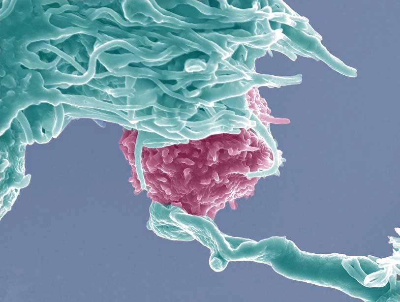
9 The immunosurveillance process and its components Initiation of the anti-tumor immune response occurs when the cells of the innate immune system become alerted to the presence of a growing tumor, at least in part owing to the local tissue disruption that occurs as a result of the stromal remodeling processes integral to the basic physiology of solid tumor development. This stromal remodeling could result from two of the six hallmarks of cancer : angiogenesis and tissue invasive growth (66-69). The stromal remodeling induced during these processes could produce proinflammatory molecules that together with chemokines that may be produced by the tumor cells themselves, summon cells of the innate immune system to this new source of local danger (70-72). The innate immune system constitute several cellular components such as NK cells, NKT cells, γδ T cells, Macrophages, Granulocytes and dendritic cells. Once recruited to the developing tumor mass, the above mentioned innate cells may recognize molecules that have been induced on tumor cells either by the incipient inflammation or the cellular transformation process itself. Regardless of the precise mechanism of recognition, these events lead to a common outcome that is critical for progression of the anti-tumor response-the production of IFN-γ. In the next step, the effects of innate immune recognition of the tumor are amplified. The initial amount of IFN-γ released at the tumor site induces the local production of chemokines that recruit more cells of the innate immune system to the tumor. Products generated during remodeling of the extracellular matrix may induce tumor-infiltrating macrophages to produce low amount of IL-12 that stimulate tumor-infiltrating NK cells to produce low amounts of IFN-γ, which in turn activate macrophages in the tumor to produce more IL- 12, leading to increased IFN-γ production by NK cells (73). In addition to this positive feedback mechanism, the binding of NK cell activating receptors to their cognate ligands on tumor cells stimulates even more NK cells IFN-γ production that can now activate a number of IFN-γ dependent processes-including antiproliferative, proapoptotic, and angiostatic effects that result in the killing of a proportion of the tumor (74-80). 14
10 Normal cells Transformation Transformed cells Innate & Adaptive immunity NK DC CTL Mφ NK NKT Elimination Figure 1. The tumor immunosurveillance process In addition macrophages activated by IFN-γ that express tumoricidal products such as reactive oxygen and reactive nitrogen intermediates and NK cells activated either by IFN-γ or via engagement of their activating receptors can kill tumor cells (81, 82). As a result of these processes, a source of tumor antigens from dead tumor cells becomes available and the adaptive immune system is recruited to the process. The tumor antigens liberated by the effects of innate immunity on the tumor drive the development of tumor-specific adaptive immune responses. Immature dendritic cells that have been recruited to the tumor site become activated by either by exposure to the cytokine milieu created during the ongoing attack on the tumor by innate immunity or by interacting with tumor-infiltrating NK cells (83). The activated DCs can acquire tumor antigens then migrate to the draining lymph node, where they induce the activation of naïve tumor-specific Th1 CD4 + cells. Th1 cells facilitate the development of 15
11 tumor-specific CD8 + CTL induced via cross-presentation of antigenic tumor peptides on DC MHC class I molecules (84-86). The development of tumor-specific adaptive immunity provides the host with a capacity to eliminate the developing tumor (Figure 1). NK cell CD4+T Eosinophil Dendritic cell NKT Th1 Th2 Basophil γδ Τ CD8+T CTL Neutrophil Macrophage B cell Plasma cell Innate Components Adaptive Components Figure 2. The components of tumor immunosurveillance process Innate components of tumor immunosurveillance The innate immune system is characterized by its rapid responses to pathogens and tumor. As discussed earlier, there have been many cell types found to play a role in innate immunity against tumor (Figure 2). Among the cell types that have been postulated to serve as innate components of tumor immune system NK cells, NKT cells and γδ T cells are compelling candidates, being able to respond rapidly and subsequently activate other cell types. 16
12 NK cells A role for NK cells in the rejection of tumors in vivo had been proposed shortly after their discovery as a unique lymphocyte subset (87, 88). Subsequent studies demonstrated that mice carrying the homozygous beige mutation, which are defective in granule exocytosis and have a profound defect in NK cell cytolytic function, develop virus-induced and carcinogen-induced tumors at a higher frequency (89, 90). However, these studies did not provide a role for NK cells in tumor immunity, because the defects caused by the beige mutation are not limited to NK cells. Other studies demonstrated an increased rate of carcinogen-induced sarcomas in mice lacking perforin, the pore forming granule protein involved in cytotoxicity by NK cells and CTLs. In contrast to perforin-deficient mice, mice genetically deficient for CD8 + T cells did not show significant defects in the control of carcinogen-induced tumor growth, suggesting a possible role of NK cells in tumor immunosurveillance (91). Two general mechanisms have been implicated in studies performed to date: (i) NK cells preferentially recognize cells, such as many tumor cells, that down regulate class I MHC molecule ( missing self recognition ), and (ii) NK cells recognize structures that are specifically up-regulated on tumor cells, including a group of recently proteins that are distantly related to MHC class I molecules and bind to a stimulatory receptor called NKG2D. The Missing-Self hypothesis for tumor cell recognition This phenomenon that NK cells preferentially attack cells in which class I MHC expression is reduced or abolished was first clearly demonstrated by comparing the activity of NK cells against wild type and class I-low variants of MHC positive tumor cell lines (92, 93). The enhanced sensitivity of class-i low tumor cells to NK cells was reversed by transfections that restored class I expression (94, 95). Cells in advanced tumors frequently extinguish class I expression, suggesting one possible rationale for 17
13 NK cell recognition of class I-low cells (96). The recognition of class I deficient cells by NK cells has been mechanistically explained by the discovery of receptors that are specific for class I molecules and inhibitory in function. Normal cells expressing high levels of class I molecules inhibit NK cells, while class I-deficient cells do not. Three families of inhibitory class I specific receptors have been identified: Ly49 (in mice), KIR (in humans) and the CD94/NKG2A receptor (in both mice and humans) (97-105). Ly49 and CD94/NKG2A receptors are related to C-type lectins in structure, while KIR are immunoglobulin (Ig)-superfamily members. Ly49 and KIR bind directly to intact classical class I molecules. CD94/NKG2A, in contrast, binds to a peptide derived from the signal sequence of classical class I molecules that is presented in the groove of a nonclassical class I molecule (Qa-1 in mice and HLA-E in humans) ( ). The various class I MHC-specific inhibitory receptors are expressed on overlapping subsets of NK cells, leading to a complex combinatorial repertoire of NK specificities for MHC class I molecules. The inhibitory receptor repertoire is shaped by an education mechanism that guarantees that NK cells are self-tolerant but are unleashed against target cells that down-regulate some or all self-class I molecules (109). The notion that missing self recognition by NK cells functions to eliminate MHC class I-loss variants is supported by studies in mice and (less directly) in humans. Studies demonstrated that cells that lack MHC class I molecules but are otherwise normal, such as bone marrow cells from MHC class I molecules bur are otherwise normal, such as bone marrow cells from class I- deficient mice, are strongly rejected when transplanted into normal mice (110).The rejection is mediated by NK cells. Subsequent studies indicated that a similar type of rejection occurs in a non-transplant scenario, in the case of variant class I- deficient cells that arise in vivo as a result of mutations. The collective results suggest that missing self recognition plays an important role in the elimination of cells that have downregulated class I MHC expression in vivo. With respect to tumors many studies have clearly demonstrated that class I-deficient transplantable tumor lines are rejected by NK cells (92, 93). 18
14 Inhibitory Inhibitory receptor receptor No MHC class I NK No activating ligands Activating receptor Inhibitory receptor MHC class I NK Activating receptor Inhibitory receptor MHC class I NK No activating ligands Activating receptoractivating ligand Inhibitory receptor MHC class I NK Activating receptor Activating ligand No response No response NK attacks target cell Balancing signal determine out come Figure 3. Missing self recognition of tumor/normal cells by NK cells Restoration of MHC class I expression on the tumor cells reversed this effect (94, 95). NK cells appear to be actively inhibited from responding when they encounter certain tumor cells that express MHC class I. Subsequently, the ability of NK cells to recognize and eliminate normal host hematopoietic cells that lack MHC class I was substantiated by demonstrating NK-cell dependent rejection of bone marrow cells from β2 microglobulin-deficient syngenic mice. According to the missing self hypothesis, NK cells were proposed to provide immune surveillance for cells that had downregulated MHC class I, an event that frequently accompanies cellular transformation or infection with certain viruses ( ). Until recently, a common misconception has been that NK cells attack any cell lacking MHC molecules because the potential target cell cannot engage an inhibitory NK cell receptor for MHC class I. This notion is counterintuitive given documentation of the events involving cell-cell binding, Ca 2+ mobilization, and synapse formation when NK cells encounter susceptible 19
15 target cells that lack MHC class I. A contemporary modification of the missing-self hypothesis might state, NK cells patrol for abnormal cells that lack MHC class I or overexpress ligands for activating NK cells receptors. In essence, the inhibitory MHC class I receptors on NK cells can serve as a rheostat, regulating and dampening signals transduced through activating receptors. When NK cells and potential target cells meet, the information is interpreted by an analog, not a binary process. Experimental evidence suggests that the MHC class I inhibitory receptors may serve only to dampen, rather than completely terminate, NK cell effector function and that the amount of MHC class I on the surface of the target is proportional to the degree of inhibition ( ) (Figure 3). Before missing self recognition was proposed, it was assumed that NK cells would recognize target cells in much the same way as other lymphocytes with stimulatory receptors specific for antigens expressed on target cells. Once the various families of MHC class I specific inhibitory receptors were discovered, the misapprehension grew that missing self recognition could account in general terms for tumor cell recognition by NK cells. Numerous examples had been reported previously of NK-susceptible tumor cells with apparently normal class I expression, suggesting the existence of additional NK recognition systems ( ). The recent discovery of the activating NK receptor NKG2D and its ligands on primary tumors has further provided a potential mechanism by which NK cells participate in tumor surveillance. It has been shown that transfection of the mouse NKG2D ligand RAE-1or H60 into MHC class I positive tumors resulted in NKG2D-mediated NK cell killing and macrophages activation in vitro. In addition, ectopic expression of RAE-1 or H60 in certain MHC class I positive tumors rendered them susceptible to rejection by NK cells and CD8 + T cells in syngenic mice, demonstrating that the NKG2D-mediated activation is able to overcome the MHC class I-mediated inhibitory signaling in responding NK cells. Therefore, it has been postulated that selective upregulation of the NKG2D ligands on stressed cells may allow NK cells to shift from immune tolerance to activation, resulting in elimination of precancerous 20
16 cells. Supporting this hypothesis, expression of retinoic acid early-1 protein (RAE-1) was found to be induced by carcinogens in skin cells, and skin-associated γδ Τ cells killed these cancer cells in vitro via NKG2D. Interestingly, in some cases mice that rejected RAE-1 or H60-expressing tumors were immune to rechallenge with the same tumors not expressing these NKG2D ligands ( ). NKT cells The term Natural killer T cell (NKT) has been broadly used to describe a diverse range of T cell subsets (127). At least two populations of CD1d-restricted NKT cells exist and these can be distinguished based on their TCR repertoire. The most prevalent and extensively studied member expresses an invariant TCR, which in the mouse consists of a Vα14Jα18 TCR-α chain, preferentially paired with either Vβ8, Vβ7 or Vβ2.These cells, which recognize the glycolipid antigen α-galactosylceramide (α-gal Cer), have been defined as type I NKT cells (128). Type I NKT cells are best known for their ability to promptly provide copious quantities of a range of cytokines upon primary stimulation, making them key regulators of tumor immunity. Other CD1d-restricted NKT cells have been identified in both humans and mice and these cells express a more diverse TCR repertoire, and have been referred to as type II NKT cells ( ). A role for NKT cells in innate immunity against tumor was implicated when Jα28 -/- mice, which lack a large population of invariant NKT cells, were found to develop MCA induced sarcoma at higher incidence than wild type counterparts. Treatment of CD1d -/- (which lack both type-i and type-ii CD1d restricted NKT cells) and TCR Jα18 -/- (which lack only type-i CD1d restricted NKT cells) with the carcinogen, MCA, resulted in these mice developing tumors faster and at a greater frequency than their wild type counterparts (132). Furthermore, when tumors derived from CD1d -/- or Jα18 -/- mice were inoculated back into CD1d -/-, Jα18 -/-, or WT mice, they grew readily in NKT cell deficient mice but were rejected in WT mice, suggesting an important role for NKT cells in this tumor rejection. This hypothesis was supported when transfer of NKT cells from WT mice with Jα18 -/- mice efficiently protected recipients from tumor growth, demonstrating an essential role 21
17 for NKT cells in tumor immunosurveillance (133). Subsequent studies have supported these findings with the demonstration that MCA-induced fibrosarcomas derived from immunodeficient RAG -/- mice can be divided into progressor or regressors (which grow or are rejected by WT mice, respectively), and that expression of CD1d by these fibrosarcomas correlates with a regressor phenotype (134). This finding provides further evidence that CD1d-restricted T cells play a role in surveying the development of carcinogen-induced fibrosarcomas. Type I NKT cells are also beneficial in some cancer therapy models. In the B16 mouse melanoma model, immunization of mice with irradiated GM-CSF secreting tumor cells protected mice from subsequent tumor challenge (135). This protection was abrogated in CD1d and Jα18 -/- deficient mice. Preliminary studies in humans with malignant melanomas, primary lung cancers, advanced prostate cancers and a range of solid progressing tumors demonstrate a numerical deficiency of NKT cells in the peripheral blood when compared with healthy volunteers, suggesting NKT cells may play a role in promoting anti-tumor immunity (136, 137). γ δ T cells Where as most mature T cells express the αβ T-cell receptor (TCR) heterodimer, a small proportion express an alternative γδ ΤCR heterodimer. Unlike αβ T cells, which recognize specific processed peptide antigens presented on major histocompatibility complex (MHC) molecules by antigen presenting cells, γδ T cells seem to directly recognize and respond to a variety of MHC-like stress induced self-antigens expressed by malignant cells. Thus, γδ Τ cells can recognize malignant cells through less specific mechanisms that require no prior antigen exposure or priming, a function that is shared by other innate immune cells such as NK cells. Although γδ Τ cells comprise less than 10% of total peripheral blood T cells, they are present in substantially greater numbers within epithelial tissues such as skin, intestine, and lung, contrasting with αβ T cells, most of which either circulate in the peripheral blood or are resident in lymphoid organs ( ). The process by which γδ Τ cells recognize stressed or malignant cells is not 22
18 completely understood. Although the TCR is involved in antigen recognition, the mechanism by which antigens are recognized by γδ T cells is fundamentally different from αβ T cells (143, 144). γδ T cells express surface NKG2D. Engagement of NKG2D by one of it several ligands, including MHC class I chain related A and B (MICA and MICB) in humans and retinoic acid early-1 (RAE-1) in mice tumor cells provides a costimulatory function and targets cellular destruction (145, 146). Several lines of evidence point to a role for γδ Τ cells in tumor immunosurveillance. It has recently been shown that mice lacking γδ Τ cells are highly susceptible to multiple regimens of cutaneous carcinogenesis (147). In clinical studies, γδ Τ cells have been shown to infiltrate a variety of tumors, including lung cancer, renal cell carcinoma and breast cancer ( ). The most common circulating γδ Τ cells, ie, those expressing the Vγ9/Vδ2 TCR heterodimer, recognize several known tumor associated ligands and cell lines. These include HSP-60, Daudi Burkitt lymphoma and glial cells. Vγ9/Vδ2 γδ T cells recognize and lyse glioblastoma, neuroblastoma, multiple myeloma and lung cancer ( ). γ9/vδ2 T cells have been isolated from patients with Hodgkins disease and Vγ9/Vδ2 T cells recognize cells with mevalonate metabolites, which are over-expressed in hematologic malignancies and mammary carcinoma (159). Vγ1 + T cells are less frequent, comprising up to 10% of all γδ T cells. They seem to recognize a different set of ligands and tumors, although there is some overlap with Vγ2 + cells. A high proportion of Vγ1 + γδ T cells appear in epitheial tumors from lung, breast, kidney, ovary and prostate that express the stress-induced antigens MICA and MICB, a non classic stress-related MHC antigen recognized by Vγ1 + cells. It was observed that primary leukemias are also killed by γδ Τ cells (160). In addition to the potential of γδ T cells to directly lyse transformed cells in an NKG2D-dependent fashion, infiltrating γδ T cells have also been shown to produce IFN-γ early in tumor development (161). 23
19 Adaptive components of tumor immunosurveillance T cells Historically, the mature T cell population has been divided into three groups. These groups are αβ T cell receptor (TCR) expressing CD4 + and CD8 + T cells and the CD4-/ CD8-γδ TCR-expressing cells. The distinct function of CD4 + and CD8 + T cells is dictated by the expression of these coreceptors. The ligand for CD4 is the β2 domain of the Major Histocompatibility Complex (MHC) class II molecule and CD8 binds to the α3 domain of MHC class I molecules (162, 163). Due to these specificities, αβ TCR of CD8 + T cells is restricted to the recognition of antigens presented by MHC class I molecules and the αβ TCR of CD4 + T cells to antigens presented by MHC class II molecules. CD4+ T cells A critical role for CD4 + T cells in anti-tumor immunity has been demonstrated in multiple settings. Anti-tumor immunity has been shown to be abrogated in CD4 knockout mice or mice depleted of CD4 + T cells via antibody depletion in cases of cell based vaccines and recombinant viral vaccines (164). In addition, CD4 + T cells that are restricted to class II MHC have been documented against human melanomas, lymphomas, colon cancers and others. The role of CD4 + T cells in anti-tumor immune response has been shown to be multifaceted and often essential to tumor rejection in many tumor models. The multiple roles of tumor specific CD4 + T cells are illustrated in Figure.4. Activation of dendritic cells Although CD4 + T cells have been shown to be effective in eliminating tumor cells in the absence of CD8 + T cells in some tumor models more often a requirement is found for both CD4 + T and CD8 + T cells for tumor rejection to occur ( ). Because most tumor cells express class I MHC molecules-the restricting element for CD8 + T cell 24
20 recognition but not class II molecules, it has been assumed that the predominant effector mechanism in tumor immunity is direct lysis of tumor cells by CD8 + CTL. Indeed, often the role of CD4 + T cells in both effective viral and tumor CTL responses is to aid activation of CD8 + T cells, leading to destruction of the virus or tumor by CD8 + CTL. The role of CD4 + T cells in the priming phase of a tumor specific CTL response is thought be at least partially at the level of activating DC to allow these cells to effectively activate naïve CD8 + T cells. There is accumulating evidence suggesting that cross-presentation of tumor antigens by professional antigen presenting cells such as DCs is important for the induction of class I MHC restricted tumor specific immunity ( ). It has been suggested that such cross priming requires CD4 + T cells help for the induction of CTL immunity. In one model, priming of tumor specific CTL was shown to require recognition of antigen by CD4 + T cells on the same APC as the CTL epitope (173). It is believed that activated CD4 + T cells license APC, which in turn expresses costimulatory molecules essential in CD8 + T cell activation (174). There is now evidence for a broader role for CD4 + T cells in anti-tumor immunity in addition to providing help for CD8 + T cells. In fact, in the absence of CD8 + T cells, adoptive transfer of CD4 + T cells can lead to the elimination of tumor cells that do not express detectable levels of class II MHC antigens, as well as tumor cells expressing class II molecules, suggesting a much larger role for CD4 + T cells than simply being help for CTL (175). Direct tumor cell killing Tumor specific CD4 + T cells recognize tumor peptide/class II MHC complexes expressed either on the surface of tumor cells or professional APC that have taken up and processed tumor antigen shed from tumor cells. Recognition of peptides/class II MHC complexes on the surface of tumor cells by CD4 + T cells can result in direct tumor cell lysis by these T cells. Cytolytic CD4 + tumor specific T cells capable of lysing class II MHC expressing tumor cells have been identified in both rodents and humans ( ). In addition, the existence of unusual CD4 + T cells that are tumor specific but not MHCrestricted have also been reported (180, 181). However, since such MHC unrestricted 25
21 CD4 + T cells are rare and the majority of tumors do not express class II molecules, direct destruction of tumor cells by CD4 + T cells is not a generally applied function of these cells. Rather, tumor cell killing by CD4 + T cells is largely by indirect mechanisms. Macrophage activation Direct tumor Killing DC maturation CD4+T NK cell activation B cell activation CD8+T cell activation Figure 4. Multifaceted role of CD4 + T cells in anti-tumor immunity B cell activation and antibody secretion One possible indirect pathway of CD4 + T cell mediated tumor destruction is through B cell activation. A critical role for CD4 + T cells in immune responses in general is to activate B cells and induce isotype class switching. In some tumor models a humoral response has been shown to be involved in tumor destruction, and in these cases CD4 + T cells are required for the antibody production by B cells. These activated B cells produce tumor-specific antibodies that may contribute to therapeutic anti-tumor immunity (182, 183). Rejection of murine kidney sarcoma following immunization with soluble T antigen has been shown to be at least partly mediated by antibody-dependent cell cytotoxicity (ADCC) (184). There is a large body of evidence in the literature to support the idea that antibodies specific for HER-2/neu can inhibit the growth of tumor cells expressing the neu gene through ADCC, complement fixation, or inhibition of 26
22 signal transduction through neu ( ). In addition, in some models, collaboration between humoral and cellular immunity has been shown to be required for tumor rejection (188). Activation of nonspecific immune cells Another indirect method of CD4+ T cell mediated tumor destruction is through activation of nonspecific immune cells that then mediate tumor cell lysis. CD4 + T cell, via secretion of IFN-γ, can activate macrophages to secrete TNF-α and nitric oxide and thus become tumoricidal (189, 190). In one model, depletion of CD4 + T cells resulted in abrogation of tumor rejection, which correlated with the absence of tumor-infiltrating macrophages (191). CD4 + T cells have also been shown to exert their anti-tumor effect by attracting natural killer cells to the tumor site where the NK cells destroy the tumor (165). Depletion of either CD4 + T cells or NK cells, but not CD8 + T cell, resulted in the rejection of a class I negative tumor (192). While these responses mediated by the innate immune cells are not antigen specific, antigen specificity is nonetheless achieved by localization of the CD4 + T cells and the effector cells in the local tumor environment. These data suggest that tumor-specific CD4 + T cells are able to induce a delayed-type hypersensitivity-like reaction during which inflammatory cells like macrophages, granulocytes, or NK cells are attracted and activated by the T cells to kill tumor cells. Th1 versus Th2 response CD4 + T cell responses are described as either Th1 or Th2 like based on the cytokines produced by the T cells. The balance between Th1/Th2 cytokines has been shown to be critically important in various immune responses (193). Th1 cells produce IFN-γ and IL-2, thereby inducing cellular immunity, whereas Th2 cells produce IL-4, IL- 5, IL-10 and IL-13, inducing humoral immunity. Because a cellular immune response is thought have the greatest potential for tumor destruction, it has been proposed that a Th1 like response is required for tumor rejection. And, indeed, a number reports have 27
23 demonstrated a correlation between the generation of Th1 type response and effective anti-tumor immunity ( ). In further support of the hypothesis that a Th1 response is critical for anti-tumor immunity, Th2 cytokines have been shown to down-regulate anti-tumor immunity (199, 200). It has been suggested that the failure of T cell-mediated immunity against tumors is not due to the inability of T cells to recognize tumor antigens, but rather to the induction of an ineffective tumor-specific immune response via immune deviation. In support of this notion, a shift from Th1 to Th2 cytokine production has been reported in progressive cancer patients, and a vaccine-induced Th2 to Th1 shift in a murine tumor model was shown to induce tumor rejection. In addition, Th2 cytokines have been shown to accelerate tumor growth in multiple experimental models ( ). IFN-γ mediated anti-tumor effects One reason a Th1 response is thought to have great potential for tumor destruction is due to secretion of IFN-γ by these cells. This cytokine, which is also secreted by activated CD8 + T cells and NK cells, can act in several ways to increase anti-tumor immunity. IFN-γ can increase MHC I and MHC II expression on tumor cells to increase the immunogenicity of the tumor and has also been shown to be critical in the process of inducing T cell migration to tumor sites (206). In addition to enhancing destruction of tumor cells by specific immune cells, IFN-γ can aid in tumor cell destruction by means outside of specific immunity. IFN-γ can often in conjunction with TNF, have direct cytotoxic activity in some tumor cells can inhibit tumor cell proliferation, can activate macrophages and can enhance the secretion of antiangiogenic chemokines (207). Some of these effects of IFN-γ require IFN-γ sensitivity of tumor cells. In one model, by expressing a dominant-negative form of the IFN-γ receptor in tumor cells, it has been shown that loss of sensitivity of tumor cells to IFN-γ can decrease immunogenicity and prevent tumor cell recognition and elimination (208). 28
24 Antiangiogenesis As mentioned above, one method by which IFN-γ secretion from CD4 + T cells can inhibit tumor cell growth is by enhancing secretion of antiangiogenic factors. IFN-γ has been shown to induce monocytes, macrophages, fibroblasts, and even certain tumor cells to produce Mig and IP-10, two CXC chemokines that are antiangiogenic. These chemokines have potent antiangiogenic activity that induce damage to tumor vasculature and results in tumor cell growth inhibition and tumor necrosis. Furthermore, anti-angiogenic substances have been shown to be able to arrest the growth of established tumors ( ). CD8 + T cells CD8 + T lymphocytes have been accepted as the most potent effectors against tumor cells. CD8 + T cells with killing potential recognize peptides generated from endogenous proteins in the context of major histocompatibility complex (MHC) class I membrane proteins expressed by eventually all tissues. Specific recognition of foreign peptide antigens results in the differentiation of CD8 + T cytotoxic T cells, which kill their cellular targets directly. Mechanism of Cytolysis by CTLs More than 30 years ago, CTLs were first discovered as the lymphocytes that could kill tumor or virally infected cells through a mechanism independent of antibody (212, 213). Naive CD8 + T cells have no lytic granules and must first undergo proliferation and differentiation into CTLs, which takes several days, before they acquire the ability to kill. Proliferation is triggered by the recognition of peptide antigen presented on the surface of a cell by self-major histocompatibility complex molecules by the TCR of naïve CD8 + T cells (214). Thus, CD8 + T cells are part of the adaptive immune system, which requires several days of induction but has a high degree of antigen specificity and the capacity for immunological memory through potent secondary responses. An activated CTL, 29
25 upon encountering an antigen-bearing cell, reorients its granules to the region of receptor activation and releases the granule components into the region of contact between the killer and the target. This region of contact, referred to as the immunological synapse, is initiated by receptor contact and maintained by adhesion molecules such as LFA-1and ICAM-1 (215). CTLs can kill multiple cells by reorienting their granules to another region of contact. An extensive series of studies has revealed that cytotoxic T lymphocytes are capable of inducing the death of target cells by at least three distinct mechanisms- secretion of cytotoxic cytokines and calcium dependent dependent or calcium independent contact dependent cytotoxicity (216). CTLs can secrete cytotoxic cytokines (such as TNF-α and IFN-γ) in the region of their targets. Alternatively, cytotoxic lymphocytes can make direct contact with target cells using recognition machinery provided by the T cell receptor and members of the integrin family. The calcium-dependent pathway relies on the secretion of cytotoxic granules onto the surface of the target cell and is therefore known as the granule exocytosis pathway (217). The calcium-independent pathway has now been shown to cause target cell death via the interaction of the Fas ligand on the CTL and the Fas receptor on the target cell (218). One of the critical molecules of cytotoxic granules is Perforin, a protein that is similar to some of the terminal components of the complement cascade. Perforin can polymerize, in the presence of calcium, to form channel-like structures in target cell membranes. The other key granule components are the granzymes, which are neutral serine proteases that activate the death machinery of appropriate target cells. Recent studies have suggested that each of the cytotoxic pathways may operate in specific types of CTL in specific settings (219). Role of perforin In the original paradigm of perforin function, perforin was thought to directly induce target cell death by damaging target cell membranes, causing cell lysis similar to that induced by complement. The similarity of perforin to that of complement component 9 further suggested that this molecule was the key granule component that 30
26 cause target cell death. However, the discovery that perforin-induced membrane damage was not sufficient to cause apoptosis, the hallmark of CTL-induced target cell death, suggested that it may instead act as a portal of entry for other cytotoxic molecules that induced the apoptotic hits (220). Upon release of perforin from cytotoxic granules, the molecule rapidly polymerizes in the presence of calcium to form a ring-like structure that apparently contains a central pore when it is inserted into the target cell membrane. The idea that insertion of polymerized perforin into target cell membranes may not be a random process, but instead may be mediated by receptors was advanced in a study by Tschopp et al (221, 222). It was observed that phosphocholine on target cell membranes acted as a specific, calcium-dependent receptor molecule for perforin (223). The concept of a perforin receptor has been further explored in recent observations, suggesting that NK cell can release the lysolipid platelet activating factor (PAF), which may act as a chaperone for perforin, synergizing with it to produce membrane damage (224). PAF receptors were found on perforin-sensitive cell lines, but not on perforin-resistant lines; interferon-γ induced PAF receptor expression correlated with the induction of perforinsensitivity. The idea that perforin created a channel through which granzymes could pass into target cells was widely held in However, the channels created by polyperforin range in size up to 16 nm, which are not large enough to permit diffusion of even small proteins (i.e., 8kDa) into cells. The granzymes range in size from approximately 30 to 65 kda, and they are probably complexed with serglycin upon secretion. These observations suggested that the granzymes cannot enter target cells directly via a perforin pore. A second hypothesis for granzymes entry, namely reparative endocytosis, was offered to explain this paradox (225). In this model, perforin entry into the target cell membrane creates a signal for the target cell to repair the damage by endocytosing the perforin and surrounding plasma cell membrane; granzymes in the vicinity of the lesion are also endocytosed and ultimately delivered to the target cell cytoplasm and nucleus, where they deliver the apoptotic hits. It is not known whether the models 31
27 provided by these studies are physiologically relevant; in vivo, cytotoxic lymphocytes secrete large amounts of granule proteins onto a very small patch of target cell membrane defined by the conjunction site. Recent reports demonstrated that purified granzyme B can enter target cells without any chaperones or perforin (226, 227). Role of granzymes The cytotoxic granules of CTLs contain a family of serine proteases known as granzymes. Granzyme A and B are the most abundant granzymes in mice and humans and are the best characterized (228). Granzyme B activates the caspase dependent pathway of apoptosis (229). After granzyme B is released from its endolysosomal compartment, it rapidly trafficks to the nucleus and initiates death by cleaving protein substrates that either directly or indirectly lead to DNA fragmentation and cell death. Many studies have shown that a number of procaspases (including caspases 2, 3, 7, 8, 9 and 10) are the substrates of granzyme B both in vitro and in vivo ( ). Genetic or pharmacological inhibition studies have demonstrated that caspases contribute to but are not absolutely required for the induction of DNA fragmentation and apoptosis by granzyme B. The ability of granzyme B to cause DNA fragmentation in cells that lack functional caspases suggests that granzyme B can directly activate apoptotic nucleases (233). Caspase activated DNase (CAD) exists as a dimer with ICAD (Inhibitor of CAD) in the cytoplasm, where CAD is retained in its inactivate form. After a cell receives a death signal, activated caspase3 cleaves ICAD, which facilitates the assembly of CAD into its active form and subsequent DNA digestion. ICAD is a substrate of granzyme B, and so CAD can be activated directly by granzyme B in the absence of activated caspase ( ). The physiological relevance of the activation of CAD by granzyme B has been confirmed by the observation that ICAD is not cleaved in target cells incubated with granzyme B deficient CTLs and that ICAD-deficient cells are partially resistant to granzyme B mediated killing (237). However, because a large percentage of cells that contain neither ICAD nor functional caspases are susceptible to granzyme B mediated death, additional death pathways must also exist. Because Bcl2 can block granzyme B 32
28 mediated death (238). It has been suggested that in addition to activation of CAD and caspase 3, granzyme B can also induce the mitochondrial pathway of death. Granzyme B is capable of directly cleaving Bid upon target cell entry. Cleavage of Bid generates a truncated form (tbid), which translocates into the mitochondria, where it interacts with its receptors Bax and Bak to cause cytochrome c release into the cytoplasm and the activation of caspase 9 and ultimately caspase 3 (239). However, it would appear that Bid-dependent cytochrome c release is not required for granzyme B mediated apoptosis because granzyme B can cause mitochondrial depolarization and death in Bid-deficient and Bax/Bad doubly deficient mice (240). Although granzyme A was the first serine protease to be discovered in CTL granules, far less is known about the mechanism by which it kills cells than that by granzyme B kills cells (241). Although it clearly has been demonstrated that granzyme A is pro-apoptotic enzyme, there are obvious differences between it and granzyme B (242). Nevertheless, perforin loaded granzyme A is equally as effective as granzyme B at inducing target cell lysis (243). Granzyme-B deficient mice harbor CTLs that are impaired in their ability to generate oligo-nucleosomal DNA but are unimpaired in their ability to lyse target cells (244). Although the granzyme A deficient mice are cytotoxic in vitro, they failed to clear pox virus ectromelia or herpes simplex virus as effectively as wild-type mice ( ). CTLs from granzyme A x granzyme B doubly deficient mice have a profound defect in cytotoxicity, similar to that observed in perforin deficient mice (249). Taken together, these results suggest that granzyme A induces a distinct cell death pathway from granzyme B, which is important for host defense. This view is supported by the observation that. Although granzyme A deficient and granzyme B deficient CTLs induced similar early apoptotic features ( such as mitochondrial depolarization and ROS production), later granzyme B killing was caspase 3/9 dependent, where as granzyme A killing was ROS dependent (250). Granzyme A is a tryptase and not an aspase like granzyme B and so predictably does not appear to cleave any of the known substrates of granzyme B, such as Procaspase 3 (243). Thus, unlike granzyme B mediated cell death, 33
29 loading of cells with granzyme A induces apoptosis completely independent of caspase activity. Indeed, a recent study shows that caspase independent mitochondrial damage is a required first step for granzyme A induced PCD (251). Unlike granzyme B, which induces oligo-nucleosomal DNA degradation, granzyme A causes single-stranded DNA breaks, suggesting the activation of different nucleases. This explains early observations that killing by granzyme A, as measured by DNA fragmentation, was thought to be slow. CTL Cytotoxic granules FAS L FAS Endocytosis Procaspase-8 Granule exocytosis Perforin pore Granzyme B Granzyme A Bid tbid Tumor cell Caspase-8 Mitochondria Procaspase-3 Cytochrome c Caspase-3 Caspase-9 Apaf1 Apaf1 ICAD CAD SET complex Procaspase-9 Granzyme A Lamin fragments DNA fragmentation Figure: 5. Mechanism of target cell lysis by Cytotoxic T lymphocytes Granzyme A targets endoplasmic reticulum-associated complex known as the SET complex, which is composed of two tumor suppressor proteins (pp32 and NM23-H1) and three granzyme A substrates (the nucleosome-assembly protein SET, the DNAbinding protein high-mobility group protein 2 HMG2, and the rate limiting base- 34
30 excision repair enzyme apurinic/apyrimidinic endonuclease APE1) ( ). The granzyme A induced DNase (GADD) nicks DNA. It is inhibited by SET (IGADD). Granzyme A translocates to the nucleus of a target cell during CTL killing, degrades SET, and so activates GADD, resulting in the nicking of chromosomal DNA. In addition, granzyme A can cleave both lamins A and B and some caspases indicates that the disruption of the nuclear envelope is a critical event in the induction of target cell apoptosis by CTLs (254). Fas-Fas L system The Fas system employs the Fas receptor- a member of the TNF family of death receptors and the Fas ligand, a membrane associated ligand which is synthesized within several hours after T cell receptor stimulation. Display of the Fas ligand on the T cell surface makes it possible for the Fas ligand to interact with Fas receptors on the surface of target cells. Engagement of the Fas receptor results in the aggregation of its intracellular death domains, leading to the recruitment of Fas associated death domain (FADD) and procaspase-8 to form a death inducing signaling complex (DISC) (255). In the DISC, FADD and Fas interact via their death domains; procaspase-8 then associates with FADD via the death receptor domains of these two molecules (256). Procaspase-8, a member of the caspase family of cysteine proteases, is then activated by proteolytic cleavage after specific aspartic acid residues. Activated caspase-8 is then activates additional downstream caspases by cleaving them after specific aspartic acid residues, ultimately leading to the activation of Procaspase-3 (257, 258) (Figure 5). Dendritic cell as a link between innate and adaptive immunity against tumor Dendritic cells in innate immunity to tumor Strong evidence has been accumulated demonstrating that tumor cells in humans and animals are recognized in general as non-self by the immune system ( ). An anti-tumor immune response is initiated when the cells of the innate immune system 35
31 become alerted to the presence of a growing tumor, at least in part owing to the local tissue disruption that occurs as a result of the stromal remodeling process integral to the basic physiology of solid tumor development (262, 263). The stromal remodeling could produce inflammatory molecules that, together with chemokines that may be produced by the tumor cells themselves, summon the cells of the innate immune system to this new source of local danger. The innate response includes soluble factors and several cellular factors, including natural killer (NK) cells, natural killer T (NKT) cells, γδ Τ cells, macrophages, and dendritic cells. The components of innate immunity use pattern recognition receptors and other cell surface molecules to directly detect tumor cells. Cancer cells frequently express families of stress-related genes, such MICA and MICB, which function as ligands for NKG2D receptors that are expressed by NK cells and other cytotoxic lymphocytes (264, 265). NK cells also monitor for the loss of MHC class I molecules from the surface of tumor cells (266). It was thought that NK cells, NKT cells, and γδ Τ cells and some soluble factors are the major components of innate effectors playing a role in innate immunity against tumor. Several lines of evidence from recent years suggest that dendritic cells have crucial role in regulating these innate effectors against tumor immunity. Here are some evidences to support the notion. The initial study by Fernandez et al, revealed that the adoptive transfer of DC-or fms like tyrosine kinase 3-ligand (FLT3-L) expansion of DC in mice bearing MHC class-i negative tumors promote NK cell dependent anti-tumor effects (267). In vitro studies demonstrated that the co-culture of DCs and resting NK cells results in a substantial increase in both NK cell cytolytic activity and IFN- γ production in an IL-12 and type I IFN-independent manner (267). Subsequent in vitro investigations identified that NK cell activation induced by DCs requires both cytokine signals and contact between NK cells and DCs. Various types of DC, depending on their activation state, are able to produce cytokines, such as IL-12, IL-15, IL-18, and type I IFN, all demonstrated to act on NK cells ( ). NK cells can be triggered for cytolytic activity by both murine and human DC depending on direct cell contact through their expression of cell surface molecules such as CD48, and CD70, which are ligands for the NK cell activating receptors 2B4 and 36
32 CD27, respectively, or by DC-derived cytokines ( ). The co-stimulatory molecules CD80, and CD86, expressed on DC were shown to trigger cytolysis by murine and human NK cells ( ). In addition, activation of NK cells by DC is potentially important for the promotion of tumor regression. For example, it was shown that anti- CD40 antibody therapy induces substantial NK cell-mediated anti-tumor and antimetastatic effects. As CD40 is not expressed by NK cells or by tumor cells used in their study, it was suggested that NK cell activation is mediated by increased cytokine production upon CD40 ligation on DCs (277). It was found that DC/NK cell interaction was bi-directional and complex, as it could result not only in NK cell activation, but also in DC maturation (278, 279). This is evident from another study, where culture of activated human NK cells with immature monocyte derived DC, at low DC/NK ratio (1:5), led to an increase in DC cytokine production (280). In this context, soluble factors such as TNF-α and IFN-γ, as well as cell-cell contacts are required for NK cell mediated DC activation. NKT cells represent a cellular population of the innate immune compartment that were shown to protect the host from tumor formation (281). A role for NKT cells in innate immunity against tumor was implicated when Jα28 -/- mice, which lack a large population of invariant NKT cells were found to develop MCA induced sarcoma at higher incidence than wild type counterparts (282). DC mediated NKT cell activation has been shown in an array of recent studies, which supports a role for both cytokine and cell-cell contact in the enhancement of NKT cell mediated anti-tumor function. For example, tumor antigen-pulsed DCs efficiently suppressed the growth of hepatocellular carcinoma in mice; the effect was mediated by enhanced NKT cell function (283). The NK cell ligand alpha-galactoceramide demonstrates its immunopotentiating anti-tumor activities in vivo that resemble IL-12 mediated anti-tumor activities. Production of IFN-γ by NKT cells in response to alpha-galactoceramide required IL-12 production by dendritic cells and direct contact between NKT cells through CD40/CD40L interaction (284). The above findings indicate an important role for DC produced IL-12 in the 37
33 activation of NKT cells and suggest that NKT cells may be able to condition DCs for subsequent immune responses. Which is evident from an in vivo study, in which administration of alpha-galactoceramide, caused activation of NKT cells to induce strong NK activity and cytokine production by CD1 restricted mechanisms. Surprisingly, it was observed that alpha-galactoceramide induced the activation of immunoregulatory cells involved in acquired immunity such as CD69 overexpressing CD4+ T cells and CD8+ T cells, indicating an essential role for the interaction between NKT cells CD1d expressing dendritic cells in the activation of acquired immunity (285). In addition, soluble antigen presentation by dendritic cells to NKT cells induces the differentiation of antigen specific cytotoxic T lymphocytes (286, 287). By activating NK and NKT cells DCs might enhance adaptive immunity to tumors in part; the killing of tumor target by NK and NKT cells could provide DCs with increased access to tumor antigens. In summary the cross talk between DC and other innate effectors now regarded as a main stage in the initiation of innate and adaptive immunity to tumors. Dendritic cells as innate effectors against tumor It has been shown that DC can produce TNF-α express membrane FasL and a high level expression of nitric oxide synthase, suggesting that dendritic cells may participate in innate defense against malignancies ( ). DC can infiltrate solid tumors, but their T cell stimulatory ability is often suppressed (292, 293). However, the density of DC infiltration may be associated with enhanced patient survival in certain tumors (294, 295). In addition several studies in mouse tumor models indicate that fusion of dendritic cells without loading specific antigens can also decrease growth of tumors to a certain degree ( ). These observations lead to investigations into direct inhibition of tumor growth by DC. Here are some evidences to support the tumoricidal activity of dendritic cells without the participation of other innate effectors. Human PBMC-derived dendritic cells were shown to inhibit the growth of a wide spectrum of tumor cell lines of different tissue origin in a cell-cell contact dependent manner (299). The addition of LPS pretreatment induced DC to secrete soluble factors that could inhibit tumor growth. 38
34 In contrast to the cytostatic activity in the previous study, peripheral blood DC were shown to lyse a number of breast cancer lines in vitro, and this activity could be neutralized by the addition of anti-tnf-α antibodies (300). DC mediated cytotoxicity was augmented by the addition of maturation or activation factors such as IFN-α, IL-15 and LPS. Peripheral blood myeloid DC were also shown to induce apoptosis in lymphoma, ovarian, prostate and melanoma cell lines (301, 302). Significantly, plasmacytoid DC did not share this property. This cytolytic activity was found to be mediated by the DC expressed TRAIL (TNF related apoptosis inducing ligand) (303). In addition, it was observed that activated human umbilical cord blood dendritic cells can serve as cytotoxic cells against hematological tumor cells without damaging the normal hematopoietic progenitor cells (304). Another study by Yang et al, demonstrated that the immature DC could exert a significant cytotoxicity towards autologous and allogenic ovarian tumor cells. This cytotoxicity was independent of Ca 2+ and could be inhibited by anti-fasl monoclonal antibody, indicating the involvement of Fas/FasL pathway in the cytotoxic mechanism (305). Finally a recent report described a new subset of CD11C+ myeloid DC that express high levels of FCγR II and FCγR III, could efficiently lyse HER-2 neu expressing breast and colon cancer cells in the presence of herceptin, an antibody directed against this tumor associated antigen. This antibody dependent cell mediated cytotoxicity (ADCC) could be inhibited by blocking antibody against FC receptors and antibody against TNF-α (306). Taken together, these results suggests that in addition to their predominant role as immune regulatory cells, DC could serve as innate effector cells in tumor immunity. Dendritic cells in adaptive immunity to tumor It is well established that the immune system recognizes tumor associated antigen (TAA) and tumor specific antigens (TSA). It has been proven that effector mechanisms of the adaptive immune system are able to eliminate tumor cells in vitro and in vivo, however require activation by antigen presenting cells, and it is the DC, which is pivotal for this process. 39


35 Bone marrow Stromal cells Macrophage Peripheral Tissue Tumor GM-CSF, IL-3, FLT3-L Tumor Ag Immature DC Mature DC Granulocyte Perforin Granymes & IFN γ CTL IL-2,IFN-γ CD8+T B cell Th1 CD4+T Tumor Th2 Peripheral Tissue Antibodies IL-4,IL-5,IL-10 Plasma cell Lymphoid Organ Figure 6. Dendritic cell play a central role in initiation of adaptive antitumor immunity The DCs that have been recruited to the tumor site become activated either by exposure to the cytokine milieu created during the ongoing attack on the tumor by innate immunity or by interacting with tumor infiltrating NK cells. The activated DCs can acquire tumor antigens directly by ingestion of tumor cell debris or potentially through indirect mechanisms involving transfer of tumor cell derived heat shock protein/ tumor antigen complexes. Activated antigen bearing mature DCs then migrate to the draining lymphnode, where they present peptides to naïve T cells, thereby inducing cellular immune response that involves both CD4+ T helper (Th1) cells and cytolytic CD8+T cells (307). DCs are famous for their T cell stimulatory properties, are now known to have major effects on B cell growth and immunoglobulin secretion. DC can directly activate both naïve and memory B cells [ ]. DC2 secretes IL-6, a cytokine known to promote plasma cell differentiation (311). IL-12 produced by activated DC, supports the differentiation of CD40 activated B cells to plasma cells (312, 313). These results suggest the DC mediated direct activation of naïve B cells during the 40
36 initiation of the immune response and the involvement of DCs in the development of humoral immune response (Figure 6). Role of dendritic cell expressed CD40 in anti-tumor immunity CD40 and its Ligand CD40 is a 48 kda transmembrane glycoprotein cell surface receptor that shares sequence homology with the tumor necrosis factor a (TNF-α) receptor family and was initially identified as a B cell surface molecule that induced B-cell growth upon ligation with monoclonal antibodies. DCs, macrophages, epithelial cells, hematopoietic progenitors have been shown to express CD40 (314, 315). Its ligand, CD154, is a kda type II integral membrane protein expressed on activated but not resting T cells (316). CD40 has emerged as a key signaling pathway for the function of dendritic cells, B cells and macrophages. The importance of CD40 in the immune system was established a decade ago with the identification of naturally occurring genetic mutations of CD154 in patients with X-linked hyper IgM syndrome (XHIM). XHIM patients have deficiencies in B cell isotype switching, cell mediated immune responses, including increased susceptibility to respiratory and gastrointestinal infections, malignancy. These deficiencies suggested an important role for CD40/CD154 in DC biology because of the requirement for DC in initiating antigen specific T cell responses ( ). CD40 signaling in dendritic cells. Both CD40 and CD40154 form homotypic trimers, and signaling is initiated by binding of a CD154 trimer to preformed trimeric CD40 (321). Confirmational rearrangements in the cytoplasmic domain of the CD40 trimeric complex results in binding of TRAF molecules. Trimerized TRAF proteins are able to bind and activate protein kinases through RING finger domains. CD40 may associate with TRAF2, 3, 5, and 6 ( ). CD40 mediated TRAF signaling activates the NF-kB and/or mitogenactivated protein kinase (MAPK) pathways, including the p38 MAPK, c-jun N-terminal 41
37 kinases (JNK), and extracellular regulated kinase (ERK) ( ). In addition to TRAF adapter molecules, CD40 may bind and activate janus like kinas (JAK) 3, which activates signal transducers and activators of transcription (STATs) ( ). The NF-kB family is a key transducer of inflammatory signals to the DC maturation program. The NF-kB complex comprises homodimers and heterodimers of the structurally related proteins p50, p52, RelA (p65), c-rel, and RelB. NF-kB proteins are present in an inactive form in the cytosol, bound to inhibitor proteins called IkB. Signaling through NF-kB-inducing kinase and other kinases induces phosphorylation of IkB and nuclear translocation of NF-kB. Blockade of all NF-kB subunits by a variety of modalities has been shown to prevent DC maturation and IL-12 secretion. Distinct roles for the five NF-kB molecules, p50, p52, RelA (p65), c-rel, and RelB have been identified in DC. Surprisingly, p50-/-, c-rel-/-, and Rel-A deficient mice were reported to have normal splenic DC development, maturation and T cell stimulatory capacity (331). RelBdeficient mice were reported to have impaired development of CD8α-DC but normal CD8α + DC development (332, 333). RelB is required for DC differentiation, as RelBdeficient mice have normal numbers of peripheral immature DC, such as Langerhans cells, but lack mature myeloid DC ( ). In contrast to single knockout studies, p50/rela-deficient mice have normal CD8α + and CD8α- CD11C + DC development. However, the ability of DC generated from these doubly deficient BM to produce IL-12 is significantly impaired in response to CD154 even though CD40 is normally expressed, implying a requirement for c-rel/p50 in CD40 mediated IL-12 production. Indeed, DC differentiated from BM progenitors in the presence of GM-CSF and IL-4 lack CD40 only in the absence of RelB subunit, but not if the mice are doubly deficient in either c-rel and p50, or RelA and p50 (331, 337). Therefore BMDC generated from RelB deficient mice have an inherent inability to respond to CD154. BMDC generated from RelA/p50 doubly deficient mice were prone to death, and BMDC generated from c-rel/p50- deficient mice showed intact APC function in MLR when corrected for viability. Taken together, the knockout data indicate that RelB/p50 specifically controls functional 42
38 myeloid DC differentiation and CD40 expression. Since IL-12 and CD40 have a particular role in CD8 + T cell proliferation and Th1 skewing, it is not surprising that c- Rel- and RelB-deficient mice have a reduced capacity for T cell activation. CD4+ T cell CD40L CD40 TRAF NIK, IKK MAPK P-Ub-Ub-Ub Dendritic cell p38 JNK ERK NF-kB NF-kB p50/rel-a RelA/RelA c-rel p50/c-rel p50/rel B CD40 DC differentiation IL-12 Production DC Survival Figure 7. CD40 signaling in dendritic cells Similar to NF-kB, the 3 MAPK pathways (ERK, JNK, p38) are activated by CD40 stimulation of DC (328). In addition to playing an important role in IL-12 production, p38 MAPK is also critical for induction of co-stimulatory molecule expression by DC. Neither CD86 nor CD40 is upregulated when an inhibitor of the p38 MAPK (SB203580) is added at the time of microbial challenge to DC in vitro (338, 339). The importance of p38 MAPK and NF-kB pathways to IL-12 production by DC suggests synergism between two pathways. This was confirmed in a recent study in which p38 MAPK enhanced the 43
39 accessibility of NF-kB binding sites within the IL-12 promoter by a mechanism involved phosphorylation and phospho-acetylation of histone H3 within the promoters of NF-kB inducible genes (340) (Figure 7). Dendritic cell function in response to CD40 signaling (A) CD40 ligation signals DC maturation and effector function DC capture and process antigens in the periphery and present antigens in an MHC-specific context in the lymph node (341). Priming of naïve CD4 + and/or CD8 + T cells is initiated by mature, activated, or differentiated DC. Ligation of CD40 either by cognate T cells or other cells, such as NK cells as well as inflammatory signals and microbial signals, can all induce a program of maturation in DC (342). Thus, downstream of proinflammatory signals- transduced by pattern recognition receptors, including TLR or CD40 signaling- the expression of adhesion and co-stimulatory molecules, including CD40, CD58, CD80, and CD86, and the secretion of proinflammatory cytokines and chemokines by DC is enhanced (343, 344). Cytokines and chemokines themselves produced in response to CD40 ligation may have important downstream effector function in both the adaptive and innate immune responses. Among the cytokines secreted by DC upon CD40 ligation, IL-12 is an obligatory signal for the development of Th1 cells and efficient CD8+T cell differentiation and function, which are necessary to contain the immunity against cancer. DC derived IL-12 can activate innate effectors such as NK and NKT cells to upregulate their cytolytic activity and secretion of IFN-γ, can then promote the development of CTLs and antigen specific Th1 cells. Immature and mature DC also responds to IL-12 by expressing MHC and costimulatory molecules (Figure 8). The combination of these events results in robust anti-tumor immunity (348). IL-15 is secreted in response to CD40 ligation, and this cytokine augments antigen-specific memory CD8+ T cell responses as a result of effects on proliferation and survival of 44
40 memory CD8+ T cells ( ). Secretion of proinflammatory cytokines and chemokines, including IL-1, IL-6, TNF-a, MIP-1α, MIP-1β, IL-8, RANTES, and MCP-1 is also induced by CD40 ligation, leading to enhancement of inflammation and chemoattraction of other immunocytes to sites at which mature DC are located (355, 356). Signaling lymphocytic activation molecule (SLAM) may play an important role intermediate role in proinflammatory cytokine secretion by DC, since its expression is markedly upregulated by CD40 ligation, and SLAM ligation leads to secretion of proinflammatory cytokines by DC, particularly IL-12p70. In this way, expression of SLAM by DC and T cells provides a further example of a growing number of bidirectional signaling mechanisms in which interacting DC and T cells are simultaneously activated to mount proinflammatory Th1 response (357). CD4+ T cell NKT cell Th1 polarization Cytolytic activity Cytokine synthesis Ag presentation Costimulation Maturation Immature DC CTL activity IL-12 Mature DC Cytolytic activity Cytokine synthesis Ag presentation Costimulation CD8+ T cell NK cell Figure 8. Auto-paracrine function of DC derived IL-12. CD40 ligation also profoundly alters the trafficking capacity of DC. Immature DC express functional chemokine receptors for inflammation induced chemokines such as CCR1, CCR2, CCR3, CCR5 and CXCR1. However, upon maturation, DC downregulate expression of inflammatory chemokine receptors and upregulate CCR7 expression (358, 45
41 359). This has significance for the attraction of mature DC to the T cell areas of lymphoid organs, since the CCR7 ligands CCL19 and CCL21, by afferent lymphatic endothelium (360). Upregulation of CCR7 can be mediated through an NF-kB dependent as well as NF-kB independent pathway; the latter is signaled through the cell surface triggering receptor expressed on myeloid cells (TREM)-2 and its adaptor molecule DAP12. Of relevance of these signaling pathways, DAP12 mutant mice accumulate DC in skin and gut, and have impaired contact hypersensitivity. (B) CD40 Ligation conditions DC for Effective APC function for CD4+ T cells and CTL. In 1998, ligation of CD40 was shown to condition or licence DC for effective APC function, particularly CTL induction (174, 361). Activating with CD40 antibody could substitute for T cell help when inducing CTL in vivo. In keeping with the importance of CD40 ligation in the induction of CTL effectors, tumor antigen pulsed BMDC treated ex vivo with soluble CD154 led to tumor protection compared with DC pretreated without soluble CD154 or pretreated with LPS (362). Since, NF-kB, and particularly RelB, is a critical control pathway for myeloid DC differentiation pathway, it was hypothesized that proinflammatory signals that differ in their capacity to condition DC for CTL induction would also differ in their capacity to activate NF-kB. DC differentiation for 2 days from monocytes in the presence of GM-CSF and IL-4 were used as a model of immature DC in these studies because their constitutive activation of NF-kB is low. Blockade of the NF-kB pathway was sufficient to prevent DC maturation, induced by maximal doses of TNF-α or CD154, and associated enhanced capacity for T cell activation. Thus NF-kB is strongly implicated in the coordinate upregulation of cell surface molecules associated with enhanced APC function (363). This reinforces previous observations that the NF-kB pathway is activated during DC maturation by diverse signals, such as microbial products, inflammatory cytokines and CD154, and that maturation of DC can be blocked by overexpression of IkBα or by use of a cell-permeable peptide to prevent NF-kB nuclear localization (364, 365). Not all 46
42 signals induce equivalent DC activation, however, and therefore, different proinflammatory signals translate to different T cell outcomes. TNF-α induces reversible DC maturation, low levels of IL-12, weak CTL, whereas CD154 stimulates high levels of IL-12, and both CD154 and IL-12 are key conditioning stimuli for CTL activation (366). The molecular mechanisms for these differences are beginning to become clear through study of the downstream signaling pathway. Thus, CD154 induced sustained, high levels of RelB/50 activation in the nucleus of DC. This occurred by several mechanisms. First, RelB, p50, and c-rel mrna were induced by CD154 more slowly than by TNF-α. Second, TNF-α and CD154 also differed in their capacity to degrade IkB. The potent and sustained effect of CD40 ligation on NF-kB nuclear translocation provides at least one explanation for the exquisite sensitivity of CD154 for CTL activation in draining lymph nodes, since NF-kB, particularly RelB, transcriptionally activate MHC class I and CD40 itself (367, 368). This positive feedback loop linking NF-kB signaling of CD40 expression by CD40 signaling provides an extremely important, efficient, and effective means to upor down-regulate DC function. Moreover, IL-12 upregulates CD154 expression on T cells, thus reinforcing the feedback loop (369). It is clear that CD40 cross linking strongly upregulate expression of CD80 and CD86, and that signaling of CD8+ T cells through the CD28 is essential for CTL effector induction. Thus, a key downstream effect of CD40 conditioning of DC is induction of enhanced capacity of that DC to signal through CD28. A critical role for the B7-CD28 pathway has been confirmed in CD80/CD86-or-CD28 deficient mice, in which CTL could not be cross-primed even if CD40 was ligated (370). Taken together, these studies support the concept that Upregulation of CD80/86 following CD40 ligation of APC provides a critical level of signaling through CD28 required for CTL cross priming. (C) CD40 and Tolerance The mechanisms for controlling immune responses to self in the periphery have converged on the role of the immature DC in managing tolerance ( ). A body of 47
43 evidence has emerged to show that persistent presentation of self-antigens by immature DCs maintains self-tolerance (375). In addition evidences from recent studies suggested the capacity of immature DCs to sustain peripheral tolerance. In particular, studies show how immature DCs are capable of consuming cell-associated antigens and inducing T cell anergy or deletion as long as they are not induced to mature via CD40 ligation (376). With regard to the capacity of immature DCs to elicit expansion of regulatory subsets, less evidence is available. Tr1 cells have been shown to be generated in vitro when antigen is presented in presence of IL-10 (377). Recent experiments have been shown that CD40 -/- and RelB -/- DCs are capable of inducing Tr1 CD4 + T cells in vivo, therefore making a case for the capacity of immature DCs to sustain the generation of regulatory T cells (374). Therefore, a link between CD40 signaling of immature DCs and the activities of regulatory T cells can be envisioned. Owing to the profound impact on DC maturation, CD40/CD154 interactions are decisive in the regulation of peripheral selftolerance. It has been repeatedly demonstrated that induction of anergy by immature DCs can be overridden by the administration of an agonistic αcd40 antibody. In most of these studies, it was shown that the delivery of a peptide induces CD8 + T cell tolerance. The coadministration of αcd40 resulted in productive CD8 + T cell responses to the administered peptide (379, 380). (D) Direct anti-tumor effects of CD40 ligation Experimental and clinical observations suggest that high level transient activation of CD40 confers direct anti-proliferative effects in CD40-positive carcinomas and certain B cell malignancies. Thus, treatment of Burkitt lymphoma, primary high-grade B cell lymphoma and multiple myeloma cells with CD40L results in a marked decrease in proliferation both in vitro and in mouse models (381, 382). This contrasts with the proliferative effects of CD40 ligation in normal B cells, as well as in low-grade B cell malignancies such as chronic lymphocytic leukaemia, hairy cell leukemia and follicular lymphoma, indicating a differentiation state-dependent response to CD40 stimulation (383). Recent evidence suggests that the level of CD40 engagement influences the 48
44 outcome of lymphoma growth in vitro. Thus, whereas high concentrations of exogenous CD40L suppress the proliferation of Burkitt lymphoma cells, low level CD40 engagement promotes their survival (384). Significant differences in the ability of CD40 to regulate apoptosis have also been noted in different types of leukemias and lymphomas. In the majority of these malignancies, as well as in normal germinal centre B cells, CD40 ligation induces the expression of survival proteins (e.g. Bcl-2, A20, Bcl-xL) and protects from apoptosis induced by serum withdrawal, IgM, anti-fas, TNF-related apoptosis-inducing ligand (TRAIL) or genotoxic agents. Most of these survival proteins are regulated by NF-kB (385). (E) Indirect effects CD40 ligation on the induction of anti-tumor immunity. In addition to the direct anti-proliferative effect of CD40 ligation in carcinoma cells, the influence of CD40L on immune recognition and the generation of cytotoxic T lymphocyte responses might provide further therapeutic opportunities. Apart from its central role in promoting B cell growth and differentiation, the CD40/CD40L interaction is crucial for the delivery of T cell help during CTL responses. This effect is mediated by the engagement of CD40 on the surface of professional antigen presenting cells, such as DCs, with CD40L expressed on antigen-stimulated T helper cells (361, 174). The critical role of the CD40/CD40L interaction in tumor immunology was first suggested by studies of protective immunity against syngenic tumors in mice. This work clearly demonstrated that blockade of the pathway with anti-cd40l antibodies inhibited the generation of protective immune responses against potent tumor vaccines, and that CD40-deficient mice were unable to generate an anti-tumor immune response (386). A growing number of reports have confirmed that CD40 ligation augments anti-tumor immune responses independent of the expression of CD40 on the tumor cells; these effects result from the ability of CD40L to induce DC maturation (387). Although these studies have focused on the contribution of the CD40 pathway to the activation of tumor-specific T cells, other effects of CD40 stimulation, such as the upregulation of antigen processing and presentation on CD40-positive tumor cells. Studies using agonist 49


45 anti-cd40 monoclonal antibodies either systemically or by local intra-tumoral administration have shown that strong systemic anti-tumor CTL activity can be elicited in tumor bearing mice. In syngenic lymphoma models, antibodies against CD40 resulted in a rapid expansion of CTLs that was able to clear tumor cells and provide long-term protection against tumor rechallenge. This effect was more manifest in CD40-positive lymphomas, suggesting a contribution from direct stimulation of antigen processing and presentation pathways in neoplastic B cells (388, 389). Tumor immune evasion Ignorance Tumor NK NKT CTL CT X X X Deletion of innate and adaptive immune effectors through cell-cell contact and secreted factors by tumor Tumor Treg X X Down regulation of immune effectors by Treg Figure 9. Overview of tumor immune evasion X x Inhibition of initial DC mediated immune recognition and activation Despite this well-orchestrated surveillance operation, the presence of a tumor indicates that the developing cancer was able to avoid detection or to escape or overwhelm the immune response. Tumor evasion strategies can either be pre-existing or arise through outgrowth of escape mutants. There are a number of mechanisms by which tumor may actively escape/suppress immune responses. Since, both the innate and adaptive 50
46 components of the immune system function in cancer immunosurveillance network, tumors most likely would have to circumvent both arms of immunity in order to achieve progressive tumor growth. Some of these mechanisms target immune anti-tumor effector cells. First of all, the tumor escape can result from changes that occur directly at the level of the tumor. For example, loss of antigen expression, loss of MHC components and shedding NKG2D ligands represents some of the mechanism by which a tumor may go unnoticed by the immune effectors cells such as NK cells and Cytotoxic T lymphocytes ( ). During the progressive growth phase, tumors may become more immune activating for a variety of reasons. Tumors can damage surrounding tissue or trigger a stress response causing upregulation of stress factors, such as heat shock proteins and death by necrosis or apoptosis ( ). In addition, as tumors grow progressively, dysregulated genetic epigenetic events lead to the expression of large number of neoantigens (398). Collectively, all these events make tumors more accessible to the activated innate and adaptive effectors. In this occasion tumor cells have additional mechanisms to escape from these immune effectors, the process employs a simple deletion of those effectors. The limited number of T cells and NK cells detectable in tumor site suggests that mechanisms acting in down modulating anti-tumor immune response are likely to be at work in tumor. Mechanisms that have been implicated in the deletion of tumor reactive T cells include secretion of the immunosuppressive cytokines IL-10 and TGF-β and the expression of apoptosis inducing Fas ligand, resulting in apoptosis of tumor reactive T cells ( ). In addition, it has been recently demonstrated that expression by tumor cells of human B7-H1, a member of the B7 family of costimulatory molecules, leads to induction of T cell apoptosis (405). Another recently described receptor, RCAS1, which is expressed by tumors has been shown to induce apoptosis in T and NK cells (406). Additionally, the exploitation of IDO expression, an important enzyme in pregnancy-related immunosuppression by cancer cells known to induce apoptosis of tumor reactive T cells (407, 408). Other mechanism of down regulation of tumor reactive effector cells responses against tumors might be the presence of regulatory T cells within the tumor (409, 410). It appears to be an additional 51

.")

47 mechanism to suppress immune response when the tumors can hide/avoid notice by immune effectors (Figure 9).Thus, tumor cells may stop the anti-tumor immune responses in one particular step, which may be the initial stage of immune recognition and activation by dendritic cells. Given the central role of dendritic cells in regulating the innate and adaptive immunity to tumors, tumors have developed strategies to successfully evade DC mediated initial stage of immune recognition and activation by interfering with their three entire phases of life cycle, such as generation from hematopoietic progenitors, maturation and their effector function (Figure 10). Generation DC Life Cycle Secreted factors CD4+T Th2 Th1 Tumor Maturation Function CD8+T CTL Tumor secreted DC Suppressors Figure 10. Tumor evasion from dendritic cell mediated initial stage of immune recognition and activation by their secreted immunosuppressive factors. DC immunotherapy Immunotherapy offers an attractive alternative to, and the potential to augment conventional chemotherapy and radiotherapy. It aims to exploit body s natural antitumor defenses by stimulating immunity above a threshold level, necessary to overcome self-tolerance, and thus lead to tumor regression. Using the body s own protective mechanisms is attractive for a number of reasons, including low toxicity, a high degree of specificity, and the avoidance of cytotoxic drugs. Immunotherapy is generally thought of as conferring either passive or active immunity. Passive immunity supplies 52
Scott Abrams, Ph.D. Professor of Oncology, x4375 Kuby Immunology SEVENTH EDITION
 Scott Abrams, Ph.D. Professor of Oncology, x4375 scott.abrams@roswellpark.org Kuby Immunology SEVENTH EDITION CHAPTER 13 Effector Responses: Cell- and Antibody-Mediated Immunity Copyright 2013 by W. H.
ACTIVATION OF T LYMPHOCYTES AND CELL MEDIATED IMMUNITY
 ACTIVATION OF T LYMPHOCYTES AND CELL MEDIATED IMMUNITY The recognition of specific antigen by naïve T cell induces its own activation and effector phases. T helper cells recognize peptide antigens through
COURSE: Medical Microbiology, PAMB 650/720 - Fall 2008 Lecture 16
 COURSE: Medical Microbiology, PAMB 650/720 - Fall 2008 Lecture 16 Tumor Immunology M. Nagarkatti Teaching Objectives: Introduction to Cancer Immunology Know the antigens expressed by cancer cells Understand
TCR, MHC and coreceptors
 Cooperation In Immune Responses Antigen processing how peptides get into MHC Antigen processing involves the intracellular proteolytic generation of MHC binding proteins Protein antigens may be processed
Chapter 10 (pages ): Differentiation and Functions of CD4+ Effector T Cells Prepared by Kristen Dazy, MD, Scripps Clinic Medical Group
: Differentiation and Functions of CD4+ Effector T Cells Prepared by Kristen Dazy, MD, Scripps Clinic Medical Group") FIT Board Review Corner September 2015 Welcome to the FIT Board Review Corner, prepared by Andrew Nickels, MD, and Sarah Spriet, DO, senior and junior representatives of ACAAI's Fellows-In-Training (FITs)
FIT Board Review Corner September 2015 Welcome to the FIT Board Review Corner, prepared by Andrew Nickels, MD, and Sarah Spriet, DO, senior and junior representatives of ACAAI's Fellows-In-Training (FITs)
Tumor Immunology. Wirsma Arif Harahap Surgical Oncology Consultant
 Tumor Immunology Wirsma Arif Harahap Surgical Oncology Consultant 1) Immune responses that develop to cancer cells 2) Escape of cancer cells 3) Therapies: clinical and experimental Cancer cells can be
The Adaptive Immune Responses
 The Adaptive Immune Responses The two arms of the immune responses are; 1) the cell mediated, and 2) the humoral responses. In this chapter we will discuss the two responses in detail and we will start
Darwinian selection and Newtonian physics wrapped up in systems biology
 Darwinian selection and Newtonian physics wrapped up in systems biology Concept published in 1957* by Macfarland Burnet (1960 Nobel Laureate for the theory of induced immune tolerance, leading to solid
CANCER IMMUNOPATHOLOGY. Eryati Darwin Faculty of Medicine Andalas University
 CANCER IMMUNOPATHOLOGY Eryati Darwin Faculty of Medicine Andalas University Padang 18 Mei 2013 INTRODUCTION Tumor: cells that continue to replicate, fail to differentiate into specialized cells, and become
ACTIVATION AND EFFECTOR FUNCTIONS OF CELL-MEDIATED IMMUNITY AND NK CELLS. Choompone Sakonwasun, MD (Hons), FRCPT
, FRCPT") ACTIVATION AND EFFECTOR FUNCTIONS OF CELL-MEDIATED IMMUNITY AND NK CELLS Choompone Sakonwasun, MD (Hons), FRCPT Types of Adaptive Immunity Types of T Cell-mediated Immune Reactions CTLs = cytotoxic T lymphocytes
ACTIVATION AND EFFECTOR FUNCTIONS OF CELL-MEDIATED IMMUNITY AND NK CELLS Choompone Sakonwasun, MD (Hons), FRCPT Types of Adaptive Immunity Types of T Cell-mediated Immune Reactions CTLs = cytotoxic T lymphocytes
T-cell activation T cells migrate to secondary lymphoid tissues where they interact with antigen, antigen-presenting cells, and other lymphocytes:
 Interactions between innate immunity & adaptive immunity What happens to T cells after they leave the thymus? Naïve T cells exit the thymus and enter the bloodstream. If they remain in the bloodstream,
Adaptive immune responses: T cell-mediated immunity
 MICR2209 Adaptive immune responses: T cell-mediated immunity Dr Allison Imrie allison.imrie@uwa.edu.au 1 Synopsis: In this lecture we will discuss the T-cell mediated immune response, how it is activated,
Immunology Lecture 4. Clinical Relevance of the Immune System
 Immunology Lecture 4 The Well Patient: How innate and adaptive immune responses maintain health - 13, pg 169-181, 191-195. Immune Deficiency - 15 Autoimmunity - 16 Transplantation - 17, pg 260-270 Tumor
Medical Virology Immunology. Dr. Sameer Naji, MB, BCh, PhD (UK) Head of Basic Medical Sciences Dept. Faculty of Medicine The Hashemite University
 Head of Basic Medical Sciences Dept. Faculty of Medicine The Hashemite University") Medical Virology Immunology Dr. Sameer Naji, MB, BCh, PhD (UK) Head of Basic Medical Sciences Dept. Faculty of Medicine The Hashemite University Human blood cells Phases of immune responses Microbe Naïve
Medical Virology Immunology Dr. Sameer Naji, MB, BCh, PhD (UK) Head of Basic Medical Sciences Dept. Faculty of Medicine The Hashemite University Human blood cells Phases of immune responses Microbe Naïve
Test Bank for Basic Immunology Functions and Disorders of the Immune System 4th Edition by Abbas
 Test Bank for Basic Immunology Functions and Disorders of the Immune System 4th Edition by Abbas Chapter 04: Antigen Recognition in the Adaptive Immune System Test Bank MULTIPLE CHOICE 1. Most T lymphocytes
Third line of Defense
 Chapter 15 Specific Immunity and Immunization Topics -3 rd of Defense - B cells - T cells - Specific Immunities Third line of Defense Specific immunity is a complex interaction of immune cells (leukocytes)
Immunology - Lecture 2 Adaptive Immune System 1
 Immunology - Lecture 2 Adaptive Immune System 1 Book chapters: Molecules of the Adaptive Immunity 6 Adaptive Cells and Organs 7 Generation of Immune Diversity Lymphocyte Antigen Receptors - 8 CD markers
C. Incorrect! MHC class I molecules are not involved in the process of bridging in ADCC.
 Immunology - Problem Drill 13: T- Cell Mediated Immunity Question No. 1 of 10 1. During Antibody-dependent cell mediated cytotoxicity (ADCC), the antibody acts like a bridge between the specific antigen
The Immune System. These are classified as the Innate and Adaptive Immune Responses. Innate Immunity
 The Immune System Biological mechanisms that defend an organism must be 1. triggered by a stimulus upon injury or pathogen attack 2. able to counteract the injury or invasion 3. able to recognise foreign
M.Sc. III Semester Biotechnology End Semester Examination, 2013 Model Answer LBTM: 302 Advanced Immunology
 Code : AS-2246 M.Sc. III Semester Biotechnology End Semester Examination, 2013 Model Answer LBTM: 302 Advanced Immunology A. Select one correct option for each of the following questions:- 2X10=10 1. (b)
THE THREE ES OFCANCER IMMUNOEDITING
 Annu. Rev. Immunol. 2004. 22:329 60 doi: 10.1146/annurev.immunol.22.012703.104803 Copyright c 2004 by Annual Reviews. All rights reserved First published online as a Review in Advance on October 15, 2003
CELL BIOLOGY - CLUTCH CH THE IMMUNE SYSTEM.
 !! www.clutchprep.com CONCEPT: OVERVIEW OF HOST DEFENSES The human body contains three lines of against infectious agents (pathogens) 1. Mechanical and chemical boundaries (part of the innate immune system)
Structure and Function of Antigen Recognition Molecules
 MICR2209 Structure and Function of Antigen Recognition Molecules Dr Allison Imrie allison.imrie@uwa.edu.au 1 Synopsis: In this lecture we will examine the major receptors used by cells of the innate and
Immune response. This overview figure summarizes simply how our body responds to foreign molecules that enter to it.
 Immune response This overview figure summarizes simply how our body responds to foreign molecules that enter to it. It s highly recommended to watch Dr Najeeb s lecture that s titled T Helper cells and
Major Histocompatibility Complex (MHC) and T Cell Receptors
 and T Cell Receptors") Major Histocompatibility Complex (MHC) and T Cell Receptors Historical Background Genes in the MHC were first identified as being important genes in rejection of transplanted tissues Genes within the MHC
Major Histocompatibility Complex (MHC) and T Cell Receptors Historical Background Genes in the MHC were first identified as being important genes in rejection of transplanted tissues Genes within the MHC
The Innate Immune Response
 The Innate Immune Response FUNCTIONS OF THE IMMUNE SYSTEM: Recognize, destroy and clear a diversity of pathogens. Initiate tissue and wound healing processes. Recognize and clear damaged self components.
Antigen Presentation and T Lymphocyte Activation. Abul K. Abbas UCSF. FOCiS
 1 Antigen Presentation and T Lymphocyte Activation Abul K. Abbas UCSF FOCiS 2 Lecture outline Dendritic cells and antigen presentation The role of the MHC T cell activation Costimulation, the B7:CD28 family
Tumors arise from accumulated genetic mutations. Tumor Immunology (Cancer)
") Tumor Immunology (Cancer) Tumors arise from accumulated genetic mutations Robert Beatty MCB150 Mutations Usually have >6 mutations in both activation/growth factors and tumor suppressor genes. Types of
Tumor Immunology (Cancer) Tumors arise from accumulated genetic mutations Robert Beatty MCB150 Mutations Usually have >6 mutations in both activation/growth factors and tumor suppressor genes. Types of
There are 2 major lines of defense: Non-specific (Innate Immunity) and. Specific. (Adaptive Immunity) Photo of macrophage cell
 and. Specific. (Adaptive Immunity) Photo of macrophage cell") There are 2 major lines of defense: Non-specific (Innate Immunity) and Specific (Adaptive Immunity) Photo of macrophage cell Development of the Immune System ery pl neu mφ nk CD8 + CTL CD4 + thy TH1 mye
There are 2 major lines of defense: Non-specific (Innate Immunity) and Specific (Adaptive Immunity) Photo of macrophage cell Development of the Immune System ery pl neu mφ nk CD8 + CTL CD4 + thy TH1 mye
Micro 204. Cytotoxic T Lymphocytes (CTL) Lewis Lanier
 Lewis Lanier") Micro 204 Cytotoxic T Lymphocytes (CTL) Lewis Lanier Lewis.Lanier@ucsf.edu Lymphocyte-mediated Cytotoxicity CD8 + αβ-tcr + T cells CD4 + αβ-tcr + T cells γδ-tcr + T cells Natural Killer cells CD8 + αβ-tcr
Micro 204 Cytotoxic T Lymphocytes (CTL) Lewis Lanier Lewis.Lanier@ucsf.edu Lymphocyte-mediated Cytotoxicity CD8 + αβ-tcr + T cells CD4 + αβ-tcr + T cells γδ-tcr + T cells Natural Killer cells CD8 + αβ-tcr
Shiv Pillai Ragon Institute, Massachusetts General Hospital Harvard Medical School
 CTLs, Natural Killers and NKTs 1 Shiv Pillai Ragon Institute, Massachusetts General Hospital Harvard Medical School CTL inducing tumor apoptosis 3 Lecture outline CD8 + Cytotoxic T lymphocytes (CTL) Activation/differentiation
Third line of Defense. Topic 8 Specific Immunity (adaptive) (18) 3 rd Line = Prophylaxis via Immunization!
 (18) 3 rd Line = Prophylaxis via Immunization!") Topic 8 Specific Immunity (adaptive) (18) Topics - 3 rd Line of Defense - B cells - T cells - Specific Immunities 1 3 rd Line = Prophylaxis via Immunization! (a) A painting of Edward Jenner depicts a cow
Topic 8 Specific Immunity (adaptive) (18) Topics - 3 rd Line of Defense - B cells - T cells - Specific Immunities 1 3 rd Line = Prophylaxis via Immunization! (a) A painting of Edward Jenner depicts a cow
T cell-mediated immunity
 T cell-mediated immunity Overview For microbes within phagosomes in phagocytes.cd4+ T lymphocytes (TH1) Activate phagocyte by cytokines studies on Listeria monocytogenes For microbes infecting and replicating
General Overview of Immunology. Kimberly S. Schluns, Ph.D. Associate Professor Department of Immunology UT MD Anderson Cancer Center
 General Overview of Immunology Kimberly S. Schluns, Ph.D. Associate Professor Department of Immunology UT MD Anderson Cancer Center Objectives Describe differences between innate and adaptive immune responses
Immunology for the Rheumatologist
 Immunology for the Rheumatologist Rheumatologists frequently deal with the immune system gone awry, rarely studying normal immunology. This program is an overview and discussion of the function of the
The development of T cells in the thymus
 T cells rearrange their receptors in the thymus whereas B cells do so in the bone marrow. The development of T cells in the thymus The lobular/cellular organization of the thymus Immature cells are called
The Immune System: Innate and Adaptive Body Defenses Outline PART 1: INNATE DEFENSES 21.1 Surface barriers act as the first line of defense to keep
 The Immune System: Innate and Adaptive Body Defenses Outline PART 1: INNATE DEFENSES 21.1 Surface barriers act as the first line of defense to keep invaders out of the body (pp. 772 773; Fig. 21.1; Table
Bihong Zhao, M.D, Ph.D Department of Pathology
 Bihong Zhao, M.D, Ph.D Department of Pathology 04-28-2009 Is tumor self or non-self? How are tumor antigens generated? What are they? How does immune system respond? Introduction Tumor Antigens/Categories
Principles of Adaptive Immunity
 Principles of Adaptive Immunity Chapter 3 Parham Hans de Haard 17 th of May 2010 Agenda Recognition molecules of adaptive immune system Features adaptive immune system Immunoglobulins and T-cell receptors
The Adaptive Immune Response. T-cells
 The Adaptive Immune Response T-cells T Lymphocytes T lymphocytes develop from precursors in the thymus. Mature T cells are found in the blood, where they constitute 60% to 70% of lymphocytes, and in T-cell
Adaptive Immunity. Jeffrey K. Actor, Ph.D. MSB 2.214,
 Adaptive Immunity Jeffrey K. Actor, Ph.D. MSB 2.214, 500-5344 Lecture Objectives: Understand role of various molecules including cytokines, chemokines, costimulatory and adhesion molecules in the development
Spontaneous. Tumour induction. Immunosuppression
 Tumor immunology is the study of : 1- The antigenic properties of the tumor cells 2- The host IR to these tumor cell 3- The immunologic consequences to the host of the growth of the malignant cells 4-
Innate immunity. Abul K. Abbas University of California San Francisco. FOCiS
 1 Innate immunity Abul K. Abbas University of California San Francisco FOCiS 2 Lecture outline Components of innate immunity Recognition of microbes and dead cells Toll Like Receptors NOD Like Receptors/Inflammasome
Scott Abrams, Ph.D. Professor of Oncology, x4375 Kuby Immunology SEVENTH EDITION
 Scott Abrams, Ph.D. Professor of Oncology, x4375 scott.abrams@roswellpark.org Kuby Immunology SEVENTH EDITION CHAPTER 11 T-Cell Activation, Differentiation, and Memory Copyright 2013 by W. H. Freeman and
Title: NATURAL KILLER CELL FUNCTIONS AND SURFACE RECEPTORS
 LECTURE: 14 Title: NATURAL KILLER CELL FUNCTIONS AND SURFACE RECEPTORS LEARNING OBJECTIVES: The student should be able to: Describe the general morphology of the NK-cells. Enumerate the different functions
General Biology. A summary of innate and acquired immunity. 11. The Immune System. Repetition. The Lymphatic System. Course No: BNG2003 Credits: 3.
 A summary of innate and acquired immunity General iology INNATE IMMUNITY Rapid responses to a broad range of microbes Course No: NG00 Credits:.00 External defenses Invading microbes (pathogens). The Immune
5/1/13. The proportion of thymus that produces T cells decreases with age. The cellular organization of the thymus
 T cell precursors migrate from the bone marrow via the blood to the thymus to mature 1 2 The cellular organization of the thymus The proportion of thymus that produces T cells decreases with age 3 4 1
Cytokines modulate the functional activities of individual cells and tissues both under normal and pathologic conditions Interleukins,
 Cytokines http://highered.mcgraw-hill.com/sites/0072507470/student_view0/chapter22/animation the_immune_response.html Cytokines modulate the functional activities of individual cells and tissues both under
Putting it Together. Stephen Canfield Secondary Lymphoid System. Tonsil Anterior Cervical LN s
 Putting it Together Stephen Canfield smc12@columbia.edu Secondary Lymphoid System Tonsil Anterior Cervical LN s Axillary LN s Mediastinal/Retroperitoneal LN s Thoracic Duct Appendix Spleen Inguinal LN
LYMPHOCYTES & IMMUNOGLOBULINS. Dr Mere Kende, Lecturer SMHS
 LYMPHOCYTES & IMMUNOGLOBULINS Dr Mere Kende, Lecturer SMHS Immunity Immune- protection against dangers of non-self/invader eg organism 3 components of immune system 1 st line: skin/mucosa/cilia/hair/saliva/fatty
Immunology CANCER IMMUNOLOGY
 Immunology د. عائدة الدرزي Lec. 6 CANCER IMMUNOLOGY Oncogenesis (passes through two stages): 1- Reversible change Normal transformed cells 2- Irreversible change Transformed oncogenic cells Factors causing
Cellular Immune response. Jianzhong Chen, Ph.D Institute of immunology, ZJU
 Cellular Immune response Jianzhong Chen, Ph.D Institute of immunology, ZJU Concept of adaptive immune response T cell-mediated adaptive immune response I. Concept of immune response A collective and coordinated
Adaptive Immunity: Specific Defenses of the Host
 17 Adaptive Immunity: Specific Defenses of the Host SLOs Differentiate between innate and adaptive immunity, and humoral and cellular immunity. Define antigen, epitope, and hapten. Explain the function
RAISON D ETRE OF THE IMMUNE SYSTEM:
 RAISON D ETRE OF THE IMMUNE SYSTEM: To Distinguish Self from Non-Self Thereby Protecting Us From Our Hostile Environment. Innate Immunity Acquired Immunity Innate immunity: (Antigen nonspecific) defense
The T cell receptor for MHC-associated peptide antigens
 1 The T cell receptor for MHC-associated peptide antigens T lymphocytes have a dual specificity: they recognize polymporphic residues of self MHC molecules, and they also recognize residues of peptide
All animals have innate immunity, a defense active immediately upon infection Vertebrates also have adaptive immunity
 1 2 3 4 5 6 7 8 9 The Immune System All animals have innate immunity, a defense active immediately upon infection Vertebrates also have adaptive immunity Figure 43.2 In innate immunity, recognition and
Tumor Immunology. Tumor (latin) = swelling
 = swelling") Tumor Immunology Tumor (latin) = swelling benign tumor malignant tumor Tumor immunology : the study of the types of antigens that are expressed by tumors how the immune system recognizes and responds to
Tumor Immunology Tumor (latin) = swelling benign tumor malignant tumor Tumor immunology : the study of the types of antigens that are expressed by tumors how the immune system recognizes and responds to
Defense mechanism against pathogens
 Defense mechanism against pathogens Immune System What is immune system? Cells and organs within an animal s body that contribute to immune defenses against pathogens ( ) Bacteria -Major entry points ;open
Adaptive Immunity: Humoral Immune Responses
 MICR2209 Adaptive Immunity: Humoral Immune Responses Dr Allison Imrie 1 Synopsis: In this lecture we will review the different mechanisms which constitute the humoral immune response, and examine the antibody
Dr. Yi-chi M. Kong August 8, 2001 Benjamini. Ch. 19, Pgs Page 1 of 10 TRANSPLANTATION
 Benjamini. Ch. 19, Pgs 379-399 Page 1 of 10 TRANSPLANTATION I. KINDS OF GRAFTS II. RELATIONSHIPS BETWEEN DONOR AND RECIPIENT Benjamini. Ch. 19, Pgs 379-399 Page 2 of 10 II.GRAFT REJECTION IS IMMUNOLOGIC
I. Lines of Defense Pathogen: Table 1: Types of Immune Mechanisms. Table 2: Innate Immunity: First Lines of Defense
 I. Lines of Defense Pathogen: Table 1: Types of Immune Mechanisms Table 2: Innate Immunity: First Lines of Defense Innate Immunity involves nonspecific physical & chemical barriers that are adapted for
Question 1. Kupffer cells, microglial cells and osteoclasts are all examples of what type of immune system cell?
 Abbas Chapter 2: Sarah Spriet February 8, 2015 Question 1. Kupffer cells, microglial cells and osteoclasts are all examples of what type of immune system cell? a. Dendritic cells b. Macrophages c. Monocytes
T Cell Effector Mechanisms I: B cell Help & DTH
 T Cell Effector Mechanisms I: B cell Help & DTH Ned Braunstein, MD The Major T Cell Subsets p56 lck + T cells γ δ ε ζ ζ p56 lck CD8+ T cells γ δ ε ζ ζ Cα Cβ Vα Vβ CD3 CD8 Cα Cβ Vα Vβ CD3 MHC II peptide
LESSON 2: THE ADAPTIVE IMMUNITY
 Introduction to immunology. LESSON 2: THE ADAPTIVE IMMUNITY Today we will get to know: The adaptive immunity T- and B-cells Antigens and their recognition How T-cells work 1 The adaptive immunity Unlike
Immunology. T-Lymphocytes. 16. Oktober 2014, Ruhr-Universität Bochum Karin Peters,
 Immunology T-Lymphocytes 16. Oktober 2014, Ruhr-Universität Bochum Karin Peters, karin.peters@rub.de The role of T-effector cells in the immune response against microbes cellular immunity humoral immunity
Mon, Wed, Fri 11:00 AM-12:00 PM. Owen, Judy, Jenni Punt, and Sharon Stranford Kuby-Immunology, 7th. Edition. W.H. Freeman and Co., New York.
 Course Title: Course Number: Immunology Biol-341/541 Semester: Fall 2013 Location: HS 268 Time: Instructor: 8:00-9:30 AM Tue/Thur Dr. Colleen M. McDermott Office: Nursing Ed 101 (424-1217) E-mail*: mcdermot@uwosh.edu
SEVENTH EDITION CHAPTER
 Judy Owen Jenni Punt Sharon Stranford Kuby Immunology SEVENTH EDITION CHAPTER 16 Tolerance, Autoimmunity, and Transplantation Copyright 2013 by W. H. Freeman and Company Immune tolerance: history * Some
WHY IS THIS IMPORTANT?
 CHAPTER 16 THE ADAPTIVE IMMUNE RESPONSE WHY IS THIS IMPORTANT? The adaptive immune system protects us from many infections The adaptive immune system has memory so we are not infected by the same pathogen
Chapter 17B: Adaptive Immunity Part II
 Chapter 17B: Adaptive Immunity Part II 1. Cell-Mediated Immune Response 2. Humoral Immune Response 3. Antibodies 1. The Cell-Mediated Immune Response Basic Steps of Cell-Mediated IR 1 2a CD4 + MHC cl.
Hematopoiesis. Hematopoiesis. Hematopoiesis
 Chapter. Cells and Organs of the Immune System Hematopoiesis Hematopoiesis- formation and development of WBC and RBC bone marrow. Hematopoietic stem cell- give rise to any blood cells (constant number,
1. Overview of Adaptive Immunity
 Chapter 17A: Adaptive Immunity Part I 1. Overview of Adaptive Immunity 2. T and B Cell Production 3. Antigens & Antigen Presentation 4. Helper T cells 1. Overview of Adaptive Immunity The Nature of Adaptive
Chapter 22: The Lymphatic System and Immunity
 Bio40C schedule Lecture Immune system Lab Quiz 2 this week; bring a scantron! Study guide on my website (see lab assignments) Extra credit Critical thinking questions at end of chapters 5 pts/chapter Due
The Adaptive Immune Response. B-cells
 The Adaptive Immune Response B-cells The innate immune system provides immediate protection. The adaptive response takes time to develop and is antigen specific. Activation of B and T lymphocytes Naive
2014 Pearson Education, Inc. Exposure to pathogens naturally activates the immune system. Takes days to be effective Pearson Education, Inc.
 The innate immune interact with the adaptive immune system 1. Damage to skin causes bleeding = bradykinin activated, resulting in inflammation 2. Dendritic phagocytose pathogens Adaptive immunity 4. Dendritic
T cell development October 28, Dan Stetson
 T cell development October 28, 2016 Dan Stetson stetson@uw.edu 441 Lecture #13 Slide 1 of 29 Three lectures on T cells (Chapters 8, 9) Part 1 (Today): T cell development in the thymus Chapter 8, pages
Immune system. Aims. Immune system. Lymphatic organs. Inflammation. Natural immune system. Adaptive immune system
 Aims Immune system Lymphatic organs Inflammation Natural immune system Adaptive immune system Major histocompatibility complex (MHC) Disorders of the immune system 1 2 Immune system Lymphoid organs Immune
Cell Mediated Immunity CELL MEDIATED IMMUNITY. Basic Elements of Cell Mediated Immunity (CMI) Antibody-dependent cell-mediated cytotoxicity (ADCC)
 Antibody-dependent cell-mediated cytotoxicity (ADCC)") Chapter 16 CELL MEDIATED IMMUNITY Cell Mediated Immunity Also known as Cellular Immunity or CMI The effector phase T cells Specificity for immune recognition reactions TH provide cytokines CTLs do the
Chapter 16 CELL MEDIATED IMMUNITY Cell Mediated Immunity Also known as Cellular Immunity or CMI The effector phase T cells Specificity for immune recognition reactions TH provide cytokines CTLs do the
محاضرة مناعت مدرس المادة :ا.م. هدى عبدالهادي علي النصراوي Immunity to Infectious Diseases
 محاضرة مناعت مدرس المادة :ا.م. هدى عبدالهادي علي النصراوي Immunity to Infectious Diseases Immunity to infection depends on a combination of innate mechanisms (phagocytosis, complement, etc.) and antigen
chapter 17: specific/adaptable defenses of the host: the immune response
 chapter 17: specific/adaptable defenses of the host: the immune response defense against infection & illness body defenses innate/ non-specific adaptable/ specific epithelium, fever, inflammation, complement,
The Adaptive Immune Response: T lymphocytes and Their Functional Types *
 OpenStax-CNX module: m46560 1 The Adaptive Immune Response: T lymphocytes and Their Functional Types * OpenStax This work is produced by OpenStax-CNX and licensed under the Creative Commons Attribution
White Blood Cells (WBCs)
") YOUR ACTIVE IMMUNE DEFENSES 1 ADAPTIVE IMMUNE RESPONSE 2! Innate Immunity - invariant (generalized) - early, limited specificity - the first line of defense 1. Barriers - skin, tears 2. Phagocytes - neutrophils,
YOUR ACTIVE IMMUNE DEFENSES 1 ADAPTIVE IMMUNE RESPONSE 2! Innate Immunity - invariant (generalized) - early, limited specificity - the first line of defense 1. Barriers - skin, tears 2. Phagocytes - neutrophils,
Defensive mechanisms include :
 Acquired Immunity Defensive mechanisms include : 1) Innate immunity (Natural or Non specific) 2) Acquired immunity (Adaptive or Specific) Cell-mediated immunity Humoral immunity Two mechanisms 1) Humoral
Effector T Cells and
 1 Effector T Cells and Cytokines Andrew Lichtman, MD PhD Brigham and Women's Hospital Harvard Medical School 2 Lecture outline Cytokines Subsets of CD4+ T cells: definitions, functions, development New
Clinical Basis of the Immune Response and the Complement Cascade
 Clinical Basis of the Immune Response and the Complement Cascade Bryan L. Martin, DO, MMAS, FACAAI, FAAAAI, FACOI, FACP Emeritus Professor of Medicine and Pediatrics President, American College of Allergy,
Chapter 21: Innate and Adaptive Body Defenses
 Chapter 21: Innate and Adaptive Body Defenses I. 2 main types of body defenses A. Innate (nonspecific) defense: not to a specific microorganism or substance B. Adaptive (specific) defense: immunity to
Blood and Immune system Acquired Immunity
 Blood and Immune system Acquired Immunity Immunity Acquired (Adaptive) Immunity Defensive mechanisms include : 1) Innate immunity (Natural or Non specific) 2) Acquired immunity (Adaptive or Specific) Cell-mediated
Immune System AP SBI4UP
 Immune System AP SBI4UP TYPES OF IMMUNITY INNATE IMMUNITY ACQUIRED IMMUNITY EXTERNAL DEFENCES INTERNAL DEFENCES HUMORAL RESPONSE Skin Phagocytic Cells CELL- MEDIATED RESPONSE Mucus layer Antimicrobial
The Major Histocompatibility Complex (MHC)
") The Major Histocompatibility Complex (MHC) An introduction to adaptive immune system before we discuss MHC B cells The main cells of adaptive immune system are: -B cells -T cells B cells: Recognize antigens
The Major Histocompatibility Complex (MHC) An introduction to adaptive immune system before we discuss MHC B cells The main cells of adaptive immune system are: -B cells -T cells B cells: Recognize antigens
RAISON D ETRE OF THE IMMUNE SYSTEM:
 RAISON D ETRE OF THE IMMUNE SYSTEM: To Distinguish Self from Non-Self Thereby Protecting Us From Our Hostile Environment. Innate Immunity Adaptive Immunity Innate immunity: (Antigen - nonspecific) defense
Chapter 23 Immunity Exam Study Questions
 Chapter 23 Immunity Exam Study Questions 1. Define 1) Immunity 2) Neutrophils 3) Macrophage 4) Epitopes 5) Interferon 6) Complement system 7) Histamine 8) Mast cells 9) Antigen 10) Antigens receptors 11)
T cell maturation. T-cell Maturation. What allows T cell maturation?
 T-cell Maturation What allows T cell maturation? Direct contact with thymic epithelial cells Influence of thymic hormones Growth factors (cytokines, CSF) T cell maturation T cell progenitor DN DP SP 2ry
HLA and antigen presentation. Department of Immunology Charles University, 2nd Medical School University Hospital Motol
 HLA and antigen presentation Department of Immunology Charles University, 2nd Medical School University Hospital Motol MHC in adaptive immunity Characteristics Specificity Innate For structures shared
CHAPTER 9 BIOLOGY OF THE T LYMPHOCYTE
 CHAPTER 9 BIOLOGY OF THE T LYMPHOCYTE Coico, R., Sunshine, G., (2009) Immunology : a short course, 6 th Ed., Wiley-Blackwell 1 CHAPTER 9 : Biology of The T Lymphocytes 1. 2. 3. 4. 5. 6. 7. Introduction
Immune surveillance hypothesis (Macfarlane Burnet, 1950s)
") TUMOR-IMMUNITÄT A.K. Abbas, A.H. Lichtman, S. Pillai (6th edition, 2007) Cellular and Molecular Immunology Saunders Elsevier Chapter 17, immunity to tumors Immune surveillance hypothesis (Macfarlane Burnet,
TUMOR-IMMUNITÄT A.K. Abbas, A.H. Lichtman, S. Pillai (6th edition, 2007) Cellular and Molecular Immunology Saunders Elsevier Chapter 17, immunity to tumors Immune surveillance hypothesis (Macfarlane Burnet,
Introduction to Immune System
 Introduction to Immune System Learning outcome You will be able to understand, at a fundamental level, the STRUCTURES and FUNCTIONS of cell surface and soluble molecules involved in recognition of foreign
Immunity to Viruses. Patricia Fitzgerald-Bocarsly September 25, 2008
 Immunity to Viruses Patricia Fitzgerald-Bocarsly September 25, 2008 The Immune System Deals with a Huge Range of Pathogens Roitt, 2003 Immune Responses to Viruses Viruses are dependent on the host cell
IMMUNOTHERAPY FOR CANCER A NEW HORIZON. Ekaterini Boleti MD, PhD, FRCP Consultant in Medical Oncology Royal Free London NHS Foundation Trust
 IMMUNOTHERAPY FOR CANCER A NEW HORIZON Ekaterini Boleti MD, PhD, FRCP Consultant in Medical Oncology Royal Free London NHS Foundation Trust ASCO Names Advance of the Year: Cancer Immunotherapy No recent
Immunity and Cancer. Doriana Fruci. Lab di Immuno-Oncologia
 Immunity and Cancer Doriana Fruci Lab di Immuno-Oncologia Immune System is a network of cells, tissues and organs that work together to defend the body against attacks of foreign invaders (pathogens, cancer
Cytokines (II) Dr. Aws Alshamsan Department of Pharmaceu5cs Office: AA87 Tel:
 Dr. Aws Alshamsan Department of Pharmaceu5cs Office: AA87 Tel:") Cytokines (II) Dr. Aws Alshamsan Department of Pharmaceu5cs Office: AA87 Tel: 4677363 aalshamsan@ksu.edu.sa Learning Objectives By the end of this lecture you will be able to: 1 Understand the physiological
Cytokines (II) Dr. Aws Alshamsan Department of Pharmaceu5cs Office: AA87 Tel: 4677363 aalshamsan@ksu.edu.sa Learning Objectives By the end of this lecture you will be able to: 1 Understand the physiological