PERSISTENCE OF EBV IN THE CANCER STEM CELL FRACTION OF MULTIPLE MYELOMA
|
|
|
- Jewel Phillips
- 6 years ago
- Views:
Transcription
1 PERSISTENCE OF EBV IN THE CANCER STEM CELL FRACTION OF MULTIPLE MYELOMA by Sunetra Biswas A dissertation submitted to Johns Hopkins University in conformity with the requirements for the degree of Doctor of Philosophy Baltimore, Maryland October 2013
2 ABSTRACT Epstein Barr Virus (EBV) is a human herpesvirus that infects most of the world s adult population. It is associated with several malignancies including Burkitt lymphoma and nasopharyngeal carcinoma. EBV infects B cells and establishes a latent life cycle as an extra-chromosomal plasmid or produces mature virions by undergoing lytic replication. We show that EBV is present in some multiple myeloma cell lines and patients and when present, it is detected in a subpopulation of cells. Previous investigators, noting the phenotypic heterogeneity of clonal myeloma cell lines, have presented evidence that cells with a plasma cell-like phenotype develop from cells with a B cell-like phenotype in culture and that a similar process may occur in patients. In this report, we present evidence that EBV persists in a phenotypically distinct cellular fraction. We have investigated cells with a cancer stem cell-like mature B cell phenotype in the peripheral blood of patients with multiple myeloma (MM) and in MM cell lines. We investigated in tissue culture and found that in commonly studied MM cell lines (MM.1S, RPMI-8226, NCI-H929, KMS-11), EBV was also present, but almost exclusively in the subpopulation of cells with a mature B cell phenotype. Limiting dilution analysis shows that the frequency of cells that harbor EBV in these lines was less than 1 in 100. However, the presence of rare EBV infected cells was confirmed by fluorescence in situ hybridization and immunofluorescence for viral nuclear antigens. Gene expression of EBV nuclear antigen (EBNA) 1 and 2 as well as latency membrane ii
3 protein (LMP)-2 was detected by RT-PCR and confirmed by immunofluorescence. The subpopulation of cells that harbor virus were phenotypically distinct CD19 + CD138 - cells. Blocking plasma cell differentiation led to a higher frequency of CD19 + B cells and subsequently an increased frequency of EBV infected cells, thereby confirming that in MM, EBV was harbored in the B cell-like population. Introduction of a dominant negative inhibitor of the EBV latency protein EBNA-1 affected cell growth kinetics in EBV(+) MM cells but not in EBV(-) cell lines. These results suggest that in some MM cell lines, EBV is harbored in a B cell-like progenitor fraction that may overlap with the putative cancer stem cell fraction. EBV may be lost during differentiation to plasma cells. Treatment with antiviral drugs like acyclovir stabilized EBV viral copy number over months showing that the virus was present in a latent plasmid form. Targeting the EBNA- 2 protein function slowed down the growth kinetics of the myeloma cell lines that were EBV positive. Knock down of a pro-differentiation transcription factor (XBP-1) led to an increase in the frequency of EBV-infected cells. Expression of a transdominant inhibitor of latency viral replication and segregation was accompanied by a slowing of cell growth as measured by doubling time in myeloma cell lines that harbor EBV but not in cell lines lacking the virus consistent with the interpretation that the viral genome plays an important role in the growth kinetics of the MM cell lines in which it is found. Eviction of the EBV nuclear plasmid or inhibition of EBV-growth signaling slows the growth of the culture as a whole consistent with the possibility that EBV plays a role in maintaining the growth of MM cancer stem cells. iii
4 In peripheral blood lymphocytes from MM patients, EBV DNA is enriched 10 fold in aldehyde dehydrogenase (ALD) rich-mature memory B cells (CD20+CD27+) expressing the same light chain type as malignant plasma cells, and enriched approximately 100 fold over the CD19+ peripheral B cell population. The presence of EBV was confirmed CD20+CD27+ALD high cells by quantitative PCR for EBV DNA and immunofluorescence. In contrast EBV is absent from these patients plasma cells. The phenotypic characteristics of this CD20+CD27+ALD high fraction overlap with the putative cancer stem cell fraction. These results taken together suggest a new model of EBV associated multiple myeloma. Advisor: Dr. Richard Ambinder Reader: Dr. S. Diane Hayward Committee: Dr. Richard Ambinder Dr. S. Diane Hayward Dr. Richard J. Jones Dr. Christopher Gocke iv
5 ACKNOWLEDGEMENTS I joined the Ambinder Lab as a rotation student in Within 2 weeks, I decided that this was the lab for me as a graduate student, a decision prompted by the personal interaction I had with every member of the lab. This is the moment to thank Rich, for many reasons - letting me be a part of his group, offering me the most rewarding experience, both professionally and personally, and inspiring me with his work ethic, creative thinking and exceptional scientific knowledge. These were the strengths that helped me work on such a challenging project. Rich's dedication to all aspects of his work has been one of the greatest lessons I have imbibed from him. I am also grateful to him for being patient with me while I honed my skills. Meir deserves special mention for being a guiding light to everyone in the lab. He is one of the most pivotal teachers I have ever had, someone who has taught me everything I needed to know about scientific techniques and also helped me turn my ideas into achievable goals. I would also like to thank Dr. Diane Hayward for reading my thesis and for her invaluable advice throughout my graduate career. This is also an opportunity to thank all the Ambinder lab members whom I have met and worked with. The laughs, the lunches and the support have been invaluable. In fact, it is here that I met two of my closest friends - Courtney and Nene - both of whom have been great influences with their positivity and unmatched support. This is also an opportune moment to thank Gautam for being my best friend and my biggest source of strength. Thanks also to my parents, Shobhan and Aparajita Biswas, for their belief in me, their unwavering support, unconditional love and constant encouragement. v
6 TABLE OF CONTENTS ABSTRACT ii ACKNOWLEDGEMENTS v TABLE OF CONTENTS vi LIST OF TABLES x LIST OF FIGURES xi INTRODUCTION 1 CHAPTER 1: EBV IS PRESENT IN A SUBPOPULATION OF CELLS IN MULTIPLE MYELOMA 11 SUMMARY 11 INTRODUCTION 12 MATERIALS AND METHODS 15 RESULTS 18 DISCUSSION 28 CHAPTER 2: EBV IS PRESENT IN A PHENOTYPICALLY DISTINCT POPULATION OF CELLS IN MULTIPLE MYELOMA 30 SUMMARY 30 vi
7 INTRODUCTION 32 MATERIALS AND METHODS 35 RESULTS 38 DISCUSSION 44 CHAPTER 3: VIRAL GENE EXPRESSION PATTERN IN MULTIPLE MYELOMA 46 SUMMARY 46 INTRODUCTION 48 MATERIALS AND METHODS 51 RESULTS 55 DISCUSSION 63 CHAPTER 4: EBV PERSISTENCE AND THE DIFFERENTIATION PATHWAY 65 SUMMARY 65 INTRODUCTION 67 MATERIALS AND METHODS 70 RESULTS 74 vii
8 DISCUSSION 90 CHAPTER 5: ROLE OF EBV IN PROLIFERATION OF MULTIPLE MYELOMA CELL LINES 93 SUMMARY 93 INTRODUCTION 94 MATERIALS AND METHODS 96 RESULTS 99 DISCUSSION 108 CHAPTER 6: EFFECTS OF DRUGS ON EBV POSITIVE MULTIPLE MYELOMA 110 SUMMARY 110 INTRODUCTION 112 MATERIALS AND METHODS 115 RESULTS 120 DISCUSSION 138 CHAPTER 7: PRELIMINARY STUDIES IN PATIENT SAMPLES 141 SUMMARY 141 viii
9 INTRODUCTION 142 MATERIALS AND METHODS 143 RESULTS 145 DISCUSSION 150 DISCUSSION 152 REFERENCES 162 CURRICULUM VITAE 175 ix
10 LIST OF TABLES Table 1 10 Table Table Table Table Table x
11 LIST OF FIGURES Figure 1-1. Schematic of limiting dilution analyses of MM.1S 22 Figure 1-2 Fluorescence in situ hybridization to confirm presence of EBV in myeloma 24 Figure 1-3 Immunofluorescence shows EBNA-1 expression in rare cells in MM1S 26 Figure 2-1 MM1S cells are phenotypically heterogenous 39 Figure 2-2. Frequency of EBV positive cells in cellular subpopulations 41 Figure 3-1: Viral gene expression 57 Figure 3-2: Heat map of unsupervised hierarchical clustering of EBV mrna 61 Figure 4-1 Schematic representation of transcription factors involved in B-cell to plasma cell differentiation pathway 77 Figure 4-2 Endogenous levels of differentiation factors 79 Figure 4-3 Knockdown of XBP-1 plasma differentiation factor in MM.1S cells leads to an increase in detection of EBV DNA 82 Figure 4-4 Knockdown of XBP-1 makes the cell line more B-cell like in phenotype 84 Figure 4-5 XBP-1 knockdown decreased the expression of the EBV gene, ZTA 86 Figure 5-1 Confirmation of dominant negative EBNA-1 induction on cell lines 102 Figure 5-2 The effects of EBV eviction on cell lines 103 xi
12 Figure 5-3 Effect of induction of dominant negative EBNA-1 on cell lines 105 Figure 6-1 EBNA2-TAT affects growth of EBNA-2(+) cell lines only 125 Figure 6-2 Bortezomib only leads to an increase in lytic gene expression 127 Figure 6-3 Knockdown of XBP-1 abrogates effects of bortezomib induced lytic reactivation in MM.1S cell line 129 Figure 6-4 Acyclovir stabilizes EBV copy number in cell lines that harbor the virus as a latent plasmid 132 Figure 6-5 Dexamethasone affects cell survival in EBV positive cell lines only 134 Figure 6-6 Dexamethasone induced killing is abrogated when EBV is lost from cell lines 136 Figure 7-1 EBV is present in the CD19 + /CD20 + /27 + /ALD + population 148 Figure 1 Model of EBV persistence in cancer stem cells of multiple myeloma 160 xii
13 INTRODUCTION Epstein-Barr virus (EBV), one of eight human herpesviruses, infects over ninety percent of humans and maintains infection throughout the lifetime of the person. EBV is a gamma herpesvirus and is considered to be one of the most successful human parasites. EBV has been associated with several diseases including infectious mononucleosis, cancers and autoimmune diseases 1, 2. EBV is an archetype for human tumor viruses and was the first virus to be associated with cancers. EBV was discovered by M. Anthony Epstein, Bert Achong and Yvonne Barr in 1964 using electron microscopy 3 on cells isolated from a Burkitt lymphoma tissue sample. Since then, EBV has been implicated in infectious mononucleiosis, nasopharyngeal carcinoma, non-hodgkins lymphoma, AIDS related malignancies, and several other malignancies 1, 4. EBV genome EBV is designated as human herpesvirus 4 (HHV-4). The EBV genome consists of 172 to 184 kilobase pairs of double stranded DNA 5.The virus exists in a latent plasmid form in infected cells or a lytic form which leads to production of infectious virions 3. EBV virions have linear double stranded genomes that encode nearly 100 proteins. The EBV genome is surrounded by a nucleocapsid consisting of 1 major capsid protein and 6 capsid proteins. The nucleocapsid is surrounded by a protein tegument which in turn is surrounded by a viral envelope consisting of glycoproteins. These glycoproteins are important for host-receptor recognition and cell tropism 6. 1
14 There are two subtypes of EBV: type-1 and type-2 differ from each mainly in the sequence of EBNA-2 and EBNA-3A, 3B and 3C sequences 78. EBV type-1 is predominant in the Western hemisphere and Southeast Asia whereas both the types are equally prevalent in Africa Latent EBV infection In vitro, EBV infects B-cells. Some of the infected cells are driven to constant proliferation and are said to be immortalized 3. In vivo, initial EBV infection occurs in the oropharynx. EBV is transmitted through saliva and infects tonsillar tissue 11. Although EBV infects lymphocytes and epithelial cells, the long term reservoir is believed to be in B lymphocytes 12. The viral glycoprotein, gp350, binds to the complement receptor, CD21, on B- cells 13 following which another viral protein, gp42, interacts with the B-cell major histocompatibility complex (MHC) class II molecules and triggers virus host membrane fusion 14. In epithelial cells, the EBV BMRF-2 protein interacts with β1 integrins and the EBV envelope protein, gh/gl, triggers virus-cell fusion via integrin ανβ6/8 interaction The nucleocapsid containing the EBV genome is released into the cytoplasm following vesicular endocytosis of the virus and its subsequent fusion with the vesicular membrane. The genome is transported to the nucleus after dissolution of the viral nucleocapsid. In the nucleus, DNA polymerases aid viral replication. EBV establishes latency in memory B-cells 7. This is a quiescent state wherein EBV expresses very few viral proteins and does not produce any infectious virus particles. The number of EBV infected cells and the number of genomes per infected cell, also known as the viral load, is kept low by circulating cytotoxic T-cells 16. 2
15 EBV latency is classified into 4 types depending on the latent gene expression patterns. The viral genes expressed during the latent life cycle include six EBV nuclear antigens (EBNA), three latent membrane proteins (LMP), two EBV encoded RNAs (EBER) and a family of BamHI A transcripts (BART). EBNA-1 protein interacts with viral DNA and allows for EBV plasmid maintenance in B-cells 17. EBNA-2 up-regulates the expression of LMP-1, LMP-2 and cellular proteins that aid B-cell transformation and growth EBNA-3 subtypes A, B and C together regulate the expression of cellular genes 19. LMP- 1 has oncogenic properties 20. LMP-2 prevents lytic reactivation of EBV by blocking phosphorylation of a tyrosine kinase 21. The EBERs do not encode any proteins but may play a role in resistance to apoptosis or other properties relevant to oncogenesis 22. These genes are differentially expressed during the 4 types of latency programs. Table 1 summarizes the current latency classification system and the associated diseases. In latency I, only EBNA-1 and the EBERs are expressed whereas EBNA-1, the EBERs, LMP-1 and LMP-2 are expressed in latency II. In the third type, latency III, all the viral latency genes are expressed. Resting mature B-cells from healthy donors (the main EBV reservoir) infected with EBV show a fourth latency subtype in which only the EBERs and LMP-2 are detected 3. EBV Nuclear Antigen-1 (EBNA-1) EBNA-1 is expressed in all latently infected proliferating cells. It is a crucial latent life cycle protein that is required for maintenance and replication of the EBV genome. EBNA-1 binds to the origin of plasmid replication (orip) in a sequence specific manner 12 The orip is a 1.7 kbps long contiguous EBV DNA fragment that supports autonomous 3
16 extrachromosomal replication of the EBV genome EBNA-1 binds to orip and recruits cellular DNA machinery and activates several viral promoters 24. EBNA-1 is separated into amino- and carboxy terminal domains by a Glycine-Alanine (Gly-Ala) repeat sequence. This repeat sequence stabilizes the mature EBNA-1 protein by preventing its breakdown by the proteasome 7. EBV Nuclear Antigen-2 (EBNA-2) EBNA-2 plays a crucial role in the transformation of B-cells in vitro 7. EBNA-2 interacts with a sequence specific DNA binding protein, CS l1 (also known as CBF1 or RBP- Јκ) which leads to transcriptional activation of cellular genes such as CD23 and LMP-1 and LMP-2A Latent Membrane Protein 2 (LMP2) The LMP2 proteins LMP2A and LMP2B are not essential for EBV transformation in vitro 29. LMP2A expression abrogates normal B-cell development in transgenic mice 30. LMP-2A is capable of driving proliferation and survival of B-cells in the absence of the B-cell receptor (BCR) signaling. LMP-2A has been found to induce expression of genes involved in cell-cycle induction, immune evasion and inhibition of apoptosis 7. EBV lytic replication Rare infected cells in a population can undergo spontaneous or exogenously induced lytic reactivation 12. Chemical agents like anti-immunoglobulin (anti-igg), phorbol esters, sodium butyrate (NaB) and trichostatin A (TSA) 3, and 5-aza-2 -deoxycytidine (5-Aza) increase lytic gene expression. Anti-IgG activates the B-cell receptor by crosslinking of 4
17 cell surface immunoglobulins 32. Phorbol esters like 12-O-tetradecanoylphorbol-13-acetate (TPA) are protein kinase C (PKC) agonists 33 and NaB and TSA are histone deacetylase (HDAC) inhibitors Aza is a DNA methyltransferase inhibitor 35. ER stress inducers like thapsigargin and bortezomib also induce lytic reactivation Lytic gene expression is tightly controlled. Three phases of expression are recognized: immediate early (IE), early (E) and late (L). The IE genes are BZLF1 and BRLF1. These immediate early genes are transactivators that are transcribed even in the presence of protein synthesis inhibitors 37. These genes activate the expression of early and then late EBV genes. The majority of early genes are required for viral DNA replication and evasion of cellular immune response. Late genes products are mainly structural proteins that are expressed after viral DNA synthesis 38. This sequence and nomenclature of gene expression was determined using DNA replication inhibitors such as phosphophonoacetic acid. The delayed early viral genes can be expressed in the presence of viral DNA replication inhibitors but not in the presence of protein synthesis inhibitors. The late viral genes cannot be expressed in the presence of inhibitors of viral DNA polymerase. Expression of Zta protein is sufficient to trigger the lytic gene signaling cascade and its expression is often monitored as an indicator of lytic activation 39. ZTA The BZLF1 gene encodes for Zta protein (also denoted as Z, ZEBRA or EB-1). It is a basic leucine zipper (bzip) transcription factor and acts as a dimer in its DNA-bound and unbound forms 21, 40. The ZTA promoter (Zp) contains many transcription factor binding sites that coordinate Zp activation in response to lytic inducers. The factors that mediate 5
18 Zp activation or transcription include CCAAT-box/enhancer binding protein (C/EBP), activator protein (AP) 1, myocyte enhancer factor 2D (MEF2D), camp-response element binding protein (CREB) as well as ZTA itself. ZTA binds to promoters containing the ZTA-responsive elsement (ZRE). Once ZTA is expressed, it binds to its own promoter and activates the another EBV immediate early transcription factor, RTA. RTA RTA is encoded by the gene BRLF1 that contains the ZRE binding site 41. ZTA and RTA then synergistically activate transcription of downstream lytic genes. ZTA also acts as a replication factor by binding to the ZRE consensus binding sites in the origin of replication (orilyt) 12. The orilyt-zta interaction is required for replication of the EBV genome. The cell transcription factor XBP-1 plays a key role in the production of plasma cells 42 and was found to contribute to regulation of the activity of the Zp promoter, from which BZLF1 is expressed 43 6
19 EBV associated diseases Burkitt lymphoma EBV was discovered using tumor cells isolated from Burkitt lymphoma (BL) 3. The oncogene c-myc has a reciprocal translocation in an immunoglobulin gene that is a hallmark of the cancer. 4546, 47. In African BL virtually every tumor is EBV-associated. The great majority of tumor cells harbor viral genomes 48. EBNA-1 is the only latent gene expressed in BL. EBV evades elimination via immune response by restricting its expression in a non-immunogenic B-cell 49. B cell environment EBV is found in the germinal center B-cells and persists in the memory B-cells. Immature B-cells originate in the bone marrow. B cells can develop into memory B cells and memory plasma cells (PCs). Human memory B cells are predominantly identified by surface expression of CD27. Mutated immunoglobulin sequences are found almost exclusively in CD27 + B cells in humans 50. In particular, CD27 is induced on B cells as they enter as well as leave germinal centers (GCs). B-cell survival can also be achieved by infection with EBV. Infected cells overcome insufficient or unstable availability of the extrinsic ligands required for stimulation of B-cell receptor (BCR) or BAFF-R (B-cell activating factor) by inducing the expression of viral surrogate receptors on the B-cell surface such as latent membrane protein-1 (LMP-1) and LMP-2a 30. LMP-2a can substitute for BCR signaling 30 and LMP1 mimics signals received normally via CD40 increasing proliferation 30, 51 and survival 52 of infected B cells. Reactivated memory B 7
20 cells can differentiate into large antibody-secreting PCs 53. Human PC can be identified by an upregulation of surface markers such as CD38 and CD27 and a concomitant downregulation of CD Mature plasma cells express CD138 on their surface. Multiple Myeloma (MM) MM is a neoplasm that is characterized by malignant plasma cells 55. MM accounts for 1% of all cancers and is the second most common hematologic malignancy 55. The diagnosis of myeloma is based on the presence of clonal bone plasma cells in bone marrow (making up at least 10% of marrow cells) and/or other evidence of clonal plasma cells such as the presence of clonal immunoglobulin molecules in serum and urine. Over the last few years, evidence has emerged suggesting the presence of cancer stem cells (CSCs) that resemble mature B lymphocytes as being responsible for myeloma initiation and maintenance 56, 57. The scientific relevance of the cancer stem cell theory is based on both, the existence of a hierarchically organized, self renewing malignant progenitor cells within each tumor as well as studies showing that their drug resistance may account for the high frequency of relapse that renders myeloma mostly incurable in spite of the options of several cytotoxic drugs 56. The research presented in this dissertation suggests that EBV persists in cancer stem cells in MM patients and in four out of seven commonly studied MM cell lines. The phenotypic characteristics of this fraction overlap with the putative cancer stem cell fraction. Knock down of a pro-differentiation transcription factor (XBP-1) leads to an increase in the frequency of EBV-infected cells. Expression of a trans-dominant inhibitor of latency viral replication and segregation was accompanied by a slowing of cell growth 8
21 as measured by doubling time in myeloma cell lines that harbor EBV but not in cell lines lacking the virus consistent with the interpretation that the viral genome plays an important role in the growth kinetics of the MM cell lines in which it is found. 9
22 Table 1 EBV latency patterns and disease associations 3 Latency EBNA- EBNA- EBNA- LMP- LMP- EBER Associated Disease Type I Burkitt lymphoma II Nasopharyngeal carcinoma, Hodgkin Disease, peripheral T- cell lymphoma III Lymphoproliferative disease, infectious mononucleosis Other ± Healthy carrier 10
23 CHAPTER 1: EBV IS PRESENT IN A SUBPOPULATION OF CELLS IN MULTIPLE MYELOMA SUMMARY Esptein Barr virus is a gamma-herpesvirus that is associated with several malignancies. It has been shown to be associated with lymphomas like Burkitt, Hodgkin, peripheral T cell/nk cell lymphomas; B cell lymphoproliferative disease; and, gastric and nasopharyngeal carcinomas. EBV primarily infects naïve B cells and establishes a latent infection in resting memory B cells. Multiple myeloma is a neoplasm of the plasma cells that has not been previously shown to be associated with EBV. Plasma cells are terminally differentiated B cells. We identified EBV in 4 out of 7 multiple myeloma cell lines. We used quantitative real time PCR to show that in the EBV-associated MM cell lines, EBV was present at less than 1 copy per cell. This was unlike other EBV-positive tumor cell lines wherein the virus is present in the great majority of cells, typically in multiple copies. We found that EBV was present in only a subpopulation of cells using limiting dilution analyses. We quantified EBV frequency and copy number per infected cell in the EBV-positive myeloma cell lines using quantitative PCR and Poisson analysis. In the cells that harbor virus, the genome is present in multiple copies. We confirmed the limiting dilution PCR data using fluorescence in situ hybridization (FISH) techniques to identify cells with the EBV genome. We similarly confirmed the presence of virus in rare cells by immunofluorescence to show the presence of viral latency antigens in rare cells. 11
24 INTRODUCTION EBV is a human herpesvirus that affects 90% of the world s adult population. The infection with EBV is benign in acute stages and latent in chronic stages. However in some cases, EBV has been demonstrated to be involved in the development of many malignancies, both hematologic and epithelial. The first association was with endemic Burkitt lymphoma. Subsequently other lymphomas like Hodgkin and non-hodgkin lymphomas as well as epithelial malignancies like nasopharyngeal carcinoma and gastric carcinoma have been classified as EBV associated malignancies. AIDS-associated lymphomas and post-transplant lymphoproliferative disease have also been shown to be associated with EBV 16. The relatively low incidence of EBV-related tumors compared to the high prevalence rate of EBV infection shows that there are many factors that contribute to EBV tumorigenesis, not just persistent EBV infection. EBV has a biphasic life cycle: latent and lytic. During latency, very few viral genes are expressed. The latent EBV genome can undergo spontaneous or externally induced lytic replication. In the lytic life cycle, the circular EBV genome becomes linear and expresses a cascade of viral genes that ultimately lead to the production of infectious virions. A full cascade of viral genes is transcribed during lytic replication cycle. Among these, the viral DNA polymerase is targeted by several antiviral agents such as acyclovir and ganciclovir 3, 11. The cascade of EBV lytic gene expression follows a hierarchy. The lytic gene transactivator, ZTA (BZLF1, ZEBRA, Z) is the first protein to be expressed. ZTA serves as a transcription factor as well as a replication factor. ZTA activates transcription of other early and late viral genes. It also binds to the origin of lytic replication (ori-lyt) 12
25 12 {{30 Rennekamp,Andrew J. July 15, 2010}}The ZTA-Ori-Lyt interaction is required for EBV genome replication. Multiple myeloma is the second most common hematologic malignancy in the United States of America. It is recognized in part by the presence of clonal plasma cells. It remains incurable, although the results of chemotherapy have markedly improved over the last decade. Molecular investigations of MM cell populations have shown that the malignant clones are not limited to cells with the plasma cell like phenotype but include a smaller population of cells with a mature B cell like phenotype. These have been identified as the putative cancer stem cells of MM 55, 57. Thorley-Lawson, et al. investigated the role of EBV lytic induction in a multiple myeloma cell line system 43. He used a MM cell line (that he found harbored EBV) to show that EBV underwent lytic replication concurrently with B cell differentiation. He showed that a transcription factor, XBP-1 required for plasma cell differentiation activates an EBV lytic gene, ZTA which then sets off the cascade for lytic replication. EBV genome has been detected in cancer cell lines using a variety of techniques that include polymerase chain reaction (PCR) analysis, Southern hybridization and immunohistochemistry specific for EBV protein EBNA (i.e., EBV nuclear antigen)-1. In situ hybridization using a probe that detects the EBV-encoded RNAs (EBERs) is commonly used for localizing latent EBV in tissue samples 58. EBV copy number is determined using standard DNA isolated from a unique Burkitt lymphoma cell line, Namalwa. This cell line has 2 copies of the EBV genome integrated in chromosome 1 22,
26 We wanted to investigate whether EBV was present in other commonly used multiple myeloma cell lines. We used the American Type Culture Collection (ATCC) for all the multiple myeloma cell lines. We conducted experiments to detect for EBV prevalence using quantitative PCR techniques. We quantified EBV copy number using laboratory standards. We performed limiting dilution analyses to calculate EBV frequency. In this report, we show that EBV is present in several commonly used multiple myeloma cell lines. When present, it persists in only rare cells of the cell line. We show here that EBV exists in a range of copy numbers in a subpopulation of some myeloma cell lines. 14
27 MATERIALS AND METHODS Cell lines MM.1S, RPMI-8226, NCI-H929, U266, KMS11, KMS12, MM.1R are human plasma cell lines derived from multiple myeloma. The panel of EBV associated Burkitt lymphoma (BL) cell lines include Namalwa, Raji and Akata. The virus genome is integrated in the human chromosome in Namalwa cells while Akata and Raji harbor the episomal form. Lymphoblastoid cell lines (LCLs) are EBV immortalized PBMCs. K562 is an EBV negative human myeloid leukemia cell line. These cell lines were maintained in RPMI 1640 medium containing 10% fetal bovine serum, 100 units/ml penicillin, 100 μg/ml streptomycin, and 100mM L-glutamine. Each of these cell lines tested negative for mycoplasma. DNA isolation and quantification of EBV viral load by quantitative PCR Genomic DNA was isolated used the QIAamp DNA micro kit (QIAGEN) according to the manufacturer's protocol. To detect EBV, BamH-W primers (fwd 5 CCCAACACTCCACCACACC 3 and rev 5 TCTTAGGAGCTGTCCGAGGG 3 ), and the BamH-W fluorescent probe (5 (FAM) CACACACTACACACACCCACCCGTCTC (BH1) 3 ) were used. EBV copy number was determined using DNA from the Namalwa cell line as standard. Each 20 μl of real-time qpcr reaction contained 1 SsoFast Probes SuperMix (Bio-Rad), a 250 nm concentration of each primer, a 200 nm concentration of the probe, and 2 μl of DNA. Thermocycling conditions were 95 C for 2 minutes for 1 cycle and 95 C for 5 seconds and 60 C for 10 seconds for 40 cycles. 15
28 Limiting Dilution Analysis To measure the frequency of EBV positive cells in culture, a limiting dilution analysis was performed. Note that serial dilutions were never performed on cell extracts or DNA but always on cells. These were serially diluted to 1000, 500 and 100 cells/ml with 10 replicates each. DNA was isolated separately from each dilution replicate. Quantitative real time PCR was carried out as described above. Poisson distribution analysis was used to calculate the frequency of EBV-infected cells using Poisson distribution software ELDA 59 Fluorescence in situ hybridization The assay utilized a plasmid (psl76, kindly provided by S.D. Hayward) that contained two copies of the BamHI-W repeat fragment (6.2 Kb insert) as EBV probe. DNA isolated from plasmid was directly labeled with Green-dUTP (Abbott Molecular) in a standard nick translation reaction. The labeled DNA was co-precipitated with salmon sperm DNA and the pellet was resuspended in 2X SSC containing 10% dextran sulfate and 50% formamide. Cell cultures from cell lines, Namalwa, K562 and MM.1S were treated with hypotonic KCl (0.075 M) and fixed in Carnoy s fixative (3:1 methanol: glacial acetic acid) and the cell suspension was dropped onto clean slides. Slides and probe were codenatured at 75 C for 90 seconds and hybridization continued over night at 37 C. Slides were washed in 2X SSC/0.3% NP-40 at 60 C for 3 min and in two washes of 2X SSC at room temperature, 3 minutes each. The slides were counterstained with DAPI, air dried and mounted with Vectashield antifade under a coverslip. The slides were imaged under 16
29 Nikon Eclispse TE2000E confocal microscope using a Plan Apo 60x 1.4 oil-immersion objective and 0.5uM z-stacks. EZ-C1 was used software analysis. 17
30 RESULTS EBV is present in a subpopulation of some myeloma cell lines. We obtained 7 multiple myeloma cell lines from the American Type Culture Collection (ATCC ). We first wanted to determine if any of the commonly used cell lines were positive for EBV presence. We isolated DNA from 1 million cells of each of the multiple myeloma cell lines and Burkitt lymphoma cell lines, Akata and Namalwa. We also isolated DNA from lymphoblastoid cell lines (LCLs). We performed a quantitative PCR on the bulk DNA from all the cell lines. We quantified EBV copy number using laboratory standard DNA from Namalwa cells. Namalwa is a Burkitt lymphoma cell line that has 2 copies of the EBV genome integrated into chromosome 1p35. The viral genomes are separated by 340 kb of cellular DNA. Therefore, Namalwa always carries 2 copies of EBV in every cell and is useful as a standard to calculate EBV copies in other cell line. Quantitative real time PCR on the cell lines confirmed previous results that Akata cells had the most EBV copies per cell. Akata cells harbored EBV in a range of 100 to 200 copies per cell. These results are similar to previous reports from multiple labs. The presence of varying copy number over a broad range reflects the presence of latent viral genomes as well as occasional cells undergoing spontaneous lytic replication leading to much higher copy numbers. Real-time PCR analysis of DNA isolated from a panel of standard myeloma cell lines identified EBV DNA in 4 of 7 cell lines. However, the copy number was very low in every cell line tested as compared to the known EBV positive cell lines. The EBV(+)MM cell lines all carried less than 1 copy of EBV DNA per 200 cells (Table 1-1). 18
31 To determine the frequency of infected cells, limiting dilution analyses (LDA) was performed (Figure 1-1). Limiting dilution is useful to determine the frequency of cells having a particular trait in a mixed population of cells. In this case, we used it to determine the frequency of EBV infected cells. We first tested the suitability of this method by diluting EBV positive Akata cells with EBV-negative cells (K562) in the ratio of 1:500 to resemble a cell line with few EBV positive cells. We then performed limiting dilution analyses and PCR for EBV DNA per cell in the mixed (Akata-K562). Using Poisson distribution analysis, we found that in this mixed cell line, an EBV positive cell was present every 1 in every 400 cells which was close to the estimated dilution. The copy number per infected cell was 175 which was in the expected range for Akata cells. This validated the limiting dilution system. Limiting dilution analysis was performed on all the EBV positive myeloma cell lines. As can be seen in Table 1, in the MM1S cell line, cells harboring EBV DNA was present at a frequency of 1 in 395. In other positive cell lines, EBV was present at even lower frequencies. In wells with 100 cells (the lowest dilution studied), EBV was not detected by PCR in most wells. In those where it was detected, Poisson probability considerations predicted that only a single infected cell should be present in most such wells. Therefore, we quantified copy number in these cells to provide an approximation of the numbers of EBV genomes per infected cell. In MM1S the median copy number was 33 viral genomes per infected cell. 19
32 Fluorescence in situ hybridization confirms presence of EBV DNA in MM cell lines We confirmed the presence of EBV genomes in the MM1S cell line in rare cells by fluorescence in-situ hybridization (FISH). As can be seen in Figure 1-2, most cells in the MM1S cell line show no hybridization signal, but rare cells show many signals. In contrast, FISH of a Burkitt cell line Namalwa that carries 2 copies of the viral genome shows 1 or 2 closely spaced signals per cell 60. Another Burkitt lymphoma cell line, Akata shows multiple EBV signals in almost every cell. Immunofluorescence confirms expression of EBV antigen in rare cells of MM cell lines We also checked for EBV nuclear antigen-1 (EBNA-1) expression in a representative myeloma cell line, MM.1S. Immunofluorescence shows EBNA-1 expression in rare cells in MM1S but no viral antigen expression in the great majority of cells as shown in Figure
33 Table 1-1 EBV is present in a subpopulation of some multiple myeloma cell lines Cell line EBV copies/µg Frequency of Median EBV copies/ DNA (EBV infected cells (95% infected cell ± S.D. copies/cell) C.I.) MM.1S 3x10 4 (0.2) 1 in 395 ( ) 33±1.9 RPMI x10 4 (0.12) 1 in 648 ( ) 12±4.5 NCI-H x10 4 (0.11) 1 in 565 ( ) 22±8 KMS x10 4 (0.09) 1 in 941 ( ) 10±3.7 MM.1R ND ND ND KMS-12 ND ND ND U266 ND ND ND ND: Not Detected; C.I.: Confidence Interval 21


34 A) B) C) Figure1-1. Schematic of limiting dilution analyses of MM.1S. EBV positive wells are indicated by green. 22
35 Figure 1-1 MM.1S cellular DNA was isolated from 10 replicates for each dilution. Quantitative PCR was performed for the BamW region of EBV for every well. A) When DNA from 1000 cells was present in each well, 9 out of 10 wells were positive for EBV. B) When DNA from 500 cells was present in each well, 7 out of 10 wells were positive for EBV. C) When DNA from 100 cells was present in each well, 3 out of 10 wells were positive for EBV. Poisson distribution showed EBV was present in 1 in every 395 cells in the MM.1S cell line. 23

36 Figure 1-2 Fluorescence in situ hybridization to confirm presence of EBV in myeloma 24
37 Figure 1-2 Fluorescence in situ hybridization for EBV DNA shows the presence of EBV in rare cells of MM.1S myeloma cell line. Control cell line, Namalwa shows signal from 2 copies of integrated EBV DNA signal in every cell. Burkitt lymphoma cell line, Akata shows multiple signals from EBV latent genomes in every cell. 25

38 Figure 1-3 Immunofluorescence shows EBNA-1 expression in rare cells in MM1S 26
39 Figure 1-3 Immunofluorescence for EBNA-1 antigen expression shows only rare cells positive for EBV. Majority of the cells were negative for any antigen expression. 27
40 DISCUSSION Multiple myeloma is the second most common hematological malignancy in the US. It has a high rate of relapse in spite of several drugs that are used currently for chemotherapy. This high rate of disease relapse suggests that the cells responsible for tumor regeneration are relatively drug resistant. This is the first report to show the presence of EBV in small subpopulations of cells in tumor cell lines. We used sensitive PCR techniques and limiting dilution to quantify EBV frequency and copy number present in rare cells of multiple myeloma cell lines (Table 1). We visually confirmed these results using fluorescence in situ hybridization (FISH) techniques. EBV has been associated with a number of lymphoid malignancies, all of which harbor the virus in every cell of the tumor. Our results show that a subset of multiple myeloma cell lines is associated with EBV in a minority of the cell populations. The first evidence of a distinctive relationship between virus and tumor cell line was the result of quantitative PCR in combination with limiting dilution analysis which demonstrated that in these MM cell lines, viral genomes are present at less than one copy per cell. The rare cells that carry EBV had at least several copies (Table 1). Copy number is stable over months in acyclovir. Taken together, this data suggests that the viral genome is maintained in these cell lines by latency replication and that the tiny percentage of cells that harbor virus are stably maintained over months or years. This is contrary to the present understanding of other well-characterized EBV-associated tumor 28
41 cell lines wherein multiple copies of the viral genomes are found to be present in every cell of the tumor. 29
42 CHAPTER 2: EBV IS PRESENT IN A PHENOTYPICALLY DISTINCT POPULATION OF CELLS IN MULTIPLE MYELOMA SUMMARY EBV is a human herpesvirus that latently affects the B cells of 90% of the world s adult population. EBV enters the host through the epithelial lining of the oropharynx and establishes a long term quiescent life cycle in the B cell population of the human host. B cells are lymphocytes involved in immune response to antigens. Immature B-cells are made in the bone marrow and then migrate to lymphoid tissue where they develop into memory B cells or plasma cells. The B-cells can be identified based on their surface markers some of which include CD19 and CD20. Plasma cells are identified by CD138 expression on their surface. Previous reports have shown that multiple myeloma cells expressing memory B cell markers, CD19, CD20 and DCD27 give rise to clonogenic multiple myeloma growth in vitro and in transplanted mice They concluded that these circulating clonotypic memory B cell populations within multiple myeloma plasma cells were the cancer stem cells. We confirmed the presence of a heterogeneous cell population within multiple myeloma cell lines using fluorescence activated cell sorting (FACS). These cells were positive for CD19 cell surface marker expression. Most of the myeloma cells were plasma cells that expressed CD138 on their surface. We used magnetic activated cell sorting technique (MACS) to separate out the CD19 + CD138 - fractions from the CD19 - CD138 + fractions. We also found a dual positive CD19 + CD138 + population. We saw that the CD19 + CD138-30
43 B cell population comprised only a minority of the total cell population. The B cells were only 2.5% of the entire cell line. The plasma cells comprised 72% and the dual positive CD19 + CD138 + cells were around 25% of the cell line population. We performed limiting dilution analyses on all 4 of the positive myeloma cell lines. They followed the same trend. We then performed limiting dilution analyses on the sorted fractions of the myeloma cells. We found that even though the CD19 + CD138 - population comprised only 2.5% of the cell line, EBV-infected cells were present at the highest frequency in this small subpopulation of cells. The frequency of EBV-infected cells decreased as the cells became more plasma cell-like. This suggests that the rare cells that harbor the virus are B cell like and express CD19 + CD138 - markers on their surface. 31
44 INTRODUCTION Several investigators have identified the presence of a phenotypically distinct cell population in multiple myeloma cell lines and patient samples 56. They showed evidence that several human cancers including multiple myeloma have functionally heterogeneous cells within the neoplasm that share biological features with normal adult stem cells including drug resistance, self regulation and self renewal. These cells are classified as cancer stem cells that are responsible for sustaining the tumor. They are highly clonogenic cell populations that can phenotypically recapitulate the tumor in cell lines and mouse models 55, 57, 61 In multiple myeloma, the existence of cancer stem cells was proposed several years ago 62, 63. The exact nature of the myeloma stem cell and its relationship to normal plasma cell and B cell development has been elucidated by Matsui et al. They showed that clonotypic cells isolated from multiple myeloma patients and cell lines expressing normal B cell surface antigens were capable of producing myeloma in NOD/SCID mice upon transplantation. They showed the cancer stem cell fraction of multiple myeloma to have distinct B-cell properties. These stem like B cells differentiate to give rise to the neoplastic plasma cell in myeloma. The clonotypic cells express CD19, CD20 and CD27 surface markers. The non clonotypic plasma cells are positive for CD138 expression on their surface. These cells comprise majority of the tumor cells with the B cell being a rare population. EBV infects B cells both in vitro and in vivo. In vitro, EBV infects B cells and immortalizes them giving rise to a lymphoblastoid cell line. In vivo, EBV also infects B 32
45 cells. Early events are poorly understood, but the viral genome persists in the mature resting memory B cells in healthy individual. These B cells are marked by the surface expression of CD19, CD20 and CD27. These memory B cell markers are also seen in the clonotypic cell population of multiple myeloma. Since we found some multiple myeloma cell lines to harbor EBV in rare cells, we wanted to investigate the characteristics of these cells that carry the virus. We performed fluorescence activated cell sorting (FACS) and magnetic activated cell sorting (MACS) to separate the EBV positive cell lines into B cell and plasma cell fractions. We performed limiting dilutions analyses and quantitative PCR on DNA isolated from the separated fractions. We wanted to identify which fraction of the EBV positive myeloma cell lines harbored the virus. We show here that the cell fraction that is positive for surface expression of CD19 harbors the greatest frequency of virus infected cells. The CD138 + plasma cell population had the lowest frequency of EBV infected cells. In addition, we analyzed the phenotypically distinct subpopulations of cells by single nucleotide polymorphism (SNP) analysis to determine whether the different phenotypes might differ with regard to polymorphism and so suggest an admixture of cell lines from different individuals. However, cell line fractions enriched for CD19 + CD138 - and CD19 - CD138 + were genetically indistinguishable thus lending no support to cell line contamination as an explanation for phenotypic heterogeneity. In contrast, SNP analysis did identify several genomic differences between MM.1S and MM.1R. The differences were consistent with cancer evolution within a particular individual (or in culture) as had 33
46 been suggested in previous reports [35]. Thus the findings cannot be readily accounted for by either PCR contamination or cell line contamination. 34
47 MATERIALS AND METHODS Cell lines MM.1S, RPMI-8226, NCI-H929, U266, KMS11, KMS12, MM.1R are human plasma cell lines derived from multiple myeloma. The panel of EBV associated Burkitt lymphoma (BL) cell lines include Namalwa, Raji and Akata. The virus genome is integrated in the human chromosome in Namalwa cells while Akata and Raji harbor the episomal form. Lymphoblastoid cell lines (LCLs) are EBV immortalized PBMCs. K562 is an EBV negative human myeloid leukemia cell line. These cell lines were maintained in RPMI 1640 medium containing 10% fetal bovine serum, 100 units/ml penicillin, 100 μg/ml streptomycin, and 100 mm L-glutamine. Each of these cell lines tested negative for mycoplasma. MACS isolation of EBV harboring cells CD138 or CD19 subsets were isolated from MM.1S cell line using mouse anti-human antibodies coupled to magnetic microbeads (Miltenyi Biotec, Auburn, CA) followed by magnetic column depletion (Miltenyi Biotec, Auburn, CA). Fluorescence-activated cell sorting and flow cytometry Cell lines were centrifuged to eliminate necrotic cells and then stained with fluorescein isothiocyanate (FITC) conjugated mouse antihuman CD138 and allophycocyanin (APC)- conjugated mouse anti-human CD19 antibodies (BD Pharmingen) for 20 minutes on ice. 35
48 Cells were washed, then resuspended in stain buffer (Pharmingen) and sorted on a BD Aria II cell sorter and analyzed using FACSDiva Version 6.1 software. Knockdown cell lines were analyzed using FACSCalibur cytometer for RFP, FITC and APC expression according to manufacturer s protocol. The data were analyzed using the FlowJo software (Tree Star, Inc., Ashland, OR, USA). DNA isolation and quantification of EBV viral load by quantitative PCR Genomic DNA was isolated used the QIAamp DNA micro kit (QIAGEN) according to the manufacturer's protocol. To detect EBV, BamH-W primers (fwd 5 CCCAACACTCCACCACACC 3 and rev 5 TCTTAGGAGCTGTCCGAGGG 3 ), and the BamH-W fluorescent probe (5 (FAM) CACACACTACACACACCCACCCGTCTC (BH1) 3 ) were used. EBV copy number was determined using DNA from the Namalwa cell line as standard. Each 20 μl of real-time qpcr reaction contained 1 SsoFast Probes SuperMix (Bio-Rad), a 250 nm concentration of each primer, a 200 nm concentration of the probe, and 2 μl of DNA. Thermocycling conditions were 95 C for 2 minutes for 1 cycle and 95 C for 5 seconds and 60 C for 10 seconds for 40 cycles. Limiting Dilution Analysis To measure the frequency of EBV positive cells in culture, a limiting dilution analysis was performed. Note that serial dilutions were never performed on cell extracts or DNA but always on cells. These were serially diluted to 1000, 500 and 100 cells/ml with 10 replicates each. DNA was isolated separately from each dilution replicate. Quantitative real time PCR was carried out as described above. Poisson distribution analysis was used 36
49 to calculate the frequency of EBV-infected cells using Poisson distribution software ELDA 59 37
50 RESULTS Multiple myeloma cell lines are phenotypically heterogeneous We labeled EBV-positive myeloma cell lines with fluorescent markers and performed fluorescence activated cell sorting (FACS) to look for phenotypically distinct subpopulations. We found that all the myeloma cell lines had a minority subset of CD19 positive cells with the majority of the cells being CD138 positive cells (Figure 2-1) EBV is present in the CD19 + CD138 - fraction of the positive myeloma cell lines. Some clonal MM cell lines are phenotypically heterogeneous with a mix of progenitor B and plasma cells 56, 56, 57, 62. In order to determine whether the distribution of EBV differed among phenotypically distinct subpopulations, we fractionated cells with magnetic activated cell sorting (MACS) beads. CD19 + and CD138 + fractions of myeloma cell lines were separated. Consistent with previously published data 56, 56, 57, 62, the CD19 + cells comprised approximately 2% of cells (Fig 2). The bulk of the cell population expressed CD138. We also detected cells positive for both CD19 and CD138 markers. Limiting dilution on the separated fractions showed greatest frequency of EBV in the CD19 + CD138 - population (1 in 162). The frequency of EBV harboring cells was decreased in those expressing CD138 (1 in 2539) (Fig 2-2). This result was verified in all the EBV (+) myeloma cell lines. 38
51 Figure 2-1 MM1S cells are phenotypically heterogenous. 39
52 Figure 2-1 MM1S cells are phenotypically heterogenous. FACS data shows a subset of cells positive for CD19 expression on their surface. Majority of the cells are positive for CD138 surface expression. 40

53 Figure 2. Frequency of EBV positive cells in cellular subpopulations 41
54 Figure 2. Frequency of EBV positive cells in cellular subpopulations Magnetic beads were used to enrich the subpopulations. Limiting dilution was performed on separated fractions. 42
55 Table 2-1 Genomic differences in MM.1S and MM.1R sorted fractions Chromosome Location MM.1R CD19(+)CD138(-) MM.1S CD19(+)CD138(-) 2 231,064, ,517,966 Deletion 4 Extra copy of chromosome 4 in low % of cells 10 Extra copy of chromosome 10 in low % of cells 11 62,685,230- No increase in region 118,795, ,039- Increase in region in high 2438,119 percentage of cells X No deletion in the same region 53,329,263 No deletion in same region no extra copy no extra copy Increase in region Increase in region in low percentage of cells Deletion The differences were identical in the plasma cell population CD19(-)CD138(+) within the two cell lines. There was no difference in the fractions within each cell line. 43
56 DISCUSSION Multiple myeloma is characterized by functionally and phenotypically heterogeneous cells. The majority of cells are malignant plasma cells 55, 56, 63, 64 that lack replicative ability whereas the clonogenic capacity is attributed to the small B-cell subpopulation of cells 56, 57, 65, 66. Some have suggested that cancer stem cells can be identified in the small subpopulation of cells with a B-cell phenotype and results show that these B-cells are important for sustaining myeloma cell lines 56, 67. We sought to characterize the subpopulation of cells that are positive for EBV. Using, flow cytometry and surface marker recognition assays, we found that the cells that harbor the virus are positive for CD19 expression on their surface. CD19 + cells have been characterized as the B-cell population in multiple myeloma and differentiate into plasma cells that are characterized by CD138 surface expression 56, 57, 65, 66. The frequency of EBV infected cells was much greater in the B cell-like population (CD19 + CD138 - ) than the plasma cell-like (CD19 - CD138 + ) population. We also detected a dually positive cell population (CD19 + CD138 + ) that harbored the virus at an intermediate frequency. The presence of EBV in a small phenotypically distinct fraction of cells in some myeloma cell lines raised the question as to whether this phenotypic heterogeneity reflected a mix of distinct cell lines or the presence of cells at various stages of differentiation. In order to address this question, we separated cell lines into CD19 + CD138 - and CD19 - CD138 + enriched fractions and used single nucleotide polymorphism (SNP) analysis to demonstrate that both the fractions were genetically indistinguishable. SNP arrays can also be useful for whole-genome screening to allow discovery of new, 44
57 significant loci in disease specimens 68. Using this technology, we set out to identify 68 genomic differences between EBV positive and negative cell lines MM.1S and MM.1R. We saw that the EBV negative cell lines are genomically different from EBV positive cell lines even though these were isolated from a single MM patient 69. This suggests EBV negative cell lines may have mutations that allow them to survive without EBV. 45
58 CHAPTER 3: VIRAL GENE EXPRESSION PATTERN IN MULTIPLE MYELOMA SUMMARY In tumors, EBV persists as a nuclear plasmid in a latent state. Viral gene expression in latency varies among cell types. A common classification of patterns of gene expression is shown in Table-1 3, 6, 70. We characterized the viral gene expression patterns in the EBV-positive myeloma cell lines. Our results show a pattern of latency gene expression that differs from any of the standard in vitro cell lines. Thus Burkitt lymphoma cell lines such as Akata (latency I), show EBNA-1 expression (and LMP-2A RNA is expressed in at least some cells) 3, 71. EBV immortalized lymphoblastoid cell lines, LCLs, express all the EBNA proteins, EBNA-1, 2, 3A, 3B, 3C and LMP-1 and LMP-2A, 2B and are classified as latency III 3, 7258, 73, 74. There are no well-established latency II cell lines, but in primary tumor tissue, latency II expression (EBNA-1, LMP-1, LMP 2A/B) is characteristic of EBV-associated Hodgkin lymphoma and nasopharyngeal carcinoma. The EBV-associated myeloma cell lines express EBNA-1 and EBNA-2 along with low levels of LMP-2A. None of the EBNA-3 gene transcripts, nor LMP-1 nor LMP-2B were detected. Low levels expression of the lytic gene mrna ZTA were also detected. To understand the gene expression characteristics, we fractionated the cell lines into (CD19 + CD138 - ) and (CD19 - CD138 + ) populations and found that the latency genes are expressed in only the CD19 + B cell population. The CD138 + cells were only positive for ZTA expression. This indicates that in myeloma, EBV persists in a latent form in the 46
59 CD19 + CD138 - B-cell progenitor population and undergoes lytic reactivation in tandem as the cell becomes a CD19 - CD138 + plasma cell. 47
60 INTRODUCTION EBV has distinct latent and lytic programs of viral gene expression 3. In the latent program, the viral DNA is generally maintained as extra-chromosomal plasmids. These plasmids are replicated by cellular enzymes in tandem with the replication of chromosomal DNA with each cell cycle. Only a single viral protein the Epstein-Barr virus nuclear antigen 1 (EBNA-1) is required for this process 75. The EBNA-1 protein binds to specific sites in the viral genome and helps to activate DNA replication. It also binds to chromatin and tethers viral plasmids to chromosomes and so facilitates segregation of viral plasmids to daughter cells following mitosis 12, 17. In the lytic program, viral plasmids serve as templates for replication of DNA to generate linear concatemers of viral genomes that are cleaved to genome length and packaged into virions 12. In vitro EBV infection of B-cells leads to lymphoblastoid cell lines that are referred to as immortalized because they grow indefinitely in culture 49, 72. In healthy seropositive individuals, latency viral gene expression is readily detected in resting CD19+CD20+CD27+ memory B cells 7. Lytic viral gene expression, on the other hand, is almost exclusively detected in plasma cells 76. Nearly 100 genes are expressed during the productive life cycle phase of EBV but only 10 genes are expressed in latently infected B cells in vitro 3 : 6 nuclear antigen proteins, 2 different types of nontranslated RNA and 2 membrane proteins are expressed in these latent B cells. EBV evades detection by cytotoxic T cells by limiting viral gene expression during latency 7. 48
61 EBV is maintained as an extra-chromosomal plasmid The EBV nuclear antigen (EBNA)- 1 protein tethers the circular viral genome to the host chromosome and allows EBV to be maintained as an extrachromosomal plasmid. EBNA-2 up-regulates EBV latent membrane protein (LMP) 1 and LMP-2 expression, as well as cellular proteins that contribute to B cell transformation and growth. EBNA-3 regulates cellular gene expression whereas EBNA leader protein (EBNA-LP) enhances the ability of EBNA-2 mediated up-regulation of LMP-1. LMP-1 is transforming in various tissue culture assasys and its expression in transgenic mice results in B-cell lymphomas 52. LMP-1 binds to several of the tumor necrosis factor receptor associated factors both in vitro and, in EBV-positive lymphomas, in vivo. These activities result in activation of the nuclear factor-kb (NF-kB) transcription factor, activation of c-jun, cytokine production, and B- cell proliferation. EBV LMP-2 blocks tyrosine kinase phosphorylation and prevents reactivation of EBV from latency. Expression of LMP-2 in transgenic mice allows nontransformed B cells to survive even in the absence of normal B-cell receptor signaling. The EBV-encoded RNA (EBER) does not encode proteins, but they may be important for oncogenesis and for conferring resistance to apoptosis. EBV-associated diseases generally show viral gene expression limited to one of three patterns of latency (Table 1-1). In the first form, type I, only EBNA-1 and EBER are expressed, whereas in type II, EBNA-1, LMP-1, LMP-2, and EBER are expressed. In the third pattern, type III, all the latency genes are expressed. A fourth pattern of latency is seen in B cells obtained from the peripheral blood of healthy persons infected with EBV in the past, in which only EBER and LMP-2, and in some studies, EBNA-1 RNA have been detected. 49
62 In order to characterize the pattern of viral gene expression in the MM1S cell line, we used RT-PCR for a panel of viral mrnas. We also used a cdna array for a whole genome analysis of viral gene expression. We see that the myeloma cell lines express EBNA-1 and EBNA-2 along with low levels of LMP-2A. None of the EBNA-3 gene transcripts, neither LMP-1 nor LMP-2B were detected. This suggests an intermediate latency pattern. In addition, we also saw low levels of lytic gene, ZTA. In our studies, we found that EBV persists in a latent form in the CD19 + CD138 - B-cell progenitor population and undergoes lytic reactivation in tandem as the cell becomes a CD19 - CD138 + plasma cell. 50
63 MATERIALS AND METHODS Cell lines MM.1S, RPMI-8226, NCI-H929, U266, KMS11, KMS12, MM.1R are human plasma cell lines derived from multiple myeloma. The panel of EBV associated Burkitt lymphoma (BL) cell lines include Namalwa, Raji and Akata. The virus genome is integrated in the human chromosome in Namalwa cells while Akata and Raji harbor the episomal form. Lymphoblastoid cell lines (LCLs) are EBV immortalized PBMCs. K562 is an EBV negative human myeloid leukemia cell line. These cell lines were maintained in RPMI 1640 medium containing 10% fetal bovine serum, 100 units/ml penicillin, 100 μg/ml streptomycin, and 100 mm L-glutamine. Each of these cell lines tested negative for mycoplasma. RNA isolation and real time PCR RNA was isolated using the RNeasy Plus Kit (QIAGEN) according to the manufacturer's protocol. cdna was synthesized from 1 μg of RNA using the iscript cdna Synthesis Kit (Bio-Rad) according to the manufacturer's protocol. The synthesized cdna was used as a template. Each PCR reaction contained 1 SsoFast EvaGreen Supermix (Bio-Rad), 500 nm primer concentrations, cdna corresponding to 25 ng of total RNA, and nuclease-free water to a final volume of 20 μl. Themocycler conditions were 95 C for 30 seconds for 1 cycle and 95 C for 5 seconds and 60 C for 5 seconds for 40 cycles. Latency gene expression patterns were tested using primers for EBNA-1 (fwd- 51
64 5 GATTCTGCAGCCCAGAGAGTAGTC 3 and rev-5 TCGTCAGACATGATTCACACTTAAAG 3 ), EBNA-2 (fwd-5 TAACCACCCAGCGCCAATC 3 and rev-5 GTAGGCATGATGGCG GCAG 3 ), EBNA-3A (fwd-5 GATTCTGCAGCCCAGAGAGTAGTC 3 and rev-5 GTAGGCATGATGGCGGCA G 3 ), EBNA-3B (fwd-5 GATTCTGCAGCCCAGAGAGTAGTC 3 and rev-5 CCACGCTTT CTTCATTATTCAGGT 3 ), EBNA-3C (fwd-5 GATTCTGCAGCCCAGAGAGTAGTC and rev-5 CCAGGGTCCTGATCATGCTC 3 ), LMP-1 (fwd-5 TCATCGCTCTCTGGAATTTG 3 and rev-5 TCCAGATACCTAAGACAAGTAAGCAC 3 ), LMP-2A (fwd 5 CTACTCTCCACGGGATGA CTCAT 3 and rev 5 GGCGGTCACAACGGTACTAACT 3 ), LMP-2B (fwd 5 CGGGAGGCC GTGCTTTAG 3 and rev 5 GGCGGTCACAACGGTACTAACT 3 ) and ZTA (fwd 5 ACATCTGCTTCAACAGGAGG and rev 5 AGCAGACATTGGTGTTCCAC 3 ). GAPDH was used as an internal control with primers (fwd-5 TCTTTTGCGTCGCCAGCCGA 3 ) and (rev-5 AGTTAAAAGCAGCCCTGGTGACCA 3 ). MACS isolation of EBV harboring cells CD138 or CD19 subsets were isolated from MM.1S cell line using mouse anti-human antibodies coupled to magnetic microbeads (Miltenyi Biotec, Auburn, CA) followed by magnetic column depletion (Miltenyi Biotec, Auburn, CA). 52
65 Fluorescence-activated cell sorting and flow cytometry Cell lines were centrifuged to eliminate necrotic cells and then stained with fluorescein isothiocyanate (FITC) conjugated mouse antihuman CD138 and allophycocyanin (APC)- conjugated mouse anti-human CD19 antibodies (BD Pharmingen) for 20 minutes on ice. Cells were washed, then resuspended in stain buffer (Pharmingen) and sorted on a BD Aria II cell sorter and analyzed using FACSDiva Version 6.1 software. Immunofluorescence assays. MM.1S, Akata EBV(+), LCL and K562 cells were seeded at cells per well in two-well slide chambers. Cells were washed in PBS, fixed with 4% paraformaldehyde in PBS for 10 min at room temperature, and permeabilized for 20 min on ice in 0.2% Triton X-100 in PBS. Cells were incubated with primary antibody for 60 min at 37 C and with secondary antibody at 37 C for 30 min. Between each staining step, the cells were washed in PBS three times for 5 min each time. The antibodies used were anti-ebna-1 mouse monoclonal antibody (1:200; Ab8329 from AbCam, Cambridge, United Kingdom) and anti-ebna-2 (1:200; DAKO). Cy3-conjugated goat anti-mouse immunoglobulin G (1:200) was a gift from P.Desai (Johns Hopkins Sidney Kimmel Caner Center). The cells were mounted using Vectashield mounting media with DAPI (Vector Labs, CA, USA). The slides were imaged under Leitz Wetzlar Dialux 20eb microscope using a Plan Apo 60x oil immersion objective. Image pro Plus 5.1 was used for software analysis. 53
66 cdna microarray and analysis Real-time qrt-pcr arrays containing PCR primers targeting all ORFs of the EBV genome were designed and performed as previously described (23). Azidothymidine inhibits NF-κB and induces Epstein-Barr virus gene expression in Burkitt lymphoma. RNA used in the microarray analysis was coimmunoprecipitated and purified as described above. RT of RNA was performed with reverse transcriptase (Invitrogen), 2 mm deoxyribonucleoside triphosphates, 2.5 mm MgCl 2, RNAsin (Applied Biosystems Inc.), and random hexamers. The conditions for RT were 42 C for 45 min, 52 C for 30 min, and 70 C for 10 min. Following RT, the removal of excess RNA was performed by incubation of each RT reaction mixture with 1 U of RNase H at 37 C. Real-time PCR was performed in triplicate for each sample with SYBR green PCR mix (Applied Biosystems) using universal cycling conditions (29). Raw cycle threshold (C T ) values were determined by threshold analysis and used directly to compare differences. 54
67 RESULTS In order to characterize the pattern of viral gene expression in the MM1S cell line, we used RT-PCR for a panel of viral mrnas. Previously characterized EBV cell lines, LCL, a Burkitt lymphoma cell line (Akata), and an EBV(-) leukemia cell line (K562) were included in the assay as controls. Consistent with similar assays of the control cell lines in the literature, all of the transcripts were detected in LCL s, whereas EBNA-1 and low level LMP-2A and ZTA were detected in EBV(+) BL 3, 58, In contrast to these control cell lines, MM.1S expressed EBNA-1, 2 and low level LMP-2A and ZTA (Fig 3A). Immunofluorescence for EBNA-1 and EBNA-2 confirmed the presence of expression in rare MM.1S cells. LCLs were positive for both EBNA-1 and EBNA-2 in every cell whereas most Akata cells were positive for EBNA-1 only. Therefore expression of antigens was more uniform in the control cell lines. In light of previous reports 43 that ZTA expression was characteristic of cells with plasma cell differentiation, we assayed for viral gene expression in cell fractions. The EBV latency genes EBNA-1, EBNA-2, LMP-2A were detected in the fraction enriched for CD19 + CD138 - but not in the fraction enriched for CD19 - CD138 +, and conversely ZTA was only expressed in the CD19 - CD138 + fraction. LCLs had a very low level of ZTA expression with the highest expression being in Akata cells. The pattern we see is similar to the expression pattern in cell lines established from patients with chronic lymphocytic leukemia (CLL) {{71 Klein,Eva 2013}} In contrast to normal B cells, these EBV-infected CLL B cells are not immortalized and showed only short-term growth in culture. Expression of EBNA-1 and 2 but not LMP-1 has also been reported in a humanized mouse model of EBV infection and lymphoproliferative disorder
68 We also used a qpcr based whole viral genome quantitative real time reverse transcription array to look for all the viral genes expressed in the MM.1S cdna samples and thereby independently confirm these data 78 (figure 3-2). In order to enhance the sensitivity given the presence of EBV in only rare cells, we studied the CD19 + fraction. The results confirmed the viral gene expression in this subpopulation. In contrast, no EBV gene expression was demonstrated in the CD138+ fraction. Only the MM.1S CD19 + CD138 - with reverse transcriptase (RT) sample (second row from bottom) showed consistent evidence of EBV mrnas (as measured by multiple EBV mrnas being detected). Neither the MM1S CD19 + CD138 - RT- nor the MM1S CD19 - CD138 + RT+ samples had evidence of EBV mrnas. EBV expression is detected in the CD19 + CD138 -, but not CD19 - CD138 + fraction of the MM.1S cell line. Because the CT values for EBV mrnas were so high, indicative of extremely low abundance, we scaled the CT values by primer (column). This identified for each mrna the lowest expressing sample in blue (here the NTC reactions) and the highest expressing sample in red (here the Namalwa DNA at the highest concentration). Because we were operating near the limit of detection of the assay, the scale is not linear. Orange/red signal indicates the presence of a particular target and a blue/white signal the absence of the target. We also sorted the cell line into CD19 + CD138 - and CD19 - CD138 + to look for expression levels in the sorted fractions. We confirmed the presence of EBV gene transcription in these samples in the CD19 + CD138 - fraction. 56
69 A 57
70 B 58


71 C Figure 3-1: Viral gene expression. 59
72 Figure 3-1: Viral gene expression. A) RT-PCR gene expression data for a panel of EBV genes in Akata, LCL, and MM1S. B) FACS sorted CD19 + and CD138 + fractions show ZTA expression only in the CD138 fraction. C) Immunofluorescence showing that only rare MM.1S cells express EBNA-1 and EBNA-2. Akata is used as positive control for EBNA-1 expression and LCL is used as positive control for EBNA-2. K562 is used as negative control. 60

73 Figure 3-2: Heat map of unsupervised hierarchical clustering of EBV mrna 61
74 Figure 3-2: Heat map of unsupervised hierarchical clustering of EBV mrna. Red (Namalwa DNA at the highest concentration) indicates higher and blue (NTC reactions) indicates lower levels than means of dct value. 62
75 DISCUSSION In tumors, EBV persists in a plasmid form extra-chromosomally in a latent state. EBV latency has been categorized into three viral gene expression programs 39, 79, 80. We characterized the viral gene expression patterns in the EBV positive myeloma cell lines. Our results show a latency program in these cell lines that does not fit into these standard categories. In our studies, we see that the myeloma cell lines express EBNA-1 and EBNA-2 along with low levels of LMP-2A. None of the EBNA-3 gene transcripts, nor LMP-1 or LMP-2B were detected. This suggests an intermediate latency pattern. In addition, we also saw low levels of lytic gene, ZTA. To understand the gene expression characteristics, we fractionated the cell lines into (CD19 + CD138 - ) and (CD19 - CD138 + ) populations and found that the latency genes are expressed in only the CD19 + B cell population. The CD138 + cells were positive for only ZTA expression. This indicates that in myeloma, EBV persists in a latent form in the CD19 + CD138 - B-cell progenitor population and undergoes lytic reactivation in tandem as the cell becomes a CD19 - CD138 + plasma cell. We have shown that the presence of EBV in a tiny subpopulation of cells with a B celllike phenotype is not limited to a particular EBV(+) MM cell line but was seen in all four EBV(+) MM cell lines studied. Furthermore, we have shown that the pattern of viral gene expression in cells with a B cell-like phenotype differed from that of other well characterized EBV cell lines (expressing EBNA-1 and EBNA-2 but not LMP-1) 1, 7. Burkitt lymphoma cell lines generally express EBNA-1, but not EBNA-2 or LMP-1 while lymphoblastoid cell lines express all three antigens. Expression of EBNA-1 and 63
76 EBNA-2 without LMP-1 is not characteristic of any well-studied cell line(s) other than the MM cell lines described here (Fig 3). However, this pattern has been reported in cells from patients with chronic lymphocytic leukemia (CLL) following in vitro infection 81. Interestingly enough, in contrast to normal B cells, these EBV-infected CLL B cells are not immortalized and showed only short-term growth in culture. Expression of EBNA-1 and 2 but not LMP-1 has also been reported in a humanized mouse model of EBV infection and lymphoproliferative disorder 82. A very different pattern of viral gene expression was seen in cells with a plasma cell-like phenotype. These cells expressed ZTA RNA but not latency viral RNAs. This suggests that the EBV in these myeloma cell lines is harbored in a latent plasmid state in the B cell-like population. The virus is reactivated and lost as the B cell differentiates into a plasma cell. 64
77 CHAPTER 4: EBV PERSISTENCE AND THE DIFFERENTIATION PATHWAY SUMMARY EBV infects and persists in B-cells in vivo and in vitro. B cells, on activation, differentiate terminally to form plasma cells 54. A well-regulated interplay between several transcription factors is required to decide the fate of a B cell. Blimp-1 and XBP-1 are two of the main transcription factors that drive the plasma cell differentiation process. PAX5 is required to establish and maintain B-cell identity until the plasma-cell stage ;however, Pax5 transcription must be repressed to allow plasma-cell differentiation 83. Multiple myeloma cell lines have been described as a phenotypically heterogeneous mix of cell populations 56. The majority of the cells are plasma cells that make up bulk of the tumor as well as the cell line. There is also a small population of B cells that are considered to be the progenitors of the plasma cells as well as the tumor driving cells. This phenotypic heterogeneity is attributed to this rare B cell population that is responsible for generating plasma cells via differentiation 57, 67. If differentiation is indeed the explanation for phenotypic heterogeneity, then perturbing cellular differentiation should alter the phenotype mix of cultured cells. We investigated this hypothesis by examining the transcription factors XBP-1, Blimp-1 and Pax-5 50, Induction of Blimp-1 and XBP-1 knockdown led to an increase in the fraction of cells with a B cell-like phenotype and a decrease in the fraction of cells with a plasma cell-like phenotype. With regard to EBV, we found a higher frequency of infected cells among the cells with a B cell-like phenotype and a lower frequency among those with a plasma cell- 65
78 like phenotype. With knockdown studies of XBP-1, we found an increase in the frequency of EBV harboring cells accompanied the shift toward a B cell-like phenotype as shown by limiting dilution analyses Knockdown of Pax5 had an opposite effect on the frequency of EBV harboring cells. We saw a decrease in the frequency of cells that are positive for EBV. This was also accompanied by an increase in the CD138+ cell population. Thus we see that EBV persistence is dependent on the cell type and B cell differentiation pathway. 66
79 INTRODUCTION Plasma cells typically represent less than 1% of the cells in lymphoid organs, yet they are responsible for all antibodies in circulation. Plasma cells, the sole producers of antibody, are crucial for an effective immune response, yet they can cause severe pathology in autoimmunity and multiple myeloma. Upon activation by antigen, mature B cells undergo immunoglobulin class switch recombination and differentiate into antibodysecreting plasma cells, the endpoint of the B cell developmental lineage. Activation of mature B cells, secretion of antibody and survival of plasma cells need to be tightly controlled 53, 54. B cells develop from hematopoietic stem cells in the bone marrow. Early development and commitment to the B-cell lineage depend on several transcription factors, including early B-cell factors, PU.1, E2A and paired box protein 5 (PAX 5). B cells continue their development in the spleen before becoming fully mature antigen-dependent stages of B- cell development, which leads to the formation of plasma cells. After antigen encounter, plasma cells can develop from naïve marginal zone B cells and follicular B cells, from activated germinal-center B cells and from memory B cells.which B-cell subset becomes terminally differentiated depends on the nature of the antigen, its dose and form, and the location of the encounter. Before differentiation into plasma cells, activated B cells undergo a strong proliferative burst. PAX5 activates target genes that are important for B cells, including Igα, CD19 and B-cell linker (BLNK). In addition, the PAX5- dependent repression of expression of X-box-binding protein 1 (XBP1),the IgH, the IgL and the immunoglobulin joining (J) chain provides a mechanism for how PAX5 blocks 67
80 plasma-cell development and why it must be repressed in plasma cells. However, loss of PAX5 is not sufficient to trigger BLIMP1 expression or plasma-cell formation. In its absence, expression of BLIMP1 is induced, and plasma cells form spontaneously. This activity depends on repression of interferon-regulatory factor 4 (IRF4) expression. Irf4 blocks the spontaneous development of plasma cell BCL-6. BCL-6 (B-cell lymphoma-6) was first described as a proto-oncogene that was translocated and deregulated in B-cell lymphoma 53, 54. BLIMP1 (B-lymphocyte-induced maturation protein 1) is a five-zinc-finger-containing protein that was first identified as a repressor of the human interferon-β (IFN-β) promoter and was subsequently identified as an induced transcript when BCL-1 lymphoma cells differentiate into antibody-secreting cells after cytokine treatment. The transcriptional repressor BLIMP1 is sufficient, when ectopically expressed by B cells at the appropriate developmental stage, to drive differentiation into plasma cells. BLIMP1 represses the expression of two transcription factors that are required for germinal-center reactions BCL-6 and PAX5 thereby ensuring that, after plasma-cell development is induced, B cells cannot return to an earlier developmental stage Repression of PAX5 expression is important not only for inhibiting B-cell functions but also for allowing plasma-cell development by de-repressing expression of X-box-binding protein 1 (XBP1). XBP1 was the first transcription factor that was shown to be uniquely required for plasmacytic differentiation. It is ubiquitously expressed but is present at increased levels in plasma cells. XBP1 acts downstream of BLIMP1 and seems to be the proximal regulator of the secretory phenotype in plasma cells. XBP1 is crucial for the plasma-cell secretory program and expression of mrna that encodes XBP1 is induced 68
81 by and BLIMP1-dependent repression of PAX5 expression. XBP-1 is the only transcription factor known to be selectively and specifically required for the terminal differentiation of B lymphocytes to plasma cells 42, 53, 54, 86 (Figure 4-1) Multiple myeloma is characterized by the presence of neoplastic plasma cells. The vast majority of myeloma plasma cells appear mature and quiescent. Normal plasma cells are terminally differentiated B cells that lack replicative capacity. In multiple myeloma, hierarchical B cell and plasma cell development is maintained. Some investigators believe that cells with a B cell phenotype (CD19+) are the cancer stem cells that sustain the tumor. The ability of multiple myeloma stem cells to self-renew and give rise to terminally differentiated progeny (ie, plasma cells) are two properties they share with normal adult stem cells 53, 55, 62. Since we detected EBV in the CD19+CD138- B-cell population, we wanted to determine whether disrupting the differentiation process would alter the frequency of cells that harbor virus. We created targeted knockdown systems to see the effect on EBV. We also wanted to confirm whether the cells that harbored EBV were clonogenic and whether they were the progenitors of the neoplastic plasma cells. We performed a colony forming assays and concluded that when pro-differentiation factors are knocked down, the frequency of EBV harboring cells increases and this is also the more clonogenic fraction of multiple myeloma cell lines. 69
82 MATERIALS AND METHODS Cell lines MM.1S, RPMI-8226, NCI-H929, U266, KMS11, KMS12, MM.1R are human plasma cell lines derived from multiple myeloma. The panel of EBV associated Burkitt lymphoma (BL) cell lines include Namalwa, Raji and Akata. The virus genome is integrated in the human chromosome in Namalwa cells while Akata and Raji harbor the episomal form. Lymphoblastoid cell lines (LCLs) are EBV immortalized PBMCs. K562 is an EBV negative human myeloid leukemia cell line. These cell lines were maintained in RPMI 1640 medium containing 10% fetal bovine serum, 100 units/ml penicillin, 100 μg/ml streptomycin, and 100 mm L-glutamine. Each of these cell lines tested negative for mycoplasma. Knockdown cell lines XBP-1 knock-down was performed using Expression Arrest TRIPZ lentiviral shrnamir constructs (Open Biosystems). The TRIPZ lentiviral vector is an inducible vector in which addition of doxycycline leads to expression of the shrnamir. Expression can be visualized due to the presence of the mirna which is a polycistronic RNA with red fluorescent protein. Following the packaging of the shrnamir using lentivirus, 21 μg of gag/pol, 7 μg of vesicular stomatitis virus glycoprotein, and 7 μg of shrnamir were transfected into HEK293T cells using calcium phosphate. 48 hours after transfection, packaged lentivirus was concentrated by filtering through a 0.45-μm syringe-driven filter unit into an Amicon Ultra 100,000 mw centrifugal filtering unit (Millipore ) and 70
83 centrifuged at 1750g for 30 minutes at 4 C, 2 ml of cold 1 PBS was added, and the centrifugation step was repeated. The concentrated virus was stored at 80 C. Next, lentiviral particles containing shrnamir were transduced into cell lines cells were added to each well in a 6-well plate. A final concentration of 8 μg/ml polybrene and 100 μl of concentrated packaged lentivirus were added to the cells. After incubation for 8 hours, media was changed. After 48 hours, 2 μg/ml puromycin was added to select for stably transduced cells (MM.1S/XBP-1si, Blimp-1si and Pax-5si). To induce shrnamir expression, 1 μg/ml doxycycline was added to the cells for 72 hours. Because the half-life of doxycycline is 24 hours in culture, it was refreshed every 48 hours during the duration of the experiment. Quantification of EBV viral load by quantitative PCR Genomic DNA was isolated used the QIAamp DNA micro kit (QIAGEN) according to the manufacturer's protocol. To detect EBV, BamH-W primers (fwd 5 CCCAACACTCCACCACACC 3 and rev 5 TCTTAGGAGCTGTCCGAGGG 3 ), and the BamH-W fluorescent probe (5 (FAM) CACACACTACACACACCCACCCGTCTC (BH1) 3 ) were used. EBV copy number was determined using DNA from the Namalwa cell line as standard. Each 20 μl of real-time qpcr reaction contained 1 SsoFast Probes SuperMix (Bio-Rad), a 250 nm concentration of each primer, a 200 nm concentration of the probe, and 2 μl of DNA. Thermocycling conditions were 95 C for 2 minutes for 1 cycle and 95 C for 5 seconds and 60 C for 10 seconds for 40 cycles. RNA isolation and real time PCR 71
84 RNA was isolated using the RNeasy Plus Kit (QIAGEN) according to the manufacturer's protocol. cdna was synthesized from 1 μg of RNA using the iscript cdna Synthesis Kit (Bio-Rad) according to the manufacturer's protocol. The synthesized cdna was used as a template. Each PCR reaction contained 1 SsoFast EvaGreen Supermix (Bio-Rad), 500 nm primer concentrations, cdna corresponding to 25 ng of total RNA, and nuclease-free water to a final volume of 20 μl. Themocycler conditions were 95 C for 30 seconds for 1 cycle and 95 C for 5 seconds and 60 C for 5 seconds for 40 cycles. Limiting Dilution Analysis To measure the frequency of EBV positive cells in culture, a limiting dilution analysis was performed. Note that serial dilutions were never performed on cell extracts or DNA but always on cells. These were serially diluted to 1000, 500 and 100 cells/ml with 10 replicates each. DNA was isolated separately from each dilution replicate. Quantitative real time PCR was carried out as described above. Poisson distribution analysis was used to calculate the frequency of EBV-infected cells using Poisson distribution software ELDA 59 MACS isolation of EBV harboring cells CD138 or CD19 subsets were isolated from MM.1S cell line using mouse anti-human antibodies coupled to magnetic microbeads (Miltenyi Biotec, Auburn, CA) followed by magnetic column depletion (Miltenyi Biotec, Auburn, CA). Fluorescence-activated cell sorting and flow cytometry 72
85 Cell lines were centrifuged to eliminate necrotic cells and then stained with fluorescein isothiocyanate (FITC) conjugated mouse antihuman CD138 and allophycocyanin (APC)- conjugated mouse anti-human CD19 antibodies (BD Pharmingen) for 20 minutes on ice. Cells were washed, then resuspended in stain buffer (Pharmingen) and sorted on a BD Aria II cell sorter and analyzed using FACSDiva Version 6.1 software. Knockdown cell lines were sorted using FACSCalibur cytometer for RFP high, FITC and APC expression according to manufacturer s protocol. The data was analyzed using the FlowJo software (Tree Star, Inc., Ashland, OR, USA). Clonogenicity Assay Clonogenic growth was assayed by plating +/- doxycycline induced XBP-1 shrna in MM.1S cells (500 cells/ml) in 1 ml 1.2% methylcellulose, 30% bovine serum albumin (BSA), 10-4 M 2 mercaptoethanol, and 2 mm L-glutamine. Cells were plated in triplicate onto 15 mm tissue culture dishes and incubated at 37 C and 5% CO 2. Colonies consisting of more than 20 cells were scored every 7 days and then isolated, washed and replated in the same conditions. Clonogenic potential was measured as a ratio of number of colonies scored to previous replating. 73
86 RESULTS The levels of endogenous XBP-1, Blimp-1 and Pax-5 are different in myeloma cell lines than in Burkitt lymphoma cell lines We first wanted to ascertain the endogenous levels of the transcription factors involved in multiple myeloma (plasma cells) cell lines and Burkitt lymphoma (B-cell) cell lines. RNA was isolated from the MM.1S and Akata EBV (+) cell lines. cdna was isolated from RNA and reverse transcriptase PCR was performed to detect levels of Blimp-1, Pax-5 and XBP-1. As expected, Akata had the greatest expression levels of Pax-5. Pax-5 is responsible for keeping the B-cell identity. Pax-5 expression is low in MM.1S cells. MM.1S has high expression levels of Blimp-1 and XBP-1. Knockdown of differentiation factor XBP-1 leads to increased frequency of EBV detection. Efficient knockdown of XBP-1 was achieved using a doxycycline-inducible shrna vector in MM.1S cells (Fig 4-3a) as compared to non-silencing control vector. Knockdown of XBP-1 increased the CD19 + B cell population from 1% to 94% and decreased the CD138 + plasma cell population from 89% in non-silencing control cells to 9% in induced knockdowns (Fig 4-3b). In parallel with an increase in CD19+ cells, there was an increase in EBV DNA copy number (Fig 4-3c). We hypothesized that the knockdown of XBP-1 and subsequent inhibition of the differentiation process resulted in more B phenotype cells that replicate and maintain viral nuclear plasmids in their progeny (Fig 4-4). Limiting dilution on XBP-1 knockdown cells showed an increase in EBV cell frequency with a stable and lower copy number per infected cell (14 ± 3) as compared to control (Fig 4-3e). XBP-1 has been shown to activate the immediate early 74
87 EBV lytic gene, ZTA 43. XBP-1 knockdown decreased the expression of the EBV gene, ZTA (Fig 4-5). We suggest that the inability for B cells to differentiate into plasma cells inhibits viral lytic replication and thereby results in the detection of the latent EBV plasmid. Knockdown of differentiation factor Blimp-1 leads to increased frequency of EBV detection Efficient knockdown of Blimp-1 was achieved using a doxycycline inducible shrna vector in MM.1S cells as compared to non-silencing control vector. There was an increase in the EBV load per cell and an increase in the frequency of EBV infected cells as tested by limiting dilution analyses (Table 4-1). Knockdown of differentiation factor Pax-5 leads to increased frequency of EBV detection Efficient knockdown of Pax-5 was achieved using a doxycycline inducible shrna vector in MM.1S cells as compared to non-silencing control vector. There was a decrease in the EBV load per cell and a decrease in the frequency of EBV infected cells as tested by limiting dilution analyses (Table4-1). Knockdown of XBP-1 increased the clonogenic potential of the EBV positive MM cell lines Since B-cells are the considered to be the clonogenic fraction in myeloma cell lines, we tested the clonogenic capacity of MM.1S/XBP-1 knockdown cells by measuring colony formation in methylcellulose. Doxycycline induced XBP-1 knockdown in MM.1S cells 75
88 had greater clonogenic expansion as seen after 5 serial replatings (fig 4-3d). After the fifth replating, the colonies were washed and limiting dilution was performed as described. XBP-1 knockdown cells had greater incidence of EBV than uninduced cells (Table 4-2). 76
89 Figure 4-1 Schematic representation of transcription factors involved in B-cell to plasma cell differentiation pathway 77
90 Figure 4-1 Schematic representation of transcription factors involved in B-cell to plasma cell differentiation pathway. Blimp-1 and XBP-1 promote the differentiation process. Pax-5 functions to maintain B-cell like environment. Blimp-1 represses Pax5 activity. 78
91 Figure 4-2 Endogenous levels of differentiation factors 79
92 Figure 4-2 Endogenous levels of differentiation factors in Burkitt lymphoma (Akata) and multiple myeloma (MM.1S) cell lines. MM.1S has high levels of pro-differentiation factor, Blimp1. Akata cells have high levels of Pax5 gene expression. 80
93 81
94 E MM.1S Frequency of infected cells (95% C.I.) Median EBV copies/ infected cell ± S.D. Non-silencing control 1 in 487 ( ) 30±11 (-) Doxycycline Non-silencing control 1 in 564 ( ) 36±18 (+) Doxycycline XBP-1 shrna 1 in 407 ( ) 34±6 (-) Doxycycline XBP-1 shrna 1 in 293 ( ) 14±3 (+) Doxycycline Figure 4-3: Knockdown of XBP-1 plasma differentiation factor in MM.1S cells leads to an increase in detection of EBV DNA. 82
95 Figure 4-3: Knockdown of XBP-1 plasma differentiation factor in MM.1S cells leads to an increase in detection of EBV DNA. A) XBP-1 knockdown in MM.1S with doxycycline (DOX) inducible shrna lentiviral vector. B) FACS analysis of cells expressing CD19 or CD138. The top panel shows increase in the CD19+ fraction (B celllike) when XBP-1 is inhibited compared to non-silencing control. The bottom panel shows decreased CD138+ (plasma cell-like) in parallel. C) Quantification of EBV copy number in XBP-1 knockdown in MM.1S cells. D) Clonogenic expansion of +/- doxycycline induced MM.1S-XBP-1 shrna cells by serial replating. Results represent the fold clonogenic expansion expressed as a ratio of number of colonies scored compared to the previous plating. Error bars indicate SEM. 83

96 Figure 4-4 Knockdown of XBP-1 makes the cell line more B-cell like in phenotype. 84
97 Figure 4-4 Knockdown of XBP-1 makes the cell line more B-cell like in phenotype. Schematic model of effect of knockdown of XBP-1 on differentiation and EBV harboring cells 85

98 Figure 4-5 XBP-1 knockdown decreased the expression of the EBV gene, ZTA 86
99 Figure 4-5 XBP-1 knockdown decreased the expression of the EBV gene, ZTA. EBV reactivation requires the interplay between the differentiation process and ZTA expression. Loss of XBP-1 function decreases ZTA expression and EBV lytic reactivation. 87
100 Table4-1 Limiting dilution analysis after knockdown of B-cell differentiation factors Cell Line Frequency of EBV Infected cell Avg Copy #/infected cell MM.1S-blimp1(-)dox 1 in ±14 ( ) MM.1S-blimp1(+)dox 1 in ±12 ( ) MM.1S-pax5(-)dox 1 in ±4 ( ) MM.1S-pax5(+)dox 1 in ±4 ( ) 88
101 Table 4-2: Limiting dilution analysis on DNA extracted from colonies after fifth replating. MM.1S XBP-1 shrn Frequency of EBV infected cel (95% C.I.) (-) Doxycycline 1 in 3476 ( ) (+) Doxycycline 1 in 105 (52-212) 89
102 DISCUSSION Multiple myeloma cell lines are phenotypically heterogeneous and comprise mainly plasma cells. There is also a small percentage of progenitor like B cells that terminally differentiate to plasma cells.? The B cell to plasma cell development pathway involves the interplay between several transcription factors that either drive the differentiation process or counter it. We studied two pro-differentiation factors, Blimp-1 and XBP-1. Blimp-1 is upstream of XP-1 and allows expression of XBP-1 by repressing Pax-5. Pax-5 is a transcription factor that keeps the cells more B cell like. We studied the effects of knockdown of these 3 factors to observe the effects on EBV persistence. Efficient knockdown of pro-differentiation factors resulted in the multiple myeloma cells lines becoming more B-cell like. Flow cytometry confirmed the shift in cell population. Knockdown of pro-differentiation factors, Blimp-1 and XBP-1 led to increased frequency of EBV infected cells. Conversely, knockdown of Pax-5 decreased the frequency of EBV infected cells. We observe that disrupting the B cell differentiation pathway alters the patterns of EBV persistence. We see that EBV harnesses the pathway to maintain latency. In multiple myeloma cells lines that are positive for EBV, the virus is seen in the B cell population of the cell line and is lost as the cell differentiates into a plasma cell. Since we see the presence of EBV plasmid in only a subpopulation of phenotypically distinct myeloma cells and not in the bulk plasma cells, a possible explanation for this might be that viral genomes are mostly lost as B cells differentiate to plasma cells. If the B cell and plasma cell populations reflect differentiation rather than an admixture of distinct cell types, then 90
103 modulation of this process might be expected to change the frequency of cells that harbor EBV. We tested this hypothesis by inhibiting the pro-differentiation transcription factor XBP-1 which plays an important role in driving plasma cell differentiation 50, Upon doxycycline induction of XBP-1 knockdown in MM.1S cells, we observed an increase in total B cell population and a concurrent decrease in the plasma cell population. We also see an increase in the frequency of EBV harboring cells. We suggest that inhibition of differentiation allows for detection of more EBV positive cells. Since the EBV lytic machinery uses the differentiation pathway for production of infectious virions, we see that levels of lytic gene, ZTA, decrease corresponding to XBP-1 knockdown. Limiting dilution analyses on knockdown MM.1S cells shows an increase in the EBV harboring cells with a lower and more stable copy number per positive cell. This suggests that inhibiting the differentiation pathway inhibits lytic replication of EBV and allows for detection of latent EBV genome only. If the MM cancer stem cell concept presented above is true, i.e. cells with a B cell-like phenotype are cancer stem cells that harbor viral DNA and periodically differentiate to cells with a plasma cell-like phenotype, then patterns of viral gene expression and the frequency of infected cells might be expected to change in parallel with phenotype. We found that expression of EBNA-1 and 2 in cells with a B cell-like phenotype and expression of ZTA in cells with a plasma cell like phenotype. As noted above, others have reported that lytic EBV gene expression is characteristic of cells with a plasma cell phenotype in blood or tonsils 76. In particular, XBP-1 expression drives expression of EBV ZTA 43, 87 and ZTA expression inhibits EBNA1 transcription Since the EBNA-1 protein is required for the maintenance of the EBV plasmid, inhibition of EBNA-1 91
104 transcription may be expected to lead to loss of viral plasmids from daughter cells. Thus the observations of heterogeneity in the cellular phenotype of the MM.1S cell line and of heterogeneity of viral gene expression are consistent with a differentiation model in which the cells with a B cell like phenotype sustain the culture and harbor viral genomes
105 CHAPTER 5: ROLE OF EBV IN PROLIFERATION OF MULTIPLE MYELOMA CELL LINES SUMMARY EBV is associated with the proliferation of several cancers like Burkitt lymphoma, nasopharyngeal carcinoma, etc. In these EBV associated cancers, the virus is present in every cell of the tumor and drives the growth of the tumor. In contrast, we found EBV to be present in only a subpopulation of cells in some multiple myeloma cell lines. We wanted to ascertain the role of EBV in multiple myeloma at these low concentrations. We made a dominant negative EBNA-1 construct. This doxycycline inducible shrna vector carries the carboxy terminal of EBNA-1. Expression of the carboxy terminus has a dominant negative effect on the expression of full length EBNA-1. Since EBNA-1 is critical for EBV replication and thereby its persistence, inhibition of EBNA-1 leads to loss of EBV from the cell lines. We wanted to see the effects of loss of EBV on the growth characteristics of the EBV(+) multiple myeloma cell lines. We found that loss of EBV affects the growth kinetics of the cell lines that are positive for EBV. We also inserted this construct into known EBV Burkitt lymphoma lines. The investigations presented here show that although the cells that harbor EBV are only a tiny percentage of the cells in culture, EBV is an important driver for the proliferation of the cultured cell population considered as a whole. 93
106 INTRODUCTION EBV persists in a latent form in every cell of all the known tumors associated with it. EBNA-1 is an EBV nuclear antigen that tethers to the host chromosome during cell division to ensure symmetrical replication of the EBV latent plasmid. Inhibition of EBNA-1 leads to loss of EBV replication potential and ultimately the loss of EBV. This is a useful method to determine the effects of EBV on the cell lines that it infects. Loss of EBV can be achieved by single cell cloning{{131 Shimizu,N. 1996}}. Another method is to introduce a vector containing the carboxy terminus of EBNA-1. EBNA-1 is a multifunctional dimeric viral protein that is important in maintaining latent EBV infection. EBNA-1 is a 641 amino acid sequence. EBNA-1 sequence has a glycinealanine repeat sequence that separates it into 2 domains, amino- and carboxy- terminal ends. The carboxy terminus ( aa ) has the DNA binding and dimerization domain. This region dimerizes on a specific 30 bp dyad DNA sequence during cell division 6 We made a dominant negative derivative of EBNA-1 that has the ability to inhibit the function of wild type EBNA-1. This derivative was the carboxy terminus (41-604) and lacked the linking or retention functions of EBNA-1. This derivative construct was efficiently introduced in an inducible vector. Upon induction, the carboxy terminus was overexpressed which inhibited EBNA-1 function. 94
107 We introduced this construct in all the multiple myeloma cell lines as well as known EBV positive and negative cell lines. We also introduced it into the Namalwa cell line that does not require EBNA-1 for replication. We observed that only those cell lines that had the latent EBV plasmid were affected by introduction of this dominant negative EBNA-1 vector. We measured cell line doubling time over two weeks and found that growth rate of EBV postivie cell lines except Namalwa was slowed considerably. There was no effect on the EBV negative multiple myeloma, Burkitt lymphoma or Namalwa cell line. These results show that although the cells that harbor EBV are only a tiny percentage of the cells in culture, EBV is drives the proliferation of the cell line that it infects. 95
108 Materials and Methods Cell lines MM.1S, RPMI-8226, NCI-H929, U266, KMS11, KMS12, MM.1R are human plasma cell lines derived from multiple myeloma. The panel of EBV associated Burkitt lymphoma (BL) cell lines include Namalwa, Raji and Akata. The virus genome is integrated in the human chromosome in Namalwa cells while Akata and Raji harbor the episomal form. Lymphoblastoid cell lines (LCLs) are EBV immortalized PBMCs. K562 is an EBV negative human myeloid leukemia cell line. These cell lines were maintained in RPMI 1640 medium containing 10% fetal bovine serum, 100 units/ml penicillin, 100 μg/ml streptomycin, and 100m M L-glutamine. Each of these cell lines tested negative for mycoplasma. Quantification of EBV viral load by quantitative PCR Genomic DNA was isolated used the QIAamp DNA micro kit (QIAGEN) according to the manufacturer's protocol. To detect EBV, BamH-W primers (fwd 5 CCCAACACTCCACCACACC 3 and rev 5 TCTTAGGAGCTGTCCGAGGG 3 ), and the BamH-W fluorescent probe (5 (FAM) CACACACTACACACACCCACCCGTCTC (BH1) 3 ) were used. EBV copy number was determined using DNA from the Namalwa cell line as standard. Each 20 μl of real-time qpcr reaction contained 1 SsoFast Probes SuperMix (Bio-Rad), a 250 nm concentration of each primer, a 200 nm concentration of the probe, and 2 μl of DNA. Thermocycling conditions were 95 C for 2 minutes for 1 cycle and 95 C for 5 seconds and 60 C for 10 seconds for 40 cycles. 96
109 Limiting Dilution Analysis To measure the frequency of EBV positive cells in culture, a limiting dilution analysis was performed. Cells were serially diluted to 1000, 500 and 100 cells/ml with 10 replicates for each concentration. DNA was isolated from each sample. Quantitative real time PCR was done on isolated DNA replicates as described above. Poisson distribution analysis was done using Poisson distribution software ELDA 59 Dominant negative cell lines Vectors were created using Expression Arrest TRIPZ lentiviral shrnamir constructs (Open Biosystems). To overexpress the carboxy terminal of EBNA-1, aa region was inserted into the TRIPZ plasmid. The TRIPZ lentiviral vector is an inducible vector in which addition of doxycycline leads to expression of the shrnamir. Expression can be visualized due to the presence of the mirna which is a polycistronic RNA with red fluorescent protein. Following the packaging of the shrnamir using lentivirus, 21 μg of gag/pol, 7 μg of vesicular stomatitis virus glycoprotein, and 7 μg of shrnamir were transfected into HEK293T cells using calcium phosphate. After transfection, packaged lentivirus was concentrated 48 hours. The supernatant was then filtered through a μm syringe-driven filter unit into a filtering conical tube and centrifuged at 1750g for 30 minutes at 4 C, 2 ml of cold 1 PBS was added, and the centrifugation step was repeated. The concentrated virus was stored at 80 C. Next, lentiviral particles containing shrnamir or carboxy terminus of EBNA-1 were transduced into cell lines cells were added to each well in a 6-well plate. A final concentration of 8 μg/ml 97
110 polybrene and 100 μl of concentrated packaged lentivirus were added to the cells. After incubation for 8 hours, media was changed. After 48 hours, 2 μg/ml puromycin was added to select for stably transduced cells (dominant negative (dn) EBNA-1). To induce shrnamir expression or carboxy terminus overexpression, 1 μg/ml doxycycline was added to the cells for 72 hours. Because the half-life of doxycycline is 24 hours in culture, it was refreshed every 48 hours during the duration of the experiment. 98
111 RESULTS Loss of EBNA-1 stops proliferation of EBV (+) MM cell lines. In order to explicitly test the hypothesis that EBV genomes could be lost from cells with the B cell phenotype in the absence of EBNA-1, we transfected the MM cell lines and control lines with a doxycycline inducible lentiviral vector carrying the carboxy terminal fragment of EBNA-1 (Fig 5-1A). Addition of doxycycline leads to overexpression of the carboxy terminus and dominant negative inhibition of EBNA-1 function 27, 92. We tested the effects of induction of dominant negative EBNA-1 in the BX-1 Akata cell line. This is a Burkitt Akata cell line that is engineered to carry GFP-tagged EBV genome in every cell. Induction leads to decrease in GFP signal that indicates loss of EBV plasmid (Fig 5-1B). This ultimately led to the loss of EBV in cell lines harboring EBV as nuclear plasmids (Akata, Raji, LCL (Fig 5-2a ) but not in an unusual cell line with the viral genome integrated into host cell DNA (Namalwa) as shown in Fig Fig 5-2b. We confirmed the decrease in viral load in the induced cell lines by qpcr (Fig 5-3). The effect of loss of EBV on the growth characteristics of these cell lines was studied by counting the number of doublings. Figure 5-2 shows representative results from EBV(+)cell lines (Akata(+), LCL, Raji and Namalwa (Fig 5-2A)), EBV (+) myeloma cell lines (MM.1S, RPMI-8226, NCI-H929 and KMS11 (Fig 5-2C) and EBV(-) lines, (Akata(-) and MM.1R). The EBV (+) cell lines stopped proliferating but the EBV(-) cell lines were unaffected. Namalwa was unaffected since the virus is integrated in the genome (Fig 5-2B). The slowing of cell growth in the EBV(+) MM cell lines suggests that although EBV is only present in a small fraction of cells, its presence helps drive the 99
112 proliferation of the cell line. Limiting dilution analysis confirmed the decrease in frequency of EBV infected cells as the dominant negative system was induced (Table 5-1). 100


113 A B Figure 5-1 Confirmation of dominant negative EBNA-1 induction on cell lines. 101
114 Figure 5-1 Dominant negative EBNA-1 induction on cell lines. A) Schematic representation of the carboxy terminal of EBNA-1 (aa ) that was overexpressed for dominant negative inhibition of EBNA-1, (NLS- nuclear localization signal). Representative western blot in Akata and MM.1S confirms insertion of carboxy terminus. B) Induction of dominant negative EBNA-1 leads to loss of GFP signal in BX-1 Akata cells. 102
115 A B C D Figure 5-2 The effects of EBV eviction on cell lines. 103
116 Figure 5-2 The effects of EBV eviction on cell lines. A) Panel of control EBV positive cell lines that harbor viral plasmids. B) Panel of EBV (+) and EBV (-) cell lines without EBV plasmids (EBV DNA in Namalwa cell line is integrated). C) Myeloma cell lines with and without EBV. Dotted lines represent induction of dominant negative EBNA-1 via doxycycline. Solid lines are uninduced cell lines.d) Limiting dilution analysis for frequency of EBV infected cells. 104
117 Figure 5-3 Effect of induction of dominant negative EBNA-1 on cell lines 105
118 Figure 5-3 Effect of induction of dominant negative EBNA-1 on cell lines A) Quantitative-PCR shows decrease in EBV copy number per cell in EBV positive control cell lines. B) ) Quantitative-PCR shows decrease in EBV copy number per cell in EBV positive myeloma cell lines. 106
119 Table 5-1 Limiting dilution analyses on dominant negative EBNA-1 induced MM.1S cells MM.1S Non-silencing control Frequency of infected cells (95% C.I.) 1 in 499 ( ) (-) Doxycycline Non-silencing control 1 in 462 ( ) (+) Doxycycline dnebna-1 1 in 540 ( ) (-) Doxycycline dnebna-1 1 in 1204 ( ) (+) Doxycycline 107
120 DISCUSSION Latent EBV replicates in cycling cells in synchrony with cellular DNA by a process requires the expression of EBNA-1 3. EBNA-1 protein binds to the origin of plasmid replication in a site-specific manner and recruits cellular replication machinery. In the absence of EBNA-1, the viral genomes do not replicate synchronously with cell cycle and are lost from progeny 27, 92. Previously published data has shown that expelling the virus from EBV associated disease cell lines stops their growth 27, 92. Since we detected the EBV to be present in the clonogenic B-cell population in myeloma, we evicted the EBV from the cells to determine the effect of the virus on the growth characteristics of the cell lines. We inserted a doxycycline inducible dominant negative EBNA-1 construct. Induction leads to overexpression of the carboxy terminus of EBNA-1 which has an inhibitory effect on the EBNA-1 function subsequently leading to a loss of EBV. We found that in EBV positive cell lines, induction of EBNA-1 overexpression stopped proliferation of these cell lines. Induction had no effect on the growth characteristics of EBV negative cell lines. Since the EBNA-1 protein is required for the maintenance of the EBV plasmid, inhibition of EBNA-1 transcription may be expected to lead to loss of viral plasmids from daughter cells. Thus the observations of heterogeneity in the cellular phenotype of the MM.1S cell line and of heterogeneity of viral gene expression are consistent with a differentiation model in which the cells with a B cell like phenotype sustain the culture and harbor viral genomes
121 The evidence presented thus far suggests that the cells that express B cell markers harbor viral genomes expressing latency genes and harbor cancer stem cells, but do not provide evidence that the EBV-harboring B cells and cancer stem cells are the same subset of cells. In order to directly address the role of the EBV-harboring cells in the sustenance of the culture, we utilized a dominant negative viral protein to interfere with the maintenance of EBV plasmids in the cell line. The approach has been used previously to characterize the role of EBV in a variety of cancer and other EBV cell lines 27, 92. When the dominant negative viral protein is expressed in cells, in some cell lines the doubling time is profoundly affected. In others, expression of the dominant negative has no effect at all. As originally shown by Sugden et.al 27 and confirmed here, when EBV plasmids are evicted from cells, the doubling time of the cultured cells markedly increases. However, in cell lines without EBV plasmids (either tumor cell lines without EBV or rare tumor cell lines with integrated EBV such as Namalwa that cannot be evicted by this strategy), the dominant negative has only modest or no effects (Fig 5). Those results were expected in Burkitt lymphoma and other EBV-associated tumor cell lines where a role for the virus has long been accepted. In the present work, we show that eviction of EBV from a cell line in which less than 0.5% of cells harbor EBV, nonetheless has a profound effect on the growth of the cell line as a whole. One explanation for this outcome is that there is a cancer stem cell compartment and that EBV plays an important role in the growth dynamics of that compartment. 109
122 CHAPTER 6: EFFECTS OF DRUGS ON EBV POSITIVE MULTIPLE MYELOMA SUMMARY Both traditional cytotoxic agents and an increasing number of novel compounds can produce clinical responses. However, in spite of these therapies, multiple myeloma remains largely incurable.disease relapse suggests the cells responsible for tumor regrowth are relatively drug resistant 57 We tested several agents currently used in antiviral therapy in EBV associated diseases as well as novel agents targeted against EBV EBV infection of peripheral blood mononuclear cells induces growth proliferation. This effect is mediated by the EBV latency Epstein-Barr virus-encoded nuclear antigen (EBNA)2 transcriptional transactivator. A 10-aa peptide called EBNA2-TAT peptide blocked EBNA2 induced cell proliferation in EBV positive lymphoblastoid cell lines. We used an EBNA2-TAT peptide in the EBV positive myeloma to show that MM.1S showed slowed growth when treated with the peptide. Dexamethasone is a glucocorticoid commonly used as adjuvant therapy in multiple myeloma. MM.1S has been shown to be very sensitive to dexamethasone whereas MM.1R is resistant due to the absence of a glucocorticoid response receptor. We showed that induction of dominant negative EBNA-1 in MM.1S cells reduced the sensitivity of the cell line to dexamethasone induced killing. 110
123 Acyclovir is one of a class of antiviral compounds recently shown to be effective against EBV lytic replication. We grew MM.1S in media containing acyclovir to show that in the presence of the drug, the viral copy number stabilizes over 2 weeks. The virus is not lost in the presence of the drug. Spontaneous lytic reactivation is inhibited in the presence of acyclovir when EBV is maintained as a latent plasmid. No effect is seen in the Namalwa cell line wherein the virus is integrated in the chromosome. Bortezomib is a proteasome inhibitor that is currently approved for multiple myeloma therapy. It has been show to reactivate EBV from its latency into a lytic lifecycle. We showed here that when the B-cell differentiation pathway is inhibited, the effects of bortezomib are lost. Therefore, by showing the novel presence of EBV in myeloma, we propose new avenues for MM therapy that can target EBV and the cellular compartment that harbors the virus. 111
124 INTRODUCTION Many multiple myeloma (MM) patients are initially responsive to chemotherapy. However, they later experience disease relapse because tumor cells acquire drug resistance. Drug resistance is a considerable obstacle to successful treatment for these patients. Multiple myeloma remains incurable for the vast majority of patients suggesting that cancer stem cells with the growth capacity to mediate relapse are relatively resistant to these clinical strategies. Several studies have found that circulating clonotypic B cells may persist following systemic treatment and their frequency increases during clinical relapse. These results suggest that these cells are drug resistant and mediate tumor regrowth Standard treatments include bortezomib, lenalidomide, thalidomide, chemotherapy, steroids like dexamethasone, stem cell and radiation therapy. Dexamethasone alone (DEX) was effective in about one-fourth of myeloma patients with resistant disease. The inability of dexamethasone or standard cytotoxic chemotherapy to produce sustained clinical remissions suggests that clonogenic cells responsible for tumor regrowth are insensitive to these agents and support data that multiple myeloma stem cells are not inhibited by these drugs. CD138 neg clonogenic precursors, CD19 +, are relatively resistant to most of these agents at concentrations that inhibit mature CD138 + plasma cells. These agents included glucocorticoids, alkylators, the thalidomide analogue lenalidomide, the proteosome inhibitor bortezomib that are currently used in the treatment of myeloma patients, and it is likely that these results explain the why these agents are effective at tumor debulking 112
125 but lack curative potential as single agents. The cellular processes that mediate drug resistance by cancer stem cells are poorly understood, but normal stem cells are similarly resistant to a wide variety of toxic compounds through multiple mechanisms. EBV infection of peripheral blood mononuclear cells induces growth proliferation. This effect is mediated by the EBV latency Epstein-Barr virus-encoded nuclear antigen (EBNA)2 transcriptional transactivator. A 10-aa peptide from the CBF1 interaction domain of EBNA2 was synthesized as a fusion with the protein transduction domain of HIV-1 TAT. The EBNA2-TAT peptide blocked EBNA2 induced cell proliferation in EBV positive lymphoblastoid cell lines. We used an EBNA2-TAT peptide in the EBV positive myeloma to show that MM.1S showed slowed growth when treated with the peptide. Selective prevention of virus replication can be attained by compounds that inactivate the novel enzymes necessary for virus growth. Virus-specific DNA polymerase is one of the target enzymes most suitable for this purpose. Several nucleoside analogs inhibit virus replication. Acyclovir [ACV; 9-(2-hydroxyethoxymethyl) guanine] is one of a class of antiviral compounds recently shown to be effective in a variety of herpesvirus infections in vitro and in animal models Acyclovir inhibits EBV-associated DNA polymerase to a much greater extent than it inhibits host a- and /3-polymerases. The triphosphorylated form of the drug behaves as a classical competitive inhibitor of EBV DNA polymerase. EBV DNA polymerase is extremely sensitive to ACV triphosphate and that the sensitivity of this enzyme to the drug is greater than that of the host polymerase
126 Bortezomib (Velcade is a proteasome inhibitor that is currently approved in the therapy for multiple myeloma. Bortezomib binds to the catalytic site of the 26S proteasome with high affinity and specificity. In normal cells, the proteasome regulates protein expression and function by degradation of ubiquitylated proteins, and also cleanses the cell of abnormal or misfolded proteins. Proteasome inhibition may prevent degradation of proapoptotic factors, permitting activation of programmed cell death in neoplastic cells dependent upon suppression of pro-apoptotic pathways 55. Dexamethasone is also used as a direct chemotherapeutic agent in the treatment of multiple myeloma, in which dexamethasone is given alone or in combination with other chemotherapeutic drugs, including most commonly with thalidomide (thal-dex), lenalidomide, bortezomib (Velcade; Vel-dex), or a combination of Adriamycin (doxorubicin) and vincristine (VAD) or VRD- Velcade, Revlamid and Dexamethasone. MM.1S cell line is sensitive to dexamethasone mediated killing. MM.1R, a myeloma cell line that is EBV negative, is resistant to dexamethasone. 69 We found the persistence of EBV in the cellular compartment widely considered to be the stem cell like fraction of multiple myeloma. We treated the EBV positive cell line, MM.1S with EBNA-2 TAT peptide, dexamethasone, acyclovir and bortezomib in separate experiments to look for effects on virus persistence and antitumor activity. 114
127 Materials and Methods Cell lines MM.1S, RPMI-8226, NCI-H929, U266, KMS11, KMS12, MM.1R are human plasma cell lines derived from multiple myeloma. The panel of EBV associated Burkitt lymphoma (BL) cell lines include Namalwa, Raji and Akata. The virus genome is integrated in the human chromosome in Namalwa cells while Akata and Raji harbor the episomal form. Lymphoblastoid cell lines (LCLs) are EBV immortalized PBMCs. K562 is an EBV negative human myeloid leukemia cell line. These cell lines were maintained in RPMI 1640 medium containing 10% fetal bovine serum, 100 units/ml penicillin, 100 μg/ml streptomycin, and 100 mm L-glutamine. Each of these cell lines tested negative for mycoplasma. Growth and doubling assays Cell viability was determined by using Trypan blue exclusion method. RNA isolation and real time PCR RNA was isolated using the RNeasy Plus Kit (QIAGEN) according to the manufacturer's protocol. cdna was synthesized from 1 μg of RNA using the iscript cdna Synthesis Kit (Bio-Rad) according to the manufacturer's protocol. The synthesized cdna was used as a template. Each PCR reaction contained 1 SsoFast EvaGreen Supermix (Bio-Rad), 500 nm primer concentrations, cdna corresponding to 25 ng of total RNA, and nuclease-free water to a final volume of 20 μl. Themocycler conditions were 95 C for 30 seconds for 1 115
128 cycle and 95 C for 5 seconds and 60 C for 5 seconds for 40 cycles. Latency gene expression patterns were tested using primers for EBNA-1 (fwd- 5 GATTCTGCAGCCCAGAGAGTAGTC 3 and rev-5 TCGTCAGACATGATTCACACTTAAAG 3 ), EBNA-2 (fwd-5 TAACCACCCAGCGCCAATC 3 and rev-5 GTAGGCATGATGGCG GCAG 3 ), EBNA-3A (fwd-5 GATTCTGCAGCCCAGAGAGTAGTC 3 and rev-5 GTAGGCATGATGGCGGCA G 3 ), EBNA-3B (fwd-5 GATTCTGCAGCCCAGAGAGTAGTC 3 and rev-5 CCACGCTTT CTTCATTATTCAGGT 3 ), EBNA-3C (fwd-5 GATTCTGCAGCCCAGAGAGTAGTC and rev-5 CCAGGGTCCTGATCATGCTC 3 ), LMP-1 (fwd-5 TCATCGCTCTCTGGAATTTG 3 and rev-5 TCCAGATACCTAAGACAAGTAAGCAC 3 ), LMP-2A (fwd 5 CTACTCTCCACGGGATGA CTCAT 3 and rev 5 GGCGGTCACAACGGTACTAACT 3 ), LMP-2B (fwd 5 CGGGAGGCC GTGCTTTAG 3 and rev 5 GGCGGTCACAACGGTACTAACT 3 ) and ZTA (fwd 5 ACATCTGCTTCAACAGGAGG and rev 5 AGCAGACATTGGTGTTCCAC 3 ). GAPDH was used as an internal control with primers (fwd-5 TCTTTTGCGTCGCCAGCCGA 3 ) and (rev-5 AGTTAAAAGCAGCCCTGGTGACCA 3 ). Quantification of EBV viral load by quantitative PCR 116
129 Genomic DNA was isolated used the QIAamp DNA micro kit (QIAGEN) according to the manufacturer's protocol. To detect EBV, BamH-W primers (fwd 5 CCCAACACTCCACCACACC 3 and rev 5 TCTTAGGAGCTGTCCGAGGG 3 ), and the BamH-W fluorescent probe (5 (FAM) CACACACTACACACACCCACCCGTCTC (BH1) 3 ) were used. EBV copy number was determined using DNA from the Namalwa cell line as standard. Each 20 μl of real-time qpcr reaction contained 1 SsoFast Probes SuperMix (Bio-Rad), a 250 nm concentration of each primer, a 200 nm concentration of the probe, and 2 μl of DNA. Thermocycling conditions were 95 C for 2 minutes for 1 cycle and 95 C for 5 seconds and 60 C for 10 seconds for 40 cycles. Knockdown and Dominant negative cell lines XBP-1 knock-down was performed using Expression Arrest TRIPZ lentiviral shrnamir constructs (Open Biosystems). To overexpress the carboxy terminal of EBNA-1, aa region was inserted into the TRIPZ plasmid. The TRIPZ lentiviral vector is an inducible vector in which addition of doxycycline leads to expression of the shrnamir. Expression can be visualized due to the presence of the mirna which is a polycistronic RNA with red fluorescent protein.following the packaging of the shrnamir using lentivirus, 21 μg of gag/pol, 7 μg of vesicular stomatitis virus glycoprotein, and 7 μg of shrnamir were transfected into HEK293T cells using calcium phosphate. After transfection, packaged lentivirus was concentrated 48 hours. The supernatant was then filtered through a 0.45-μm syringe-driven filter unit into a filtering conical tube and centrifuged at 1750g for 30 minutes at 4 C, 2 ml of cold 1 PBS was added, and the 117
130 centrifugation step was repeated. The concentrated virus was stored at 80 C. Next, lentiviral particles containing shrnamir or carboxy terminus of EBNA-1 were transduced into cell lines cells were added to each well in a 6-well plate. A final concentration of 8 μg/ml polybrene and 100 μl of concentrated packaged lentivirus were added to the cells. After incubation for 8 hours, media was changed. After 48 hours, 2 μg/ml puromycin was added to select for stably transduced cells (MM.1S/XBP-1si and dominant negative (dn) EBNA-1). To induce shrnamir expression or carboxy terminus overexpression, 1 μg/ml doxycycline was added to the cells for 72 hours. Because the half-life of doxycycline is 24 hours in culture, it was refreshed every 48 hours during the duration of the experiment. EBNA-2 TAT peptide treatment EBNA-2 TAT peptide and its derivatives were kindly provided by Dr. S. Diane Hayward. Intracellular localization of an N-terminal fluorescein-conjugated version of the EBNA2- TAT peptide (F-EBNA2-TAT) was performed by pulsing the cells with 100 μm peptide in PBS, ph 7.4, plus 2% FCS for 20 min at room temperature and were imaged by using a fluorescence microscope. Drug Treatment 20 nm bortezomib, 50 um dexamethasone and 50 ng/ml acyclovir were used for the experiments. Briefly, cells were grown in media containing 50 ng/ml of acyclovir for 2 weeks. The cells were counted for viability every day. Aliquots were taken for DNA quantification to track effect on EBV copy number every day. Cells were treated for
131 hours with bortezomib and RNA was isolated. cdna was synthesized from the RNA and RT-PCR was performed for lytic gene expression. Cells were treated with 50 um dexamethasone for 24 hours. Cell viability was measured using the Cell Titer Glo assay (Promega ) 119
132 RESULTS Inhibition of EBNA2 function blocks doubling times. EBNA2 plays a role in driving the proliferation and protecting against apoptosis in EBV-immortalized B lymphoblastoid cell lines. EBNA2 effects are largely mediated by protein-protein interacts with the CSL transcription factor to mimic activated Notch signaling. As previously demonstrated, a 10-aa peptide from the CSL interaction domain of EBNA2 fused with the protein transduction domain of HIV-1 TAT is efficiently taken up into cells in vitro and disrupts EBNA2:CSL protein-protein interactions. Neither EBNA2-TAT, nor a mutant peptide with a 2-aa substitution that was unable to block the EBNA2-CSL interaction, affected the growth of non-ebna2-expressing EBV(-) B cells or Burkitt lymphoma (Akata) cells. However, treatment of a MM1S with the EBNA2-TAT peptide stopped cell growth and reduced cell viability (Figure 6-1A, B). Figure 6-1 shows the entry of the peptide efficiently into all the cell lines tested after 20 minutes of treatment. 120
133 Bortezomib leads to increased expression of ZTA mrna Akata and MM.1S cell lines were treated with 20 nm bortezomib, sodium butyrate and TPA for 24 hours. RNA and cdna were synthesized and RT-PCR was performed for ZTA. ZTA levels increased in Akata after drug treatments, the highest levels being after sodium butyrate and TPA treatment. MM.1S cells did not show any effect after sodium butyrate and TPA treatment. There was an increase in ZTA expression after bortezomib treatment (Figure 6-2). Knockdown of XBP-1 abrogates effect of bortezomib MM.1S/XBPsi cell lines were induced with doxycycline for 72 hours and treated with 20 nm bortezomib for 24 hours. RNA and cdna were synthesized and RT-PCR was performed for ZTA. ZTA levels increased in uninduced knockdown cells after bortezomib treatment. MM.1S/XBPsi cells showed lower levels of ZTA expression after bortezomib treatment (Figure 6-3). 121
134 Acyclovir maintains stable EBV copy number in MM.1S cells Acyclovir selectively prevents lytic replication of EBV. It is a nucleoside analogue that targets the virus specific DNA polymerase and has minimal effect on cellular DNA polymerase. MM.1S, Akata and Namalwa cell lines were grown in media containing acyclovir over a month to track EBV copy number and maintenance in the presence of the drug. Namalwa is a Burkitt lymphoma cell line that harbors the virus in an unusual manner. Two copies of the EBV viral genome are integrated into the host chromosome. Therefore, treatment with acyclovir has no effect on viral copy number in Namalwa cells (Figure 6-4A). In Akata and MM.1S cells, we see an initial drop in EBV copy number followed by a stable copy number of the EBV. (Figure 6-4B,6-4C). The copy number in these two cell line rises after removal of the drug. 122
135 Dexamethasone affects growth and survival of EBV positive cell lines only Dexamethasone is a glucocorticoid that acts on the steroid receptor in cells and induces apoptosis in tumor cells. 57 MM.1S and MM.1R are isogenic cell lines derived from the same parent cell line called MM.1. MM.1S is sensitive to dexamethasone induced killing whereas MM.1R is resistant. MM.1R displays this resistance due to the lack of a steroid receptor. We tested two isogenic Burkitt lymphoma cell lines, Akata EBV(+) and Akata EBV (-). Akata EBV (-) cell line has been derived from Akata EBV (+). The virus was lost from this cell line by single cell cloning. We see that after 24 and 48 hours, dexamethasone was toxic only to Akata EBV (+) and MM.1S cells (Figure 6-5A, 6-5B). We treated the dominant negative EBNA-1 derivative created in MM.1S cells with dexamethasone. Effects of dexamethasone in the sensitive MM.1S cell line was abrogated when EBV was lost after induction of dominant negative EBNA-1 (Figure 6-6) 123
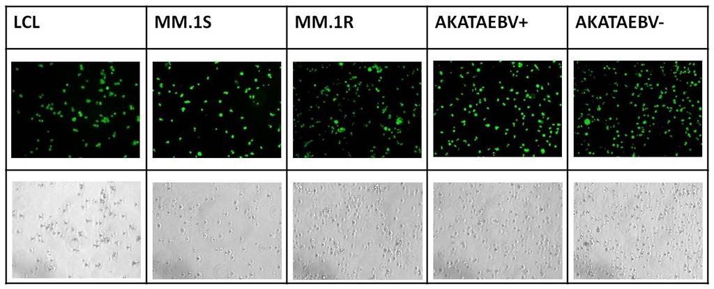
136 A B 124
137 Figure 6-1 EBNA2-TAT affects growth of EBNA-2(+) cell lines only 125
138 Figure 6-1 EBNA2-TAT affects growth of EBNA-2(+) cell lines only A) Cells after 20 minutes of peptide exposure show entry of EBNA-2 tat peptide in all cell lines. B) Proliferation assay showing effect of EBNA2-TAT on EBNA-2(+) cell lines, LCL and MM.1S and EBNA-2(-) cell lines, Akata EBV(+), Akata EBV(-) and MM.1R. 126
139 Figure 6-2 Bortezomib only leads to an increase in lytic gene expression 127
140 Figure 6-2 Bortezomib only leads to an increase in lytic gene expression in MM.1S cells. No effect is seen with sodium butyrate/tpa treatment. Error bars represent S.E.M. 128
141 Figure 6-3 Knockdown of XBP-1 abrogates effects of bortezomib induced lytic reactivation in MM.1S cell line 129
142 Figure 6-3 Knockdown of XBP-1 abrogates effects of bortezomib induced lytic reactivation in MM.1S cell line. Doxycycline induction of XBP-1 knockdown showed a smaller fold increase in lytic gene, ZTA, expression than uninduced cells. Error bars represent S.E.M. 130
143 A) B) 131
144 C) Figure 6-4 Acyclovir stabilizes EBV copy number in cell lines that harbor the virus as a latent plasmid. 132
145 Figure 6-4 Acyclovir stabilizes EBV copy number in cell lines that harbor the virus as a latent plasmid. A) Acyclovir has no effect on Namalwa. B) Akata and, C) MM.1S that carry latent virus show stable EBV copy number after treatment with acyclovir. 133
146 A B Figure 6-5 Dexamethasone affects cell survival in EBV positive cell lines only 134
147 Figure 6-5 Dexamethasone affects cell survival in EBV positive cell lines only A) 24 hour toxicity and B) 48 hour toxicity studies on EBV positive cell lines (Akata+ and MM.1S) and EBV negative cell line (Akata- and MM.1R). Only EBV positive cell lines were affected by dexamethsone. Error bars represent S.E.M. 135
148 Figure 6-6 Dexamethasone induced killing is abrogated when EBV is lost from cell lines. 136
149 Figure 6-6 Dexamethasone induced killing is abrogated when EBV is lost from cell lines. Induction of dominant negative EBNA-1 makes the MM.1S cell line resistant to dexamethasone induced toxicity. Error bars represent S.E.M. 137
150 DISCUSSION Multiple myeloma is the second most common hematologic malignancy in the US. 55. Several first line therapeutic options are available for the treatment of multiple myeloma but drug resistance and disease relapse are common. We discovered the presence of EBV in several multiple myeloma cell lines and found that targeting the EBV in the cell lines can be used as a useful therapeutic option in multiple myeloma. EBNA-2 gene expression was detected in MM.1S cells. EBNA-2 is an EBV nuclear antigen needed for proliferation of EBV positive cells. We used a 10 amino acid peptide, EBNA-2 TAT to disrupt the function of EBNA-2 and thereby affect growth of the cell line. Peptides have the ability to mediate cell entry in a concentration-dependent manner that is independent of receptors or transporters and consequently operates in a wide variety of cell types. In control EBV infected lymphoblastoid cell lines (LCLs), the EBNA-2-TAT peptide prevented expansion of newly infected B cells 94. The presence of EBNA-2 in the myeloma cell line suggests a novel pattern of viral latency pattern. EBNA-2 has not been associated with any other EBV positive malignancy. Due to this heterogeneity in viral gene expression, targeting EBNA-2 is a viable therapeutic option in MM. The effect was specific in that the EBNA2(mt)-TAT peptide did not have this property and continuous exposure to peptide was required to block cell line growth. Bortezomib is an approved therapeutic agent in multiple myeloma. However, reports have shown that it is effective against the tumor bulk cells comprising CD138 expression on their surface 56, 57, 67. We tested for the effects of bortezomib on the EBV positive myeloma cell line, MM.1S. Bortezomib has been shown to increase the expression of the 138
151 EBV lytic gene, ZTA, in Burkitt lymphoma 36. We showed a similar increase in ZTA expression level in the presence of bortezomib in MM.1S. However, other known lytic inducers, sodium butyrate and TPA combined had no effect on MM.1S. XBP-1 is a transcription factor that promotes differentiation of B-cells to plasma cells. Plasma cells comprise the tumor bulk in multiple myeloma. Loss of XBP-1 activity leads to an increase in B cells and EBV harboring cells in MM.1S cells. Knockdown of XBP-1 abrogated the effect of bortezomib mediated EBV lytic reactivation in MM.1S cell line. Loss of EBV using the dominant negative EBNA-1 expression system also weakened the effect of bortezomib on ZTA expression. This implied that EBV plays a role in bortezomib effect. Acyclovir is a nucleoside analogue that interferes with EBV lytic replication. We saw that MM.1S cells grown in media containing acyclovir had stable EBV copy number over months. This data suggests that the viral genome is maintained in these cell lines by latency replication and that the tiny percentage of cells that harbor virus are stably maintained over months or years. This is contrary to the present understanding of other well-characterized EBV-associated tumor cell lines. The EBV genome most commonly persists as a nuclear plasmid. Dexamethasone is used in the first line therapy of multiple myeloma. We tested its effects on other known EBV positive control cell lines. We showed that only the EBV positive cell lines showed cell killing by dexamethasone. It had no effect on cell lines that were negative for the virus. We showed that loss of EBV in the EBV positive cell line, MM.1S 139
152 abrogates effect of dexamethasone. This data suggests that the steroid receptor plays a role in the maintenance of EBV in the cell line. 140
153 CHAPTER 7: PRELIMINARY STUDIES IN PATIENT SAMPLES SUMMARY We have investigated cells with a cancer stem cell-like mature B cell phenotype in the peripheral blood of patients with multiple myeloma (MM) and in MM cell lines. In peripheral blood lymphocytes from MM patients, EBV DNA is enriched 10 fold in aldehyde dehydrogenase rich-mature memory B cells (CD20+CD27+) expressing the same light chain type as malignant plasma cells, and enriched approximately 100 fold over the CD19+ peripheral B cell population. The presence of EBV was confirmed by immunofluorescence. In contrast EBV is absent from these patients plasma cells. 141
154 INTRODUCTION Molecular investigations of patients and cell lines have shown that clonally-related cells are not limited to cells with a plasma cell-like phenotype but often include a population of cells with a mature B cell-like phenotype 56, 65, 66. These have been identified as putative cancer stem cells. Isolated directly from the peripheral blood of patients with MM, these cells with a mature B cell like phenotype grow as clonotypic plasma cell tumors corresponding to the patients plasma cell tumors. We tested putative cancer stem cells for presence of EBV. Multiple myeloma patient blood was first sorted for CD19 + B-cells. These B-cells were further sorted for the memory B cell compartment identified for the presence of CD20 + /27 + surface expression. These cells were then stained with Aldefluor that can stains cells that produce aldehyde dehydrogenase. This is a marker for stem cell activity. Cells that are ALD high are considered to be cancer stem cells. The cells were also sorted based on their light chain expression. MM patients can be diagnosed by clonal light chain rearrangement. The ratio of kappa to lambda light chain is often skewed in MM. The CD20/27+ cells were also sorted as CD kappa and CD lambda. We used immunofluorescence techniques and quantitative PCR for EBV DNA to test for the presence of EBV in these highly enriched cells. We show that EBV was present in the putative cancer stem cell fraction in these patient samples. 142
155 Materials and Methods Cell sorting Multiple myeloma patient blood was sorted for CD19+ cells using the MACS technique. The CD19+ cells were further sorted using FACS into i) CD20+/27+ cells which were further sorted into ii) CD20+/27+/Aldefluor+ and iii) CD20+/27+/Aldefluor- cells. They were also sorted into iv) CD20+/27+/kappa and v) CD20+/27+/lambda populations. MACS isolation of EBV harboring cells CD19 subsets were isolated from MM.1S cell line using mouse antihuman antibodies coupled to magnetic microbeads (Miltenyi Biotec, Auburn, CA) followed by magnetic column depletion (Miltenyi Biotec, Auburn, CA). Quantification of EBV viral load by quantitative PCR Genomic DNA was isolated used the QIAamp DNA micro kit (QIAGEN) according to the manufacturer's protocol. To detect EBV, BamH-W primers (fwd 5 CCCAACACTCCACCACACC 3 and rev 5 TCTTAGGAGCTGTCCGAGGG 3 ), and the BamH-W fluorescent probe (5 (FAM) CACACACTACACACACCCACCCGTCTC (BH1) 3 ) were used. EBV copy number was determined using DNA from the Namalwa cell line as standard. Each 20 μl of real-time qpcr reaction contained 1 SsoFast Probes SuperMix (Bio-Rad), a 250 nm concentration of each primer, a 200 nm concentration of the probe, and 2 μl of DNA. Thermocycling conditions were 95 C for 2 minutes for 1 cycle and 95 C for 5 seconds and 60 C for 10 seconds for 40 cycles. 143
156 Immunofluorescence assays Cells were seeded at cells per well in two-well slide chambers. Cells were washed in PBS, fixed with 4% paraformaldehyde in PBS for 10 min at room temperature, and permeabilized for 20 min on ice in 0.2% Triton X-100 in PBS. Cells were incubated with primary antibody for 60 min at 37 C and with secondary antibody at 37 C for 30 min. Between each staining step, the cells were washed in PBS three times for 5 min each time. The antibodies used were anti-ebna-1 mouse monoclonal antibody (1:200; Ab8329 from AbCam, Cambridge, United Kingdom) and anti-ebna-2 (1:200; DAKO). Cy3-conjugated goat anti-mouse immunoglobulin G (1:200) was a gift from P.Desai (Johns Hopkins Sidney Kimmel Caner Center). The cells were mounted using Vectashield mounting media with DAPI (Vector Labs, CA, USA). The slides were imaged under Leitz Wetzlar Dialux 20eb microscope using a Plan Apo 60x oil immersion objective. Image pro Plus 5.1 was used for software analysis. 144
157 RESULTS EBV is present in the nominal cancer stem cell fraction of MM patients. We studied EBV in cell populations isolated from peripheral blood from 7 MM patients (Fig.1). EBV was present by immunofluorescence and PCR in the population of cells expressing light chains corresponding to tumor and was completely absent from the population of cells expressing the non-tumor light chain. EBV was present in more than 50% of cells in the nominal stem cell fraction (CD20+CD27+ aldehyde dehydrogenase rich) but only in rare cells in the CD19+ fraction as assessed by EBNA1 immunofluorescence. PCR quantitation showed more than 5 copies per cell in the nominal stem cell fraction versus 0.05 and 0.1 copies per cell in the CD19+ fraction. EBV was not detected in plasma cell populations isolated from bone marrow. These results are consistent with the possibility that a population of cells that harbors virus overlaps with the cancer stem cell population. 145
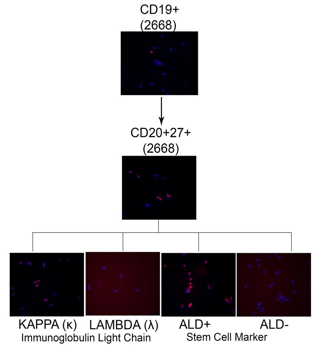
158 A 146
EBV infection B cells and lymphomagenesis. Sridhar Chaganti
 EBV infection B cells and lymphomagenesis Sridhar Chaganti How EBV infects B-cells How viral genes influence the infected B cell Differences and similarities between in vitro and in vivo infection How
Herpesviruses. Virion. Genome. Genes and proteins. Viruses and hosts. Diseases. Distinctive characteristics
 Herpesviruses Virion Genome Genes and proteins Viruses and hosts Diseases Distinctive characteristics Virion Enveloped icosahedral capsid (T=16), diameter 125 nm Diameter of enveloped virion 200 nm Capsid
Determination of the temporal pattern and importance of BALF1 expression in Epstein-Barr viral infection
 Determination of the temporal pattern and importance of BALF1 expression in Epstein-Barr viral infection Melissa Mihelidakis May 6, 2004 7.340 Research Proposal Introduction Apoptosis, or programmed cell
EBV Infection and Immunity. Andrew Hislop Institute for Cancer Studies University of Birmingham
 EBV Infection and Immunity Andrew Hislop Institute for Cancer Studies University of Birmingham EBV Introduction Large ds DNA virus Spread by saliva contact Lifelong infection Predominantly B-lymphotropic
BIT 120. Copy of Cancer/HIV Lecture
 BIT 120 Copy of Cancer/HIV Lecture Cancer DEFINITION Any abnormal growth of cells that has malignant potential i.e.. Leukemia Uncontrolled mitosis in WBC Genetic disease caused by an accumulation of mutations
Identification of the Site of Epstein-Barr Virus Persistence In Vivo as a Resting B Cell
 JOURNAL OF VIROLOGY, July 1997, p. 4882 4891 Vol. 71, No. 7 0022-538X/97/$04.00 0 Copyright 1997, American Society for Microbiology Identification of the Site of Epstein-Barr Virus Persistence In Vivo
Epstein-Barr Virus: Stimulation By 5 '-Iododeoxy uridine or 5 '-Brom odeoxy uridine in Human Lymphoblastoid Cells F ro m a Rhabdom yosarcom a*
 A n n a ls o f C l i n i c a l L a b o r a t o r y S c i e n c e, Vol. 3, No. 6 Copyright 1973, Institute for Clinical Science Epstein-Barr Virus: Stimulation By 5 '-Iododeoxy uridine or 5 '-Brom odeoxy
Chapter 4 Cellular Oncogenes ~ 4.6 -
 Chapter 4 Cellular Oncogenes - 4.2 ~ 4.6 - Many retroviruses carrying oncogenes have been found in chickens and mice However, attempts undertaken during the 1970s to isolate viruses from most types of
Human Immunodeficiency Virus
 Human Immunodeficiency Virus Virion Genome Genes and proteins Viruses and hosts Diseases Distinctive characteristics Viruses and hosts Lentivirus from Latin lentis (slow), for slow progression of disease
Polyomaviridae. Spring
 Polyomaviridae Spring 2002 331 Antibody Prevalence for BK & JC Viruses Spring 2002 332 Polyoma Viruses General characteristics Papovaviridae: PA - papilloma; PO - polyoma; VA - vacuolating agent a. 45nm
Fayth K. Yoshimura, Ph.D. September 7, of 7 HIV - BASIC PROPERTIES
 1 of 7 I. Viral Origin. A. Retrovirus - animal lentiviruses. HIV - BASIC PROPERTIES 1. HIV is a member of the Retrovirus family and more specifically it is a member of the Lentivirus genus of this family.
Large DNA viruses: Herpesviruses, Poxviruses, Baculoviruses and Giant viruses
 Large DNA viruses: Herpesviruses, Poxviruses, Baculoviruses and Giant viruses Viruses are the only obstacles to the domination of the Earth by mankind. -Joshua Lederberg Recommended reading: Field s Virology
mirna Dr. S Hosseini-Asl
 mirna Dr. S Hosseini-Asl 1 2 MicroRNAs (mirnas) are small noncoding RNAs which enhance the cleavage or translational repression of specific mrna with recognition site(s) in the 3 - untranslated region
Chapter13 Characterizing and Classifying Viruses, Viroids, and Prions
 Chapter13 Characterizing and Classifying Viruses, Viroids, and Prions 11/20/2017 MDufilho 1 Characteristics of Viruses Viruses Minuscule, acellular, infectious agent having either DNA or RNA Cause infections
Viruses Tomasz Kordula, Ph.D.
 Viruses Tomasz Kordula, Ph.D. Resources: Alberts et al., Molecular Biology of the Cell, pp. 295, 1330, 1431 1433; Lehninger CD Movie A0002201. Learning Objectives: 1. Understand parasitic life cycle of
LESSON 4.6 WORKBOOK. Designing an antiviral drug The challenge of HIV
 LESSON 4.6 WORKBOOK Designing an antiviral drug The challenge of HIV In the last two lessons we discussed the how the viral life cycle causes host cell damage. But is there anything we can do to prevent
Epstein-Barr Virus: Cell Trafficking Is Crucial for Persistence
 Epstein-Barr Virus: Cell Trafficking Is Crucial for Persistence This virus moves between host lymphoid and epithelial cells, switching its tropism while enhancing persistence Lindsey Hutt-Fletcher Herpesviruses,
Overview of primary HHV-8 infection
 Overview of primary HHV-8 infection HHV-8, also known as Kaposi sarcoma-associated herpesvirus (KSHV), is a gamma herpesvirus primarily transmitted through saliva. The virus initially replicates in epithelial
Principles of Adaptive Immunity
 Principles of Adaptive Immunity Chapter 3 Parham Hans de Haard 17 th of May 2010 Agenda Recognition molecules of adaptive immune system Features adaptive immune system Immunoglobulins and T-cell receptors
7.012 Quiz 3 Answers
 MIT Biology Department 7.012: Introductory Biology - Fall 2004 Instructors: Professor Eric Lander, Professor Robert A. Weinberg, Dr. Claudette Gardel Friday 11/12/04 7.012 Quiz 3 Answers A > 85 B 72-84
Lecture 2: Virology. I. Background
 Lecture 2: Virology I. Background A. Properties 1. Simple biological systems a. Aggregates of nucleic acids and protein 2. Non-living a. Cannot reproduce or carry out metabolic activities outside of a
VIRUSES AND CANCER Michael Lea
 VIRUSES AND CANCER 2010 Michael Lea VIRAL ONCOLOGY - LECTURE OUTLINE 1. Historical Review 2. Viruses Associated with Cancer 3. RNA Tumor Viruses 4. DNA Tumor Viruses HISTORICAL REVIEW Historical Review
19/06/2013. Viruses are not organisms (do not belong to any kingdom). Viruses are not made of cells, have no cytoplasm, and no membranes.
. Viruses are not made of cells, have no cytoplasm, and no membranes.") VIRUSES Many diseases of plants and animals are caused by bacteria or viruses that invade the body. Bacteria and viruses are NOT similar kinds of micro-organisms. Bacteria are classified as living organisms,
VIRUSES Many diseases of plants and animals are caused by bacteria or viruses that invade the body. Bacteria and viruses are NOT similar kinds of micro-organisms. Bacteria are classified as living organisms,
Epstein-Barr virus and immunity
 Epstein-Barr virus and immunity Elena Kashuba, PhD Associate Professor Department of Microbiology, Tumor and Cell Biology (MTC) Karolinska Institutet 1 Epstein-Barr virus EBV Everybody s virus 200 nm Enveloped
Egr-1 regulates RTA transcription through a cooperative involvement of transcriptional regulators
 /, 2017, Vol. 8, (No. 53), pp: 91425-91444 Egr-1 regulates RTA transcription through a cooperative involvement of transcriptional regulators Roni Sarkar 1 and Subhash C. Verma 1 1 Department of Microbiology
Persistent Infections
 Persistent Infections Lecture 17 Biology 3310/4310 Virology Spring 2017 Paralyze resistance with persistence WOODY HAYES Acute vs persistent infections Acute infection - rapid and self-limiting Persistent
MHC class I MHC class II Structure of MHC antigens:
 MHC class I MHC class II Structure of MHC antigens: MHC class I antigens consist of a transmembrane heavy chain (α chain) that is non-covalently associated with β2- microglobulin. Membrane proximal domain
Epstein-Barr Virus 1
 Epstein-Barr Virus 1 Herpesviruses dsdna, linear, enveloped, 180-200 nm Large genome, codes for 75 viral proteins 50-70% similarity Cross reactivity between HSV and VZV HSV-2 virus particle. Note that
MedChem 401~ Retroviridae. Retroviridae
 MedChem 401~ Retroviridae Retroviruses plus-sense RNA genome (!8-10 kb) protein capsid lipid envelop envelope glycoproteins reverse transcriptase enzyme integrase enzyme protease enzyme Retroviridae The
Clinical Aspect and Application of Laboratory Test in Herpes Virus Infection. Masoud Mardani M.D,FIDSA
 Clinical Aspect and Application of Laboratory Test in Herpes Virus Infection Masoud Mardani M.D,FIDSA Shahidhid Bh BeheshtiMdi Medical lui Universityit Cytomegalovirus (CMV), Epstein Barr Virus(EBV), Herpes
Clever tricks EBV employed to modulate innate immunity during latency and lytic infection
 Clever tricks EBV employed to modulate innate immunity during latency and lytic infection Kirsten Kuipers December 22, 2011 About the cover: Upon recognition of PAMPs, activated PRRs initiate innate immune
COURSE: Medical Microbiology, PAMB 650/720 - Fall 2008 Lecture 16
 COURSE: Medical Microbiology, PAMB 650/720 - Fall 2008 Lecture 16 Tumor Immunology M. Nagarkatti Teaching Objectives: Introduction to Cancer Immunology Know the antigens expressed by cancer cells Understand
COMMENT LES VIRUS INDUISENT-ILS LA LYMPHOMAGENESE?
 COMMENT LES VIRUS INDUISENT-ILS LA LYMPHOMAGENESE? Yegor Vassetzky, CNRS UMR8126 IGR - UPS VIRAL THEORY OF CANCER: UPS AND DOWNS Peyton Rouss 1911: discovery of RSV 1966: Nobel Prize Denis Burkitt 1957:
VZV, EBV, and HHV-6-8
 VZV, EBV, and HHV-6-8 Anne Gershon Common Features of Herpesviruses Morphology Basic mode of replication Primary infection followed by latency Ubiquitous Ability to cause recurrent infections (reactivation
Hepatitis B Antiviral Drug Development Multi-Marker Screening Assay
 Hepatitis B Antiviral Drug Development Multi-Marker Screening Assay Background ImQuest BioSciences has developed and qualified a single-plate method to expedite the screening of antiviral agents against
Melatonin and its Role in the Inhibition of Breast Cancer Ciara Nicol Ross Copyright 2014 by Ciara Ross and Koni Stone
 1 Melatonin and its Role in the Inhibition of Breast Cancer Ciara Nicol Ross Copyright 2014 by Ciara Ross and Koni Stone Cancer is a disease caused by out of control division of abnormal cells within a
The Development of Lymphocytes: B Cell Development in the Bone Marrow & Peripheral Lymphoid Tissue Deborah A. Lebman, Ph.D.
 The Development of Lymphocytes: B Cell Development in the Bone Marrow & Peripheral Lymphoid Tissue Deborah A. Lebman, Ph.D. OBJECTIVES 1. To understand how ordered Ig gene rearrangements lead to the development
Hepadnaviruses: Variations on the Retrovirus Theme
 WBV21 6/27/03 11:34 PM Page 377 Hepadnaviruses: Variations on the Retrovirus Theme 21 CHAPTER The virion and the viral genome The viral replication cycle The pathogenesis of hepatitis B virus A plant hepadnavirus
VIRUSES. 1. Describe the structure of a virus by completing the following chart.
 AP BIOLOGY MOLECULAR GENETICS ACTIVITY #3 NAME DATE HOUR VIRUSES 1. Describe the structure of a virus by completing the following chart. Viral Part Description of Part 2. Some viruses have an envelope
Supplementary Information
 Supplementary Information Supplementary Figure 1. EBV-gB 23-431 mainly exists as trimer in HEK 293FT cells. (a) Western blotting analysis for DSS crosslinked FLAG-gB 23-431. HEK 293FT cells transfected
Medical Virology. Herpesviruses, Orthomyxoviruses, and Retro virus. - Herpesviruses Structure & Composition: Herpesviruses
 Medical Virology Lecture 2 Asst. Prof. Dr. Dalya Basil Herpesviruses, Orthomyxoviruses, and Retro virus - Herpesviruses Structure & Composition: Herpesviruses Enveloped DNA viruses. All herpesviruses have
Hepatitis B Virus infection: virology
 Hepatitis B Virus infection: virology 167 Falk Symposium: Liver under constant attack from fat to viruses III Falk Gastro-Konferenz 17.-21. September 2008 Congress Centrum Mainz Maura Dandri Department
number Done by Corrected by Doctor Maha Shomaf
 number 19 Done by Waseem Abo-Obeida Corrected by Abdullah Zreiqat Doctor Maha Shomaf Carcinogenesis: the molecular basis of cancer. Non-lethal genetic damage lies at the heart of carcinogenesis and leads
Chapter 18. Viral Genetics. AP Biology
 Chapter 18. Viral Genetics 2003-2004 1 A sense of size Comparing eukaryote bacterium virus 2 What is a virus? Is it alive? DNA or RNA enclosed in a protein coat Viruses are not cells Extremely tiny electron
Yamaguchi University School of Medicine, Kogushi, Ube, Yamaguchi. growth in low serum, anchorage-independent growth in soft
 JOURNAL OF VIROLOGY, Sept. 1994, p. 6069-6073 Vol. 68, No. 9 0022-538X/94/$04.00+0 Copyright 1994, American Society for Microbiology Isolation of Epstein-Barr Virus (EBV)-Negative Cell Clones from the
Received 16 January 2009/Accepted 5 May 2009
 JOURNAL OF VIROLOGY, Aug. 2009, p. 7749 7760 Vol. 83, No. 15 0022-538X/09/$08.00 0 doi:10.1128/jvi.00108-09 Copyright 2009, American Society for Microbiology. All Rights Reserved. Features Distinguishing
Chapter 19: Viruses. 1. Viral Structure & Reproduction. 2. Bacteriophages. 3. Animal Viruses. 4. Viroids & Prions
 Chapter 19: Viruses 1. Viral Structure & Reproduction 2. Bacteriophages 3. Animal Viruses 4. Viroids & Prions 1. Viral Structure & Reproduction Chapter Reading pp. 393-396 What exactly is a Virus? Viruses
Viruses. Poxviridae. DNA viruses: 6 families. Herpesviridae Adenoviridae. Hepadnaviridae Papovaviridae Parvoviridae
 Viruses DNA viruses: 6 families Poxviridae Herpesviridae Adenoviridae Hepadnaviridae Papovaviridae Parvoviridae Human herpesviruses Three subfamilies (genome structure, tissue tropism, cytopathologic effect,
A Schematic Of The Life Cycle Of The
 A Schematic Of The Life Cycle Of The Lymphocytes Involved A wonderful and informative website to visit is from Johns Hopkins Medical Center Called "The Body". The ability of these viruses to infect lymphocytes
Determination Differentiation. determinated precursor specialized cell
 Biology of Cancer -Developmental Biology: Determination and Differentiation -Cell Cycle Regulation -Tumor genes: Proto-Oncogenes, Tumor supressor genes -Tumor-Progression -Example for Tumor-Progression:
T-cell activation T cells migrate to secondary lymphoid tissues where they interact with antigen, antigen-presenting cells, and other lymphocytes:
 Interactions between innate immunity & adaptive immunity What happens to T cells after they leave the thymus? Naïve T cells exit the thymus and enter the bloodstream. If they remain in the bloodstream,
Unit 13.2: Viruses. Vocabulary capsid latency vaccine virion
 Unit 13.2: Viruses Lesson Objectives Describe the structure of viruses. Outline the discovery and origins of viruses. Explain how viruses replicate. Explain how viruses cause human disease. Describe how
Chapter 19: Viruses. 1. Viral Structure & Reproduction. What exactly is a Virus? 11/7/ Viral Structure & Reproduction. 2.
 Chapter 19: Viruses 1. Viral Structure & Reproduction 2. Bacteriophages 3. Animal Viruses 4. Viroids & Prions 1. Viral Structure & Reproduction Chapter Reading pp. 393-396 What exactly is a Virus? Viruses
Primary Adult Naïve CD4+ CD45RA+ Cells. Prepared by: David Randolph at University of Alabama, Birmingham
 Primary Adult Naïve CD4+ CD45RA+ Cells Prepared by: David Randolph (drdrdr@uab.edu) at University of Alabama, Birmingham Goal: To obtain large numbers of highly pure primary CD4+ CD45RO- CD25- cells from
Activation of cellular proto-oncogenes to oncogenes. How was active Ras identified?
 Dominant Acting Oncogenes Eugene E. Marcantonio, M.D. Ph.D. Oncogenes are altered forms of normal cellular genes called proto-oncogenes that are involved in pathways regulating cell growth, differentiation,
Immunology - Lecture 2 Adaptive Immune System 1
 Immunology - Lecture 2 Adaptive Immune System 1 Book chapters: Molecules of the Adaptive Immunity 6 Adaptive Cells and Organs 7 Generation of Immune Diversity Lymphocyte Antigen Receptors - 8 CD markers
Properties of Herpesviruses
 Herpesviruses Properties of Herpesviruses Structure and Composition Spherical icosahedron, 150-200 nm Double-stranded DNA, linear More than 35 proteins Enveloped Replication from nucleus (budding) Features
Human Herpesviruses. VZV, EBV, and HHV-6-8. The rash of VZV is vesicular. MID 34
 VZV, EBV, and HHV-6-8 Anne Gershon Human Herpesviruses Replication (lytic infection) occurs in a cascade Latency occurs when the cascade is interrupted Transcription of viral genome and protein synthesis
Section 6. Junaid Malek, M.D.
 Section 6 Junaid Malek, M.D. The Golgi and gp160 gp160 transported from ER to the Golgi in coated vesicles These coated vesicles fuse to the cis portion of the Golgi and deposit their cargo in the cisternae
Effective activity of cytokine-induced killer cells against autologous metastatic melanoma including cells with stemness features
 Effective activity of cytokine-induced killer cells against autologous metastatic melanoma including cells with stemness features Loretta Gammaitoni, Lidia Giraudo, Valeria Leuci, et al. Clin Cancer Res
Immunopathology of Lymphoma
 Immunopathology of Lymphoma Noraidah Masir MBBCh, M.Med (Pathology), D.Phil. Department of Pathology Faculty of Medicine Universiti Kebangsaan Malaysia Lymphoma classification has been challenging to pathologists.
Last time we talked about the few steps in viral replication cycle and the un-coating stage:
 Zeina Al-Momani Last time we talked about the few steps in viral replication cycle and the un-coating stage: Un-coating: is a general term for the events which occur after penetration, we talked about
General information. Cell mediated immunity. 455 LSA, Tuesday 11 to noon. Anytime after class.
 General information Cell mediated immunity 455 LSA, Tuesday 11 to noon Anytime after class T-cell precursors Thymus Naive T-cells (CD8 or CD4) email: lcoscoy@berkeley.edu edu Use MCB150 as subject line
Instructions for Use. APO-AB Annexin V-Biotin Apoptosis Detection Kit 100 tests
 3URGXFW,QIRUPDWLRQ Sigma TACS Annexin V Apoptosis Detection Kits Instructions for Use APO-AB Annexin V-Biotin Apoptosis Detection Kit 100 tests For Research Use Only. Not for use in diagnostic procedures.
Foundations in Microbiology
 Foundations in Microbiology Fifth Edition Talaro Chapter 15 The Acquisition of Specific Immunity and Its Applications Chapter 15 2 Chapter Overview 1. Development of the Dual Lymphocyte System 2. Entrance
Cancer. The fundamental defect is. unregulated cell division. Properties of Cancerous Cells. Causes of Cancer. Altered growth and proliferation
 Cancer The fundamental defect is unregulated cell division. Properties of Cancerous Cells Altered growth and proliferation Loss of growth factor dependence Loss of contact inhibition Immortalization Alterated
HIV INFECTION: An Overview
 HIV INFECTION: An Overview UNIVERSITY OF PAPUA NEW GUINEA SCHOOL OF MEDICINE AND HEALTH SCIENCES DIVISION OF BASIC MEDICAL SCIENCES DISCIPLINE OF BIOCHEMISTRY & MOLECULAR BIOLOGY PBL MBBS II SEMINAR VJ
Nanoparticles and persistent virus infection a dangerous liaison for the development of chronic lung disease(s)? Tobias Stöger
? Tobias Stöger") Nanoparticles and persistent virus infection a dangerous liaison for the development of chronic lung disease(s)? Tobias Stöger Herpesviruses and lung disease Double-stranded DNA-viruses (a, b, g- herpesviruses)
Nanoparticles and persistent virus infection a dangerous liaison for the development of chronic lung disease(s)? Tobias Stöger Herpesviruses and lung disease Double-stranded DNA-viruses (a, b, g- herpesviruses)
Acta Medica Okayama AUGUST A spectrum of clinical manifestations caused by host immune responses against Epstein-Barr virus infections.
 Acta Medica Okayama Volume 58, Issue 4 2004 Article 1 AUGUST 2004 A spectrum of clinical manifestations caused by host immune responses against Epstein-Barr virus infections. Keiji Iwatsuki Takenobu Yamamoto
Corrigenda. WHO Classification of Tumours of Haematopoietic and Lymphoid Tissues (revised 4th edition): corrections made in second print run
: corrections made in second print run") Corrigenda WHO Classification of Tumours of Haematopoietic and Lymphoid Tissues (revised 4th edition): corrections made in second print run In addition to corrections of minor typographical errors, corrections
Corrigenda WHO Classification of Tumours of Haematopoietic and Lymphoid Tissues (revised 4th edition): corrections made in second print run In addition to corrections of minor typographical errors, corrections
Molecular Markers. Marcie Riches, MD, MS Associate Professor University of North Carolina Scientific Director, Infection and Immune Reconstitution WC
 Molecular Markers Marcie Riches, MD, MS Associate Professor University of North Carolina Scientific Director, Infection and Immune Reconstitution WC Overview Testing methods Rationale for molecular testing
This electronic thesis or dissertation has been downloaded from the King s Research Portal at
 This electronic thesis or dissertation has been downloaded from the King s Research Portal at https://kclpure.kcl.ac.uk/portal/ Evaluation of the Role of Epstein-Barr virus and Cellular Gene Expression
What causes cancer? Physical factors (radiation, ionization) Chemical factors (carcinogens) Biological factors (virus, bacteria, parasite)
 Chemical factors (carcinogens) Biological factors (virus, bacteria, parasite)") Oncogenes What causes cancer? Chemical factors (carcinogens) Physical factors (radiation, ionization) Biological factors (virus, bacteria, parasite) DNA Mutation or damage Oncogenes Tumor suppressor genes
Oncogenes What causes cancer? Chemical factors (carcinogens) Physical factors (radiation, ionization) Biological factors (virus, bacteria, parasite) DNA Mutation or damage Oncogenes Tumor suppressor genes
11/15/2011. Outline. Structural Features and Characteristics. The Good the Bad and the Ugly. Viral Genomes. Structural Features and Characteristics
 Chapter 19 - Viruses Outline I. Viruses A. Structure of viruses B. Common Characteristics of Viruses C. Viral replication D. HIV II. Prions The Good the Bad and the Ugly Viruses fit into the bad category
York criteria, 6 RA patients and 10 age- and gender-matched healthy controls (HCs).
.") MATERIALS AND METHODS Study population Blood samples were obtained from 15 patients with AS fulfilling the modified New York criteria, 6 RA patients and 10 age- and gender-matched healthy controls (HCs).
MATERIALS AND METHODS Study population Blood samples were obtained from 15 patients with AS fulfilling the modified New York criteria, 6 RA patients and 10 age- and gender-matched healthy controls (HCs).
Cancer. The fundamental defect is. unregulated cell division. Properties of Cancerous Cells. Causes of Cancer. Altered growth and proliferation
 Cancer The fundamental defect is unregulated cell division. Properties of Cancerous Cells Altered growth and proliferation Loss of growth factor dependence Loss of contact inhibition Immortalization Alterated
Under the Radar Screen: How Bugs Trick Our Immune Defenses
 Under the Radar Screen: How Bugs Trick Our Immune Defenses Session 7: Cytokines Marie-Eve Paquet and Gijsbert Grotenbreg Whitehead Institute for Biomedical Research HHV-8 Discovered in the 1980 s at the
HIV Immunopathogenesis. Modeling the Immune System May 2, 2007
 HIV Immunopathogenesis Modeling the Immune System May 2, 2007 Question 1 : Explain how HIV infects the host Zafer Iscan Yuanjian Wang Zufferey Abhishek Garg How does HIV infect the host? HIV infection
Chapter 6. Antigen Presentation to T lymphocytes
 Chapter 6 Antigen Presentation to T lymphocytes Generation of T-cell Receptor Ligands T cells only recognize Ags displayed on cell surfaces These Ags may be derived from pathogens that replicate within
Recombinant Protein Expression Retroviral system
 Recombinant Protein Expression Retroviral system Viruses Contains genome DNA or RNA Genome encased in a protein coat or capsid. Some viruses have membrane covering protein coat enveloped virus Ø Essential
VIRUSES. Biology Applications Control. David R. Harper. Garland Science Taylor & Francis Group NEW YORK AND LONDON
 VIRUSES Biology Applications Control David R. Harper GS Garland Science Taylor & Francis Group NEW YORK AND LONDON vii Chapter 1 Virus Structure and 2.2 VIRUS MORPHOLOGY 26 Infection 1 2.3 VIRAL CLASSIFICATION
An Epstein-Barr virus-encoded microrna targets PUMA to promote host cell survival
 An Epstein-Barr virus-encoded microrna targets to promote host cell survival The Journal of Experimental Medicine 205(11): 2551-2560, 2008. 1 Elizabeth Yee-Wai Choy, Kam-Leung Siu, Kin-Hang Kok, Raymond
Chapter 22: The Lymphatic System and Immunity
 Bio40C schedule Lecture Immune system Lab Quiz 2 this week; bring a scantron! Study guide on my website (see lab assignments) Extra credit Critical thinking questions at end of chapters 5 pts/chapter Due
LESSON 4.4 WORKBOOK. How viruses make us sick: Viral Replication
 DEFINITIONS OF TERMS Eukaryotic: Non-bacterial cell type (bacteria are prokaryotes).. LESSON 4.4 WORKBOOK How viruses make us sick: Viral Replication This lesson extends the principles we learned in Unit
2017 CST-Astellas Canadian Transplant Fellows Symposium. EBV Post Transplantation Implications and Approach to Management
 2017 CST-Astellas Canadian Transplant Fellows Symposium EBV Post Transplantation Implications and Approach to Management Atul Humar, MD Atul Humar is a Professor in the Department of Medicine, University
Problem Set 8 Key 1 of 8
 7.06 2003 Problem Set 8 Key 1 of 8 7.06 2003 Problem Set 8 Key 1. As a bright MD/PhD, you are interested in questions about the control of cell number in the body. Recently, you've seen three patients
Problem Set 5 KEY
 2006 7.012 Problem Set 5 KEY ** Due before 5 PM on THURSDAY, November 9, 2006. ** Turn answers in to the box outside of 68-120. PLEASE WRITE YOUR ANSWERS ON THIS PRINTOUT. 1. You are studying the development
Supplementary Data 1. Alanine substitutions and position variants of APNCYGNIPL. Applied in
 Supplementary Data 1. Alanine substitutions and position variants of APNCYGNIPL. Applied in Supplementary Fig. 2 Substitution Sequence Position variant Sequence original APNCYGNIPL original APNCYGNIPL
Direct ex vivo characterization of human antigen-specific CD154 + CD4 + T cells Rapid antigen-reactive T cell enrichment (Rapid ARTE)
") Direct ex vivo characterization of human antigen-specific CD154 + CD4 + T cells Rapid antigen-reactive T cell enrichment (Rapid ARTE) Introduction Workflow Antigen (ag)-specific T cells play a central
Direct ex vivo characterization of human antigen-specific CD154 + CD4 + T cells Rapid antigen-reactive T cell enrichment (Rapid ARTE) Introduction Workflow Antigen (ag)-specific T cells play a central
VIII Curso Internacional del PIRRECV. Some molecular mechanisms of cancer
 VIII Curso Internacional del PIRRECV Some molecular mechanisms of cancer Laboratorio de Comunicaciones Celulares, Centro FONDAP Estudios Moleculares de la Celula (CEMC), ICBM, Facultad de Medicina, Universidad
Third line of Defense
 Chapter 15 Specific Immunity and Immunization Topics -3 rd of Defense - B cells - T cells - Specific Immunities Third line of Defense Specific immunity is a complex interaction of immune cells (leukocytes)
T Cell Effector Mechanisms I: B cell Help & DTH
 T Cell Effector Mechanisms I: B cell Help & DTH Ned Braunstein, MD The Major T Cell Subsets p56 lck + T cells γ δ ε ζ ζ p56 lck CD8+ T cells γ δ ε ζ ζ Cα Cβ Vα Vβ CD3 CD8 Cα Cβ Vα Vβ CD3 MHC II peptide
Supplemental Materials and Methods Plasmids and viruses Quantitative Reverse Transcription PCR Generation of molecular standard for quantitative PCR
 Supplemental Materials and Methods Plasmids and viruses To generate pseudotyped viruses, the previously described recombinant plasmids pnl4-3-δnef-gfp or pnl4-3-δ6-drgfp and a vector expressing HIV-1 X4
cure research HIV & AIDS
 Glossary of terms HIV & AIDS cure research Antiretroviral Therapy (ART) ART involves the use of several (usually a cocktail of three or more) antiretroviral drugs to halt HIV replication. ART drugs may
Antigen Receptor Structures October 14, Ram Savan
 Antigen Receptor Structures October 14, 2016 Ram Savan savanram@uw.edu 441 Lecture #8 Slide 1 of 28 Three lectures on antigen receptors Part 1 (Today): Structural features of the BCR and TCR Janeway Chapter
MicroRNA and Male Infertility: A Potential for Diagnosis
 Review Article MicroRNA and Male Infertility: A Potential for Diagnosis * Abstract MicroRNAs (mirnas) are small non-coding single stranded RNA molecules that are physiologically produced in eukaryotic
Persistent Infections
 Persistent Infections Lecture 16 Virology W3310/4310 Spring 2013 1 Breaking Up Is Hard To Do Neil Sedaka 1962 2 Acute vs. Persistent Infections Acute - a natural infection that usually is rapid and self
Karyotype analysis reveals transloction of chromosome 22 to 9 in CML chronic myelogenous leukemia has fusion protein Bcr-Abl
 Chapt. 18 Cancer Molecular Biology of Cancer Student Learning Outcomes: Describe cancer diseases in which cells no longer respond Describe how cancers come from genomic mutations (inherited or somatic)
K R N Baumforth, L S Young, K J Flavell, C Constandinou, P G Murray
 J Clin Pathol: Mol Pathol 1999;52:307 322 307 Demystified... Biomedical Research Laboratories, School of Health Sciences, University of Wolverhampton, Wolverhampton WV1 1DJ, UK K R N Baumforth K J Flavell