Maintenance of immune fitness during reconstitution from T cell lymphopenia by CD4-positive, CD25-positive, and Foxp3-positive regulatory T cells
|
|
|
- Tyler Booth
- 6 years ago
- Views:
Transcription
1 Maintenance of immune fitness during reconstitution from T cell lymphopenia by CD4-positive, CD25-positive, and Foxp3-positive regulatory T cells A DISSERTATION SUBMITTED TO THE FACULTY OF THE GRADUATE SCHOOL OF THE UNIVERSITY OF MINNESOTA BY Colleen Jean Winstead IN PARTIAL FULFILLMENT OF THE REQUIREMENTS FOR THE DEGREE OF DOCTOR OF PHILOSOPHY Alexander Khoruts, M.D., Advisor July, 2009
2 Colleen Jean Winstead 2009
3 Acknowledgements Thanks are due to my advisor, Dr. Alexander Khoruts, and the other members of my thesis committee, Dr. Michael A. Farrar, Dr. Ameeta Kelekar, Dr. Daniel L. Mueller, and Dr. Stephen C. Jameson for their suggestions, guidance, and patience, which made this degree possible. I am also grateful to Dr. Marc K. Jenkins for his mentorship and generosity in sharing reagents and animals used in research for this thesis. i
4 Dedication This thesis is dedicated to my parents, Jean and Theodore Winstead, who taught me the value of integrity and hard work, and through much personal sacrifice made my higher education, this thesis, and my degree possible. ii
5 Abstract Work presented in this doctoral thesis focuses on the role of regulatory T cells (Tregs) in controlling T cell homeostasis and emergence of autoimmunity during immune reconstitution from lymphopenia. It is recognized that lymphopenia may be a common trigger of many autoimmune diseases due to oligoclonal expansion of selfreactive T cells with an effector phenotype. It is also a clinical fact that autoimmunity is often associated with immune deficiency and poor responsiveness to vaccines and infections. Research supporting work presented in chapter 2 of this thesis was based on the hypothesis that poor Treg function may play a central role in these phenomena. This work, published in June of 2008 in the Journal of Immunology, clearly demonstrated that Tregs selectively restrain one specific form of lymphopenia-induced proliferation characterized by burst-like cell cycle activity and effector T cell differentiation (spontaneous proliferation). The spontaneous form of lymphopenia-induced proliferation is the likely source of oligoclonal expansion of self-reactive T cells that drive autoimmunity. Work presented in chapter 3 of this thesis addresses the hypothesis that such oligoclonal expansion by a few T cell clones consumes resources away from the rest of the T cell population, which ultimately results in loss of T cell diversity. Using the technique of T cell adoptive transfer, we measured immune responses to infection with the gram negative bacteria Listeria monocytogenes in lymphopenic mice reconstituted in the presence or absence of Tregs by analysis of T cell receptor (TCR) Vβ chain usage and repertoire sampling using Vβ-Jβ chain TCR spectratyping and magnetic bead enrichment with specific major histocompatibility (MHC) class I and II tetramers. Experimental results suggest that the presence of Tregs during immune iii
6 reconstitution preserves TCR structural diversity and allows for more accumulation of pathogen-associated antigen-specific T cells in secondary lymphoid tissues following clearance of the infection. This result may otherwise seem paradoxical as Tregs are typically thought of as generalized suppressors of the immune system. Regardless, we believe this unappreciated ability to maintain T cell homeostasis through preservation of peripheral diversity will shed considerable insight into the role of Tregs in the immune system, vaccine responsiveness, pathogenesis of autoimmunity, and immune senescence. iv
7 Table of Contents Page Number(s) Acknowledgements Dedication Abstract List of Tables List of Figures i ii iii-iv vi vii-ix Chapter 1: Introduction 1-19 Chapter 2: Regulatory CD4+CD25+Foxp3+ T cells selectively inhibit the spontaneous form of lymphopenia-induced proliferation of naïve T cells Chapter 3: CD4+CD25+Foxp3+ regulatory T cells maintain optimal responsiveness of the Foxp3- T cell population during reconstitution from lymphopenia Chapter 4: Regulatory CD4+CD25+Foxp3+ T cell suppression of conventional T cell LIP: potential mechanisms Chapter 5: Discussion Bibliography Journal of Immunology Copyright Permission Letter 165 v
8 List of Tables 1-1. Potential Treg mechanisms of suppression LIP and subsequent bacterial infection do not induce 97 significant peripheral conversion of conventional T cells to Foxp3+. Page Number(s) Supplemental 3-1. Primers used for TCR spectratyping (5 3 ). 104 vi
9 List of Figures Page Number(s) 1-1. Homeostatic and Spontaneous LIP Naïve polyclonal and monoclonal transgenic OT-I CD8 T cells undergo homeostatic and spontaneous forms of lymphopenia-induced proliferation (LIP) Tregs selectively suppress the spontaneous form of LIP experienced by naïve CD8 T cells Tregs suppress expression of effector characteristics by CD8 T cells undergoing spontaneous LIP CD4+CD25- T cells enhance spontaneous LIP of CD8 T cells Wild-type and IL-10-/- mice Tregs from young mice are phenotypically similar Suppression of CD8 T cell spontaneous proliferation by Tregs is not IL-10 dependent Suppression of CD4 T cell spontaneous proliferation by Tregs is not IL-10 dependent IL-10-deficient Tregs cannot completely prevent colitis caused by adoptive transfer of CD4 T cells into RAG-/- mice IL-10-deficient Tregs are impaired in their ability to suppress effector CD4 T cells in the colon When co-transferred, large numbers of Tregs are required to suppress infiltration of effector CD4 T cells in the colon Model of Treg-mediated control of responder T cells in lymphopenia Filling of the T cell niche in RAG -/- mice through adoptive transfer Assessing T cell diversity post-lip at the level of 90 TCR Vβ chain usage. vii
10 3-3. Tregs prevent emergence of holes in the T cell repertoire 91 during LIP Tregs preserve the diversity of foreign Ag-reactive T cells during LIP The presence of Tregs during LIP ensures responsiveness of 96 foreign Ag-reactive T cells to bacterial infection 3-6. Tregs preserve immune fitness of conventional T cells 98 undergoing LIP. Supplemental 3-1. Tregs preserve the diversity of foreign 99 Ag-reactive T cells during LIP in TCRα -/- hosts. Supplemental 3-2. Tregs concomitantly transferred with 100 conventional T cells preserve the diversity of conventional CD4 T cell foreign Ag-reactivity during LIP. Supplemental 3-3. Tregs preserve T cell diversity during LIP 101 as assessed by correlative measures. Supplemental 3-4. Isolated and re-transferred LIP cells respond to challenge in a secondary, lymphoreplete host CD28 -/- T cells undergoing LIP in a B7 -/- host are insensitive to Treg-mediated suppression Treg-mediated suppression of spontaneous LIP is IDO-independent Protection from adoptive transfer-induced colitis is partially IDO-dependent IDO expression in radio-resistant cells protects from colitis via Treg-induction in the colon MyD88-/- Tregs are competent suppressors of polyclonal 130 CD8 T cell spontaneous LIP Loss of TLR signaling in responder CD4 T cells confers some 131 protection from adoptive transfer-induced colitis without preventing activation and differentiation to an effector phenotype. viii
11 4-7. Tregs are partially dependent on IL-2 for LIP, whereas IL-2-/- CD8 T cells exhibit IL-2-insensitive hyperproliferation in WT lymphopenic hosts Bim-/- T cells are sensitive to suppression of spontaneous LIP 134 by Tregs IL-2-/- CD8 T cells undergoing LIP in an IL-2-/- host are partially defective for effector function but sensitive to Treg-mediated suppression Polyclonal and TCR Tg OT-I cells have different IL requirements for spontaneous LIP but are both sensitive to Treg-mediated suppression in IL-15-/- hosts. 2
12 Chapter 1 Introduction 1
13 Establishing a competent immune system: basic concepts and components The purpose of the mammalian immune system, in simple terms, is to defend the host organism against pathogenic invaders while maintaining tolerance to self. A competent mammalian immune system is comprised of two basic branches: the innate and the adaptive. The innate branch of the immune system serves as first line of defense to pathogenic invasion, in that cellular components are sensitive and responsive to inflammation and infectious signals without the requirement for specificity. Innate immunity also primes the adaptive immune system, establishing specificity and memory essential to defense against future insult as well as prevent autoimmunity. Cellular components of innate immunity essential to coordination with adaptive immune cells include the antigen-presenting cells (APCs). Virtually all nucleated cells can function as antigen presenters, however APCs expressing proteins necessary for direct cell-to-cell interaction and activation of adaptive cells are termed professional APC. The three major cell types meeting the requirements for this classification include macrophages, dendritic cells (DC), and B cells. Though macrophages and B cells play necessary roles in establishing adaptive immunity and express the necessary receptors for direct recognition of pathogen products, DCs are arguably the most important APCs (1). DC are exquisitely sensitive to inflammatory signals encountered at the site of pathogen invasion. As immature cells, they express a wide range of pathogen recognition receptors (PRRs), of which toll-like receptors (TLRs) are the best studied. DC biology is tightly controlled. Maturation in response to inflammatory signals at the site of pathogen invasion induces up-regulation of receptors necessary for migration to 2
14 lymphoid tissues and interaction with adaptive immune cells, and their ability to process and present antigens in the context of cytokine signals that direct the activation and differentiation of B and T cells (2). Activation and differentiation of CD8-expressing cytotoxic T lymphocytes (CTLs) and CD4-expressing, T helper (Th)-type effector cells is implemented through three stimulatory signals provided by primed APC (3, 4). Signal one constitutes the ligation of the T cell receptor (TCR) with foreign peptide presented in the context of self-major histocompatibility complexes (MHC) on DCs. Generally, peptides generated from processing of intracellular pathogens, including viruses and some species of bacteria, are presented in the context of MHC class I and induce activation of CD8 T cells. Recognition of extracellular bacteria, fungi, and metazoans generally results in processing and presentation of peptides on MHC class II to CD4 T cells (5). Signal two constitutes up-regulated expression of co-stimulatory molecules on the surface of mature DCs (e.g. B7) and ligation of T cell co-stimulatory receptors (e.g. CD28) (6). The final signal provided by CTL differentiation and Th cell polarizing molecules is largely dependent on the conditions under which DCs have been primed (i.e. what the milieu looks like to DC) (7-9). An emerging area of study regarding DC biology seeks to define requirements for TLR (inflammatory) and Notch (non-inflammatory) signaling, and resultant cytokine production, in regulation of T cell differentiation (10). 3
15 T Cell Homeostasis Central tolerance is established in the thymus through the processes of positive and negative selection for T cells with weak recognition of self-peptide antigen in the context of self peptide-mhc. Mature T cells emigrate to the periphery where they retain enough self-reactivity for survival and basal proliferation, while at the same time exhibiting strong reactivity to foreign, pathogen-derived antigens. Secondary to structural T cell diversity established in the thymus is the diversity determined through homeostasis in the periphery. T cell homeostasis in the periphery is determined over time and space by the niche, which is in turn determined largely by availability and recognition of two categories of cell-extrinsic signals: cognate antigen and homeostatic cytokines. Survival and basal proliferation signal requirements for a naïve T cell differ from those of a cell that has encountered its foreign antigen in the context of an antigen presenting cell (effector - T eff, effector memory - T EM, or central memory T CM cell) (11). Peripheral homeostasis of naïve T cells, the magnitude of a T cell response to a particular pathogen, and the quality and quantity of memory established is regulated in large part by cytokine signals delivered through the γc chain cytokine receptors (IL-7, - 2, and -15) (12). IL-7, a cytokine produced and constitutively presented by thymic and lymphoid tissue stroma, is essential to survival of naïve CD4 and CD8 T cells (13). Intermittent recognition of IL-7 in the periphery prevents naïve T cell apoptosis, stimulates basal cell cycling, and, in collaboration with self-p-mhc, establishes the peripheral, naïve T cell niche (14, 15). Although T cells lose sensitivity to IL-7 upon 4
16 activation, it again becomes an important survival signal for memory T cells, in that they again up-regulate expression of the receptor. Memory T cells, in addition to IL-7, are also very sensitive to IL-15 signaling. Recognition of IL-15 trans-presented as a complex with the receptor α chain on the surface of bone-marrow-derived cells by T cells bearing the low affinity IL-2 receptor (IL-2Rβ:γc) plays an essential part in homeostasis of central memory CD8 T cells and may play a part in regulating pathogenspecific CD4 T cell responses. The role IL-2 plays in homeostasis of T cells is, as well as IL-15, highly complex and lacking in complete definition. Both cytokines share the same low-affinity receptor and both support memory T cell homeostasis, however IL-2 favors support of the effector memory T cell phenotype, by virtue of the fact that IL-2 is produced primarily by activated CD4 T cells. In addition, IL-2 plays a major part in maintenance of peripheral T cell tolerance through mediation of activation-induced cell death (AICD), a process whereby self-reactive cells are eliminated (16). Important aspects of peripheral tolerance will be discussed in more detail below. T cell structural repertoire diversity The first step in establishing T cell repertoire diversity takes place in the thymus during development (17). Here, a developing αβ T cell is selected for survival based on successful expression of a structural complex receptor on its surface that recognizes MHC presented by thymic epithelia. This receptor (TCR) is composed of an α and β chain encoded by several dozen variable, randomly arranged gene segments denoted as variable (V), diverse (D), and joining (J) (18). Specificity is conferred to the receptor 5
17 primarily through deletion and addition of nucleotides at the junctions between these fragments (complementarity determining region 3 or CDR3). Receptors that recognize self-peptides in the context of self-mhc weakly confer survival signals to the cell and allow maturation. The outcome of thymic selection is seeding of the periphery with strongly foreign antigen-reactive, self-tolerant T cells, each of which expresses ~20,000 receptors all with the same peptide antigen specificity. Estimates of αβ T cell receptor diversity based solely on potential gene segment combinations range between and (mouse and human). Restrictions imposed by thymic selection and peripheral survival signals reduce this estimate to roughly 10 8, or % of the potential genetic combinations. This is likely a gross overestimate of the actual functional repertoire in any given individual, however, based on further requirements for affinity and avidity of TCRs for peripheral self-antigens supporting homeostasis (19). T cell functional repertoire diversity As already mentioned briefly above, naïve CD4 and CD8 T cells, upon antigen encounter in the presence of co-stimulation and cytokine recognition, undergo a process of activation and differentiation. The goal, and usual outcome, of this activity is maintenance of T cell repertoire fitness. Both CD4 and CD8 T cells adopt effector phenotypes to facilitate clearance of a pathogen. Following pathogen clearance, many of the effector T cells die, and some effector cells transition to a memory phenotype. Central memory cells are thought to reside in the secondary lymphoid tissue (lymph nodes and spleen), whilst the effector memory population functions primarily in the 6
18 capacity of peripheral tissue immunosurveillance. The kinetics of each programming step differ for CD4 and CD8 T cells and are dictated by several complex factors, including the site and degree of infection, and by the biology of the infecting organism. Generally, CD8 T cells adopt a cytolytic phenotype for clearance of intracellular pathogens (viruses and bacteria). CD4 T cells, on the other hand, may adopt one of many defined phenotypes. In addition, CD4 T cells provide help in the form of cellsurface and secreted signals to CD8 T cells and B cells as part of their activation program. CD4 T cells respond to most extra-cellular, bacterial pathogens by adopting a helper type 1 phenotype (Th1) and secreting the cytotoxic molecule interferon-gamma (IFN-γ). Most responses to fungal pathogens are thought to elicit differentiation of IL- 17-secreting, Th17 CD4 T cells. This complex cytokine is thought to target endothelial and epithelial cells, as well as other T cells, and has been reported to both exacerbate and attenuate inflammation (20). Allergens are traditionally believed to stimulate a Th2 differentiation program in responding T cells, exemplified by production and secretion of interleukins 4, 5, and 13 that serve to recruit other innate immune cells to the site of exposure. What is perhaps the most complex phenotype CD4 T cells may adopt is one that functions to regulate other innate and adoptive immune responses. This particular CD4 T cell phenotype is a focus of work presented here and will be discussed in detail below. 7
19 Techniques to study T cell diversity Studies focusing on T cell repertoire diversity include attempts to reduce this overwhelming complexity through manipulation of antigen receptor genes (21). The best examples are the generation of TCR Tg animals in which the majority of T cells harbor receptors with the same specificity, and the pairing of a TCRβ chain transgene with a TCRα minilocus comprised of a limited number of variable and junctional gene segments. These models of limited diversity have been extremely useful for studies of both thymic selection and peripheral activation and differentiation (22). They are appealing in the sense that the cells retain diversity in their ability to differentiate while at the same time being easy to track over time. The modern technique of TCR CDR3 spectratyping was pioneered by Pannetier and colleagues in 1993 (23). This PCR (polymerase chain reaction)-based technique allows detection of patterns in the repertoire based on structural determination without the need for determinations of antigen specificity. One may, therefore, tackle the issue of diversity without the need for transgenics. It also avoids the complexity involved with sequencing of the TCR (determinations of contact footprint with MHC), which often require isolation of single cells by limiting dilution and amplification of template material through cloning. The transfer of immune cells from one animal to another, or adoptive transfer, as a concept was first introduced by Medawar in 1899 and refined by Mitchison in 1957 for improvements in engraftment (isogenic transfers into lymphopenic hosts) (24, 25). The technique has evolved over the years alongside methods for purification and sorting of donor tissue, and the generation of genetically altered mice allowing manipulation of 8
20 engraftment. This was especially useful for studies involving deconstruction of complex human immune diseases. Until the recent advent of MHC tetramer technology and the reliable detection of antigen-specific lymphocytes within unsorted populations, however, questions regarding repertoire diversity and precursor frequency of specific cells in wild-type, un-manipulated animals went largely unanswered (26-28). The concept of immune cell diversity as a countermeasure against pathogen diversity Much controversy exists regarding the role commensal bacterial and fungal antigens play in regulation and maintenance of mammalian peripheral T cell homeostasis. This controversy stems in part from the fact that tolerance is maintained to gut, skin, and other mucosal flora antigens in absence of expression in the thymus during negative selection of T cells. It is at present unknown how many, and to what extent, flora antigens cross-react to self-antigen-specific T cells that have undergone negative selection in the thymus, however published evidence suggests absence of flora antigens alone results in dysregulated T cell homeostasis (29). The hygiene hypothesis postulates that our chronic exposure to microbes, be they commensal or environmental, shapes and maintains the quality of our immune system (30, 31). Though conjectural, there is evidence to support this theory based on correlations of autoimmune disease and allergy in the western world with cleanliness of our living environment (32). Published evidence also suggests chronic helminth infections in mice and man are associated with protection from pathogenic microbial 9
21 and viral infection (33). On the other hand, exposure to viral pathogens early in life, such as those western world children are vaccinated against (e.g. measles), is positively correlated with inflammatory bowel disease (IBD) in adulthood. The Firmicutes and the Bacteroidetes are the predominant bacterial phyla colonizing the mammalian gut with as many as 100 trillion microorganisms (34, 35). Data on the use of probiotics to treat gastrointestinal conditions is, as of yet, largely circumstantial. However, existing data suggests a potential prophylactic treatment for allergy is establishing or preventing disruption of enteric flora including these phyla and the other 4, relatively underrepresented phyla, known to colonize the human gut (36, 37). Much of the conjecture regarding the balance of commensal bacteria with healthy immunity is based on potential for differentiation of anti-inflammatory innate and adaptive cellular responses. Peripheral T cell tolerance: the role of regulatory T cells The processes of clonal deletion implemented by selection in the thymus and BM are imperfect. There must be additional peripheral mechanisms by which lymphocytes are rendered non-functional and tolerant to self and/or inappropriately MHC-presented antigens (38). Mechanisms of peripheral T cell tolerance are varied and complex, but can generally be categorized as passive or active. Passive tolerance or suppression is based on competition between conventional cells for homeostatic signals (i.e. self p-mhc, costimulation, and cytokines like IL-7 and IL-15). Failure to receive context-dependent homeostatic signals in many cases results in anergy 10
22 (unresponsiveness) or apoptosis (cell death) (39). A third party mediates active tolerance either directly or indirectly. DC are an essential third party mediator to active tolerance, however the cell population perhaps best defined for its active tolerizing function is the CD4 + Foxp3 + regulatory T cell (Treg). Regulatory CD4 + Foxp3 + T cells (Tregs) constitute 5-15% of peripheral CD4 T cells in healthy, adult mice and humans (40), and are functionally suppressive in vitro and in vivo toward a range of immunologic responses (41). Tregs require signaling through the co-stimulatory receptor CD28, the γc chain, and the β chain of the IL-2 receptor, as well as TCR engagement for development, function, and peripheral homeostasis (42-45). They are characterized by constitutive expression of IL-2Rα (CD25), the cytotoxic T lymphocyte antigen 4 (CTLA-4), the tumor necrosis factor (TNF)-receptor family members GITR (glucocorticoid-induced TNF receptor-related protein) and OX40, and the CD4 homolog LAG-3 (46, 47). The transcription factor Foxp3 is necessary and sufficient for Treg lineage commitment and suppressive function, and serves as their best phenotypic marker (48-50). Though our knowledge regarding thymic development is by no means complete, we can now link intracellular IL-2Rβ/signal transducer and activator of transcription 5 (STAT5) and TCR/nuclear factor-kb (NFkB) directly to expression of Foxp3 in precursor T cells (51). The primary function of Tregs is to allow induction and resolution of host protective immune responses. These cells play a critical role in prevention of autoimmunity and possibly have therapeutic potential for established autoimmune disease (52). Treg deficiencies generally manifest in one of two ways: reduction in number or reduction in function. 11
23 Well-defined spontaneous Treg deficiency models include the scurfy mouse and the human counterpart: immune dysregulation, polyendocrinopathy, enteropathy, X-linked (IPEX) syndrome. In both cases, the deficiency in Tregs results from point mutations in the Foxp3 gene. Some other notable models well studied in mice include defects in expression of CTLA-4, CD28 (on T cells) and B7 (on DC), and CD25. Controversy regarding Treg development: Thymus-derived (ttregs) vs. peripherally-induced (itregs) Like conventional CD4 T cells, Treg TCR diversity is established by selection in the thymus. As already discussed, negative selection (deletion) of conventional T cells occurs following encounter with self-antigens expressed on thymic medullary epithelial cells. Though controversial, some published evidence suggests regulatory T cells are selected by the same mechanism, in conjunction with signals received through γcbinding cytokines that are also necessary for homeostasis of the cells in the periphery (53-56). Some of the controversy is inherent to the fact that a small percentage of the total Treg population originates from converted conventional T cells in the periphery. A significant amount of published work regarding the role of anti-inflammatory signaling in CD4 T cells through receptors for TGF-β (and STAT3/NFAT downstream) and retinoic acid (RA) suggests conversion takes place primarily in mucosal tissues/areas of exposure to commensal pathobionts (e.g. skin, lungs, and gut) (57-59). The converted cells localize to these areas and produce anti-inflammatory TGF-β and IL-10, contributing to maintenance of barrier homeostasis. 12
24 Treg mechanisms of suppression Much of the research published in the last 10 years in the Treg field has focused on defining mechanisms of suppression, a daunting task given the number and variety of mechanisms proposed. A short list of well defined and intensely studied mechanisms involving either direct cell-to-cell (Treg-to-T conv ) or indirect (via APC) suppression by Tregs of both innate and adaptive immunity is outlined in Table 1-1. Work presented in this thesis focuses on three Treg-mediated suppressive mechanisms: IL-10, CTLA-4, and competition with conventional cells for IL-2. A more detailed discussion of these mechanisms will follow at the end of this chapter and in chapters 2 and 4. Published evidence suggests Tregs exert their suppressive functions both directly and indirectly depending on context (47, 60). There is also evidence to suggest different Treg populations may work concomitantly at different sites or sequentially through different mechanisms e.g. over the course of a particular infection to limit bystander inflammatory damage inflicted by innate immunity (early) and suppress cellular immunity after infection is cleared (late) (61, 62). Defining a single suppressive mechanism necessary for clearance of a particular infection or tolerance in a particular alloimmune transplant model is therefore unlikely. Suppressive mechanisms likely overlap, but the degree of overlap may be different under different autoimmune or infectious conditions making study of each alone or in combination (coinfection/ heterologous infection/autoimmune manifestation as a result of infection clearance) warranted. 13
25 Like conventional T cells, the biology of Tregs is impacted heavily by cellextrinsic signaling that dictates complex distribution in time and space. This type of complex analysis, used liberally in recent years to study conventional T cell biology, is just beginning to be applied to the study of Tregs. Some extrinsic signals that have garnered attention include responsiveness to chemokines and mutations that effect trafficking ability (63, 64). Treg-expressed marker Target Outcome Disease models CTLA-4 B7 on DC Induction of IDO Tumor, colitis, GVHD (indoleamine 2,3- (graft versus host deoxygenase) in DC disease) CD39/CD73 ATP/AMP Decreased Skin allograft, released by co-stimulation provided gastritis, contact damaged DC by DC hypersensitivity, colitis, multiple sclerosis (MS) LAG-3 MHC II on DC Decreased antigen IL-10, IL-35, TGF-β (complexed on cell surface or secreted) IL-2 Granzyme-B/ IFN-γ Multiple (Teff, DC, etc.) Competition w/ Teff Teff presentation Cell cycle arrest / induction of an immunosuppressive phenotype Induction of Teff apoptosis Induction of Teff apoptosis Table 1-1. Potential Treg mechanisms of suppression. NA = not applicable. Salient References (65-67) (68, 69) NA (46) Tumor, colitis, collagen-induced arthritis (70-73) colitis (74, 75) Tumor, GVHD (76-78) T cell lymphopenia-induced proliferation (LIP) and lymphopenia-driven autoimmunity. Peripheral T cell deficiency and resultant LIP can occur in a number of physiological and pathological situations. Some of these situations include natural lymphopenic neonatal/prenatal status, involution of the thymus due to aging, iatrogenic 14
26 causes such as chemotherapy for cancer and immunotherapy with depleting antibody, and transient or chronic lymphopenia as a result of viral infection (e.g. influenza or HIV) (79, 80). The two general outcomes of T cell LIP are either 1) the loss of TCR diversity, accumulation of autoreactive T cell clones and development of autoimmunity, or 2) homeostasis of cell numbers and suppression of potential autoimmunity by peripheral tolerance mechanisms (81-83). The concept of T cell reconstitution in response to lymphopenia has been studied extensively using adoptive transfer of naïve T cells into genetically T celldeficient mice (e.g. mice deficient in recombinase activating gene (RAG), T cell receptor α chain or associated CD3) or mice with induced lymphopenia (irradiation models). Recovery from lymphopenia is characterized by two qualitatively different types of proliferation (84, 85). Homeostatic, or slow T cell proliferation, is driven by available resources in the microenvironment, such as the cytokine IL-7 and selfpeptide/mhc. Spontaneous, or fast T cell LIP, however, is characterized by significant, selective expansion of individual antigen-specific T cell clones with autoaggressive/pathogenic potential, and can be detected in animals free of foreign pathogens (86). Either or both IL-2 and IL-15 may function as stimulators of spontaneous T cell LIP, as mrna expression of both is up-regulated in mature, TLRexperienced DC, and receptors for which are up-regulated on T cells upon activation (12, 87, 88). T cells that undergo spontaneous LIP, if driven to do so by recognition of microbial antigens (Fig. 1-1), may contribute essential stimulatory cytokines to a subsequent appropriate, foreign pathogen-driven immune response by naïve T cells (89). If peripheral tolerance mechanisms are lost, and if these microbial antigens are 15
27 cross-reactive to self, an inappropriate immune response may ensue with autoimmunity as an end result (81, 90). Spontaneous: burst-like Homeostatic: slow and steady -IL-7 independent -IL-7 dependent for both -benefits from B7/CD28 costimulation CD4 and CD8 T cells -correlated with high CD5 expression (in adults) (84) -regulated by repertoire complexity of pre- -self-peptide-mhcexisting memory cells (91) dependent -likely driven by recognition of antigens from -dependent on CD11c+ DCs commensal flora (86) (89, 91-93) -memory T cells are sensitive to IL-2 and CFSE -predominant when IL-15: definitive role for spontaneous LIP peripheral tolerance has not yet been shown mechanisms are preserved Figure 1-1. Homeostatic and Spontaneous LIP. LIP is typically observed in animals where lymphopenia is caused by gene disruption. Adoptive transfer of naïve CD4 or CD8 T cells into lymphocyte-deficient recipient animals initiates their proliferation, which is most commonly measured by dilution of the dye carboxy-fluorescein diacetate succinimidyl ester (CFSE) (94). The homeostatic form of LIP is characterized by slow and steady proliferation, whereas the spontaneous form is characterized by a rapid burst-like proliferative response that leads to high numbers of cell divisions. These high dividers promptly acquire properties of memory T cells as defined by surface markers, cytokine production potential, and enhanced ability to traffic into peripheral tissues. Though the validity of genetically lymphopenic mice as physiological models has been called into question, an enormous amount of research on complex aspects of T cell homeostasis has been enabled (95). Autoimmunity involves defects in both branches of the immune response: activation and regulation. The first requirement for development of an autoimmune response is the existence of peripheral T cells with selfreactivity. The second basic requirement is a means for breaking peripheral tolerance. The lymphopenic mouse model is a good one to study autoimmunity in a reductionist way because basic elements fulfilling these requirements can be added individually and events arranged temporally. Several autoimmune models defined in mice that mimic human diseases are characterized by lymphopenia and LIP, including autoimmune gastritis (AIG), thyroiditis, and diabetes (IDDM) (96-99). What may be the most used 16
28 and best studied LIP-induced autoimmune mouse model in existence today, however, is the adoptive transfer colitis model pioneered by Fiona Powrie in 1994 (100), in which transfer of CD4 + CD45RB high T cells from a normal, healthy wild-type mouse into syngeneic, lymphopenic recipients results in inflammatory bowel disease (IBD) 5-8 weeks later. The important elements of this model Th1 CD4 T cell responses to enteric flora and dependence of homeostatic cytokines including IL-2 make it an ideal one for investigation of T cell responses to self-antigens and inflammatory cytokines associated with infection. Listeria monocytogenes infection model Listeria monocytogenes (LM) is a gram positive, intracellular bacterial pathogen known to colonize the human gut after ingestion of contaminated food. Individuals who are immunocompromised are especially sensitive to infection with LM, in that both innate and adaptive immunity are important for clearance. In these individuals, LM may quickly spread to other trophic sites, including the spleen, liver, brain, and lymphatics, causing serious illness and death. Trophism in the mouse is very different, in that common lab strains of LM are unable to adhere to the intestinal epithelium (101). Systemic infection in mice mimics that in humans, however, and has demonstrated great utility in assessment of both T cell-dependent and independent immunity. Injected LM are phagocytosed by macrophages, where they promptly escape the vacuole by secreting the pore-forming haemolysin listeriolysin O (LLO). Once in the cytoplasm, the bacteria replicate and hijack host actin by surface expression of the polymerization 17
29 protein ActA, allowing motile bacteria to spread from cell to cell. Escape from the vacuole to the cytoplasm and spread to other APC (namely, DC) exposes LM to the adaptive immune system via presentation of primarily secreted bacterial products to both CD4 and CD8 T cells. An avirulent strain of LM that is deficient in ActA, used liberally for research in recent years, retains its ability to induce memory T cell responses in the mouse (102). Potential Treg Suppressive Mechanisms Secretion of immunosuppressive cytokines (e.g. IL-10) IL-10 plays a part in dampening autoimmune inflammation and in inhibition of antimicrobial immunity. These are related if one considers the best anatomical location for IL-10 detection: the gut (103). IL-10 is crucial to the health of the gut and the best example of this is the transfer colitis model, wherein Asseman and Powrie demonstrated that IL-10 is essential to disease prevention (70). This is a complex immunosuppressive mediator because many cell types, both innate and adaptive, may respond to it (via expression of the four-polypeptide receptor complex), including cells actually making the cytokine. A wealth of published information on the topics of autoimmunity and tumor escape from immunosurveillance suggest Tregs, both thymus-derived and peripherally induced, are a major cellular source of IL-10. Even certain viruses (e.g. EBV) have IL-10 gene homologs and use widespread expression of its receptor to hide from the immune system during the lytic phase (104). 18
30 Controversy regarding major cellular sources of IL-10 has been addressed experimentally in recent years with the advent of genetic reporter mouse systems. In 2006, Richard Flavell and colleagues generated and published work on an IL-10 reporter mouse (bred to a Foxp3 knock-in mouse background), which answered many questions regarding Treg-specific IL-10 expression. Specifically, this tiger mouse system allowed the Flavell lab to demonstrate conversion of conventional T cells into IL-10-expressing, immuno-regulatory cells in the intestines of lymphopenic hosts upon adoptive transfer (105). In 2007, Casey Weaver and colleagues generated an IL- 10/Thy1 T cell reporter mouse to show that Foxp3-negative CD4 T cells also make IL- 10 and contribute to dampening gut inflammation (106). Alexander Rudensky complemented the tiger model in 2008 by making a Foxp3-conditional IL-10 ablation mouse (107). The ablation mouse model results suggest IL-10 is not required for controlling systemic autoimmunity, but is required for controlling inflammation at sites exposed to environmental antigens (i.e. gut, skin, and lungs). 19
31 Chapter 2 Regulatory CD4+CD25+Foxp3+ T cells selectively inhibit the spontaneous form of lymphopenia-induced proliferation of naïve T cells 1 1 Reprinted from the Journal of Immunology, Volume 180, pp , 2008, Winstead CJ, Fraser JM, Khoruts A. Regulatory CD4+CD25+Foxp3+ T cells selectively inhibit the spontaneous form of lymphopenia-induced proliferation of naïve T cells, Copyright The American Association of Immunologists, Inc. 20
32 Abstract Regulatory CD4+CD25+Foxp3+ T cells play a critical role in controlling autoimmunity and T cell homeostasis. However, their role in regulation of lymphopenia-induced proliferation (LIP), a potential mechanism for generation of auto-aggressive T cells, has been poorly defined. Currently, two forms of LIP are recognized: spontaneous and homeostatic. Spontaneous LIP is characterized by fast, burst-like cell cycle activity, and may allow effector T cell differentiation. Homeostatic LIP is characterized by slow and steady cell cycle activity and is not associated with acquisition of an effector phenotype. In this study, we demonstrate that CD4+CD25+Foxp3+ T cells suppress the spontaneous, but not homeostatic, LIP of naïve CD8 and CD4 T cells. However, selective inhibition of spontaneous LIP does not fully explain the tolerogenic role of Tregs in lymphopenia-associated autoimmunity. We show here that suppression of LIP in the lymphoid tissues is independent of Treg-derived IL-10. However, IL-10- deficient Tregs are partially defective in their ability to prevent colitis caused by adoptive transfer of CD4 T cells into RAG-/- mice. We propose that Tregs may inhibit emergence of effector T cells during the inductive phase of the immune response in the secondary lymphoid tissues by IL-10-independent mechanisms. In constrast, Tregmediated inhibition of established effector T cells does require IL-10. Both Treg functions appear to be important in control of lymphopenia-associated autoimmunity. 21
33 Introduction Peripheral regulation of T cell homeostasis is a critical feature of the adaptive immune system, which ensures adequate T cell population size, TCR diversity, responsiveness to foreign antigens, and self-tolerance. Insults to the immune system that result in transient lymphopenia are common events in the lives of vertebrate organisms, and are typically followed by uneventful immune reconstitution. However, in some clinical settings the drive to re-establish the T cell population size may compromise the diversity of the TCR repertoire and allow emergence of auto-reactive clones, which in turn may lead to immunodeficiency and autoimmunity (81, ). Thus, understanding the mechanisms that regulate T cell homeostasis has important clinical applications. Experimentally, lymphopenia-induced proliferation (LIP) of naïve T cells is commonly studied using adoptive transfer systems with T cell-deficient recipients. Recently, two forms of LIP have been defined: homeostatic and spontaneous (84). Homeostatic LIP is slow and dependent on IL-7. In contrast, spontaneous LIP is rapid and independent of IL-7. Self-peptide/MHC ligand stimulation benefits both forms of LIP. However, the two forms of LIP may differ in the strength of TCR stimulation experience. In general, T cells with higher avidity for self-ligands undergo more vigorous LIP ( ). Thus, it is possible that the experience of T cells undergoing spontaneous LIP may be comparable to full cognate antigenic stimulation, which benefits from CD28 co-stimulation (79, 86, 89, 114, 115). In fact, the highly divided T cells emerging from LIP preferentially acquire ability to produce effector cytokines and 22
34 migrate into peripheral tissues (79, 84, 86, 89, 114, 115). This phenotype, combined with likely higher degree of self-reactivity among the high dividers, suggests that the majority of auto-reactive pathogenic T cell clones are generated by spontaneous proliferation during LIP. While competition for positive resources such as cytokines and self-ligands in T cell homeostasis has been studied in some detail, much less is known about potential inhibitory factors. Regulatory CD4+CD25+Foxp3+ T cells (Tregs) have long been suspected to play a role in T cell homeostasis, and have been demonstrated to be suppressive in multiple animal models of autoimmunity that involve lymphopenic hosts. However, their role in controlling LIP has been unclear and controversial (116). Initial studies failed to demonstrate an effect of co-transfer of Tregs on the LIP of CFSElabeled CD4+CD25- T cells as measured by dilution of the dye (117). Other studies noted that co-transfer of Tregs markedly decreased the homeostatic plateau population size of CD4+CD25- T cells, but allowed at least some LIP (118, 119). Finally, recent studies demonstrated that Tregs inhibit proliferation, survival and differentiation of CD4+CD25- responder T cells following adoptive transfer into lymphopenic hosts (114, 120). Failure to distinguish between homeostatic and spontaneous forms of LIP is one potential reason for conflicting reports on the suppressive potential of Tregs. In this study, we report that Tregs can selectively inhibit the spontaneous form of LIP, but have little effect on the homeostatic form. This suppressive ability cannot be attributed to mere competition for TCR signals, because CD4 Tregs inhibit spontaneous LIP of CD8 T cells, which receive TCR signals from MHC class II and MHC class I molecules, 23
35 respectively. Previously, IL-10-deficient Tregs have been reported to be unable to inhibit expansion of CD4 T cells in RAG-/- hosts (118). Therefore, we tested whether Treg-derived IL-10 could mediate inhibition of LIP. We found that IL-10-deficient and wild-type Tregs are indistinguishable in their ability to suppress LIP within secondary lymphoid tissues. Interestingly, IL-10-deficient Tregs were only partially effective in preventing colitis caused by CD4 T cell responders. Incomplete suppressive ability of IL-10-deficient Tregs is consistent with conflicting earlier reports on requirements for Treg-derived IL-10 control of colitis (119, 121). Our results suggest a two-step model for the role of Tregs in lymphopenia-associated autoimmunity. First, by selective inhibition of spontaneous LIP, Tregs limit emergence of pathogenic T cells. Second, Tregs act within sites of inflammation to suppress activity of pathogenic T cells that do escape into the periphery. The two suppressive functions appear to be mediated by different mechanisms, and both may be required for optimal control of autoimmunity associated with lymphopenia. Materials and Methods Mice C57BL6 (B6) and CD45.1 congenic mice were purchased from the National Cancer Institute (Frederick, MD). Recombinase-deficient (RAG-1 -/- ; hereafter referred to as RAG -/- ), Thy1.1 congenic, and IL-10 -/- mice on the B6 background were obtained from The Jackson Laboratory (Bar Harbor, ME). RAG-1 -/- OT-I TCR-transgenic mice (Tg) 24
36 (122) were bred onto CD45.1 and Thy1.1 congenic backgrounds. All mice used were generally 6-12 weeks of age. All animals were maintained in a specific pathogen-free facility in microisolator cages with filtered air according to the National Institutes of Health guidelines. All experiments were approved by the University of Minnesota Institutional Animal Care and Use Committee. Adoptive transfer and cell preparations Unless otherwise specified, donor T cells were collected from secondary lymphoid tissues (axillary, brachial, cervical, mesenteric, and inguinal lymph nodes, and spleen). CD44 low CD8 and control CD44 low CD4 T cells were purified in two stages: first, CD8 or CD4 T cells were prepared by negative selection against CD8 or CD4, MHC class II, CD11b, and B220 (all Abs labeled with FITC) using anti-fitc BioMag particles (Polysciences, Warrington, PA), and in some cases also anti-cd25-, glucocorticoidinduced TNFR (GITR), and CD103-FITC, followed by depletion of CD44 high cells using magnetic microbeads (Miltenyi Biotec, Auburn, CA), as previously described (123). Briefly, the purified CD8 or CD4 T cells were suspended in labeling buffer (2% FCS in PBS), incubated with µg anti-cd44-fitc (ebioscience, San Diego, CA) per 10 6 cells for 15 minutes, washed, and labeled with anti-fitc magnetic microbeads. The negative fraction was collected following Miltenyi Biotec magnetic column separation. CD25 + T cells were prepared using positive selection with anti-cd25 biotinylated mab, PC61, and streptavidin-labeled magnetic microbeads (Miltenyi Biotec). RAG -/- OT-I lymph nodes and spleens or lymph nodes alone were harvested, and purified CD8 T cells were labeled with CFSE and i.v. injected into host 25
37 mice. Labeling with CFSE was done using a technique described previously (94). FACS analysis Mice were sacrificed and perfused with PBS before removal of lymph nodes, spleen, liver, lungs, and colons. All secondary lymphoid tissues were disrupted by mashing with a syringe. Livers and lungs were digested in collagenase D (Roche Applied Science), 400 U/ml - 1 for 30 min at 37 C. In some cases, the collagenase medium was supplemented with 10 µg/ml - 1 brefeldin A (Sigma-Aldrich) to prevent lymphokine secretion. Liver cells were separated using a 40/100 Percoll gradient, and the hematopoietic cells were collected from the interface. Colons were washed with PBS, cut into small segments, and digested before staining for FACS. Intracellular staining for cytokines was done following stimulation with PMA/ionomycin and fixation. Specific T cell subsets were identified using fluorochrome-labeled Abs against CD4, CD8, and congenic markers such as anti-thy1.1/thy1.2 and anti-cd45.1/cd45.2. All anti-cytokine Abs (IL-2, IL-17A, IFN-γ, and isotype controls) were purchased from ebioscience. Granzyme B was stained intracellularly using anti-human granzyme B Abs (Caltag Laboratories). Staining for Foxp3 was done using an ebioscience kit and instructions provided by the manufacturer. Absolute numbers of T cells within various tissues were calculated using PKH reference beads (Sigma-Aldrich). Relative numbers of CD4 T cells of specific subtype were calculated in comparison to the number of total CD4 T cells in recipient groups that did not receive Tregs. 26
38 Induction and assessment of colitis Colitis was induced by i.v. adoptive transfer of 1 x 10 5 CD4 + CD25 - CD103 - CD44 low GITR low cells purified using magnetic microbeads from wild-type donor mice, as described above, into RAG -/- animals. For some of the recipient mice, x 10 6 CD4 + CD25 + Tregs were transferred 1 wk before the transfer of naïve CD4 T cells, unless indicated otherwise. The animals were monitored clinically by weights and signs of colitis and systemic toxicity. After sacrifice, a 1 cm segment of the distal-most part of the colon was removed and fixed in 10% buffered formalin. Parrafin-embedded sections were cut and stained with H & E. Severity of colitis was scored in a blinded fashion according to a scale similar to that previously described (121): grade 0, normal histologic appearance; grade 1, minimal scattered inflammatory cell infiltrates within the lamina propria, with or without minimal epithelial hyperplasia; grade 2, mild to moderate, scattered inflammatory infiltrates, sometimes extending into the submucosa, with mild to moderate epithelial hyperplasia and and mild to moderate depletion of mucin from goblet cells; grade 3, moderate inflammation in the lamina propria, sometimes transmural, occasional crypt abcesses, moderate to severe epithelial hyperplasia and mucin depletion; grade 4, severe inflammatory infiltration of lamina propria, transmural inflammation, abundant crypt abcesses, occasional ulceration, marked epithelial hyperplasia, and mucin depletion; grade 5, severe inflammation with diffuse loss of epithelium. 27
39 Results Tregs inhibit spontaneous LIP of naïve CD8 T cells Adoptive transfer of naïve CD4 or CD8 T cells into lymphocyte-deficient recipient animals initiates their proliferation. The use of CFSE-labeled T cells in adoptive transfer protocols allows their proliferation to be easily measured by dilution of the CFSE dye (94). Two forms of LIP are currently recognized and are typically observed in animals where lymphopenia is caused by genetic disruption of T cell development: homeostatic and spontaneous (84). The rapid, burst-like proliferation characteristic of spontaneous LIP leads to loss of detectable CFSE in responder T cells (CFSE - ). In contrast, T cells undergoing homeostatic LIP, which is slow and steady, have detectable CFSE (CFSE + ). To demonstrate these two forms of LIP, we measured CFSE content subsequent to adoptive transfer of CFSE-labeled naive CD8 T cells into lymphocytedeficient RAG / mice. We used two different responder CD8 T cell populations: naive polyclonal T cells and monoclonal OT-I TCR Tg RAG / T cells (OT-I T cells). The two distinct forms of LIP were exhibited by both responder populations. Ten days after transfer, two-thirds of polyclonal CD8 responder T cells had lost detectable CFSE signal, indicating that they were undergoing spontaneous LIP (Fig. 1A, left panel). A significant fraction of OT-I T cells had also lost detectable CFSE signal 8 days following adoptive transfer into RAG / mice, although the majority of these responders retained measurable CFSE content characteristic of homeostatic LIP (Fig. 1A, right panel). T cells undergoing spontaneous LIP have been shown to promptly acquire 28
40 properties of memory T cells as defined by surface markers, cytokine production potential, and enhanced ability to traffic into peripheral tissues (79, 115, ). It is likely that this fraction of T cells experiences activation similar to that of cognate Ag stimulation, and is enriched for autoreactive specificities. Therefore, we wished to test whether Tregs have different effects on the two different forms of LIP. We focused the initial studies on CD8 responder T cells to avoid effects of Tregs that could be attributed to competition for self-ligand stimulation. To measure LIP, we transferred varying numbers of CFSE-labeled polyclonal naive CD8 T cells or monoclonal OT-I T cells into RAG / recipients. Eight or 10 days later, we measured the CFSE dye content of adoptively transferred T cells in the secondary lymphoid tissues. Interestingly, regardless of the number of transferred polyclonal CD8 T cells, we always observed a similar percentage of CFSE T cell responders. (Fig. 1B, left panel). However, in the case of OT-I T cells, the emergence of the CFSE fraction was more sensitive to the input number, and it could not be detected if we transferred >5 x 10 6 T cells per recipient (Fig. 1B, right panel). The high input of OT-I T cells only allowed homeostatic LIP to take place. This observation is consistent with the idea that spontaneous LIP requires strong TCR stimulation, which can become limiting for large numbers of monoclonal T cells competing for the same peptide/mhc signals. Next, we tested the effects of Treg cells on LIP by first transferring Treg cells into RAG / recipients, then 1 wk later transferring CFSE-labeled naive CD8 T cell responders into the same recipients, RAG / mice that had not received a pretransfer of Treg cells, or wild-type mice. Presence of Tregs greatly decreased the proportion of 29
41 CFSE T cells arising from the polyclonal input CD8 T cell population or OT-I cells transferred in relatively low numbers (Fig. 2, A, left and middle panels, and B, top left and middle panels). In contrast, Tregs had little effect on the CFSE content of T cells undergoing the slower homeostatic form of LIP (Fig. 2B, lower left and middle panels). Furthermore, Tregs did not inhibit LIP of OT-I T cells transferred at high input number (Fig. 2, A, right panel, and B, bottom right panel), a condition that only allows the homeostatic form of LIP. These results indicated that Tregs selectively inhibited emergence of the CFSE fraction, but had no effect on the profile of the CFSE + responders. In addition, we tested the effects of Tregs on some functional characteristics of responder CD8 T cells undergoing LIP. This included expression of granzyme B and homing into peripheral tissues. We first transferred Treg cells into RAG / recipients, then one week later transferred CFSE-labeled naive polyclonal CD8 T cell responders into the same recipients or into RAG / mice that had not received a pretransfer of Treg cells. In the absence of Tregs, the CFSE fraction formed the majority in the secondary lymphoid tissues and was overwhelmingly dominant in nonlymphoid tissues such as liver and lung (Fig. 3A, top panels). In the presence of Tregs the fraction of CFSE T cells significantly diminished (Fig. 3, A, bottom panels, and B, top panels), and the total numbers of CFSE T cells decreased more than an order of magnitude in both lymphoid and nonlymphoid tissues (Fig. 3C, left panel). In contrast, the numbers of CFSE + T cells were not altered by the presence of Tregs (Fig. 3C, right panel). Furthermore, the presence of Tregs did not alter the CFSE content of CFSE + T cells in the lymphoid tissues, although it did seem to increase the CFSE content of CFSE + T cells slightly in 30
42 the nonlymphoid tissues (Fig. 3B, bottom panel). Notably, only the CFSE fraction of polyclonal CD8 T cells expressed significant levels of granzyme B (Fig. 3A), suggestive of their more differentiated, effector phenotype (127). Interestingly, both low and high dividers found in the liver in the presence of Tregs expressed granzyme B, which was not the case for other peripheral tissue sites, such as lungs (Fig. 3A, rightmost lower panel). This may relate to the observation of liver being a site of accumulation for effector T cells destined to undergo apoptosis (128). Naïve CD4+CD25- T cells enhance spontaneous LIP of naïve CD8 T cells We considered that the observed inhibition of spontaneous LIP by Tregs may be due to consumption of some limiting resources, such as cytokines, that are common to all T cells. Thus, we next tested the effects of conventional CD4 T cells on LIP of naive CD8 T cells by first transferring naive CD4 + CD25 T cells into RAG / recipients, then 1 wk later transferring CFSE-labeled naive polyclonal CD8 or OT-1 responders into the same recipients or into RAG / mice that had not received a pre-transfer of CD4 cells. The conventional CD4 T cells rapidly expanded during the week before the transfer of CD8 T cells to match or exceed the numbers of cells we observed following Treg transfers (data not shown). Notably, the mice did not display any clinical signs of autoimmunity, such as colitis, that may be seen over a longer time course (129). In contrast to the effects we observed with Tregs, we found that the conventional CD4 T cells actually enhanced spontaneous proliferation of CD8 T cells. In fact, on day 10 the CFSE fraction dwarfed the CFSE + population in mice that received conventional CD4 T cells (Fig. 4A), and the total numbers of CD8 T cells were considerably greater in these 31
43 animals compared with animals that did not receive a pretransfer of conventional CD4 T cells (Fig. 4B, center panel). The results were similar for polyclonal and OT-1 CD8 T cell responders. Furthermore, CD8 T cells that underwent spontaneous LIP in the presence of conventional CD4 T cells also benefited in terms of differentiation, as measured by greater expression of granzyme B (Fig. 4B, right panel). This differentiation was evident even for highly divided OT-1 cells, which express only minimal levels of granzyme B in absence of CD4 help (Fig. 4B, right panel). Interestingly, the CFSE content of the CFSE + CD8 T cells was greater in the presence of conventional CD4 T cells (Fig. 4, A and B, left panel), suggesting that the two T cell populations do compete for common resources that are important for the homeostatic form of LIP. IL-10 production by Tregs is not required to inhibit LIP of CD8 T cells Next, we wished to probe the mechanism of Treg-mediated inhibition of spontaneous LIP. Although multiple mechanisms for suppressive function of Tregs have been proposed to operate in different systems, these mechanisms remain very poorly understood. We decided that IL-10 production by Tregs is one important mechanism to explore, because it has been reported that IL-10-deficient Tregs cannot inhibit expansion of CD4 T cells following adoptive transfer into RAG-/- hosts (118). Thus, we tested whether IL-10-deficient Tregs could inhibit LIP of naïve CD8 T cells. Because IL-10-deficient mice develop inflammatory bowel disease over time, we exclusively used relatively young mice at 6 weeks of age or less as donors of Tregs. Analyses of Tregs from the lymph nodes and spleens of IL-10-deficient mice and wild- 32
44 type mice indicated that the purity of Tregs isolated from both donors and the levels of Foxp3 expression in both types of mice were comparable (Fig. 5A). In addition, wildtype and IL-10-deficient Tregs expressed equivalent levels of CTLA-4, GITR, and CD69 (Fig. 5B), and had similar ability to undergo their own homeostatic proliferation as evidence by comparable CFSE profiles (Fig. 5C, left panel), and numbers of cells recovered following expansion (Fig. 5C, right panel). After confirming that Tregs from IL-10-deficient mice were phenotypically similar to those from wild-type mice, we examined the effects of IL-10 / Tregs on LIP of naive polyclonal CD8 T cells in RAG / mice. We first transferred wild-type or IL- 10 / Treg cells into RAG / recipients, then 1 wk later transferred CFSE-labeled naive CD8 T cell responders into the same recipients or into RAG / mice that had not received a pretransfer of Treg cells. We found that wild-type and IL-10-deficient Tregs were comparable in their ability to suppress spontaneous LIP of CD8 T cells, as evidenced by similarly decreased fraction of CFSE responders in presence of either type of Tregs (Fig. 6A). The suppression resulting in decreased total numbers of CD8 T cells in the presence of either wild-type or IL-10-deficient Tregs in the secondary lymphoid tissues was even more dramatic in the periphery, where in absence of Tregs the overwhelming majority of responders belonged to the CFSE fraction (Fig. 6B). The experiments suggest that inhibition of spontaneous LIP by Tregs within the lymphoid tissues limits emergence of effector T cells capable of trafficking into the periphery. 33
45 IL-10 production by Tregs is not required to inhibit LIP of CD4 T cells We considered that the lack of any defect in suppressive effects of IL-10-deficient Tregs on LIP of CD8 T cells might be due to a fundamental difference between CD8 and CD4 responders. This includes their relative pathogenicity. Adoptive transfer of naive polyclonal CD4 T cells can lead to fatal autoimmunity or immunopathology, which includes colitis and pneumonitis (129, 130). In contrast, we observed RAG / recipients of naive polyclonal CD8 T cells for 3 mo and did not note any disease (data not shown). Thus, we tested the ability of Tregs to inhibit LIP of polyclonal CD4 T cells using the identical experimental design we used for CD8 responders above. We first transferred wild-type or IL-10 / Treg cells into RAG / recipients, then 1 wk later transferred CFSE-labeled naive CD4 T cell responders into the same recipients or into RAG / mice that had not received a pretransfer of Treg cells. CFSE content was measured 7 days after the adoptive transfer of CD4 responders (Fig. 7A). Presence of either IL-10- deficient or wild-type Tregs resulted in a decreased proportion of CFSE CD8 T cell responders (Fig. 7B, upper panels). Interestingly, there was a trend toward greater CFSE content in the CFSE + fraction in the presence of Tregs, at least in the mesenteric lymph nodes (Fig. 7B, lower right panel). This suggests that there may be some competition for common resources among Tregs and conventional CD4 T cells. However, the most marked effect of Tregs, either IL-10-deficient or wild type, was on the numbers of CFSE responders, which were significantly decreased in the presence of both types of Tregs (Fig. 7C). These data indicated that Tregs selectively inhibited the spontaneous form of LIP of naive CD4 cells, and that wild-type and IL-10-deficient Tregs were no different in their suppressive potency. 34
46 Treg-derived IL-10 plays an important role in control of colitis induced by CD4 T cells in RAG-/- mice Although our data demonstrated that Treg-derived IL-10 is not involved in control of LIP within lymphoid tissues in the absence of disease, we questioned whether it may become important after onset of inflammatory disease, which itself may perpetuate expansion of CD4 T cells if left uncontrolled. To test this possibility, we first transferred wild-type or IL-10 / Treg cells into RAG / recipients, then 1 wk later transferred naive CD4 T cell responders into the same recipients or into RAG / mice that had not received a pretransfer of Treg cells. We then assessed the severity of colitis following adoptive transfer. RAG / mice that received no Tregs developed severe colitis and had to be sacrificed at 6 9 wk after transfer of conventional CD4 T cells, according to preset criteria. RAG / mice that received IL-10-deficient Tregs before the transfer of CD4 T cells developed colitis of intermediate severity, while RAG / mice that received wildtype Tregs before the transfer of CD4 T cells developed no disease (Fig. 8). Significantly different weights were noted among the groups, expressed as percent of initial weight (Fig. 8A, left panel), and they correlated well with the colitis histology scores (Fig. 8, A, right panel, and B). In addition to measuring the effects of wild-type and IL-10-deficient Tregs on clinical colitis caused by CD4 T cells in RAG / mice, we analyzed the effector cytokine profiles generated by CD4 T cells in the same animals. T cells taken from mesenteric lymph nodes or colons were stimulated with PMA/ionomycin in vitro, and cytokine production was measured by intracellular staining. Most cells produced either IFN- or 35
47 IL-17, although some cells produced both cytokines simultaneously (Fig. 9, A and black bars in B). Tregs, regardless of their ability to produce IL-10, markedly inhibited expansion of IFN--producing CD4 T cells. This was evidenced by smaller fractions and total numbers of IFN--producing CD4 T cells in the mesenteric lymph nodes (Fig. 9B, top left panels). However, IL-10-deficient Tregs were less effective than wild-type Tregs in suppressing infiltration of colons by IFN--producing CD4 T cells (Fig. 9B, bottom left columns). Interestingly, both types of Tregs were only modestly effective in reducing the numbers of Th17 cells compared with Th1 cells. In fact, the presence of wild-type or IL-10-deficient Tregs inverted the IFN--producer/IL-17-producer ratio in favor of Th17 T cells. Nevertheless, similarly to the effect seen on IFN--producing CD4 T cells, wild-type Tregs effectively prevented infiltration of colons by IL-17-producing CD4 T cells, while IL-10-deficient Tregs failed to do so. These findings, combined with no demonstrable difference in the ability of wild-type and IL-10-deficient Tregs to control LIP within lymphoid tissues, suggest that Treg-derived IL-10 is important primarily in control of established pathogenic effectors in the peripheral tissues. It is important to note that suppressive function of Tregs in experimental colitis experiments is typically measured following a cotransfer with 10 5 conventional CD4 T cells in a 1:1 or even 1:10 Treg-responder ratio. This contrasts with our staggered protocol using more Treg cells. However, we had determined early in our work that cotransfer of Tregs in relatively small numbers does not inhibit LIP (data not shown). We did the above colitis experiments specifically trying to tease out the potential contribution of Treg-mediated inhibition of spontaneous LIP in pathogenesis of experimental disease. However, we did attempt to compare our results reported above 36
48 (Fig. 8) with those that would be obtained using a cotransfer protocol. We ran pretransfer and cotransfer Treg/responder T cell adoptive transfer protocols in parallel. In one set of mice, we first transferred 1 x 10 6 wild-type or IL-10 / Treg cells into RAG / recipients (on day 7), then 1 wk later transferred naive CD4 T cell responders into the same recipients (on day 0). A second set of RAG / mice that had not received a pretransfer of Treg cells received a cotransfer of 5 x 10 5 or 1 x 10 5 wild-type or IL-10 / Treg cells and 1 x 10 5 naive CD4 T cell responders on day 0. A control set of RAG / mice received only naive CD4 T cell responders on day 0. When assayed simply by weight, the mice benefited from both wild-type and IL-10-deficient Tregs received through both the pretransfer or the cotransfer protocols (Fig. 10A, left panel). However, in both adoptive transfer protocols, IL-10-deficient Tregs failed to prevent significant histological colitis activity, while wild-type Tregs completely abolished histologic colitis (Fig. 10A, right panel). As found above, these data indicate that IL-10-deficient Tregs at all doses tested were less successful in prevention of disease than wild-type Tregs. Higher histology scores correlated with higher total numbers of responder T cells isolated from the colon (Fig. 10B, left panel), as well as with higher numbers of T cells capable of producing IFN- and/or IL-17 (data not shown). An interesting caveat of these titration colitis experiments, which were performed in the later phase of this project, was that a longer period was required for colitis induction than in earlier studies. In our early experiments shown in previous figures, diarrhea was evident by 3 4 wk after transfer of responder T cells. Interestingly, we saw that the ability of Tregs to prevent colitis in early experiments was relatively inefficient, when simultaneously cotransferred with responders in 1:1 ratio (data not 37
49 shown). However, in these later experiments (Fig. 10), the colitis induction period was 9 10 wk. This longer induction period was consistently observed by several individual workers in the laboratory in various individual experiments, and may reflect a change in enteric flora in our animal facility. During this long induction period, Tregs likely have sufficient time to expand even from low input numbers to occupy their homeostatic niche, which is reflected by similar numbers of Tregs found in the lymphoid tissues at the end of the experiment (Fig. 10B, right panel). Discussion Although regulatory CD4+CD25+ T cells were originally discovered in the context of autoimmunity associated with lymphopenia ( ), surprisingly they were also initially reported to have no inhibitory effects on T cell LIP (116, 117). At least two distinct forms of LIP have been recognized since: spontaneous and homeostatic (84). These are readily distinguished in short-term experiments by the content of CFSE dye subsequent to the adoptive transfer of CFSE-labeled T cells. The spontaneous form is characterized by rapid, burst-like T cell proliferation that results in complete loss of CFSE. It is also associated with acquisition of potentially pathogenic effector phenotypes. In contrast, the homeostatic form is characterized by a slow and steady rate of cell division and does not usually lead to acquisition of effector functions. We show here that Tregs selectively inhibit the spontaneous form of LIP, but spare the homeostatic form of LIP. This finding does resolve some of the conflict in the literature on the potential role of Tregs in controlling LIP. First, the spontaneous form of LIP can 38
50 be easily missed if CFSE is used as the sole marker of adoptively transferred cells. Second, not all models of lymphopenia support spontaneous LIP. Although spontaneous LIP is readily seen following adoptive transfer of T cells into animals with a complete congenital deficiency of T cells, it is largely absent in irradiation-induced lymphopenia (86, 89, 134). In fact, our results provide one explanation for the latter finding: residual endogenous Tregs may be sufficient to block spontaneous LIP following irradiation. After our original observation that Tregs can inhibit LIP of CD4 T cells (114), we considered that Tregs may simply compete for TCR signals with the CD4 responders. Indeed, Treg TCRs are generally thought to have higher affinities for selfpeptide/mhc complexes compared with TCRs of conventional CD4 T cells (135). To test the hypothesis of competition for peptide/mhc signals, we focused our initial studies here on CD8 T cells as responders, because it is unlikely that there is substantial overlap between peptide/mhc signals seen by CD8 T cells and CD4 Tregs. Indeed, a different mechanism is likely responsible for the ability of Tregs to inhibit the spontaneous form of LIP experienced by CD8 T cells. However, competition for peptide/mhc signals may be one reason for the very modest inhibition of homeostatic LIP experienced by naïve CD4 T cells (Fig. 2-7B). It might also explain complete Treg-mediated inhibition of LIP by several monoclonal CD4 T cell populations reported previously (114, 120). However, our results here show that Tregs inhibit the spontaneous form of LIP experienced by polyclonal CD4 T cells more than the homeostatic form, and it is reasonable to speculate that this inhibition is mediated by a mechanism common for both CD4 and CD8 responders. 39
51 Molecular mechanisms of Treg-mediated suppression are still poorly understood. It is likely that multiple mechanisms operate simultaneously, and those that are critical in vitro may play only secondary roles in vivo. For example, Tregs were described to suppress IL-2 production and expression of CD25 by responder T cells during in vitro culture by a mechanism that required direct cell-cell contact (136). However, direct in vivo imaging studies failed to detect physical interactions between Tregs and responder T cells. Instead, Tregs were seen to limit stable conjugation between responder T cells and dendritic cells (DCs) (137, 138). Interestingly, these effects do not interfere with early activation events in the responder T cells, including production of IL-2 (139, 140). Some of the inhibitory mechanisms mediated by Tregs that have been shown to play important roles in vivo include production of immunosuppressive cytokines such as IL-10 (141, 142) and TGF-β (73, 143), induction of indoleamine 2,3-dioxygenase (IDO) via CTLA-4:B7 engagement in certain DC subsets (144, 145), and generation of anti-proliferative molecules such as adenosine (69, 146, 147). We decided to focus our initial exploration of Treg-mediated suppression of LIP on IL-10 because it has been reported previously that IL-10-deficient Tregs fail to inhibit expansion of CD4 T cells in RAG-/- hosts (118). However, we found no defect in the ability of IL-10-deficient Tregs to control LIP of CD8 or CD4 T cells measured within the secondary lymphoid tissues. Interestingly, IL-10-deficient Tregs were significantly impaired in their ability to control colitis. This result can be understood if we recognize that Tregs function differently in different anatomic locations. We propose the following model (Fig. 2-11). During the inductive phase of the immune 40
52 response, which takes place within the secondary lymphoid tissues, Tregs limit oligoclonal expansion of T cells experiencing cognate antigen stimulation. This function is IL-10-independent; in fact, Tregs localized within secondary lymphoid tissues produce little IL-10 (139, 148). However, the suppressive effects of Tregs in the lymphoid tissues are incomplete, and some effector T cells are generated even in their presence. These effectors leave the lymphoid tissues and migrate into the periphery. Once there, established effectors can become activated again and trigger inflammation. At this phase of the immune response Tregs suppress the activity of established pathogenic effectors by mechanisms that at least partially depend on their ability to produce IL-10. This model fits with the observation that Tregs produce IL-10 within inflamed tissues, but not within secondary lymphoid tissues (139, 148). This model predicts that wild-type Tregs limit emergence of pathogenic T cells from the lymphoid tissues and inhibit their activity in the periphery. Therefore, no colitis is observed in their presence. However, IL-10-deficient Tregs are only partially effective. Although they limit emergence of pathogenic T cell in the lymphoid tissues, they fail to suppress pathogenic activity of effector T cells in the periphery. The model explains the intermediate severity of colitis observed in our experiments with IL-10-deficient Tregs (Figs. 2-8 through 2-10). The model is also consistent with failure to rescue scurfy mice (Foxp3 sf ) from fatal autoimmunity by adoptive transfer of Tregs after the immediate neonatal period. Adoptive transfer of wild-type Tregs into neonatal Foxp3 sf mice fully protects them from disease (49, 149, 150). However, adoptive transfer of Tregs into Foxp3 sf mice on day 3 of life or later merely delays their eventual death from autoimmune disease (151). We suspect that in the first case, Tregs limit emergence of 41
53 pathogenic T cells and further suppress activity of the few that do escape into the peripheral tissues. However, in the second case, the large number of differentiated pathogenic effectors cannot be completely controlled in the periphery. It is important to note that our experimental system differs somewhat from the usual protocols for adoptive transfer with Tregs for disease suppression. Typically, Tregs are co-transferred simultaneously along with the responder T cells into the RAG- /- recipients. In contrast, in most experiments here we transferred x 10 6 Tregs one week before the responder T cells were transferred. Tregs occupy a homeostatic compartment separate from other T cells (119, ) and during this week they expand to reach near plateau numbers (data not shown). Our staggered adoptive transfer protocol is similar to one used by Shen et al. who were also able to demonstrate control of LIP by Tregs (120). The relatively large number of polyclonal Tregs is important for revealing their ability to suppress LIP. In contrast, co-injection of a relatively small number of Tregs fails to control the initial proliferation and expansion of CD4 responders (155). Therefore, it is likely that in co-transfer experiments, Tregs exert most of their suppressive effects directly in the tissues. This may explain the ability of Tregs to cure established CD4 T cell-induced colitis in RAG-/- mice, and the failure of IL-10-deficient Tregs to treat this disease (148). It is reasonable to ask whether inhibition of LIP by Tregs plays a significant role in inhibition of experimental colitis and other types of autoimmunity. We did see a trend toward less efficient disease suppression when the lowest numbers of Tregs were used, as evidenced by greater numbers of responder T cells in the colon (Fig. 10B) and greater numbers of polarized Th1 and Th17 effectors (data not shown). Nevertheless, in their 42
54 totality our results can also be interpreted to suggest that inhibition of LIP within lymphoid tissues is not an essential mechanism of disease suppression. Instead, suppression of established effector T cells may be sufficient. However, this would contradict recent demonstration of poor suppression of colitis by CCR7-deficient Tregs, which fail to migrate into lymph nodes and fail to inhibit generation and expansion of colitogenic T cell clones (64). These data do support our interpretation: Tregs do play an important role during the inductive phase of the immune response within the lymphoid tissues, and they have to be present in large numbers to be effective during this stage. This condition was likely unmet in simultaneous cotransfer experiments where there was insufficient time for Treg expansion before colitis induction. It should be noted that clinically important lymphopenic states with relative sparing of the Treg compartment are common. We have already mentioned that irradiation-induced lymphopenia, common in cancer clinics, is rarely associated with autoimmunity. Similarly, Tregs may be spared in lymphopenia caused by viral infections, which results in impaired viral clearing (156). Increased numbers of Tregs have been observed in patients with HIV infection (157, 158). In this case, we speculate that residual Tregs may protect patients being treated by highly active antiretroviral therapy against the immune reconstitution syndrome. Finally, it is interesting to consider that selective inhibition of spontaneous LIP might also benefit the overall TCR diversity achievable during recovery from a lymphopenic state by LIP. Currently, it is not known whether responders undergoing spontaneous and homeostatic forms of LIP compete for any overlapping resources. If they do, then it is possible for a relatively small number of T cells prone to spontaneous 43
55 LIP to dominate the T cell repertoire following recovery from a lymphopenic state. For example, although T cells undergoing spontaneous LIP do not require IL-7, they still express similar levels of the IL-7Rα as T cells undergoing homeostatic LIP, and theoretically might steal IL-7 away from them (84). It is also possible that T cell undergoing spontaneous LIP may compete for TCR signals. In fact, it has been shown that at least some TCR Tg T cells with relatively high avidity for self-ligands can inhibit homeostatic proliferation of other TCR Tg T cells with lower avidity for self-ligands following co-transfer into lymphopenic hosts (112). Furthermore, memory T cells generated by immunization with antigen or LIP in RAG-/- recipients can inhibit LIP of naïve responders if the two populations have an overlap in TCR specificity (91, 159). Therefore, it is possible that the expansion of T cells by spontaneous proliferation can occur at the expense of homeostatic proliferation. In this case, the spontaneous form of LIP may be detrimental not only because it is more likely to give rise to pathogenic T cells, but also because it may limit immune fitness by constraining the homeostatic form of LIP. Future studies are needed to test this hypothesis. 44
56 FIGURE 2-1. Naive polyclonal and monoclonal transgenic OT-1 CD8 T cells undergo homeostatic and spontaneous forms of LIP. OT-1 RAG / or CD44 low CD8 T cells were CFSE-labeled and injected in varying numbers into RAG / recipients on day 0. Lymph nodes were harvested and analyzed by FACS on day 8 (OT-1 cells) or 10 (polyclonal cells). CD8 responders were gated using CD45.1 or Thy1.1 congenic markers and CD8. A, Representative CFSE profiles of CD8 responder T cells in the lymph nodes from individual animals following adoptive transfer of 1 x 10 6 cells. The arrows within histograms indicate the control location of the signal generated by control, undivided T cells 1 day after transfer. B, Data shown are the mean percentage of responder T cells that have lost detectable CFSE signal ± variance of two animals per group. Two separate experiments are shown for both CD8 responder populations. 45
57 46
58 FIGURE 2-2. Tregs selectively suppress the spontaneous form of LIP experienced by naive CD8 T cells. CD25 + cells were injected via tail vein into RAG / recipients on day 7 (2 x 10 6 cells/animal). Approximately 1 x 10 7 OT-1 RAG / (high transfer) or 1 x 10 6 (low transfer) each of OT-1 or naive polyclonal CD8 T cells were CFSE-labeled and injected on day 0 into the same recipients, RAG / mice that had not received CD25 + cells on day 7, or wild-type mice. Lymph nodes were harvested for FACS analysis on day 10. Responder T cells were gated using staining for CD45.1 or Thy1.1 congenic markers and CD8. A, Representative CFSE profiles of responding CD8 T cells in the lymph nodes from individual animals. Solid line histograms show CFSE content of CD8 T cells in RAG / animals in absence of Tregs. Stippled line histograms show CFSE content of CD8 T cells in RAG / animals in the presence of Tregs. Shaded histograms show CFSE content of CD8 T cells transferred into wild-type recipients. Similar results were obtained from more than two independent experiments (two or more individual mice per experiment). B, The data for the RAG / recipient mice were analyzed for percentage of CFSE fractions in different responder CD8 T cell populations, as well as the CFSE content in the CFSE + CD8 T cells, a measure of overall cell cycle activity in this subpopulation of responders, represented by the mean fluorescence intensity (MFI). Each bar represents the mean ± SD of four animals. Statistical significance was determined using the two-tailed Student s t test, comparing the group with pretransfer of Tregs with the control group without pretransfer of Tregs. Stars represent a statistical significance with p <
59 48
60 FIGURE 2-3. Tregs suppress expression of effector characteristics by CD8 T cells undergoing spontaneous LIP. Approximately 2 x 10 6 CD25 + cells were transferred into RAG / recipients on day 7. Approximately 3 x 10 6 naive polyclonal CD8 T cells were CFSE-labeled and injected on day 0 into the same recipients (or into RAG / mice that had not received CD25 + cells on day 7). Tissues were harvested on day 10, stained for granzyme B, and analyzed by FACS. A, Each plot is gated on the responder CD8 T cells and represents data from a single animal. The vertical line in each plot represents separation between CFSE and CFSE + responder T cells. Numbers in plots represent percentages of CFSE cells in the sample. B, Data from two independent experiments were pooled and analyzed for percentage of CFSE cells in different responder CD8 T cell populations. CFSE content in the CFSE + fraction is represented by the MFI. Each bar represents the mean of four animals ± SD. Statistical significance was determined using the two-tailed Student s t test. Stars represent a statistical significance with p < C, Absolute number of CFSE and CFSE + responder CD8 T cells in the indicated tissues. Green bars represent the number of responder cells in absence of Tregs, while red bars represent the number of responder cells in the presence of Tregs. 49
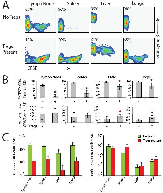
61 50
62 FIGURE 2-4. CD4 + CD25 T cells enhance spontaneous LIP of CD8 T cells. Approximately 2.5 x 10 5 naive CD4 + CD25 T cells were transferred into RAG / recipients on day 7. Approximately 1 x 10 5 OT-1 RAG / or naive polyclonal CD8 T cells were CFSE-labeled and injected into the same recipients on day 0 (or into RAG / mice that had not received CD4 + CD25 cells on day 7). Lymph node cells were harvested on day 10 and stained for granzyme B, and analyzed by FACS. A, Representative CFSE profiles of responding CD8 T cells in the lymph nodes from individual animals. Histograms are shown for total (upper panels) and only the CFSE + (lower panels) responder CD8 T cells. Solid line histograms show CFSE content of responders in RAG / animals in the absence of CD4 T cells, and stippled line histograms show CFSE content of responders in RAG / animals in the presence of CD4 T cells. B, Left panel, The CFSE content of CD8 T cells undergoing homeostatic proliferation (CFSE + cells), represented by the MFI, in the presence or absence of CD4 T cells. Center panel, The absolute number of total CD8 T cells in the presence or absence of CD4 T cells. Right panel, The fraction of CD8 responders undergoing spontaneous proliferation (CFSE ) (gray bar), and the subfraction of these that express granzyme B (black bar). Each bar represents the mean ± SD of three animals. Statistical significance was determined using the two-tailed Student t test comparing groups with pretransfer of CD4 T cells with the control groups without pretransfer of CD4 T cells. Stars represent a statistical significance with p <
63 52
64 FIGURE 2-5. Tregs from young wild-type and IL-10 / mice are phenotypically similar. A, FACS analysis of suspensions of pooled lymph nodes and spleens from four wild-type and four IL-10 / mice before and after purification by positive selection with anti-cd25 mab (PC61). Numbers in plots represent gated cells as a percent of total. B, Histograms show surface staining for CD69, GITR, and intracellular staining for CTLA-4 in wild-type (blue lines) and IL-10 / (red lines) Tregs isolated as described in A, and Foxp3 CD4 T cells (shaded). C, Approximately 6 x 10 5 wild-type or IL-10 / CD25 + cells were CFSE-labeled and injected into RAG / recipients on day 0. Lymph nodes were harvested on day 7 and analyzed by FACS. Histograms show representative CFSE profiles of wild-type (blue histograms) and IL-10 / (red histograms) Tregs transferred into RAG / recipients. The bar graph represents absolute numbers of CD4 + Foxp3 + cells in the lymph nodes (pooled axial, brachial, inguinal, and mesenteric) of RAG / animals that received either wild-type or IL-10 / CD25 + cells. 53
65 54
66 FIGURE 2-6. Suppression of CD8 T cell spontaneous LIP by Tregs is not IL-10 dependent. Approximately 2 x 10 6 wild-type or IL-10 / CD25 + cells were transferred into RAG / recipients on day 7. Approximately 3 x 10 6 naive polyclonal CD8 T cells were CFSE-labeled and injected on day 0 into the same recipients (or into RAG / mice that had not received Treg cells on day 7). Tissues were harvested on day 10 and analyzed by FACS. A, Representative CFSE profiles of CD8 responder T cells in indicated groups of mice and tissues. The numbers are percentages of responder CD8 T cells undergoing spontaneous LIP (CFSE cells). B, Total numbers of CD8 responders recovered from different tissues in indicated groups of mice. Each bar shows the average of four animals pooled from two separate experiments. Stars represent a statistical significance with p < 0.05 by Student s two-tailed t test when comparing the number of cells isolated from tissues of animals receiving a pretransfer of Tregs with those from animals without pretransfer of Tregs. 55
67 56
68 FIGURE 2-7. Suppression of CD4 T cell spontaneous LIP by Tregs is not IL-10 dependent. Approximately 5 x 10 5 wild-type or IL-10 / CD25 + cells were transferred into RAG / recipients on day 7. Approximately 2.5 x 10 6 naive polyclonal CD4 T cells were CFSE-labeled and injected on day 0 into the same recipients (or into RAG / mice that had not received Treg cells on day 7). Lymph nodes were harvested on day 7 and analyzed by FACS. A, Representative CSFE profiles of responding CD4 T cells in the lymph nodes. Solid line histograms represent CFSE content of CD4 responders in the absence of Tregs. Stippled line histograms represent CFSE content of CD4 responders in the presence of Tregs. B, Percentages of CFSE responder CD4 T cells in indicated groups of mice, as well as the CFSE content in the CFSE + CD4 T cell responders represented by the MFI. C, Relative numbers of indicated CD4 T cells responders in different groups of mice. Each bar represents the mean ± SD of three animals. Statistical significance was determined using the two-tailed Student t test. One star represents a statistical significance with p < 0.05; Two stars represent a statistical significance with p < Skin-draining lymph nodes (LN) include axial, brachial, cervical, and inguinal. 57
69 58
70 FIGURE 2-8. IL-10-deficient Tregs cannot completely prevent colitis caused by adoptive transfer of CD4 T cells into RAG / mice. Approximately 5 x 10 5 wild-type or IL-10 / CD25 + cells were transferred into RAG / recipients on day 7. Approximately 1 x 10 5 naive polyclonal CD4 T cells were injected on day 0 into the same recipients (or into RAG / mice that had not received Treg cells on day 7). Mice were sacrificed when the group of RAG / mice without pretransfer of Tregs was showing obvious clinical signs of disease, including diarrhea and hunched posture. Three separate experiments were harvested 6, 8, or 9 wk after transfer and the results were pooled here. Histologic scores were determined on H&E sections of distal colons blinded to the identity of the experimental groups. A, Mean changes ± SD in weight of the animals and histologic scores ± SD in indicated groups of mice. The pooled experiments together totaled 13 mice per group. Statistical significance was determined using the two-tailed Student t test. B, Representative H&E sections from each of the experimental groups. 59
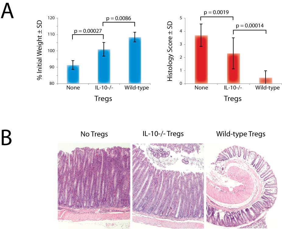
71 60

72 FIGURE 2-9. IL-10-deficient Tregs are impaired in their ability to suppress effector CD4 T cells in the colon. Approximately 5 x 10 5 wild-type or IL-10 / CD4 + CD25 + T cells were transferred into RAG / recipients on day 7. Approximately 1 x 10 5 naive polyclonal CD4 T cells were injected on day 0 into the same recipients (or into RAG / mice that had not received Treg cells on day 7). Mice were sacrificed at 9 wk, and cells isolated from the lymph nodes and colons were stimulated with PMA/ionomycin in vitro, stained for cytokine expression, and subjected to FACS analysis. A, Representative FACS plots of gated CD4 T cells in the mesenteric lymph nodes of individual animals. Percent numbers show fractions of total CD4 T cells expressing indicated cytokines. B, Number of effector cytokine-producing CD4 T cells in mesenteric lymph nodes and colons. Relative CD4 T cell numbers were calculated in comparison to the group that did not receive a pretransfer of Tregs. Each bar represents the mean ± SD of at least six animals. Statistical significance was determined using the two-tailed Student t test. The black and red stars indicate statistical significance with p < 0.05 and p < 0.01, respectively. WT, Wild type. 61
73 62
74 FIGURE When co-transferred, large numbers of Tregs are required to suppress infiltration of effector CD4 T cells in the colon. In one set of mice, indicated numbers of wild-type or IL-10 / CD4 + CD25 + T cells were transferred into RAG / recipients on day 7, then 1 x 10 5 naive polyclonal CD4 T cells were injected into the same recipients on day 0. In a second set of mice, indicated numbers of wild-type or IL-10 / CD4 + CD25 + T cells were co-transferred with 1 x 10 5 naive polyclonal CD4 T cells on day 0. Control RAG / mice received only 1 x 10 5 naive CD4 T cell responders on day 0. Mice were sacrificed at 12 wk following adoptive transfer of CD4 T cell responders. A, Mean changes in weight of the animals and histologic scores for colon sections for the indicated groups of mice. Histologic scores were determined on H&E sections of distal colons blinded to the identity of the experimental groups. Each bar represents the mean ± SEM of four animals. B, Relative CD4 responder T cell and Treg numbers were calculated based on total lymphocyte numbers. WT, Wild type; MLN, mesenteric lymph nodes. 63
75 64
76 FIGURE Model of Treg-mediated control of responder T cells in lymphopenia. The top half of the figure illustrates control of LIP by Tregs within the secondary lymphoid tissues. Responder T cells and Tregs are shown to exist in different compartments, although it is possible that Tregs (shaded circles) and responder T cells (black circles) undergoing spontaneous LIP compete for some common resources. The presence of Tregs limits spontaneous LIP, which in turn limits the number of potentially autoreactive T cells capable of causing autoimmunity. This function of Tregs is independent of their ability to produce IL-10. The bottom half of the figure illustrates the second phase of Treg-mediated suppression, which happens in the periphery. Some effector T cells generated in the course of spontaneous LIP leave the lymphoid tissues. However, their local activity and ability to trigger inflammation in the periphery is inhibited by Tregs in an IL-10-dependent manner. Finally, we speculate at this time that T cells undergoing spontaneous proliferation compete for resources with T cells undergoing homeostatic proliferation (white circles). Therefore, uncontrolled spontaneous LIP may limit homeostatic proliferation and potentially impair overall TCR diversity during immune reconstitution. 65
77 66
78 Chapter 3 CD4+CD25+Foxp3+ regulatory T cells maintain optimal responsiveness of the Foxp3- T cell population during reconstitution from lymphopenia 67
79 Abstract T cell diversity is a key feature of the adaptive immune system. Lymphopenia-induced proliferation (LIP) can trigger oligoclonal expansion of T cells reacting to foreign or self-antigens. This can result in loss of T cell diversity and compromise immune fitness that can be achieved following reconstitution by LIP. We have demonstrated that CD4+CD25+Fopx3+ regulatory T cells (Tregs) selectively inhibit the spontaneous (fast) form of LIP, which is associated with differentiation into memory/effector-like cells, and is likely to contain the most reactive T cell clones. Selective suppression of these clones is likely to leave more resources for T cells undergoing the slower homeostatic form of LIP. We hypothesize, therefore, that one of the functions of Tregs is to maintain the T cell diversity and overall immune fitness during reconstitution from lymphopenia. Structural data generated using flow cytometric analysis of TCR Vβ usage and PCR-based TCR Jβ spectratyping suggest Tregs effectively prevent the emergence of 'holes' in the T cell repertoire following LIP. Furthermore, using the Listeria monocytogenes infection model and magnetic bead enrichment with specific MHC class I and II tetramers, we demonstrate that the presence of Tregs during immune reconstitution allows for more accumulation of pathogen-associated antigenspecific T cells in secondary lymphoid tissues following clearance of the bacteria. This result may otherwise seem paradoxical as Tregs are typically thought of as generalized suppressors of the immune system. We believe these results provide some insight into the central role of Tregs in the immune system, vaccine responsiveness, pathogenesis of autoimmunity, and immune senescence. 68
80 Introduction Maintenance of peripheral T cell homeostasis is a critical feature of the adaptive immune system (82). Strict control of T cell homeostasis ensures adequate peripheral cell numbers, T cell receptor (TCR) diversity, responsiveness to foreign antigens, and self-tolerance. If a state of peripheral T cell deficiency is created, the residual T cells are driven to undergo proliferation by a process termed lymphopenia-induced proliferation (LIP) to reconstitute the optimal T cell numbers. LIP can be observed in a number of physiological and pathological situations (81). Lymphopenia is physiologic during the prenatal and neonatal periods, and LIP may contribute to generation of sufficient numbers of T cells with a memory phenotype (79). In addition, as the thymus involutes with increasing age, LIP assumes greater importance in maintaining T cell population size (80). Transient lymphopenia can accompany virtually any viral infection, and some viral infections, e.g., HIV, lead to chronic and progressive lymphopenia. In addition, there are multiple iatrogenic causes of lymphopenia, including cytodepletion by radiation, chemotherapy, and depleting antibodies. Notably, lymphopenia can be a trigger to autoimmunity, which is at least in part explained by T cell resistance to tolerance induction during LIP (83). Furthermore, as individual T cell clones expand at different rates during LIP, the emerging T cell population risks loss of TCR diversity and diminished immune fitness. However, although lymphopenic states occur multiple times during lifetimes of all individuals, autoimmunity and immune deficiency do not manifest in most people. Therefore, there must be mechanisms that help to maintain optimal immunologic tolerance and immune fitness during immune reconstitution. 69
81 Regulatory CD4 + CD25 + Foxp3 + T cells (Tregs) constitute 5-15% of peripheral CD4 T cells in healthy, adult mice and humans (40), and are critical in the maintenance of immunologic tolerance and peripheral T cell homeostasis. These cells are able to suppress a range of immunologic responses in vitro and in vivo. Their discovery was aided by a number of animal models of autoimmunity associated with lymphopenia (neonatal thymectomy, the SCID adoptive transfer colitis model, and autoimmune thyroiditis) (79, 97, 100). However, early studies failed to demonstrate their ability to suppress LIP. Since then at least two major forms of LIP were recognized homeostatic and spontaneous (89). Homeostatic LIP is driven primarily by cytokines such as IL-7 and IL-15, and is generally slow and steady. Spontaneous LIP is burst-like and is likely driven by cognate antigen stimulation, and is the most likely source of pathogenic T cells with higher TCR affinity for self- and commensal flora antigens. We have shown previously that while Tregs do not suppress homeostatic LIP, they do suppress the spontaneous form of LIP (160). This likely contributes to their protective function against development of autoimmunity during LIP. We also thought it was possible that selective suppression of spontaneous LIP could benefit T cells with lower TCR affinity for self antigens by allowing greater access to cytokine resources important for their survival and homeostatic LIP. Therefore, we predicted that presence of Tregs during immune reconstitution by LIP should lead to preservation of greater diversity of TCR repertoire, and consequently, better immune fitness. Here, we characterized the TCR repertoire in T cell populations expanded by LIP in the presence or absence of Tregs following adoptive transfer into recombinase activating gene-deficient (RAG-/-) or TCRα-/- recipients. Using TCR Vβ CDR3 70
82 spectratyping we demonstrated significantly enhanced preservation of structural TCR diversity in the presence of Tregs during LIP. The reduction of TCR diversity in absence of Tregs resulted in development of functional holes in the repertoire that were revealed by enumerating antigen-specific T cells following infection with Listeria monocytogenes. Thus, markedly greater numbers of Listeria-specific responding T cells were found if Tregs were present than if they were absent during LIP. These findings suggest that Tregs preserve TCR diversity and immune responsiveness during immune reconstitution from lymphopenia. This positive effect of Tregs on immunity contrasts with their generally accepted immunosuppressive function. Materials and Methods Mice C57BL6 (B6) and CD45.1 congenic mice were purchased from the National Cancer Institute (Frederick, MD). Some B6 mice used were wild-type littermates to IL-10 -/- originally obtained from The Jackson Laboratory (Bar Harbor, ME) and CTLA-4 +/- mice, which were a generous gift from Dr. J. Allison (Memorial Sloan-Kettering Cancer Center, New York, NY), respectively. Recombinase gene 1-deficient (RAG-1 -/- ), T-cell receptor alpha chain-deficient (TCRα -/- ), and Thy1.1 congenic on the B6 background were obtained from Jackson Labs. B6 RAG-1 -/- were bred onto the CD45.1 congenic background in our facility. Thy1.1 BALB/c mice were originally provided by Dr. L. Turka (University of Pennsylvania, Philadelphia, PA). RAG-2 -/- BALB/c mice were purchased from NCI and bred onto the congenic Thy1.1 background in our facility. All 71
83 mice used were generally 4-20 weeks of age. All animals were maintained in a specific pathogen-free facility in microisolator cages with filtered air according to the National Institutes of Health guidelines. Cell Isolation, labeling, adoptive transfer, and antibody-mediated in vivo depletion Donor T cells were collected from secondary lymphoid tissues (axillary, brachial, cervical, mesenteric, and inguinal lymph nodes, and spleen). CD44 low CD4, CD8, and bulk (both CD4 and CD8) T cells were purified in two stages first, CD8 or CD4 T cells were prepared by negative selection against CD8 or CD4, MHC class II, CD11b, B220, CD25, and in some cases CD103 (all Abs labeled with FITC) using anti-fitc BioMag particles (Polysciences, PA), followed by depletion of CD44 high cells (B6 only) using magnetic microbeads (Miltenyi Biotec, CA), as previously described (123). Briefly, the purified T cells were suspended in labeling buffer (2% FBS in PBS), incubated with µg anti-cd44-fitc (ebioscience, CA) per 10 6 cells for 20 minutes, washed, and labeled with anti-fitc magnetic microbeads. The negative fraction was collected following Miltenyi Biotec magnetic column separation. CD25 + T cells were prepared using positive selection with anti-cd25 biotinylated mab, PC61, and streptavidin-labeled magnetic microbeads (Miltenyi). Purified cells were transferred to RAG -/- or TCRα -/- hosts via intravenous (i.v.) tail injection. In some cases cells were labeled with 7uM CFSE or SNARF-1 prior to transfer. Regulatory T cells were depleted in vivo using a single i.p. injection of µg anti-thy1.1 mab (clone 1A14). 72
84 TCR Vβ and Jβ spectratyping Regulatory and conventional T cells collected from secondary lymphoid tissues were MACS sorted by positive selection for Thy1.1, as described above. Total RNA was extracted from positive (Thy1.1+/Treg) and negative (Thy1.1-/Tconv) fractions with an RNeasy Mini Kit (Qiagen, CA), and first-strand cdna was generated from total RNA with Oligo(dT) 20 and SuperScript III reverse transcriptase (Invitrogen, CA). Each cdna sample was checked for integrity and for detectable TCR Cβ chain using β actin and Cβ-specific primers (5 Cβ1A; 3 Cβ3C) under PCR amplification conditions described below (161). The CDR3 size distribution PCR assay was performed as described (23) (control and spectratyping primers listed in supplemental Table 3-1). Briefly, each TCR Vβ was amplified with Vβ8.1, 8.3 and/or Vβ10-specific sense primers and a Cβ antisense primer (162). For generation of Vβ spectratypes, the Cβ antisense primer was labeled with 6-fluorescein phosphoramidite (6-FAM) on the 5 end. One ul of the previous synthesis, corresponding to the reverse transcription of ug of total RNA was used in the amplification. cdna was added to tubes already containing a mixture of Taq Master Mix (Qiagen), DNAse/RNAse-free water, Vβspecific and Cβ-specific primers (10-20uM) and amplification was performed as follows: thirty cycles of 95 o C for 1 min/52 o C for 1 min/72 o C for 1 min, followed by 5 min at 72 o C. A second, nested amplification for specific Jβ sequences was performed using one ul of Vβ-specific product. Cycling conditions remained the same, with the exceptions of Master Mix containing 5 6-FAM labeled Jβ-specific sense primers and twenty cycles of amplification. The size distribution of each fluorescent PCR product was determined by electrophoresis on an Applied Biosystems 3130xl Genetic Analyzer 73
85 (Foster City, CA), and data were analyzed using Genoprofiler 2.1 (163, 164). TCR Spectratyping Complexity Scores To assess each individual sample diversity profile as normal or limited, we used a previously reported complexity scoring system (165); that is, a complexity score = (sum of all the peak areas/sum of the major peak areas) x (number of the major peaks). Major peaks were defined as those on the spectratype histogram whose area was at least 10% of the sum of all the peak areas. Bacterial infections and peptide:mhc tetramer-based enrichment protocol Wild-type B6 mice were immunized intravenously with approximately 1x10 7 CFU of a ActA strain of L. monocytogenes transformed by a plasmid containing ovalbumin and an I-A b -specific mutant epitope of I-Eα ( 2W1S - courtesy of Dr. SingSing Way at the U. of Minnesota) (166). Bacteria were grown in LB media with 24 ug/ml of chloramphenicol to an absorbance at 600 nm of about 0.1. The actual number of bacteria injected was confirmed by dilution and growth on LB agar plates containing chloramphenicol. K b -OVA and I-A b -2W1S tetramers, respectively, were produced in the laboratories of Drs. Stephen Jameson and Marc Jenkins, respectively, as previously described (26, 167). Lymph nodes and spleens were harvested together and processed for K b -OVA and I-A b -2W1S double-tetramer enrichment according to the protocol established by Moon et al. (26, 168). 74
86 FACS analysis Mice were sacrificed with CO 2 before spleen and lymph nodes were removed. All secondary lymphoid tissues were disrupted by mashing with a syringe. In some cases, complete media (10% FBS) was supplemented with 10 µg ml -1 brefeldin A (Sigma, MO) to prevent lymphokine secretion. Fixation and intracellular staining for cytokines was done following 2-3 hours in vivo challenge with approximately 5x10 7 CFU LM- 2W1S-OVA. All antibodies used for cell surface staining, negative selection, and tetramer enrichment were purchased from ebioscience, with the exceptions of anti- B220 and anti-cd11c (Biolegend, CA), anti-cd8a (Invitrogen), and anti-cd4 singlestain controls (BD Biosciences, CA). Specific T cell subsets were identified using fluorochrome-labeled antibodies against a panel of TCR Vβ receptors (15 in total), CD3e, CD4, CD8, CD44, and congenic markers such as anti-thy1.1/thy1.2 and anti- CD45.1/CD45.2. Anti-IFN-γ antibodies were purchased from ebioscience. Staining for Foxp3 was done using an ebioscience kit and instructions provided by the manufacturer. Absolute numbers of T cells were calculated using PKH reference beads (Sigma-Aldrich). Relative numbers of T cells of specific subtype were calculated in comparison to the number of total T cells in the group that did not receive Tregs. All flow cytometry data was acquired on a FACS Calibur, an LSR, or an LSRII (BD Immunocytometry Systems, CA) and analyzed with FlowJo software (Tree Star, OR). Statistical analysis All error bars represent the standard error of the mean (SEM), unless noted otherwise. For data in Figures 2, 3, and 5, a two-tailed, paired Student's t test was used for 75
87 assessment of the differences between groups. Prism (Graphpad Software, La Jolla, CA) was used for graphs and statistical analysis. Differences were considered to be significant when the p value was less than Results T cell adoptive transfer system for studies of the TCR repertoire during LIP. Tregs are well established in their ability to prevent and treat immunopathology arising in the course of immune reconstitution from lymphopenia by suppression of emergence and reactivity of pathogenic T cells. Here, we set out to test the idea that another function of Tregs during immune reconstitution is preservation of immune fitness by maintaining optimal structural TCR diversity of the residual T cell population. In order to establish LIP we used the RAG-/- adoptive transfer model. First, we titrated numbers of bulk, CD44 lo CD25 - T cells transferred on day 0 and measured the number of T cells expressing each TCR Vβ chain on days 7 and 30 (Fig. 3-1). Similarly to what has been reported previously (91), we found that the ultimate number of T cells in the lymphoid periphery is independent of the initial input cell number. The T cell population size at 30 days was the same for inputs of T cells, and it was still rising following adoptive transfer of 10 4 T cells. We decided to exclude T cell transfers from future experiments because any potential TCR repertoire that could emerge from these would by definition be severely limited. Furthermore, adoptive transfer of very low numbers of T cells is associated with development of immunopathology (85, 91). We also excluded 10 7 T cell transfers because these filled the T cell niche within a mere 7 days, and we didn t think that 76
88 significant distortion of the TCR repertoire could be established to allow testing of our hypothesis. We chose adoptive transfer of 10 6 T cells as optimal for our experiments because more than one week was required to reach the T cell population plateau, and only mild immunopathology manifesting as dermatitis was noted with this input number after one month following adoptive transfer. Colitis was not detected clinically or by histopathology, in agreement with previous reports (85). In addition, the T cell populations emerging from 10 6 T cell transfers were indistinguishable from the T cell populations derived from 10 7 T cell transfers at least by rough diversity measures such as Vβ repertoire usage. In order to test the effects of Tregs on the responder T cell population during LIP, we constructed the following experimental system (Fig. 3-2A). First, 10 6 CD25+Tregs are purified from Thy1.1 BALB/c mice and transferred into RAG-/- recipients and allowed to expand for 7-10 days, after which 10 6 Thy1.2 CD44 lo CD25 - responder T cells are adoptively transferred. The lymphoid tissues were harvested for T cell population analysis 30 days after the responder T cell transfer. We used this protocol previously to show that Tregs fill their own T cell niche and selectively suppress only the spontaneous form of LIP by the responder T cells. The TCR Vβ usage of the reconstituted population was similar to the initial population regardless of the presence or absence of Tregs in the host (Fig. 3-2B). Although simple and robust, flow cytometric measurements of TCR Vβ usage are relatively insensitive, in that they do not allow determination of clonal dominance or restriction within populations of cells expressing the same Vβ chain. In order to probe the TCR repertoire further, we generated spectratypes at the level of a particular Vβ 77
89 chain, e.g., Vβ10 shown in Fig. 3-2C. This assay did suggest that CD4 T cells undergoing LIP in absence of Tregs do experience contraction in TCR diversity compared to that measured before LIP or after LIP in presence of Tregs. However, we could not detect decreased TCR diversity when CD8 T cells or unfractionated CD25- responder T cells were allowed to undergo LIP under the same conditions. Presence of Tregs during immune reconstitution protects structural diversity of the responder T cell population. Although Vβ spectratyping does reflect some level of structural TCR diversity within a T cell population, it isn t particularly sensitive because it is not focused on the hypervariable regions of the TCR. In contrast, TCR Vβ-Jβ spectratyping is based on measurement of complementarity-determining region 3 (CDR3) size distribution within a population of T cells, and provides a more accurate reflection of its clonality. We performed TCR Vβ10 Jβ spectratyping of CD25- responder T cells that had undergone LIP in the presence or absence of Tregs. We measured spectratypes of CD4 T cells, CD8 T cells, and unfractionated T cells (bulk) under these conditions. In all cases the diversity of the TCR repertoire became severely constricted if Tregs were absent during LIP (Fig. 3-3A). Visual inspection of spectratyping histograms from several TCR Vβ families also allowed us to visualize actual holes in the reconstituted repertoire, as defined by specific Jβ chain representation below our spectratyping limit of detection (approximately 10,000 cells) (Fig. 3-3B). These holes appeared only if Tregs were absent during LIP. In contrast, relative preservation of TCR complexity was noted if Tregs were present during LIP (Fig. 3-3B). 78
90 Presence of Tregs during immune reconstitution prevents emergence of repertoire holes that can limit antigen-specific responsiveness to microbial pathogens. The value of a diverse, polyclonal T cell repertoire is well appreciated in the context of infection by a microbial pathogen. Therefore, we wished to test the biological significance of LIP-associated TCR repertoire constriction in a model of infectious disease such as Listeriosis (infection with the pathogenic bacteria Listeria monocytogenes). Extensive TCR sequence analysis by Casrouge and colleagues demonstrated that in steady state a mouse spleen contains about 2 x 10 6 distinct naïve αβ TCR clones. Each T cell clone might be able to recognize more than one antigen, and it has been estimated that lymphoid tissues of a mouse contain distinct T cell clones in the naïve T cell repertoire able to recognize a specific peptide/mhc epitope (26, 169). Work published by Moon et al. using tetramer-based enrichment of epitope-specific T cells estimated the number of IA b -2W1S-specific CD4 + cells in a naïve C57BL/6 mouse to be 200 (26). Using the same basic methodology, the naïve (precursor) frequency of K b -OVA-specific CD8 + cells was found by Obar et al. to be approximately 100 cells per mouse (28). One would, therefore, expect that adoptive transfer of 1 x 10 6 naïve donor T cells into RAG-/- or TCRα-/- recipients should introduce approximately 2-4 T cells with specificity for K b -OVA and IA b -2W1S, respectively. Our hypothesis, however, predicts that the fate of these naïve precursor cells during LIP may be different depending on the presence or absence of Tregs. Specifically, we thought that since these T cells have foreign antigen specificity, they would be less likely to undergo 79
91 spontaneous LIP, and would, in fact, benefit from suppression of spontaneous LIP by Tregs. In order to test the hypothesis, we reconstituted RAG-/- or TCRα-/- mice by adoptive transfer of Thy1.2 CD25-CD44 low CD4 T cells (Fig. 3-4A). Some of the recipient animals were pre-transferred with Thy1.1 CD25+ Tregs, and some were not. After 30 days of reconstitution the mice were infected with attenuated L. monocytogenes bacteria expressing 2W1S and OVA epitopes. The variable we wanted to test was presence or absence of Tregs during LIP. Obviously, presence or absence of Tregs during the infection could also alter the T cell response. Therefore, we added two additional experimental groups. Some of the mice that received Tregs before the adoptive transfer of naïve responder T cells were treated with depleting anti-thy1.1 antibody several days before the infection. In this group transferred Tregs were present only during immune reconstitution, but not the infection. Another group of mice was adoptively transferred Tregs only after immune reconstitution was complete so that Tregs would be absent during LIP, but present during the infection (Fig. 3-4A). We found that the total numbers of responder T cells in the lymphoid tissues after LIP and L. monocytogenes infection were identical. However, the percentages and absolute numbers (Fig. 3-5) of IA b -2W1S-specific CD4 T cells and K b -OVA-specific CD8 T cells, enumerated using magnetic enrichment and flow cytometry, revealed that presence of Tregs during LIP significantly enhanced expansion of antigen-specific T cells. In fact, the percentage of antigen-specific T cells in reconstituted RAG-/- mice that had Tregs present during LIP was similar to those seen in wild-type animals, although the absolute numbers were somewhat lower reflective of the lower total T cell 80
92 populations in reconstituted RAG-/- animals. Depletion of Tregs by anti-thy1.1 mab after T cell reconstitution was completed did not impact expansion of Listeria-specific responder T cells during the infection. In contrast, RAG-/- mice allowed to reconstitute in absence of Tregs had very few antigen-specific T cells, and there was no significant difference seen between animals that never received Tregs or received Tregs after LIP was already completed. Virtually all measured pathogen-specific T cells derived from the naïve responder T cell population and not the Thy1.1+ Tregs. This observation is consistent with the results reported by Ertelt et al. where infection by the same attenuated strain of Listeria used in our experiments was not seen to cause expansion of Listeria-specific Foxp3+ T cells. Presence of Tregs during immune reconstitution preserves functional responsiveness of the T cell population. Production of interferon-g by adaptive and innate immune systems contributes significantly to clearance of intracellular pathogens such as L. monocytogenes. Therefore, it would be reasonable to presume that numbers of Listeria-specific CD4 T cells that can produce IFN-γ relate to the immune fitness of an individual animal challenged with this infection. In order to examine this question, we repeated some experiments described above using TCRα-/- recipients (Supplemental Fig. 3-1). Once again, animals were reconstituted with conventional naïve T cells in the presence or absence of Tregs. Some animals that received Tregs before T cell reconstitution were treated with the depleting antibody following completion of LIP. Some animals that did not have Tregs during the period of LIP, were adoptively transferred with Tregs 81
93 following LIP completion. IFN-γ production was stimulated in vivo by intravenous injection of 5 x 10 7 CFU of live ActA - LM-2W1S-OVA 2-3 hours prior to harvest of the lymphoid tissues. We found that if Tregs were present during LIP, but not after, the reconstituted animals had more antigen-specific CD4 T cells overall, and more antigenspecific CD4 T cells capable of producing IFN-γ. Finally, we tested whether Listeria-specific effector T cells generated within T cell populations that emerged in the course of immune reconstitution in the presence or absence of Tregs were capable of expansion when re-challenged with the pathogen (Supplemental Fig. 3-4). RAG-/- mice were repopulated with naïve Thy1.1 T cells in the presence or absence of Thy1.2 Tregs. After 30 days of reconstitution all animals were infected with 10 7 ActA - LM-2W1S-OVA bacteria. 10 days after infection, which is the peak of expansion by antigen-specific T cells in this system, lymphoid tissues of the animals were harvested and T cells that derived from the conventional T cell inoculum (Thy1.2 - T cells in this experiment) were purified by negative selection. New wild-type recipients received 3 x 10 6 of these purified T cells, and were challenged with 10 7 ActA - LM-2W1S-OVA bacteria. Some wild-type recipients did not receive T cells from reconstituted RAG-/- animals and were used as controls. All mice were sacrificed on day 5 after the infection and lymphoid tissues were harvested for analysis. The results were dramatic. If Tregs were present during LIP, we could detect significant numbers of Thy1.1 CD4 T cells, most of which were specific for the 2W1S epitope. If Tregs were absent during LIP the numbers of 2W1S-specific CD4 T cells were indistinguishable from the numbers in the pre-immune repertoire. Similar general pattern was seen for the K b -OVA-specific CD8 T cells. 82
94 Discussion Discovery and characterization of CD25+Foxp3+ T cells in recent years has resurrected the concept of suppressor T cells playing a critical role in the maintenance of immunologic tolerance. These cells were termed regulatory to be more encompassing of their potential functions, although they were shown to be truly suppressive to virtually every immunologic response studied. Multiple reasons can be cited to explain the abandonment of the suppressor T cell paradigm in 1980s, but one of them is its apparent conceptual inferiority in comparison to antigen-specific recessive mechanisms of tolerance such as clonal anergy and deletion. Today the existence of regulatory/suppressor T cells is no longer in question since we have physical markers that identify them, and although our mechanistic understanding for their suppressive activity remain incompletely understood, numerous potential mechanisms have been described. Nevertheless, the same theoretical question can be raised why are suppressor T cells needed in our immune system? The idea of suppressor T cells may seem more appealing if their antigen specificity compliments the antigen specificity of purely recessive mechanisms of tolerance. In fact, the repertoire of Tregs appears to be at least as diverse as that of conventional T cells, but it is also largely different. Most evidence suggests that the TCRs of thymic-derived Tregs have higher affinities for self-antigens than conventional T cells (55). Therefore, one could speculate that since Tregs need to be activated by their TCR to be functional, they may have the capacity to preferentially dampen immune responses in the settings where greater amounts of self-antigen are being 83
95 presented. Certainly, there is a great deal of clinical interest in antigen-specific Treg induction because antigen specificity can increase their suppressive potency by targeting their activity. The function of suppressor T cells in infectious disease may be more nuanced. Tregs can inhibit or delay pathogen clearance, which at first glance may seem detrimental to the host. However, in some models, such as leishmaniasis and schistosomiasis, antigen persistence ensured by Tregs contributes to maintenance of immunity to reinfection. In other models, such as HSV-1 infection, Tregs were shown to inhibit immunopathology caused by inflammatory response that would otherwise severely disable the host (170). These are some examples that illustrate inadequacy of a simplistic view of the immune response, where stronger reaction is assumed to be better. In fact, in these cases of chronic infections Tregs may prevent winning a battle with a pathogen, but at the same time help to win the war. Of course, the paradigm of war does not apply well to most of the activity of the immune system. The majority of the immune effort is concentrated at the mucosal surfaces, where it helps to maintain homeostasis with the commensal microbial flora and aid the physical barrier to exclude pathogen entry. That is one of the main functions of the secreted IgA, for example, which requires transforming growth factor-β (TGF-β) for class switching, one of the cytokines elaborated by Tregs (171). At the outset we were intrigued by the differential ability of Tregs to inhibit spontaneous versus homeostatic forms of LIP in our experiments. This finding suggested that Tregs could play another positive role in the function of the immune system. Specifically, we thought they might optimize the immune fitness of the 84
96 residual T cell population during its recovery from lymphopenia by shaping its TCR repertoire. Lymphopenia is a common occurrence during lifetimes of individuals and peripheral mechanisms of immune reconstitution assume increasing importance with age as the thymus involutes. Our experimental adoptive transfer system models absence of the thymus and allows recovery of the T cell population exclusively by LIP. We specifically wanted to start out with only minimally restricted potential TCR repertoire in order to both mimic realistic clinical scenarios, e.g., recovery of the T cell population in AIDS patients treated with highly active anti-retroviral therapy (HAART), and provide an experimental window to measure significant differences (172). A normal mouse contains about 2 x 10 6 T cell clones within its naïve TCR repertoire, and a starting population significantly below that would ensure severely restricted responsiveness to potential foreign antigens. In contrast, a starting population significant above that would limit LIP because there would be little lymphopenia. We employed p-mhc tetramers to probe the responsiveness of T cell populations to two foreign peptides, OVA and 2W1S, expressed within an infectious pathogen, L. monocytogenes. Enumeration of K b -OVA- and IA b -2W1S-specific T cells in the naïve repertoire reported previously and reproduced in our own work suggested that adoptive transfer of 1 x 10 6 T cells would result in transfer of about 2-4 T cells per mouse specific for these antigens. In fact, it has been shown that a single precursor T cells is capable of massive clonal expansion and functional subset diversification following immunization (173). Therefore, we expected to detect significant antigen-specific T cell expansion in all our experimental conditions in absence of significant distortion of the T cell repertoire during the period of immune reconstitution. 85
97 We found that the greatest numbers of responder foreign antigen-specific T cells recovered after infection were seen only in mice that had Tregs during the period of LIP. The simplest explanation for this finding is that some antigen-specific precursors from the naïve T cell inoculum did not survive the immune reconstitution period in absence of Tregs. In fact, in some experiments when Tregs were absent during LIP we couldn t detect any antigen-specific responder T cells at all following infection, and in others they were markedly decreased in numbers. This idea is also supported by the results of characterization of the TCR repertoire obtained by TCR Vβ-Jβ spectratyping, which demonstrated that absence of Tregs during immune reconstitution correlated with development of marked loss of structural TCR diversity. One would predict that the diversity of the few recovered antigen-specific responder T cells from populations established in the absence of Tregs would be extremely constricted and likely monoclonal. In fact, we have done several experiments using flow cytometric analysis Vβ chain usage by antigen-specific responders, and these were compatible with that prediction. Specifically, the Vβ chain usage of antigen-specific 2W1S-specific responders in mice reconstituted in presence of Tregs was comparable to the repertoire of 2W1S-specific responders in wild-type mice. The Vβ chain repertoire of 2W1Sspecific responders in mice reconstituted in absence of Tregs appeared distorted. However, because of the small numbers of antigen-specific T cells in these mice and their complete absence in some experiments, the total number of mice necessary to characterize the Vβ usage by these cells was prohibitive. It should be interesting to focus future studies on the characteristics of the residual T cells that persist within the T cell population established by LIP in absence of Tregs. We might anticipate, for 86
98 example, that these cells have greater affinity for self-antigens that provide them with more tonic survival signals. Such cells might not necessarily have highest affinity for foreign antigens that the organism may encounter. Our results indicate that the beneficial role of Tregs on the antigen-specific T cell response is exerted during the period of LIP (Fig. 3-6). Tregs adoptively transferred after completion of immune reconstitution had no beneficial effect. Furthermore, if Tregs were depleted following immune reconstitution, the beneficial effect persisted. It should be noted that Tregs were depleted using an antibody directed against congenic marker, Thy1.1. While that was very effective in depleting Thy1.1+ T cells, early presence of Tregs also appeared to encouraged some emergence of Foxp3+ T cells derived from the naive responder T cell population (Table 3-1). This might represent an example of infectious tolerance, and is consistent with the idea that factors produced by Tregs are somehow needed for Treg induction. However, the effect was relatively small and the numbers of induced Tregs in this group was considerably smaller than that produced by late adoptive transfer of Tregs. In both cases Tregs appearing after completion of LIP did not enhance the diversity of the reconstituted responder T cell population, and thereby its immune fitness. The mechanisms of how Tregs may shape the TCR repertoire during LIP remain unclear at this time. At least several attractive models seem to be reasonable and nonmutually exclusive candidates. We started these studies after documenting that Tregs selectively inhibit the spontaneous form of LIP. Spontaneous LIP likely results from cognate antigen stimulation because it is inhibited by intraclonal competition and requires co-stimulation by B7:CD28 signaling. In contrast, co-stimulatory signals are 87
99 not needed for the homeostatic form of LIP, which is in turn driven mainly by mere tonic TCR signals and cytokines such as IL-7. It is reasonable to speculate that T cells undergoing both forms of LIP do utilize some common resources, e.g., IL-7, which may not be used significantly by Tregs. At the same time, Tregs and T cells undergoing spontaneous LIP may compete for another common resource, e.g., IL-2, which is known to be important for both Tregs and memory CD8 T cells. In this situation, suppression of spontaneous LIP by Tregs will liberate more resources for the rest of the T cells and prevent their extinction during immune reconstitution. Alternatively, Tregs may somehow selectively identify T cells undergoing spontaneous LIP and suppress them by blocking their cell cycle progression or inducing their apoptosis. Once again, more resources will be made available the majority of T cells that are not undergoing spontaneous LIP. Investigation of mechanism awaits future studies, and these phenomena may be in play in emerging clinical protocols. Thus, T cell depletion is commonly used in solid organ transplantation and we anticipate that costimulatory B7 blockade used in that setting may have protective effects on the T cell repertoire during recovery from lymphopenia. In addition, therapeutic adoptive transfer of Tregs is being developed for clinical use in organ transplantation, and their effects on immune fitness would be interesting and important to measure. 88
.")
100 FIGURE 3-1. Filling of the T cell niche in RAG -/- mice through adoptive transfer. A titration of CD44 lo T cell numbers were transferred into C57BL/6 RAG-1 -/- recipients on Day 0 (two recipients per condition). Major lymph nodes (cervical, brachial, axillary, inguinal, and mesenteric) and spleens were harvested on Days 7 and 30, stained for T cell markers (CD4 and CD8) and a panel of antibodies reactive to 14 known mouse TCR Vβ chains, and analyzed by FACS. 89
101 FIGURE 3-2. Assessing T cell diversity post-lip at the level of TCR Vβ chain usage. A. The experiment protocol for assessment of structural T cell diversity post-lip in the presence or absence of Tregs involves two separate measurements from each sample: TCR Vβ surface staining for FACS, and RNA isolation and spectratyping. B. Animals were sacrificed and lymphoid tissue samples were processed on Day 30. Half of each processed sample was treated to cell surface staining with antibodies specific to T cell markers (CD4, CD8, Thy1.2) and FACS analysis. C. The remaining half of each sample was processed for RNA and TCR Vβ spectratyping. Each graphed data bar or point represents an average of 3-4 animals total from 2 independent experiments. Complexity scores were calculated from spectratyping of Vβ 10 and 8.1 (experiment #1), and from Vβ 10 and 8.3 (experiment #2). 90
102 FIGURE 3-3. Tregs prevent emergence of holes in the T cell repertoire during LIP. A. Samples from the experiments described in Fig. 3-2 PCR amplified for Vβ10 expression were spectratyped for CDR3 diversity by further PCR amplification for Jβ1.5. Each bar graph depicts a single, representative animal sample. B. An average complexity score for each Jβ chain was calculated based on spectratyping samples from experiments described above. A mean value representing data from all 12 chains under each treatment condition (LIP +/- Tregs) was used to determine statistical significance from before transfer samples. All conditions were significant with respect to before transfer scores. 91
103 FIGURE 3-4. Tregs preserve the diversity of foreign Ag-reactive T cells during LIP. A. The basic experiment protocol for assessment of functional diversity post-lip in the presence or absence of Tregs involves lymphoid tissue harvest and tetramer basedenrichment for Ag-specific cells post-bacterial infection. B. Plots depict the gating strategy using the control group samples from one representative experiment in which two animals per group were processed, enriched, acquired by FACS, and analyzed together. C. Depicted are samples from one representative experiment. Numbers represent percent gated of total population within each plot. Tr late = Tregs added after LIP, but before infection (Day 28); Tregs were depleted from the third group of animals on Day
104 93
105 94
106 95
107 FIGURE 3-5. The presence of Tregs during LIP ensures responsiveness of foreign Agreactive T cells to bacterial infection. Data represent total and tetramer-enriched T cells from six independent experiments of 1 to 2 animals per group. Note: no K b -OVA specific events were found in LIP hosts from one of six experiments. 96
108 TABLE 3-1. LIP and subsequent bacterial infection do not induce significant peripheral conversion of conventional T cells to Foxp3+. Data represents mean +/- standard deviation of two independent experiments of two animals per group, conducted as described in Fig
109 FIGURE 3-6. Tregs preserve immune fitness of conventional T cells undergoing LIP. The CFSE profile depicted on the left side of this model figure illustrates control of T cell LIP by Tregs within the secondary lymphoid tissues. The presence of Tregs limits spontaneous LIP, which in turn limits the potential for holes in the repertoire to develop and preserves diversity upon reconstitution (colored squares). As depicted on the right side of the figure, the outcome of preserved T cell repertoire fitness is invading pathogen clearance and/or suppression of damaging inflammation and breach of tolerance to commensals. 98
110 SUPPLEMENTAL FIGURE 3-1. Tregs preserve the diversity of foreign Ag-reactive T cells during LIP in TCRα -/- hosts. LIP experiments were conducted as described in Fig. 3-4, with two exceptions. First, TCRα -/- hosts were used in place of RAG -/- hosts for LIP. Second, host mice were challenged with LM as described in the Materials and Methods section for the purpose of intracellular staining for IFN-γ and assessment of functional Ag-specific T cell fitness. A. Each data point in the plots represents the average of 2 animals per group from 2 independent experiments (4 animals total per condition). B. Each set of 2 FACS plots reflects a representative sample from each group (tissue from 2 animals processed, stained, and acquired together). Numbers represent percent gated of total population within each plot. 99

111 SUPPLEMENTAL FIGURE 3-2. Tregs concomitantly transferred with conventional T cells preserve the diversity of conventional CD4 T cell foreign Ag-reactivity during LIP. Depicted are samples from a single experiment conducted as in Fig Numbers represent percent gated of total population within each plot. Highlighted with a black box are groups corresponding to concomitant transfers of 1x10 6 Treg and 1x10 6 Tconv on day
Adaptive immune responses: T cell-mediated immunity
 MICR2209 Adaptive immune responses: T cell-mediated immunity Dr Allison Imrie allison.imrie@uwa.edu.au 1 Synopsis: In this lecture we will discuss the T-cell mediated immune response, how it is activated,
Tolerance, autoimmunity and the pathogenesis of immunemediated inflammatory diseases. Abul K. Abbas UCSF
 Tolerance, autoimmunity and the pathogenesis of immunemediated inflammatory diseases Abul K. Abbas UCSF Balancing lymphocyte activation and control Activation Effector T cells Tolerance Regulatory T cells
Effector mechanisms of cell-mediated immunity: Properties of effector, memory and regulatory T cells
 ICI Basic Immunology course Effector mechanisms of cell-mediated immunity: Properties of effector, memory and regulatory T cells Abul K. Abbas, MD UCSF Stages in the development of T cell responses: induction
The development of T cells in the thymus
 T cells rearrange their receptors in the thymus whereas B cells do so in the bone marrow. The development of T cells in the thymus The lobular/cellular organization of the thymus Immature cells are called
Central tolerance. Mechanisms of Immune Tolerance. Regulation of the T cell response
 Immunoregulation: A balance between activation and suppression that achieves an efficient immune response without damaging the host. Mechanisms of Immune Tolerance ACTIVATION (immunity) SUPPRESSION (tolerance)
Mechanisms of Immune Tolerance
 Immunoregulation: A balance between activation and suppression that achieves an efficient immune response without damaging the host. ACTIVATION (immunity) SUPPRESSION (tolerance) Autoimmunity Immunodeficiency
Immune Regulation and Tolerance
 Immune Regulation and Tolerance Immunoregulation: A balance between activation and suppression of effector cells to achieve an efficient immune response without damaging the host. Activation (immunity)
Lecture outline. Immunological tolerance and immune regulation. Central and peripheral tolerance. Inhibitory receptors of T cells. Regulatory T cells
 1 Immunological tolerance and immune regulation Abul K. Abbas UCSF 2 Lecture outline Central and peripheral tolerance Inhibitory receptors of T cells Regulatory T cells 1 The immunological equilibrium:
Immunological Tolerance
 Immunological Tolerance Introduction Definition: Unresponsiveness to an antigen that is induced by exposure to that antigen Tolerogen = tolerogenic antigen = antigen that induces tolerance Important for
T-cell activation T cells migrate to secondary lymphoid tissues where they interact with antigen, antigen-presenting cells, and other lymphocytes:
 Interactions between innate immunity & adaptive immunity What happens to T cells after they leave the thymus? Naïve T cells exit the thymus and enter the bloodstream. If they remain in the bloodstream,
Medical Virology Immunology. Dr. Sameer Naji, MB, BCh, PhD (UK) Head of Basic Medical Sciences Dept. Faculty of Medicine The Hashemite University
 Head of Basic Medical Sciences Dept. Faculty of Medicine The Hashemite University") Medical Virology Immunology Dr. Sameer Naji, MB, BCh, PhD (UK) Head of Basic Medical Sciences Dept. Faculty of Medicine The Hashemite University Human blood cells Phases of immune responses Microbe Naïve
Medical Virology Immunology Dr. Sameer Naji, MB, BCh, PhD (UK) Head of Basic Medical Sciences Dept. Faculty of Medicine The Hashemite University Human blood cells Phases of immune responses Microbe Naïve
Tolerance 2. Regulatory T cells; why tolerance fails. Abul K. Abbas UCSF. FOCiS
 1 Tolerance 2. Regulatory T cells; why tolerance fails Abul K. Abbas UCSF FOCiS 2 Lecture outline Regulatory T cells: functions and clinical relevance Pathogenesis of autoimmunity: why selftolerance fails
5/1/13. The proportion of thymus that produces T cells decreases with age. The cellular organization of the thymus
 T cell precursors migrate from the bone marrow via the blood to the thymus to mature 1 2 The cellular organization of the thymus The proportion of thymus that produces T cells decreases with age 3 4 1
T Cell Receptor & T Cell Development
 T Cell Receptor & T Cell Development Questions for the next 2 lectures: How do you generate a diverse T cell population with functional TCR rearrangements? How do you generate a T cell population that
Tolerance 2. Regulatory T cells; why tolerance fails. FOCiS. Lecture outline. Regulatory T cells. Regulatory T cells: functions and clinical relevance
 1 Tolerance 2. Regulatory T cells; why tolerance fails Abul K. Abbas UCSF FOCiS 2 Lecture outline Regulatory T cells: functions and clinical relevance Pathogenesis of autoimmunity: why selftolerance fails
General Overview of Immunology. Kimberly S. Schluns, Ph.D. Associate Professor Department of Immunology UT MD Anderson Cancer Center
 General Overview of Immunology Kimberly S. Schluns, Ph.D. Associate Professor Department of Immunology UT MD Anderson Cancer Center Objectives Describe differences between innate and adaptive immune responses
Effector T Cells and
 1 Effector T Cells and Cytokines Andrew Lichtman, MD PhD Brigham and Women's Hospital Harvard Medical School 2 Lecture outline Cytokines Subsets of CD4+ T cells: definitions, functions, development New
The Adaptive Immune Responses
 The Adaptive Immune Responses The two arms of the immune responses are; 1) the cell mediated, and 2) the humoral responses. In this chapter we will discuss the two responses in detail and we will start
Immunology Lecture 4. Clinical Relevance of the Immune System
 Immunology Lecture 4 The Well Patient: How innate and adaptive immune responses maintain health - 13, pg 169-181, 191-195. Immune Deficiency - 15 Autoimmunity - 16 Transplantation - 17, pg 260-270 Tumor
COURSE: Medical Microbiology, MBIM 650/720 - Fall TOPIC: Antigen Processing, MHC Restriction, & Role of Thymus Lecture 12
 COURSE: Medical Microbiology, MBIM 650/720 - Fall 2008 TOPIC: Antigen Processing, MHC Restriction, & Role of Thymus Lecture 12 FACULTY: Dr. Mayer Office: Bldg. #1, Rm B32 Phone: 733-3281 Email: MAYER@MED.SC.EDU
LESSON 2: THE ADAPTIVE IMMUNITY
 Introduction to immunology. LESSON 2: THE ADAPTIVE IMMUNITY Today we will get to know: The adaptive immunity T- and B-cells Antigens and their recognition How T-cells work 1 The adaptive immunity Unlike
Innate immune regulation of T-helper (Th) cell homeostasis in the intestine
 cell homeostasis in the intestine") Innate immune regulation of T-helper (Th) cell homeostasis in the intestine Masayuki Fukata, MD, Ph.D. Research Scientist II Division of Gastroenterology, Department of Medicine, F. Widjaja Foundation,
Innate immune regulation of T-helper (Th) cell homeostasis in the intestine Masayuki Fukata, MD, Ph.D. Research Scientist II Division of Gastroenterology, Department of Medicine, F. Widjaja Foundation,
ACTIVATION OF T LYMPHOCYTES AND CELL MEDIATED IMMUNITY
 ACTIVATION OF T LYMPHOCYTES AND CELL MEDIATED IMMUNITY The recognition of specific antigen by naïve T cell induces its own activation and effector phases. T helper cells recognize peptide antigens through
FOCiS. Lecture outline. The immunological equilibrium: balancing lymphocyte activation and control. Immunological tolerance and immune regulation -- 1
 1 Immunological tolerance and immune regulation -- 1 Abul K. Abbas UCSF FOCiS 2 Lecture outline Principles of immune regulation Self-tolerance; mechanisms of central and peripheral tolerance Inhibitory
The Immune System: Innate and Adaptive Body Defenses Outline PART 1: INNATE DEFENSES 21.1 Surface barriers act as the first line of defense to keep
 The Immune System: Innate and Adaptive Body Defenses Outline PART 1: INNATE DEFENSES 21.1 Surface barriers act as the first line of defense to keep invaders out of the body (pp. 772 773; Fig. 21.1; Table
Objectives. Abbas Chapter 11: Immunological Tolerance. Question 1. Question 2. Question 3. Definitions
 Objectives Abbas Chapter 11: Immunological Tolerance Christina Ciaccio, MD Children s Mercy Hospitals and Clinics February 1, 2010 To introduce the concept of immunologic tolerance To understand what factors
All animals have innate immunity, a defense active immediately upon infection Vertebrates also have adaptive immunity
 1 2 3 4 5 6 7 8 9 The Immune System All animals have innate immunity, a defense active immediately upon infection Vertebrates also have adaptive immunity Figure 43.2 In innate immunity, recognition and
SEVENTH EDITION CHAPTER
 Judy Owen Jenni Punt Sharon Stranford Kuby Immunology SEVENTH EDITION CHAPTER 16 Tolerance, Autoimmunity, and Transplantation Copyright 2013 by W. H. Freeman and Company Immune tolerance: history * Some
Micro 204. Cytotoxic T Lymphocytes (CTL) Lewis Lanier
 Lewis Lanier") Micro 204 Cytotoxic T Lymphocytes (CTL) Lewis Lanier Lewis.Lanier@ucsf.edu Lymphocyte-mediated Cytotoxicity CD8 + αβ-tcr + T cells CD4 + αβ-tcr + T cells γδ-tcr + T cells Natural Killer cells CD8 + αβ-tcr
Micro 204 Cytotoxic T Lymphocytes (CTL) Lewis Lanier Lewis.Lanier@ucsf.edu Lymphocyte-mediated Cytotoxicity CD8 + αβ-tcr + T cells CD4 + αβ-tcr + T cells γδ-tcr + T cells Natural Killer cells CD8 + αβ-tcr
Determinants of Immunogenicity and Tolerance. Abul K. Abbas, MD Department of Pathology University of California San Francisco
 Determinants of Immunogenicity and Tolerance Abul K. Abbas, MD Department of Pathology University of California San Francisco EIP Symposium Feb 2016 Why do some people respond to therapeutic proteins?
TCR, MHC and coreceptors
 Cooperation In Immune Responses Antigen processing how peptides get into MHC Antigen processing involves the intracellular proteolytic generation of MHC binding proteins Protein antigens may be processed
Scott Abrams, Ph.D. Professor of Oncology, x4375 Kuby Immunology SEVENTH EDITION
 Scott Abrams, Ph.D. Professor of Oncology, x4375 scott.abrams@roswellpark.org Kuby Immunology SEVENTH EDITION CHAPTER 13 Effector Responses: Cell- and Antibody-Mediated Immunity Copyright 2013 by W. H.
DNA vaccine, peripheral T-cell tolerance modulation 185
 Subject Index Airway hyperresponsiveness (AHR) animal models 41 43 asthma inhibition 45 overview 41 mast cell modulation of T-cells 62 64 respiratory tolerance 40, 41 Tregs inhibition role 44 respiratory
PBS Class #2 Introduction to the Immune System part II Suggested reading: Abbas, pgs , 27-30
 PBS 803 - Class #2 Introduction to the Immune System part II Suggested reading: Abbas, pgs. 15-25, 27-30 Learning Objectives Compare and contrast the maturation of B and T lymphocytes Compare and contrast
1. Overview of Adaptive Immunity
 Chapter 17A: Adaptive Immunity Part I 1. Overview of Adaptive Immunity 2. T and B Cell Production 3. Antigens & Antigen Presentation 4. Helper T cells 1. Overview of Adaptive Immunity The Nature of Adaptive
Scott Abrams, Ph.D. Professor of Oncology, x4375 Kuby Immunology SEVENTH EDITION
 Scott Abrams, Ph.D. Professor of Oncology, x4375 scott.abrams@roswellpark.org Kuby Immunology SEVENTH EDITION CHAPTER 11 T-Cell Activation, Differentiation, and Memory Copyright 2013 by W. H. Freeman and
Structure and Function of Antigen Recognition Molecules
 MICR2209 Structure and Function of Antigen Recognition Molecules Dr Allison Imrie allison.imrie@uwa.edu.au 1 Synopsis: In this lecture we will examine the major receptors used by cells of the innate and
T Cell Development. Xuefang Cao, MD, PhD. November 3, 2015
 T Cell Development Xuefang Cao, MD, PhD November 3, 2015 Thymocytes in the cortex of the thymus Early thymocytes development Positive and negative selection Lineage commitment Exit from the thymus and
Antigen Presentation and T Lymphocyte Activation. Abul K. Abbas UCSF. FOCiS
 1 Antigen Presentation and T Lymphocyte Activation Abul K. Abbas UCSF FOCiS 2 Lecture outline Dendritic cells and antigen presentation The role of the MHC T cell activation Costimulation, the B7:CD28 family
Defensive mechanisms include :
 Acquired Immunity Defensive mechanisms include : 1) Innate immunity (Natural or Non specific) 2) Acquired immunity (Adaptive or Specific) Cell-mediated immunity Humoral immunity Two mechanisms 1) Humoral
Allergy and Immunology Review Corner: Chapter 19 of Immunology IV: Clinical Applications in Health and Disease, by Joseph A. Bellanti, MD.
 Allergy and Immunology Review Corner: Chapter 19 of Immunology IV: Clinical Applications in Health and Disease, by Joseph A. Bellanti, MD. Chapter 19: Tolerance, Autoimmunity, and Autoinflammation Prepared
Immune Tolerance. Kyeong Cheon Jung. Department of Pathology Seoul National University College of Medicine
 Immune Tolerance Kyeong Cheon Jung Department of Pathology Seoul National University College of Medicine Immune tolerance Unresponsiveness to an antigen that is induced by previous exposure to that antigen
T Cell Activation, Costimulation and Regulation
 1 T Cell Activation, Costimulation and Regulation Abul K. Abbas, MD University of California San Francisco 2 Lecture outline T cell antigen recognition and activation Costimulation, the B7:CD28 family
Introduction to Immunology Part 2 September 30, Dan Stetson
 Introduction to Immunology Part 2 September 30, 2016 Dan Stetson stetson@uw.edu 441 Lecture #2 Slide 1 of 26 CLASS ANNOUNCEMENT PLEASE NO TREE NUTS IN CLASS!!! (Peanuts, walnuts, almonds, cashews, etc)
ACTIVATION AND EFFECTOR FUNCTIONS OF CELL-MEDIATED IMMUNITY AND NK CELLS. Choompone Sakonwasun, MD (Hons), FRCPT
, FRCPT") ACTIVATION AND EFFECTOR FUNCTIONS OF CELL-MEDIATED IMMUNITY AND NK CELLS Choompone Sakonwasun, MD (Hons), FRCPT Types of Adaptive Immunity Types of T Cell-mediated Immune Reactions CTLs = cytotoxic T lymphocytes
ACTIVATION AND EFFECTOR FUNCTIONS OF CELL-MEDIATED IMMUNITY AND NK CELLS Choompone Sakonwasun, MD (Hons), FRCPT Types of Adaptive Immunity Types of T Cell-mediated Immune Reactions CTLs = cytotoxic T lymphocytes
Immune System AP SBI4UP
 Immune System AP SBI4UP TYPES OF IMMUNITY INNATE IMMUNITY ACQUIRED IMMUNITY EXTERNAL DEFENCES INTERNAL DEFENCES HUMORAL RESPONSE Skin Phagocytic Cells CELL- MEDIATED RESPONSE Mucus layer Antimicrobial
T cell maturation. T-cell Maturation. What allows T cell maturation?
 T-cell Maturation What allows T cell maturation? Direct contact with thymic epithelial cells Influence of thymic hormones Growth factors (cytokines, CSF) T cell maturation T cell progenitor DN DP SP 2ry
The Adaptive Immune Response. B-cells
 The Adaptive Immune Response B-cells The innate immune system provides immediate protection. The adaptive response takes time to develop and is antigen specific. Activation of B and T lymphocytes Naive
Chapter 22: The Lymphatic System and Immunity
 Bio40C schedule Lecture Immune system Lab Quiz 2 this week; bring a scantron! Study guide on my website (see lab assignments) Extra credit Critical thinking questions at end of chapters 5 pts/chapter Due
Cover Page. The handle holds various files of this Leiden University dissertation.
 Cover Page The handle http://hdl.handle.net/1887/23854 holds various files of this Leiden University dissertation. Author: Marel, Sander van der Title: Gene and cell therapy based treatment strategies
TITLE: MODULATION OF T CELL TOLERANCE IN A MURINE MODEL FOR IMMUNOTHERAPY OF PROSTATIC ADENOCARCINOMA
 AD Award Number: DAMD17-01-1-0085 TITLE: MODULATION OF T CELL TOLERANCE IN A MURINE MODEL FOR IMMUNOTHERAPY OF PROSTATIC ADENOCARCINOMA PRINCIPAL INVESTIGATOR: ARTHUR A HURWITZ, Ph.d. CONTRACTING ORGANIZATION:
Immunology for the Rheumatologist
 Immunology for the Rheumatologist Rheumatologists frequently deal with the immune system gone awry, rarely studying normal immunology. This program is an overview and discussion of the function of the
Overview of the Lymphoid System
 Overview of the Lymphoid System The Lymphoid System Protects us against disease Lymphoid system cells respond to Environmental pathogens Toxins Abnormal body cells, such as cancers Overview of the Lymphoid
Chapter 13 Lymphatic and Immune Systems
 The Chapter 13 Lymphatic and Immune Systems 1 The Lymphatic Vessels Lymphoid Organs Three functions contribute to homeostasis 1. Return excess tissue fluid to the bloodstream 2. Help defend the body against
SUPPLEMENTARY INFORMATION
 Complete but curtailed T-cell response to very-low-affinity antigen Dietmar Zehn, Sarah Y. Lee & Michael J. Bevan Supp. Fig. 1: TCR chain usage among endogenous K b /Ova reactive T cells. C57BL/6 mice
Autoimmunity. Autoimmunity arises because of defects in central or peripheral tolerance of lymphocytes to selfantigens
 Autoimmunity Autoimmunity arises because of defects in central or peripheral tolerance of lymphocytes to selfantigens Autoimmune disease can be caused to primary defects in B cells, T cells and possibly
Prof. Ibtesam Kamel Afifi Professor of Medical Microbiology & Immunology
 By Prof. Ibtesam Kamel Afifi Professor of Medical Microbiology & Immunology Lecture objectives: At the end of the lecture you should be able to: Enumerate features that characterize acquired immune response
S. No Topic Class No Date
 S. No Topic Class No Date 1 Introduction 2 3 4 5 6 7 Fundamentals of Immunology Definitions and basic terms Types of immunity Organs of immune system Cells of immune system Innate immunity PAMPs PRRs Phagocytosis
Primer on Tumor Immunology. International Society for Biological Therapy of Cancer. C. H. June, M.D. November 10, 2005
 Primer on Tumor Immunology International Society for Biological Therapy of Cancer C. H. June, M.D. November 10, 2005 Outline: Primer on Tumor Immunology T Cell Receptors T Cell Biology Tumor immunology
CELL BIOLOGY - CLUTCH CH THE IMMUNE SYSTEM.
 !! www.clutchprep.com CONCEPT: OVERVIEW OF HOST DEFENSES The human body contains three lines of against infectious agents (pathogens) 1. Mechanical and chemical boundaries (part of the innate immune system)
Putting it Together. Stephen Canfield Secondary Lymphoid System. Tonsil Anterior Cervical LN s
 Putting it Together Stephen Canfield smc12@columbia.edu Secondary Lymphoid System Tonsil Anterior Cervical LN s Axillary LN s Mediastinal/Retroperitoneal LN s Thoracic Duct Appendix Spleen Inguinal LN
FOR OPTIMAL GUT HEALTH KEMIN.COM/GUTHEALTH
 FOR OPTIMAL GUT HEALTH KEMIN.COM/GUTHEALTH ALETA A SOURCE OF 1,3-BETA GLUCANS Aleta is highly bioavailable, offering a concentration greater than 5% of 1,3-beta glucans. Aleta provides a consistent response
Animal Models to Understand Immunity
 Animal Models to Understand Immunity Hussein El Saghire hesaghir@sckcen.be Innate Adaptive immunity Immunity MAPK and NF-kB TLR pathways receptors Fast Slow Non-specific Specific NOD-like receptors T-cell
The Adaptive Immune Response: T lymphocytes and Their Functional Types *
 OpenStax-CNX module: m46560 1 The Adaptive Immune Response: T lymphocytes and Their Functional Types * OpenStax This work is produced by OpenStax-CNX and licensed under the Creative Commons Attribution
MATERIALS AND METHODS. Neutralizing antibodies specific to mouse Dll1, Dll4, J1 and J2 were prepared as described. 1,2 All
 MATERIALS AND METHODS Antibodies (Abs), flow cytometry analysis and cell lines Neutralizing antibodies specific to mouse Dll1, Dll4, J1 and J2 were prepared as described. 1,2 All other antibodies used
This is a free sample of content from Immune Tolerance. Click here for more information or to buy the book.
 A ACPAs. See Antibodies to citrullinated peptide antigens Activation-induced cell death (AICD), 25 AICD. See Activation-induced cell death AIRE, 3, 18 19, 24, 88, 103 104 AKT, 35 Alefacept, 136 ALPS. See
Fluid movement in capillaries. Not all fluid is reclaimed at the venous end of the capillaries; that is the job of the lymphatic system
 Capillary exchange Fluid movement in capillaries Not all fluid is reclaimed at the venous end of the capillaries; that is the job of the lymphatic system Lymphatic vessels Lymphatic capillaries permeate
There are 2 major lines of defense: Non-specific (Innate Immunity) and. Specific. (Adaptive Immunity) Photo of macrophage cell
 and. Specific. (Adaptive Immunity) Photo of macrophage cell") There are 2 major lines of defense: Non-specific (Innate Immunity) and Specific (Adaptive Immunity) Photo of macrophage cell Development of the Immune System ery pl neu mφ nk CD8 + CTL CD4 + thy TH1 mye
There are 2 major lines of defense: Non-specific (Innate Immunity) and Specific (Adaptive Immunity) Photo of macrophage cell Development of the Immune System ery pl neu mφ nk CD8 + CTL CD4 + thy TH1 mye
T Cell Effector Mechanisms I: B cell Help & DTH
 T Cell Effector Mechanisms I: B cell Help & DTH Ned Braunstein, MD The Major T Cell Subsets p56 lck + T cells γ δ ε ζ ζ p56 lck CD8+ T cells γ δ ε ζ ζ Cα Cβ Vα Vβ CD3 CD8 Cα Cβ Vα Vβ CD3 MHC II peptide
C. Incorrect! MHC class I molecules are not involved in the process of bridging in ADCC.
 Immunology - Problem Drill 13: T- Cell Mediated Immunity Question No. 1 of 10 1. During Antibody-dependent cell mediated cytotoxicity (ADCC), the antibody acts like a bridge between the specific antigen
Principles of Adaptive Immunity
 Principles of Adaptive Immunity Chapter 3 Parham Hans de Haard 17 th of May 2010 Agenda Recognition molecules of adaptive immune system Features adaptive immune system Immunoglobulins and T-cell receptors
Supplemental Table I.
 Supplemental Table I Male / Mean ± SEM n Mean ± SEM n Body weight, g 29.2±0.4 17 29.7±0.5 17 Total cholesterol, mg/dl 534.0±30.8 17 561.6±26.1 17 HDL-cholesterol, mg/dl 9.6±0.8 17 10.1±0.7 17 Triglycerides,
Examples of questions for Cellular Immunology/Cellular Biology and Immunology
 Examples of questions for Cellular Immunology/Cellular Biology and Immunology Each student gets a set of 6 questions, so that each set contains different types of questions and that the set of questions
RAISON D ETRE OF THE IMMUNE SYSTEM:
 RAISON D ETRE OF THE IMMUNE SYSTEM: To Distinguish Self from Non-Self Thereby Protecting Us From Our Hostile Environment. Innate Immunity Acquired Immunity Innate immunity: (Antigen nonspecific) defense
Guarding the immune system: Suppression of autoimmunity by CD4 1 CD25 1 immunoregulatory T cells
 Immunology and Cell Biology (2006) 84, 487 501 doi:10.1111/j.1440-1711.2006.01471.x Review Article Guarding the immune system: Suppression of autoimmunity by CD4 1 CD25 1 immunoregulatory T cells TRICIA
Blocking antibodies and peptides. Rat anti-mouse PD-1 (29F.1A12, rat IgG2a, k), PD-
, PD-") Supplementary Methods Blocking antibodies and peptides. Rat anti-mouse PD-1 (29F.1A12, rat IgG2a, k), PD- L1 (10F.9G2, rat IgG2b, k), and PD-L2 (3.2, mouse IgG1) have been described (24). Anti-CTLA-4 (clone
Supplementary Methods Blocking antibodies and peptides. Rat anti-mouse PD-1 (29F.1A12, rat IgG2a, k), PD- L1 (10F.9G2, rat IgG2b, k), and PD-L2 (3.2, mouse IgG1) have been described (24). Anti-CTLA-4 (clone
Topic (Final-02): Immunologic Tolerance
: Immunologic Tolerance") Topic (Final-02): Immunologic Tolerance Immunologic tolerance is defined as unresponsiveness to an antigen that is induced by previous exposure to that antigen. The term arose from the experimental observation
Topic (Final-02): Immunologic Tolerance Immunologic tolerance is defined as unresponsiveness to an antigen that is induced by previous exposure to that antigen. The term arose from the experimental observation
Physiology Unit 3. ADAPTIVE IMMUNITY The Specific Immune Response
 Physiology Unit 3 ADAPTIVE IMMUNITY The Specific Immune Response In Physiology Today The Adaptive Arm of the Immune System Specific Immune Response Internal defense against a specific pathogen Acquired
T Lymphocyte Activation and Costimulation. FOCiS. Lecture outline
 1 T Lymphocyte Activation and Costimulation Abul K. Abbas, MD UCSF FOCiS 2 Lecture outline T cell activation Costimulation, the B7:CD28 family Inhibitory receptors of T cells Targeting costimulators for
Cellular Immune response. Jianzhong Chen, Ph.D Institute of immunology, ZJU
 Cellular Immune response Jianzhong Chen, Ph.D Institute of immunology, ZJU Concept of adaptive immune response T cell-mediated adaptive immune response I. Concept of immune response A collective and coordinated
General Biology. A summary of innate and acquired immunity. 11. The Immune System. Repetition. The Lymphatic System. Course No: BNG2003 Credits: 3.
 A summary of innate and acquired immunity General iology INNATE IMMUNITY Rapid responses to a broad range of microbes Course No: NG00 Credits:.00 External defenses Invading microbes (pathogens). The Immune
I. Lines of Defense Pathogen: Table 1: Types of Immune Mechanisms. Table 2: Innate Immunity: First Lines of Defense
 I. Lines of Defense Pathogen: Table 1: Types of Immune Mechanisms Table 2: Innate Immunity: First Lines of Defense Innate Immunity involves nonspecific physical & chemical barriers that are adapted for
The Adaptive Immune Response. T-cells
 The Adaptive Immune Response T-cells T Lymphocytes T lymphocytes develop from precursors in the thymus. Mature T cells are found in the blood, where they constitute 60% to 70% of lymphocytes, and in T-cell
CHAPTER 9 BIOLOGY OF THE T LYMPHOCYTE
 CHAPTER 9 BIOLOGY OF THE T LYMPHOCYTE Coico, R., Sunshine, G., (2009) Immunology : a short course, 6 th Ed., Wiley-Blackwell 1 CHAPTER 9 : Biology of The T Lymphocytes 1. 2. 3. 4. 5. 6. 7. Introduction
11/25/2017. THE IMMUNE SYSTEM Chapter 43 IMMUNITY INNATE IMMUNITY EXAMPLE IN INSECTS BARRIER DEFENSES INNATE IMMUNITY OF VERTEBRATES
 THE IMMUNE SYSTEM Chapter 43 IMMUNITY INNATE IMMUNITY EXAMPLE IN INSECTS Exoskeleton made of chitin forms the first barrier to pathogens Digestive system is protected by a chitin-based barrier and lysozyme,
生命科学基础 (21)- 动物的免疫器官. The Immune System. KE, Yuehai 柯越海. Zhejiang University, School of Basic Medical Sciences (BMS-ZJU) 浙江大学基础医学院
- 动物的免疫器官. The Immune System. KE, Yuehai 柯越海. Zhejiang University, School of Basic Medical Sciences (BMS-ZJU) 浙江大学基础医学院") 生命科学基础 (21)- 动物的免疫器官 The Immune System KE, Yuehai 柯越海 Zhejiang University, School of Basic Medical Sciences (BMS-ZJU) 浙江大学基础医学院 Outlines The Immune System 1. Innate immunity 2. Adaptive immunity 3. Immune
生命科学基础 (21)- 动物的免疫器官 The Immune System KE, Yuehai 柯越海 Zhejiang University, School of Basic Medical Sciences (BMS-ZJU) 浙江大学基础医学院 Outlines The Immune System 1. Innate immunity 2. Adaptive immunity 3. Immune
Immune response to infection
 Immune response to infection Dr. Sandra Nitsche (Sandra.Nitsche@rub.de ) 20.06.2018 1 Course of acute infection Typical acute infection that is cleared by an adaptive immune reaction 1. invasion of pathogen
Chapter 1. Full file at
 Chapter 1 1. Which is the best definition of immunity? Answer: B A. The state of having been exposed to a pathogen repeatedly B. The state of being resistant to reinfection with a pathogen C. When an individual
T Cell Differentiation
 T Cell Differentiation Ned Braunstein, MD MHC control of Immune Responsiveness: Concept Whether or not an individual makes an immune response to a particular antigen depends on what MHC alleles an individual
Optimizing Intracellular Flow Cytometry:
 Optimizing Intracellular Flow Cytometry: Simultaneous Detection of Cytokines and Transcription Factors An encore presentation by Jurg Rohrer, PhD, BD Biosciences 10.26.10 Outline Introduction Cytokines
The Immune System. These are classified as the Innate and Adaptive Immune Responses. Innate Immunity
 The Immune System Biological mechanisms that defend an organism must be 1. triggered by a stimulus upon injury or pathogen attack 2. able to counteract the injury or invasion 3. able to recognise foreign
Immunology MIMM-314 MID-TERM II EXAMINATION. 1 hour between 8:30 a.m. - 10:00 a.m. McIntyre Medical Rm 504 (Martin Amphitheatre)
") GROUP (Version) 1 Annotated version April 8, 2011, RGEP DEPARTMENT OF MICROBIOLOGY AND IMMUNOLOGY Immunology MIMM-314 MID-TERM II EXAMINATION Course Coordinator: Dr. Roger Palfree Date: Thursday, March
GROUP (Version) 1 Annotated version April 8, 2011, RGEP DEPARTMENT OF MICROBIOLOGY AND IMMUNOLOGY Immunology MIMM-314 MID-TERM II EXAMINATION Course Coordinator: Dr. Roger Palfree Date: Thursday, March
T Cell Development II: Positive and Negative Selection
 T Cell Development II: Positive and Negative Selection 8 88 The two phases of thymic development: - production of T cell receptors for antigen, by rearrangement of the TCR genes CD4 - selection of T cells
chapter 17: specific/adaptable defenses of the host: the immune response
 chapter 17: specific/adaptable defenses of the host: the immune response defense against infection & illness body defenses innate/ non-specific adaptable/ specific epithelium, fever, inflammation, complement,
I. Critical Vocabulary
 I. Critical Vocabulary A. Immune System: a set of glands, tissues, cells, and dissolved proteins that combine to defend against non-self entities B. Antigen: any non-self chemical that triggers an immune
Darwinian selection and Newtonian physics wrapped up in systems biology
 Darwinian selection and Newtonian physics wrapped up in systems biology Concept published in 1957* by Macfarland Burnet (1960 Nobel Laureate for the theory of induced immune tolerance, leading to solid
Properties & Overview of IRs Dr. Nasser M. Kaplan JUST, Jordan. 10-Jul-16 NM Kaplan 1
 Properties & Overview of IRs Dr. Nasser M. Kaplan JUST, Jordan 10-Jul-16 NM Kaplan 1 Major components of IS & their properties Definitions IS = cells & molecules responsible for: 1- Physiologic; protective
Advances in Cancer Immunotherapy
 Advances in Cancer Immunotherapy Immunology 101 for the Non-Immunologist Arnold H. Zea, PhD azea@lsuhsc.edu Disclosures No relevant financial relationships to disclose This presentation does not contain
Optimizing Intracellular Flow Cytometry:
 Optimizing Intracellular Flow Cytometry: Simultaneous Detection of Cytokines and Transcription Factors Presented by Jurg Rohrer, PhD, BD Biosciences 23-10780-00 Outline Introduction Cytokines Transcription