How to set up a good protocol or Polychromatic flow cytometry: Advantages and pitfalls
|
|
|
- Jerome Johns
- 5 years ago
- Views:
Transcription

1 Universität Leipzig How to set up a good protocol or Polychromatic flow cytometry: Advantages and pitfalls Attila Tarnok UNIVERSITÄT LEIPZIG H E R Z Z E N T R U M Dept. of Pediatric Cardiology, Heart Center, and Translational Center for Regenerative Medicine, University Leipzig 13 th ESCCA Conference, Luxembourg, November 2013 Thanks to: Prof. M Roederer, NIH, Bethesda USA
2 What is polychromatic flow cytometry and why is it needed?

3 17-Color Flow Cytometry Roederer, Nature Rev - Immunology 2004


4 2-colors = 2 tubes 2 cell types T-cytotox. T-helper Adding more colors increases depth of information and sensitivity.


5 3-colors = one tube > 7 populations T-cytotox double pos. T-helper double neg.
6 Complete differential blood picture and normal distribution of different sub-sets Plasmacytoid cells, %
7 all CD45+ Markers Category Parent population Subset name CD3+ T cells lymphocytes (CD45+, low SSC) T cells CD3+,CD4+, CD8- T cells T-cells ( CD45+,CD3+) gated region T helper CD3+,CD4+, CD8+ T cells T-cells ( CD45+,CD3+) gated region Double positive CD3+,CD4-, CD8+ T cells T-cells ( CD45+,CD3+) gated region T cytotoxic CD3+,CD4-, CD8- T cells T-cells ( CD45+,CD3+) gated region T immature CD3+,CD4+,CD8- T helper cells (CD45+,CD3+) gated region T helper IL7 r on T helper cells CD3+,CD4+,CD8-,CD127+ T helper cells (CD45+,CD3+, CD4+,CD8-) gated region (activated and Treg) CD3+,CD4+,CD8-,CD25high+,CD127low+ T regulatory cells (CD45+,CD3+, CD4+,CD8-) gated region Treg T cytotoxic CD3+,CD8+,CD4- cells (CD45+,CD3+) gated region T cytotoxic T cytotoxic Activated T cytotoxic CD3+CD8+CD4-CD25+ cells (CD45+,CD3+,CD8+,CD4-) gated region CD25+ CD3+CD8+CD4-CD25high, CD127low T cytotoxic reg. cells (CD45+,CD3+,CD8+,CD4-) gated region Tcreg
8 How to set up a comprehensive polychromatic panel.
9 Designing a Multicolor Panel Considerations: 1. What do you want to identify? Minimum set of necessary markers Multiple panels vs. single panel 2. What do you want to exclude? Dump channel Negative markers 3. What additional markers might you use? Rank: Is it useful, or is it luxury?
10 How Many Markers to Use? It is always tempting (and in fact desirable) to use as many markers as possible. However, this must be balanced against the overriding tenet of multicolor flow cytometry The more colors you use, the more problems you will have Problems include: Loss of sensitivity (from spectral crossover) Unwanted FRET Reagent interactions
11 How Many Markers to Use? Divide your potential reagents into three groups: (1) Absolutely necessary (2) Important (3) Luxury Always consider splitting panels if the information content not overlapping (for example, if you are separately interrogating B cells and T cells). You will optimize in same order as your list, being careful to validate each step against the previous.
12 Selection of Marker/Color Combinations All colors are not created equal. Same monoclonal antibody conjugated to FITC, PE, Cy5PE, APC, Cy7APC can show apparently different distributions on singly-stained cells. Two facets contribute to this: Reagent brightness: Compared to autofluroescence, dimly stained cells may resolve with some colors but not others (combination of brightness, AF, sensitivity) Absolute signal: PE yields many more photons per antibody-conjugate than Cy7PE, hence the width (CV) of distributions is narrower, providing better separation even for brightly-stained cells.

13 <PE-A> <PE-A> Sensitivity for FITC, PE <PE-A> <PE-A> <PE-A> <PE-A> <PE-A> <PE-A> <FITC-A> <FITC-A> <FITC-A> <FITC-A> <FITC-A> <FITC-A> <FITC-A> <FITC-A>
14 10 4 Panel Development: Effect of Spreading Error Uncompensated Compensated Spillover Fluorescence Dim Populations Primary Fluorescence Spreading error makes it difficult to detect dimlystaining populations
15 Selection of Marker/Color Combinations Given the difficulty in predicting how color selection for each reagent will perform in the final panel, it is necessary to perform panel optimization empirically and iteratively. The iterative process should be performed step-wise: begin with a subset of the reagents in the panel, and then add the other reagents one or two at a time. At each step, validate the combination to make sure the performance is what you expect. Fortunately, this process is not pure guess-work
16 Selection of Marker/Color Combinations We divide reagents into three categories: Primary Well-characterized, identify broad subsets of cells, expression is usually on/off. Fluorochrome selected: Lowest e.g., CD3, CD4, CD8, CD14, CD19, CD20 Typically used as parent gates in analysis Secondary Well-characterized, bright expression patterns e.g., CD27, CD28, CD45RA/RO, IFN, perforin Expression levels can be a continuum. Fluorochrome: Medium Tertiary Low-expression levels or uncharacterized. Fluorochrome : Best e.g., CD25, CCRs, X
17 Reagent Inventory In order to test multiple combinations and iteratively improve your panels, you will need to have multiple colors of each conjugate available! This is expensive. (Hopefully, the reagent manufacturers will help). Our approach is to have as many combinations of Primary reagents as possible, less for Secondary, and only one or a few for Tertiary.
18 General Approach 1. Test all conjugates of Secondary reagents to determine how good they are. 2. Choose 3-4 best conjugates, and construct panels with Primary reagents slotted in. 3. Evaluate expression patterns to ensure appropriate identification of naïve/memory subsets. 4. Evaluate potential sensitivity of FITC and PE channels (where CXCR3 and CCR4 will be used).
19 First set of panels TRPE Cy5PE Cy55PE Cy7PE APC Cy55APC Ax680 Cy7APC CB QD655 1 CD45RA CD4 CD27 CD62L CD11a CD45RO CD3 2 CD45RO CD4 CD27 CD45RA CD11a CD62L CD3 3 CD45RO CD45RA CD62L CD27 CD4 CD11a CD3 4 CD45RA CD62L CD4 CD45RO CD3 5 CD62L CD4 CD45RA CD45RO CD3 6 CD45RA CD11a CD27 CD62L CD4 CD45RO CD3 7 CD4 CD45RA CD62L CD27 CD28 CD11a CD45RO CD3 8 CD45RO CD3 CD62L CD28 CD11a CD4 CD27 CD45RA


20 <Cblue-A>: CD45RO <Cblue-A>: CD45RO <Cblue-A>: CD45RO <TRPE-A>: CD45RO <Cblue-A>: CD45RO <TRPE-A>: CD45RO <TRPE-A>: CD45RO <Cblue-A>: CD45RO Panel Evaluation: CD45RO vs. CD62L <Ax680-A>: CD62L <Cblue-A>: CD62L <Cy7PE-A>: CD62L <Ax680-A>: CD62L <Cy7PE-A>: CD62L <Ax680-A>: CD62L <Cy7PE-A>: CD62L <Cy7PE-A>: CD62L Cy5.5APC CD62L: Too much smearing in some panels. CD45RO: Looks good in all panels
21 Is a long, complicated, iterative process. Plan to spend 5 experiments minimum. (1): Survey range of reagents Panel Optimization (2): Construct 8-12 possible multicolor combinations (3): Rank each combination, deriving rules about reagents and combinations. Construct 4-6 derivative combinations (4): Repeat step 3, winnowing down the combinations. Record the process as you go along!
22 Quality Control, Standardization and Data Analysis.
23 FITC Single Stain Control FITC PE Argon Laser FL1 FL
24 PE - no stain PE - no stain FITC Compensation Control Uncompensated Compensated FL2-15%FL1 FITC CD3 FITC CD3


25 Compensation in 2 colors: Mostly aesthetic Accurate identification and enumeration of subsets is still easy in two color experiments 10 4 Uncompensated 10 4 Compensated CD CD3 CD3
26 Compensation: Mostly aesthetic Accurate discrimination of subsets is possible with uncompensated data However, this is true only when the expression of all antigens is uniform on each subset (e.g., CD45 / CD3 / CD4 / CD8) Otherwise, it may not be possible to gate on subsets (with current tools) New automated software is on the way for unbiased analysis (no gating).
27 Impact of Compensation on Visualization and Analysis of Data Visualization artifacts lead to: Manual overcompensation Incorrect gate settings Specific staining controls become essential What causes this artifact?
28 Spreading due to Measurement Error Why do these populations look funny?


29 Cy7PE-A: CD20 <Cy7PE-A>: CD20 Multicolor Compensation Uncompensated Compensated 10 5 Lymphocytes 10 5 Lymphocytes PE-A: CD <PE-A>: CD8


30 Log Transformation of Data Display Leads to Manual Overcompensation Events in channel 0 (out of 2446 total): A: 30 B: 475 C: 933 D: Spillover Fluorescence Spillover Fluorescence
31 Compensation Does NOT Introduce or Increase Error: Compensation Only Reveals It!
32 Spread of Compensated Data Properly compensated data may not appear rectilinear ( rectangular ), because of measurement errors. This effect on compensated data is unavoidable, and it cannot be corrected. It is important to distinguish between incorrect compensation and the effects of measurement errors.
33 Controls Staining controls fall into three categories: Instrument setup and validation (compensation, brightness) Staining/gating controls (Viability, FMO) Biological
34 Instrument Setup Controls Typically, fluorescent beads with a range of fluorescences from negative to very bright. Use these to validate: Laser stability & focusing Filter performance PMT sensitivity (voltage) Fluidics performance Daily variability Consider setting target fluorescences for alignment: this allows for greatest consistency in analysis (gating) between experiments.

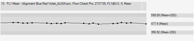
35 Stability of instrumentation
36 Compensation Controls Single-stained samples must be at least as bright as the reagent you are using in the experiment! Can use any carrier, as long as the positive & negative populations have the same fluorescence when unstained: Cells (mix stained & unstained) Subpopulations (CD8 within total T) Beads (antibody-capture) One compensation for every color and one for each unique lot of a tandem (Cy5PE, Cy7PE, Cy7APC, TRPE)

37 Staining Controls Staining controls are necessary to identify cells which do or do not express a given antigen. The threshold for positivity may depend on the amount of fluorescence in other channels!
38 Staining Controls Unstained cells or complete isotype control stains are improper controls for determining positive vs. negative expression in multi-color experiments. The best control is to stain cells with all reagents except the one of interest. FMO Control Fluorescence Minus One


39 Identifying CD4 cells with 4 colors PBMC were stained as shown in a 4-color experiment. Compensation was properly set for all spillovers FITC PE Cy5PE Cy7PE 10 5 Unstained Control FMO Control Fully Stained CD3 CD3 CD4 CD8 CD8 CD45RO CD45RO 10 4 PE 10 3 Isotype Bounds FMO Bounds FITC

40 FMO controls aid even when compensation is improper Incorrect Cy5PE into Cy7PE compensation 10 5 Unstained Control FMO Control Fully Stained FITC PE Cy5PE Cy7PE Š Š Š Š CD3 Š CD8 CD45RO CD3 CD4 CD8 CD45RO 10 4 PE 10 3 Isotype Bounds FMO Bounds FITC
41 FMO Controls are a much better way to identify positive vs. negative cells can also help identify problems in compensation that are not immediately visible should be used whenever accurate discrimination is essential or when antigen expression is relatively low

42 Cy7PE-A Why Bright Comp Controls? Estimating a low spillover fluorescence accurately is impossible (autofluorescence). Therefore, compensation is generally only valid for samples that are duller than the compensation control Unstained Bright cells cells Dimmer cells FITC spillover into Cy7PE (1%) Autofluorescence FITC-A
43 Different lots of tandems can require different compensation! TR-PE reagent 1 Median = 21,100 Compensation Required ( PE / TRPE) PE Median = % TR-PE reagent 2 Median = 8,720 PE Median = %
44 Advantage of More-Than-Minimal Markers Two extremes of gating strategy: Conservative - drawn to be very tight around the visuallydefined populations Greatest purity of subset Lowest sensitivity Liberal - drawn to include much larger areas than visually appear to belong to a subset. Greatest sensitivity Greatest chance of contamination BUT: multiple rounds of Liberal gating based on multiple parameters results in excellent purity and sensitivity.
45 Polychromatic panels Development is time-consuming, expensive and requires substantial expertise. Fortunately, you do not always need to reinvent the wheal because many optimized panels are already published ( OMIPs)
46 OMIPs Optimized Multicolor Immunofluorescence Panels Mario Roederer, NIH, Bethesda
47 OMIPs A new publication type exclusive to Cytometry A. Proposed in 2010, with guidelines for publication: Publication of optimized multicolor immunofluorescence panels, Mahnke, Chattopadhyay, and Roederer. Cytometry A. 2010;77:814 The first two OMIPs in 2010: OMIP-001: Quality and phenotype of Ag-responsive human T- cells. Mahnke, Roederer. Cytometry A 2010;77:819 OMIP-002: Phenotypic analysis of specific human CD8+ T-cells using peptide-mhc class I multimers for any of four epitopes. Chattopadhyay, Roederer, Price. Cytometry A 2010;77:821. A total of >18 OMIPs now in published and more to come
48 OMIPs OMIPs have 2 parts A brief (2 page only!) printed version that summarizes information and shows an example. An extended online version that has multiple required tables and information pieces. The format and content, even of the online material, is fairly well specified and must be followed.
49
50 LIFE-Study LIFE - Leipzig Research Center for Civilization Diseases LIFE-study individuals (5 % of population) Aims: Influence of health status and life style - Identification of risk factors - Innovative ways to predict disease development and early diagnosis -Improvement of German healthcare Methods: Complex medical, psychological and laboratory analysis and questionnaires. Follow up studies.
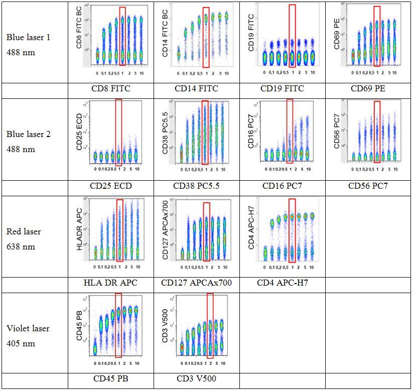
51 Specificity MAB Ab Clone Fluorochrome Purpose Isotype CD8 B9.11 FITC T-cytotoxic cells IgG1 CD14 RMO52 FITC LPS Rec. Monocytes IgG2a CD19 J3-119 FITC B-cells IgG1kappa CD69 TP PE Early activation IgG2b CD25 B ECD IL-2 Receptor a IgG2a CD38 LS PC5.5 Activated T and B-cells IgG1 CD16 3G8 PC7 Fcγ Rec III IgG1 CD56 N901(NKH-1) PC7 N-Cam IgG1 HLA DR Immu-357 APC MHC-II IgG1 CD127 R APCAx700 IL-7 Receptor a IgG1 kappa CD4 SK3 APC-H7 T-helper cells IgG1 kappa CD45 J.33 Pacific Blue PanLeukocyte antigen IgG1 kappa CD3 SP34-2 V500 T-cells IgG1 lamda Print Table 1B: Antibodies used for OMIP-BJ-AT 30 defined cell phenotypes >> 5 functional information in one run!
52

53 Combination of many markers on one color Single CD14 FITC Staining Single CD8 FITC Staining 4. Combined Staining CD8/14/19 FITC Single CD19 FITC Staining


 7. 3.")
54 CD16/56 PC7 Combination of many markers on one color 4. CD14 FITC on Monocytes CD8 FITC on CD3+ Lymphocytes 6. CD8 FITC on NK-cells (CD3-, CD16/56+) HLA-DR APC CD19 FITC on B-cells (CD3-, CD16/56-, HLA DR+)
55
56 Stability of pre-analytics Relative percentage Intra-assay variance II Normalized antigen expression for the main parameters Intra-Assay-Variance Cell X Data types 1 - Neutrophil CD16 2 Eosinophil CD Monocyte CD Lymphocyte CD T-Lymphocyte CD3 6 NK-cells CD16/ B-Lymphocytes HLA DR 8 - Plasma Cells CD B cells CD T-cytotoxic cells ++ CD T-helper cells CD Treg cells CD25 13 NKT cells CD16/ Monocyte Atypic CD Monocyte Typic CD14
57 Cocktail stability
58 Stability of manual analysis
59 Mean count/µl Stability of cell counts WBC neutrophils T-cells Th Date
60 Organism Published OMIPS Cell-subtype 1 human CD8+ T-cells 2 human CD4+, CD8+ T-cells (HIV+) 3 human Memory B cells 4 human Regulatory T-cells 5 Rhesus macaque T-cells 6 human Regulatory T-cells 7 human NK cells 8 human T-cells 9 human CD4+, CD8+ T-cells 10 human lymphoma cells (leukemia) 11 human circulating endothelial cells (CECs) 12 mouse leukocytes 13 human T-cells 14 human T-cells 15 human Regulatory T-cells 16 Cynomolgus macaque/human CD4+, CD8+ T-cells 17 human CD4+ T-helper-cells 18 human CD4 T-cells 19 human gd T-cells, inkt-cells, haematopoietic precursors

61 Since Oct required for Cytometry A publications. MIFlowCyt: Minimum Information about a Flow Cytometry Experiment Ryan Brinkman Department of Medical Genetics, University of British Columbia BC Cancer Research Center

62 Flow Repository Website

63 The Journal for quantitative single cell science and cell systems biology Impact Factor 2011: (2012 exp.: ~3.7) Transition time 1st submission to 1st decision: < 30 days Papers published/year ~ 100
64
65 Thank you
66 References and examples Manuscript examples are found on the Cytometry Part A Wiley-Blackwell Website. MIFlowCyt: the minimum information about a Flow Cytometry Experiment. Lee et al. Cytometry A. 2008;73:926.
67 MI For experimental publications a minimum information (MI) has to be provided so that the experiments can be understood and repeated Promoting coherent minimum reporting guidelines for biological and biomedical investigations: the MIBBI project. Taylor CF et al. Nat Biotechnol. 2008;26:889. Usage of these guidelines is now obligatory for many journals. ~ 100% of FCM submissions to us claim MIFlowCyt compliance.
68 Advantage of More-Than-Minimal Markers When designing your panels, try to include reagent combinations that will allow you a combination of positive and negative expression gates for every subset of interest. Note that there is almost never a downside to including additional markers that are negative gates--the lack of this fluorescence signal on your cells of interest cannot alter the sensitivity of your measurements. Dump channels and viability channels are virtually always a good thing!
69 Example Optimization In this example, we wished to evaluate the expression of CXCR3 and CCR4 on naïve (CD62L + CD45RA + CD45RO ) CD4 T cells. What fraction of naïve T cells express these molecules? If possible: are those cells truly naïve (CD28 + CD11a dim CD27 + )? Requirements: CD4, CD3 = Primary reagents CD45RO/RA, CD62L = Secondary (need excellent separation) CXCR3, CCR4 = Tertiary reagents CD27, CD11a, CD28 = Luxury reagents
70 Selection of Marker/Color Combinations Primary Well-characterized, identify broad subsets of cells, expression is usually on/off. e.g., CD3, CD4, CD8, CD14, CD19, CD20 Typically used as parent gates in analysis These reagents are usually assigned to dimmer colors and colors that exhibit the greatest spillover problems e.g., Cy5.5PE, Cy7PE, Cy7APC, AmCyan
71 Selection of Marker/Color Combinations Secondary Well-characterized, bright expression patterns e.g., CD27, CD28, CD45RA/RO, IFN, perforin Expression levels can be a continuum These are usually assigned to the next tier of colors, those that perform well with little spillover problems e.g., FITC, TRPE, Cy5PE/PerCP, Alexa 405, Alexa 690
72 Selection of Marker/Color Combinations Tertiary Low-expression levels or uncharacterized e.g., CD25, CCRs, X These require the absolutely brightest colors, with the least spillover problems possible e.g. PE, APC, QD655
73 FITC Single Stain Control FITC PE Total signal detected in FL1 Unwanted signal detected in FL2 = roughly 15% FL1 FL2 True PE = Total FL2 15% FL1

74 Cblue-A Cblue-A Cblue-A Cblue-A Cblue-A Cblue-A Cblue-A Cblue-A Cblue-A Cblue-A Cblue-A Cblue-A APC-A CD45RO Example Stains For panel Ax680 CD45RO; V905 APC CD45RO; V736 Cy55APC CD45RO; V615 CB CD45RO; V209 Ax680-A APC-A Ax680-A Cblue-A Cy55PE CD45RO; V613 Cy55PE-A Cy5PE CD45RO; V612 Cy5PE-A Cy7APC CD45RO; V616 Cy7APC-A FITC CD45RO; V39 FITC-A FITC CD45RO; V975 FITC-A Cy7APC CD45RO; V909 Cy7APC CD45RO; V913 Cy7PE CD45RO; V614 TRPE CD45RO; Co/13 Cy7APC-A Cy7APC-A Cy7PE-A TRPE-A


75 Using Beads to Compensate Gate on Singlets ; then gate on singlestained beads. FITC PE 1000 PE 800 FS SS 67.9 FITC Comp PE Comp Cy5PE comp APC comp Unst ained Cy5PE Cy5PE APC


76 Result A B C 31 CD4 + T Cells D E F C B A D E F CD62L CXCR3 Final panel worked very well--in fact, identified expression of CCR4 not previously seen on FACSCalibur!


77 Complex Interactions in Compensation The same data is shown with correct or wrong Cy5PE->Cy7PE comp setting. Note that neither of these channels is shown here!
78 Imperfect Measurement Leads to Apparent Spread in Compensation 10 4 Uncompensated Compensated Spillover Fluorescence Primary Fluorescence (-200) Why is there a 400-unit spread? Photon counting statistics.
79 Cy5PE CD16 Selection of Marker/Color Combinations (2) All colors are not created equal. The same monoclonal antibody conjugated to FITC, PE, Cy5PE, APC, Cy7APC can show apparently different distributions on multiply-stained cells This is due to spectral-spillover, and the propagation of the error in those measurements QD 655 CD45RA
80 Cy5PE CD16 Selection of Marker/Color Combinations (2) Prediction of the spillover effect is very difficult. You need to know three different aspects: (1) The brightness of the other reagents in your panel (2) The spillover of these reagents into your channel (3) The absolute brightness of every measurement Amount of spread in your measurement channel is equal to the sum of all other reagents brightnesses multiplied by their spillover coefficient and by the inverse square root of the absolute brightness QD 655 CD45RA


81 Fix/Perm Changes Cy7APC Compensation Requirement The longer Cy7APC is in fixative, the more it falls apart, leading to more APC compensation Note that this exacerbates the higher IL4+ gate required for CD8 cells. The undercompensation would not have been detected except by looking at the APC vs. Cy7APC graphic
82 Insufficiently-Bright Comp Control Is. Bad! Note that either under- or over-compensation can result from using comp controls that are too dim!
83 Good Instrument Alignment Is Critical! Uncompensated PE Compensated Day 1 Day 2 While the amount of compensation did not differ, the measurement error (correlation) decreased leading to much better visualization of the population! TR-PE
84 Compensation for more colors: It s not just pretty pictures Spillover from unviewed measurement channel can alter event positions without obvious visual evidence (no diagnostic diagonals!) Thus, gate positions may depend on unviewed measurement channels and be different for various tubes in a panel Separation of populations may require multidimensional surfaces.
85 Using Beads to Compensate Antibody-capture beads Use reagent in use Lots positive Small CV, bright Sonicate Some reagents won t work (IgL, non mouse, too dim, EMA/PI)--mix with regular comps
86 Final Panels Based on the evaluation of the first sets of panels, certain combinations were eliminated. The good aspects of other combinations were combined and fine-tuned. TRPE Cy5PE Cy55PE Cy7PE APC Cy55APC Ax680 Cy7APC CB QD655 1 CD62L CD4 CD45RO CD45RA 2 CD45RO CD3 CD62L CD28 CD11a CD4 CD27 CD45RA 3 CD45RO CD27 CD4 CD11a CD62L CD45RA Note: CD3 was dropped from 1 & 3 as CD4 staining was deemed good enough to identify CD4 T cells. Panel 2 will validate this assertion! Panels 2 & 3 add more memory markers to verify the final phenotype of the chemokine-expressing cells.

87 Compensating with the wrong TRPE Wrong TR-PE comp control Right TR-PE comp control
88 Some Examples of Problems The following four examples illustrate some types of problems that can be occur related to compensation. In each case, compensation itself is not the problem: there is an underlying reagent, instrumentation, or analysis problem. However, the manifestation of this problem is an apparent incorrect compensation!
89 Design of panels Since optimal sensitivity was desired, I tried to minimize reagents that would have spillover-spreading into FITC and PE. Optimal separation of CD62L and CD45Rx was required. Other memory markers were less important: therefore, some panels were designed to test minimal requirements, and others were part of the wish list.
90 Why do we need OMIPs? Developing a multicolor panel is extremely labor-intensive. The complexity increases geometrically with number of colors it often takes us 4 months to develop a color panel. Publishing these panels accomplishes two goals: (1) Sharing the panel for others to use, adapt, or build upon (2) Providing a mechanism by which recognition for panel development is achieved (attribution by citation)
91 OMIPs: OPTIMIZED The key part of OMIPs is the optimization. Without optimization, there is no intellectual contribution, nor is their evidence that the panel should not be improved! Optimization includes: Comparing as many variations of each reagent as possible (choosing the best and why!) Comparing variations of combinations of reagents Showing that each reagent is optimal (titration!) Lack of optimization demonstration is the most frequent reason for rejection!
 Print")
92 Example OMIP (OMIP-001) Print portion
 Print")
93 Example OMIP (OMIP-001) Print portion
94 Example OMIP (OMIP-001) Online material Required tables include: Instrument configuration (lasers/optics) Commercial reagents (fluorochrome, vendor, clone, catalog number, dilution, staining conditions) In-house synthesized reagents (no proprietary materials)


95 Example OMIP (OMIP-001) Online material Development strategy: how many (which) reagents were tested. Why was each chosen or eliminated?
96 Example OMIP (OMIP-001) Online material Detailed staining protocol
97 The Future of OMIPs The collection of published OMIPs will provide a valuable resource for development of new panels. Using an existing OMIP gives you an assurance that the panel is likely to work well on your instrument. OMIPs are living as new reagents come about, we expect to update the online portion of OMIPs so as to always have the most recent optimized version available. OMIPs provide a mechanism to credit the huge amount of work that goes into a panel.
98 Advantage of More-Than-Minimal Markers Two extremes of gating strategy: Conservative - drawn to be very tight around the visuallydefined populations Greatest purity of subset Lowest sensitivity Liberal - drawn to include much larger areas than visually appear to belong to a subset. Greatest sensitivity Greatest chance of contamination BUT: multiple rounds of Liberal gating based on multiple parameters results in excellent purity and sensitivity.
 - Identification of risk factors - Establishment of effective forms of prevention and early diagnosis -Improvement of German healthcare")
99 LIFE-Study LIFE - Leipzig Research Center for Civilization Diseases Aims: - to explain the causes of widespread common diseases (metabolic & cardiovascular diseases, heart attack, diabetes, depression, dementia, head- and neck cancer, allergies ) - Identification of risk factors - Establishment of effective forms of prevention and early diagnosis -Improvement of German healthcare Methods: Complex medical analysis and questionnaires Leipzig population (12/2011) with a density of 1,787/km 2 LIFE-study (5 % of population)
100 Cytomics for LIFE cytometric analysis of 1200 EDTA-anticoagulated fresh blood samples over a 3 year period complex antibody panel with 13 fluorescent antibodies on 10 colours immunophenotyping: differentiation of over 30 leukocyte subpopulations and activity Calculation of reference intervals for leukocyte subpopulations for adults (20-80 years) Correlation of biological variability with lifestyle and diseases
A step-by-step approach to build and analyze a multicolor panel
 Analyze A step-by-step approach to build and analyze a multicolor panel For Research Use Only. Not for use in diagnostic or therapeutic procedures. Alexa Fluor is a registered trademark of Life Technologies
Naive, memory and regulatory T lymphocytes populations analysis
 Naive, memory and regulatory T lymphocytes populations analysis Jaen Olivier, PhD ojaen@beckmancoulter.com Cellular Analysis application specialist Beckman Coulter France Introduction Flow cytometric analysis
BD Flow Cytometry Reagents Multicolor Panels Designed for Optimal Resolution with the BD LSRFortessa X-20 Cell Analyzer
 Multicolor Panels Designed for Optimal Resolution with the BD LSRFortessa X-2 Cell Analyzer Proper multicolor panel design takes into account fluorochrome brightness, antigen density, co-expression, and
What to Measure, How to Measure It
 Dale and Betty Bumpers Vaccine Research Center National Institute of Allergy and Infectious Diseases National Institutes of Health Monitoring Memory T-cells: What to Measure, How to Measure It Pratip K.
Do Your Flow Cytometric LDTs. Validation Guidelines? Fiona E. Craig, MD University of Pittsburgh School of Medicine
 Do Your Flow Cytometric LDTs Conform to the ICSH ICCS Validation Guidelines? Fiona E. Craig, MD University of Pittsburgh School of Medicine How should LDTs be validated? Accuracy Specificity Sensitivity
Human Progenitor Cell Enumeration by Flow Cytometry Practical Considerations
 Human Progenitor Cell Enumeration by Flow Cytometry Practical Considerations Bruce Briggs, M.S., S.I. (A.S.C.P.) OSU Medical Center Clinical Flow Cytometry Objectives Conceptual overview: How the Flow
Measuring Dendritic Cells
 Measuring Dendritic Cells A.D. Donnenberg, V.S. Donnenberg UNIVERSITY of PITTSBURGH CANCER INSTITUTE CCS Longbeach 10_04 Measuring DC Rare event detection The basics of DC measurement Applications Cancer
DURACLONE IF BE CERTAIN ABOUT THE RESPONSE. l res. a il n c n. For Research Use Only - Not for use in Diagnostic procedures
 DURACLONE IF earch tria l res lc a om ic il n c n nio pa Yo ur BE CERTAIN ABOUT THE RESPONSE For Research Use Only - Not for use in Diagnostic procedures BE CERTAIN ABOUT THE RESPONSE The sensitive and
DURACLONE RE THERE ARE EVENTS YOU CANNOT AFFORD TO MISS
 DURACLONE RE THERE ARE EVENTS YOU CANNOT AFFORD TO MISS Your clinical research trial companion For Reseach Use Only - Not for use in Diagnostic procedures THERE ARE EVENTS YOU CANNOT AFFORD TO MISS Rare
a Beckman Coulter Life Sciences: White Paper
 a Beckman Coulter Life Sciences: White Paper An 8-color DuraClone IM panel for detection of Human blood dendritic cells by flow cytometry Nathalie Dupas 1, Snehita Sattiraju 2, Neha Girish 2, Murthy Pendyala
VUmc Basispresentatie
 Clinical diagnostic cytometry Gerrit J Schuurhuis Dept of Hematology VU University Medical Center Amsterdam, Netherlands Use of immunophenotyping at diagnosis to trace residual disease after therapy 1.
The CAPRI-T was one of two (along with the CAPRI-NK, see reference. [29]) basic immunological studies nested within the CAMELIA trial.
![The CAPRI-T was one of two (along with the CAPRI-NK, see reference. [29]) basic immunological studies nested within the CAMELIA trial.](/thumbs/88/117921481.jpg "The CAPRI-T was one of two (along with the CAPRI-NK, see reference. [29]) basic immunological studies nested within the CAMELIA trial.") 1 SUPPLEMENTARY INFORMATION Patient description and recruitment The CAPRI-T was one of two (along with the CAPRI-NK, see reference [29]) basic immunological studies nested within the CAMELIA trial. Patients
1 SUPPLEMENTARY INFORMATION Patient description and recruitment The CAPRI-T was one of two (along with the CAPRI-NK, see reference [29]) basic immunological studies nested within the CAMELIA trial. Patients
Flow Cytometric Analysis of Cerebral Spinal Fluid Involvement by Leukemia or Lymphoma
 Flow Cytometric Analysis of Cerebral Spinal Fluid Involvement by Leukemia or Lymphoma Maryalice Stetler-Stevenson, M.D., Ph.D. Flow Cytometry Unit, Laboratory of Pathology, DCS, NCI, NIH DEPARTMENT OF
CHAPTER 3 LABORATORY PROCEDURES
 CHAPTER 3 LABORATORY PROCEDURES CHAPTER 3 LABORATORY PROCEDURES 3.1 HLA TYPING Molecular HLA typing will be performed for all donor cord blood units and patients in the three reference laboratories identified
Fluorochrome Panel 1 Panel 2 Panel 3 Panel 4 Panel 5 CTLA-4 CTLA-4 CD15 CD3 FITC. Bio) PD-1 (MIH4, BD) ICOS (C398.4A, Biolegend) PD-L1 (MIH1, BD)
 PD-1 (MIH4, BD) ICOS (C398.4A, Biolegend) PD-L1 (MIH1, BD)") Additional file : Table S. Antibodies used for panel stain to identify peripheral immune cell subsets. Panel : PD- signaling; Panel : CD + T cells, CD + T cells, B cells; Panel : Tregs; Panel :, -T, cdc,
Additional file : Table S. Antibodies used for panel stain to identify peripheral immune cell subsets. Panel : PD- signaling; Panel : CD + T cells, CD + T cells, B cells; Panel : Tregs; Panel :, -T, cdc,
The spectrum of flow cytometry of the bone marrow
 The spectrum of flow cytometry of the bone marrow Anna Porwit Lund University Faculty of Medicine Dept. of Clinical Sciences Div. Oncology and Pathology anna.porwit@med.lu.se Disclosure of speaker s interests
Phagocytosis of FITC labelled opsonized and non-opsonized E. coli bacteria by monocytes and granulocytes in a whole blood assay
 Phagocytosis of FITC labelled opsonized and non-opsonized E. coli bacteria by monocytes and granulocytes in a whole blood assay APPLICATION NOTE Author: Andreas Spittler, MD, Associate Professor for Pathophysiology
Establishing a Pure Lymphocyte Gate for Subset Analysis by Flow Cytometry
 Cytornetry (Communications in Clinical Cytometry) 26:172-177 (1996) Establishing a Pure Lymphocyte Gate for Subset nalysis by Flow Cytometry Joseph M. Homtinovich, Sara D. Sparks, and Karen P. Mann Clinical
DuraClone IM. Standardized phenotyping panels for studies of the human immune system
 DuraClone IM Standardized phenotyping panels for studies of the human immune system Standardize with the experts, adopt DuraClone IM Tubes. Clinical research studies require accurate, reproducible results
Supplementary Figure 1. Enhanced detection of CTLA-4 on the surface of HIV-specific
 SUPPLEMENTARY FIGURE LEGEND Supplementary Figure 1. Enhanced detection of CTLA-4 on the surface of HIV-specific CD4 + T cells correlates with intracellular CTLA-4 levels. (a) Comparative CTLA-4 levels
MHC Tetramers and Monomers for Immuno-Oncology and Autoimmunity Drug Discovery
 MHC Tetramers and Monomers for Immuno-Oncology and Autoimmunity Drug Discovery Your Partner in Drug Discovery and Research MHC Tetramer Background T-Cell Receptors recognize and bind to complexes composed
Supplementary Data. Treg phenotype
 Supplementary Data Additional Experiment An additional experiment was performed using cryopreserved peripheral blood mononuclear cells (PBMC) derived from five renal cell carcinoma (RCC) patients [see
B and T-Cells. CD8+ Cytotoxic T Cells
 Sony Biotechnology Inc. Application Data, SA38 Spectral Analyzer TBNK Panel Page 1 TBNK panel on the SA38 Spectral Analyzer TBNK (B-Cell, T-Cell, and natural killer cells) panels are frequently used to
Application Information Bulletin: Human NK Cells Phenotypic characterizing of human Natural Killer (NK) cell populations in peripheral blood
 cell populations in peripheral blood") Application Information Bulletin: Human NK Cells Phenotypic characterizing of human Natural Killer (NK) cell populations in peripheral blood Christopher A Fraker, Ph.D., University of Miami - Miami, Florida
Application Information Bulletin: Human NK Cells Phenotypic characterizing of human Natural Killer (NK) cell populations in peripheral blood Christopher A Fraker, Ph.D., University of Miami - Miami, Florida
Fast and Easy Isolation of T Cells
 Fast and Easy Isolation of T Cells Isolate T Cells In As Little As 25 Minutes Isolate whole T cell populations as well as various T cell subsets with high purity and recovery using the fast and easy T
BD CBA on the BD Accuri C6: Bringing Multiplexed Cytokine Detection to the Benchtop
 BD CBA on the BD Accuri C6: Bringing Multiplexed Cytokine Detection to the Benchtop Maria Dinkelmann, PhD Senior Marketing Applications Specialist BD Biosciences, Ann Arbor, MI 23-14380-00 Cellular Communication
Tube 1 : FLAER/CD24/CD16/CD15/CD45
 Gating procedure for white blood cells (WBC) on FC500 The French PNH diagnosis working group has developed an optimized gating strategy adapted to a two-tubes diagnostic test (PMN, MO), evaluated in a
CLINICAL USE OF CELLULAR SUBPOPULATION ANALYSIS IN BM
 CLINICAL USE OF CELLULAR SUBPOPULATION ANALYSIS IN BM CANCER RESEARCH CENTRE, UNIVERSITY AND UNIVERSITY HOSPITAL OF SALAMANCA (SPAIN)( Sao Paulo, 18th of April, 2009 IDENTIFICATION OF HPC (I) 1.- In vivo
Technical Bulletin No. 157
 CPAL Central Pennsylvania Alliance Laboratory Technical Bulletin No. 157 January 11, 2017 Flow Cytometry Lymphocyte Subset Analysis Testing Platform Change Change Effective Date: February 6, 2017 Methods
IMMUNOLOGY. Source, Isolate, Culture, And Analyze Immune Cells. Scientists Helping Scientists
 IMMUNOLOGY Source, Isolate, Culture, And Analyze Immune Cells Scientists Helping Scientists WWW.STEMCELL.COM TABLE OF CONTENTS Tools For Your Immunology Research 4 Primary Cells: It All Starts with The
were isolated from the freshly drawn blood of healthy donors and ACS patients using the
 Supplemental Figure 1. Quality control of CD4 + T-cell purification. CD4 + T cells were isolated from the freshly drawn blood of healthy donors and ACS patients using the RosetteSep CD4 + T Cell Enrichment
Supporting Information
 Supporting Information Idoyaga et al. 10.1073/pnas.0812247106 SSC a) Single cell suspension 99 Aqua b) Live cells 96 -W c) Singlets 92 -A CD19+ER119 d) CD19 ER119 cells 97 CD3 e) CD3 cells 27 f) DX5 cells
BD Multitest IMK Kit
 BD Multitest IMK Kit 50 Tests Catalog No. 340503 50 Tests with BD Trucount Tubes Catalog No. 340504 IVD 2016 BD. BD, the BD Logo and all other trademarks are property of Becton, Dickinson and Company.
Commercially available HLA Class II tetramers (Beckman Coulter) conjugated to
 conjugated to") Class II tetramer staining Commercially available HLA Class II tetramers (Beckman Coulter) conjugated to PE were combined with dominant HIV epitopes (DRB1*0101-DRFYKTLRAEQASQEV, DRB1*0301- PEKEVLVWKFDSRLAFHH,
Class II tetramer staining Commercially available HLA Class II tetramers (Beckman Coulter) conjugated to PE were combined with dominant HIV epitopes (DRB1*0101-DRFYKTLRAEQASQEV, DRB1*0301- PEKEVLVWKFDSRLAFHH,
Clinical question. Screening tube. Diagnostic panel MRD. Clinical question
 OW CYTOMETRY UPDATES IN LYMPHOPROLIFERATIVE DISORDERS CANCER RESEARCH CENTER IBSAL UNIVERSITY & UNIVERSITY HOSPITAL, SALAMANCA (SPAIN) DISCLOSURES The EuroFlow Scientific Consortium Iamco-chairof receives
BD FACSCanto. BD FACSCanto. 488nm 20mw 633nm 17mw 0.1% 120uL/min 10,000events/sec. BD Biosciences 1
 BD FACSCanto BD FACSCanto 488nm 20mw 633nm 17mw 6 2 0.1% 120uL/min 10,000events/sec BD Biosciences 1 BD FACSCCanto BD FACStation 1.1 FACSCanto 1.1.1 Fluidics Sample injection tube Aspirator arm flow cell
PanLeucogating Akin Abayomi
 PanLeucogating Akin Abayomi Cost effective CD4 counting at a central laboratory. The Barbados experience. CCAS Surinam 2008 Appreciation Frank Mandy George Janossy Phil McCoy John Codrington Clive Landis
Cytotoxicity assays. Rory D. de Vries, PhD 1. Viroscience lab, Erasmus MC, Rotterdam, the Netherlands
 Cytotoxicity assays Rory D. de Vries, PhD 1 1 Viroscience lab, Erasmus MC, Rotterdam, the Netherlands Anti-influenza immunity Humoral / CD4+ / CD8+ / NK? Function of CTL Elimination of virus-infected cells?
Attribution: University of Michigan Medical School, Department of Microbiology and Immunology
 Attribution: University of Michigan Medical School, Department of Microbiology and Immunology License: Unless otherwise noted, this material is made available under the terms of the Creative Commons Attribution
FLOW CYTOMETRIC ANALYSIS OF NORMAL BONE MARROW
 XI International Conference Hematopoiesis Immunology Budapest, June 6-7, 2014 FLO CYTOMETRIC ANALYSIS OF NORMAL BONE MARRO Bruno Brando and Arianna Gatti Hematology Laboratory and Transfusion Center Legnano
Bead Based Assays for Cytokine Detection
 Bead Based Assays for Cytokine Detection September 27, 2014 6 th EFIS-EJI South East European Immunology School SEEIS 2014 Timisoara, Romania The Cells of the Immune System The Immune Reaction (Th2) (Th1)
ACS GUIDELINE FOR LYMPHOCYTE SUBSET IMMUNOPHENOTYPING. Second Edition Australasian Cytometry Society Guideline Document
 ACS GUIDELINE FOR LYMPHOCYTE SUBSET IMMUNOPHENOTYPING Second Edition 2017 Australasian Cytometry Society Guideline Document Paper-based publications This work is copyright. You may reproduce the whole
x Lymphocyte count /µl CD8+ count/µl 800 Calculated
 % Lymphocyte in CBC A. 50 40 30 20 10 Lymphocyte count /µl B. x10 3 2.5 1.5 C. 50 D. 1000 % CD3+CD8+ Cells 40 30 20 Calculated CD8+ count/µl 800 600 400 200 10 0 #61 #63 #64 #65 #68 #71 #72 #75 Figure
BD Pharmingen. Human Th1/Th2/Th17 Phenotyping Kit. Technical Data Sheet. Product Information. Description Components:
 Technical Data Sheet Human Th1/Th2/Th17 Phenotyping Kit Product Information Material Number: Size: Description Components: 560751 50 Tests BD Pharmingen 51-9006615 Human Th1/Th2/Th17 Phenotyping Cocktail
ACTIVATION AND EFFECTOR FUNCTIONS OF CELL-MEDIATED IMMUNITY AND NK CELLS. Choompone Sakonwasun, MD (Hons), FRCPT
, FRCPT") ACTIVATION AND EFFECTOR FUNCTIONS OF CELL-MEDIATED IMMUNITY AND NK CELLS Choompone Sakonwasun, MD (Hons), FRCPT Types of Adaptive Immunity Types of T Cell-mediated Immune Reactions CTLs = cytotoxic T lymphocytes
ACTIVATION AND EFFECTOR FUNCTIONS OF CELL-MEDIATED IMMUNITY AND NK CELLS Choompone Sakonwasun, MD (Hons), FRCPT Types of Adaptive Immunity Types of T Cell-mediated Immune Reactions CTLs = cytotoxic T lymphocytes
ab Exosome Isolation and Analysis Kit - Flow Cytometry, Plasma
 Version 1 Last updated 25 May 2018 ab228565 Exosome Isolation and Analysis Kit - Flow Cytometry, Plasma For the isolation/detection of exosomes from human plasma, urine or cell culture media. This product
Pearson r = P (one-tailed) = n = 9
 = n = 9") 8F4-Specific Lysis, % 1 UPN1 UPN3 8 UPN7 6 Pearson r =.69 UPN2 UPN5 P (one-tailed) =.192 4 UPN8 n = 9 2 UPN9 UPN4 UPN6 5 1 15 2 25 8 8F4, % Max MFI Supplementary Figure S1. AML samples UPN1-UPN9 show variable
8F4-Specific Lysis, % 1 UPN1 UPN3 8 UPN7 6 Pearson r =.69 UPN2 UPN5 P (one-tailed) =.192 4 UPN8 n = 9 2 UPN9 UPN4 UPN6 5 1 15 2 25 8 8F4, % Max MFI Supplementary Figure S1. AML samples UPN1-UPN9 show variable
In vitro human regulatory T cell expansion
 - 1 - Human CD4 + CD25 + CD127 dim/- regulatory T cell Workflow isolation, in vitro expansion and analysis In vitro human regulatory T cell expansion Introduction Regulatory T (Treg) cells are a subpopulation
Supplementary Fig. 1: Ex vivo tetramer enrichment with anti-c-myc beads
 Supplementary Fig. 1: Ex vivo tetramer enrichment with anti-c-myc beads Representative example of comparative ex vivo tetramer enrichment performed in three independent experiments with either conventional
Nine-Color Flow Cytometry for Accurate Measurement of T Cell Subsets and Cytokine Responses. Part I: Panel Design by an Empiric Approach
 Original Article Nine-Color Flow Cytometry for Accurate Measurement of T Cell Subsets and Cytokine Responses. Part I: Panel Design by an Empiric Approach Bridget E. McLaughlin, 1 Nicole Baumgarth, 2 Martin
Effector T Cells and
 1 Effector T Cells and Cytokines Andrew Lichtman, MD PhD Brigham and Women's Hospital Harvard Medical School 2 Lecture outline Cytokines Subsets of CD4+ T cells: definitions, functions, development New
Flow cytometry leukocyte differential : a critical appraisal
 Flow cytometry leukocyte differential : a critical appraisal Francis Lacombe Flow cytometry department University Hospital of Bordeaux, Pessac, France francis.lacombe@chu-bordeaux.fr 2008 HORIBA ABX, All
MHC Multimer staining (6 colours, with fixation)
") v MH Multimer staining (6 colours, with fixation) Authors M. Odendahl,. Blum,. Busch ate 27-04-2007 Version 1.0 REAGENTS: - PBMs after Ficoll - EMA - Phosphate buffered saline (PBS), ph 7.45 - FAS staining
v MH Multimer staining (6 colours, with fixation) Authors M. Odendahl,. Blum,. Busch ate 27-04-2007 Version 1.0 REAGENTS: - PBMs after Ficoll - EMA - Phosphate buffered saline (PBS), ph 7.45 - FAS staining
ab Exosome Isolation and Analysis Kit - Flow Cytometry, Cell culture
 Version 1 Last updated 14 March 2018 ab228564 Exosome Isolation and Analysis Kit - Flow Cytometry, Cell culture For the measurement of human exosomes in cell culture. This product is for research use only
HD1 (FLU) HD2 (EBV) HD2 (FLU)
 HD2 (EBV) HD2 (FLU)") ramer staining + anti-pe beads ramer staining a HD1 (FLU) HD2 (EBV) HD2 (FLU).73.11.56.46.24 1.12 b CD127 + c CD127 + d CD127 - e CD127 - PD1 - PD1 + PD1 + PD1-1 1 1 1 %CD127 + PD1-8 6 4 2 + anti-pe %CD127
ramer staining + anti-pe beads ramer staining a HD1 (FLU) HD2 (EBV) HD2 (FLU).73.11.56.46.24 1.12 b CD127 + c CD127 + d CD127 - e CD127 - PD1 - PD1 + PD1 + PD1-1 1 1 1 %CD127 + PD1-8 6 4 2 + anti-pe %CD127
Alessandra Franco MD PhD UCSD School of Medicine Department of Pediatrics Division of Allergy Immunology and Rheumatology
 Immunodominant peptides derived from the heavy constant region of IgG1 stimulate natural regulatory T cells: identification of pan- HLA binders for clinical translation Alessandra Franco MD PhD UCSD School
ab Exosome Isolation and Analysis Kit - Flow Cytometry, Cell Culture (CD63 / CD81)
") Version 1 Last updated 26 September 2018 ab239682 Exosome Isolation and Analysis Kit - Flow Cytometry, Cell Culture (CD63 / For the isolation and analysis of exosome from cell culture. This product is
Version 1 Last updated 26 September 2018 ab239682 Exosome Isolation and Analysis Kit - Flow Cytometry, Cell Culture (CD63 / For the isolation and analysis of exosome from cell culture. This product is
Immune response. This overview figure summarizes simply how our body responds to foreign molecules that enter to it.
 Immune response This overview figure summarizes simply how our body responds to foreign molecules that enter to it. It s highly recommended to watch Dr Najeeb s lecture that s titled T Helper cells and
Supplementary Table 1: Summary of Reactogenicity Events by Group and Time of Onset in Entebbe
 Supplementary Table 1: Summary of Reactogenicity Events by Group and Time of Onset in Group A (n=39) B (n=11) A (n=39) B (n=11) Onset Post-Vaccination Event 3 Minutes 3-14 days Pain 5 (13%) 1 (1%) 6 (15%)
Dr Prashant Tembhare
 Dr Prashant Tembhare docprt@gmail.com FCM very powerful technology in Identification and characterization of neoplastic plasma cells as it allows - simultaneous assessment of multiple antigens large numbers
Clinico-cytometric classification of PNH
 Clinico-cytometric classification of PNH Definition Clone size (by FCM) Hemolysis BMF Classic (or florid) Large + - PNH in the setting of other BM disorders Small, unable to counterbalance BMF + + Subclinical
Adaptive Immunity. Jeffrey K. Actor, Ph.D. MSB 2.214,
 Adaptive Immunity Jeffrey K. Actor, Ph.D. MSB 2.214, 500-5344 Lecture Objectives: Understand role of various molecules including cytokines, chemokines, costimulatory and adhesion molecules in the development
MHC MULTIMER PROFICIENCY PANEL 2017
 MHC MULTIMER PROFICIENCY PANEL 2017 August 2017 CONTACT Charlotte Halgreen ProficiencyPanel@immudex.com FOR MORE INFORMATION www.proficiencypanel.com MHC MULTIMER PROFICIENCY PANEL 2017 This report summarizes
Children's Hospital of Pittsburgh Annual Progress Report: 2011 Formula Grant
 Children's Hospital of Pittsburgh Annual Progress Report: 2011 Formula Grant Reporting Period July 1, 2012 June 30, 2013 Formula Grant Overview The Children's Hospital of Pittsburgh received $228,401 in
CELL-DYN 3700 Strength in Technology, Proven Reliability
 CELL-DYN 3700 Strength in Technology, Proven Reliability Optical WBC Technology Patented MAPSS Differential Multiple Technologies CELL-DYN 3700 Multiple Technologies One Superior Result Multiple Technologies
A Multifaceted Immunomonitoring to Identify Predictive Biomarkers for the Clinical Outcome of Immunotherapy-Treated Melanoma Patients
 A Multifaceted Immunomonitoring to Identify Predictive Biomarkers for the Clinical Outcome of Immunotherapy-Treated Melanoma Patients Immunotherapy Biomarkers: Overcoming the Barriers NIH, Bethesda, April
Technical Resources. BD Immunocytometry Systems. FastImmune Intracellular Cytokine Staining Procedures
 FastImmune Intracellular Cytokine Staining Procedures BD has developed protocols for the detection of intracellular cytokines in activated lymphocytes and in activated monocytes. The procedures have been
CD25-PE (BD Biosciences) and labeled with anti-pe-microbeads (Miltenyi Biotec) for depletion of CD25 +
 and labeled with anti-pe-microbeads (Miltenyi Biotec) for depletion of CD25 +") Supplements Supplemental Materials and Methods Depletion of CD25 + T-cells from PBMC. Fresh or HD precultured PBMC were stained with the conjugate CD25-PE (BD Biosciences) and labeled with anti-pe-microbeads
Supplements Supplemental Materials and Methods Depletion of CD25 + T-cells from PBMC. Fresh or HD precultured PBMC were stained with the conjugate CD25-PE (BD Biosciences) and labeled with anti-pe-microbeads
Nature Immunology: doi: /ni Supplementary Figure 1. Examples of staining for each antibody used for the mass cytometry analysis.
 Supplementary Figure 1 Examples of staining for each antibody used for the mass cytometry analysis. To illustrate the functionality of each antibody probe, representative plots illustrating the expected
In vitro human regulatory T cell expansion
 - 1 - Human CD4 + CD25 + regulatory T cell isolation, Workflow in vitro expansion and analysis In vitro human regulatory T cell expansion Introduction Regulatory T (Treg) cells are a subpopulation of T
Supplementary Table e-1. Flow cytometry reagents and staining combinations
 Supplementary data Supplementary Table e-1. Flow cytometry reagents and staining combinations Reagents Antibody Fluorochrome Clone Source conjugation CD3 FITC UCHT1 BD Biosciences CD3 PerCP-Cy5.5 SK7 Biolegend
Standardizing immunophenotyping for the Human Immunology Project
 Standardizing immunophenotyping for the Human Immunology Project Holden T. Maecker 1, J. Philip McCoy 2,3 and Robert Nussenblatt 3,4 Abstract The heterogeneity in the healthy human immune system, and the
Page 1 of 2. Product Information Contents: ezkine Th1 Activation 2 Whole Blood Intracellular Cytokine Kit
 Page 1 of 2 ezkine Th1 Activation 2 Whole Blood Intracellular Cytokine Kit Catalog Number: 8822-6852 RUO: For Research Use Only. Not for use in diagnostic procedures. Staining of human whole blood with
Getting Beyond the Flags: Quantitative assessment of immature granulocyte (IG) populations may improve the assessment of sepsis and inflammation.
 populations may improve the assessment of sepsis and inflammation.") Getting Beyond the Flags: Quantitative assessment of immature granulocyte (IG) populations may improve the assessment of sepsis and inflammation. Sysmex America White Paper One Nelson C. White Parkway,
Getting Beyond the Flags: Quantitative assessment of immature granulocyte (IG) populations may improve the assessment of sepsis and inflammation. Sysmex America White Paper One Nelson C. White Parkway,
MRD Evaluation The Austrian experience
 MRD Evaluation The Austrian experience Gabriele Brachtl Molekularzytologisches Labor III. Medical Department of Hematology, Oncology, Hemostaseology, Rheumatology and Infectiology Head: Univ. Prof. Dr.
Categorical analysis of human T cell heterogeneity with One-SENSE
 1 2 3 4 Supplementary Information Categorical analysis of human T cell heterogeneity with One-SENSE 5 6 Running title: T cell Analysis by One-SENSE 7 8 9 1 11 12 13 14 15 16 17 18 19 2 21 22 23 24 25 26
Lu SX, 1 Na I-K, 1 Howe L, 2 McIntyre CA, 2 Sasaki D, 3 Balderas R, 3 and van den Brink MRM 1 1. BD Biosciences, San Jose, CA
 Construction of multicolor antibody panels for the flow cytometric analysis of murine thymic Lu SX, 1 Na I-K, 1 Howe L, 2 McIntyre CA, 2 Sasaki D, 3 Balderas R, 3 and van den Brink MRM 1 1 Department of
Immune response to infection
 Immune response to infection Dr. Sandra Nitsche (Sandra.Nitsche@rub.de ) 20.06.2018 1 Course of acute infection Typical acute infection that is cleared by an adaptive immune reaction 1. invasion of pathogen
Manuscript: OX40 signaling is involved in the autoactivation of CD4 + CD28 - T cells and contributes to pathogenesis of autoimmune arthritis
 Manuscript: OX40 signaling is involved in the autoactivation of CD4 + CD28 - T cells and contributes to pathogenesis of autoimmune arthritis Vincent Laufer Rheumatology JC October 17 Disclosures NIH 2
Supplementary Table; Supplementary Figures and legends S1-S21; Supplementary Materials and Methods
 Silva et al. PTEN posttranslational inactivation and hyperactivation of the PI3K/Akt pathway sustain primary T cell leukemia viability Supplementary Table; Supplementary Figures and legends S1-S21; Supplementary
Katie Ridge, Noel Downes & Brenda Finney
 Effects of strain, sex and age on immunophenotyping parameters in the rat and mouse Katie Ridge, Noel Downes & Brenda Finney Comparative Clinical Pathology ISSN 1618-5641 DOI 10.1007/s00580-018-2713-6
COMPONENT NAME COMPONENT # QUANTITY STORAGE SHELF LIFE FORMAT. Store at 2-8 C. Do not freeze. Store at 2-8 C. Do not freeze.
 This document is available at www.stemcell.com/pis Catalog #18765 EasySep Mouse CD4+CD62L+ T Cell Isolation Kit For processing 1x 10^9 cells Description Isolate highly purified naïve CD4+ T cells (CD4+CD62L+)
7-AAD/CFSE Cell-Mediated Cytotoxicity Assay Kit
 7-AAD/CFSE Cell-Mediated Cytotoxicity Assay Kit Item No. 600120 www.caymanchem.com Customer Service 800.364.9897 Technical Support 888.526.5351 1180 E. Ellsworth Rd Ann Arbor, MI USA TABLE OF CONTENTS
Table S1. Viral load and CD4 count of HIV-infected patient population
 Table S1. Viral load and CD4 count of HIV-infected patient population Subject ID Viral load (No. of copies per ml of plasma) CD4 count (No. of cells/µl of blood) 28 7, 14 29 7, 23 21 361,99 94 217 7, 11
Supplementary Materials
 Supplementary Materials 43 Figure S1. CD123 in acute lymphoblastic leukemia and leukemia-initiating cells. A. CD123 (histograms) is highly and homogenously expressed in B-ALL blasts (as defined by live,
Supplementary Figure 1
 Supplementary Figure 1 FMO control (no HLA-DR antibody) FMO control (no HLA-DR antibody) Supplementary Figure 1. Gating strategy for HLA-DR+ T-cells. A gate was drawn around lymphocytes based on cell size
ISCT Workshop #7 Perspectives in Cell Selection Immunomagnetic Selection
 ISCT Workshop #7 Perspectives in Cell Selection Immunomagnetic Selection Carolyn A. Keever-Taylor, PhD Medical College of Wisconsin June 7, 2012 History of Available Devices CellPro CEPRATE Avidin/Biotin
Glow. Save. Let it. and. Get a Discount of 10% BD Biosciences Year End Promotion on a huge Selection of Reagent Solutions
 Let it Glow and Save BD Biosciences Year End Promotion 2014 Get a Discount of 10% on a huge Selection of Reagent Solutions All fluorochrome labeled antibodies The complete BD CBA portfolio Kits and reagents
Abbott Cell-Dyn Reticulocyte Method Comparison and Reticulocyte Normal Reference Range Evaluation
 Laboratory Hematology 8:85-90 2002 Carden Jennings Publishing Co.. Ltd. Abbott Cell-Dyn Reticulocyte Method Comparison and Reticulocyte Normal Reference Range Evaluation T. SCHISANO, L. VAN HOVE Abbott
Immune Cell Phenotyping in Solid Tumors using Quantitative Pathology
 Immune Cell Phenotyping in Solid Tumors using Quantitative Pathology James R. Mansfield Director of Quantitative Pathology Applications 2009 PerkinElmer What is Quantitative Pathology? Quantitative Pathology
Application Note. Productivity and Efficiency of 6-Color BD Multitest and BD Trucount Technologies for Enumeration of Lymphocyte Subsets
 Sept 27 Productivity and Efficiency of 6-Color BD Multitest and BD Trucount Technologies for Enumeration of Lymphocyte Subsets Phillip Ruiz, MD, PhD*, Michael J. Borowitz, MD, PhD**, Henok Tilahun***,
Supplemental Figures Supplemental Figure 1:
 Supplemental Figures Supplemental Figure 1: Representative FACS data showing Concurrent Brain cell type Acquisition using either Percoll PLUS (top row) or myelin removal beads (bottom two rows). Debris
Additional file 6. Summary of experiments characterizing development of adaptive immune response
 Additional file 6. Summary of experiments characterizing development of adaptive immune response Tests performed to evaluate the development of adaptive immunity in patients are summarized in Table 1.
Evaluation of Monoclonal Antibody Reagents from Three Different Manufacturers Using Flow Cytometric Two-Color Immunophenotyping
 ASIAN PACIFIC JOURNAL OF ALLERGYAND IMMUNOLOGY (1998) 16: 185-192 Evaluation of Monoclonal Antibody Reagents from Three Different Manufacturers Using Flow Cytometric Two-Color Immunophenotyping 1 2 2 1
Start your T cell research right
 Cell Isolation and Expansion Start your T cell research right Dynabeads tube-based cell isolation Cell Isolation and Expansion When you need healthy, pure, and viable T cells Dynabeads for human and mouse
COMPONENT NAME COMPONENT # QUANTITY STORAGE SHELF LIFE FORMAT. Store at 2-8 C. Do not freeze. Store at 2-8 C. Do not freeze.
 This document is available at www.stemcell.com/pis Catalog #18765 EasySep Mouse CD4+CD62L+ T Cell Isolation Kit For processing 1x 10^9 cells Description Isolate highly purified naïve CD4+ T cells (CD4+CD62L+)
HIV-infected subjects were recruited from an ongoing clinic-based cohort (SCOPE)
") SUPPLEMENTAL METHODS Populations studied HIV-infected subjects were recruited from an ongoing clinic-based cohort (SCOPE) based at the University of California, San Francisco. From this cohort, we recruited
SUPPLEMENTAL METHODS Populations studied HIV-infected subjects were recruited from an ongoing clinic-based cohort (SCOPE) based at the University of California, San Francisco. From this cohort, we recruited
HumaCount 5D. Outperforming 5-part Hematology System. CoreLab DX. Hematology
 HumaCount 5D Outperforming 5-part Hematology System Direct Capillary Process by OptimalCount Technology Distinct 5-part Differentiation Definite Immature Cell Count (LIC, ALY) CoreLab DX Hematology 5-part
Rapid Flow Cytometry Method for Quantitation of LFA-1-Adhesive T Cells
 CLINICAL AND VACCINE IMMUNOLOGY, Mar. 2006, p. 403 408 Vol. 13, No. 3 1556-6811/06/$08.00 0 doi:10.1128/cvi.13.3.403 408.2006 Rapid Flow Cytometry Method for Quantitation of LFA-1-Adhesive T Cells Brian
CONTRACTING ORGANIZATION: Johns Hopkins University School of Medicine Baltimore, MD 21205
 AD Award Number: DAMD7---7 TITLE: Development of Artificial Antigen Presenting Cells for Prostate Cancer Immunotherapy PRINCIPAL INVESTIGATOR: Jonathan P. Schneck, M.D., Ph.D. Mathias Oelke, Ph.D. CONTRACTING