HIV-infected subjects were recruited from an ongoing clinic-based cohort (SCOPE)
|
|
|
- Shawn Hodges
- 5 years ago
- Views:
Transcription
1 SUPPLEMENTAL METHODS Populations studied HIV-infected subjects were recruited from an ongoing clinic-based cohort (SCOPE) based at the University of California, San Francisco. From this cohort, we recruited individuals who were either off antiretroviral therapy for at least one year or on a stable antiretroviral regimen for at least one year. We excluded individuals who had experienced an acute infection or vaccination within the prior eight weeks or who were receiving ongoing treatment with an immune-based therapy such as interferon-alpha. Carotid intima-media thickness (IMT) measurements were available for 29 previously published individuals (8) that were separated in two groups: one group of 16 subjects with a low IMT (below 1 mm) and one group of 13 subjects with a high IMT (over 1 mm) at baseline. Control subjects were selected mainly from those answering advertisements to participate in clinical studies who were of similar age and gender to the HIV-infected participants. Eligible individuals underwent an extensive interview regarding prior cardiovascular risk factors and an in-depth assessment including a detailed interview and structured questionnaire covering HIV disease history (for the HIV-infected subjects), medication exposure, and cardiac risk factors. Total and HDL cholesterol, triglycerides, and glucose were measured from blood obtained in the fasting state. HIV RNA levels were measured using branched chain DNA method (Quantiplex HIV RNA, ChironVersion3.: Chiron Corporation, Emeryville, California, USA). Antibodies (IgG) against CMV were measured using enzyme immunoassay (Quest Diagnostics, Nichols Institute, San Juan Capistrano, California, USA). All HIV-infected subjects tested in this study were CMV-positive. The University of California, San
2 Francisco Committee on Human Research approved the study and all subjects provided written informed consent. Carotid Intima-Media Thickness measurements Carotid IMT was measured by high resolution ultrasound with the GE VividSeven Imaging System and a 1MHz linear array probe, as described previously (1). Briefly, carotid IMT was measured in 12 predefined segments (six segments per side) using the standardized protocol of the Atherosclerosis Risk in Communities (ARIC) Study, which includes measurements of the near and far wall of the common carotid, the carotid bifurcation, and the internal carotid (13). The mean value of 12 segments was calculated. All scans were performed and measurements obtained on digital images using manual calipers by a single experienced vascular technician who was blinded to the subject s clinical status. The measurement reproducibility in our laboratory has been described previously and is greater than.9 (1). Baseline carotid IMT measurements were obtained within one month of the immunophenotyping analysis. Each subject had a mean of measurements for a mean follow-up of years. IMT progression was calculated as the mean IMT at follow up mean IMT at baseline / duration of follow up (in years). Flow cytometry The panels of antibodies used for phenotypic detection and intracellular cytokine detection are described in Table S1. Cytokine assays were performed in vitro on cells after no stimulation or stimulation with media or anti-cd3 (1 µg/ml) anti-
3 CD28 (2.5 µg/ml) antibodies (BD Pharmingen) for 6 hours at 37 C in the presence of brefeldin A (GolgiPlug, BD Pharmingen). A pool of 138 peptide sequences (15-mers) spanning the entire amino acid sequence of the CMV pp65 protein, overlapping by 11 amino acid residues, was used for testing the CMV specificity of T cells (final concentration: 2 µg/ml). Cytokine detection and phenotyping were performed by cell surface staining and subsequent intracellular staining following the manufacturer s instruction. FACS analysis was performed on one four-laser BD LSR-II flow cytometers and data were analyzed with FlowJo software v8-6 (Treestar) and transferred into analysis and graphic software including Excel (Windows), and/or GraphPad Prism4. All analyses were carried out without knowledge of the subject s clinical status, including IMT measurements. Enzyme-linked immunosorbent assay Soluble CX3CL1 was detected in culture supernatants with a commercially available enzyme-linked immunosorbent assay (ELISA) detection kit (R&D Systems), performed according to the standard protocol. All samples were prepared in duplicate and graphed as the mean and standard deviation. Cell culture Human artery endothelial cells (HAEC) were purchased from Clonetics (BioWhittaker) and maintained with EGM-2-MV BulletKit (Clonetics). Cells from passages 2 4 were used for experiments. The purity of endothelial cells was verified in each passage by immunofluorescence staining of paraformaldehyde-fixed slide cultures with rabbit
4 polyclonal anti-human Von Willebrand Factor (Abcam). To form polarized monolayers, the cells were seeded on 12-mm-diameter permeable filters (3 µm pore size, Transwell; Corning) coated with fibronectin (1 µg/cm2; Sigma) and maintained for 2 4 weeks. The formation of confluent monolayers was monitored by staining the cells with mouse antihuman MAb to VE-cadherin (R&D sytems) In vitro CMV infection VR1814, an endothelial cell tropic clinical strain of CMV isolated from the cervix, was adapted for growth in HAEC (14). HAEC were infected (1 PFU/cell) in EBM Basal Medium (Clonetics) containing 1% fetal bovine serum (HyClone) for 2 hours; the inoculum was then removed, replaced with fresh medium, and the cells were monitored daily. CMV infection was identified by production of its characteristic cytopathic effects (CPE) and immunostaining for CMV immediate early proteins (Millipore). Co-cultures of PBMC or subset populations with HAEC monolayers Co-cultures were set up with resting confluent primary HAEC monolayers that had been infected or not by CMV and that were overlaid with either 1 6 PBMC or CD4 + T cells. CMV infection of HAEC was assessed when more than 5% of the cells showed CPE. The CD4 + T cells were depleted from PBMCs according to the manufacturer s instructions by using a positive selection MidiMACS system and LD immunomagnetic columns (Miltenyi Biotech). Co-cultured cells were incubated in a humidified incubator overnight at 37 C in 5% CO 2. Co-cultures were maintained in a 5:5 mix of RPMI and EGM-2 medium (Clonetics) to support both cell types.
5 Neutralization assays Neutralization assays with antibodies specific for TNFα and IFNγ were carried out by adding the specific antibodies at the time of the co-cultivations of PBMC with CMVinfected HAEC. Commercially available antibodies used in the neutralization assays were anti-ifnγ monoclonal antibody B27 (BD Pharmingen), anti-tnfα monoclonal antibody MABTNF-A5 (BD Pharmingen), and purified mouse IgG1 antibody clone 17.3 and mouse IgG2a,K as isotype controls (BD Pharmingen). All antibodies were prepared without azide. The manufacturer s recommended concentrations were used for neutralization assays. Transendothelial migration assay The transwell inserts used were 12 mm in diameter and had 3 µm pore size polycarbonate membranes in 12-well polystyrene plates (Corning). HAEC were seeded on the membrane to form polarized monolayers. CD4 + T cells were enriched from PBMC populations according to the manufacturer s instructions by using a negative selection MidiMACS system and LS immunomagnetic columns (Miltenyi Biotech). Recombinant human CX3CL1 or control media were loaded into lower well chambers using a volume of 1ml. The upper insert was placed into the well, and enriched CD4 + T cells (1 5 ) in a 25 µl volume of control media were placed into the upper-chamber well inserts with the lower compartment containing varying concentrations (, 1, 5 and 2 ng/ml) of human recombinant CX3CL1. Transwell assays were placed in a 5% CO 2 /37 C humidified incubator for four hours. The number and percentage of input CD4 + T cells and of CD4 + CX3CR1 + T cells deposited in the upper compartment were determined by
6 flow cytometric analysis. For each assay, three replicate wells were set up, and the transmigrated populations were counted from each well independently. Results are expressed as the ratio and percentage of migrated cells for each dilution of chemotractant and control media. Immunohistochemistry assay Specimens of coronary arteries were collected from autopsies (NCI AIDS and Cancer Specimen Resource, UCSF). They were fixed in buffered formalin, embedded in paraffin, sectioned, and stained with hematoxylin and eosin. Sections were examined by hematoxylin eosin stain, and classified into minimal atherosclerotic and diffuse atherosclerotic coronary arteries. Five µm-thick sections were prepared for staining. Tissue sections were rehydrated, incubated in a pressure cooker with EDTA buffer for antigen retrieval, and treated with 3% H 2 O 2 to block endogenous peroxidase activity and with TBS with 1% BSA to block nonspecific conjugation. Slides were incubated with a mouse monoclonal antibody to CD4 (clone 1F6, 2 hours; Thermoscientific), a mouse monoclonal antibody to CD3 (clone PS1, 1 hour; Thermoscientific), and a rabbit polyclonal antibody to CX3CR1 (1 hour; Thermoscientific) at room temperature. Detection of primary antibodies was performed with horseradish peroxidase polymer (DAKO Envision kit) conjugated antibodies to mouse or rabbit and developed with 3,3 - diaminobenzidine. Counterstains were done with Mayer s hematoxylin. Paraffin sections of tonsils served as positive control. Negative controls included replacement of the primary antibody by TBS/BSA.
7 Statistical analyses Exact nonparametric two-tailed tests were used. The nonparametric Mann-Whitney test was used to compare continuous variables. The Fisher s exact test was used to compare dichotomous variables. The Spearman rank correlation test was used to determine correlations between variables, with r being the Spearman correlation coefficient. Statistical analysis was performed with GraphPad Prism 5.1 software. Multiple linear regression was used to assess the strength of association between pairs of variables while adjusting for confounders (e.g., age). P values of <.5 were considered statistically significant.
8 SUPPLEMENTAL FIGURE LEGENDS Figure S1. Flow cytometry gating strategy Gating strategy by flow cytometry for the detection of T cells [singlet (forward scatter A {FSC-A} FSC-H diagonal), live [Aqua]-, CD3 + cells)], B cells (singlet, live, CD19 + cells), CD4 + T cells (singlet, live, CD3 + CD4 + cells), CD8 + T cells (singlet, live, CD3 + CD8 + cells), monocytes (singlet, live, CD3 - CD19 - CD14 + cells), and NK cells (singlet, live, CD3 - CD19 - CD16 + cells). Figure S2. HIV viral load is not correlated with CD4 + CXCR1 + T cells or with IMT and IMT is not associated with protease inhibitor treatment (A) Absence of correlation between the frequency of CD4 + T cells expressing CX3CR1 and HIV RNA measurement in plasma (log1 copies/ml) at baseline. (B) Comparison of the HIV RNA measurement in plasma (log1 copies/ml) between HIV-infected subjects separated into two groups: one with low (<1 mm) and another with high ( 1 mm) IMT. (C) No statistical difference in baseline IMT measurement (mm) between HIV-infected subjects treated (PI, protease inhibitor) or not (no PI) with a protease inhibitor. ART refers to antiretroviral therapy. Figure S3. CD4 + CX3CR1 + T cells are CD57 +, CCR7 -, and PD-1 + (A) Flow cytometric analysis of CD57 (top), CCR7 (middle), and PD-1 (bottom) expression on CD4 + CX3CR1 - T cells (left) and on CD4 + CX3CR1 + T cells (right) from a representative subject. (B) Statistical comparison of the expression of CD57 (top), CCR7 (middle), and PD-1 (bottom) on CD4 + T cells that express and that do not express CX3CR1. The analysis was carried out on PBMCs from 16 HIV-infected subjects.
9 Figure S4. CMV-pp65-specific CD4 + T cells Flow cytometric analysis of IFNγ and TNFα production after CMV-pp65 stimulation of CD4 + T cells (left panel) from a representative subject. Mean production of IFNγ and TNFα (right panel) by CD4 + T cells upon CMV-pp65 stimulation, as analyzed in 16 HIV-infected subjects. Figure S5. Immunohistochemical detection of CX3CR1 at a late stage of atherosclerotic disease. Immunohistochemical staining of CD3, CD4, and CX3CR1 in adjacent sections of the same coronary artery showing late atheromatous lesions from an HIV + subject. CX3CR1 + cells (x2, bottom) are present in the lumen of a small blood vessel running in the adventitia of the artery. No CD3 + ( 2, top) or CD4 + immunoreactive cells (x2, middle) were detected. Figure S6. A model for CMV-induced T cell immunopathology in HIV-associated atherosclerosis Immune dysfunction associated with HIV infection is associated with: (1) an increased frequency of circulating CD4 + CX3CR1 + T cells, some of which are specific for CMV, (2) increased presentation of CMV by endothelial cells to T cells, (3) increased production of TNFα and IFNγ by CMV-stimulated CD4 + T cells, which then induce production of the ligand CX3CL1 in the endothelium, and (4) migration of pro-inflammatory CD4 + CX3CR1 + T cells through the endothelium.
10 Table S1. Panels of antibodies used for flow cytometry PBMC subpopulations T subpopulations/ TCR stimulation Dye Antigen Clone Manufacture Dilution Antigen Clone Manufacture Dilution FITC CX3CR1 2A9-1 MBL 25 CX3CR1 2A9-1 MBL 25 PerCP CD4 SK3 BD 2 CD4 SK3 BD 2 PE-Cy7 HLA-DR L243 BD 25 TNFα Mab11 BD 1 PE-TR CD14 RM-52 Coulter 5 CD45RA 2H4 Coulter 5 Al7 CD19 HIB19 BD 5 CD27 O323 ebioscience 25 APC CD8 RPA-T8 BD 5 CD8 RPA-T8 BD 5 APC-Cy7 CD3 SP34-2 BD 25 CD3 SP34-2 BD 25 PB CD16 3G8 BD 5 IFNγ 4SB3 ebioscience 1 Amcyan Live/Dead Invitrogen 25 Live/Dead Invitrogen 25


11 S1 25K Singlet Cells Live K K K 2K 2K FSC-H 15K SSC-A 15K SSC-A 15K 1K 1K 1K 5K 5K 5K 5K 1K 15K 2K 25K FSC-A 5K 1K 15K 2K 25K FSC-A <AmCyan-A>: Live 1 5 <Alexa7-A>: CD CD CD <APC-Cy7-A>: CD3 1 5 <Pacific Blue-A>: CD CD <PerCP-A>: CD4 1 4 CD CD14 <ECD-A>: CD CD8 <APC-A>: CD8
12 S2 A B HIV+ Low IMT HIV+ High IMT C


13 S3 A CD B CX3CR1 CD4+CX3CR1- CD4+CX3CR CD CD CCR PD-1
14 S4 CMV-pp65 stimulation IFNγ TNFα

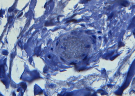
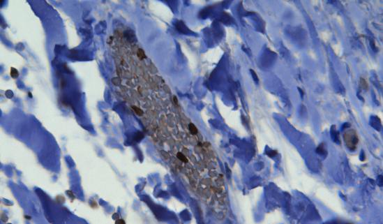
15 S5 CD3 CD4 CX3CR1
16 S6 CD4 + CX3CR1 + T cells 1 CMV-specific T cells CMV 2 TNFα IFNγ 3 CX3CL1 4 Endothelium Arterial wall CMV-antigen presentation Transendothelial migration
Blocking antibodies and peptides. Rat anti-mouse PD-1 (29F.1A12, rat IgG2a, k), PD-
, PD-") Supplementary Methods Blocking antibodies and peptides. Rat anti-mouse PD-1 (29F.1A12, rat IgG2a, k), PD- L1 (10F.9G2, rat IgG2b, k), and PD-L2 (3.2, mouse IgG1) have been described (24). Anti-CTLA-4 (clone
Supplementary Methods Blocking antibodies and peptides. Rat anti-mouse PD-1 (29F.1A12, rat IgG2a, k), PD- L1 (10F.9G2, rat IgG2b, k), and PD-L2 (3.2, mouse IgG1) have been described (24). Anti-CTLA-4 (clone
Commercially available HLA Class II tetramers (Beckman Coulter) conjugated to
 conjugated to") Class II tetramer staining Commercially available HLA Class II tetramers (Beckman Coulter) conjugated to PE were combined with dominant HIV epitopes (DRB1*0101-DRFYKTLRAEQASQEV, DRB1*0301- PEKEVLVWKFDSRLAFHH,
Class II tetramer staining Commercially available HLA Class II tetramers (Beckman Coulter) conjugated to PE were combined with dominant HIV epitopes (DRB1*0101-DRFYKTLRAEQASQEV, DRB1*0301- PEKEVLVWKFDSRLAFHH,
Gladstone Institutes, University of California (UCSF), San Francisco, USA
, San Francisco, USA") Fluorescence-linked Antigen Quantification (FLAQ) Assay for Fast Quantification of HIV-1 p24 Gag Marianne Gesner, Mekhala Maiti, Robert Grant and Marielle Cavrois * Gladstone Institutes, University of
Fluorescence-linked Antigen Quantification (FLAQ) Assay for Fast Quantification of HIV-1 p24 Gag Marianne Gesner, Mekhala Maiti, Robert Grant and Marielle Cavrois * Gladstone Institutes, University of
Supplementary Data 1. Alanine substitutions and position variants of APNCYGNIPL. Applied in
 Supplementary Data 1. Alanine substitutions and position variants of APNCYGNIPL. Applied in Supplementary Fig. 2 Substitution Sequence Position variant Sequence original APNCYGNIPL original APNCYGNIPL
Optimizing Intracellular Flow Cytometry:
 Optimizing Intracellular Flow Cytometry: Simultaneous Detection of Cytokines and Transcription Factors An encore presentation by Jurg Rohrer, PhD, BD Biosciences 10.26.10 Outline Introduction Cytokines
Primary Adult Naïve CD4+ CD45RA+ Cells. Prepared by: David Randolph at University of Alabama, Birmingham
 Primary Adult Naïve CD4+ CD45RA+ Cells Prepared by: David Randolph (drdrdr@uab.edu) at University of Alabama, Birmingham Goal: To obtain large numbers of highly pure primary CD4+ CD45RO- CD25- cells from
PBMC from each patient were suspended in AIM V medium (Invitrogen) with 5% human
 with 5% human") Anti-CD19-CAR transduced T-cell preparation PBMC from each patient were suspended in AIM V medium (Invitrogen) with 5% human AB serum (Gemini) and 300 international units/ml IL-2 (Novartis). T cell proliferation
Anti-CD19-CAR transduced T-cell preparation PBMC from each patient were suspended in AIM V medium (Invitrogen) with 5% human AB serum (Gemini) and 300 international units/ml IL-2 (Novartis). T cell proliferation
Rapid antigen-specific T cell enrichment (Rapid ARTE)
") Direct ex vivo characterization of human antigen-specific CD154+CD4+ T cell Rapid antigen-specific T cell enrichment (Rapid ARTE) Introduction Workflow Antigen (ag)-specific T cells play a central role
Direct ex vivo characterization of human antigen-specific CD154+CD4+ T cell Rapid antigen-specific T cell enrichment (Rapid ARTE) Introduction Workflow Antigen (ag)-specific T cells play a central role
Suppl Video: Tumor cells (green) and monocytes (white) are seeded on a confluent endothelial
 and monocytes (white) are seeded on a confluent endothelial") Supplementary Information Häuselmann et al. Monocyte induction of E-selectin-mediated endothelial activation releases VE-cadherin junctions to promote tumor cell extravasation in the metastasis cascade
Supplementary Information Häuselmann et al. Monocyte induction of E-selectin-mediated endothelial activation releases VE-cadherin junctions to promote tumor cell extravasation in the metastasis cascade
Nature Medicine: doi: /nm.2109
 HIV 1 Infects Multipotent Progenitor Cells Causing Cell Death and Establishing Latent Cellular Reservoirs Christoph C. Carter, Adewunmi Onafuwa Nuga, Lucy A. M c Namara, James Riddell IV, Dale Bixby, Michael
Optimizing Intracellular Flow Cytometry:
 Optimizing Intracellular Flow Cytometry: Simultaneous Detection of Cytokines and Transcription Factors Presented by Jurg Rohrer, PhD, BD Biosciences 23-10780-00 Outline Introduction Cytokines Transcription
Application Information Bulletin: Human NK Cells Phenotypic characterizing of human Natural Killer (NK) cell populations in peripheral blood
 cell populations in peripheral blood") Application Information Bulletin: Human NK Cells Phenotypic characterizing of human Natural Killer (NK) cell populations in peripheral blood Christopher A Fraker, Ph.D., University of Miami - Miami, Florida
Application Information Bulletin: Human NK Cells Phenotypic characterizing of human Natural Killer (NK) cell populations in peripheral blood Christopher A Fraker, Ph.D., University of Miami - Miami, Florida
Detailed step-by-step operating procedures for NK cell and CTL degranulation assays
 Supplemental methods Detailed step-by-step operating procedures for NK cell and CTL degranulation assays Materials PBMC isolated from patients, relatives and healthy donors as control K562 cells (ATCC,
Low Avidity CMV + T Cells accumulate in Old Humans
 Supplementary Figure Legends Supplementary Figure 1. CD45RA expressing CMVpp65-specific T cell populations accumulate within HLA-A*0201 and HLA-B*0701 individuals Pooled data showing the size of the NLV/HLA-A*0201-specific
Ex vivo Human Antigen-specific T Cell Proliferation and Degranulation Willemijn Hobo 1, Wieger Norde 1 and Harry Dolstra 2*
 Ex vivo Human Antigen-specific T Cell Proliferation and Degranulation Willemijn Hobo 1, Wieger Norde 1 and Harry Dolstra 2* 1 Department of Laboratory Medicine - Laboratory of Hematology, Radboud University
Direct ex vivo characterization of human antigen-specific CD154 + CD4 + T cells Rapid antigen-reactive T cell enrichment (Rapid ARTE)
") Direct ex vivo characterization of human antigen-specific CD154 + CD4 + T cells Rapid antigen-reactive T cell enrichment (Rapid ARTE) Introduction Workflow Antigen (ag)-specific T cells play a central
Direct ex vivo characterization of human antigen-specific CD154 + CD4 + T cells Rapid antigen-reactive T cell enrichment (Rapid ARTE) Introduction Workflow Antigen (ag)-specific T cells play a central
Supplementary Figure 1
 Supplementary Figure 1 Identification of IFN-γ-producing CD8 + and CD4 + T cells with naive phenotype by alternative gating and sample-processing strategies. a. Contour 5% probability plots show definition
VEGFR2-Mediated Vascular Dilation as a Mechanism of VEGF-Induced Anemia and Bone Marrow Cell Mobilization
 Cell Reports, Volume 9 Supplemental Information VEGFR2-Mediated Vascular Dilation as a Mechanism of VEGF-Induced Anemia and Bone Marrow Cell Mobilization Sharon Lim, Yin Zhang, Danfang Zhang, Fang Chen,
Human Immunodeficiency Virus Type-1 Myeloid Derived Suppressor Cells Inhibit Cytomegalovirus Inflammation through Interleukin-27 and B7-H4
 Human Immunodeficiency Virus Type-1 Myeloid Derived Suppressor Cells Inhibit Cytomegalovirus Inflammation through Interleukin-27 and B7-H4 Ankita Garg, Rodney Trout and Stephen A. Spector,,* Department
Supplementary Data Table of Contents:
 Supplementary Data Table of Contents: - Supplementary Methods - Supplementary Figures S1(A-B) - Supplementary Figures S2 (A-B) - Supplementary Figures S3 - Supplementary Figures S4(A-B) - Supplementary
Peptide stimulation and Intracellular Cytokine Staining
 v Peptide stimulation and Intracellular Cytokine Staining Authors A. Cosma, S. Allgayer MATERIALS Date 01-02-2007 REAGENTS: Version 1.0 - PBMCs - Culture medium - Costimulating antibodies - Peptide pools
Supplementary Figures
 Inhibition of Pulmonary Anti Bacterial Defense by IFN γ During Recovery from Influenza Infection By Keer Sun and Dennis W. Metzger Supplementary Figures d a Ly6G Percentage survival f 1 75 5 1 25 1 5 1
Supplemental figure 1. PDGFRα is expressed dominantly by stromal cells surrounding mammary ducts and alveoli. A) IHC staining of PDGFRα in
 IHC staining of PDGFRα in") Supplemental figure 1. PDGFRα is expressed dominantly by stromal cells surrounding mammary ducts and alveoli. A) IHC staining of PDGFRα in nulliparous (left panel) and InvD6 mouse mammary glands (right
Supplemental figure 1. PDGFRα is expressed dominantly by stromal cells surrounding mammary ducts and alveoli. A) IHC staining of PDGFRα in nulliparous (left panel) and InvD6 mouse mammary glands (right
Technical Resources. BD Immunocytometry Systems. FastImmune Intracellular Cytokine Staining Procedures
 FastImmune Intracellular Cytokine Staining Procedures BD has developed protocols for the detection of intracellular cytokines in activated lymphocytes and in activated monocytes. The procedures have been
York criteria, 6 RA patients and 10 age- and gender-matched healthy controls (HCs).
.") MATERIALS AND METHODS Study population Blood samples were obtained from 15 patients with AS fulfilling the modified New York criteria, 6 RA patients and 10 age- and gender-matched healthy controls (HCs).
MATERIALS AND METHODS Study population Blood samples were obtained from 15 patients with AS fulfilling the modified New York criteria, 6 RA patients and 10 age- and gender-matched healthy controls (HCs).
MATERIALS. Peptide stimulation and Intracellular Cytokine Staining- EFFECTOR 45RA PANEL
 v Peptide stimulation and Intracellular Cytokine Staining- EFFECTOR 45RA PANEL Authors S. Kutscher, A. Cosma, MATERIALS REAGENTS: - PBMCs - Culture medium - Costimulating antibodies - Peptide pools including
Supplementary Fig. 1: Ex vivo tetramer enrichment with anti-c-myc beads
 Supplementary Fig. 1: Ex vivo tetramer enrichment with anti-c-myc beads Representative example of comparative ex vivo tetramer enrichment performed in three independent experiments with either conventional
In vitro human regulatory T cell suppression assay
 Human CD4 + CD25 + regulatory T cell isolation, in vitro suppression assay and analysis In vitro human regulatory T cell suppression assay Introduction Regulatory T (Treg) cells are a subpopulation of
ImageStream cytometer analysis. Cells were cultured as described above in vented-cap
 ImageStream cytometer analysis. Cells were cultured as described above in vented-cap polypropylene tubes, stained with αcd66b-fitc, αm-dc8-pe and αcd56-pe-cy5.5 mabs, washed and fixed with 4 % (w/v) paraformaldehyde.
In vitro human regulatory T cell expansion
 - 1 - Human CD4 + CD25 + regulatory T cell isolation, Workflow in vitro expansion and analysis In vitro human regulatory T cell expansion Introduction Regulatory T (Treg) cells are a subpopulation of T
SUPPLEMENTARY INFORMATION
 Complete but curtailed T-cell response to very-low-affinity antigen Dietmar Zehn, Sarah Y. Lee & Michael J. Bevan Supp. Fig. 1: TCR chain usage among endogenous K b /Ova reactive T cells. C57BL/6 mice
In vitro human regulatory T cell expansion
 - 1 - Human CD4 + CD25 + CD127 dim/- regulatory T cell Workflow isolation, in vitro expansion and analysis In vitro human regulatory T cell expansion Introduction Regulatory T (Treg) cells are a subpopulation
Fluorochrome Panel 1 Panel 2 Panel 3 Panel 4 Panel 5 CTLA-4 CTLA-4 CD15 CD3 FITC. Bio) PD-1 (MIH4, BD) ICOS (C398.4A, Biolegend) PD-L1 (MIH1, BD)
 PD-1 (MIH4, BD) ICOS (C398.4A, Biolegend) PD-L1 (MIH1, BD)") Additional file : Table S. Antibodies used for panel stain to identify peripheral immune cell subsets. Panel : PD- signaling; Panel : CD + T cells, CD + T cells, B cells; Panel : Tregs; Panel :, -T, cdc,
Additional file : Table S. Antibodies used for panel stain to identify peripheral immune cell subsets. Panel : PD- signaling; Panel : CD + T cells, CD + T cells, B cells; Panel : Tregs; Panel :, -T, cdc,
January 25, 2017 Scientific Research Process Name of Journal: ESPS Manuscript NO: Manuscript Type: Title: Authors: Correspondence to
 January 25, 2017 Scientific Research Process Name of Journal: World Journal of Gastroenterology ESPS Manuscript NO: 31928 Manuscript Type: ORIGINAL ARTICLE Title: Thiopurine use associated with reduced
Supporting Information
 Supporting Information Desnues et al. 10.1073/pnas.1314121111 SI Materials and Methods Mice. Toll-like receptor (TLR)8 / and TLR9 / mice were generated as described previously (1, 2). TLR9 / mice were
Pearson r = P (one-tailed) = n = 9
 = n = 9") 8F4-Specific Lysis, % 1 UPN1 UPN3 8 UPN7 6 Pearson r =.69 UPN2 UPN5 P (one-tailed) =.192 4 UPN8 n = 9 2 UPN9 UPN4 UPN6 5 1 15 2 25 8 8F4, % Max MFI Supplementary Figure S1. AML samples UPN1-UPN9 show variable
8F4-Specific Lysis, % 1 UPN1 UPN3 8 UPN7 6 Pearson r =.69 UPN2 UPN5 P (one-tailed) =.192 4 UPN8 n = 9 2 UPN9 UPN4 UPN6 5 1 15 2 25 8 8F4, % Max MFI Supplementary Figure S1. AML samples UPN1-UPN9 show variable
Supporting Online Material for
 www.sciencemag.org/cgi/content/full/1175194/dc1 Supporting Online Material for A Vital Role for Interleukin-21 in the Control of a Chronic Viral Infection John S. Yi, Ming Du, Allan J. Zajac* *To whom
Figure S1. PMVs from THP-1 cells expose phosphatidylserine and carry actin. A) Flow
 Flow") SUPPLEMENTARY DATA Supplementary Figure Legends Figure S1. PMVs from THP-1 cells expose phosphatidylserine and carry actin. A) Flow cytometry analysis of PMVs labelled with annexin-v-pe (Guava technologies)
SUPPLEMENTARY DATA Supplementary Figure Legends Figure S1. PMVs from THP-1 cells expose phosphatidylserine and carry actin. A) Flow cytometry analysis of PMVs labelled with annexin-v-pe (Guava technologies)
Hua Tang, Weiping Cao, Sudhir Pai Kasturi, Rajesh Ravindran, Helder I Nakaya, Kousik
 SUPPLEMENTARY FIGURES 1-19 T H 2 response to cysteine-proteases requires dendritic cell-basophil cooperation via ROS mediated signaling Hua Tang, Weiping Cao, Sudhir Pai Kasturi, Rajesh Ravindran, Helder
BD CBA on the BD Accuri C6: Bringing Multiplexed Cytokine Detection to the Benchtop
 BD CBA on the BD Accuri C6: Bringing Multiplexed Cytokine Detection to the Benchtop Maria Dinkelmann, PhD Senior Marketing Applications Specialist BD Biosciences, Ann Arbor, MI 23-14380-00 Cellular Communication
Optimizing Intracellular Flow Cytometry
 Optimizing Intracellular Flow Cytometry Detection of Cytokines, Transcription Factors, and Phosphoprotein by Flow Cytometry Presented by Erika O Donnell, PhD, BD Biosciences 23-14876-00 Outline Basic principles
Supplementary Table e-1. Flow cytometry reagents and staining combinations
 Supplementary data Supplementary Table e-1. Flow cytometry reagents and staining combinations Reagents Antibody Fluorochrome Clone Source conjugation CD3 FITC UCHT1 BD Biosciences CD3 PerCP-Cy5.5 SK7 Biolegend
CD14 + S100A9 + Monocytic Myeloid-Derived Suppressor Cells and Their Clinical Relevance in Non-Small Cell Lung Cancer
 CD14 + S1A9 + Monocytic Myeloid-Derived Suppressor Cells and Their Clinical Relevance in Non-Small Cell Lung Cancer Po-Hao, Feng M.D., Kang-Yun, Lee, M.D. Ph.D., Ya-Ling Chang, Yao-Fei Chan, Lu- Wei, Kuo,Ting-Yu
Effect of Antiretroviral Therapy on HIV-mediated Impairment of the Neutrophil
 Online Data Supplement Effect of Antiretroviral Therapy on HIV-mediated Impairment of the Neutrophil Antimycobacterial Response David M Lowe, Nonzwakazi Bangani, Rene Goliath, Beate Kampmann, Katalin A
Interleukin-2-Dependent Allergen-Specific Tissue-Resident Memory Cells Drive Asthma
 Immunity Supplemental Information Interleukin-2-Dependent Allergen-Specific Tissue-Resident Memory Cells Drive Asthma Brian D. Hondowicz, Dowon An, Jason M. Schenkel, Karen S. Kim, Holly R. Steach, Akshay
IL-6Rα IL-6RαT-KO KO. IL-6Rα f/f bp. f/f 628 bp deleted 368 bp. 500 bp
 STD H 2 O WT KO IL-6Rα f/f IL-6Rα IL-6RαT-KO KO 1000 bp 500 bp f/f 628 bp deleted 368 bp Supplementary Figure 1 Confirmation of T-cell IL-6Rα deficiency. (a) Representative histograms and (b) quantification
Supplementary Table; Supplementary Figures and legends S1-S21; Supplementary Materials and Methods
 Silva et al. PTEN posttranslational inactivation and hyperactivation of the PI3K/Akt pathway sustain primary T cell leukemia viability Supplementary Table; Supplementary Figures and legends S1-S21; Supplementary
Supplementary Figure 1. Enhanced detection of CTLA-4 on the surface of HIV-specific
 SUPPLEMENTARY FIGURE LEGEND Supplementary Figure 1. Enhanced detection of CTLA-4 on the surface of HIV-specific CD4 + T cells correlates with intracellular CTLA-4 levels. (a) Comparative CTLA-4 levels
T H 1, T H 2 and T H 17 polarization of naïve CD4 + mouse T cells
 A complete workflow for cell preparation, isolation, polarization and analysis T H 1, T H 2 and T H 17 polarization of naïve CD4 + mouse T cells Introduction Workflow CD4 + T helper (T H) cells play a
MATERIALS AND METHODS. Neutralizing antibodies specific to mouse Dll1, Dll4, J1 and J2 were prepared as described. 1,2 All
 MATERIALS AND METHODS Antibodies (Abs), flow cytometry analysis and cell lines Neutralizing antibodies specific to mouse Dll1, Dll4, J1 and J2 were prepared as described. 1,2 All other antibodies used
Supplementary Materials for
 immunology.sciencemag.org/cgi/content/full/2/16/eaan6049/dc1 Supplementary Materials for Enzymatic synthesis of core 2 O-glycans governs the tissue-trafficking potential of memory CD8 + T cells Jossef
Challenges in Development and Validation of an Intracellular Cytokine Staining assay
 Challenges in Development and Validation of an Intracellular Cytokine Staining assay Jenny Hendriks, Crucell Hatching @ EBF, Brussels, June 202 www.crucell.com Vaccines vs Protein therapeutics Protein
HD1 (FLU) HD2 (EBV) HD2 (FLU)
 HD2 (EBV) HD2 (FLU)") ramer staining + anti-pe beads ramer staining a HD1 (FLU) HD2 (EBV) HD2 (FLU).73.11.56.46.24 1.12 b CD127 + c CD127 + d CD127 - e CD127 - PD1 - PD1 + PD1 + PD1-1 1 1 1 %CD127 + PD1-8 6 4 2 + anti-pe %CD127
ramer staining + anti-pe beads ramer staining a HD1 (FLU) HD2 (EBV) HD2 (FLU).73.11.56.46.24 1.12 b CD127 + c CD127 + d CD127 - e CD127 - PD1 - PD1 + PD1 + PD1-1 1 1 1 %CD127 + PD1-8 6 4 2 + anti-pe %CD127
Supplementary Table 1: Summary of Reactogenicity Events by Group and Time of Onset in Entebbe
 Supplementary Table 1: Summary of Reactogenicity Events by Group and Time of Onset in Group A (n=39) B (n=11) A (n=39) B (n=11) Onset Post-Vaccination Event 3 Minutes 3-14 days Pain 5 (13%) 1 (1%) 6 (15%)
ab Exosome Isolation and Analysis Kit - Flow Cytometry, Cell Culture (CD63 / CD81)
") Version 1 Last updated 26 September 2018 ab239682 Exosome Isolation and Analysis Kit - Flow Cytometry, Cell Culture (CD63 / For the isolation and analysis of exosome from cell culture. This product is
Version 1 Last updated 26 September 2018 ab239682 Exosome Isolation and Analysis Kit - Flow Cytometry, Cell Culture (CD63 / For the isolation and analysis of exosome from cell culture. This product is
BD Flow Cytometry Reagents Multicolor Panels Designed for Optimal Resolution with the BD LSRFortessa X-20 Cell Analyzer
 Multicolor Panels Designed for Optimal Resolution with the BD LSRFortessa X-2 Cell Analyzer Proper multicolor panel design takes into account fluorochrome brightness, antigen density, co-expression, and
Supplementary Data. Treg phenotype
 Supplementary Data Additional Experiment An additional experiment was performed using cryopreserved peripheral blood mononuclear cells (PBMC) derived from five renal cell carcinoma (RCC) patients [see
SUPPLEMENTARY FIGURES
 SUPPLEMENTARY FIGURES Supplementary Figure 1: Chemokine receptor expression profiles of CCR6 + and CCR6 - CD4 + IL-17A +/ex and Treg cells. Quantitative PCR analysis of chemokine receptor transcript abundance
Table S1. Viral load and CD4 count of HIV-infected patient population
 Table S1. Viral load and CD4 count of HIV-infected patient population Subject ID Viral load (No. of copies per ml of plasma) CD4 count (No. of cells/µl of blood) 28 7, 14 29 7, 23 21 361,99 94 217 7, 11
MAIT cell function is modulated by PD-1 signaling in patients with active
 MAIT cell function is modulated by PD-1 signaling in patients with active tuberculosis Jing Jiang, M.D., Xinjing Wang, M.D., Hongjuan An, M.Sc., Bingfen Yang, Ph.D., Zhihong Cao, M.Sc., Yanhua Liu, Ph.D.,
Supplementary Figure 1. Example of gating strategy
 Supplementary Figure 1. Example of gating strategy Legend Supplementary Figure 1: First, gating is performed to include only single cells (singlets) (A) and CD3+ cells (B). After gating on the lymphocyte
Human Immunodeficiency Virus type 1 (HIV-1) gp120 / Glycoprotein 120 ELISA Pair Set
 gp120 / Glycoprotein 120 ELISA Pair Set") Human Immunodeficiency Virus type 1 (HIV-1) gp120 / Glycoprotein 120 ELISA Pair Set Catalog Number : SEK11233 To achieve the best assay results, this manual must be read carefully before using this product
Human Immunodeficiency Virus type 1 (HIV-1) gp120 / Glycoprotein 120 ELISA Pair Set Catalog Number : SEK11233 To achieve the best assay results, this manual must be read carefully before using this product
Influenza A H1N1 (Swine Flu 2009) Hemagglutinin / HA ELISA Pair Set
 Hemagglutinin / HA ELISA Pair Set") Influenza A H1N1 (Swine Flu 2009) Hemagglutinin / HA ELISA Pair Set Catalog Number : SEK001 To achieve the best assay results, this manual must be read carefully before using this product and the assay
Influenza A H1N1 (Swine Flu 2009) Hemagglutinin / HA ELISA Pair Set Catalog Number : SEK001 To achieve the best assay results, this manual must be read carefully before using this product and the assay
SUPPORTING INFORMATIONS
 SUPPORTING INFORMATIONS Mice MT/ret RetCD3ε KO α-cd25 treated MT/ret Age 1 month 3 mnths 6 months 1 month 3 months 6 months 1 month 3 months 6 months 2/87 Survival 87/87 incidence of 17/87 1 ary tumor
(A) PCR primers (arrows) designed to distinguish wild type (P1+P2), targeted (P1+P2) and excised (P1+P3)14-
 PCR primers (arrows) designed to distinguish wild type (P1+P2), targeted (P1+P2) and excised (P1+P3)14-") 1 Supplemental Figure Legends Figure S1. Mammary tumors of ErbB2 KI mice with 14-3-3σ ablation have elevated ErbB2 transcript levels and cell proliferation (A) PCR primers (arrows) designed to distinguish
1 Supplemental Figure Legends Figure S1. Mammary tumors of ErbB2 KI mice with 14-3-3σ ablation have elevated ErbB2 transcript levels and cell proliferation (A) PCR primers (arrows) designed to distinguish
Cover Page. The handle holds various files of this Leiden University dissertation.
 Cover Page The handle http://hdl.handle.net/1887/23854 holds various files of this Leiden University dissertation. Author: Marel, Sander van der Title: Gene and cell therapy based treatment strategies
A step-by-step approach to build and analyze a multicolor panel
 Analyze A step-by-step approach to build and analyze a multicolor panel For Research Use Only. Not for use in diagnostic or therapeutic procedures. Alexa Fluor is a registered trademark of Life Technologies
Supplemental Methods. CD107a assay
 Supplemental Methods CD107a assay For each T cell culture that was tested, two tubes were prepared. One tube contained BCMA-K562 cells, and the other tube contained NGFR-K562 cells. Both tubes contained
x Lymphocyte count /µl CD8+ count/µl 800 Calculated
 % Lymphocyte in CBC A. 50 40 30 20 10 Lymphocyte count /µl B. x10 3 2.5 1.5 C. 50 D. 1000 % CD3+CD8+ Cells 40 30 20 Calculated CD8+ count/µl 800 600 400 200 10 0 #61 #63 #64 #65 #68 #71 #72 #75 Figure
Human and mouse T cell regulation mediated by soluble CD52 interaction with Siglec-10. Esther Bandala-Sanchez, Yuxia Zhang, Simone Reinwald,
 Human and mouse T cell regulation mediated by soluble CD52 interaction with Siglec-1 Esther Bandala-Sanchez, Yuxia Zhang, Simone Reinwald, James A. Dromey, Bo Han Lee, Junyan Qian, Ralph M Böhmer and Leonard
Monocyte subsets in health and disease. Marion Frankenberger
 Monocyte subsets in health and disease Marion Frankenberger main cellular components: Leukocytes Erythrocytes Composition of whole blood Monocytes belong to the cellular components of peripheral blood
Islet viability assay and Glucose Stimulated Insulin Secretion assay RT-PCR and Western Blot
 Islet viability assay and Glucose Stimulated Insulin Secretion assay Islet cell viability was determined by colorimetric (3-(4,5-dimethylthiazol-2-yl)-2,5- diphenyltetrazolium bromide assay using CellTiter
SUPPLEMENTARY METHODS
 SUPPLEMENTARY METHODS Histological analysis. Colonic tissues were collected from 5 parts of the middle colon on day 7 after the start of DSS treatment, and then were cut into segments, fixed with 4% paraformaldehyde,
Interferon γ regulates idiopathic pneumonia syndrome, a. Th17 + CD4 + T-cell-mediated GvH disease
 Interferon γ regulates idiopathic pneumonia syndrome, a Th17 + CD4 + T-cell-mediated GvH disease Nora Mauermann, Julia Burian, Christophe von Garnier, Stefan Dirnhofer, Davide Germano, Christine Schuett,
Supplementary Figure 1. IL-12 serum levels and frequency of subsets in FL patients. (A) IL-12
 IL-12") 1 Supplementary Data Figure legends Supplementary Figure 1. IL-12 serum levels and frequency of subsets in FL patients. (A) IL-12 serum levels measured by multiplex ELISA (Luminex) in FL patients before
1 Supplementary Data Figure legends Supplementary Figure 1. IL-12 serum levels and frequency of subsets in FL patients. (A) IL-12 serum levels measured by multiplex ELISA (Luminex) in FL patients before
Cell isolation. Spleen and lymph nodes (axillary, inguinal) were removed from mice
 were removed from mice") Supplementary Methods: Cell isolation. Spleen and lymph nodes (axillary, inguinal) were removed from mice and gently meshed in DMEM containing 10% FBS to prepare for single cell suspensions. CD4 + CD25
Supplementary Methods: Cell isolation. Spleen and lymph nodes (axillary, inguinal) were removed from mice and gently meshed in DMEM containing 10% FBS to prepare for single cell suspensions. CD4 + CD25
Supplementary Figure S1. Flow cytometric analysis of the expression of Thy1 in NH cells. Flow cytometric analysis of the expression of T1/ST2 and
 Supplementary Figure S1. Flow cytometric analysis of the expression of Thy1 in NH cells. Flow cytometric analysis of the expression of T1/ST2 and Thy1 in NH cells derived from the lungs of naïve mice.
Human LDL Receptor / LDLR ELISA Pair Set
 Human LDL Receptor / LDLR ELISA Pair Set Catalog Number : SEK10231 To achieve the best assay results, this manual must be read carefully before using this product and the assay is run as summarized in
Supporting Information
 Supporting Information Pang et al. 10.1073/pnas.1322009111 SI Materials and Methods ELISAs. These assays were performed as previously described (1). ELISA plates (MaxiSorp Nunc; Thermo Fisher Scientific)
ab LDL Uptake Assay Kit (Cell-Based)
") ab133127 LDL Uptake Assay Kit (Cell-Based) Instructions for Use For the detection of LDL uptake into cultured cells. This product is for research use only and is not intended for diagnostic use. Version
ab133127 LDL Uptake Assay Kit (Cell-Based) Instructions for Use For the detection of LDL uptake into cultured cells. This product is for research use only and is not intended for diagnostic use. Version
Supplemental Information. Tissue Myeloid Progenitors Differentiate. into Pericytes through TGF-b Signaling. in Developing Skin Vasculature
 Cell Reports, Volume 18 Supplemental Information Tissue Myeloid Progenitors Differentiate into Pericytes through TGF-b Signaling in Developing Skin Vasculature Tomoko Yamazaki, Ani Nalbandian, Yutaka Uchida,
Figure S1. Generation of inducible PTEN deficient mice and the BMMCs (A) B6.129 Pten loxp/loxp mice were mated with B6.
 B6.129 Pten loxp/loxp mice were mated with B6.") Figure S1. Generation of inducible PTEN deficient mice and the BMMCs (A) B6.129 Pten loxp/loxp mice were mated with B6.129-Gt(ROSA)26Sor tm1(cre/ert2)tyj /J mice. To induce deletion of the Pten locus,
Figure S1. Generation of inducible PTEN deficient mice and the BMMCs (A) B6.129 Pten loxp/loxp mice were mated with B6.129-Gt(ROSA)26Sor tm1(cre/ert2)tyj /J mice. To induce deletion of the Pten locus,
Evaluation of directed and random motility in microslides Assessment of leukocyte adhesion in flow chambers
 Evaluation of directed and random motility in microslides Motility experiments in IBIDI microslides, image acquisition and processing were performed as described. PMN, which ended up in an angle < 180
Supporting Information
 Supporting Information Idoyaga et al. 10.1073/pnas.0812247106 SSC a) Single cell suspension 99 Aqua b) Live cells 96 -W c) Singlets 92 -A CD19+ER119 d) CD19 ER119 cells 97 CD3 e) CD3 cells 27 f) DX5 cells
SUPPLEMENTARY INFORMATION. Involvement of IL-21 in the epidermal hyperplasia of psoriasis
 SUPPLEMENTARY INFORMATION Involvement of IL-21 in the epidermal hyperplasia of psoriasis Roberta Caruso 1, Elisabetta Botti 2, Massimiliano Sarra 1, Maria Esposito 2, Carmine Stolfi 1, Laura Diluvio 2,
Superior Fluorescent Labeling Dyes Spanning the Full Visible Spectrum...1. Trademarks: HiLyte Fluor (AnaSpec, Inc.)
") Table of Contents Fluor TM Labeling Dyes Superior Fluorescent Labeling Dyes Spanning the Full Visible Spectrum....1 Fluor TM 405 Dye, an Excellent Alternative to Alexa Fluor 405 & DyLight 405....2 Fluor
Table of Contents Fluor TM Labeling Dyes Superior Fluorescent Labeling Dyes Spanning the Full Visible Spectrum....1 Fluor TM 405 Dye, an Excellent Alternative to Alexa Fluor 405 & DyLight 405....2 Fluor
Supplementary Figure 1 Lymphocytes can be tracked for at least 4 weeks after
 Supplementary Figure 1 Lymphocytes can be tracked for at least 4 weeks after photoconversion by using H2B-Dendra2. 4-5 PPs of H2B-Dendra2 BM chimeras were photoconverted and analyzed 7 days (upper panel)
For research or further manufacturing use only. Not for injection or diagnostic procedures.
 PRIME-XV T cell Expansion XSFM PRIME-XV T Cell Expansion XSFM is a xeno-free, serum-free medium optimized for the activation and expansion of human T lymphocytes. This medium contains gentamicin and requires
2009 H1N1 Influenza ( Swine Flu ) Hemagglutinin ELISA kit
 Hemagglutinin ELISA kit") 2009 H1N1 Influenza ( Swine Flu ) Hemagglutinin ELISA kit Catalog Number : SEK001 To achieve the best assay results, this manual must be read carefully before using this product and the assay is run as
2009 H1N1 Influenza ( Swine Flu ) Hemagglutinin ELISA kit Catalog Number : SEK001 To achieve the best assay results, this manual must be read carefully before using this product and the assay is run as
Activating and propagating polyclonal gamma delta T cells with broad specificity for
 SUPPLEMENTAL MATERIAL: Activating and propagating polyclonal gamma delta T cells with broad specificity for malignancies ADDITIONAL MATERIALS AND METHODS Tumor cell line culture conditions Cell cultures
ab Exosome Isolation and Analysis Kit - Flow Cytometry, Cell culture
 Version 1 Last updated 14 March 2018 ab228564 Exosome Isolation and Analysis Kit - Flow Cytometry, Cell culture For the measurement of human exosomes in cell culture. This product is for research use only
BMDCs were generated in vitro from bone marrow cells cultured in 10 % RPMI supplemented
 Supplemental Materials Figure S1. Cultured BMDCs express CD11c BMDCs were generated in vitro from bone marrow cells cultured in 10 % RPMI supplemented with 15 ng/ml GM-CSF. Media was changed and fresh
HIV-1 p24 ELISA Pair Set Cat#: orb54951 (ELISA Manual)
") HIV-1 p24 ELISA Pair Set Cat#: orb54951 (ELISA Manual) BACKGROUND Human Immunodeficiency Virus ( HIV ) can be divided into two major types, HIV type 1 (HIV-1) and HIV type 2 (HIV-2). HIV-1 is related to
HIV-1 p24 ELISA Pair Set Cat#: orb54951 (ELISA Manual) BACKGROUND Human Immunodeficiency Virus ( HIV ) can be divided into two major types, HIV type 1 (HIV-1) and HIV type 2 (HIV-2). HIV-1 is related to
Supplementary Table 1
 Supplementary Table 1 Flow Cytometry Antibodies Antibody Fluorochrome Clone Vendor CD45 PE-cyanine 7 30-F11 D ioscience CD3 Pacific lue 17A2 iolegend (San Diego, CA) CD11b APC M1/70 iolegend (San Diego,
ab Exosome Isolation and Analysis Kit - Flow Cytometry, Plasma
 Version 1 Last updated 25 May 2018 ab228565 Exosome Isolation and Analysis Kit - Flow Cytometry, Plasma For the isolation/detection of exosomes from human plasma, urine or cell culture media. This product
well for 2 h at rt. Each dot represents an individual mouse and bar is the mean ±
 Supplementary data: Control DC Blimp-1 ko DC 8 6 4 2-2 IL-1β p=.5 medium 8 6 4 2 IL-2 Medium p=.16 8 6 4 2 IL-6 medium p=.3 5 4 3 2 1-1 medium IL-1 n.s. 25 2 15 1 5 IL-12(p7) p=.15 5 IFNγ p=.65 4 3 2 1
Essential Medium, containing 10% fetal bovine serum, 100 U/ml penicillin and 100 µg/ml streptomycin. Huvec were cultured in
 Supplemental data Methods Cell culture media formulations A-431 and U-87 MG cells were maintained in Dulbecco s Modified Eagle s Medium. FaDu cells were cultured in Eagle's Minimum Essential Medium, containing
Supporting Information
 Supporting Information Bellora et al. 10.1073/pnas.1007654108 SI Materials and Methods Cells. NK cells purified from peripheral blood mononuclear cells (PBMC) of healthy donors (Human NK Cell Isolation
Influenza B Hemagglutinin / HA ELISA Pair Set
 Influenza B Hemagglutinin / HA ELISA Pair Set Catalog Number : SEK11053 To achieve the best assay results, this manual must be read carefully before using this product and the assay is run as summarized
HCC1937 is the HCC1937-pcDNA3 cell line, which was derived from a breast cancer with a mutation
 SUPPLEMENTARY INFORMATION Materials and Methods Human cell lines and culture conditions HCC1937 is the HCC1937-pcDNA3 cell line, which was derived from a breast cancer with a mutation in exon 20 of BRCA1