The basis of Disease
|
|
|
- Jared Jefferson
- 5 years ago
- Views:
Transcription
1 General Curriculum The basis of Disease ZHOU REN 周韧 Prof., M.D., Ph.D. Institute of Pathology & Forensic Medicine Department of Pathology & Patho-physiology Zhenjiang University Judicial Evidence & Evaluation Center Zhejiang University School of Medicine
2 (2). Cell injury
3 Form and Morphology of cell injury 1. Intracellular Accumulations (degeneration) (1)Cellular swelling Conception: Influx of H 2 O and abnormal accumulation within cell whenever cells are incapable of maintaining ionic and fluid homeostasis. A common form of cell injury. The most often affected organ are liver, kidney, heart etc.

4 Causes and mechanism: hypoxia, intoxication, infection. G: Pallor, loss of normal glistening transluscency as if scalded in hot water.

5 H: Swollen of affected cells, with fine granules and vacuoles filling the cytoplasm. The severe form of this type of cell injury is known as hydropic change, even ballooning change.
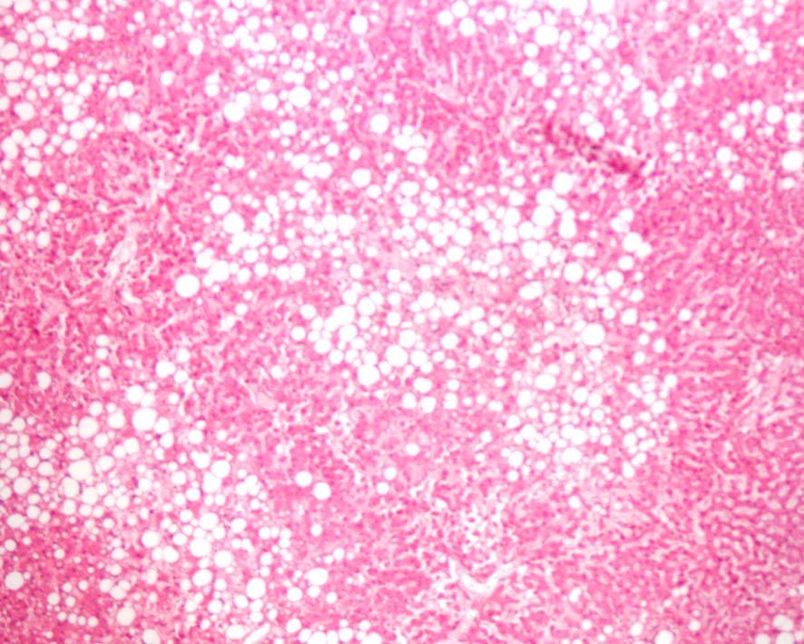
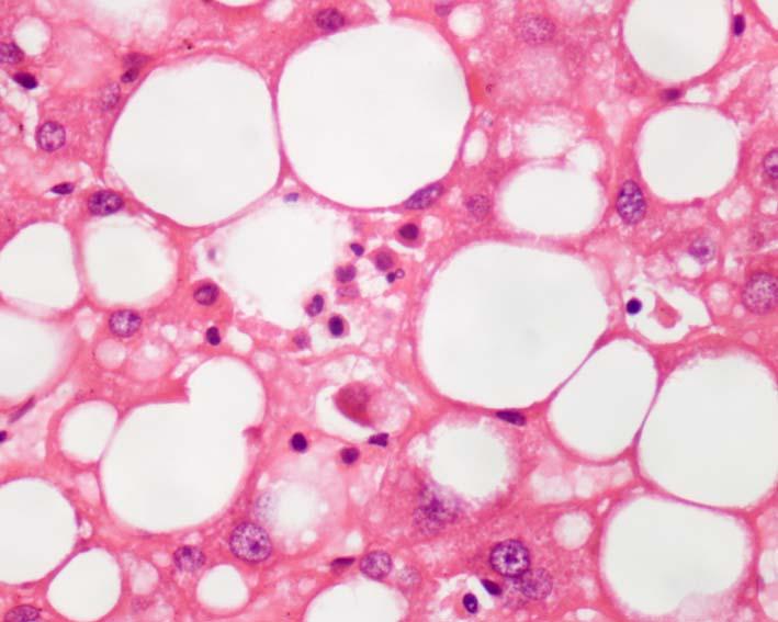
6 (2)Fatty change Conception: Fatty change refers to any abnormal accumulation of fat within parenchymal cells. Most affected organ: liver
7 Causes and mechanism of fatty change of liver:

8 Morphology G: Liver enlargement, yellow in color

9 H: Small vacuoles in the cytoplasm. In severe case, the vacuoles coalesce to large vacuole pushing the nucleus against the cell membrane.
10
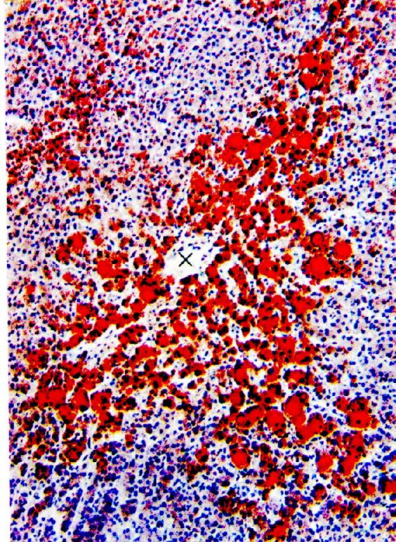
11 Fatty change(sudaniii)

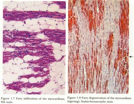
12 Fatty change of cardiac muscle G. Tigroid appearance. Fatty change is more severe in cardiac muscle remote from the blood vessel than in those near the vessel.

13 H. Fat globules within the muscle fibers.
14

15 (3)Hyaline change Conception: Appearance of homogenous, refractive, eosinophilic, hyaline material in connective tissue, vascular wall and cell is known as hyaline change.
16
17

18 (4)Amyloid change(amyloidosis) Conception: Amyloid is an abnormal proteinaceous substance which is deposited between cells in many tissue and organs in a variety of clinical settings. At one time it was thought to be starch-like substance, hence the designation amyloid, however, it is now known to be protein in nature.
19 Morphology: With usual tissue stains, amyloid appears as an intercellular pink translucent material.
20 Systemic amyloidosis: Follows chronic infection (osteomyelitis, tuberculosis). Spleen and kidney are the most prominently affected organs. Local amyloidosis: Occurs in sites with numerous plasma cell infiltration. (eyelid and upper respiratory tract).
21

22
23 (5) Mucoid degeneration Mucoid accumulation mucin Myxedema The diseases usually involved are: Rhumatic disease Tumour
24 (6)Intracellular pigment accumulation (i)hemosiderin Conception: Hemosiderin is a goldenyellow to brown iron containing pigment readily visible with the light microscope and shows positive Prussian blue reaction.
25
26 Morphology:

27 (ii)lipofuscin Conception: Lipofuscin is an insoluble pigment also known as wear-and tear or aging pigment. It represents the indigestible residues of autophagic vacuoles formed during aging or atrophy. Morphology:
28 (iii)melanin Conception: Melanin is an endogenous, nonhemoglobin-derived, brown- black pigment formed when the enzyme tyrosinase catalyzes the oxidation of tyrosine to dihydroxyphenylalanine(dopa). In humans, melanin synthesis is under adrenal and pituitary control. Albinos suffer from a hereditary lack of tyrosinase.
29 Morphology:
Dystrophic calcification: Deposition of calcium in dead or dying tissue despite normal serum calcium level and normal calcium")
30 (7) Pathological calcification Conception: The deposition of calcium in cells and tissues other than bone, cartilage and teeth. (1)Dystrophic calcification: Deposition of calcium in dead or dying tissue despite normal serum calcium level and normal calcium metabolism

31 Course of Basic Medical Sciences Chapter 1
32 (2)Metastatic calcification: Metastatic calcification is always in association with deranged calcium metabolism, leading to hypercalcemia (hyperparathyroidism, widespread metastasis of bone cancers and vit. D intoxication).
33 2. Cell Death I. Necrosis Def. Necrosis may be defined as the morphology of cell death in living body. Autolysis Heterolysis

34 Fundamental changes 1Nuclear changes: Pyknosis, karyorrhexis, Karyolysis. 2Cytoplasmic changes: acidophilic, granular, opaque mass.
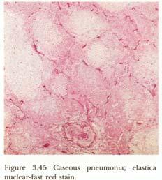
35 Gross appearances of necrosis in earlier stage
36 Classification of necrosis 1Coagulative necrosis 2Caseous necrosis 3Liquefactive necrosis 4 Fat necrosis Fibrinoid necrosis (Fibrinoid change ) Gangrene a)dry gangrene b)moist gangrene c)gas gangrene
37 1 Coagulative necrosis 2 Liquefactive necrosis 3 Types of Special necrosis Caseous necrosis Gangrene a) Dry gangrene b) Moist gangrene c) Gas gangrene Fat necrosis Fibrinoid necrosis (Fibrinoid change )

38 Types of necrosis. 1Coagulative necrosis Conception: Coagulative necrosis implies preservation of the basic outline of the coagulated cell for some days with the disappearance of fine structures (cellular details) within cell.

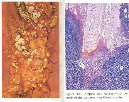
39
40

41
42 Mechanism: Intracellular acidosis denatures not only structural proteins but also enzymic proteins and so blocks the proteolysis of the cell.

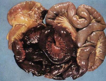
43 Morphology: 2Caseous necrosis Conception: A distinctive form of coagulative necrosis, most often occurs in tuberculosis. The term caseous is derived from the gross appearance of the necrotic area. i.e. white and cheesy. Characteristic morphology: disappearance of both basic outline and fine structures (cellular details) within cell of the affected cell.
44
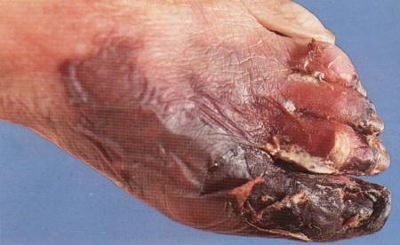
45

46 3Liquefactive necrosis Conception: The damaged cells are lysed by autolysis or heterolysis leaving amorphous, fluid or semifluid material. Morphology:
traumatic fat necrosis: Trauma induce rupture of fat cells.")
47 4 Fat necrosis a)enzymatic fat necrosis: In acute pancreatitis, activated pancreatic enzymes are released from pancreas resulting in destruction of fat tissue. b)traumatic fat necrosis: Trauma induce rupture of fat cells. The released fat causes chronic inflammation and foreign body reaction.
48

49 Fibrinoid necrosis(fibrinoid change) Conception: Accumulation of pink staining homogenous masses of fibrin, immunoglobulins, and other plasma proteins within the vascular wall, connetive tissue is known as fibrinoid change.
50 Gangrene Def. Massive necrosis of body surface or organs with a passage to the body surface with putrefaction superadded is known as gangrene.
51 Classification: a)dry gangrene Conception: Dry gangrene occurs in the extremities due to occlusion of large artery with intact venous returns. The necrotic area is well dermacated black and mummification.
52 Morphology:
53 b)moist gangrene Conception: Blockage of venous return and artery supply massive necrosis followed by liquefaction caused by bacterial enzyme purplish black color with fouel smelling.
54
 + clostridia group bacterial infection gas production through the action of")
55 c)gas gangrene Conception: A serious complication of war wounds. deep contaminated wound(massive necrosis) + clostridia group bacterial infection gas production through the action of saccharolytic and proteolytic enzyme released from bacteria.
56
The basis of Disease
 General Curriculum The basis of Disease ZHOU REN 周韧 Prof., M.D., Ph.D. Institute of Pathology & Forensic Medicine Department of Pathology & Patho-physiology Zhenjiang University Judicial Evidence & Evaluation
SECTION 2 CELL INJURY
 Adapted myocyte Normal myocyte Reversibly-injured myocyte SECTION 2 CELL INJURY Cell death 5/4/2014 1 5/4/2014 2 Reversible Degeneration Irreversible Cellular Swelling Fatty Change Hyaline Change Amyloid
PATHOLOGY Intracellular Degeneration LAB 1
 PATHOLOGY Intracellular Degeneration LAB 1 Cellular swelling Liver Organ :- Liver Lesion :- 1. Narrowing of hepatic sinusoids due to the swelling of hepatocyte. 2. The cytoplasm of affected hepatocyte
number Done by Corrected by Doctor Heyam Awad
 number 4 Done by Waseem Abu Obeida Corrected by Saad Al-Hayek Doctor Heyam Awad Cell injury -in the previous lectures we talked about the causes (etiology) and the mechanism (pathogenesis) of cell injury.
Cellular responses to stress
 Cellular responses to stress (Adaptations, injury and death) (2 of 5) Most injurious stimuli are grouped into: Oxygen deprivation Chemical agents Infectious agents Immunologic reactions Genetic factors
Cell injury, adaptation and death. Unite one Second Lab.
 Cell injury, adaptation and death Unite one Second Lab. The two lung abscesses seen here are examples of liquefactive necrosis in which there is a liquid center in an area of tissue injury. One abscess
Necrosis is death of cells and tissues in the living animal. Focal/ Multifocal necrosis- terms used for one
 Necrosis Necrosis Necrosis is death of cells and tissues in the living animal. Focal/ Multifocal necrosis- terms used for one or more, small, clearly defined areas of necrosis. Diffuse necrosis- term used
PREPARED BY P.DHARANI PRASAD II YEAR B.PHARM II SEM SUB:PATHOPHYSIOLOGY
 CELL INJURY UNIT I PREPARED BY P.DHARANI PRASAD II YEAR B.PHARM II SEM SUB:PATHOPHYSIOLOGY DETECTION OF CELLULAR CHANGES AFTER INJURY BY: LIGHT MICROSCOPY OR GROSS EXAMINATION DETECT CHANGES HOURS TO DAYS
INTRODUCTION TO HEALTH AND DISEASE BLOCK
 MBBS 1 st Yr. Lecture Dr. Annie Cheung September 25, 2002, 8:30AM 9:30 AM LT1, G/F, Academic and Administration Block Faculty of Medicine Building INTRODUCTION TO HEALTH AND DISEASE BLOCK CELL INJURY AND
Histopathology: Cell necrosis and cytoplasmic accumulations
 Histopathology: Cell necrosis and cytoplasmic accumulations These presentations are to help you identify basic histopathological features. They do not contain the additional factual information that you
Cell Adaptation, Cell Injury and Cell Death
 Cell Adaptation, Cell Injury and Cell Death Pathology:- is the study of structural and functional abnormalities that are expressed as diseases of organs and systems. Modern pathology, proposed that injury
DEGENERATION NECROSIS AND INFILTRATION
 DEGENERATION NECROSIS AND INFILTRATION Cellular Degenerations and Infiltrations 1. Cloudy swelling and hydropic degeneration Cloudy swelling and hydropic degeneration occur when the regulatory mechanisms
Extracellular degeneration
 Extracellular degeneration By Dr. Hemn Hassan Othman PhD, Pathology Fall 2016 1/17/2017 1 Extracellular Degenerations I / Hyaline Degeneration (Hyalinization): The ward hyaline is derived from the Latin
Mechanisms of Cell Injury
 Causes of Cell Injury 1- oxygen deprivation (anoxia) 2- physical agents 3- chemical agents 4- infections agents 5- immunologic reactions 6- genetic defects 7- nutritional imbalances Mechanisms of Cell
Cellular Injury, Necrosis, Apoptosis
 Cellular Injury, Necrosis, Apoptosis Cell injury results when cells are stressed and can no longer adapt Injury may progress through a reversible stage Reversible Cell Injury Reduced oxidative phosphorylation
[General Pathology] Introduction to Pathology
![[General Pathology] Introduction to Pathology](/thumbs/73/69585662.jpg "[General Pathology] Introduction to Pathology") Introduction to Pathology Pathology: Literally translated, pathology is the study (logos) of disease (pathos, suffering). It involves the investigation of the causes of disease and the associated changes
Introduction to Pathology Pathology: Literally translated, pathology is the study (logos) of disease (pathos, suffering). It involves the investigation of the causes of disease and the associated changes
I. ADAPTATION TO ENVIRONMENTAL STRESS. A. Hypertrophy:
 د.جواهر محي الدين Lec:2&3 Cellular Reaction to Injury I. ADAPTATION TO ENVIRONMENTAL STRESS II. HYPOXIC CELL INJURY III. FREE RADICAL INJURY IV. CHEMICAL CELL INJURY V. NECROSIS VI. APOPTOSIS VII. REVERSIBLE
NECROSIS, GANGRENE. I. practical training 2 rd year Dentistry
 NECROSIS, GANGRENE. I. practical training 2 rd year Dentistry Signs of death Cardiac arrest (no pulse) Pallor mortis, paleness which happens in the 15 120 minutes after death Livor mortis, a settling of
Cellular Injury. Intracellular degeneration. By Dr. Hemn Hassan Othman PhD, Pathology Fall /20/2018 1
 Cellular Injury Intracellular degeneration By Dr. Hemn Hassan Othman PhD, Pathology Fall 2018 10/20/2018 1 Types of cell injury Cell injury is divided into: 1. Reversible cell injury 2. Irreversible cell
This is Learning Component 6 in Learning Module 1. We will show examples of features ( things ) including mineral deposits, urates, pigments, dust,
 including mineral deposits, urates, pigments, dust,") This is Learning Component 6 in Learning Module 1. We will show examples of features ( things ) including mineral deposits, urates, pigments, dust, plant material, and amyloid. 1 Calcium salts are the
This is Learning Component 6 in Learning Module 1. We will show examples of features ( things ) including mineral deposits, urates, pigments, dust, plant material, and amyloid. 1 Calcium salts are the
APOPTOSIS, NECROSIS AND CANCER. Dr. S. P. Pattanayak
 APOPTOSIS, NECROSIS AND CANCER Dr. S. P. Pattanayak LEARNING OBJECTIVES At the end of the lecture, students should be able to: Know the importance of cell death. Define various modes of cell death. Identify
Lecture-2 / Dr Hussain Abady Aljebori Over view of cell injury and cell death; Cell injury results when: a. cells are stressed so severely that they
 Lecture-2 / Dr Hussain Abady Aljebori Over view of cell injury and cell death; Cell injury results when: a. cells are stressed so severely that they are no longer able to adapt or b. when cells are exposed
Coagulative Necrosis of Myocardium. Dr Rodney Itaki Division of Pathology
 Coagulative Necrosis of Myocardium Dr Rodney Itaki Division of Pathology Coagulative Necrosis Gross pathology: 3 day old infarct: Yellow necrosis surrounded by hyperemic borders. Arrow points to a transmural
Cellular Pathology. Histopathology Lab #2 (web) Paul Hanna Jan 2018
 Paul Hanna Jan 2018") Cellular Pathology Histopathology Lab #2 (web) Paul Hanna Jan 2018 Slide #91 Clinical History: a necropsy was performed on an aged cat the gross pathological changes included: widespread subcutaneous edema
Cellular Pathology Histopathology Lab #2 (web) Paul Hanna Jan 2018 Slide #91 Clinical History: a necropsy was performed on an aged cat the gross pathological changes included: widespread subcutaneous edema
Quiz 1 Review. More Cowbell
 Quiz 1 Review More Cowbell Quiz 1 review Inflamma7on Repair Cell Injury and Adapta7on Quiz 1 review Inflamma7on Injury Acute inflammation Chronic inflammation Abscess Resolution Repair Time course Inflammation
Pigments and accumulations
 Pigments and accumulations Intracellular Accumulations Normal cellular constituent vs. abnormal substance Transient vs. permanent Harmless vs. toxic Cytoplasm vs. nucleus Cell produced vs. produced other
Hemosiderin. Livia Vida 2018
 Hemosiderin Livia Vida 2018 Questions Histochemical caracteristics of the different pigments. Exogenous pigments. Hemoglobinogenic pigments. Causes and forms of jaundice. Hemoglobinogenic pigments. Pathological
Avian Pathology. Bacterial diseases: histo slides. ECVP-ESVP Summer School 2012 Frédérique NGUYEN
 Avian Pathology Bacterial diseases: histo slides ECVP-ESVP Summer School 2012 Frédérique NGUYEN Bacterial diseases: histo slides B1. Turkey. Organs? Morphologic diagnosis? Special procedure? B2. Hen. Organ?
Cellular Injury and Adaptation
 General pathology Introduction to pathology Literal translation of the word pathology is the study (logos) of suffering (Pathos). It is a discipline that bridges clinical practice and basic sciences. Pathology
6 DISTURBANCES IN CELL METABOLISM
 6 DISTURBANCES IN CELL METABOLISM Cloudy Swelling Hydropic Degeneration Mucinous Degeneration Mucoid Degeneration Psuedomucin Amyloid Infilteration Hyaline Degeneration Fatty Changes Glycogen Infilteration
Types of insult - hypoxia
 Introduction This presentation will be a guide to cell injury and cell death outline causes and pathogenesis of cell injury/death describe the morphological changes of cell injury/death Describe the process
Cellular response to stress
 Cellular pathology - cell injury, death and adaptations Pathology Göran Andersson Cellular response to stress Cells differ in their capacity to tolerate changes in their microenvironment Acute, severe
Place and role of the pathology in the medicine. Structure of pathology and methods of investigation
 Place and role of the pathology in the medicine. Structure of pathology and methods of investigation Dr. Attila Zalatnai (Just for educational purposes) Without pathology there is no modern diagnostics!
Lysosomes. Gr: lysis solution, soma body. Membrane bounded vesicles. Usually round ovoid or irregular electron dense bodies m.
 Lysosomes Gr: lysis solution, soma body Membrane bounded vesicles Usually round ovoid or irregular electron dense bodies 0.05 0.5 m. Lysosomes No. varies from a few to several hundred per cell, in different
Morphological changes (accumulations) occur inside and outside cells
 occur inside and outside cells") MIXED ACCUMULATIONS (DEGENERATIONS) Morphological changes (accumulations) occur inside and outside cells The group includes: - chromoproteins metabolism disturbances; - lipoproteins metabolism disturbances;
MIXED ACCUMULATIONS (DEGENERATIONS) Morphological changes (accumulations) occur inside and outside cells The group includes: - chromoproteins metabolism disturbances; - lipoproteins metabolism disturbances;
Cell Adaptations and Responses to Injury
 Topic 2: Introduction Cell Adaptations Atrophy Hyperplasia and Hypertrophy General Features Hyperplasia Hypertrophy Metaplasia Developmental Adaptations Aplasia (Agenesis) Hypoplasia Reversible and Irreversible
Cellular Pathology (VPM 152) Lecture 4 (Web) Paul Hanna Jan 2018
 Lecture 4 (Web) Paul Hanna Jan 2018") Cellular Pathology (VPM 152) Lecture 4 (Web) Paul Hanna Jan 2018 IRREVERSIBLE CELL INJURY 1) Necrosis describes the range of morphologic changes that follow cell death in living tissue the morphologic
Cellular Pathology (VPM 152) Lecture 4 (Web) Paul Hanna Jan 2018 IRREVERSIBLE CELL INJURY 1) Necrosis describes the range of morphologic changes that follow cell death in living tissue the morphologic
Atrophy. Dystrophy. II. practical training 2 rd year Dentistry. Lucie Tučková
 Atrophy. Dystrophy. II. practical training 2 rd year Dentistry Lucie Tučková Atrophy Decrease in size of the cell or organ Reduction in cell size and/or cell number, or both Atrophic cells may have diminished
General Pathology Theory Syllabus for II B.D.S.
 General Pathology Theory Syllabus for II B.D.S. Sr. No. Topic (Must Know) (Desirable to know) 1.Introduction to Pathology - Different sections in pathology - The Cell in health - Normal cell structure
Histopathology: Glomerulonephritis and other renal pathology
 Histopathology: Glomerulonephritis and other renal pathology These presentations are to help you identify basic histopathological features. They do not contain the additional factual information that you
Cellular Pathology Gross Pathology Laboratory 2 Cell Injury. VPM 152: General Pathology Instructor: Chelsea Martin Winter 2016
 Cellular Pathology Gross Pathology Laboratory 2 Cell Injury VPM 152: General Pathology Instructor: Chelsea Martin Winter 2016 Gross Specimens The following slides consist of images from the specimens presented
ECVP/ESVP Summer School in Veterinary Pathology Summer School 2015 Histology Case 5 DOG HD: Kidney.
 Case 5 DOG HD: Kidney. 100% of mid to deep renal cortex is characterized by coagulative necrosis/infarction, linear widespread haemorrhages and multifocal vasculitis with thrombosis. Throughout the section
7 NECROSIS, GANGRENE AND POST-MORTEM CHANGES
 7 NECROSIS, GANGRENE AND POST-MORTEM CHANGES Necrosis Coagulative Necrosis Caseative Necrosis Liquifactive Necrosis Fat Necrosis Apoptosis Gangrene Post Mortem Changes Autolysis Putrefaction Rigor Mortis
Pathological Pigmentation
 Pathological Pigmentation By Dr. Hemn Hassan Othman PhD, Pathology, Fall 2018 10/20/2018 1 Pathological Pigmentation: Pigments: Pigments are colored substances accumulate abnormally within the tissue and
Pathology MCQs. lipid. protein. glycogen. lipofuscin. water. Karyolysis. Cellular swelling. Involvement of a large number of cells
 Pathology MCQs 1. In hypoxic cell injury, cell swelling occurs because of increased intracellular: lipid protein glycogen lipofuscin water 2. Which of the following is a feature of apoptosis? Karyolysis
Ischaemia It means local anemia, it is characterized by a decrease amount of blood in an organ or region. Causes of Ischemia: *1.
 المرحلة الثالثة م. هالة عباس ناجي Ischaemia It means local anemia, it is characterized by a decrease amount of blood in an organ or region. Causes of Ischemia: *1.External pressure upon an artery e.g:
2 nd Practice. Cell injury, adaptation, storage disorders. Semmelweis University 2nd Department of Pathology
 2 nd Practice Cell injury, adaptation, storage disorders Semmelweis University 2nd Department of Pathology Cell and tissue injury Cellular response to injury depends on the type, the duration and the severity
Stages in the cellular response to stress & injurious stimuli
 Blok BBS 2 Departemen Patologi Anatomi Fakultas Kedokteran Universitas Sumatera Utara Medan -2011 Stages in the cellular response to stress & injurious stimuli 3/28/2011 2 1 Table 1-1.Cellular Responses
MORPHOLOGIC DIAGNOSIS: Liver: Hepatitis, necrotizing, multifocal to coalescing, severe, with numerous trichomonads. (3 pt)
") Case 1. Tissue from a pelican. MICROSCOPIC DESCRIPTION: Liver: Approximately 80% (1 pt) of the liver is replaced by multifocal to coalescing areas of coagulative and lytic necrosis. Centrally, within these
Case 1. Tissue from a pelican. MICROSCOPIC DESCRIPTION: Liver: Approximately 80% (1 pt) of the liver is replaced by multifocal to coalescing areas of coagulative and lytic necrosis. Centrally, within these
CELL INJURY AND CELL DEATH
 CELL INJURY AND CELL DEATH INTRODUCTION Cell Injury is a result of the sequence of events that occur if the limits of the adaptive capability of cells are exceeded or there is no adaptive response is possible,
Pathophysiology lab 2. Cellular injury and adaptation
 Pathophysiology lab 2 Cellular injury and adaptation Adaptation Cellular changes that aim to preserve cell viability and prevent cell injury. The adaptive responses include: 1. Atrophy 2. Hypertrophy 3.
WSC , Conference 9, Case 1. Tissue from a nyala.
 WSC 2009-2010, Conference 9, Case 1. Tissue from a nyala. MICROSCOPIC DESCRIPTION: Heart, atrium (1 pt.): Approximately 40% of the atrial myocardium is replaced by areas of fibrous connective tissue (1
Non-hematogenous endogenous pigments
 Non-hematogenous endogenous pigments 0 This group contains the following : 1. Melanins. 2. Lipofuscins. 3. Chromaffin. 4. Pseudomelanosis. 5. Dubin-Johnson pigments. 6. Ceroid-type lipofuscins. 7. Hamazaki-Weisenberg
Disturbances of Circulation, Lab 1: Edema and Congestion/Hyperemia. Shannon Martinson, Feb
 Disturbances of Circulation, Lab 1: Edema and Congestion/Hyperemia Shannon Martinson, Feb 2017 http://people.upei.ca/smartinson/ Case #1 Signalment and History: 6-month old feeder lamb found dead on pasture
Chapter 1 CELL INJURY CELL DEATH CELL ADAPTATIONS. M.G.Rajanandh, Dept. of Pharmacy Practice, SRM College of Pharmacy, SRM University.
 Chapter 1 CELL INJURY CELL DEATH CELL ADAPTATIONS M.G.Rajanandh, Dept. of Pharmacy Practice, SRM College of Pharmacy, SRM University. CONCEPTS IN CELL INJURY The clinical signs and symptoms are several
PLATES 24 TO 26. (Received for publication, December 4, 1935)
") Published Online: 1 March, 1936 Supp Info: http://doi.org/10.1084/jem.63.3.303 Downloaded from jem.rupress.org on January 19, 2019 THE VISCERAL LESIONS PRODUCED IN MICE BY THE SALIVARY GLAND VIRUS OF MICE*
Published Online: 1 March, 1936 Supp Info: http://doi.org/10.1084/jem.63.3.303 Downloaded from jem.rupress.org on January 19, 2019 THE VISCERAL LESIONS PRODUCED IN MICE BY THE SALIVARY GLAND VIRUS OF MICE*
Average adult = 8-10 pints of blood. Functions:
 Average adult = 8-10 pints of blood Functions: Transports nutrients, oxygen, cellular waste products, and hormones Aids in distribution of heat Regulates acid-base balance Helps protect against infection
Hemodynamic Disorders, Thrombosis, and Shock. Richard A. McPherson, M.D.
 Hemodynamic Disorders, Thrombosis, and Shock Richard A. McPherson, M.D. Edema The accumulation of abnormal amounts of fluid in intercellular spaces of body cavities. Inflammation and release of mediators
VETERINARY HEMATOLOGY ATLAS OF COMMON DOMESTIC AND NON-DOMESTIC SPECIES COPYRIGHTED MATERIAL SECOND EDITION
 VETERINARY HEMATOLOGY ATLAS OF COMMON DOMESTIC AND NON-DOMESTIC SPECIES SECOND EDITION COPYRIGHTED MATERIAL CHAPTER ONE HEMATOPOIESIS GENERAL FEATURES All blood cells have a finite life span, but in normal
CELL INJURY, DEATH, AND ADAPTATION
 CELL INJURY, DEATH, AND ADAPTATION Definitons Pathology is a dicipline bridging clinical practice and basic sience To render diagnosis and guide therapy - Identity changes in gross - Morphology ( microscopy
Histopathology: healing
 Histopathology: healing These presentations are to help you identify, and to test yourself on identifying, basic histopathological features. They do not contain the additional factual information that
Mechanisms of disease
 PP Mechanisms of disease Stress and disease Homeostasis - Responsible for maintaining a constant, safe internal environment - Controlled by feedback loops o Negative feedback loop: temperature, blood glucose
CELL INJURY. Severity of Cell Injury
 GENERAL PATHOLOGY LECTURE - 3 DR. M. TARIQ JAVED Professor Department of Pathology, Faculty of Veterinary Science, University of Agriculture, Faisalabad, Pakistan. 9/11/2009 1 CELL INJURY No adaptive response
Connective tissue Histology lab 6 Notes by Omar Sami
 Connective tissue Histology lab 6 Notes by Omar Sami The connective tissue is composed of: 1- Cells. 2- Extra Cellular Matrix; fibers & ground substance. Ground substance is where you find both Cells &
Degenerations. Conditions with cloudy cornea at birth or in infancy
 Dermoids The lesions are choristomas, which are congenital masses of tissue that have been dislocated from their normal position Limbal dermoids--overlapping the cornea and sclera, often inferotemporally
VPM Pigment and other tissue deposits. Shannon Martinson
 VPM 152 - Pigment and other tissue deposits Shannon Martinson http://people.upei.ca/smartinson/ Case 1 Signalment: 2 month old heifer beef calf Clinical History: Lateral recumbency for 4 days Tachycardia,
POST-INJURY INTERVALS 1
 POST-INJURY INTERVALS 1 Introduction 1 Contusion dating 2 Skin 2 Brain 5 Hypoxic/ischemic injury and increased intracranial pressure 18 Brain incidentals (non-injurious) 21 Sexual violence 27 INTRODUCTION
CONNECTIVE TISSUE (C.T.)
") CONNECTIVE TISSUE (C.T.) Objectives: By the end of this lecture, the student should be able to: 1. Enumerate the general characteristics of C.T. 2. Classify C.T into C.T. proper and special types of C.T.
CONNECTIVE TISSUE (C.T.) Objectives: By the end of this lecture, the student should be able to: 1. Enumerate the general characteristics of C.T. 2. Classify C.T into C.T. proper and special types of C.T.
HYPEREMIA AND CONGESTION
 HYPEREMIA AND CONGESTION Learning Objectives Define congestion and hyperemia Differentiate between the two with regard to: Mechanisms / underlying causes Appearance (gross and histologic) Effects Differentiate
2015 Descriptive Vet Path Course. Histo Exam #3 KEY
 2015 Descriptive Vet Path Course Histo Exam #3 KEY Test 3, Slide 1 Tissue from a guinea pig. MORPHOLOGIC DIAGNOSIS: Heart: Multifocally and randomly (1 pt), within the left and right ventricular myocardium
EDUCATIONAL COMMENTARY BLOOD CELL IDENTIFICATION
 EDUCATIONAL COMMENTARY BLOOD CELL IDENTIFICATION Educational commentary is provided through our affiliation with the American Society for Clinical Pathology (ASCP). To obtain FREE CME/CMLE credits click
Hematology. The Study of blood
 Hematology The Study of blood Average adult = 8-10 pints of blood Composition: PLASMA liquid portion of blood without cellular components Serum plasma after a blood clot is formed Cellular elements are
What is the composition of blood, including blood cells? What organs and structures control the flow of blood throughout the body?
 3 Chapter 10: Circulatory System and Lymphatic System In this chapter, you will learn about the structure and function of the circulatory system and lymphatic system. What is the composition of blood,
Cerebrovascular diseases-2
 Cerebrovascular diseases-2 Primary angiitis of CNS - Other causes of infarction i. Hypercoagulable states ii. Drug-abuse such as amphetamine, heroin and cocain Note - The venous side of the circulation
Disturbances of Circulation. Histopathology Lab #2 (Web)
") Disturbances of Circulation Histopathology Lab #2 (Web) Paul Hanna Winter 2015 Slide #96 History: pig was fine in the morning & found dead in the afternoon there was ~100 mls of clear fluid in the pericardial
Disturbances of Circulation Histopathology Lab #2 (Web) Paul Hanna Winter 2015 Slide #96 History: pig was fine in the morning & found dead in the afternoon there was ~100 mls of clear fluid in the pericardial
Histologic Comparison of Pressure and Autoimmune Wounds
 Histologic Comparison of Pressure and Autoimmune Wounds Item Type Thesis Authors Nanda, Alisha Publisher The University of Arizona. Rights Copyright is held by the author. Digital access to this material
INFLAMMATION & REPAIR
 INFLAMMATION & REPAIR Histopath Laboratory 1 Winter 2013 Chelsea Martin Special thanks to Drs. Hanna and Forzan Goals: Examine Tissue and Identify the Organ Describe the lesion, grossly and histologically
CEREBROVASCULAR DISEASES. By: Shifaa AlQa qa
 CEREBROVASCULAR DISEASES By: Shifaa AlQa qa Cerebrovascular diseases Brain disorders caused by pathologic processes involving blood vessels 3 pathogenic mechanisms (1) thrombotic occlusion, (2) embolic
Glistening, Skin-Colored Nodule
 To Print: Click your browser's PRINT button. NOTE: To view the article with Web enhancements, go to: http://www.medscape.com/viewarticle/436334 Medscape Dermatology Clinic Glistening, Skin-Colored Nodule
HYPERTENSIVE VASCULAR DISEASE
 HYPERTENSIVE VASCULAR DISEASE Cutoffs in diagnosing hypertension in clinical practice sustained diastolic pressures >90 mm Hg, or sustained systolic pressures >140 mm Hg Malignant hypertension A small
Assisted Living Resident Assessment (To be used when yes is indicated for skin issues under Section 5 of Assisted Living Resident Assessment)
") Skin Assessment Current open skin areas: Yes No Current pressure ulcer: Yes No A. Stage 1 Ulcers Report based on highest stage of existing ulcers at its worst; do not reverse stage. Number of existing
Skin Assessment Current open skin areas: Yes No Current pressure ulcer: Yes No A. Stage 1 Ulcers Report based on highest stage of existing ulcers at its worst; do not reverse stage. Number of existing
Special Staining (I)
") Special Staining (I) Carbohydrates 1- PERIODIC ACID SCHIFF'S (PAS ) Purpose: Glycogen is present in liver, kidney, skeletal and cardiac muscle. The PAS stain is used to demonstrate neutral polysaccharides
Special Staining (I) Carbohydrates 1- PERIODIC ACID SCHIFF'S (PAS ) Purpose: Glycogen is present in liver, kidney, skeletal and cardiac muscle. The PAS stain is used to demonstrate neutral polysaccharides
While corps examination it was noticed: turbid cornea, dry skin integument with yellowbrownish spots of
 While corps examination it was noticed: turbid cornea, dry skin integument with yellowbrownish spots of parchment-like look. Indicate the kind of postmortal changes. + putrid drying out - redistribution
I. Concepts: Fill in the following sections with information from the text and lecture.
 Name: Period: 10 Blood Study Guide I. Concepts: Fill in the following sections with information from the text and lecture. 1. Composition and Function of Blood: 2. Hematopoiesis: 1 Miss School, Miss Out
Epithelia will be discussed according to the following scheme: Type Number of layers Shape Line drawing. Squamous Cuboidal Columnar
 Epithelia Epithelia will be discussed according to the following scheme: Type Number of layers Shape Line drawing Simple Squamous Cuboidal Columnar Covering and Lining epithelium Pseudostratified Stratified
Pathology of Hypertension
 2016-03-07 Pathology of Hypertension Honghe Zhang honghezhang@zju.edu.cn Tel:88208199 Department of Pathology ❶ Genetic predisposition ❷ Dietary factors ❸ Environmental factors ❹ Others Definition and
EDUCATIONAL COMMENTARY MORPHOLOGIC CHANGES IN PERIPHERAL BLOOD CELLS
 EDUCATIONAL COMMENTARY MORPHOLOGIC CHANGES IN PERIPHERAL BLOOD CELLS Educational commentary is provided through our affiliation with the American Society for Clinical Pathology (ASCP). To obtain FREE CME/CMLE
Blood. The only fluid tissue in the human body Classified as a connective tissue. Living cells = formed elements Non-living matrix = plasma
 Blood Blood The only fluid tissue in the human body Classified as a connective tissue Living cells = formed elements Non-living matrix = plasma Blood Physical Characteristics of Blood Color range Oxygen-rich
Ibrahim Awaisheh ALI KILANY ... Mousa Al-Abbadi
 13 Ibrahim Awaisheh ALI KILANY... Mousa Al-Abbadi FACTORS THAT IMPAIR TISSUE REPAIR: Are also called co-morbidities and can act individually or in combination. All can delay repair (causes 2 nd intention
DIAGNOSTIC CHALLENGES Pancreas FNAB. Dr. M. Weir Oct 2017
 DIAGNOSTIC CHALLENGES Pancreas FNAB Dr. M. Weir Oct 2017 CONFLICT OF INTEREST DISCLOSURE I have not had in the past 3 years, a financial interest, arrangement or affiliation with one or more organizations
Interpretation of the liver hypertrophy in the toxicological evaluation of veterinary medicinal products
 Provisional translation The Food Safety Commission Final decision on September 7, 2017 This English version of the Commission Decision is intended to be reference material to provide convenience for users.
The Endocrine System Pituitary
 The Endocrine System Pituitary Look at your slide of the human pituitary with your naked eye. You should see a cellular region and a more fibrous region. Then view each region with your microscope under
ATHEROSCLEROSIS. Secondary changes are found in other coats of the vessel wall.
 ATHEROSCLEROSIS Atherosclerosis Atherosclerosis is a disease process affecting the intima of the aorta and large and medium arteries, taking the form of focal thickening or plaques of fibrous tissue and
Wound Jeopardy: Name That Wound Session 142 Saturday, September 10 th 2011
 Initial Wound Care Consult History Physical Examination Detailed examination of the wound Photographs Cultures Procedures TCOM ABI Debridement Management Decisions A Detailed History and Physical (wound)
HISTOPATHOLOGY. Shannon Martinson
 HISTOPATHOLOGY Shannon Martinson March 2013 Case #1 History: 8 year old beagle Neck pain for the past couple of weeks Paresis, followed by paralysis developed over the past few days Gross Description courtesy
Blood ESSENTIALS OF HUMAN ANATOMY & PHYSIOLOGY ELAINE N. MARIEB EIGHTH EDITION
 10 Blood PowerPoint Lecture Slide Presentation by Jerry L. Cook, Sam Houston University ESSENTIALS OF HUMAN ANATOMY & PHYSIOLOGY EIGHTH EDITION ELAINE N. MARIEB Blood The only fluid tissue in the human
Mechanisms of Disease
 Chapter 2 Mechanisms of Disease Causes of Disease Heredity Trauma Inflammation/infection Hyperplasias/neoplasms Nutritional imbalance Impaired immunity Heredity Hereditary diseases Error in individual
Histopathology: Vascular pathology
 Histopathology: Vascular pathology These presentations are to help you identify basic histopathological features. They do not contain the additional factual information that you need to learn about these
Blood ESSENTIALS OF HUMAN ANATOMY & PHYSIOLOGY ELAINE N. MARIEB EIGHTH EDITION
 10 Blood PowerPoint Lecture Slide Presentation by Jerry L. Cook, Sam Houston University ESSENTIALS OF HUMAN ANATOMY & PHYSIOLOGY EIGHTH EDITION ELAINE N. MARIEB Blood The only fluid tissue in the human