C. Imaging the spine in arthritis
|
|
|
- Patience Sullivan
- 5 years ago
- Views:
Transcription
1 C. Imaging the spine in arthritis Poster No.: A-173 Congress: ECR 2010 Type: Invited Speaker Topic: Musculoskeletal - Without Subtopic Authors: A. G. Jurik; Århus C/DK Keywords: arthritis, spondyloarthritis, spine Keywords: Musculoskeletal system DOI: /ecr2010/A-173 Any information contained in this pdf file is automatically generated from digital material submitted to EPOS by third parties in the form of scientific presentations. References to any names, marks, products, or services of third parties or hypertext links to thirdparty sites or information are provided solely as a convenience to you and do not in any way constitute or imply ECR's endorsement, sponsorship or recommendation of the third party, information, product or service. ECR is not responsible for the content of these pages and does not make any representations regarding the content or accuracy of material in this file. As per copyright regulations, any unauthorised use of the material or parts thereof as well as commercial reproduction or multiple distribution by any traditional or electronically based reproduction/publication method ist strictly prohibited. You agree to defend, indemnify, and hold ECR harmless from and against any and all claims, damages, costs, and expenses, including attorneys' fees, arising from or related to your use of these pages. Please note: Links to movies, ppt slideshows and any other multimedia files are not available in the pdf version of presentations. Page 1 of 37
2 Learning objectives Learning objectives: To become familiar with the radiographic features of spinal involvement in rheumatoid arthritis (RA) and seronegative spondyloarthropathy (SpA). To know the diagnostic advantages of MRI and CT. To know the MRI and CT features in RA and SpA. Page 2 of 37
3 Images for this section: Page 3 of 37
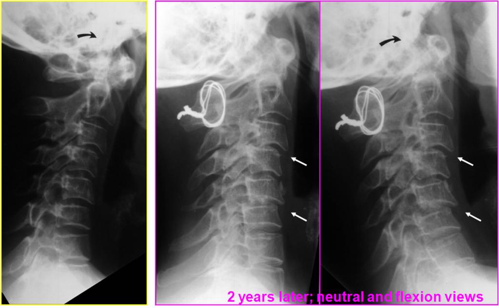
4 Main The spine can be involved in most inflammatory disorders encompassing Rheumatoid arthritis Seronegative spondyloarthropathies Ankylosing spondylitis Psoriatic arthritis Reactive arthritis Enteropathic arthritis Undifferentiated spondyloarthropathy Juvenile arthritides Rare disorders (gout, SAPHO etc.) Inflammatory spinal changes are most frequent in rheumatoid arthritis (RA) and seronegative spondyloarthropathies (SpA). The presentation will therefore focus on these disorders, which display somewhat different imaging features. Rheumatoid arthritis (RA) Involvement in RA is usually located to the cervical spine where erosive changes are predominantly seen in the atlanto-axial (C1-C2) region. Pannus formation around the odontoid process (dens) (Fig. 1) may cause bone erosion and destruction of surrounding ligaments; most seriously if the posterior transverse ligament is involved. The diagnosis of cervical RA changes is important [1]. The changes can cause instability, potentially risking spinal cord injury. Radiography of the cervical spine is therefore mandatory in RA patients with neck pain. It should always include a lateral view during flexion compared with neutral position in addition to a special view of the dens (Fig. 2) to detect any lesions and/or instability. Arthritis of apophyseal joints with instability in the C2-Th1 region may also occur, but less frequently. It may progress over time, especially if the C1-C2 region is stabilized surgically [2](Fig. 3). Cross sectional imaging in the form of computed tomography (CT) and magnetic resonance imaging (MRI) can improve the detection of RA changes in the atlanto-axial region resulting in the following advantages: Elimination of overprojecting structures. Detection of signs of involvement before they are visible by radiography. Clear delineation by CT of osseous structures - erosions etc. Visualization by MRI of: Page 4 of 37
5 Soft tissue structures - pannus; spinal cord etc. Signs of disease activity. Sequels of inflammation - fibrous pannus and fat deposition in the bone marrow. The advantages of CT and MRI are demonstrated in Figures 4-6. Radiography (Fig. 4) shows erosion at the base of dens (arrow) and instability with a distance between the dens and the anterior arc of C2 exceeding 3 mm (normal upper limit). CT (Fig. 5) demonstrates extensive erosion not only at the base of dens, but also at the tip and at the atlanto-axial and atlanto-occipital joints. MRI, post-contrast sagittal T1 weighted fat saturated (T1FS) sequence (Fig. 6) demonstrates enhancing pannus tissue surrounding the dens, which is a sign of active inflammation. MRI visualization of the spinal cord is important to detect cord injury or risk of injury. It is recommended that MRI should be performed in all RA patients with neck pain and neurological symptoms [3;4]. Risk of cord compression/injury occurs especially in patients with flexion instability accompanied by erosive changes in the atlanto-axial and/ or atlanto-occipital joints causing dens to protrude into the occipital foramen (basilar impression). Figure 7, sagittal and axial images show erosion of the dens and protrusion of the tip into the occipital foramen causing cord compression (white arrows). There is a mixture of fibrotic end enhancing pannus tissue (black arrow) in the widened space between the dens and the anterior arc of C2. MRI also gives the possibility of detecting signs of arthritis before the occurrence of erosive changes [4]. Figure 8 illustrates MRI of a RA patient with neck pain and normal radiography. There are signs of active arthritis with synovial contrast enhancement (synovitis) at the left atlanto-axial joint in addition to enhancing pannus tissue at the left side of the dens (white arrows) and a subchondral enhancing area in the axis (black arrow). Seronegative spondyloarthropathy (SpA) According to the European classification criteria [5] SpA is divided into: Ankylosing spondylitis (AS) Psoriatic arthritis Reactive arthritis Arthritis associated with inflammatory bowel disorders (enteropathic arthritis) Undifferentiated spondyloarthropathy. Inflammatory changes at the sacroiliac joints always occur in AS and is part of most other forms of SpA. Spinal changes are also a feature of SpA, especially in late stages of AS. Page 5 of 37
6 Radiographic features Manifest structural sacroiliac and spinal changes can be diagnosed at radiography. It is, however, not possible to differentiate between the different forms of SpA based on radiography of the sacroiliac joints although symmetric involvement is more frequent in AS than in other forms of SpA [6]. Spinal changes are more specific. Syndesmophytes and involvement of apophyseal joints are more frequent in AS than in the other forms of SpA [6]. Vertebral squaring, syndesmophytes and ankylosis of apophyseal joints are characteristic of AS whereas voluminous paravertebral new bone formation occur in psoriatic and reactive arthritis. The features of enteropathic arthritis often resemble those of AS. Figure 9 illustrates the slim ossification (named syndesmophytes) at the periphery of annulus fibrous characteristic in AS. Figure 8 illustrates the voluminous paravertebral new bone formation (parasyndesmophytes) characteristic in psoriatic and reactive arthritis. Ankylosing spondylitis (AS) The most frequent and usually the most disabling form of SpA is AS. It is therefore important to know the radiographic features characteristic of AS. According to the Modified New York Criteria [7] the diagnosis of definite AS requires the following: Positive radiography: Grade >2 bilateral sacroiliitis or unilateral grade 3-4 sacroiliitis. At least one of the following clinical criteria: Low back pain and stiffness for more than 3 months which improves by activity. Limited movement of the lumbar spine in both the sagittal and frontal plane. Reduced chest expansion. These criteria are still used in the diagnosis of AS despite the increasing use of CT and MRI in the diagnosis of SpA. Figure 11 illustrates typical definite bilateral AS sacroiliitis (grade 3) in the form of bilateral moderate joint erosion, especially at the iliac side of the joints, accompanied by subchondral sclerosis and focal widening of the joint space. Accompanying initial spinal changes are illustrated in Figure 12. They consist of vertebral squaring due to bone apposition at the anterior aspect of the vertebral bodies (arrowhead) and erosion of vertebral corners which can appear condensed (shiny corners (arrow)). Initial development of syndesmophytes (curved arrow) is also seen. The spine gradually fuses due to syndesmophytes crossing the intervertebral spaces in addition to fusion of apophyseal joints (Figure 13). Also the interspinous ligaments Page 6 of 37
7 posteriorly may ossify and be visible at frontal radiographs as a slim ossified streak (Figure 14, arrows). Erosive changes within intervertebral spaces occur in about 10% of patients with AS and are sometimes accompanied by subchondral sclerosis. Such changes can heal with ankylosis (Fig. 15), but there may occasionally be persisted movement at single intervertebral spaces. This can result in pseudo-arthrosis like changes with the formation of reactive osteophytes due to excessive mechanical load at single movable areas. The diagnosis of such changes may require CT for adequate visualization (Fig. 16). Advantages of CT and MRI CT gives a clear delineation of osseous structures - posterior joints, fracture, pseudo-arthrosis etc. MRI can visualize: Signs of disease activity. Signs of previous inflammation - fatty depositions in bone marrow. Chronic structural changes. CT is the modality of choice to detect fractures and is superior to MRI in detecting osseous lesions such as erosion and ankylosis of costo-vertebral and costo-transversal joints (Fig. 17, arrows). CT is not able to detect signs of disease activity in the form of inflammatory edema. This can be visualized by MRI which has gained a central role in the evaluation of disease activity [8]. The most frequently used MR sequences are: Sagittal STIR or T2FS and T1 weighted sequences of the entire spine. Supplementary axial slices can be necessary for visualizing involvement of apophyseal, costo-vertebral and costo-transversal joints [9;10]. T1FS sequences after intravenous Gadolinium can be advantageous and may provide a better anatomic delineation [11]. Early activity changes mainly consist of edema at vertebral corners and/or costo-vertebral joints (Fig. 18). However, during the course of disease signs of activity can also be seen at syndesmophytes (A), apophyseal joints (B) and interspinous ligaments (C)(Fig. 19). Chronic AS changes detectable by MRI mainly consist of fatty marrow deposition at vertebral corners (Fig. 20), erosion which may be surrounded by signs of active inflammation and/or fatty marrow deposition and vertebral fusion in advanced disease. Syndesmophytes may not always be visible by MRI (Fig. 21) [8]. Complications in AS Page 7 of 37
8 One of the feared complications in AS is spinal fracture. Non-fatal fractures have been reported to occur in up to 6% of AS patients and especially in patients with long disease duration [12;13]. Fracture may occur after minor trauma due to the spinal stiffness and a frequent accompanying osteoporosis. Fractures often occur at intervertebral spaces, but usually involve the ankylosed posterior structures and are thereby unstable. Obvious fractures can be diagnosed by radiography (Fig. 22), but fractures may be obscured. It is therefore mandatory to supplement a negative radiography with CT if fracture is suspected (trauma or change of spinal symptoms) (Fig. 23). Cervico-thoracic fractures may cause spinal cord injury which can be visualized by MRI (Fig 24, arrow). The fractures may be lethal even after minor trauma [14]. Other forms of SpA In psoriatic and reactive arthritis the spinal changes are often characterized by voluminous paravertebral new bone formation (parasyndesmophytes) or coalescing ossification of the paravertebral ligaments (Fig. 25). However, in psoriatic arthritis the changes may also present as erosion of vertebral plates often accompanied by subchondral sclerosis and sometimes signs of multiple osseous inflammation by MRI. Such osseous lesions are especially seen in patients with pustular psoriasis or pustulosis palmoplantaris (Fig. 26). In patients with reactive arthritis and HLA B27 the spinal changes may progress to changes indistinguishable from those seen in AS (Fig. 27). In patients with enteropathic arthritis the spine is often osteoporotic with various accompanying features characteristic of SpA, most often AS-like features. However, by MRI there is usually more pronounced inflammation in the posterior ligaments than seen in the other forms of SpA (Fig. 28). Grading of SpA changes The purposes of grading inflammatory spinal changes are to: Achieve a quantitative value for Disease activity. Chronic changes. This is important for Therapeutic decisions. Monitoring the disease/therapy. Grading by radiography can either be done using the Bath Ankylosing Spondylitis Index (BASRI) (Fig. 29) [15] or Modified Stoke Ankylosing Spondylitis Spine Score (msasss) (Fig. 30) [16] both developed by rheumatologists. Page 8 of 37
9 Grading of activity by MRI has become important after the introduction of anti-tumor necrosis factor alpha (anti-tnf#) agents that have proven promising for alleviating inflammatory symptoms of AS and possibly preventing structural damage [17]. There is no accepted universal grading method, but one of the three following methods are commonly used: The SPARCC MRI spinal index (Spondyloarthritis Research Consortium of Canada) - six most involved discovertebral units scored dichotomously for activity (Fig. 31) [18]. ASspiMRIa (Ankylosing Spondylitis Spine MRI Activity Score) with modifications - all discovertebral units (Fig. 32) [19;20]. The Aarhus method - all discovertebral units (Fig. 33) [8]. It is possible to gain a quantitative value for the effect of anti-tnf# using one of these methods. Chronic changes can also be quantified using the Aarhus method (Fig. 34) [8]. Conclusion. Radiography is still valuable in the diagnosis of spinal inflammatory disorders. Is necessary for visualizing instability. Is superior to MRI for detecting syndesmophytes. MRI can add information on: Signs of activity; soft tissue changes. Fat deposition - a sign of chronic disease. MRI is widely used to monitor disease activity during anti-tnf therapy. CT is especially valuable in the detection of fracture. MRI and CT can detect changes before they can be visualized by radiography. Page 9 of 37

10 Images for this section: Ross et al. Diagnostic Imaging, Spine Page 10 of 37

11 Page 11 of 37

12 Page 12 of 37


13 Page 13 of 37


14 Page 14 of 37

15 Page 15 of 37

16 Page 16 of 37

17 Page 17 of 37

18 Page 18 of 37

19 Page 19 of 37


20 Page 20 of 37


21 Page 21 of 37

22 Page 22 of 37

23 Page 23 of 37

24 Page 24 of 37

25 Page 25 of 37


26 Page 26 of 37

27 Page 27 of 37


28 Page 28 of 37

29 Page 29 of 37

30 Page 30 of 37

31 Page 31 of 37
32 Braun J et al. Arthritis Rheum 2003;48(4): , and Rudwaleit M et al. Ann Rheum Dis 2008;67(9): Page 32 of 37

33 Madsen KB & Jurik AG. Clin Radiol 2009;65:6-14. Page 33 of 37

34 Madsen KB & Jurik AG. Clin Radiol 2009;65:6-14. Page 34 of 37
35 References Kim DH, Hilibrand AS. Rheumatoid arthritis in the cervical spine. J Am Acad Orthop Surg 2005;13(7): Ishii K, Matsumoto M, Takahashi Y, Okada E, Watanabe K, Tsuji T, et al. Risk Factors for Development of Subaxial Subluxations Following Atlantoaxial Arthrodesis for Atlantoaxial Subluxations in Rheumatoid Arthritis. Spine;2010 Jan 12; Epubl in head of print. Narvaez JA, Narvaez J, Serrallonga M, De Lama E, de AM, Mast R, et al. Cervical spine involvement in rheumatoid arthritis: correlation between neurological manifestations and magnetic resonance imaging findings. Rheumatology (Oxford) 2008;47(12): Younes M, Belghali S, Kriaa S, Zrour S, Bejia I, Touzi M, et al. Compared imaging of the rheumatoid cervical spine: prevalence study and associated factors. Joint Bone Spine 2009;76(4): Dougados M, van der Linden S, Juhlin R, Huitfeldt B, Amor B, Calin A, et al. The European Spondylarthropathy Study Group preliminary criteria for the classification of spondylarthropathy. Arthritis Rheum 1991;34: Helliwell PS, Hickling P, Wright V. Do the radiological changes of classic ankylosing spondylitis differ from the changes found in the spondylitis associated with inflammatory bowel disease, psoriasis, and reactive arthritis? Ann Rheum Dis 1998;57(3): van der Linden S, Valkenburg HA, Cats A. Evaluation of diagnostic criteria for ankylosing spondylitis. A proposal for modification of the New York criteria. Arthritis Rheum 1984;27: Madsen KB, Jurik AG. MRI grading method for active and chronic spinal changes in spondyloarthritis. Clin Radiol 2009;65:6-14. Khanna M, Keightley A. MRI of the axial skeleton manifestations of ankylosing spondylitis. Clin Radiol 2005;60(1): Levine DS, Forbat SM, Saifuddin A. MRI of the axial skeletal manifestations of ankylosing spondylitis. Clin Radiol 2004;59(5): Baraliakos X, Hermann KG, Landewe R, Listing J, Golder W, Brandt J, et al. Assessment of acute spinal inflammation in patients with ankylosing spondylitis by magnetic resonance imaging: a comparison between contrast enhanced T1 and short tau inversion recovery (STIR) sequences. Ann Rheum Dis 2005;64(8): Feldtkeller E, Vosse D, Geusens P, van der Linden S. Prevalence and annual incidence of vertebral fractures in patients with ankylosing spondylitis. Rheumatol Int 2006;26(3): Vosse D, Feldtkeller E, Erlendsson J, Geusens P, van der Linden S. Clinical vertebral fractures in patients with ankylosing spondylitis. J Rheumatol 2004;31(10): Thomsen AH, Uhreholt L, Jurik AG, Vesterby A. Traumatic Death in Ankylosing Spondylitis - A Case Report. J Forensic Sci (In press) Page 35 of 37
36 15. MacKay K, Mack C, Brophy S, Calin A. The Bath Ankylosing Spondylitis Radiology Index (BASRI) : a new, validated approach to disease assessment. Arthritis Rheum 1998;41(12): Creemers MC, Franssen MJ, van't Hof MA, Gribnau FW, van de Putte LB, van Riel PL. Assessment of outcome in ankylosing spondylitis: an extended radiographic scoring system. Ann Rheum Dis 2005;64(1): Baraliakos X, Listing J, Brandt J, Haibel H, Rudwaleit M, Sieper J, et al. Radiographic progression in patients with ankylosing spondylitis after 4 yrs of treatment with the anti-tnf-alpha antibody infliximab. Rheumatology (Oxford) 2007;46(9): Maksymowych WP, Inman RD, Salonen D, Dhillon SS, Krishnananthan R, Stone M, et al. Spondyloarthritis Research Consortium of Canada magnetic resonance imaging index for assessment of spinal inflammation in ankylosing spondylitis. Arthritis Rheum 2005;53(4): Braun J, Baraliakos X, Golder W, Brandt J, Rudwaleit M, Listing J, et al. Magnetic resonance imaging examinations of the spine in patients with ankylosing spondylitis, before and after successful therapy with infliximab: evaluation of a new scoring system. Arthritis Rheum 2003;48(4): Rudwaleit M, Schwarzlose S, Hilgert ES, Listing J, Braun J, Sieper J. MRI in predicting a major clinical response to anti-tumour necrosis factor treatment in ankylosing spondylitis. Ann Rheum Dis 2008;67(9): Page 36 of 37
37 Personal Information The author has for the last 20 years worked within the field of imaging seronegative spondyloarthropathies using the available imaging modalities. During 6 years participated in international OMERACT (Outcome Measures in Rheumatology Clinical Trials) analyses and member of Assessment of Ankylosing Spondylitis (ASAS) working groups. Page 37 of 37
Imaging the spine in arthritis a pictorial review
 Insights Imaging (2011) 2:177 191 DOI 10.1007/s13244-010-0061-4 PICTORIAL REVIEW Imaging the spine in arthritis a pictorial review Anne Grethe Jurik Received: 3 August 2010 /Revised: 6 November 2010 /Accepted:
MRI of the sacroiliac joints: what to report and its pitfalls
 MRI of the sacroiliac joints: what to report and its pitfalls Poster No.: C-1920 Congress: ECR 2016 Type: Educational Exhibit Authors: J. Goncalves, A. Y. Aihara, C. Longo, H. Guidorizzi, P. Aguiar, 1
Seronegative spondyloarthropathies : A Pictorial Review
 Seronegative spondyloarthropathies : A Pictorial Review Poster No.: P-0008 Congress: ESSR 2012 Type: Scientific Exhibit Authors: J. Acosta Batlle, B. Palomino Aguado, M. D. Lopez Parra, S. 1 2 3 2 4 1
Ankylosing spondylitis: A Pictorial Review
 Ankylosing spondylitis: A Pictorial Review Poster No.: P-0009 Congress: ESSR 2012 Type: Scientific Exhibit Authors: J. Acosta Batlle, B. Palomino Aguado, M. D. Lopez Parra, S. 1 2 3 2 4 1 2 Hernandez Muñiz,
Spinal injury is very common in Ireland: 19 per 100,000 (1). It poses a significant disease burden.
. It poses a significant disease burden.") MRI in traumatic spinal cord injury: a single national spinal centre experience and study of imaging features with clinical correlation with ASIA score and outcome Poster No.: C-1235 Congress: ECR 2011
MRI in traumatic spinal cord injury: a single national spinal centre experience and study of imaging features with clinical correlation with ASIA score and outcome Poster No.: C-1235 Congress: ECR 2011
Diagnostic accuracy of MRI in detecting posterior ligamentous complex injury in thoracolumbar vertebral fractures
 Diagnostic accuracy of MRI in detecting posterior ligamentous complex injury in thoracolumbar vertebral fractures Poster No.: C-1726 Congress: ECR 2011 Type: Scientific Exhibit Authors: E. Aguirre, P.
Articular disease of the hand - the target joint approach
 Articular disease of the hand - the target joint approach Poster No.: C-1817 Congress: ECR 2016 Type: Educational Exhibit Authors: R. R. Domingues Madaleno 1, A. P. Pissarra 1, I. Abreu 2, A. Canelas 1,
Seronegative Spondyloarthropathies: A Radiological Persepctive
 Seronegative Spondyloarthropathies: A Radiological Persepctive Poster No.: C-1816 Congress: ECR 2016 Type: Educational Exhibit Authors: K. Shindi, H. Nejadhamzeeigilani, P. Nagtode, C. Nel ; 1 1 2 2 3
Seemingly isolated greater trochanter fractures do not exist
 Seemingly isolated greater trochanter fractures do not exist Poster No.: B-0950 Congress: ECR 2012 Type: Scientific Paper Authors: D. Dunker, J. H. Göthlin, M. Geijer ; Gothenburg/SE, Lund/SE Keywords:
Persistent ankle pain after inversion lesions: what the radiologist must look for
 Persistent ankle pain after inversion lesions: what the radiologist must look for Poster No.: P-0118 Congress: ESSR 2016 Type: Authors: Keywords: DOI: Educational Poster R. Leao, L. C. Zattar-Ramos, E.
T he spondyloarthritides (SpA) comprise five subtypes:
 comprise five subtypes:") 1305 EXTENDED REPORT Magnetic resonance imaging of the spine and the sacroiliac joints in ankylosing spondylitis and undifferentiated spondyloarthritis during treatment with etanercept M Rudwaleit*, X
1305 EXTENDED REPORT Magnetic resonance imaging of the spine and the sacroiliac joints in ankylosing spondylitis and undifferentiated spondyloarthritis during treatment with etanercept M Rudwaleit*, X
Active (acute) inflammation on MRI highly suggestive of sacroiliitis associated with SpA
 inflammation on MRI highly suggestive of sacroiliitis associated with SpA") MRI findings of active and chronic sacroiliitis in light of recent ASAS criteria for diagnosing axial spondyloarthritis: what the radiologist should know Poster No.: C-1955 Congress: ECR 2012 Type: Educational
MRI findings of active and chronic sacroiliitis in light of recent ASAS criteria for diagnosing axial spondyloarthritis: what the radiologist should know Poster No.: C-1955 Congress: ECR 2012 Type: Educational
Cierny-Mader classification of chronic osteomyelitis: Preoperative evaluation with cross-sectional imaging
 Cierny-Mader classification of chronic osteomyelitis: Preoperative evaluation with cross-sectional imaging Poster No.: C-590 Congress: ECR 2009 Type: Topic: Educational Exhibit Musculoskeletal Authors:
Imaging and intervention of sacroiliac joint. Dr Ryan Lee Ka Lok Associate Consultant Prince of Wales Hospital
 Imaging and intervention of sacroiliac joint Dr Ryan Lee Ka Lok Associate Consultant Prince of Wales Hospital Introduction 15%-25% of low back pain is related to sacroiliac joint (SIJ) pain SIJ pain is
Dynamic CT Assessment of Distal Radioulnar Instability
 Dynamic CT Assessment of Distal Radioulnar Instability Poster No.: P-0114 Congress: ESSR 2016 Type: Educational Poster Authors: S. Dumonteil, M. A. Shah, A. Srikanthan, V. Ejindu, N. Papadakos; London/UK
MR imaging the post operative spine - What to expect!
 MR imaging the post operative spine - What to expect! Poster No.: C-2334 Congress: ECR 2012 Type: Educational Exhibit Authors: A. Jain, M. Paravasthu, M. Bhojak, K. Das ; Warrington/UK, 1 1 1 2 1 2 Liverpool/UK
Computed tomography for the detection of thumb base osteoarthritis, comparison with digital radiography.
 Computed tomography for the detection of thumb base osteoarthritis, comparison with digital radiography. Poster No.: C-1981 Congress: ECR 2012 Type: Scientific Exhibit Authors: M. S. Saltzherr, J. W. van
MR imaging features of paralabral ganglion cyst of the shoulder
 MR imaging features of paralabral ganglion cyst of the shoulder Poster No.: C-1482 Congress: ECR 2016 Type: Educational Exhibit Authors: M. Bartocci, C. Dell'atti, E. Federici, D. Beomonte Zobel, V. Martinelli,
Identification and numbering of lumbar vertebrae using various anatomical landmarks on MRI of lumbosacral spine
 Identification and numbering of lumbar vertebrae using various anatomical landmarks on MRI of lumbosacral spine Poster No.: C-2125 Congress: ECR 2015 Type: Authors: Scientific Exhibit S. patil 1, A. M.
Cervical spine degenerative disease: a comparative study between computed tomography and magnetic resonance imaging findings
 Cervical spine degenerative disease: a comparative study between computed tomography and magnetic resonance imaging findings Poster No.: C-2517 Congress: ECR 2012 Type: Scientific Paper Authors: M. Papavasilopoulou,
Diffusion-weighted MRI (DWI) "claw sign" is useful in differentiation of infectious from degenerative Modic I signal changes of the spine
 claw sign is useful in differentiation of infectious from degenerative Modic I signal changes of the spine") Diffusion-weighted MRI (DWI) "claw sign" is useful in differentiation of infectious from degenerative Modic I signal changes of the spine Poster No.: C-0894 Congress: ECR 2012 Type: Scientific Exhibit
Diffusion-weighted MRI (DWI) "claw sign" is useful in differentiation of infectious from degenerative Modic I signal changes of the spine Poster No.: C-0894 Congress: ECR 2012 Type: Scientific Exhibit
Psoriatic arthritis: early ultrasound findings
 Psoriatic arthritis: early ultrasound findings Poster No.: C-0399 Congress: ECR 2014 Type: Educational Exhibit Authors: R. Persechino 1, L. Cristiano 1, A. Bartoloni 1, C. Cantone 2, A. Keywords: DOI:
Anatomical Variations of the Levator Scapulae Muscle - an MR Imaging Study
 Anatomical Variations of the Levator Scapulae Muscle - an MR Imaging Study Poster No.: R-0016 Congress: 2015 ASM Type: Scientific Exhibit Authors: J. Au, A. Webb, G. Buirski, P. Smith, M. Pickering, D.
Extrapulmonary Manifestations of Tuberculosis: A Radiologic Review
 Extrapulmonary Manifestations of Tuberculosis: A Radiologic Review Poster No.: C-1958 Congress: ECR 2014 Type: Authors: Educational Exhibit J. Isern 1, S. Llaverias Borrell 1, A. Olarte 1, E. Grive 1,
Cover Page. The handle holds various files of this Leiden University dissertation
 Cover Page The handle http://hdl.handle.net/1887/43590 holds various files of this Leiden University dissertation Author: Machado, Pedro Title: Health and imaging outcomes in axial spondyloarthritis Issue
Sacroiliac joints MR: Finally a universal language for the sacroiliitis diagnosis
 Sacroiliac joints MR: Finally a universal language for the sacroiliitis diagnosis Poster No.: C-1836 Congress: ECR 2013 Type: Scientific Exhibit Authors: M. E. Banegas Illescas, C. López Menéndez, M. L.
Extraarticular Lateral Ankle Impingement
 Extraarticular Lateral Ankle Impingement Poster No.: C-1282 Congress: ECR 2016 Type: Educational Exhibit Authors: C. Cevikol; Keywords: Trauma, Diagnostic procedure, MR, CT, Musculoskeletal system, Musculoskeletal
Long bones manifestations of congenital syphilis
 Long bones manifestations of congenital syphilis Poster No.: C-0139 Congress: ECR 2011 Type: Educational Exhibit Authors: T. F. de Souza 1, P. P. Collier 1, E. J. M. Bronzatto 1, G. L. P. Keywords: DOI:
Chronic knee pain in adults - a multimodality approach or which modality to choose and when?
 Chronic knee pain in adults - a multimodality approach or which modality to choose and when? Poster No.: P-0157 Congress: ESSR 2013 Type: Authors: Keywords: DOI: Scientific Exhibit E. Ilieva, V. Tasseva,
Optimal Site for Bone Graft Harvesting from the Iliac Bone
 Optimal Site for Bone Graft Harvesting from the Iliac Bone Poster No.: P-0095 Congress: ESSR 2015 Type: Scientific Poster Authors: B. Batohi 1, A. Isaac 1, J. Edwin 1, A. Hussain 1, J. Kumaraguru 1, L.
Synovial hemangioma of the suprapatellar bursa
 Synovial hemangioma of the suprapatellar bursa Poster No.: P-0040 Congress: ESSR 2013 Type: Authors: Keywords: DOI: Scientific Exhibit A. YESILDAG, S. Keskin, H. Kalkan, S. Kucuksen, U. Kerimoglu; Konya/TR
Radiological features of Legionella Pneumophila Pneumonia
 Radiological features of Legionella Pneumophila Pneumonia Poster No.: E-0048 Congress: ESTI 2012 Type: Scientific Exhibit Authors: M. Vinciguerra, L. Stefanetti, E. Teti, G. Argentieri, L. G. 1 1 1 1 1
MRI in Patients with Forefoot Pain Involving the Metatarsal Region
 MRI in Patients with Forefoot Pain Involving the Metatarsal Region Poster No.: C-0151 Congress: ECR 2015 Type: Authors: Keywords: DOI: Scientific Exhibit R. Vukojevi#, M. Mustapic, D. Marjan; Zagreb/HR
Trabecular bone analysis with tomosynthesis in diabetic patients: comparison with CT-based finite-element method
 Trabecular bone analysis with tomosynthesis in diabetic patients: comparison with CT-based finite-element method Poster No.: C-1789 Congress: ECR 2015 Type: Scientific Exhibit Authors: M. Fujii, T. Aoki,
Figuring out the "fronds"-synovial proliferative disorders of the knee.
 Figuring out the "fronds"-synovial proliferative disorders of the knee. Poster No.: C-1209 Congress: ECR 2014 Type: Educational Exhibit Authors: S. Sivasubramanian; Tamil Nadu/IN Keywords: Imaging sequences,
ARDS - a must know. Page 1 of 14
 ARDS - a must know Poster No.: C-1683 Congress: ECR 2016 Type: Authors: Keywords: DOI: Educational Exhibit M. Cristian; Turda/RO Education and training, Edema, Acute, Localisation, Education, Digital radiography,
Reliability of the pronator quadratus fat pad sign to predict the severity of distal radius fractures
 Reliability of the pronator quadratus fat pad sign to predict the severity of distal radius fractures Poster No.: C-0669 Congress: ECR 2014 Type: Scientific Exhibit Authors: J. Tonak, I. Wobbe, R. L. Duschka,
Update - Imaging of the Spondyloarthropathies. Spondyloarthropathies. Spondyloarthropathies
 Update - Imaging of the Spondyloarthropathies Donald J. Flemming, M.D. Dept of Radiology Penn State Hershey Medical Center Spondyloarthropathies Family of inflammatory arthritides of synovium and entheses
Digital tomosynthesis in diagnosis of occult hip fractures
 Digital tomosynthesis in diagnosis of occult hip fractures Poster No.: B-0781 Congress: ECR 2013 Type: Authors: Keywords: DOI: Scientific Paper M. Geijer 1, D. Collin 2, J. H. Göthlin 2 ; 1 Lund/SE, 2
Evaluation of MRI diffusion in spondylarthropathy axial skeleton lesions
 Evaluation of MRI diffusion in spondylarthropathy axial skeleton lesions Poster No.: C-2353 Congress: ECR 2012 Type: Scientific Paper Authors: B. Dallaudiere, R. Dautry, A. Felter, J. LINCOT, P. Koch,
Spinal meningioma imaging
 Spinal meningioma imaging Poster No.: C-0448 Congress: ECR 2018 Type: Educational Exhibit Authors: M. Smoljan, D. Zadravec ; Zagreb/HR, Zageb/HR Keywords: Neoplasia, Imaging sequences, Education, MR, CT,
Delayed Vertebral Augmentation With Spinejack Technique in A3 Type Vertebral Compression Fractures
 Delayed Vertebral Augmentation With Spinejack Technique in A3 Type Vertebral Compression Fractures Poster No.: C-0358 Congress: ECR 2016 Type: Authors: Keywords: DOI: Scientific Exhibit J. Chiras; Paris
"D10-D11 Facet enlargement"
 "D10-D11 Facet enlargement" Poster No.: C-0898 Congress: ECR 2011 Type: Scientific Exhibit Authors: J. Jalal Shokouhi, A. A. Ameri, H. Mohammad pour ; Tehran/IR, 1 2 2 1 2 tehran/ir Keywords: Spine, Neuroradiology
The posterolateral corner of the knee: the normal and the pathological
 The posterolateral corner of the knee: the normal and the pathological Poster No.: P-0104 Congress: ESSR 2014 Type: Educational Poster Authors: M. Bartocci 1, C. Dell'atti 2, E. Federici 1, V. Martinelli
Color duplex Doppler ultrasound in rheumatoid arthritis
 Color duplex Doppler ultrasound in rheumatoid arthritis Poster No.: C-1505 Congress: ECR 2012 Type: Authors: Keywords: DOI: Scientific Paper G. Ivanac, J. Morovic Vergles, M. Cavka, N. Radovic, B. Brkljacic;
Lumbosacral Transitional Vertebrae
 Lumbosacral Transitional Vertebrae Poster No.: C-073 Congress: ECR 206 Type: Educational Exhibit Authors: M. Mustapic, R. Vukojevi#, M. Gulin, D. Marjan, I. Boric ; 2 2 Zagreb/HR, Zabok/HR Keywords: Congenital,
Ultrasonography in early diagnosis of acromioclavicular joint degeneration: comparison with plain radiography
 Ultrasonography in early diagnosis of acromioclavicular joint degeneration: comparison with plain radiography Poster No.: C-2400 Congress: ECR 2013 Type: Authors: Scientific Exhibit E. Sanverdi 1, N. Aydin
MRI Findings of Posterolateral Corner Injury on Threedimensional
 MRI Findings of Posterolateral Corner Injury on Threedimensional Isotropic SPACE. Poster No.: C-1792 Congress: ECR 2013 Type: Scientific Exhibit Authors: S.-W. Lee, Y. M. Jeong, J. A. Sim, S. Ahn; Incheon/KR
Reliability of change in lumbar MRI findings
 Reliability of change in lumbar MRI findings Poster No.: C-0539 Congress: ECR 2012 Type: Scientific Exhibit Authors: L. Berg 1, O. Gjertsen 2, C. Hellum 2, G. F. Neckelmann 1, L. G. Johnsen 3, G. E. Eide
A Pictorial Review of Congenital Tarsal Coalition
 A Pictorial Review of Congenital Tarsal Coalition Poster No.: C-2305 Congress: ECR 2011 Type: Educational Exhibit Authors: J. Jethwa, M. Tapp; Torquay/UK Keywords: Musculoskeletal joint, Musculoskeletal
Significance of MRI in diagnostics, outcome prognosis and definition the therapeutic tactics for cases of aseptic necrosis of the femoral head
 Significance of MRI in diagnostics, outcome prognosis and definition the therapeutic tactics for cases of aseptic necrosis of the femoral head Abstract: 539 Congress: ESMRMB 2013 Type: Scientific Poster
Carpal bossing - review and an unrecognized variation.
 Carpal bossing - review and an unrecognized variation. Poster No.: P-0053 Congress: ESSR 2014 Type: Authors: Keywords: DOI: Educational Poster K. B. Puhakka, L. Roemer, B. Munk; Aarhus C/DK Developmental
"Ultrasound measurements of the lateral ventricles in neonates: A comparison of multiple measurements methods."
 "Ultrasound measurements of the lateral ventricles in neonates: A comparison of multiple measurements methods." Poster No.: C-1557 Congress: ECR 2014 Type: Authors: Keywords: DOI: Educational Exhibit I.
Tuberculosis afeccting the central nervous sistem and spine: CT and MR imaging implications for diagnosis and treatment
 Tuberculosis afeccting the central nervous sistem and spine: CT and MR imaging implications for diagnosis and treatment Poster No.: C-1854 Congress: ECR 2012 Type: Educational Exhibit Authors: S. G. Trigo,
A pictorial review of normal anatomical appearences of Pericardial recesses on multislice Computed Tomography.
 A pictorial review of normal anatomical appearences of Pericardial recesses on multislice Computed Tomography. Poster No.: C-1787 Congress: ECR 2012 Type: Educational Exhibit Authors: N. Ahmed 1, G. Avery
What is Axial Spondyloarthritis?
 Physiotherapist Module 2 What is Axial Spondyloarthritis? How does it apply to physiotherapists? Claire Harris, Senior Physiotherapist, London North West Healthcare NHS Trust Susan Gurden, Advanced Physiotherapy
Complications of Perianal Crohn s Disease - Adenocarcinoma & Extensive Fistulization
 Complications of Perianal Crohn s Disease - Adenocarcinoma & Extensive Fistulization Poster No.: C-0711 Congress: ECR 2013 Type: Educational Exhibit Authors: P. Faria João 1, D. Penha 2, P. Cabral 1, E.
64-MDCT imaging of the pancreas: Scan protocol optimisation by different scan delay regimes
 64-MDCT imaging of the pancreas: Scan protocol optimisation by different scan delay regimes Poster No.: C-051 Congress: ECR 2009 Type: Scientific Exhibit Topic: Abdominal and Gastrointestinal Authors:
B. CT protocols for the spine
 B. CT protocols for the spine Poster No.: A-003 Congress: ECR 2010 Type: Invited Speaker Topic: Neuro Authors: B. Tins; Oswestry/UK Keywords: CT, spine, diagnostic imaging protocol DOI: 10.1594/ecr2010/A-003
Resuscitation lateral cervical spine X-ray (LSCX): A useful mandatory screening tool in acute trauma?
: A useful mandatory screening tool in acute trauma?") Resuscitation lateral cervical spine X-ray (LSCX): A useful mandatory screening tool in acute trauma? Poster No.: C-2397 Congress: ECR 2010 Type: Topic: Authors: Keywords: DOI: Scientific Exhibit Musculoskeletal
Resuscitation lateral cervical spine X-ray (LSCX): A useful mandatory screening tool in acute trauma? Poster No.: C-2397 Congress: ECR 2010 Type: Topic: Authors: Keywords: DOI: Scientific Exhibit Musculoskeletal
CT-guided percutaneous intraspinal needle aspiration for the diagnosis and treatment of epidural collections
 CT-guided percutaneous intraspinal needle aspiration for the diagnosis and treatment of epidural collections Poster No.: P-0064 Congress: ESSR 2013 Type: Scientific Exhibit Authors: G. Petrocheilou, I.
Monophasic versus biphasic contrast application in CT of patients with head and neck tumour
 Monophasic versus biphasic contrast application in CT of patients with head and neck tumour Poster No.: C-3331 Congress: ECR 2010 Type: Topic: Authors: Keywords: DOI: Scientific Exhibit Head and Neck G.
Cognitive target MRI-TRUS fusion biopsies of MRI detected PIRADS 4 and 5 lesions
 Cognitive target MRI-TRUS fusion biopsies of MRI detected PIRADS 4 and 5 lesions Poster No.: B-0704 Congress: ECR 2015 Type: Scientific Paper Authors: P. P. van Westerveld, J. Vriesema, J. H. W. van den
A retrospective audit of General Practitioner (GP) referrals for musculoskeletal radiographs.
 referrals for musculoskeletal radiographs.") A retrospective audit of General Practitioner (GP) referrals for musculoskeletal radiographs. Poster No.: C-1902 Congress: ECR 2015 Type: Authors: Keywords: DOI: Scientific Exhibit J. Jacob 1, H. Thampy
A retrospective audit of General Practitioner (GP) referrals for musculoskeletal radiographs. Poster No.: C-1902 Congress: ECR 2015 Type: Authors: Keywords: DOI: Scientific Exhibit J. Jacob 1, H. Thampy
MRI assessment of the plantar fascia in diabetic versus nondiabetic patients: How thick should it be?
 MRI assessment of the plantar fascia in diabetic versus nondiabetic patients: How thick should it be? Poster No.: C-2324 Congress: ECR 2010 Type: Scientific Exhibit Topic: Musculoskeletal Authors: C. Pierre-Jerome
DEXA Scores and Bone Density Measured on Routine CT Scans
 DEXA Scores and Bone Density Measured on Routine CT Scans Poster No.: R-0072 Congress: 2015 ASM Type: Scientific Exhibit Authors: C. Annabattula, P. Phadke; Sydney/AU Keywords: Screening, Nuclear medicine
Chapter 2. Overview of ankylosing spondylitis
 Chapter 2 Overview of ankylosing spondylitis The concept and classification of spondyloarthritis The term spondyloarthritis (SpA) comprises AS, reactive arthritis, arthritis/spondylitis associated with
High density thrombi of pulmonary embolism on precontrast CT scan: Is it dangerous?
 High density thrombi of pulmonary embolism on precontrast CT scan: Is it dangerous? Poster No.: C-1753 Congress: ECR 2013 Type: Authors: Keywords: DOI: Scientific Exhibit B. Y. Lee, H. R. KIM, J. I. Jung,
Imaging the post-operative spine - are we united in where we stand?
 Imaging the post-operative spine - are we united in where we stand? Poster No.: C-2424 Congress: ECR 2015 Type: Authors: Keywords: DOI: Scientific Exhibit J. Kumaraguru, N. Kandasamy, D. A. Elias, J. M.
MRI evaluation of TMJ condylar angulations
 MRI evaluation of TMJ condylar angulations Poster No.: C-2272 Congress: ECR 2010 Type: Topic: Authors: Keywords: DOI: Scientific Exhibit Musculoskeletal M. Pregarz 1, C. Bodin 2 ; 1 Peschiera del Garda/IT,
Medial tibial condyle friction syndrome: MRI study of a new entity
 Medial tibial condyle friction syndrome: MRI study of a new entity Poster No.: C-0420 Congress: ECR 2013 Type: Authors: Keywords: DOI: Scientific Exhibit M. Klontzas, I. Akoumianakis, I. Vagios, A. H.
Unenhanced and dynamic contrast enhanced (DCE) MRI in assessment of scaphoid fracture non-union revisited: role in pre-operative planning
 MRI in assessment of scaphoid fracture non-union revisited: role in pre-operative planning") Unenhanced and dynamic contrast enhanced (DCE) MRI in assessment of scaphoid fracture non-union revisited: role in pre-operative planning Poster No.: B-0440 Congress: ECR 2014 Type: Authors: Keywords:
Unenhanced and dynamic contrast enhanced (DCE) MRI in assessment of scaphoid fracture non-union revisited: role in pre-operative planning Poster No.: B-0440 Congress: ECR 2014 Type: Authors: Keywords:
Basic low - field MR imaging of meniscal injuries in children.
 Basic low - field MR imaging of meniscal injuries in children. Poster No.: C-2365 Congress: ECR 2012 Type: Authors: Keywords: DOI: Scientific Exhibit A. Yakimov, M. Nikonova, E. Prokhorova, D. Vybornov,
Differentiation of osteoporosis from metastasis in the vertebral fracture using chemical shift and diffusion weighted imaging
 Differentiation of osteoporosis from metastasis in the vertebral fracture using chemical shift and diffusion weighted imaging Poster No.: C-0444 Congress: ECR 2012 Type: Educational Exhibit Authors: H.
Computed tomography and Modified RECIST criteria for assessment of response in malignant pleural mesothelioma
 Computed tomography and Modified RECIST criteria for assessment of response in malignant pleural mesothelioma Poster No.: C-0729 Congress: ECR 2013 Type: Scientific Exhibit Authors: A. Marin, I. Pozek,
PI-RADS classification: prognostic value for prostate cancer grading
 PI-RADS classification: prognostic value for prostate cancer grading Poster No.: C-1622 Congress: ECR 2014 Type: Scientific Exhibit Authors: I. Platzek, A. Borkowetz, T. Paulus, T. Brauer, M. Wirth, M.
Diffuse high-attenuation within mediastinal lymph nodes on non-enhanced CT scan: Usefulness in the prediction of benignancy
 Diffuse high-attenuation within mediastinal lymph nodes on non-enhanced CT scan: Usefulness in the prediction of benignancy Poster No.: C-1785 Congress: ECR 2012 Type: Authors: Keywords: DOI: Scientific
Single cold nodule in Graves' disease: benign vs malignant
 Single cold nodule in Graves' disease: benign vs malignant Poster No.: C-0073 Congress: ECR 2011 Type: Authors: Keywords: DOI: Scientific Paper L. I. Sonoda, M. Halim, K. Balan; Cambridge/UK Head and neck,
The imaging features of spondylolisthesis : what the clinician needs to know
 The imaging features of spondylolisthesis : what the clinician needs to know Poster No.: C-1018 Congress: ECR 2011 Type: Authors: Educational Exhibit D. Shah 1, C. J. Burke 1, A. C. andi 2, R. Houghton
 www.fisiokinesiterapia.biz Peak onset between 20 and 30 years Form of spondyloarthritis (cause inflammation around site of ligament insertion into bone) and association with HLA-B27 Prevalence as high
MRI grading of postero-lateral corner and anterior cruciate ligament injuries
 MRI grading of postero-lateral corner and anterior cruciate ligament injuries Poster No.: C-2533 Congress: ECR 2012 Type: Educational Exhibit Authors: J. Lopes Dias, J. A. Sousa Pereira, L. Fernandes,
Spinal and para-spinal plexiform neurofibromas in NF1 patients, a clinical-radiological correlation study
 Spinal and para-spinal plexiform neurofibromas in NF1 patients, a clinical-radiological correlation study Poster No.: C-1846 Congress: ECR 2015 Type: Scientific Exhibit Authors: M. Mauda-Havakuk, B. Shofty,
Magnetic Resonance Imaging of Perianal Fistulas
 Magnetic Resonance Imaging of Perianal Fistulas Poster No.: C-0317 Congress: ECR 2014 Type: Authors: Keywords: DOI: Educational Exhibit A. P. Sathe, E. Soh, K. Y. Seto, B. Yeh, D. W. Y. chee, R. Quah,
NICE Guidelines for C-Spine Imaging: Real Life Impact
 NICE Guidelines for C-Spine Imaging: Real Life Impact Poster No.: C-1367 Congress: ECR 2017 Type: Scientific Exhibit Authors: D. Weinberg, I. Djoukhadar, G. Potter; Salford/UK Keywords: Trauma, Audit and
Idiopathic dilatation of the pulmonary artery : radiographic and MDCT features in 6 cases
 Idiopathic dilatation of the pulmonary artery : radiographic and MDCT features in 6 cases Poster No.: P-0075 Congress: ESTI 2014 Type: Authors: Educational Poster J. J. Woo 1, K. Y. Lee 2, Y. Cho 1, J.
Hip pain rating after preforming MRI with gadolinium arthrography and intra-articular lidocaine
 Hip pain rating after preforming MRI with gadolinium arthrography and intra-articular lidocaine Poster No.: C-1352 Congress: ECR 2014 Type: Scientific Exhibit Authors: J. García Yavar, J. Cabezudo, S.
Soft tissues lymphoma, the great pretender. MRI diagnostic keys.
 Soft tissues lymphoma, the great pretender. MRI diagnostic keys. Poster No.: C-2133 Congress: ECR 2015 Type: Educational Exhibit Authors: L. Caminero, M. E. Banegas Illescas, M. L. Rozas, M. Y. Torres,
Slowly growing malignant nodules and rapidly growing benign nodules: Evaluation of the value of volume doubling time
 Slowly growing malignant nodules and rapidly growing benign nodules: Evaluation of the value of volume doubling time Poster No.: C-208 Congress: ECR 2009 Type: Educational Exhibit Topic: Chest Authors:
Clinical utility of tomosynthesis in suspected scaphoid fracture: Preliminary results evaluating the VolumeRad technique
 Clinical utility of tomosynthesis in suspected scaphoid fracture: Preliminary results evaluating the VolumeRad technique Poster No.: C-2193 Congress: ECR 2010 Type: Scientific Exhibit Topic: Musculoskeletal
Reporting of Spinal Fractures
 Reporting of Spinal Fractures Poster No.: P-0026 Congress: ESSR 2014 Type: Authors: Keywords: DOI: Scientific Poster H. Al-Chalabi, C. Groves; Bradford/UK Osteoporosis, Structured reporting, Observer performance,
The Radiologic Features of Xanthogranulomatous Cholecystitis: An Important Mimic of Gallbladder Carcinoma
 The Radiologic Features of Xanthogranulomatous Cholecystitis: An Important Mimic of Gallbladder Carcinoma Poster No.: C-0691 Congress: ECR 2014 Type: Authors: Keywords: DOI: Educational Exhibit H. L. khosa
Sronegative Spondyloarthropathies. Dr. M Jokar
 Sronegative Spondyloarthropathies Dr. M Jokar 1 Definition The spondyloarthropathies are a group of disorders that share certain clinical features and an association with the HLA-B27 allele 2 Spondyloarthropathies
Apophysiolysis of the pelvic area in adolescents.
 Apophysiolysis of the pelvic area in adolescents. Poster No.: C-0077 Congress: ECR 2014 Type: Educational Exhibit Authors: R. Derks, R. E. Westerbeek, R. Van Dijk; Deventer/NL Keywords: Musculoskeletal
Imaging Gorham's disease (vanishing bone)
") Imaging Gorham's disease (vanishing bone) Poster No.: C-2201 Congress: ECR 2010 Type: Scientific Exhibit Topic: Musculoskeletal Authors: D. Vanel, P. Ruggieri, M. Alberghini; Bologna/IT Keywords: Gorham,
Imaging Gorham's disease (vanishing bone) Poster No.: C-2201 Congress: ECR 2010 Type: Scientific Exhibit Topic: Musculoskeletal Authors: D. Vanel, P. Ruggieri, M. Alberghini; Bologna/IT Keywords: Gorham,
Marked changes in intervertebral disc morphology in ochronosis of alkaptonuria (AKU): a quantitative analysis of lumbar spine MR images
: a quantitative analysis of lumbar spine MR images") Marked changes in intervertebral disc morphology in ochronosis of alkaptonuria (AKU): a quantitative analysis of lumbar spine MR images Poster No.: C-2314 Congress: ECR 2011 Type: Authors: Keywords: DOI:
Marked changes in intervertebral disc morphology in ochronosis of alkaptonuria (AKU): a quantitative analysis of lumbar spine MR images Poster No.: C-2314 Congress: ECR 2011 Type: Authors: Keywords: DOI:
Comparison of Image quality in temporal bone MRI at 3T using 2D selective RF excitation versus a routine SPACE sequence
 Comparison of Image quality in temporal bone MRI at 3T using 2D selective RF excitation versus a routine SPACE sequence Poster No.: C-1065 Congress: ECR 2015 Type: Authors: Keywords: DOI: Scientific Exhibit
Bolus administration of esmolol allows for safe and effective heart rate control during coronary computed tomography angiography
 Bolus administration of esmolol allows for safe and effective heart rate control during coronary computed tomography angiography Poster No.: C-1342 Congress: ECR 2013 Type: Scientific Exhibit Authors:
Unlocking the locked Knee
 Unlocking the locked Knee Poster No.: P-0027 Congress: ESSR 2013 Type: Scientific Exhibit Authors: J. P. SINGH, S. Srivastava, S. S. BAIJAL ; Gurgaon, Delhi 1 1 2 1 2 NCR/IN, LUCKNOW, UTTAR PRADESH/IN
Popliteal pterygium syndrome
 Popliteal pterygium syndrome Poster No.: C-1816 Congress: ECR 2011 Type: Educational Exhibit Authors: L. B. S. Santos, J. L. D. O. Schiavon, O. O. Guimaraes Neto, 1 1 2 3 1 1 C. A. P. Braga, R. S. LEMOS,
CT findings of osteoradionecrosis of the mandible
 CT findings of osteoradionecrosis of the mandible Poster No.: C-2569 Congress: ECR 2015 Type: Educational Exhibit Authors: J. Abreu e Silva, M. J. Magalhães, N. Costa, S. Ramos Alves, M. V. P. P. Gouvea;