Extra articular Extra-synovial Solitary Osteochondromatosis of the Ankle S Madi 1, S Vijayan 2, MA Naik 3, SK Rao 4 ABSTRACT
|
|
|
- Violet Simon
- 5 years ago
- Views:
Transcription
1 Extra articular Extra-synovial Solitary Osteochondromatosis of the Ankle S Madi 1, S Vijayan 2, MA Naik 3, SK Rao 4 ABSTRACT Synovial chondromatosis or osteochondromatosis is a benign neoplastic condition arising from synovial tissue of joints, tendon sheath and bursa. Commonly involved joints are the knee, hip, shoulder, elbow and ankle. According to authors knowledge, only four cases have been reported in the English literature describing the extra articular synovial chondromatosis around the ankle joint. The peculiarity of the index case lies in its subtle clinical and radiological presentations which can create a diagnostic dilemma. Keywords: Ankle, extra-articular; solitary, synovium, osteochondromatosis From: Department of Orthopaedics, Kasturba Medical College, Manipal, Manipal University, Karnataka, India Correspondence: Dr S Vijayan, Department of Orthopaedics, Kasturba Medical College, Manipal, Manipal University, Karnataka, India sandeep.vijayan18@gmail.com 1 West Indian Med J DOI: /wimj
2 Osteochondromatosis of Ankle INTRODUCTION Synovial chondromatosis is a condition characterized by the formation of multiple osteochondral loose bodies in the joints. Clinically, these loose bodies can lead to locking of the joints or restrict the range of motion. Radiological picture is not only characteristic but almost diagnostic. There have been only four case reports till date describing the extra articular synovial chondromatosis out of which two were extra articular and two had both extra as well as intra articular involvement (TABLE). Despite rarity of this condition in the ankle region, in all these cases the clinical and radiological signs were certain give away towards the diagnosis thus resulting in prompt management. Here, we describe a case of extra articular extra synovial solitary chondromatosis of the ankle, the diagnosis of which was delayed due to subtle presentation. CASE REPORT A 49 year old female without pre-existing co-morbidities presented to our foot and ankle clinic withcomplains of dull aching pain in her right ankle of more than three years duration. For the past one year, she also noticed a soft swelling over the lateral aspect of the ankle. She consulted a general practitioner who treated her with pain killers and herbal medicines diagnosing it as degenerative osteoarthritis of the ankle joint. The pain subsided with medications and rest but aggravated on prolonged standing and walking on uneven ground. There was no history of trauma, constitutional symptomsor involvement of any other joints. On examination of the right ankle joint, there was an ovoid swelling of 4x3cms located just below the lateral malleolus which was mobile, firm in consistency andnon trans-illuminant, suggestive of a lipomatous tumor. 2
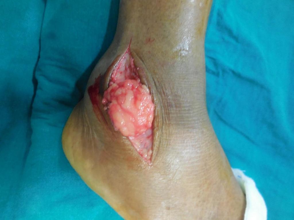
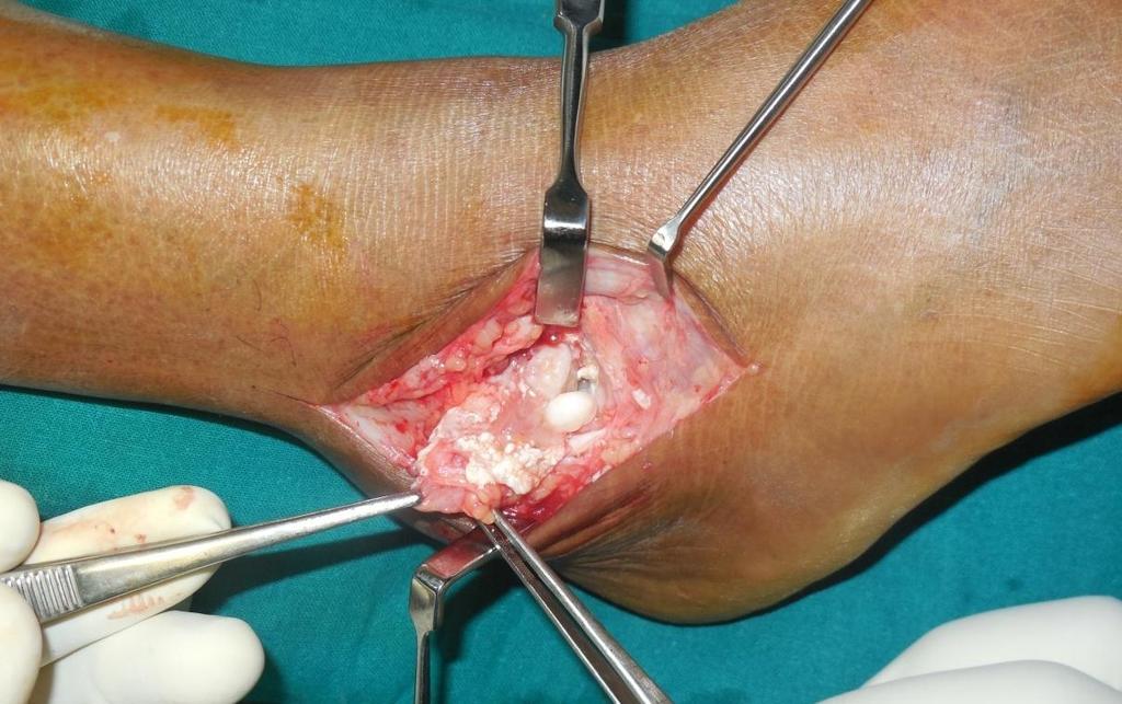
3 Madi et al Ankle range of motion was normal and no loose bodies or crepitus were palpable in the joint. Plain x-rays [antero-posterior and lateral views] of ankle joint showed a single calcified body measuring 1x1cms lying just underneath the tip oflateral malleolus (FIGURE-I). Rest of the ankle joint appeared normal except for the presence of ostrigonum but without evidence of degenerative changes. With this atypical radiological picture, we advised the patient for an MRI of the ankle, but due to financial constraints patient refused it. Regardless, we decided to proceed with excisional biopsy. A lipomatous swelling [6x5cms] was excised and beneath it was a solitary calcified body measuring 1x1cm lying outside the joint capsule (FIGURE-II). The wound was closed and the tissues were sent for Histo-Pathological Examination [HPE]. Postoperative days were uneventful. HPE reported as extra-synovial extra-articular synovial chondromatosis of the ankle (FIGURE-III). Before discharge, patient gave her consent for documenting this case for academic purpose. Unfortunately, patient never returned for follow-up after 3 months and the status of recurrence/ malignant transformations cannot be commented. DISCUSSION Primary synovial chondromatosis is a benign self-limiting lesion of unknown etiology that may recur locally. Even though, previously it was thought to be a metaplastic process of the synovial tissue, on the basis of currently known molecular abnormalities, it is now considered as a benign neoplastic disease (1). The extra-articular subtype typically arises from the synovium of the tendon sheath or bursa. The role of trauma in the development of this condition is uncertain, but history of trauma can mislead to the diagnosis of a trivial sprain. The condition is frequently seen in 3 rd to 5 th decade with a male preponderance, however when extraarticular disease is diagnosed 3
4 Osteochondromatosis of Ankle in older patients [i.e., after the fifth decade], there is a female predominance [2:1 ratio] (2). Clinical manifestations in extra articular subtype can be subtle in the form of a painless mass or mild tenderness upon palpation with seldom limitation in range of motion. The classical radiological appearance of synovial chondromatosis is multiple, calcified bodies in juxta articular location. These calcifications frequently show a pathognomonic appearance of ring and arc pattern of mineralization. Sometimes, these individual loose bodies coalesce to form a giant single chondroma and become symptomatic. Neither of these two characteristic pictures were seen in the index case.three differential diagnosis that could be made from the current clinicoradiological picture were: a calcified ganglion, fibrinous rice bodies [Tubercular arthropathy], and symptomatic os subfibulare. Ganglion and synovial cysts are the most common soft tissue lesions in the ankle and foot region and are usually associated with diseases like post-traumatic, inflammatory or degenerative joint diseases (3); moreover calcification in these cysts is not uncommon. Os subfibulare are separated ossicles at the tip of the lateral malleolus which can be anothersource of chronic ankle pain (4). Additional investigations like ultrasound, CT, and MRI further aid in diagnosing and choosing surgical approach. However,the final word on diagnosis could be made only by HPE. Microscopically, synovial osteochondromatosis is characterized bylobules of hyaline cartilage with variable degree of synovial proliferation or hyperplasia.the hyaline cartilage in primary synovial chondromatosis is often hypercellular with atypical histologic features, including multinucleation, nuclear crowding, nuclear enlargement and hyperchromasia, and mild myxoid changes, which would otherwise suggest a malignant cartilage neoplasm [grade 1 to grade 2 chondrosarcoma] (1). Associated foci of ossification is also noted in the cartilage; thereby the term osteo -chondromatosis.. Secondary synovial chondromatosis can be distinguished from primary disease both radiologically [underlying articular disease and 4
5 Madi et al fewer chondral bodies of variable size and shape]and pathologically [concentric rings of growth] (1). Open surgical excision in the preferred treatment for this condition with prompt relief of the symptoms. Recurrence rate reported for extra articular subtype is highly varied, but most recurrences occur within 5 years of initial resection. The potential of malignant transformation into chondrosarcoma is seen in upto 5% of cases which further warrants a close post-operative follow-up (5). CONCLUSION Synovial osteochondromatosis is a rare cause for chronic ankle pain. Thorough clinical evaluation supported by radiological investigations aid in clinching the diagnosis.surgical excision is the preferred modality of treatment. Long term follow-up is a must to rule out rare possibilities of recurrence or malignant transformation. ACKNOWLEDGEMENT We express our gratitude to Dr.Anuradha Rao, Professor, Department of Pathology, Kasturba Medical College, Manipal for providing the histo-pathology photograph. 5
6 Osteochondromatosis of Ankle REFERENCES 1. Murphey Mark D, Jorge A. Vidal, Julie C. Fanburg-Smith, and Donald A. Gajewski. "Imaging of Synovial Chondromatosis with Radiologic-Pathologic Correlation 1." Radiographics 27, no. 5 (2007): Fetsch John F, Tuyethoa N. Vinh, FabrizioRemotti, Eric A. Walker, Mark D. Murphey, and Donald E. Sweet. "Tenosynovial (extraarticular) chondromatosis: an analysis of 37 cases of an underrecognized clinic-pathologic entity with a strong predilection for the hands and feet and a high local recurrence rate." Ame J Surg Pathol 27, no. 9 (2003): Van Hul Erik, FilipVanhoenacker, Pieter Van Dyck, Arthur De Schepper, and Paul M. Parizel. "Pseudotumoural soft tissue lesions of the foot and ankle: a pictorial review." Insights into imaging 2, no. 4 (2011): KonoTaisuke, Mitsuo Ochi, Masato Takao, Kohei Naito, Yuji Uchio, and Kazunori Oae. "Symptomatic ossubfibulare caused by accessory ossification: a case report." Clinical Orthopaedics and Related Research 399 (2002): Davis R. I, HamiltonA, and Biggart J. D. "Primary synovial chondromatosis: a clinicopathologic review and assessment of malignant potential." Hum Pathol 29, no. 7 (1998): Tibrewal, S. B, and Iossifidis A. "Extra-articular synovial chondromatosis of the ankle." J Bone Joint Surg British Volume 77, no. 4 (1995): PathakShirish S, Clement Joseph, Aravinda M, Satish Sonar, Vinodh K, and David Rajan. "A case of primary intraarticular and extra articular synovial chondromatosis of ankle and foot." Internet J Orthopedic Surgry 4, no. 1 (2007). 6
7 Madi et al 8. Brian Carpenter D. P. M and John Clyde D. P. M. "Extrasynovial Synovial Osteochondromatosis of the Ankle: A case report." (2010). 9. Bahari, S., and J. McKenna. "Subtalar and Extra Articular Synovial Chondromatosis." Journal of Case Reports 2, no. 2 (2013):
8 Osteochondromatosis of Ankle Table: Case reports on extra-articular synovial chondromatosis of the ankle joint Sl. Author/year Age/sex Presentation Radiology Treatment Follow-up no 1. S. B. Tibrewal et al [1995](6) 44/F Painful mass, But pain-free movements Open Excision Bony erosion of tibio-fibular syndesmosis. CT revealed calcification in mass 2years. No recurrence and asymptomatic 2. S.Pathak et al [2006] (7) 32/M Restricted dorsiflexion & locking episodes Multiple loose bodies [intraarticular & extraarticular] Arthroscopic synovectomy + loose body removal. + Open excision 1 year. Pain-free full range of ankle movement 3. B.Carpenter et al [2010] (8) 56/F Painful dorsiflexion & anteromedial mass Single radioopaque nodule. MRI done. Open excision 9 months. Patient is asymptomatic 4. S. Bahari et al [2012] (9) 53/F Ankle sprain history, Pain & swelling Erosive lesion in ankle. MRI was required. Open excision 1 year. Returned to normal activity 8


9 Madi et al Fig.1: Plain x-ray antero-posteior and lateral view of ankle joint 9
10 Osteochondromatosis of Ankle 10

11 Madi et al Fig.2: Lipomatous swelling and solitary osteochondromatosis lying outside the joint. Fig.3: Histopathlogy: 5X cartilaginous cap overlying trabeculae of bone. 11
A Case Of Primary Intra-Articular And Extra Articular Synovial Chondromatosis Of Ankle And Foot
 ISPUB.COM The Internet Journal of Orthopedic Surgery Volume 4 Number 1 A Case Of Primary Intra-Articular And Extra Articular Synovial Chondromatosis Of Ankle And Foot S Pathak, C Joseph, M Aravinda, S
Monophasic Synovial Carcinoma of knee joint- A Case Report and Review of Literature
 IOSR Journal of Dental and Medical Sciences (IOSR-JDMS) e-issn: 2279-0853, p-issn: 2279-0861.Volume 17, Issue 3 Ver.5 March. (2018), PP 13-17 www.iosrjournals.org Monophasic Synovial Carcinoma of knee
Case Report Extraosseous Intra-Articular Osteochondroma
 Case Reports in Orthopedics Volume 2013, Article ID 181862, 5 pages http://dx.doi.org/10.1155/2013/181862 Case Report Extraosseous Intra-Articular Osteochondroma Pragash Mohanen, 1,2 Kumaresan Palania
Bursa Formation and Synovial Chondrometaplasia Associated with Osteochondromas
 Bursa Formation and Synovial Chondrometaplasia Associated with Osteochondromas ANITA M. BORGES, M. D., ANDREW G. HUVOS, M. D., AND JULIUS SMITH, M. D. Borges, Anita M., Huvos, Andrew G., and Smith, Julius:
A rare case of a swollen knee due to disseminated synovial chondromatosis: a case report
 JOURNAL OF MEDICAL CASE REPORTS CASE REPORT Open Access A rare case of a swollen knee due to disseminated synovial chondromatosis: a case report Hugh Mackenzie *, Vivek Gulati, Samantha Tross Abstract
Intra-articular soft tissue masses of the knee: An imaging review of biopsy proven diagnoses
 Intra-articular soft tissue masses of the knee: An imaging review of biopsy proven diagnoses Poster No.: P-0114 Congress: ESSR 2014 Type: Scientific Poster Authors: A. Kirwadi 1, S. Raniga 2, R. Hargunani
Management of Chronic Elbow Pain
 Mr. Nashat Siddiqui Consultant Upper Limb Orthopaedic Surgeon Management of Chronic Elbow Pain Patients presenting with elbow pain can pose a diagnostic challenge, especially if there is no obvious recent
Ankle Arthroscopy.
 Ankle Arthroscopy Key words: Ankle pain, ankle arthroscopy, ankle sprain, ankle stiffness, day case surgery, articular cartilage, chondral injury, chondral defect, anti-inflammatory medication Our understanding
Neglected synovial osteochondromatosis of the elbow: a rare case
 WORLD JOURNAL OF SURGICAL ONCOLOGY Neglected synovial osteochondromatosis of the elbow: a rare case Giannetti et al. Giannetti et al. World Journal of Surgical Oncology 2013, 11:233 Giannetti et al. World
Intracapsular and para- articular chondroma of knee: a report of four cases and review of the literature
 Intracapsular and para- articular chondroma of knee: a report of four cases and review of the literature Milan Samardziski, Marta Foteva, Aleksandar Adamov, George Zafiroski University Clinic for Orthopaedic
Research Article Synovial Chondrosarcoma Arising in Synovial Chondromatosis
 Sarcoma, Article ID 647939, 4 pages http://dx.doi.org/10.1155/2014/647939 Research Article Synovial Chondrosarcoma Arising in Synovial Chondromatosis Scott Evans, Michele Boffano, Samena Chaudhry, Lee
A 24 year old male patient presented with a swelling on the dorsal aspect of left foot since 3 years. He was operated thrice before, outside, for
 A 24 year old male patient presented with a swelling on the dorsal aspect of left foot since 3 years. He was operated thrice before, outside, for same. Came to us with recurrence since last one year with
Advertisement. Osteochondroma
 Advertisement Osteochondroma An osteochondroma is a benign (noncancerous) tumor that develops during childhood or adolescence. It is an abnormal growth that forms on the surface of a bone near the growth
Welcome to the: Orthopaedic Opinion Online Website The website for the answer to all your Orthopaedic Questions
 Welcome to the: Orthopaedic Opinion Online Website The website for the answer to all your Orthopaedic Questions Orthopaedic Opinion Online is a website designed to provide information to patients who have
Multicentric localized giant cell tumor of the tendon. sheath
 Multicentric localized giant cell tumor of the tendon sheath Toshihiro Akisue, Tetsuji Yamamoto ( ), Teruya Kawamoto, Toshiaki Hitora, Takashi Marui, Tetsuya Nakatani, Takafumi Onga, and Masahiro Kurosaka.
Case Report Multiple Giant Cell Tumors of Tendon Sheath Found within a Single Digit of a 9-Year-Old
 Case Reports in Orthopedics Volume 2016, Article ID 1834740, 4 pages http://dx.doi.org/10.1155/2016/1834740 Case Report Multiple Giant Cell Tumors of Tendon Sheath Found within a Single Digit of a 9-Year-Old
Case Study. Your Diagnosis?
 Case Study 35 year old man twisted his ankle while playing and was surgically for treated for ankle sprain for instability symptoms 2 years. Increasing pain around the ankle and pain present all the time
SURGERY OF THE HAND. Synovial Chondromatosis of the Ulnocarpal Joint INTRODUCTION CASE REPORT CASE REPORT. Sung-Guk Kim
 CSE REPORT pissn 1598-3889 eissn 2234-0998 J Korean Soc Surg Hand 2016;21(1):50-54. http://dx.doi.org/10.12790/jkssh.2016.21.1.50 JOURNL OF THE KOREN SOCIETY FOR SURGERY OF THE HND Synovial Chondromatosis
Calcifying Aponeurotic Fibroma of the Knee: a Case Report with Radiographic and MRI Finding
 pissn 2384-1095 eissn 2384-1109 imri 2017;21:259-263 Calcifying Aponeurotic Fibroma of the Knee: a Case Report with Radiographic and MRI Finding Seung Hyun Lee 1,2, In Sook Lee 1,2, You Seon Song 1,2,
Giant solitary synovial osteochondromatosis of the elbow causing ulnar nerve neuropathy: a case report and review of literature
 Al-Najjim et al. Journal of Brachial Plexus and Peripheral Nerve Injury 2013, 8:1 JOURNAL OF BRACHIAL PLEXUS AND PERIPHERAL NERVE INJURY CASE REPORT Open Access Giant solitary synovial osteochondromatosis
Case 8 Soft tissue swelling
 Case 8 Soft tissue swelling 26-year-old female presented with a swelling on the back of the left knee joint since the last 6 months and chronic pain in the calf and foot since the last 2 months. Pain in
Figuring out the "fronds"-synovial proliferative disorders of the knee.
 Figuring out the "fronds"-synovial proliferative disorders of the knee. Poster No.: C-1209 Congress: ECR 2014 Type: Educational Exhibit Authors: S. Sivasubramanian; Tamil Nadu/IN Keywords: Imaging sequences,
Delayed presentation of osteochondroma at superior angle of scapula- a case report
 Article ID: ISSN 2046-1690 Delayed presentation of osteochondroma at superior angle of scapula- a case report Peer review status: No Corresponding Author: Dr. Mohit K Jindal, Senior Resident, ESI PGIMSR
GIANT CELL TUMOR OF TENDON SHEATH A CYTO HISTO CORRELATION
 GIANT CELL TUMOR OF TENDON SHEATH A CYTO HISTO CORRELATION Dr.S.SRIKANTH, Assistant Professor.Dept of Patholgy. Dr.SMITHA VADANA, Resident.Dept of pathology. Dr.R.SUHELA. Resident.Dept Of Pathology. Prathima
Synovial chondromatosis: a pictorial review
 Synovial chondromatosis: a pictorial review Poster No.: C-1307 Congress: ECR 2014 Type: Educational Exhibit Authors: L. Silva, M. O. E. Castro, B. M. Q. Santos, C. Bilreiro, F. Aleixo; Portimão/PT Keywords:
Synovial Hemangioma of the elbow: An uncommon lesion to be considered
 ISPUB.COM The Internet Journal of Pathology Volume 7 Number 2 Synovial Hemangioma of the elbow: An uncommon lesion to be considered A Mohammadi, M Rosa, D Wolfson Citation A Mohammadi, M Rosa, D Wolfson.
What are rice bodies?: Differential diagnosis.
 What are rice bodies?: Differential diagnosis. Poster No.: C-1605 Congress: ECR 2015 Type: Educational Exhibit Authors: S. Córdoba Rovira, A. Ramos, L. E. Guerrero, D. Villa Viñas, A. Guedea Martin, M.
Synovial Chondromatosis Associated with Polyarteritis Nodosa
 Synovial Chondromatosis Associated with Polyarteritis Nodosa Hywel Davies BSc ( ),Andrew J Unwin BSc, Nick P H Morgan BSc Windsor Knee Clinic, Windsor, United Kingdom Correspondence: Hywel Davies, Windsor
Pigmented Villonodular Synovitis PVNS
 February 2002 Pigmented Villonodular Synovitis PVNS Amy Gillis, Harvard Medical School Year III 47 year old female Our Patient Right hip pain since age 20 No history of trauma Diagnosed with DJD of R hip
Case Report Arthroscopic Treatment of a Case with Concomitant Subacromial and Subdeltoid Synovial Chondromatosis and Labrum Tear
 Case Reports in Orthopedics Volume 2013, Article ID 636747, 4 pages http://dx.doi.org/10.1155/2013/636747 Case Report Arthroscopic Treatment of a Case with Concomitant Subacromial and Subdeltoid Synovial
Message of the Month for GPs June 2013
 Message of the Month for GPs June 2013 Dr Winn : Consultant Musculoskeletal Radiologist, Manchester Royal Infirmary Imaging of the musculoskeletal system Musculoskeletal pain is a common problem in the
Interesting Case Series. Ganglion Cyst of the Peroneus Longus
 Interesting Case Series Ganglion Cyst of the Peroneus Longus Andrew A. Marano, BA, Paul J. Therattil, MD, Dare V. Ajibade, MD, PhD, MPH, and Ramazi O. Datiashvili, MD, PhD Division of Plastic and Reconstructive
Indian Journal of Medical Research and Pharmaceutical Sciences August 2015; 2(8) ISSN: ISSN: Impact Factor (PIF): 2.672
 ISSN: ISSN: Impact Factor (PIF): 2.672") CASE REPORT OF TUBERCULOUS SUBDELTOID BURSITIS WITH RICE BODIES Dr. Jhatoth Venkateshwarlu*, Dr. Tandra Venkateshwararao, Dr. K. Ramkumar Reddy, Dr. K.Venkatswamy, Dr. M.Sudhir MS Orthopaedics, Associate
CASE REPORT OF TUBERCULOUS SUBDELTOID BURSITIS WITH RICE BODIES Dr. Jhatoth Venkateshwarlu*, Dr. Tandra Venkateshwararao, Dr. K. Ramkumar Reddy, Dr. K.Venkatswamy, Dr. M.Sudhir MS Orthopaedics, Associate
Dupuytren's Contracture Assessment
 Dupuytren's Contracture Assessment Link to guidance: http://www.enhertsccg.nhs.uk/ bedfordshire-and-hertfordshire-priorities-forum Dupuytren's contracture - clinical presentation for patients History Examination
emoryhealthcare.org/ortho
 COMMON SOCCER INJURIES Oluseun A. Olufade, MD Assistant Professor, Department of Orthopedics and PM&R 1/7/18 GOALS Discuss top soccer injuries and treatment strategies Simplify hip and groin injuries in
Arthroscopy in sports medicine
 Arthroscopy in sports medicine DR PABITRA KUMAR SAHOO, ASSISTANT PROFESSOR (PMR) Sports medicine is a special branch of rehab specialty which deals with training in anatomy, biomechanics, pathophysiology
Ankle Arthroscopy PAULO ROCKETT, M.D. Porto Alegre Brazil
 Ankle Arthroscopy PAULO ROCKETT, M.D. Porto Alegre Brazil Ankle sprains are among the most common injuries in sports and at work. Between 20 and 40% of patients treated with conservative therapy may have
Dr. Ashish Shah MD Dr. John Kirchner MD Dr. Sameer Naranje MD
 Dr. Ashish Shah MD Dr. John Kirchner MD Dr. Sameer Naranje MD Assistant Professor, UAB Orthopaedic Surgery, Foot & Ankle Section, 1313 13th Street South, Birmingham, AL 35205 Clinical Instructor Fellow,
Case Report Elbow Arthroscopy: Review of the Literature and Case Reports
 Hindawi Publishing Corporation Case Reports in Orthopedics Volume 2012, Article ID 478214, 5 pages doi:10.1155/2012/478214 Case Report Elbow Arthroscopy: Review of the Literature and Case Reports Prakash
Imaging characteristics of tenosynovial and bursal chondromatosis
 DOI 10.1007/s00256-010-1012-3 SCIENTIFIC ARTICLE Imaging characteristics of tenosynovial and bursal chondromatosis Eric A. Walker & Mark D. Murphey & John F. Fetsch Received: 19 April 2010 /Revised: 15
CLINICAL PRESENTATION AND RADIOLOGY QUIZ QUESTION
 Donald L. Renfrew, MD Radiology Associates of the Fox Valley, 333 N. Commercial Street, Suite 100, Neenah, WI 54956 12/01/2012 Radiology Quiz of the Week # 101 Page 1 CLINICAL PRESENTATION AND RADIOLOGY
Sequalae of Ankle Sprains: Peri Articular Fractures of the Ankle in Sports Medicine.
 Sequalae of Ankle Sprains: Peri Articular Fractures of the Ankle in Sports Medicine www.fisiokinesiterapia.biz Chronic Ankle Pain The most common cause of chronic pain following an ankle sprain is a missed
Citation Acta medica Nagasakiensia. 1997, 42
 NAOSITE: Nagasaki University's Ac Title Author(s) Dysplasia Epiphysealis Hemimelica o Uetani, Masataka; Hashmi, Rashid; H Hayashi, Tomayoshi Citation Acta medica Nagasakiensia. 1997, 42 Issue Date 1997-12-20
Dumbbell Ganglion Of The Foot: Case Report
 Article ID: WMC001079 2046-1690 ISSN Dumbbell Ganglion Of The Foot: Case Report Author(s):Dr. S S Suresh, Dr. Hosam Zaki, Dr. Joyce Jose Corresponding Author: Dr. S S Suresh, Head of Department, Ibri Regional
Skeletally Immature Athletes Ununited Osteochondral Fractures of the Distal Fibula
 Chronic, Painful Ankle Instability in Skeletally Immature Athletes Ununited Osteochondral Fractures of the Distal Fibula Brian D. Busconi,* MD, and Arthur M. Pappas, MD From the Department of Orthopedics
Case report. A neglected case of giant synovial chondromatosis in knee joint. Open Access
 Case report Open Access A neglected case of giant synovial chondromatosis in knee joint Sancar Serbest 1,&, Ugur Tiftikçi 1, Fatih Karaaslan 2, Haci Bayram Tosun 3, Hüseyin Fatih Sevinç 1, Mahi Balci 4
Priorities Forum Statement GUIDANCE
 Priorities Forum Statement Number 21 Subject Knee Arthroscopy including arthroscopic knee washouts Date of decision November 2016 Date refreshed March 2017 Date of review November 2018 Osteoarthritis of
WORKPLACE SAFETY AND INSURANCE APPEALS TRIBUNAL DECISION NO. 1417/12
 WORKPLACE SAFETY AND INSURANCE APPEALS TRIBUNAL DECISION NO. 1417/12 BEFORE: S. Martel: Vice-Chair HEARING: July 9, 2012 at Toronto Written DATE OF DECISION: September 5, 2012 NEUTRAL CITATION: 2012 ONWSIAT
CASE REPORT Idiopathic Tumoral Calcinosis of the Nontraumatic Thumb
 CASE REPORT Idiopathic Tumoral Calcinosis of the Nontraumatic Thumb Ginard I. Henry, MD and Chad M. Teven, MD Section of Plastic and Reconstructive Surgery, University of Chicago, Chicago, IL Correspondence:
Primary bone tumors > metastases from other sites Primary bone tumors widely range -from benign to malignant. Classified according to the normal cell
 Primary bone tumors > metastases from other sites Primary bone tumors widely range -from benign to malignant. Classified according to the normal cell counterpart and line of differentiation. Among the
Gout. Crystal deposition disease: Imaging perspectives. Crystal associated arthropathies. Clinical Stages of Gout 07/06/60
 Crystal associated arthropathies Crystal deposition disease: Imaging perspectives Warapat Virayavanich, MD Ramathibodi hospital, Mahidol University Commonly seen arthropathy MSU (gout) CPPD HADD Uncommon
Recognizing Cartilaginous Tumors: Spectrum of Imaging Characteristics with Radiologic-Pathologic correlation.
 Recognizing Cartilaginous Tumors: Spectrum of Imaging Characteristics with Radiologic-Pathologic correlation. Poster No.: C-1451 Congress: ECR 2012 Type: Educational Exhibit Authors: E. Barcina García,
CASE REPORT GIANT OSTEOCHONDRAL LOOSE BODY OF THE KNEE JOINT
 Journal of Musculoskeletal Research, Vol. 4, No. 2 (2000) 145 149 World Scientific Publishing Company ORIGINAL CASE REPORT ARTICLES GIANT OSTEOCHONDRAL LOOSE BODY OF THE KNEE JOINT Mustafa Yel *,, Mustafa
ELBOW ARTHROSCOPY WHERE ARE WE NOW?
 ELBOW ARTHROSCOPY WHERE ARE WE NOW? Christian Veillette M.D., M.Sc., FRCSC Assistant Professor, University of Toronto Shoulder & Elbow Reconstructive Surgery Toronto Western Hospital @ University Health
Scrotum-like protrusion of lipoma arising from the proximal thigh
 Upsala J Med sci 109: 261 265, 2004 Scrotum-like protrusion of lipoma arising from the proximal thigh Report of two cases Koshi Hattori, 1 Masahito Hatori, 1 Mika Watanabe, 2 Toshihisa Osanai, 3 Shoichi
A case of extensive synovial involvement by tophaceous gout
 A case of extensive synovial involvement by tophaceous gout Nausheen Khan, MB BS, FCRad (D) Irma van de Werke, MB ChB, FRCR Farzanah Ismail, MB ChB, FCRad (D) Department of Radiology, Kalafong Hospital,
Unusual Lateral Presentation of Popliteal Cyst
 Unusual Lateral Presentation of Popliteal Cyst Tarek Hemmali,* Abstract: The most common cyst occurs in the popliteal region is the popliteal cyst and over the past years it has been received much clinical
Mr Simon Jennings BSc, MB BS, FRCS, Dip Sports Med FRCS (Trauma & Orthopaedics)
") Mr Simon Jennings BSc, MB BS, FRCS, Dip Sports Med FRCS (Trauma & Orthopaedics) Consultant Orthopaedic Surgeon Northwick Park Hospital 107 Harley Street RSM 16 th September 2010 Orthopaedic Surgeon Knee
Mr Simon Jennings BSc, MB BS, FRCS, Dip Sports Med FRCS (Trauma & Orthopaedics) Consultant Orthopaedic Surgeon Northwick Park Hospital 107 Harley Street RSM 16 th September 2010 Orthopaedic Surgeon Knee
University Journal of Surgery and Surgical Specialities
 University Journal of Surgery and Surgical Specialities Volume 1 Issue 1 2015 EXTRA SKELETAL MESENCHYMAL CHONDROSARCOMA :A CASE REPORT Rajaraman R Subbiah S Navin Naushad Kilpaulk Medical College Abstract:
Bilateral Shoulder Pain
 HR J Bilateral Shoulder Pain, p. 64-69 Clinical Case - Test Yourself Bilateral Shoulder Pain Musculoskeletal Eirini D. Savva, Rafaela M. Smarlamaki, Foteini I. Terezaki Department of Radiology, University
Anterior ankle impingement in sports Hrefna Thorbjorg Hakonardottir
 Anterior ankle impingement in sports Hrefna Thorbjorg Hakonardottir Anterior ankle impingement in sports Ankle impingement syndromes are classified by their anatomical location around the tibiotalar joint
RAPID MALIGNANT TRANSFORMATION OF PRIMARY SYNOVIAL CHON DROMATOSIS INTO CHONDROSARCOMA
 JR TR, 2014, 97: 303-307. RPID MLIGNNT TRNSFORMTION OF PRIMRY SYNOVIL HON DROMTOSIS INTO HONDROSROM J. Jonckheere 1, M. Shahabpour 1, I. Willekens 1, N. Pouliart 2, M. Dezillie 3, F. Vanhoenacker 4, J.
We present 12 patients with synovial
 Synovial osteochondromatosis of the elbow S. Kamineni, S. W. O Driscoll, B. F. Morrey From the Mayo Clinic, Rochester, USA We present 12 patients with synovial osteochondromatosis of the elbow treated
Case Report Intra-Articular Giant Synovial Osteochondroma: Case Reports of the Ankle and Knee Joint
 Case Reports in Orthopedics Volume 2015, Article ID 320139, 5 pages http://dx.doi.org/10.1155/2015/320139 Case Report Intra-Articular Giant Synovial Osteochondroma: Case Reports of the Ankle and Knee Joint
Tumours and tumour-like lesions of the patella : A report of eight cases
 Acta Orthop. Belg., 2008, 74, 391-396 ORIGINAL STUDY Tumours and tumour-like lesions of the patella : A report of eight cases Yener SAGLIK, Yusuf YILDIZ, Kerem BASARIR, Engin TEZEN, Derviş GÜNER From Ankara
Joint Injuries and Disorders
 Joint Injuries and Disorders Introduction A joint is where two or more bones come together. Your joints include the knees, hips, elbows and shoulders. There are many types of joint disorders, including
ELENI ANDIPA General Hospital of Athens G. Gennimatas
 ELENI ANDIPA General Hospital of Athens G. Gennimatas Technological advances over the last years have caused a dramatic improvement in ultrasound quality and resolution An established imaging modality
SYNOVIAL OSTEOCHONDROMATOSIS; SECONDARY SYNOVIAL OSTEOCHONDROMATOSIS (SOC) OF SHOULDER JOINT
 OF SHOULDER JOINT") The Professional Medical Journal DOI: 10.29309/TPMJ/18.5058 1. Doctor of Medical Imaging 2. Doctor of Medical Imaging 3. Doctor of Medical Imaging 4. Doctor of Medical Imaging 5. Doctor of Medical Imaging
The Professional Medical Journal DOI: 10.29309/TPMJ/18.5058 1. Doctor of Medical Imaging 2. Doctor of Medical Imaging 3. Doctor of Medical Imaging 4. Doctor of Medical Imaging 5. Doctor of Medical Imaging
A CASE OF A Huge Submandibular Pleomorphic Adenoma
 ISPUB.COM The Internet Journal of Head and Neck Surgery Volume 4 Number 2 S VERMA Citation S VERMA.. The Internet Journal of Head and Neck Surgery. 2009 Volume 4 Number 2. Abstract Pleomorphic adenoma
Persistent ankle pain after inversion lesions: what the radiologist must look for
 Persistent ankle pain after inversion lesions: what the radiologist must look for Poster No.: P-0118 Congress: ESSR 2016 Type: Authors: Keywords: DOI: Educational Poster R. Leao, L. C. Zattar-Ramos, E.
TUBERCULOUS OSTEOMYELITIS OF PATELLA: A CASE REPORT Babu B. Hundekar 1
 TUBERCULOUS OSTEOMYELITIS OF PATELLA: A Babu B. Hundekar 1 HOW TO CITE THIS ARTICLE: Babu B. Hundekar. Tuberculous Osteomyelitis of Patella: A Case Report. Journal of Evolution of Medical and Dental Sciences
Mr. Duy Thai Orthopaedic Surgeon, Melbourne VIC
 Mr. Duy Thai Orthopaedic Surgeon, Melbourne VIC International Convention of the Vietnamese Physicians, Dentists and Pharmacists of the Free World Melbourne 8 10 August 2014 Conflict of Interest None Subacromial
Salisbury Foundation Trust Radiology Department Referral Guidelines for Primary Care: Musculoskeletal Imaging
 Salisbury Foundation Trust Radiology Department Referral Guidelines for Primary Care: Musculoskeletal Imaging These guidelines have been issued in conjunction with the Royal College of Radiology referral
Ankle Replacement Surgery
 Ankle Replacement Surgery Ankle replacement surgery is performed to replace the damaged articular surfaces of the three bones of the ankle joint with artificial implants. This procedure is now being preferred
Pigmented Villonodular Synovitis (PVNS) A Case Report
 A Case Report") Case Report: Pigmented Villonodular Synovitis (PVNS) A Case Report Abhishek Patil 1, Sanjay Mulay 2, Chandrashekar Jaiswal 1, Uday Mahajan 1, Shriniwas Yadkikar 3, Vishnu Yadkikar 4 1Junior Resident, 2
Case Report: Pigmented Villonodular Synovitis (PVNS) A Case Report Abhishek Patil 1, Sanjay Mulay 2, Chandrashekar Jaiswal 1, Uday Mahajan 1, Shriniwas Yadkikar 3, Vishnu Yadkikar 4 1Junior Resident, 2
SICOT Online Report E057 Accepted April 23th, in Fibula and Rib
 Metachronous, multicentric giant cell tumors in Fibula and Rib Toshihiro Akisue, Tetsuji Yamamoto ( ), Teruya Kawamoto, Toshiaki Hitora, Takashi Marui, Tetsuya Nakatani, Takafumi Onga, and Masahiro Kurosaka
Index. Note: Page numbers of article titles are in boldface type.
 Magn Reson Imaging Clin N Am 12 (2004) 185 189 Index Note: Page numbers of article titles are in boldface type. A Acromioclavicular joint, MR imaging findings concerning, 161 Acromion, types of, 77 79
PEM GUIDE CHILDHOOD FRACTURES
 PEM GUIDE CHILDHOOD FRACTURES INTRODUCTION Skeletal injuries account for 10-15% of all injuries in children; 20% of those are fractures, 3 out of 4 fractures affect the physis or growth plate. Always consider
Ankle Arthritis PATIENT INFORMATION. The ankle joint. What is ankle arthritis?
 PATIENT INFORMATION Ankle Arthritis The ankle joint The ankle is a very complex joint. It is actually made up of two joints: the true ankle joint and the subtalar ankle joint. The ankle joint consists
Debridement arthroplasty for osteoarthritis of the elbow (Outerbridge-Kashiwagi procedure)
") Acta Orthop. Belg., 2004, 70, 306-310 ORIGINAL STUDIES Debridement arthroplasty for osteoarthritis of the elbow (Outerbridge-Kashiwagi procedure) Bart VINGERHOEDS, Ilse DEGREEF, Luc DE SMET From the University
Acta Orthop. Belg., 2004, 70, 306-310 ORIGINAL STUDIES Debridement arthroplasty for osteoarthritis of the elbow (Outerbridge-Kashiwagi procedure) Bart VINGERHOEDS, Ilse DEGREEF, Luc DE SMET From the University
Synovial Chondromatosis of the Temporomandibular Joint: Long-Term Postoperative Follow-Up of the Residual Calcification
 J Med Dent Sci 2003; 50: 133 137 Case Report Synovial Chondromatosis of the Temporomandibular Joint: Long-Term Postoperative Follow-Up of the Residual Calcification Junichi Ishii 1, Koji Kino 2, Junji
Arthroscopic synovectomy, removal of loose bodies and selective biceps tenodesis for. of synovial chondromatosis of the shoulder.
 Upper limb Arthroscopic synovectomy, removal of loose bodies and selective biceps for synovial chondromatosis of the shoulder J. V. Lunn, J. Castellanos- Rosas, G. Walch From Centre Orthopédique Santy,
ISPUB.COM. Spectrum Of MRI Findings In Musculoskeletal Tuberculosis: Pictoral Essay. P Chudgar INTRODUCTION SPINE
 ISPUB.COM The Internet Journal of Radiology Volume 8 Number 2 Spectrum Of MRI Findings In Musculoskeletal Tuberculosis: Pictoral Essay P Chudgar Citation P Chudgar.. The Internet Journal of Radiology.
Primary care referral criteria for musculoskeletal MRI scans
 Appendix 1 Primary care referral criteria for musculoskeletal MRI scans Accepted Criteria for Direct Access MRI Body Part Symptoms Imaging indicated Lumbar Spine Low Back Pain with adverse symptoms or
Synovial hemangioma of the suprapatellar bursa
 Synovial hemangioma of the suprapatellar bursa Poster No.: P-0040 Congress: ESSR 2013 Type: Authors: Keywords: DOI: Scientific Exhibit A. YESILDAG, S. Keskin, H. Kalkan, S. Kucuksen, U. Kerimoglu; Konya/TR
Multifocal fibrous Dysplasia with enchondroma-like areas: Fibrocartilaginous Dysplasia
 ISPUB.COM The Internet Journal of Pathology Volume 7 Number 2 Multifocal fibrous Dysplasia with enchondroma-like areas: Fibrocartilaginous Dysplasia V Monappa, R Kudva Citation V Monappa, R Kudva. Multifocal
SMF PCP Treatment & Referral Guideline Orthopedics Developed February 1, 2003 Revised: October, 2011
 SUTTER MEDICAL FOUNDATION (SMF) 2800 L Street, 7 th Floor Sacramento, CA 95816 SMF PCP Treatment & Referral Guideline Orthopedics Developed February 1, 2003 Revised: October, 2011 I. Shoulder Pain...Page
Chondrosarcoma of 5 th metatarsal Right Foot: An Unusual Presentation and Review of Literature
 IOSR Journal of Dental and Medical Sciences (IOSR-JDMS) e-issn: 2279-0853, p-issn: 2279-0861.Volume 17, Issue 8 Ver. 13 (August. 2018), PP 27-33 www.iosrjournals.org Chondrosarcoma of 5 th metatarsal Right
Ankle Ligament Injury: Don t Worry- It s Only a Sprain Wes Jackson MD Orthopaedic Foot & Ankle
 Ankle Ligament Injury: Don t Worry- It s Only a Sprain Wes Jackson MD Orthopaedic Foot & Ankle Outline I. Epidemiology II. Classification and Types of Sprains III. Anatomy IV. Clinical Assessment and Imaging
CASE REPORT PLEOMORPHIC LIPOSARCOMA OF PECTORALIS MAJOR MUSCLE IN ELDERLY MAN- CASE REPORT & REVIEW OF LITERATURE.
 PLEOMORPHIC LIPOSARCOMA OF PECTORALIS MAJOR MUSCLE IN ELDERLY MAN- CASE REPORT & REVIEW OF LITERATURE. M. Madan 1, K. Nischal 2, Sharan Basavaraj. C. J 3. HOW TO CITE THIS ARTICLE: M. Madan, K. Nischal,
MEMORANDUM 171/91. DATE: June 26, 1991 TO: ALL WCAT STAFF SUBJECT: DECISION NO. 171/91. Continuity (of treatment) - Strains and sprains (ankle).
 - Strains and sprains (ankle).") MEMORANDUM 171/91 DATE: June 26, 1991 TYPE: A TO: ALL WCAT STAFF SUBJECT: DECISION NO. 171/91 Continuity (of treatment) - Strains and sprains (ankle). The worker sprained his ankle in a compensable accident
MEMORANDUM 171/91 DATE: June 26, 1991 TYPE: A TO: ALL WCAT STAFF SUBJECT: DECISION NO. 171/91 Continuity (of treatment) - Strains and sprains (ankle). The worker sprained his ankle in a compensable accident
Sports Injuries of the Ankle and Ankle Arthritis. Mr Amit Amin Consultant Foot and Ankle Surgeon Parkside Hospital
 Sports Injuries of the Ankle and Ankle Arthritis Mr Amit Amin Consultant Foot and Ankle Surgeon Parkside Hospital Impingement Painful mechanical limitation of full ankle movement secondary to osseous
Clin Podiatr Med Surg 19 (2002) Index
 Index") Clin Podiatr Med Surg 19 (2002) 335 344 Index Note: Page numbers of article titles are in bold face type. A Accessory soleus muscle, magnetic resonance imaging of, 300 Achilles tendon injury of, magnetic
Clin Podiatr Med Surg 19 (2002) 335 344 Index Note: Page numbers of article titles are in bold face type. A Accessory soleus muscle, magnetic resonance imaging of, 300 Achilles tendon injury of, magnetic
Acute Ankle Injuries, Part 1: Office Evaluation and Management
 t June 08, 2009 Obesity [1] Each acute ankle injury commonly seen in the office has associated with it a mechanism by which it can be injured, trademark symptoms that the patient experiences during the
DISEASES AND DISORDERS
 DISEASES AND DISORDERS 9. 53 10. Rheumatoid arthritis 59 11. Spondyloarthropathies 69 12. Connective tissue diseases 77 13. Osteoporosis and metabolic bone disease 95 14. Crystal arthropathies 103 15.
Rheumatoid Arthritis
 Rheumatoid Arthritis Rheumatoid arthritis (RA) is an autoimmune disease that causes chronic inflammation of the joints. Autoimmune diseases are illnesses that occur when the body's tissues are mistakenly
Giant Pleomorphic Adenoma of the Parotid gland- A Case Report
 ISPUB.COM The Internet Journal of Otorhinolaryngology Volume 14 Number 1 Giant Pleomorphic Adenoma of the Parotid gland- A Case Report O M.E, U A.N, U Akpan, K J, I Bassey Citation O M.E, U A.N, U Akpan,
Anterior impingement syndrome in dancers
 Curr Rev Musculoskelet Med (2008) 1:12 16 DOI 10.1007/s12178-007-9001-4 Anterior impingement syndrome in dancers John William O Kane Æ Nancy Kadel Published online: 6 November 2007 Ó Humana Press 2007