MRI of Pediatric Ankle and Foot. Mahesh Thapa, MD Associate Professor Seattle Children s University of Washington School of Medicine
|
|
|
- Roderick Thompson
- 6 years ago
- Views:
Transcription
1 MRI of Pediatric Ankle and Foot Mahesh Thapa, MD Associate Professor Seattle Children s University of Washington School of Medicine
2 Disclosures Under contract with Lippincott Williams and Wilkins (LWW) for Pediatric Radiology A Teaching File Textbook
3 Acknowledgement I want to thank my coauthors for contributing to this talk and to the Pediatric Radiology manuscript Ramesh Iyer, MD Felix Chew, MD
4 MR Technique Ankle: Reference plane is the table surface Foot: Reference plane is long axis of metatarsals Supine with 20 plantar flexion Ankle surface coil; other small coils (head or extremity)
5 MR Technique T1W, T2W (or STIR) in all planes 3D GRE for cartilage cm FOV 320 x 256 matirx 1-2 signals 3-4mm T1W and STIR; 1.2 mm on 3D GRE +/- Gad; +/- MRA
6 Indications Tumor Infection Arthritis Ligamentous Injury Stress fractures Osteochondral lesions Osteonecrosis


7 Marrow Signal Best assessed with T1W and T2W/STIR Scattered T2W/STIR foci normal

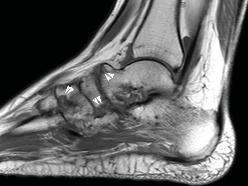
8 Tarsal Coalition Osseous, cartilaginous or fibrous Most are calcaneonavicular or talocalcaneal 50% bilateral
9 Osteochondral Lesion of the Talus OCL = Osteochondritis dissecans, osteochondral defects, transchondral fractures? Cause.? Trauma, ischemia, genetics Usu medial or lateral margins of talar dome


10 Osteochondral Lesion of the Talus
11 Osteonecrosis Chronically ill kids on long-term steroid therapy Talar neck fracture Common sites: talar dome, navicular, 2 nd /3 rd metatarsal heads


12 Talar dome osteonecrosis 19-year-old who sustained nondisplaced talar fracture while jumping on trampoline. Tehranzadeh J et al. AJR 2003;181:

13 Talus Blood Supply Tehranzadeh J et al. AJR 2003;181:


14 Hawkins Classification Type I: 0 15% AVN Type II: 20 50% AVN Pearce D H et al. Radiographics 2005;25:

15 Hawkins Classification Type III: % AVN Type IV: 100% AVN Pearce D H et al. Radiographics 2005;25:


16 Osteochondrosis of Navicular AKA Kohler disease Typically unilateral Boys > Girls Usually self-limiting with excellent prognosis
17 Stress Fracture Partial or Complete Fatigue vs Insufficiency Stress response (periosteal elevation and marrow edema) Stress Fracture Hindfoot, metatarsals, cuboid Capitate, lunate, scaphoid


18 Stress Fracture


19 Stress Fracture

20 Osteomyelitis
21 Osteomyelitis: Contrast? Controversial What I do: If noncontrast is stone-cold normal, no contrast Non-confluent STIR signal abnormality, no contrast Confluent STIR signal abnormality, or suggestion of fluid, give contrast to exclude abscess Any bony edema give contrast
22 Inflammatory Arthritis JIA Group of arthritides Younger than 16 yrs Symptoms longer than 6 weeks Oligoarticular: < 5 joints Polyartiular 5 joints
23 Inflammatory Arthritis: MR Findings Synovial maximum thickness greater than 3 mm (100% sensitive, 77% specific) with irregularity and enhancement Joint effusion Multifocal marrow edema w/ or w/o enhancement Cartilaginous high T2W signal, erosion, joint space narrowing, soft tissue edema
24 Inflammatory Arthritis: Newer MR Techniques MR spectroscopy (MRS) Diffusion-weighted imaging (DWI) Dynamic contrast-enhancement MRI, T2W cartilage mapping

25 JIA
26 Soft Tissue Neoplasms Uncommon about foot and ankle Benign much more common than malignant Contrast helps to differentiate mass from cyst and lesion extent


27 Plantar Fibromatosis Benign soft tissue growth Fibroplastic proliferation of one or more nodules along plantar aponeurosis Usu medial aspect of plantar fascia Strong male predeliction and 10-25% bilateral
 > Trunk")

28 Lipoblastoma & Lipoblastomatosis Occur almost exclusively in infants and young children Extremities (61%) > Trunk (15%) > Abdomen (14%) > Head/Neck (11%) 90% before 3 years of age; virtually all reported cases within first decade


29 Synovial Sarcoma STIR STIR Most common soft tissue malignancy of foot and ankle in children Named for histologic similarity to synovium and not its derivation Non specific appearance Smaller tumors often have nonaggressive appearance, often cyst-like on precontrast imaging There may be indolent growth Synovial sarcoma is prime diagnostic consideration in a nonspecific foot mass in children


30 Calcaneal Bone Cyst Critical cysts are at risk for fracture Extend to both medial and lateral cortices on coronal plane Involve > 30% of AP calcaneal length Treat with curretage and bone grafting (w/ or w/o symptoms)


31 Chondroblastoma 10% found in hands and feet the talus and calcaneus most common If in talus, all reported cases in the body May be associated with ABC Treat with curettage


32 Osteoid Osteoma In the distal lower extremity, most common in the hindfoot, usually the talus In tarsals, frequently juxta-articular and subperiosteal MR appearance may be confusing


33 Leukemia Mets to foot and ankle are rare but may include multifocal osteosarcoma, or hematologic or lymphatic malignancies Ewing sarcoma most common primary of foot and ankle, usually arising in the calcaneus or metatarsals
Evaluation of Pediatric Foot Pain
 May 2006 Evaluation of Pediatric Foot Pain John Flibotte, Harvard Medical School Year III Our Patient AP is a 10 year old boy with chronic R foot pain 2 Anatomy of the Foot Manusov EG, et al. (1996), Part
Imaging of Ankle and Foot pain
 Imaging of Ankle and Foot pain Pramot Tanutit, M.D. Department of Radiology Faculty of Medicine, Prince of Songkla University 1 Outlines Plain film: anatomy Common causes of ankle and foot pain Exclude:
Section 4: Tarsal Coalitions
 Case H (Figure 2): PedCat CBCT transverse plane reconstruction of right Lisfranc midfoot dislocation compared to normal left foot. Clinical Relevance of the PedCat Study: The weight bearing CBCT study
Avascular Necrosis of the Foot. Dr. Hema Choudur MD, FRCPC Associate Professor. Dept. of Radiology. McMaster University, Hamilton, Canada.
 Avascular Necrosis of the Foot Dr. Hema Choudur MD, FRCPC Associate Professor. Dept. of Radiology. McMaster University, Hamilton, Canada. Avascular Necrosis: Pathophysiology Ischemia to the bone from oxygen
Case. 15 Y old boy presented with pain in the foot. No history of injury or any constitutional symptoms. Your diagnosis?
 Case 15 Y old boy presented with pain in the foot. No history of injury or any constitutional symptoms Your diagnosis? Diagnosis: Calcaneo-navicular tarsal coalition. C sign Talar beaking Ant eaters nose
Tarsal Coalition On MR
 Tarsal Coalition On MR By William Renner, M.D. This and other topics will be discussed in Tarsal coalition is a congenital anomaly with fusion of the tarsal bones. The fusion may be bony, fibrous or cartilaginous.
A Pictorial Review of Congenital Tarsal Coalition
 A Pictorial Review of Congenital Tarsal Coalition Poster No.: C-2305 Congress: ECR 2011 Type: Educational Exhibit Authors: J. Jethwa, M. Tapp; Torquay/UK Keywords: Musculoskeletal joint, Musculoskeletal
ANKLE JOINT ANATOMY 3. TALRSALS = (FOOT BONES) Fibula. Frances Daly MSc 1 CALCANEUS 2. TALUS 3. NAVICULAR 4. CUBOID 5.
 Fibula. Frances Daly MSc 1 CALCANEUS 2. TALUS 3. NAVICULAR 4. CUBOID 5.") ANKLE JOINT ANATOMY The ankle joint is a synovial joint of the hinge type. The joint is formed by the distal end of the tibia and medial malleolus, the fibula and lateral malleolus and talus bone. It is
ANKLE JOINT ANATOMY The ankle joint is a synovial joint of the hinge type. The joint is formed by the distal end of the tibia and medial malleolus, the fibula and lateral malleolus and talus bone. It is
Radiographic Assessment of Pediatric Foot Alignment: Self-Assessment Module
 1.5 CME AJR Integrative Imaging LIFELONG LEARNING FOR RADIOLOGY Radiographic Assessment of Pediatric Foot Alignment: Self-Assessment Module Mahesh M. Thapa 1,2, Sumit Pruthi 1,2, Felix S. Chew 2 ABSTRACT
Case 1 7 yo male Right elbow injury 3 months ago Medial elbow pain and tenderness over medial epicondyle Long arm cast given but off himself 1 month a
 Case presentations Case 1 7 yo male Right elbow injury 3 months ago Medial elbow pain and tenderness over medial epicondyle Long arm cast given but off himself 1 month after Progressive limited elbow flexion
Current Thinking of the Osteochondroses. Diego Jaramillo, M.D., M.P.H. Department of Radiology Stanford Children s Hospital
 Current Thinking of the Osteochondroses Diego Jaramillo, M.D., M.P.H. Department of Radiology Stanford Children s Hospital What is an osteochondrosis? Abnormal endochondral ossification and epiphyseal
Musculoskeletal Sarcomas
 Musculoskeletal Sarcomas Robert C. Orth, M.D., Ph.D. Edward B. Singleton Department of Pediatric Radiology Texas Children s Hospital Page 0 xxx00.#####.ppt 9/23/2012 9:01:18 AM No disclosures Page 1 xxx00.#####.ppt
The Spring Ligament, PTT Tear, and Adult Acquired Flatfoot Deformity On MRI
 The Spring Ligament, PTT Tear, and Adult Acquired Flatfoot Deformity On MRI (Part 2) By William Renner, M.D. This and other topics will be discussed in: The posterior tibial tendon is the primary stabilizer
THE JOURNAL OF NUCLEAR MEDICINE Vol. 56 No. 3 March 2015 Rauscher et al.
 Supplemental Figure 1 Correlation analysis of tracer between and subsequent as assessed by SUV max in focal lesions (A). x-axis displays quantitative values as obtained by, and y-axis displays corresponding
MRI XR, CT, NM. Principal Modality (2): Case Report # 2. Date accepted: 15 March 2013
: Case Report # 2. Date accepted: 15 March 2013") Radiological Category: Musculoskeletal Principal Modality (1): Principal Modality (2): MRI XR, CT, NM Case Report # 2 Submitted by: Hannah Safia Elamir, D.O. Faculty reviewer: Naga R. Chinapuvvula, M.D.
Radiological Category: Musculoskeletal Principal Modality (1): Principal Modality (2): MRI XR, CT, NM Case Report # 2 Submitted by: Hannah Safia Elamir, D.O. Faculty reviewer: Naga R. Chinapuvvula, M.D.
What a Pain! Radiology Evaluation of Leg Complaints and Limping. I have nothing to disclose. Leg Complaints. Leg Complaints
 What a Pain! Radiology Evaluation of Leg Complaints and Limping I have nothing to disclose Maria-Gisela Mercado-Deane, MD FAAP Christus Santa Rosa Children Hospital San Antonio, TX Age groups Infant and
Computed Tomographic Imaging of Foot and Ankle trauma
 Computed Tomographic Imaging of Foot and Ankle trauma Dr. Tudor H. Hughes M.D., FRCR Department of Radiology University of California School of Medicine San Diego, California CT of Foot and Ankle Trauma
MARK D. MURPHEY MD, FACR. Physician-in-Chief, AIRP. Chief, Musculoskeletal Imaging
 ALPHABET SOUP AND CYSTIC LESIONS OF THE BONE MARK D. MURPHEY MD, FACR Physician-in-Chief, AIRP Chief, Musculoskeletal Imaging ALPHABET SOUP AND CYSTIC LESIONS OF THE BONE Giant cell tumor (GCT) Unicameral
pedcat Clinical Case Studies
 pedcat Clinical Case Studies C u r v e B e a m 1 7 5 T i t u s A v e, S u i t e 3 0 0 W a r r i n g t o n, P A 1 8 9 7 6 267-4 8 3-8081 w w w. c u r v e b e a m. c o m PedCAT: Clinical Evidence of diagnostic
The role of CT and MRI in evaluation of Osteoid Oteoma
 The role of CT and MRI in evaluation of Osteoid Oteoma Elene Iordanishvili Tbilisi Sate Medical University Instructor: Prof. Dr. Ketevan Kotetishvili Department of Physics Georgian Technical University
Imaging of posterior ankle pain : Main etiologies and differential diagnosis
 Imaging of posterior ankle pain : Main etiologies and differential diagnosis Poster No.: C-2399 Congress: ECR 2017 Type: Educational Exhibit Authors: W. Frikha, M. MECHRI, S. boukriba, H. RIAHI, M. CHELLI
EASILY MISSED FOOT AND ANKLE FRACTURES NORDIC TRAUMA COURSE 2016, AARHUS
 EASILY MISSED FOOT AND ANKLE FRACTURES NORDIC TRAUMA COURSE 2016, AARHUS Ken F. Linnau, MD, MS Emergency Radiology Harborview Medical Center University of Washington Seattle, WA Thanks to Claire K Sandstrom
ABC of Emergency Radiology
 l ja ) $% _2) < j> ~~~~~~~~~~~~~~~~~foot ABC of Emergency Radiology THE FOOT D A Nicholson, D O'Keeffe, P A Driscoll Accurate clinical assessment of injuries to the foot will avoid unnecessary exposure
Screening for and Assessment of Osteonecrosis in Oncology Patients. Sue C. Kaste, DO SPR Postgraduate Course 2015
 Screening for and Assessment of Osteonecrosis in Oncology Patients Sue C. Kaste, DO SPR Postgraduate Course 2015 The author declares no potential conflicts of interest or financial disclosures Osteonecrosis
Naviculo-Medial Cuneiform Coalition:
 Naviculo-Medial Cuneiform Coalition: Radiological Features 1 Yun Sun Choi, M.D., Sung Moon Kim, M.D. 2, Kyung Tae Lee, M.D. 3, Ki Won Young, M.D. 3, Sang Jin Bae, M.D. 2, Joong Mo Ahn, M.D. 4, Myung Jin
The Child With a Limp
 KID WITH A LIMP Common in ED, common in Exams Differential diagnosis is very wide Most causes benign, but mustn't miss Septic arthritis Osteomyelitis Fractures / NAI SUFE (older, heavier children) The
Extraarticular Lateral Ankle Impingement
 Extraarticular Lateral Ankle Impingement Poster No.: C-1282 Congress: ECR 2016 Type: Educational Exhibit Authors: C. Cevikol; Keywords: Trauma, Diagnostic procedure, MR, CT, Musculoskeletal system, Musculoskeletal
June 2013 Case Study. Author: T. Walker Robinson, MD, MPH, Nationwide Children s Hospital
 June 2013 Case Study Author: T. Walker Robinson, MD, MPH, Nationwide Children s Hospital Chief Complaint: Right ankle pain HPI: A 10 year old female dancer presents to the clinic with a five day history
2017 SAFSA CONGRESS PROGRAMME
 2017 SAFSA CONGRESS PROGRAMME THURSDAY, MAY 25 07h45 07h55: WELCOME & INTRODUCTIONS Forefoot I: Hallux Valgus and Lesser Toes (08h00-10h00 Lectures) 08h00 08h30: Surgical Management of Hallux Valgus Rippstein,
Pediatric sports injuries in foot and ankle
 Pediatric sports injuries in foot and ankle Poster No.: P-0013 Congress: ESSR 2014 Type: Scientific Poster Authors: Y. Kobashi 1, T. Mogami 1, S. Yamazoe 1, A. Baba 2, S. Ogiwara 1 ; 1 2 Chiba/JP, Tokyo/JP
APMA 2018 Radiology Track Bone Tumors When to say Gulp!
 APMA 2018 Radiology Track Bone Tumors When to say Gulp! DANIEL P. EVANS, DPM, FACFAOM Professor, Department of Podiatric Medicine and Radiology Dr. Wm. Scholl College of Podiatric Medicine Conflict of
Impingement Syndromes of the Ankle. Noaman W Siddiqi MD 5/4/2006
 Impingement Syndromes of the Ankle Noaman W Siddiqi MD 5/4/2006 Ankle Impingement Overview Clinical DX Increasingly recognized cause of chronic ankle pain Etiology can be soft tissue or osseous Professional
Acquired Hip Disorders in Children and Adolescents. Sarah D. Bixby Department of Radiology Boston Children s Hospital Boston, MA
 Acquired Hip Disorders in Children and Adolescents Sarah D. Bixby Department of Radiology Boston Children s Hospital Boston, MA Don t Miss Acquired Hip Disorders SCFE Posterior Hip Dislocation Osteoid
Publication for the Philips MRI Community
 FieldStrength Publication for the Philips MRI Community Issue 38 Summer 2009 Pediatric MSK imaging benefits from tailored scan protocols Vanderbilt University Children s Hospital builds dedicated scans
17/10/2017. Foot and Ankle
 17/10/2017 Alicia M. Yochum RN, DC, DACBR, RMSK Foot and Ankle Plantar Fasciitis Hallux Valgus Deformity Achilles Tendinosis Posterior Tibialis Tendon tendinopathy Stress Fracture Ligamentous tearing Turf
IMAGING TECHNIQUES CHAPTER 4. Imaging techniques
 IMAGING TECHNIQUES Imaging techniques 23 4.1. Conventional radiographic findings Conventional radiography, tomography, arthrography and stress views have traditionally been used for imaging the ankle and
Topics. Musculoskeletal Infection Extremities. Detection of Infection. Role of Imaging in Extremity Infection. Detection of Infection
 Topics Musculoskeletal Infection Extremities Nuttaya Pattamapaspong M.D. Department of Radiology, Faculty of Medicine, Chiang Mai University, Chiang Mai, Thailand Role of imaging in extremity infection
Bone Tumors Clues and Cues
 William Herring, M.D. 2002 Bone Tumors Clues and Cues In Slide Show mode, advance the slides by pressing the spacebar All Photos Retain the Copyright of their Authors Clues by Appearance of Lesion Patterns
Section 3: Foot Subluxations and Dislocations
 Section 3: Foot Subluxations and Dislocations Case Study F: Lisfranc s Midfoot Dislocation Clinical History: J.K. a 28 year old female presents complaining of a painful right foot. She sustained an acute
It's normal, but it hurts! Painful sesamoid and accessory bone syndromes of the foot.
 It's normal, but it hurts! Painful sesamoid and accessory bone syndromes of the foot. Poster No.: P-0120 Congress: ESSR 2016 Type: Educational Poster Authors: A. C. Vieira, A. Vieira, R. Cunha; Porto/PT
Persistent ankle pain after inversion lesions: what the radiologist must look for
 Persistent ankle pain after inversion lesions: what the radiologist must look for Poster No.: P-0118 Congress: ESSR 2016 Type: Authors: Keywords: DOI: Educational Poster R. Leao, L. C. Zattar-Ramos, E.
MRI of the Hips and Pelvis
 MRI of the Hips and Pelvis Hips and Pelvis Protocols Vascular abnormalities Fractures Soft tissues Labrum and FAI Hips and Pelvis Protocols Vascular abnormalities Fractures Soft tissues Labrum and FAI
radiologymasterclass.co.uk
 http://radiologymasterclass.co.uk Hip X-ray anatomy - Normal AP (anterior-posterior) Shenton's line is formed by the medial edge of the femoral neck and the inferior edge of the superior pubic ramus Loss
Ankle Tendons in Athletes. Laura W. Bancroft, M.D.
 Ankle Tendons in Athletes Laura W. Bancroft, M.D. Outline Protocols Normal Anatomy Tendinopathy, partial and complete tears Posterior tibial, Flexor Hallucis Longus, Achilles, Peroneal and Anterior Tibial
OVERUSE AND SPORTS-RELATED INJURIES. Overuse and sports-related injuries of the ankle and hindfoot: MR imaging findings
 OVERUSE ND SPORTS-RELTED INJURIES CHPTER 10 Overuse and sports-related injuries of the ankle and hindfoot: MR imaging findings Elizabeth S. Sijbrandij 1, d P.G. van Gils 1, Eduard E. de Lange 2 From the
Ultrasound Evaluation of Masses
 Ultrasound Evaluation of Masses Jon A. Jacobson, M.D. Professor of Radiology Director, Division of Musculoskeletal Radiology University of Michigan Disclosures: Consultant: Bioclinica Advisory Panel: GE,
 P R E S E N T S Dr. Mufa T. Ghadiali is skilled in all aspects of General Surgery. His General Surgery Services include: General Surgery Advanced Laparoscopic Surgery Surgical Oncology Gastrointestinal
Musculoskeletal MR Protocols
 Musculoskeletal MR Protocols Joint-based protocols MSK 1: Shoulder MRI MSK 1A: Shoulder MR arthrogram MSK 1AB: Shoulder MR arthrogram (instability protocol) MSK 2: Elbow MRI MSK 2A: Elbow MR arthrogram
RADIOGRAPHY OF THE ANKLE and LOWER LEG
 RADIOGRAPHY OF THE ANKLE and LOWER LEG Patient Position: ANKLE AP Projection Part Position: True Slight to place foot s long axis Center to Central Ray: to IR Midway Note: Ankle joint is to tips of malleoli
MR Imaging of Bone Marrow Changes in the Diabetic Foot
 MR Imaging of Bone Marrow Changes in the Diabetic Foot Poster No.: C-1453 Congress: ECR 2011 Type: Educational Exhibit Authors: E. A. Fatone, T. R. Toledano, A. Cotten, A. Weis, J. Beltran ; 1 1 2 2 3
Sacroiliac Joint Imaging
 Sacroiliac Joint Imaging Jacob Jaremko, MD, PhD Edmonton, Canada SPR, May 2017 Longview, Alberta Overview SI joint anatomy Sacroiliitis pathophysiology Sacroiliitis imaging Disease features Imaging protocols
MRI IN NONOSSEOUS ABNORMALITIES OF THE FOREFOOT: A PICTORIAL REVIEW
 MRI IN NONOSSEOUS ABNORMALITIES OF THE FOREFOOT: A PICTORIAL REVIEW I Delgado, P Melloni, M Veintemillas, R Valls, M Vilagran, A Valera UDIAT. Sabadell (Barcelona). Spain. PURPOSE To catalog the wide spectrum
Why? Ultrasound of the Foot. Ultrasound of the Foot. General Rules. Plantar Fascia. Plantar Fasciitis 18/09/2018
 Ultrasound of the Foot Why? Ultrasound of the Foot Plantar fasciitis Plantar fascia fibromatosis Morton s neuroma Intermetatarsal bursitis Adventitial bursitis Plantar plate tears MTP joint synovitis Ganglia
Monophasic Synovial Carcinoma of knee joint- A Case Report and Review of Literature
 IOSR Journal of Dental and Medical Sciences (IOSR-JDMS) e-issn: 2279-0853, p-issn: 2279-0861.Volume 17, Issue 3 Ver.5 March. (2018), PP 13-17 www.iosrjournals.org Monophasic Synovial Carcinoma of knee
Upper Extremity Page Lower Extremity Special Cases
 MSK MRI PROTOCOLS Contents Upper Extremity Page Shoulder Elbow Wrist Finger Thumb Lower Extremity Hip Pelvis Thigh Knee Lower Extremity/Shin Ankle Foot Special Cases Soft Tissue Mass Metal Protocol MSK
Is Attention Deficit Hyperactivity Disorder a Risk for Kohler s Disease? Osteonecrosis of Navicular Bone of Foot
 Is Attention Deficit Hyperactivity Disorder a Risk for Kohler s Disease? Osteonecrosis of Navicular Bone of Foot Ozgur Basal, Halil Burc, Tolga Atay Department of Orthopaedics and Traumatology, Faculty
Case Report Painful Os Peroneum Syndrome: Underdiagnosed Condition in the Lateral Midfoot Pain
 Case Reports in Radiology Volume 2016, Article ID 8739362, 4 pages http://dx.doi.org/10.1155/2016/8739362 Case Report Painful Os Peroneum Syndrome: Underdiagnosed Condition in the Lateral Midfoot Pain
Bone Marrow Edema Patterns in the Ankle and Hindfoot: Distinguishing MRI Features
 Musculoskeletal Imaging Pictorial Essay Rios et al. MRI of the Ankle and Hindfoot Musculoskeletal Imaging Pictorial Essay Adriana Martins Rios 1 Zehava Sadka Rosenberg 2 Jenny Teresa Bencardino 2 Silvia
Dr Nabil khouri MD. MSc. Ph.D
 Dr Nabil khouri MD. MSc. Ph.D Foot Anatomy The foot consists of 26 bones: 14 phalangeal, 5 metatarsal, and 7 tarsal. Toes are used to balance the body. Metatarsal Bones gives elasticity to the foot in
Imaging of Pediatric MSK Tumors
 Imaging of Pediatric MSK Tumors Kirsten Ecklund, M.D. Boston Children s Hospital Harvard Medical School kirsten.ecklund@childrens.harvard.edu Tumor Imaging Goals Diagnosis Lesion characterization Benign
Ankle Pain After a Sprain.
 Ankle Pain After a Sprain www.fisiokinesiterapia.biz Anterior Drawer Stress Test Talar Tilt Talar Tilt (CFL) Difficult to isolate from subtalar ROM Slight plantar flexion (dorsi = relative subtalar isolation)
Radiology 10/28/2013 COLLIMATION CAN IMPROVE YOUR IMAGES COLLIMATION CAN IMPROVE YOUR IMAGES REVIEW OF BASIC X-RAY PHYSICS
 Radiology Hector RiveraMelo, DC, DACBR Director, Center for Diagnostic Imaging Southern California University of Health Sciences COLLIMATION CAN IMPROVE YOUR IMAGES This film demonstrates limited collimation.
The Egyptian Journal of Hospital Medicine (October 2017) Vol.69 (3), Page
 Vol.69 (3), Page") The Egyptian Journal of Hospital Medicine (October 2017) Vol.69 (3), Page 2016-2024 Role of MRI in Evaluation of Traumatic Ankle Injuries Mervat Mohamed Ibrahim Ali Elgohary*, Susan Adel Ali Abdul Rahim*,
The Egyptian Journal of Hospital Medicine (October 2017) Vol.69 (3), Page 2016-2024 Role of MRI in Evaluation of Traumatic Ankle Injuries Mervat Mohamed Ibrahim Ali Elgohary*, Susan Adel Ali Abdul Rahim*,
MRI of the Ankle and Foot
 Acta Radiológica Portuguesa, Vol.XX, nº 79, pág. 55-63, Jul.-Set., 2008 MRI of the Ankle and Foot Mark Anderson University of Virginia Health Sciences Center, Charlottesville, Virginia discuss the basic
Foot Injuries. Dr R B Kalia
 Foot Injuries Dr R B Kalia Overview Dramatic impact on the overall health, activity, and emotional status More attention and aggressive management Difficult appendage to study and diagnose. Aim- a stable
Fractures in the Immature Foot
 Fractures in the Immature Foot Kaye E. Wilkins, M.D. Clinical Professor Orthopaedics & Pediatrics University of Texas Health Science Center at San Antonio San Antonio, Texas (210) 692-1613 e-mail: drkwilkins@aol.com
Case Report The Rare Cuboid-Navicular Coalition Presenting as Chronic Foot Pain
 Case Reports in Radiology Volume 2015, Article ID 625285, 4 pages http://dx.doi.org/10.1155/2015/625285 Case Report The Rare Cuboid-Navicular Coalition Presenting as Chronic Foot Pain Omer Awan 1,2,3 and
Case 8 Soft tissue swelling
 Case 8 Soft tissue swelling 26-year-old female presented with a swelling on the back of the left knee joint since the last 6 months and chronic pain in the calf and foot since the last 2 months. Pain in
Posttraumatic subchondral bone contusions and fractures of the talotibial joint: Occurrence of kissing lesions
 KISSING CONTUSIONS CHAPTER 7 Posttraumatic subchondral bone contusions and fractures of the talotibial joint: Occurrence of kissing lesions Elizabeth S. Sijbrandij 1, Ad P.G. van Gils 1, Jan Willem K.
Rippstein, Trnka, Saragas, Narramore
 THURS 25th MAY 07:45 07:55 Welcome and Introductions Paulo Ferrao Lecture 1: 08:00 10:20 Forefoot I: Hallux Valgus and Lesser Toes Mark Easley 30 mins 08:00 08:30 Surgical Management of Hallux Valgus Saragas,
Ultrasound Evaluation of Posteromedial Ankle Pathology. Andrew C Cordle, M.D., Ph.D. 9/21/2018
 Ultrasound Evaluation of Posteromedial Ankle Pathology Andrew C Cordle, M.D., Ph.D. 9/21/2018 Overview: Pathology of the Posteromedial Ankle Flexor Tendon Pathology Accessory Navicular Bone Pathology Tarsal
PEDIATRIC OVERUSE INJURIES. Nick Monson, DO Assistant Professor University of Utah Orthopedic Center U of U Sports Medicine Symposium
 PEDIATRIC OVERUSE INJURIES Nick Monson, DO Assistant Professor University of Utah Orthopedic Center U of U Sports Medicine Symposium MINI-ME Little adults Different injury patterns Ligaments > bones Changing
Talus Fractures: When and Why on Screws and Plates
 Talus Fractures: When and Why on Screws and Plates Frank A. Liporace, MD Associate Professor Director of Orthopaedic Research New York University / Hospital for Joint Diseases, NY, NY Director Orthopaedic
Upper Extremity Page Lower Extremity Special Cases
 MSK MRI PROTOCOLS Contents Upper Extremity Shoulder Elbow Wrist Finger Thumb Lower Extremity Hip Pelvis Thigh Knee Lower Extremity/Shin Ankle Foot Special Cases Soft Tissue Mass Metal Protocol Page MSK
11/4/2018 SUBTLETIES OF LOWER EXTREMITY TRAUMA IMAGING SPEAKER DISCLOSURES
 SUBTLETIES OF LOWER EXTREMITY TRAUMA IMAGING Charles S. Resnik, M.D. Professor of Radiology University of Maryland School of Medicine Upon completion of this presentation, participants will be better able
Other Congenital and Developmental Diseases of the Foot. Department of Orthopedic Surgery St. Vincent s s Hospital, The Catholic University
 Other Congenital and Developmental Diseases of the Foot Department of Orthopedic Surgery St. Vincent s s Hospital, The Catholic University Contents Metatarsus Adductus Skewfoot Hallux Valgus Hallux Valgus
Skeletally Immature Athletes Ununited Osteochondral Fractures of the Distal Fibula
 Chronic, Painful Ankle Instability in Skeletally Immature Athletes Ununited Osteochondral Fractures of the Distal Fibula Brian D. Busconi,* MD, and Arthur M. Pappas, MD From the Department of Orthopedics
Rheumatoid Arthritis 2. Inflammatory Diseases. Definition. Imaging Signs
 Rheumatoid Arthritis 2 Definition " Epidemiology Affects 2% of the population Peak incidence (diagnosis) in 4th and 5th decades Women affected 3 4 times more often than men Increased familial incidence
MR IMAGING OF THE WRIST
 MR IMAGING OF THE WRIST Wrist Instability Dissociative Pattern apparent on routine radiographs Non-dissociative Stress / positional radiographs Dynamic fluoroscopy during stress Arthrography MRI / MR arthrography
Accessory ossicles of the ankle and foot
 Accessory ossicles of the ankle and foot Poster No.: C-2598 Congress: ECR 2013 Type: Educational Exhibit Authors: Á. Gómez Trujillo; Madrid/ES Keywords: Education and training, Education, MR, Digital radiography,
Rocker bottom foot in diabetic patients - a severe deformity of the diabetic Charcot s joint with serious sequelae
 Rocker bottom foot in diabetic patients - a severe deformity of the diabetic Charcot s joint with serious sequelae Poster No.: P-0079 Congress: ESSR 2015 Type: Educational Poster Authors: J. Brtkova, T.
ISPUB.COM. Spectrum Of MRI Findings In Musculoskeletal Tuberculosis: Pictoral Essay. P Chudgar INTRODUCTION SPINE
 ISPUB.COM The Internet Journal of Radiology Volume 8 Number 2 Spectrum Of MRI Findings In Musculoskeletal Tuberculosis: Pictoral Essay P Chudgar Citation P Chudgar.. The Internet Journal of Radiology.
Ankle Arthroscopy.
 Ankle Arthroscopy Key words: Ankle pain, ankle arthroscopy, ankle sprain, ankle stiffness, day case surgery, articular cartilage, chondral injury, chondral defect, anti-inflammatory medication Our understanding
Evangelia E. Vassalou MD,PhD Radiologist Department of Medical Imaging, Heraklion University Hospital Department of Medical Imaging, Sitia General
 Evangelia E. Vassalou MD,PhD Radiologist Department of Medical Imaging, Heraklion University Hospital Department of Medical Imaging, Sitia General Hospital Osteonecrosis pathophysiology epidemiology imaging
Anesthesia Cross Coder. Essential links from CPT codes to ICD-9-CM and HCPCS codes
 Anesthesia Cross Coder Essential links from CPT codes to ICD-9-CM and HCPCS codes 2015 Contents Introduction... i CPT Anesthesia to Procedure Crosswalk...i Format...i Icon Key...ii CPT Codes...ii Resequenced
FIRST COAST SERVICE OPTIONS FLORIDA MEDICARE PART B LOCAL COVERAGE DETERMINATION
 FIRST COAST SERVICE OPTIONS FLORIDA MEDICARE PART B LOCAL COVERAGE DETERMINATION CPT/HCPCS Codes 78300 Bone and/or joint imaging; limited area 78305 multiple areas 78306 whole body 78315 three phase study
BIOMECHANICAL EXAMINATION OF THE PEDIATRIC LOWER EXTREMITY
 BIOMECHANICAL EXAMINATION OF THE PEDIATRIC LOWER EXTREMITY B.Resseque, D.P.M. ARCH HEIGHT OFF WEIGHTBEARING Evaluate arch height by placing a ruler from the heel to the first metatarsal head Compare arch
Fluid-fluid levels in bone tumors: A pictorial review
 Fluid-fluid levels in bone tumors: A pictorial review Poster No.: C-578 Congress: ECR 2009 Type: Educational Exhibit Topic: Musculoskeletal Authors: L. Figueroa Nasra, C. Martín Hervás, M. Tapia-Viñé,
*Rippstein, Trnka, Saragas, Hoffman
 THURS 25th MAY 07:00 07:10 Welcome and Introductions Paulo Ferrao Lecture 1: 07:10 09:45 Forefoot I: Hallux Valgus and Lesser Toes Mark Easley 40 mins 07:10 07:50 Surgical Management of Hallux Valgus 30
The American Academy of Foot & Ankle Osteosynthesis. presents ADVANCED TECHNIQUES IN SKELETAL FIXATION OF THE FOOT AND ANKLE
 The American Academy of Foot & Ankle Osteosynthesis presents ADVANCED TECHNIQUES IN SKELETAL FIXATION OF THE FOOT AND ANKLE Hyatt DFW Airport Dallas, Texas SCIENTIFIC CHAIRMAN John M. Schuberth, DPM San
MRI PEDIATRIC PROTOCOLS (Updated 6/19/2018)
") MRI PEDIATRIC PROTOCOLS (Updated 6/19/2018) *Please get or let us know where radiologist can review plain films. *For Texas Orthopedics and other Docs requesting only MSK section read for their pediatric
MRI PEDIATRIC PROTOCOLS (Updated 6/19/2018) *Please get or let us know where radiologist can review plain films. *For Texas Orthopedics and other Docs requesting only MSK section read for their pediatric
Osteonecrosis - Spectrum of imaging findings
 Osteonecrosis - Spectrum of imaging findings Poster No.: C-1861 Congress: ECR 2016 Type: Educational Exhibit Authors: P. Ninitas, A. L. Amado Costa, A. Duarte, I. Távora ; Lisbon/ 1 1 2 1 1 2 PT, Costa
Foot and Ankle Natalie Stork, MD
 Foot and Ankle Natalie Stork, MD Assistant Professor University of Missouri-Kansas City School of Medicine, Department of Orthopaedic Surgery and Department of Pediatrics Children s Mercy Kansas City,
분당제생병원재활의학과이태임 COMMON PAINFUL CONDITIONS OF PEDIATRIC FOOT
 분당제생병원재활의학과이태임 COMMON PAINFUL CONDITIONS OF PEDIATRIC FOOT CASE M/12 CC Insidous onset of heel pain 축구를좋아해서많이했다. 걷거나뛰면더아프다. PE pes planus heelcord tightness Tenderness over the posterior calcaneus Sqeeze
Foot and Ankle Complaints.
 Foot and Ankle Complaints www.fisiokinesiterapia.biz INTRODUCTION Anatomy and Function Foot Ankle Common complaints Common diagnoses FOOT AND ANKLE ANATOMY 26 bones and 2 sesamoids Forefoot Metatarsals
Assesment by MRI in the diagnosing of osteomyelitis in children
 Assesment by MRI in the diagnosing of osteomyelitis in children Poster No.: C-1295 Congress: ECR 2011 Type: Educational Exhibit Authors: M. Teixidor Viñas, J. L. Ribó, J. muxart, J. Blanch, L. Riaza ;
Message of the Month for GPs June 2013
 Message of the Month for GPs June 2013 Dr Winn : Consultant Musculoskeletal Radiologist, Manchester Royal Infirmary Imaging of the musculoskeletal system Musculoskeletal pain is a common problem in the
Joints and muscles of the foot. Architecture of the foot. Sándor Katz M.D.,Ph.D.
 Joints and muscles of the foot. Architecture of the foot. Sándor Katz M.D.,Ph.D. Ankle (talocrural) joint type: hinge Talocrural joint - medial collateral ligament Medial collateral = deltoid ligament
Anatomy of the lower limb
 Anatomy of the lower limb Arches & sole of the foot Dr. Hayder ARCHES OF THE FOOT The foot as a mechanical unit performs two major functions: - It acts as a pliable platform to support the body weigh during