Connective Tissue. Practice Identifying Your Tissue With This Slide Show! Then try the quiz at the bottom of this page!
|
|
|
- Chester McDonald
- 6 years ago
- Views:
Transcription


1 Connective Tissue scientistcindy.com /connective-tissue.html Practice Identifying Your Tissue With This Slide Show! Then try the quiz at the bottom of this page! The 4 Types of Connective Tissues Connective tissues include a wide variety of tissue types with a wide variety of functions, only one of those functions is to connect tissues and organs together. For example, your bones and your cartilage are connective tissues that function as the structural support of the body. Fat tissue and blood are connective tissues that function to store and carry nutrients. Connective tissue proper acts to surround delicate vessels and nerves and contains specialized cells that fight infection. The four broad categories of connective tissue are classified according to the characteristics of their ground substance and the types of fibers found within the matrix. The 4 Types of Connective Tissues Include: 1) Connective Tissue Proper 2) Cartilage 3) Bone 4) Blood Connective Tissue Proper Cartilage Bone Blood There are 3 characteristics that are shared by all of the different types of connective tissue. 1. All connective tissue contains relatively few cells with large space between them. 2. All connective tissue contains a large amount of extracellular matrix (ground substances and protein fibers). 3. All connective tissues have the same embryonic origin; the mesenchyme (comes from mesoderm germ layer of embryo). 4. Special cell that secrete extracellular matrix proteins fibroblasts for areolar tissue and reticular C.T., 1/25
2 adipocytes for adipose chondroblasts for cartilage osteoblasts for bone hematopoietic stem cell for blood Connective Tissue - The Matrix is REAL - via GIPHY Yes, the matrix is real... and it's in your connective tissue! Yes, the matrix is real! Connective tissues are composed of not only of specific cell types, but also the protein fibers and ground substance that make up the surrounding extracellular matrix. Connective tissue cells are interspersed within the unique extracellular matrix of the tissue. The composition and density of proteins and molecules that make up the matrix varies with the different types of connective tissues. It is the specific composition of the cells, ground substance and the protein fibers that make up the connective tissue that gives it the ability to provide its specific function within the body. This is the complementarity between structure and function in connective tissues. What is the "Ground Substance"? The ground substance refers to the substance that surrounds the cells and fibers of the extracellular matrix. The ground substance is a thick or jelly-like substance in connective tissues, except for in bone and cartilage in which the ground substance is calcified. What are the "Protein Fibers"? The matrix has a scaffolding made up of fibrous proteins. that provide support for the connective tissue. There are 3 types of protein fibers that can be found in connective tissues: 1) collagen fibers, 2) reticular fibers, and 3) elastic fibers. The types, density, and distribution of the protein fibers is unique in different connective tissue types. 2/25
 and most abundant type of fiber in connective tissues. They are bundles of fibers that are cross-linked to each other.")
3 The matrix of connective tissues is made up of three types of fibers. The amounts and density of collagen fibers, reticular fibers, and elastic fibers. Collagen fibers are the strongest, most visible (under the microscope) and most abundant type of fiber in connective tissues. They are bundles of fibers that are cross-linked to each other. They are resistant to being stretched and they add strength to the connective tissue. Reticular fibers are thin collagen fibrils that form a scaffolding network of fibers. The word reticulum means "network". Reticular fibers allow a lot more flexibility in being moved or stretched due to the lack of crosslinks that would hinder their movement. Elastic fibers contain the protein elastin, which allows for a great deal of stretching, while being able to return to its original non-stretched position. IMAGE Courtesy of BOUNDLESS figures.boundless-cdn.com/19479/full/figure jpeg The Common Origin of Connective Tissue It may be hard to believe that these very different tissue types are related, but it is true! 3/25
, the mesoderm (middle layer, and endoderm (inner layer) and ).")
4 The cells that make up the different connective tissue types all have the same embryonic origin. During the early stages of development, the embryo form 3 distinct layers; the ectoderm (outer layer), the mesoderm (middle layer, and endoderm (inner layer) and ). The cells of the mesoderm have mesenchymal stem cells that can differentiate into many different cell types. The Origin of Connective Tissue Cell Types: The mesenchymal stem cells give rise to the different cell types needed to form all of the different connective tissue types. Mesenchymal stem cells give rise to the osteoblasts which form your bones, the chondrocytes that form your cartilage, the fibrocytes that form your connective tissue proper, and the adipocytes that form your adipose (fat) cells. 4/25

5 The origin of blood cells - Blood cells are also considered connective tissue, but they are derived from hematopoietic stem cells that are made in your bone marrow which was derived from mesenchymal stem cells. Hematopoietic stem cells give rise to the various types of white blood cells, red blood cells and platelets. Defense cells of the connective tissue proper. The blood usually takes all of the glory when people think of the cells that fight infection, but you have many types of immune cells that are at work in your connective tissue proper screening and destroying invaders. Your immune cells include phagocytes (neutrophils, monocytes, and macrophages), inflammatory mediators (basophils, mast cells, and eosinophils), and natural killer cells (NK cells). Connective Tissue Proper The first category of connective tissue we will explore is called the connective tissue proper. The main function of connective tissue proper is to bind tissues, and to resist stress and taring due to stretching and tension placed on the tissue. Connective tissue proper, found in most organs, is characterized by a predominance of fibers (mainly type I collagen) in the extracellular matrix. Its varied functions are chiefly related to binding cells and tissues into organs and organ systems. Its sub-classes are based on the type, density, and orientation of its fibers. 5/25

6 The primary cell type in connective tissue proper is the fibroblast. The fibroblast produces components for the extracellular matrix of the connective tissue proper, which includes collagen and elastin. The fibroblast means fiber formers, and they live up to their name. Fibroblasts manufacture proteins and secrete them (via exocytosis) into the extracellular matrix where they act as building blocks for the matrix. Connective tissue proper includes a few other cell types in addition to its primary cell type, the fibroblast. These additional cells include fibrocytes, defense cells, and adipose (fat) cells. The ground substance in connective tissue proper is the consistency of jelly and is composed of collagen and elastin. What Tissue Types Are Considered "Connective Tissue Proper"? Connective tissue proper contains tissues that fall into 2 different categories. 1) Loose Connective Tissues 2) Dense Connective Tissues There are 3 types of Loose Connective Tissue. 1) Areolar Connective Tissue 2) Adipose (Fat) Tissue 3) Reticular Tissue 6/25
7 There are 3 types of Dense Connective Tissue. 1) Dense Regular Connective Tissue 2) Dense Irregular Connective Tissue 3) Elastic Connective Tissue Areolar Connective Tissue A Type of Loose Connective Tissue Here is an illustration of areolar connective tissue. Areloar connective tissue is very ' loose'. Areolar tissue is composed of a lot of matrix and relatively few cells. The primary cell of areolar connective tissue is the flibroblast. The fibroblast function in the areloar tissue to form the fibers of the matrix and the surrounding ground substance. Areolar connective tissue is a loose connective tissue.the appearance of areolar connective tissue makes me think of "paint brush strokes". Areolar tissue appears as a disorganized network of fibers with lots of space between them and a small number of spread out cells. The ground substance of the areolar tissue is a viscous liquid that surrounds the cells and fibers. Areolar tissue is strong, yet flexible and elastic and resists taring. This tissue surrounds blood vessels and nerves, is found in and around most of your organs, and is an important component for your skin where it acts to support the epithelia, and to anchor your skin to underlying muscle tissues. 7/25


8 The areolar tissue surrounds the organs provides flexible structure and support. The areolar tissue allows a high degree of movement between adjacent body parts. Know THIS Slide for the Practical! The key functions of areolar tissue can be summarized as providing: Support Strength Elasticity Areolar Connective Tissue Slide Areolar Connective Tissue Slide Areolar Connective Tissue Slide Adipose Connective Tissue A Type of Loose Connective Tissue 8/25
 cells, is considered a loose connective tissue of the")


9 Adipose (fat) tissue function to store energy and produce heat. Adipose (fat) cells, is considered a loose connective tissue of the connective tissue proper. There are actually two different types of fat tissue; white fat and brown fat. Your white fat stores energy (in the form of a lipid droplet, while brown fat functions to generate body heat. Infants have a lot more brown fat than adults do. This is one of the reasons that infants have a higher normal temperature than adults. Adipose Connective Tissue Slides Know THIS Slide for the Practical! 9/25
 droplet that flattens the nucleus and pushes the cytoplasm to one end of the cell.")
10 Under the microscope, adipose cells (adipocytes) appear as large ovals with the nucleus off to one side. This odd appearance is due to the presence of a large lipid (fat) droplet that flattens the nucleus and pushes the cytoplasm to one end of the cell. The lipid droplet that takes of most of the area within these cells is hydrophobic, so it does not take up any of the applied stain. So, a slide containing adipose cells is going to appear as mostly "white space". However, it is important to locate the cell membrane and the nuclei to ensure that the slide is of adipose cells and not just out of focus. Visceral Fat vs. Subcutaneous Fat Visceral fat is different from subcutaneous fat underneath the skin, and intramuscular fat interspersed in skeletal muscles. You may have heard that people who have a large accumulation of adipose tissue in the abdomen region are at a higher risk diabetes, heart disease, cancer and stroke. This is true, but why? "Belly fat" is an indicator of "visceral fat". Visceral fat surrounds these visceral organs and can effect their function. Remember that your visceral organs include all of the organs contained within your ventral cavity. Men are more likely to have their excess adipose tissue stored in the abdomen due to male sex hormones. Women, on the other hand, tend to carry their excess adipose tissue on the buttocks, thighs, and hips in women due to female sex hormones. When women reach menopause, the decrease in estrogen production by the ovaries induces a change in the location of fat store from the thighs, hips and buttocks to the abdomen where it is more detrimental to health. Subcutaneous fat is found just below the skin in a region called the hypodermis and is not related to the obesityrelated diseases, cancer and stroke. Reticuluar Connective Tissue A Type of Loose Connective Tissue Reticular Connective Tissue. Reticular connective tissue is a type of loose connective tissue that contains a network of reticular fibers. The word "recticulum" means (net or network). Reticular fibers are very noticeable in these tissues. These reticular fibers are synthesized by fibroblasts (also called reticular cells). Reticular connective tissue is found in bone marrow and surrounding the kidneys, the spleen, and the lymph nodes. Reticular connective tissue forms a flexible 'skeleton' or internal framework, that can support many free blood cells (largely lymphocytes) in lymph nodes, the spleen, and red bone marrow. Slides of reticular connective tissue. 10/25


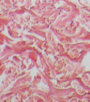
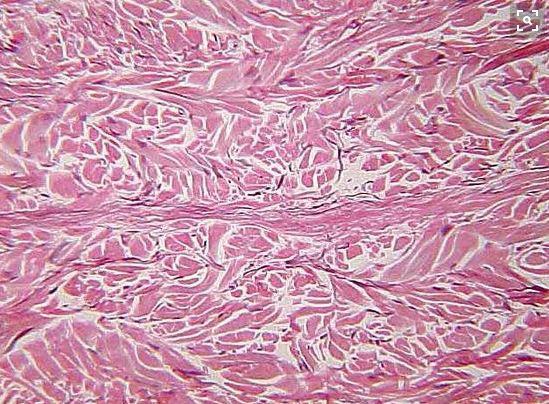
11 Know THIS Slide for the Practical! There are 3 types of Dense Connective Tissue: Dense Regular Connective Tissue - has a rope-like arrangement of fiber bundles Dense Irregular connective tissue - has a wavy, woven fabric-like arrangement Elastic Connective Tissue - has stacked swiggly wavy lines Dense Regular Connective Tissue A Type of Dense Connective Tissue Dense regular connective tissue The fibers of this tissue are tightly packed into parallel bundles, between which are a few highly attenuated, spindle-shaped fibroblasts. The small, cigar-shaped nuclei of the fibroblasts are oriented parallel to the fibers; the cytoplasm is difficult to distinguish with the light microscope. There is little room for the ground substance, which nevertheless permeates the tissue. Know THIS Slide for the Practical! Know THIS Slide for the Practical! Slide of tendon 11/25

12 via GIPHY Dense regular connective tissue is packed with collagen fibers. This makes them very strong and resistant to stretch. This unique property allows them to transit mechanical force over long distances using a minimum of material and space, while resisting mechanical forces from other directions. This tissue therefore serves to transmit the force of muscle contraction, to attach bones to one another, and to protect other tissues and organs. Dense regular connective tissue is found in tendons, ligaments, periosteum, perichondrium, deep fascia, and some organ capsules. You are already familiar with these tissues, even if you don't already know it. In some cuts of meat, you can see these tissues as the white stringy substance that has a harder consistency. In anatomy, a ligament is the dense regular connective tissue that connects bones to other bones. and a tendon is the dense regular connective tissue that connects bones to muscle (for example, the hamstring.) Dense Irregular Connective Tissue - WE DO NOT HAVE DENSE IRREGULAR TISSUE SLIDE FOR PRACTICAL - 12/25
13 13/25

14 Dense irregular connective tissue consists of tightly packer collagen fibers that appear less organized and less uniform that dense regular connective tissue tissue. The nuclei in this tissue are stretch long and the tissue may appear wavy under a microscope. The major cell on dense irregular connective tissue is the fibroblasts. Dense irregular connective tissue is able to withstand tension from multiple directions. It provides support and strength. This tissue is found in areas that experience stretching from multiple angles, such as the dermis of the skin, the submucosa of the digestive tract and the fibrous capsules of organs and joints. Elastic Connective Tissue A Type of Dense Connective Tissue 14/25


15 Elastic fibers are present in different concentrations in all of the connective tissues proper. Elastic fibers are made with elastin that has the ability to stretch and then return to its original shape after being stretched. Connective tissues that have a very large amount of elastin are referred to as elastic connective tissue. Elastic connective tissues are present in relatively high concentration in several organs, including the largest arteries in the body. Know THIS Slide for the Practical! Fibroblasts or fibrocytes are the primary cell in elastic connective tissue. The fibroblast cells make he elastin necessary for the elastic fibers and also provide other proteins and molecules that contribute to the matrix. Cartilage 15/25

16 WHAT IS CARTILAGE? Cartilage is a supportive connective tissue that functions in the body to provide structural support, protection of soft tissues and increases strength. You are born with a lot more cartilage than you have as an adult. As you grow, much of this cartilage is used to guide the growth of bones. For example, the human skull is composed of separated bones at birth and then fuses during development to form the skull. Once the bones have fused, the cartilage is broken down by the body and not replaced. This is why infants are born with a "soft spot" at the top of their head that disappears. Cartilage tissue consists of a dense collection of collagen and elastin fibers. The primary cell in cartilage are chondrocytes. The fibers and the ground substance that makes up the matrix of cartilage is made from chrondroblasts (instead of fibroblasts as seen in connective tissue proper) which are immature chondrocytes. Cartilage is usually closely associated with a dense irregular connective tissue (called perichondrium). This configuration is very important for the cartilage. Cartilage does not have blood or lymph vessels and does not have nerves (in not innervated). The cartilage gets is nutrients from dense irregular tissue of the perichondrium via diffusion through the matrix. Whenever there is an injury that occurs in an area that contains a lot of cartilage (like the Achilles heel ) the amount of time necessary for healing is greatly increased. The fact that cartilage has no nerves, means that cartilage is not sensitive to pain or pressure, Cartilage is closely related to bone. Whenever cartilage gets calcified, the chondrocytes of the cartilage die and the cartilage will be replaced by bone tissue. Cartilage has more flexibility than bone, due to a flexible material 16/25
17 called chondroitin which exists in the matrix. chondroitin is sometimes taken as a supplement for joint pain.. There are 3 types of cartilage. 1) Hyaline Cartilage 2) Fibrocartilage 3) Elastic Cartilage. 17/25
18 Hyaline Cartilage 18/25


. Know THIS Slide for the Practical! Hyaline cartilage means \"glass.")
19 Hyaline cartilage is the most abundant type of cartilage found in our body. Your entire skeleton actually started out as hyaline cartilage when you were developing in the womb. As an adult, you have hyaline cartilage covering the surfaces of your bones at each of your joints. Your ribs also contain hyaline cartilage at the anterior ends. You have hyaline cartilage in your bronchi, your bronchial tubes, nose and trachea. You even have hyaline cartilage that forms your vocal cords! via GIPHY Hyaline cartilage tissue appears as a shiny bluish-white, material that contains collagen fibrils and chondrocytes. Hyaline Cartilage functions is the body to allow for the gliding motion of adjacent structure by providing a smooth surface. It also provides flexible support. Hyaline cartilage also acts to connect the ribs to the sternum (the costal region). Know THIS Slide for the Practical! Hyaline cartilage means "glass. It gets its name from its clear glass-like appearance when observed with the naked eye (this quality is not visible on stained slides). When viewed under the light microscope, hyaline cartilage 19/25


20 has clearly defined cells (the chondrocytes) that seem round. The cells are surrounded by a blank spaces called lacunae. In histology, a lacuna is a small space containing an osteocyte in bone or chondrocyte in cartilage.the fiber portion of the matrix of hyaline cartilage contains only thin fibrils of collagen that are too thin to be seen with a light microscope. The ground substance has a jelly-like consistency and holds large amounts of water. The structural properties of hyaline cartilage given it the ability to resists compression and provide strength and flexibility. Fibrocartilage (Fibrous Cartilage) Know THIS Slide for the Practical! Fibrocartilage is a tough form of cartilage that consists of chondrocytes scattered among clearly visible dense bundles of collagen fibers within the matrix. Fibrocartilage is found in portions of your joints, your intevertebral discs, your knee (as the meniscus) and your pubic symphysis which joins the anterior portions of your hip bones together Fibrocartilage provides structure and support. It is also the strongest type of cartilage. Fibrocartilage has the ability to resist strong compression and strong tension (pushing and pulling) forces. The areas of the body that have this type of cartilage are subjected to a lot of push and pull (compression and tension) forces. its consistency lies somewhere between hyaline cartilage and dense regular connective tissue. Fibrocartilage has thick collagen fibers which makes it somewhat similar to dense regular connective tissue, however the cartilage includes chondrocytes within lacunae. In histology, a lacuna is a small space containing an osteocyte in bone or chondrocyte in cartilage. 20/25

21 Elastic Cartilage 21/25

 of the larynx each time we swallow, is made of elastic cartilage, as is the highly bendable cartilage in the outer ear.")
 Animated GIF Courtesy of Source dollysobsessionwithskulls.tumblr.com via GIPHY Bone is a tissue, also known as osseous tissue. Bone is alive and filled with blood vessels.")
22 Know THIS Slide for the Practical! Elastic cartilage contains a lot of elastic fibers with thin collagen fiberils in its matrix. As you would expect, this cartilage is more elastic than hyaline cartilage. This exatra elasticity allows for better tolerance of repeated bending. The body has elastic cartilage tissue in the eustacian tubes which regulate the inner ear pressure, it form the structure of the outer ear (the auricle) and it forms the epiglottis. The epiglottis, which bends down to cover the glottis (opening) of the larynx each time we swallow, is made of elastic cartilage, as is the highly bendable cartilage in the outer ear. Elastic cartilage contains chondrocytes that are dispersed within a a threadlike network of elastic fibrers. Elastic cartilage functions to provides support. It also provides structure to the area.. Bone (Osseous Tissue) Animated GIF Courtesy of Source dollysobsessionwithskulls.tumblr.com via GIPHY Bone is a tissue, also known as osseous tissue. Bone is alive and filled with blood vessels. It has incredible stregthe due to inorganic calcium salts that form a rigid ground substance of the matrix. Bone functions in the body to provide structural support and protection to internal body structures. Bone is able to withstand a great deal of compression and tension (pushing force and pulling force). The primary cell of bone tissue is the osteoblast. Osteoblastss secrete the collagen fibers and the materials needed for the ground substance. These materials are then calcified by the addition of calcium salts which form the rigid ground substance of the bone. When bone is mature, the matrix consists of collagen fibers surrounded by a calcified rigid ground substance. The osteoblasts mature into the osteocytes. The osteocytes reside in the lacunae filling up the space. 22/25


23 Bone tissue is a supportive type of connective tissue. It acts to shield the internal organs and to provide structure and support. Bone tissue includes compact bone and spongy bone. In bone, the matrix is rigid and described as calcified because of the deposited calcium salts. Blood Animated GIF Courtesy of Source scinerds.tumblr.com via GIPHY Hematopoiesis 23/25


24 Picture Picture Fluid connective tissue includes lymph and blood. Various specialized cells circulate in a watery fluid containing salts, nutrients, and dissolved proteins. Red blood cells do not have a nucleus! We don't see purple in the red blood cells, because there is no nucleus to take up the purple nuclear stain. Blood is pretty different from the other connective tissue types. It is liquid and it does not bind things together or provide any mechanical or structural support. However, it develops from the mesenchyme like the other connective tissue types. Blood consists of blood cells surrounded by a liquid nonliving matrix called blood plasma. Its cells and matrix are very different from those in other connective tissues. Blood functions as the transport vehicle for the cardiovascular system, carrying defense cells, nutrients, wastes, respiratory gases, and many other substances throughout the body. 24/25
25 Summary Table Picture GOT MAD TISSUE IDENTIFYING SKILLS?? Try the QUIZ! Updated 02/21/ /25
Connective Tissue. Found everywhere in the body. Most abundant and widely distributed. Never exposed to the outside environment.
 Connective Tissue Found everywhere in the body. Most abundant and widely distributed. Never exposed to the outside environment. Connective Tissue Functions Binding and support Protection Insulation Transportation
8/30/2017. Tissue: The Living Fabric. 4.3 Connective Tissue
 Chapter 4 Part B Tissue: The Living Fabric Annie Leibovitz/Contact Press Images PowerPoint Lecture Slides prepared by Karen Dunbar Kareiva Ivy Tech Community College 4.3 Connective Tissue Connective tissue
Most abundant and widely distributed tissues in the body Binds, support, and strengthen body tissues, protect and insulate internal organ, serve as
 Connective tissue Most abundant and widely distributed tissues in the body Binds, support, and strengthen body tissues, protect and insulate internal organ, serve as major transport system, compartmentalizes
Lecture Overview. Connective Tissues. Marieb s Human Anatomy and Physiology. Chapter 4 Tissues: The Living Fabric Connective Tissues Lecture 10
 Marieb s Human Anatomy and Physiology Marieb Hoehn Chapter 4 Tissues: The Living Fabric Connective Tissues Lecture 10 Lecture Overview General composition and function of connective tissue Components of
Connective Tissue. Consists of two basic elements: Cells and Extra-cellular matrix
 Connective Tissue Consists of two basic elements: Cells and Extra-cellular matrix True Connective Tissue Cells Fibroblasts: Secrete both fibers and ground substance of the matrix (wandering) Macrophages:
Bio& 241 Unit 1 / Lecture 4
 Bio& 241 Unit 1 / Lecture 4 Connective Tissue Consists of two basic elements: Cells and Extra-cellular matrix 1 True Connective Tissue Cells Fibroblasts: Secrete both fibers and ground substance of the
The Tissue Level of Organization
 The Tissue Level of Organization 4.5-4.11 August 31, 2012 4.5 Connective Tissues Describe the general features of connective Describe the structure, location, and function of the various types of connective
Connective Tissues. Copyright 2009 Pearson Education, Inc., publishing as Pearson Benjamin Cummings
 C.T. are found in all parts of the body & diverse in structure & function. C.T. Functions: -connect structures -provide support -protect vital organs -fill space b/w structures -stores fat -defends body
Epithelia of Coverings and Linings. Tissues. Tissue
 Tissue Tissues Chapter 3 Definition an aggregation of cells in which each cooperates with all others in the performance of a given function Examples of general functions Movement Protection Support Production
Practical Histology. Lab 3: Connective tissue
 Practical Histology Lab 3: Connective tissue Connective tissues Connective tissue provides structural support for the body by binding cells and tissues together to form organs. It also provides metabolic
CONNECTIVE TISSUE (Refer to pp for specific characteristics of each) VAN (**Be familiar with exceptions**)
 VAN (**Be familiar with exceptions**)") CONNECTIVE TISSUE CHARACTERISTICS: *Most abundant tissue type; Composed of ECM (GS & Protein Fibers) + Cells (Refer to pp.129-131 for specific characteristics of each) *Highly equipped with VAN assists
CONNECTIVE TISSUE CHARACTERISTICS: *Most abundant tissue type; Composed of ECM (GS & Protein Fibers) + Cells (Refer to pp.129-131 for specific characteristics of each) *Highly equipped with VAN assists
10/3/2012. Tissue: The Living Fabric: Part B. Extracellular matrix Ground substance Fibers Collagen fiber Elastic fiber Reticular fiber.
 PowerPoint Lecture Slides prepared by Janice Meeking, Mount Royal College C H A P T E R 4 Tissue: The Living Fabric: Part B Copyright 2010 Pearson Education, Inc. Copyright 2010 Pearson Education, Inc.
4 Types of Tissue. Epithelial Connective Muscle Neural
 Connective Tissue 4 Types of Tissue Epithelial Connective Muscle Neural Connective Tissue Fills internal spaces Supports & binds other tissues Transports materials Stores energy Classification of Connective
Anatomy and Physiology Tissue Review
 Anatomy and Physiology Tissue Review OVERVIEW Histology practicals can be rough, especially when access to slides is limited to the lab period. This resource provides an opportunity to learn or review
Function: Provides reserve food fuel; Copyright 2011 Pearson Education, Inc. Copyright 2011 Pearson Education, Inc. White blood cell (lymphocyte)
") Adipose Tissue Closely packed adipocytes Have nucleus pushed to one side by fat droplet Richly vascularized Provides reserve food fuel Insulates against heat loss Supports and protects organs Under skin
Adipose Tissue Closely packed adipocytes Have nucleus pushed to one side by fat droplet Richly vascularized Provides reserve food fuel Insulates against heat loss Supports and protects organs Under skin
Tejido Conectivo Parte B. Informe #3 Laboratorio Biología # 240 Profesor: Javier Cabello
 Tejido Conectivo Parte B Informe #3 Laboratorio Biología # 240 Profesor: Javier Cabello Figure 4-8 The Cells and Fibers of Connective Tissue Proper Areolar Elastic fibers Collagen fibers Fibroblast Free
Body Tissues Pearson Education, Inc.
 Body Tissues Tissues Groups of cells with similar structure and function Four primary types: Epithelial tissue (epithelium).1 Connective tissue.2 Muscle tissue.3 Nervous tissue.4 Epithelial Tissues Locations:
CONNECTIVE TISSUE (C.T.)
") CONNECTIVE TISSUE (C.T.) Objectives: By the end of this lecture, the student should be able to: 1. Enumerate the general characteristics of C.T. 2. Classify C.T into C.T. proper and special types of C.T.
CONNECTIVE TISSUE (C.T.) Objectives: By the end of this lecture, the student should be able to: 1. Enumerate the general characteristics of C.T. 2. Classify C.T into C.T. proper and special types of C.T.
4 Types of Tissue. Epithelial Connective Muscle Neural
 Connective Tissue 4 Types of Tissue Epithelial Connective Muscle Neural Connective Tissue Fills internal spaces Supports & binds other tissues Transports materials Stores energy Classification of Connective
The Tissue Level of Organization
 Tissue The Tissue Level of Organization Chapter 3 Definition an aggregation of cells in which each cooperates with all others in the performance of a given function Examples of general functions Movement
Tissues 10/21/2016. Epithelial Tissue
 Tissues This is a generalized cell diagram. It shows the anatomy of a cell, but most cells do not actually look like this. Cells can have a wide variety of shapes and sizes, depending on their function.
Cells and Tissues 3PART D. PowerPoint Lecture Slide Presentation by Patty Bostwick-Taylor, Florence-Darlington Technical College
 PowerPoint Lecture Slide Presentation by Patty Bostwick-Taylor, Florence-Darlington Technical College Cells and Tissues 3PART D Connective Tissue Found everywhere in the body Includes the most abundant
Connective Tissue. Answer Choices(In CAPITAL BOLD): RETICULAR ELASTIC. IRREGULAR Spongy bone ELASTIC BLOOD
: RETICULAR ELASTIC. IRREGULAR Spongy bone ELASTIC BLOOD") Connective Tissue Answer Choices(In CAPITAL BOLD): Proper: Specialized: Loose- Cartilage- AREOLAR HYALINE ADIPOSE FIBROCARTILAGE RETICULAR ELASTIC Dense- Bone- REGULAR COMPACT BONE IRREGULAR Spongy bone
Connective Tissue Answer Choices(In CAPITAL BOLD): Proper: Specialized: Loose- Cartilage- AREOLAR HYALINE ADIPOSE FIBROCARTILAGE RETICULAR ELASTIC Dense- Bone- REGULAR COMPACT BONE IRREGULAR Spongy bone
Lab Exercise 6a-2. Classification of connective tissues. Connective Tissue. Connective tissues. Areolar. Areolar tissue
 Classification of connective tissues Lab Exercise 6a-2 Connective Tissue Nervous Muscle Connective Tissue Connective tissues Connective tissue proper Fluid connective tissue Supportive connecting tissue
Epithelial Tissue. Simple Cuboidal Function: secretion and absorption. Simple Squamous
 Epithelial Tissue General Functions: Lines and covers organs Absorbs / secretes substances Gas exchange Protection Special Characteristics: - have an apical surface on top - have a basement membrane below
Cartilage. Dr. Heba Kalbouneh Associate Professor of Anatomy and Histology
 Cartilage Dr. Heba Kalbouneh Associate Professor of Anatomy and Histology 1 Cartilage is a specialized type of connective tissue designed to give support, bear weight and withstand tension, torsion and
HOLE S ANATOMY CHAPTER 5, PART II Lecture notes
 HOLE S ANATOMY CHAPTER 5, PART II Lecture notes I. Connective Tissue A. Structure 1. have few cells that are spaced apart and can divide; two categories: a. fixed cells cells that are present in tissue
Connective Tissue Nervous Muscle. Classification of connective tissues
 Connective Tissue Nervous Muscle Lab Exercise 6a-2 Classification of connective tissues 1 Connective Tissue Connective tissue proper Fluid connective tissue Supportive connecting tissue Connective tissues
Basic Histology. By Mrs. Bailey
 Basic Histology By Mrs. Bailey Primary Tissues 1. Epithelial Tissue 2. Connective Tissue 3. Muscle Tissue 4. Nervous Tissue Very cellular Supported by underlying connective tissue Epithelial & connective
Classification of Tissues
 6 R e v i e w S h e e t Exercise Classification of Tissues NAME LAB TIME/DATE Tissue Structure and Function General Review 1. Define tissue. A group of cells similar to one another in structure that perform
Tissues organs system organism. pg151
 Histology is the study of tissues A TISSUE is a group of cells, usually of one kind, & their intercellular substance (e.g. intercellular matrix in animal) which are linked together & perform a particular
Tissues Chapter 5...Tissue - a group or mass of similar cells working together to perform certain common functions
 Tissues Chapter 5...Tissue - a group or mass of similar cells working together to perform certain common functions There are 4 major types of tissue Epithelial Connective Muscle Nervous 1. Epithelial Tissue
BIOL 2457 CHAPTER 4 Part 2 SI All connective tissues arise from, an embryonic tissue.
 BIOL 2457 CHAPTER 4 Part 2 SI 1 1. All connective tissues arise from, an embryonic tissue. 2. Describe the vascularity of connective tissues, which are very diverse. 3. Describe the innervation of connective
Connective Tissues. 2. Describe the function of fibroblasts. 3. What is ground substance? What is its function?
 Connective Tissues Directions: Insert and install your Interactions: Foundations CD. a. Click the "Contents" button. b. Open the Tissue Level of Organization file. c. Click on Anatomy Overviews. d. Work
Histology. The study of tissues.
 Histology The study of tissues. Body Tissues Cells are specialized for particular functions Tissues Groups of cells with similar structure and function Four primary types Epithelium Connective tissue Nervous
Classification of Tissues
 M06_MARI0000_00_SE_CH06.qxd 3/28/11 4:37 PM Page 35 NAME LAB TIME/DATE R E V I E W S H E E T EXERCISE 6 Classification of Tissues Tissue Structure and Function General Review 1. Define tissue. A group
contains an antiangiogenesis factor
 CARTILAGE & BONE Cartilage and Bone objectives Student must learn :. What is the meaning of cartilage, and their function, location in human body.. To distinguish the 3 types of cartilage. And their cells,
Body Tissues. Cells are specialized for particular functions Tissues - groups of cells with similar structure. and function Four primary tissue types:
 Chapter 3 Tissues Body Tissues Cells are specialized for particular functions Tissues - groups of cells with similar structure and function Four primary tissue types: Epithelium Connective tissue Nervous
Unit II: Tissues and Integumentary System
 Unit II: Tissues and Integumentary System 2.1 - Tissues Chapter 4 Written Response #1 1. What is a tissue? 2. What are four major types of tissues? Tissue Definition: a group or mass of similar cells working
Chapter 5. Tissues. 4 Types of Body Tissues. Tissues
 Chapter 5 Tissues Tissues Tissues - groups of cells that are similar in structure & function RBC, WBC, & platelets are a group of cells working together to form BLOOD tissue Histology Pathohistology study
Histology. There are four basic tissue types in the body are :-
 Histology Lab.I There are four basic tissue types in the body are :- 1- Epithelial tissues (Epithelium) 2- Connective tissues 3- Muscular tissues 4- Nervous tissues 1-Epithelial tissues epithelial tissues
Tissue Outline (chapter 4) Tissues group of cells that perform structural and roles. List the 4 types:
 Tissues group of cells that perform structural and roles. List the 4 types:") Tissue Outline (chapter 4) Tissues group of cells that perform structural and roles. List the 4 types: 1. 2. 3. 4. I. Epithelial Tissue covers all the surfaces, inside & out. Are the major tissues of,
Tissue Outline (chapter 4) Tissues group of cells that perform structural and roles. List the 4 types: 1. 2. 3. 4. I. Epithelial Tissue covers all the surfaces, inside & out. Are the major tissues of,
HISTOLOGY. Simple squamal lungs
 HISTOLOGY Lab Objectives: Students should be able to... 1. Visually identify each class of tissue and examples within each class 2. Indicate the location (in the human body and/or organ) and function of
What is histology? HISTOLOGY
 Introduction to Histology What is histology? HISTOLOGY histo = tissue ogy = study So HISTOLOGY = the study of tissues! What is a TISSUE? Tissues are groups of cells with specialized structural and functional
Chapter 1: Cells and Tissues
 Chapter 1: Cells and Tissues Cells and Tissues Carry out all chemical activities needed to sustain life Cells are the building blocks of all living things Tissues are groups of cells that are similar in
Tissue = groups of cells that are similar in structure and function
 Tissue = groups of cells that are similar in structure and function Types Epithelial - covering Connective - support Muscle - movement Nervous - control Membranes line body cavities and hold organs together
6a A&P: Introduction to the Human Body - Tissues
 6a A&P: Introduction to the Human Body - Tissues 6a A&P: Introduction to the Human Body - Tissues! Class Outline" 5 minutes" "Attendance, Breath of Arrival, and Reminders " 10 minutes "Lecture: AOIs of
Cartilage. - Cartilage together with long bone form the skeleton and support the body.
 Cartilage - Cartilage is a special type of CT has a firm pliable matrix that can resist mechanical stress, act as a shock absorber. - Cartilage together with long bone form the skeleton and support the
Tissue Outline. Chapter 4. Tissue. Cellular Connections. I. Definitions II. Cellular Connections III. Tissue Types IV. Membranes V.
 Tissue Outline Chapter 4 The Tissue Level of Organization I. Definitions II. Cellular Connections III. Tissue Types IV. Membranes V. Tissue Repair 1 2 Tissue Cellular Connections Tissue Groups of cells
Basic Tissue Types and Functions
 Tissues Histology Basic Tissue Types and Functions 1) Epithelial tissue covering 2) Connective tissue support 3) Muscle tissue movement 4) Nervous tissue control Epithelial Tissue 1) Covers a body surface
Histology 101! !! Name:! Block: Identify and describe the functions of major tissue types including their subclasses and varieties!
 Histology 101 Identify and describe the functions of major tissue types including their subclasses and varieties Name: Block: "1 Introduction to Tissues Histology Notes Tissue (living fabric) : groups
Blood. Hematopoietic Tissue
 Blood Hematopoietic Tissue Is a type of connective tissue in which its cells are suspended in a circulating fluid. Erythrocytes+ leukocytes + platelets (thrombocytes) =formed elements of blood. These formed
Tissues. groups of cells similar in structure and function 4 types. epithelium connective muscle nervous
 Tissues groups of cells similar in structure and function 4 types epithelium connective muscle nervous Epithelial Tissue lining covering glandular Functions protection absorption filtration secretion Epithelium
Chapter 4. Cartilage and Bone. Li Shu-Lei instructor. Dept. Histology and Embryology, School of Basic Medical Sciences, Jilin University
 Chapter 4 Cartilage and Bone Li Shu-Lei instructor Dept. Histology and Embryology, School of Basic Medical Sciences, Jilin University I Cartilage a specialized connective tissue Characterizers: Cartilage
Tissues. Tissues. Four basic tissues. A collection of cells with a common function. 1. Epithelial 2. Connective 3. Muscular 4.
 Tissues Tissues A collection of cells with a common function Four basic tissues 1. Epithelial 2. Connective 3. Muscular 4. Nervous Epithelia: cells in layers Types of epithelia 1) lining Layers of cells
Chapter 4. The Tissue Level of Organization
 Chapter 4 The Tissue Level of Organization 1 Tissue Outline I. Definitions II. Cellular Connections III.Tissue Types IV. Membranes V. Tissue Repair 2 Tissue Tissue Groups of cells that are similar in structure
Collin College. BIOL Chapter 4. Tissue Levels CONNECTIVE TISSUE. C.T. derives from Mesenchyme embryonic tissue.
 Collin College BIOL. 2401 Chapter 4 Tissue Levels. CONNECTIVE TISSUE C.T. derives from Mesenchyme embryonic tissue. Depending on the stimuli, mesenchyme develops into specific cells that give rise to the
TISSUES. Objectives. Tissues
 TISSUES Objectives Introduce the four major types of tissues Describe the general characteristics and functions of epithelial & connective tissue Name the major types of epithelial & connective tissues
Which compound is reponsible for the viscous character of the ground substance?
 1 2 Which type of collagen forms the coarse collagen fibres in dense regular and irregular connective tissues? Which compound is reponsible for the viscous character of the ground substance? 3 Which class
TISSUE. A group of cells that perform a similar function within an organism. Epithelium Connective Muscle Nervous CREDITS
 TISSUE A group of cells that perform a similar function within an organism. Epithelium Connective Muscle Nervous CREDITS Epithelium Connective Muscle Nervous Epithelium Composed of a layer of cells. Lines
They cells can not function death.
 Jenna Hellack Jan 2001 Tissues What do you think happens when the cells use up their food and oxygen before there is time to replenish it? They cells can not function death. Blood Cell Cancer cell Plant
Histology. Study of body tissues
 Histology Study of body tissues 2 Introduction to Body Tissues 1. Composed of specialized cells of similar structure and perform a common function 2. Four major types (4 Cs) a. Epithelial - Cover b. Connective
Lab Animal Tissue. LEARNING OBJECTIVES: To understand the relationship between the structure and function of different animal tissues
 Name: Bio A.P. PURPOSE: HYPOTHESIS: NONE Lab Animal Tissue BACKGROUND: In animals, groups of closely related cells specialized to perform the same function are called tissues. There are four general classes
A. cells that perform related functions and are similar in structure. B. extracellular material - made by cells and secreted into interstitial space
 I. tissue components A. cells that perform related functions and are similar in structure B. extracellular material - made by cells and secreted into interstitial space II. tissue types A. epithelium (e.)
NOTES: CH 40 Introduction to Human Anatomy & Physiology
 NOTES: CH 40 Introduction to Human Anatomy & Physiology THE HUMAN BODY Anatomy Physiology (= structures) (= functions or processes) Characteristics of LIFE: 1) Made up of 1 or more CELLS. 2) Obtain and
Tissues are: group of similar or identical cells that share a common function. used to build organs
 Tissues: Four classes Epithelium Connective Muscle Nervous Tissues are: group of similar or identical cells that share a common function. used to build organs Overview: Epithelial o Line body cavities
Connective Tissue Supports and Protects
 Connective Tissue Supports and Protects Bởi: OpenStaxCollege As may be obvious from its name, one of the major functions of connective tissue is to connect tissues and organs. Unlike epithelial tissue,
Tissues Description Function(s) Locations Miscellaneous. avascular -thelium = covering
 Locations Miscellaneous. avascular -thelium = covering") Epithelial Tissue Simple Squamous flattened cells diffusion and Kidney glomeruli disc-shaped central filtration air sacs of lung Simple = Single layer nuclei secretes lubricating lining of heart, blood
Epithelial Tissue Simple Squamous flattened cells diffusion and Kidney glomeruli disc-shaped central filtration air sacs of lung Simple = Single layer nuclei secretes lubricating lining of heart, blood
Study of different tissues Abnormal cells and tissues can be compared to normal tissues to identify disease, such as cancer Being able to know and
 CHAPTER 4 Study of different tissues Abnormal cells and tissues can be compared to normal tissues to identify disease, such as cancer Being able to know and recognize normal tissues under the microscope
Hole s Human Anatomy and Physiology
 Hole s Human Anatomy and Physiology 1 Chapter 5 Tissues Four major tissue types 1. Epithelial 2. Connective 3. Muscle 4. Nervous 2 Epithelial Tissues General characteristics - cover organs and the body
Connective tissue CONNECTIVE TISSUE Part I
 Connective tissue CONNECTIVE TISSUE Part I Part 1 Connective Tissue Found everywhere in the body (app. 50% of body weight) Includes the most abundant and widely distributed tissues General features of
Histology review. Histology. Slides. Epithelial tissue. Another example - kidney. Simple cuboidal epithelium. What to look for
 Histology review Histology What to look for Histology Practical = 50 pts Some slides set up on scopes (~10) Some Powerpoint pictures on the projector Questions I will ask: What kind of tissue? General
Human Anatomy and Physiology I Laboratory
 Human Anatomy and Physiology I Laboratory Skeletal Tissue: Cartilage and Bone This lab involves study of the laboratory exercise Overview of the Skeleton, Classification and Structure of Bones and Cartilages,
Histology= the study of tissues
 Unit 3-Histology Histology= the study of tissues A tissue is a group of cells that have a similar shape and function. Different types of tissues can be found in different organs. In humans, there are four
Anatomy &- Physiology Histology Worksheet
 Anatomy &- Physiology Histology Worksheet 1. The four primary tissue types found in the human body are a) squamous, cuboidal, columnar, glandular b) adipose, elastic, reticular, cartilage c) skeletal,
Tissues. Group of cells that are similar in structure and function. 4 primary types. Epithelium (covering) Connective (support) Nervous(control)
 Connective (support) Nervous(control)") Tissues Tissues Group of cells that are similar in structure and function 4 primary types Epithelium (covering) Connective (support) Nervous(control) Epithelial tissue (epithelium) Lining, covering, and
Tissues Tissues Group of cells that are similar in structure and function 4 primary types Epithelium (covering) Connective (support) Nervous(control) Epithelial tissue (epithelium) Lining, covering, and
Human anatomy Unit III. Tissue
 Human anatomy Unit III Tissue Definition of Tissues Biological tissue is a collection of interconnected cells that perform a similar function within an organism. In other words, it is a group of cells
SKELETAL TISSUES CHAPTER 7 INTRODUCTION TO THE SKELETAL SYSTEM TYPES OF BONES
 SKELETAL TISSUES CHAPTER 7 By John McGill Supplement Outlines: Beth Wyatt Original PowerPoint: Jack Bagwell INTRODUCTION TO THE SKELETAL SYSTEM STRUCTURE Organs: Bones Related Tissues: Cartilage and Ligaments
THE TISSUE LEVEL OF ORGANIZATION PART I: EPITHELIAL TISSUE
 THE TISSUE LEVEL OF ORGANIZATION PART I: EPITHELIAL TISSUE 4 Main Tissue Types Epithelium Covers surfaces, lines cavities, forms glands Connective Tissue Support and protects body Muscular Tissue Movement
Sheet #9. Dr. Heba Kalbouneh. Dr. Heba Kalbouneh. Dr. Heba Kalbouneh
 Sheet #9 Dr. Heba Kalbouneh Dr. Heba Kalbouneh Dr. Heba Kalbouneh Elastic fibers The main function of elastic fibers is to provide elasticity. In other words these fibers are able to restore the original
HISTOLOGY Lecture TWO DR. ASHRAF SAID
 HISTOLOGY Lecture TWO DR. ASHRAF SAID Start Of this lecture TISSUES TISSUE: A DEFINITION A group of connected and interdependent cells that cooperate to perform a specific function CONNECTIVE TISSUE The
Connec<ve Tissue. Major Func<ons of Connec<ve Tissue 9/8/14. Most and widely distributed <ssue type Four classes. Tissue: The Living Fabric: Part B
 Connec
Connec<ve Tissue 9/8/14. Major Func<ons of Connec<ve Tissue. of Connec<ve Tissue. Characteris<cs of Connec<ve Tissue
 Connec
Outline. Bio 105: Tissues Laboratory. Organization of the Human Body. Tissue - Epithelium. Tissues 3/2/ Copyright 2009 Pearson Education, Inc
 Outline Bio 105: Tissues Laboratory Laboratory 5 Reading: Chapter 4 I. Cell to cell contact II. Body Cavities III. Membranes IV. Homeostasis V. Integumentary System I. Includes skin, hair and nails 1 2
Biology. Dr. Khalida Ibrahim
 Biology Dr. Khalida Ibrahim The cartilage General characteristics: 1. Cartilage is a specialized type of connective tissue (supporting connective tissue). 2. Consists, like other connective tissues, of
Growth and repair: Cartilage is a vascular tissues that receives nutrients by diffusion through its matrix, cartilage grow by 2 mechanisms:
 Skeletal connective tissues: (cartilage and bone): Cartilage and bone are specialized connective tissues both adapted to serve as skeletal framework in most vertebrates the presence of solid inter cellular
TISSUES. Dr. Gary Mumaugh
 TISSUES Dr. Gary Mumaugh Tissues Tissues - Groups of cells similar in structure and function and perform a common function Histology The study of tissues The four types of tissues Epithelial Connective
Histology= the study of tissues
 Histology 2014 Histology= the study of tissues A tissue is a group of cells that have a similar shape and function. Different types of tissues can be found in different organs. In humans, there are four
Histo lab 7. Special connective tissue is derived from the mesoderm (mesenchyme).
.") Histo lab 7 Special connective tissue is derived from the mesoderm (mesenchyme). If we have high density of fibers, we call it dense connective tissue. (Fibers are more than the ground substance). If we
Histo lab 7 Special connective tissue is derived from the mesoderm (mesenchyme). If we have high density of fibers, we call it dense connective tissue. (Fibers are more than the ground substance). If we
Introduction to Types of Body Tissue Putting it All Together. Packet #12
 Introduction to Types of Body Tissue Putting it All Together Packet #12 Introduction Body Tissues Tissues Groups of cells with similar structure and function Four primary types Epithelial tissue (epithelium)
Lab 1 ANIMAL TISSUES
 Lab 1 ANIMAL TISSUES Levels of Organization Animals are multicellular heterotrophs whose cells lack cell walls. Most animals exhibit a hierarchical level of organization: Cells are organized into tissues
Air sacs of lungs and the lining of the heart, blood vessels, and lymphatic vessels
 Cells Location Function Simple squamous epithelium Air sacs of lungs and the lining of the heart, blood vessels, and lymphatic vessels Allows materials to pass through by diffusion and filtration, and
Tissues. Cells work together in functionally related groups called tissues Types of tissues: 1. Epithelial lining and covering. 2. Connective support
 Histology Tissues Cells work together in functionally related groups called tissues Types of tissues: 1. Epithelial lining and covering 2. Connective support 3. Muscle movement 4. Nervous control Epithelial
ACTIVITY 2: HISTOLOGY AND INTEGUMENT
 ACTIVITY 2: HISTOLOGY AND INTEGUMENT Objectives: 1) How to get ready: Read Chapter 4 and 5, McKinley et al., Human Anatomy, 4e. All text references are for this textbook. 2) Identify each tissue (26 tissues)
Chapter 05. Review. Copyright The McGraw-Hill Companies, Inc. Permission required for reproduction or display.
 Chapter 05 Review 5.1: Introduction Similar cells with a common function are called tissues. The study of tissues is called histology. There are four (4) primary or major tissue types: 1. Epithelial Tissue
Connective Tissue. Red: important. Black: in male female slides. Gray: notes. Editing File
 Connective Tissue Red: important. Black: in male female slides. Gray: notes. Editing File OBJECTIVES Enumerate the general characteristics of C.T Classify C.T into C.T. proper and special types of C.T
B. Classification of epithelium: by number of cell layers present and by shape of the superficial cell layers.
 I. Introduction - tissue: group of cells that are closely associated, similar in structure and function, and perform a common or related function. - four primary tissues: epithelial tissue, connective
Tissues and Membranes
 I. In the Beginning a. Egg + sperm! Tissues and Membranes b. 1 cell divides to make 2, 2 divide to make 4, 4 divide to make 8, and then? c. d. e. Totipotent: f. Pluripotent: II. III. Tissues a. Tissues
Tissue: The Living Fabric: Part B
 PowerPoint Lecture Slides prepared by Janice Meeking, Mount Royal College CHAPTER 4 Tissue: The Living Fabric: Part B Warm Up 9/27/17 Distinguish between Connective Tissue and Epithelial Tissue: Explain