Surface characteristics of human articular cartilagea scanning electron microscope study
|
|
|
- Margery Henderson
- 6 years ago
- Views:
Transcription
 INTRODUCTION Significant advances have been made in the study of articular cartilage since the")
1 J. Anat. (1971), 108, 1, pp With 16 figures Printed in Great Britain Surface characteristics of human articular cartilagea scanning electron microscope study IAN C. CLARKE BioEngineering Unit, University of Strathclyde, Glasgow, Scotland (Received 29 December 1969) INTRODUCTION Significant advances have been made in the study of articular cartilage since the introduction of the scanning electron microscope. McCall (1968 c) described three zones in the fibrous structure of the superior aspect of the femoral head. These comprised a superficial layer of large parallel fibres, an intermediate zone of unorientated fibres, and a radial zone of tightly packed fibres. Two types of articular surface were described. Immature specimens had a fine fibrous surface mesh overlying 'cell nests', and mature specimens had a surface composed of large parallel ridges attributed to the presence of fibre bundles on or immediately below the surface layer. Walker (1969) and colleagues also assumed that the asperities on articular cartilage surfaces were due to large parallel fibre bundles underlying the surface. Localized deposits, thought to be derived from synovial fluid, were found on articular surfaces which had been loaded in contact with synovial fluid. These 'trapped pools' of synovial fluid may have a role in the lubrication of articular cartilage. J. Graham (1969, personal communication) compared the structures of loaded and unloaded articular cartilage and found that the first visible sign of failure under load was a splitting of the surface layer of the specimen. Gardner & Woodward (1969) studied the surfaces and surface replicas of guineapig articular cartilage using light microscopy, scanning and transmission electron microscopy techniques. The surface characteristics appeared to vary from region to region. Arrangements of orientated ridges were observed in some regions and were thought to be due to an orderly array of fibres underlying the articular surfaces. Other regions contained hollows 20-30,m in diameter superimposed on larger hollows ,um in diameter. It was suggested that the smaller hollows could be the site of subarticular chondrocytes but there was no definite evidence put forward for this. There also appeared to be a background of fine orientated fibres on the surface of specimens which had been treated with hyaluronidase. The present study of articular cartilage is part of a continuing research programme in progress at the University of Strathclyde and has been undertaken to determine whether the cartilage structures described to date are typical of the hip joint cartilages. The features presented here describe the surface characteristics of both the femoral and acetabular components of the human hip joint.

2 24 IAN C. CLARKE MATERIALS AND METHODS Post-mortem material, years of age, which showed no obvious signs of degeneration was used. The material was stored at -29 C prior to fixation in 100 formol saline solution. The specimens were prepared for the scanning electron microscope using the techniques developed by McCall (1968 a) and were subsequently coated with gold-palladium (60/40). The regions which were systematically examined at this stage comprised the articular surfaces on the upper half of the femoral heads and the corresponding acetabular surfaces with which these femoral regions could possibly be in contact in the normal standing position. The structures underlying the articular surface were also studied. In preparing these specimens a pair of fine forceps was used to peel off a thin translucent layer from the articular surfaces of dehydrated specimens. The surface thus revealed on the specimen will be termed the 'exposed surface' and the layer torn off will be termed the 'peeled layer'. The stub-mounted specimens were viewed at 450 inclination to the electron beam in a Cambridge Stereoscan IIA electron microscope and photographed on Ilford FP4 roll film. RESULTS Ridges and depressions on the articular surfaces A pilot study by the author seemed to indicate that the articular surfaces contained an arrangement of ridges (Fig. 1). The local periodicity of these ridges was quite regular but varied between sites and specimens. The calculated periodicity from a number of specimens ranged from 1 to 100,um. The wide variation in surface ridges suggested that the changes in surface contours were not due to normally occurring discrete fibre bundles in the underlying fibre network. In this series of studies, ridges were more frequently found adjacent to fractured edges and were always parallel to these edges (Fig. 2). The ridges did not normally extend far from the fractured edges and rapidly gave way to a surface which contained only depressions. In some instances, surface depressions were apparent on top of these ridges. These bowl-shaped depressions were apparent on the surfaces of both acetabular (Fig. 3) and femoral (Fig. 4) cartilages at low magnifications. The surfaces were otherwise featureless, apart from small artefacts on the surface. Higher magnifications revealed that the depressions had regular contours-either a compact 'figure of eight' (Fig. 5) or an elongated 'figure of eight' (Fig. 6). The diameter of the depressions varied from 15 to 30,m and their depth was estimated at from 1 to 6,tm. Shaded contour maps of these depressions derived from Figs. 5 and 6 are illustrated in Figs. 7 and 8 respectively. 'Exposed surface' lacunae The fibre network of the 'exposed surface' was visible as a fine mesh with no apparent orientation. The fibres constituting this network had a mean diameter of 200 nm (Figs. 9, 10). Lacunae with diameters ranging from 10 to 40,tm were visible in the fibre network. The central area of each lacuna frequently contained a number of fibres but the general impression was one of a non-fibrous region. Occasionally


, and ridges (B) alongside and parallel")

3 Microstructure of articular cartilage 25 these areas were occupied by apparently shrunken material which may have been derived from chondrocytes. The lacunae were shallow and lay in planes parallel to the original articular surface. They were generally arranged in pairs and the 'figures of eight' which they thus formed had a striking similarity to the depressions seen on the articular surface. I;.. I I Fig. 1. Pattern of ridges or 'bundles' visible on the surface of femoral cartilage, 64 years of age. Fig. 2. View of 32-year-old acetabular specimen with depressions visible on the articular surface (S), and ridges (B) alongside and parallel to the fractured edge (FE). Fig. 3. Surface depressions observed in acetabular cartilage, 32 years of age. Fig. 4. Surface detail of cartilage from the femoral head, 32 years of age. A layer-like appearance was visible on the 'exposed surfaces' of a few specimens (Fig. 11), but since these 'layers' followed the direction of the tear in the surface zone, it is thought that they were produced by the tearing action during specimen


4 26 IAN C. CLARKE _! _S _ l i t_ =_ r_ L _ ;;S_ /m I 0I I o p M Figs. 5, 6. Surface depressions in acetabular cartilage (32 years of age) viewed at higher magnification. Figs. 7, 8. Tracings made of the depression outlines in Figs. 3 and 4 respectively, showing the 'figure of eight' patterns. Figs. 9, 10. Outlines of lacunae visible in the exposed superficial matrix of acetabular cartilage (32 years of age) after removal of the surface layer.

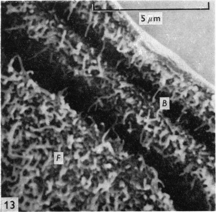
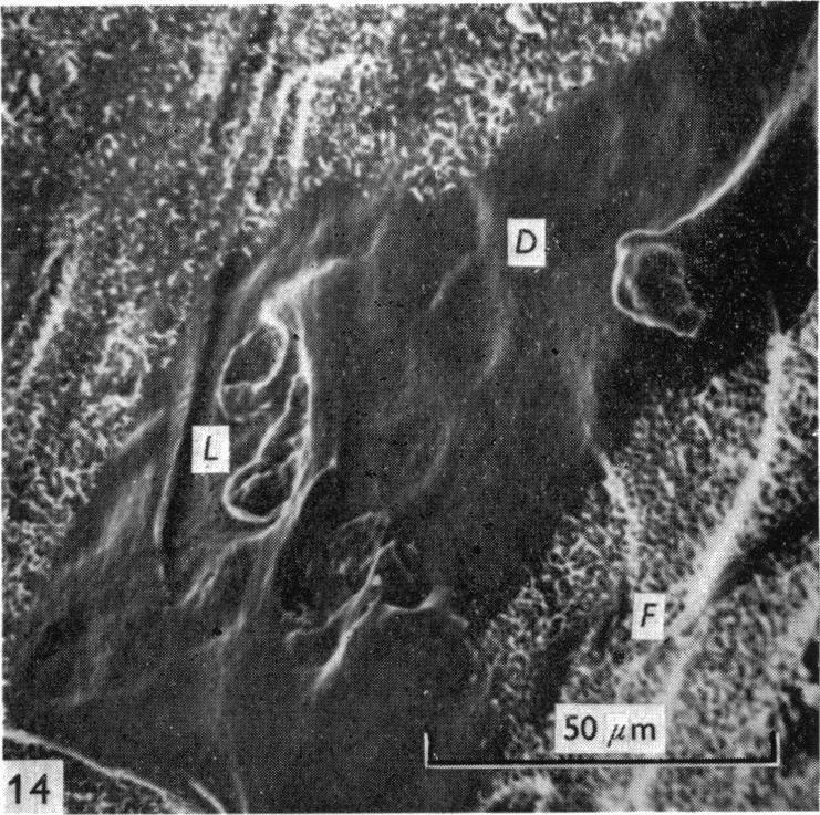
5 Microstructure of articular cartilage 27 preparation. Areas which were apparently non-fibrous, but could be artefacts, were also visible in the fibrous networks. A specimen which had been grossly torn appeared to have a multitude of 'bundles' on the 'exposed surface', but on closer examination the fibre network had clearly torn along the path of the 'peeled layer' and the resulting tendrils of torn fibre network had a bundle-like appearance (Fig. 12). Underside of the 'peeled layer' A fibre network similar to that seen on the 'exposed surface' of the specimen was observed on the underside of the 'peeled layer'. Lacunae were also present in this fibre network. Fibre 'bundles' occasionally appeared at the edges of the 'peeled layer' (Fig. 13) but at higher magnifications these bundles were seen to be folds in the fibre network. Areas which were apparently non-fibrous were also visible in the network of the 'peeled layer' (Fig. 14). Lacunae-like forms could be seen in these areas and at higher magnifications (x ), a ridging or corrugation was also visible. Whether these areas were artefacts created during preparation has not yet been resolved. DISCUSSION The results presented here show that articular cartilage specimens contained surface depressions due to the underlying lacunae and that surface ridges only occurred alongside the fractured edges of the specimens. The superficial layer was shown to consist of a fibrous network with no obvious signs of orientation. Depressions on the articular surfaces The similarity in shape and magnitude of the superficial lacunae and the surface depressions suggests that the depressions can be directly attributed to the presence of the underlyinglacunae. Meyer (193 1) has described the appearance ofdegenerate nuclei lying parallel to and just below the surface of human articular cartilage and Davies and his colleagues (1962) have described in young adult rabbit cartilage the appearance of superficial chondrocytes separated from the articular surface by an acellular layer 2-3,tm thick. The evidence suggests that the lacunae are indeed those of the superficial layer chondrocytes and that the depressions on the surface are due to the acellular surface iayer of cartilage following the outlines of these lacunae or indeed even collapsing into them. The larger type of hollow, measuring ,m, described by Gardner & Woodward (1969) was not evident in this series of experiments. Fibre bundles on the articular surfaces Although previous studies have indicated that the ridges appear alongside and parallel to surface cracks or fractured edges and sometimes occur over the total specimen surface, the fact that they only occurred alongside fractured edges of the specimens in this investigation suggests that they are artefacts created by preparation techniques. Large fibre bundles have not been observed in cross-section at the fractured edge nor has the existence of such structures been reported in the literature.


6 28 IAN C. CLARKE I 15A 15 B Lacunae

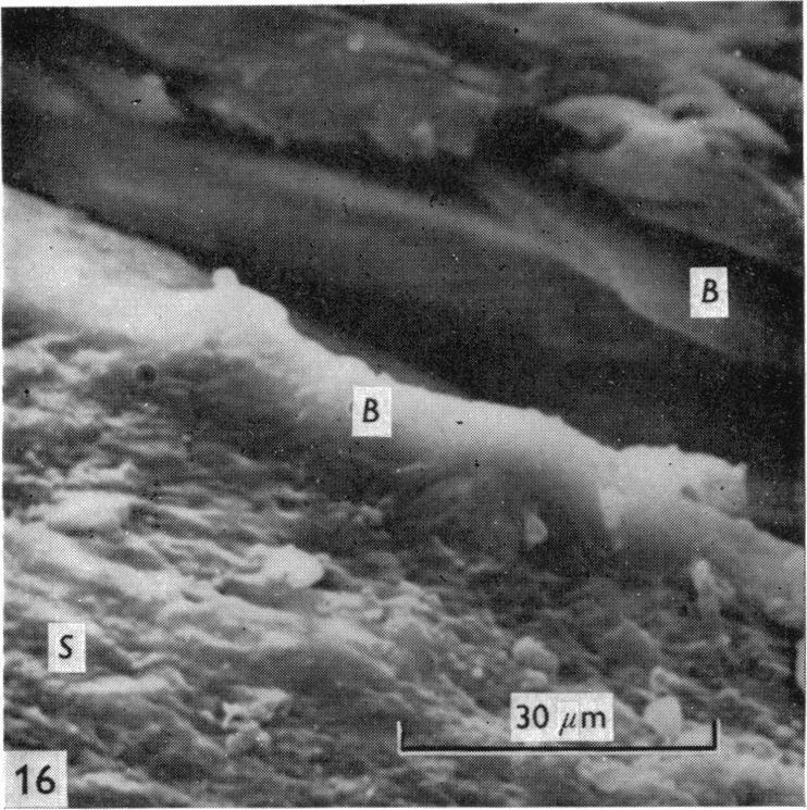
7 Microstructure of articular cartilage 29 This confirms the suggestion that the bundles do not exist in vivo, but that specimen preparation produces pseudo-bundling due to buckling of the surface layer of cartilage. This effect could occur either during fracturing of the specimens or as a result of differential shrinkage during dehydration. It is possible that during the latter effect, if the surface layer does not shrink to the same extent as the rest of the cartilage structure, then it would buckle into ridges. Walker et al. (1969 a, b) estimated that a 1000 decrease in volume occurred during preparation, accompanied by edge curling. It is conceivable that this shrinkage could buckle the surface layer. The substantial amount of subchondral bone attached to the specimens in the present series of experiments has reduced the amount of shrinkage and apparently eliminated curling. This may account for the almost total absence of surface ridges in these specimens. Relation of surface depressions to Talysurf readings The surface roughness of articular cartilage was formerly believed to be less than 0a 1,um (Davies, 1966). The Talysurf readings (Walker, Dowson, Longfield & Wright, 1968) indicated cartilage surface asperities with a mean periodicity of 25,um and a mean peak to valley depth of 2-5,am. The asperities were interpreted by Walker and his colleagues to be due to ridges 25,um apart formed by large fibre bundles underlying the surface. Valleys with a depth of 2 5,um appeared to be formed between ridges due to finer fibres separating the large fibre bundles (Fig. 15 A). However, the surface depressions previously described (Figs. 5, 6) were of similar magnitude to the Talysurf results (Fig. 15 B), and it has been demonstrated that such asperities can be attributed to the cellular structures underlying the surface of cartilage and do not indicate the existence of the fibre bundle formations as postulated by McCall (1968 a) and by Walker et al. (1969a) Surface characteristics in vivo The question now arises whether these surface irregularities actually exist in vivo. Living surfaces may well be smooth as would be indicated by Davies's figures of surface roughness. Freezing, fixation, dehydration or vacuum techniques could result in irregularities in the articular surface, including collapse of the surface layer into the underlying lacunae. The fact that the depressions were more apparent in some specimens than others would be consistent with this view. However, Gardner & Fig. 11. Superficial network of acetabular cartilage (32 years of age) revealed by peeling off the surface layer (S). Areas which appear to be non-fibrous (A) are visible in the 'exposed surface'. Lacunae (L) and rupture or tear lines (T) are also evident. Fig. 12. Superficial network of a grossly torn acetabular specimen (32 years of age). The fibrous network has ruptured to form layers of 'bundles'. Fig. 13. Underside of the 'peeled layer'. The superficial fibrous network (F) has buckled into folds or ridges (B) at the edge in this 32-year-old acetabular specimen. Fig. 14. The underside of the 'peeled layer' of a 32-year-old acetabular specimen at lower magnification. The fibrous network (F) contains areas which do not appear to be fibrous and lacunaelike forms are evident in these areas (L and D). Fig. 15. Models for a sectional view through the surface layer of cartilage: (A) as described by Walker et al. (1969 b); and (B) as interpreted by the author from the data discussed in this report and shown in Figs. 6 and 10. Fig. 16. Prick pattern in 64-year-old femoral articular surface. Ridges and bundle-like structures (B) visible both in the fissure and on the articular surface (S).

8 30 IAN C. CLARKE Woodward (1970, personal communication) have been able to show by means of light microscopy that the hollows exist in fresh specimens a few minutes after death. This implies that unless there is a sudden change following death, the hollows must exist in vivo. Orientation of the superficial layer Although no large fibre bundles have been seen in the superficial layer of articular cartilage using the scanning electron microscope, there is undeniably some orientation in the structure as the well-known phenomenon of prick patterns indicates. Bullough & Goodfellow (1968) showed that the prick pattern or split-lines were parallel to the principal orientation of fibres in this zone. The fibrous network revealed by the scanning electron microscope has so far not shown any preferred fibre orientation. The appearance of 'bundles' or ridges parallel to some prick patterns is believed to be due to the combined effects of the probe and the split distorting the surface zone adjacent to the fissure (Fig. 16). SUMMARY The articular surfaces of human femoral and acetabular cartilages have been studied using the scanning electron microscope. Surface depressions found on articular cartilage specimens are shown to be due to the collapse of the surface layer into the underlying lacunae. Published values of surface roughness correspond in magnitude to these surface depressions, but the existence of these depressions in vivo has not yet been established beyond dispute. It is suggested that the surface ridges visible in some specimens are artefacts created by preparation techniques and that the structure of the superficial zone is a fine fibre network which appears orientated in some torn surfaces but which generally has a random appearance in the plane parallel to the articular surfaces. This work was supported by a Science Research Council Grant no. B/SR/4322 and was carried out in the BioEngineering Unit at the University of Strathclyde (Director, Professor R. M. Kenedi) under the supervision of Dr P. F. Millington. The author is the holder of a Science Research Council Grant for Postgraduate Research. REFERENCES BULLOUGH, P. & GOODFELLOW, J. (1968). Significance of fine structure of articular cartilage. J. Bone it Surg. 50 B, DAVIES, D. V., BARNETT, C. H., COCHRANE, W. & PALFREY, A. J. (1962). Electron microscopy of articular cartilage in the young adult rabbit. Ann. rheum. Dis. 21, DAVIES, D. V. (1966). Synovial fluid as a lubricant. Fed. Proc. 25, GARDNER, D. L. & WOODWARD, D. (1969). Scanning electron microscopy and replica studies of articular surfaces of guinea pig synovial joints. Ann. rheum. Dis. 28, MCCALL, J. (1968a). Scanning electron microscopy study of human articular cartilage. M.Sc. Thesis, University of Strathclyde. MCCALL, J. (1968 b). Scanning electron microscopy study of articular cartilage. Lancet, (ii), MCCALL, J. (1968 c). Ultrastructure of human articular cartilage. J. Anat. 194, MEYER, A. W. (1931). Minuter anatomy of attrition lesions. J. Bone Jt Surg. 13, WALKER, P. S., DOWSON, D., LONGFIELD, M. D. & WRIGHT, V. (1968). 'Boosted lubrication' in synovial joints by fluid entrapment and enrichment. Ann. rheum. Dis. 27, WALKER, P. S., SIKORSKI, J., DOWSON, D., LONGFIELD, M. D., WRIGHT, V. & BUCKLEY, T. (1969a). Behaviour of synovial fluid on surfaces of articular cartilage. Ann. rheum. Dis. 28, WALKER, P. S., DowsoN, D., LONGFIELD, M. D., WRIGHT, V. & BUCKLEY, T. (1969b). Boosted lubrication of human joints by fluid enrichment and entrapment. Bio-med. Engng 4,
preparations of fibrillated
 Ann. rheum. Dis. (1972), 31, 457 Light microscopy of Indian ink preparations of fibrillated cartilage G. MEACHIM Department ofpathology, University ofliverpool At present it is not known whether fibrillation
Figure 4.25: SEM Photo of Hyaluronic Acid Specimen, High Load, 22X
 Figure 4.25 shows the specimen from test CS18 at a magnification of 22X. Hyaluronic acid, dissolved at a concentration of 0.375 wt.% in buffered saline solution, was the lubricant in this test, with high
Disclosures: C.B. Raub: None. B.C. Hansen: None. T. Yamaguchi: None. M.M. Temple-Wong: None. K. Masuda: None. R.L. Sah: None.
 En Face Microscopy of Rabbit Knee Articular Cartilage Following Anterior Cruciate Ligament Transection Reveals Early Matrix Damage, Chondrocyte Loss and Cloning Christopher B. Raub, PhD, Bradley C. Hansen,
AET-treated normal red cells (PNH-like cells)
") J. clin. Path., 1971, 24, 677-684 Electron microscope study of PNH red cells and AET-treated normal red cells (PNH-like cells) S. M. LEWIS, G. LAMBERTENGHI, S. FERRONE, AND G. SIRCHIA From the Department
J. clin. Path., 1971, 24, 677-684 Electron microscope study of PNH red cells and AET-treated normal red cells (PNH-like cells) S. M. LEWIS, G. LAMBERTENGHI, S. FERRONE, AND G. SIRCHIA From the Department
Department of Anatomy, Glasgow University
 THE UPTAKE OF LABELLED SULPHATE INJECTED INTO THE HOST ANIMAL BY CARTILAGE HOMOGRAFTS By G. M. WYBURN, D.Sc., F.R.F.P.S.G., and P. BACSICH, D.Sc., M.D. Department of Anatomy, Glasgow University INTRODUCTION
[1920], in studies on the human pleural membrane, pointed out the
![[1920], in studies on the human pleural membrane, pointed out the](/thumbs/84/89127006.jpg "[1920], in studies on the human pleural membrane, pointed out the") 'ca -.101 6II.25:6II.OI8.86 NERVES AND NERVE ENDINGS IN THE VISCERAL PLEURA OF THE CAT. BY A. I. G. McLAUGHLIN. (From the Unit Laboratories, University College Hospital Medical School.) (Received September
'ca -.101 6II.25:6II.OI8.86 NERVES AND NERVE ENDINGS IN THE VISCERAL PLEURA OF THE CAT. BY A. I. G. McLAUGHLIN. (From the Unit Laboratories, University College Hospital Medical School.) (Received September
The purpose of this practical session is to demonstrate cartilage and bone as specialized connective tissues to the student.
 1 CARTILAGE AND BONE The purpose of this practical session is to demonstrate cartilage and bone as specialized connective tissues to the student. 1. Hyaline cartilage Slide 73 This is a cross section through
Determination of the Distribution of Cilia on the Surface of the Mantle of Cypraea caputserpentis utilizing Scanning Electron Microscopy
 Determination of the Distribution of Cilia on the Surface of the Mantle of Cypraea caputserpentis utilizing Scanning Electron Microscopy DURATION September 10, 1990- May 7, 1991 Tracie A. Yokoi Advisor
micromechanical studies
 Annals of the Rheumatic Diseases, 1984; 43, 320-326 Hyaline articular cartilage dissected by papain: light and scanning electron microscopy and micromechanical studies PATRICIA O'CONNOR, JANET D. BRERETON,
Scanning electron microscopy of pulmonary alveolar capillary vessels
 Thorax (1973), 28, 222. Scanning electron microscopy of pulmonary alveolar capillary vessels I. G. S. ALEXANDER', B. C. RITCHIE, and J. E. MALONEY Departments of Anatomy and Medicine, Monash University,
The surface contour of articular cartilage in an intact, loaded joint
 J. Anat. (1999) 195, pp. 45 56, with 7 figures Printed in the United Kingdom 45 The surface contour of articular cartilage in an intact, loaded joint J. M. CLARK, 1 A. G. NORMAN, 1 M. J. KA A B 3 AND H.
Yara Saddam. Amr Alkhatib. Ihsan
 1 Yara Saddam Amr Alkhatib Ihsan NOTE: Yellow highlighting=correction/addition to the previous version of the sheet. Histology (micro anatomy) :- the study of tissues and how they are arranged into organs.
Growth and repair: Cartilage is a vascular tissues that receives nutrients by diffusion through its matrix, cartilage grow by 2 mechanisms:
 Skeletal connective tissues: (cartilage and bone): Cartilage and bone are specialized connective tissues both adapted to serve as skeletal framework in most vertebrates the presence of solid inter cellular
Department of Plastic Surgery, Royal Melbourne Hospital, Australia
 ARTICULAR CARTILAGE LOSS IN LONG-STANDING IMMOBILISATION OF INTERPHALANGEAL JOINTS By P. L. FIELD, F.R.C.S., and J. T. HUESTON,/Vi.S., F.R.C.S., F.R.A.C.S. Department of Plastic Surgery, Royal Melbourne
(From The Rockefeller Institute) Materials and Methods. Observations with the Electron Microscope
 Materials and Methods. Observations with the Electron Microscope") ELECTRON MICROSCOPE STUDY OF THE DEVELOPMENT OF THE PAPILLOMA VIRUS IN THE SKIN OF THE RABBIT* BY ROBERT S. STONE,~ M.D., RICHARD E. SHOPE, M.D., DAN H. MOORE, P,~.D. (From The Rockefeller Institute) PLATES
ELECTRON MICROSCOPE STUDY OF THE DEVELOPMENT OF THE PAPILLOMA VIRUS IN THE SKIN OF THE RABBIT* BY ROBERT S. STONE,~ M.D., RICHARD E. SHOPE, M.D., DAN H. MOORE, P,~.D. (From The Rockefeller Institute) PLATES
Chapter 6: Skeletal System: Bones and Bone Tissue
 Chapter 6: Skeletal System: Bones and Bone Tissue I. Functions A. List and describe the five major functions of the skeletal system: 1. 2. 3.. 4. 5.. II. Cartilage A. What do chondroblasts do? B. When
Skeletal Muscle : Structure
 1 Skeletal Muscle : Structure Dr.Viral I. Champaneri, MD Assistant Professor Department of Physiology 2 Learning objectives 1. Gross anatomy of the skeletal muscle 2. Myofilaments & their molecular structure
Introduction to Biomedical Engineering
 Introduction to Biomedical Engineering FW 16/17, AUT Biomechanics of tendons and ligaments G. Rouhi Biomechanics of tendons and ligaments Biomechanics of soft tissues The major soft tissues in musculoskeletal
Dentin Formation(Dentinogenesis)
") Lecture four Dr. Wajnaa Oral Histology Dentin Formation(Dentinogenesis) Dentinogenesis begins at the cusp tips after the odontoblasts have differentiated and begin collagen production. Dentinogenesis growth
Lecture four Dr. Wajnaa Oral Histology Dentin Formation(Dentinogenesis) Dentinogenesis begins at the cusp tips after the odontoblasts have differentiated and begin collagen production. Dentinogenesis growth
Why the dog? Analogy of the anatomy
 Why the dog? Analogy of the anatomy Surgically Induced canine OA models: Anterior (cranial) cruciate ligament transection model Pond MJ, Nuki G. Ann Rheum Dis 1973 (and > 100 others) Meniscal disruption
OSTEOPHYTOSIS OF THE FEMORAL HEAD AND NECK
 908 RDIOLOGIC VIGNETTE OSTEOPHYTOSIS OF THE FEMORL HED ND NECK DONLD RESNICK Osteophytes are frequently considered the most characteristic abnormality of degenerative joint disease. In patients with osteoarthritis,
FURTHER STUDIES OF THE CONDUCTING SYSTEM OF THE BIRD'S HEART
 FURTHER STUDIES OF THE CONDUCTING SYSTEM OF THE BIRD'S HEART By FRANCIS DAVIES, M.D. (LONDON) Anatomy Department, University College, London INTRODUCTION T1HE histological investigation of the conducting
Summary. Introduction. Materials and methods
 Osteoarthritis and Cartilage (2000) 8, 303 308 2000 OsteoArthritis Research Society International 1063 4584/00/040303+06 $35.00/0 doi:10.1053/joca.1999.0305, available online at http://www.idealibrary.com
Medical Biology. Dr. Khalida Ibrahim
 Dr. Khalida Ibrahim Medical Biology MUSCLE TISSUE 1. Muscle tissue is characterized by its well-developed properties of contraction. 2. Muscle is responsible for the movements of the body and the various
Arthrographic study of the rheumatoid knee.
 Annals of the Rheumatic Diseases, 1981, 40, 344-349 Arthrographic study of the rheumatoid knee. Part 2. Articular cartilage and menisci KYOSUKE FUJIKAWA, YOSHINORI TANAKA, TSUNEYO MATSUBAYASHI, AND FUJIO
Experimental Prediction of Contact Area in Hip Replacement and Hemi- Arthroplasty
 Experimental Prediction of Contact Area in Hip Replacement and Hemi- Arthroplasty Qianqian Wang, John Fisher, Sophie Williams. Institute of Medical and Biological Engineering, School of Mechanical Engineering,
Pathology of the intima in coarctation of the aorta: a study using light and scanning electron microscopy
 Thorax, 1979, 34, 366-374 Pathology of the intima in coarctation of the aorta: a study using light and scanning electron microscopy ALEXANDER KENNEDY, D G TAYLOR, AND T E DURRANT From the Departments of
Classification of Acetabular Cartilage Lesions. Claudio Mella, MD
 Classification of Acetabular Cartilage Lesions Claudio Mella, MD Acetabular cartilage lesions are frequently found during hip arthroscopy. The arthroscopic view offers an exceptional perspective to assess
Arch. histol. jap. Vol. 30, No. 5 (1969) p
 p") Arch. histol. jap. Vol. 30, No. 5 (1969) p. 425-435 Department of Anatomy (Prof. H. OUTI) Department of Orthopedic Surgery (Prof. T. KODAMA), Okayama University Medical School, Okayama, Japan; Department
Arch. histol. jap. Vol. 30, No. 5 (1969) p. 425-435 Department of Anatomy (Prof. H. OUTI) Department of Orthopedic Surgery (Prof. T. KODAMA), Okayama University Medical School, Okayama, Japan; Department
Evaluation of the Quality of Thick Fibre Composites Using Immersion and Air- Coupled Ultrasonic Techniques
 ECNDT 2006 - We.1.6.4 Evaluation of the Quality of Thick Fibre Composites Using Immersion and Air- Coupled Ultrasonic Techniques Kaj K. BORUM, Risø National Laboratory, Materials Research Department, Roskilde,
Ultrastructure of Mycoplasmatales Virus laidlawii x
 J. gen. Virol. (1972), I6, 215-22I Printed in Great Britain 2I 5 Ultrastructure of Mycoplasmatales Virus laidlawii x By JUDY BRUCE, R. N. GOURLAY, AND D. J. GARWES R. HULL* Agricultural Research Council,
Principles of Ultrasound. Cara C. Prideaux, M.D. University of Utah PM&R Sports Medicine Fellow March 14, 2012
 Principles of Ultrasound Cara C. Prideaux, M.D. University of Utah PM&R Sports Medicine Fellow March 14, 2012 None Disclosures Outline Introduction Benefits and Limitations of US Ultrasound (US) Physics
Lab Animal Tissue. LEARNING OBJECTIVES: To understand the relationship between the structure and function of different animal tissues
 Name: Bio A.P. PURPOSE: HYPOTHESIS: NONE Lab Animal Tissue BACKGROUND: In animals, groups of closely related cells specialized to perform the same function are called tissues. There are four general classes
A COMPARISON OF MEMBRANE FRACTURE FACES OF FIXED AND UNFIXED GLYCERINATED TISSUE
 J. Cell Set. 21, 437-448 (1976) 43-7 Printed in Great Britain A COMPARISON OF MEMBRANE FRACTURE FACES OF FIXED AND UNFIXED GLYCERINATED TISSUE A. S. BREATHNACH, M. GROSS, B. MARTIN AND C. STOLINSKI Department
Tissues. Tissues - Overview. Bio211 Laboratory 2. Epithelial and Connective Tissues
 Bio211 Laboratory 2 Epithelial and Connective Tissues 1 Tissues Tissues to be examined under the microscope Epithelial Tissue (p. 79 Lab Manual) [TODAY] Connective Tissue (p. 93 Lab Manual) [TODAY] Muscle/Nervous
RECENT ADVANCES IN CLINICAL MR OF ARTICULAR CARTILAGE
 In Practice RECENT ADVANCES IN CLINICAL MR OF ARTICULAR CARTILAGE By Atsuya Watanabe, MD, PhD, Director, Advanced Diagnostic Imaging Center and Associate Professor, Department of Orthopedic Surgery, Teikyo
Cartilage. Dr. Heba Kalbouneh Associate Professor of Anatomy and Histology
 Cartilage Dr. Heba Kalbouneh Associate Professor of Anatomy and Histology 1 Cartilage is a specialized type of connective tissue designed to give support, bear weight and withstand tension, torsion and
ARTICLE IN PRESS. Surface Oxidized Zirconium Total Hip Arthroplasty Head Damage Due to Closed Reduction. Effects on Polyethylene Wear
 The Journal of Arthroplasty Vol. 00 No. 0 2008 Surface Oxidized Zirconium Total Hip Arthroplasty Head Damage Due to Closed Reduction Effects on Polyethylene Wear William L. Jaffe, MD,* Eric J. Strauss,
Connective Tissues. Copyright 2009 Pearson Education, Inc., publishing as Pearson Benjamin Cummings
 C.T. are found in all parts of the body & diverse in structure & function. C.T. Functions: -connect structures -provide support -protect vital organs -fill space b/w structures -stores fat -defends body
FORMATION OF BONE. Intramembranous Ossification. Bone-Lec-10-Prof.Dr.Adnan Albideri
 FORMATION OF BONE All bones are of mesodermal origin. The process of bone formation is called ossification. We have seen that formation of most bones is preceded by the formation of a cartilaginous model,
ABNORMAL SOFTENING IN ARTICULAR CARTILAGE
 1209 ABNORMAL SOFTENING IN ARTICULAR CARTILAGE Its Relationship to the Collagen Framework NEIL D. BROOM Abnormal softening in articular cartilage is related to the presence of collagen fibers strongly
The fibrous flexor sheaths of the fingers
 J. Anat. (1988), 156, pp. 185-196 185 With 9 figures Printed in Great Britain The fibrous flexor sheaths of the fingers MARILYN M. JONES AND A. A. AMIS* Division of Anatomy, United Medical and Dental Schools,
8/4/2012. Causes and Cures. Nucleus pulposus. Annulus fibrosis. Vertebral end plate % water. Deforms under pressure
 Causes and Cures Intervertebral discs Facet (zygopophyseal) joints Inter body joints Spinal nerve roots Nerve compression Pathological conditions Video Causes of back pain Nucleus pulposus Annulus fibrosis
******************************************************************************************************* MUSCLE CYTOLOGY AND HISTOLOGY
 BIOLOGY 211: HUMAN ANATOMY & PHYSIOLOGY ******************************************************************************************************* MUSCLE CYTOLOGY AND HISTOLOGY *******************************************************************************************************
ELECTRON MICROSCOPIC STUDY OF EPIDERMAL PRICKLE CELLS*
 ELECTRON MCROSCOPC STUDY OF EPDERMAL PRCKLE CELLS* EDWARD L. LADEN, M.D., JOHN 0. ERCKSON, Pn.D., AND DOROTHY ARMEN The introduction of the electron microscope by Knoll and Rnska (1) in 1931 has led to
Microscopic Anatomy of Inferior Medullary Velum Of Cerebellum
 32 J Anat. Soc. India 51(1) 32-34 (2002) Microscopic Anatomy of Of Cerebellum Arora, N.K. Department of Anatomy, Government Medical College, Chandigarh INDIA. Abstract. A study of the inferior medullary
THE ELECTRON-MICROSCOPIC STUDIES AND ROUGHNESS-TESTING OF NATURAL SURFACE C-AVITAMINOSIS
 236 THE ELECTRON-MICROSCOPIC STUDIES AND ROUGHNESS-TESTING OF NATURAL SURFACE OF TEETH BY EXPERIMENTAL C-AVITAMINOSIS BY ICHIRO SUMr I INTRODUCTORY Despite the fact that vitamin C had been discovered comparatively
Bone Tissue- Chapter 5 5-1
 Bone Tissue- Chapter 5 5-1 Bone Functions Support Protection Assistance in movement Mineral storage and release Blood cell production Triglyceride storage 5-2 Bone Chemistry Water (25%) Organic Constituent
A close look at pollen grains
 A close look at pollen grains Elizabeth M A Hirst Duncan Shaw/SPL The photographs on pages 10-11 show pollen grains from several different plant species. They were made using a scanning electron microscope.
Elastic Skeleton of Intracranial Cerebral Aneurysms in Rats
 1722 Elastic Skeleton of Intracranial Cerebral Aneurysms in Rats Naohiro Yamazoe, MD, Nobuo Hashimoto, MD, Haruhiko Kikuchi, MD, and Fumitada Hazama, MD In an attempt to clarify the developmental mechanism
Appendix : Dermoscopy
 Go Back to the Top To Order, Visit the Purchasing Page for Details APP Appendix : Dermoscopy Dermoscopy, also known as dermatoscopy, epiluminoscopy and epiluminescent microscopy, is an effective non-invasive
Skeletal muscle. General features :
 Muscular tissues In the first embryonic life the muscular tissues arise from mesoderm, The function of movement in multicellular organisms is usually assumed by specialized cells called muscle fibers which
API. Defined Procedure. for. Ultrasonic Thickness Measurement API-UT-21
 API Defined Procedure for Ultrasonic Thickness Measurement API-UT-21 This Procedure Defines the Recommended Ultrasonic Methods and Techniques for Thickness Measurements Page 1 1.0 PURPOSE 1.1 This procedure
THE FORM OF HAEMOGLOBIN IN THE ERYTHROCYTES OF THE COD, GADUS CALLARIAS
 J. Cell Set. 8, 407-412 (1971) 407 Printed in Great Britain THE FORM OF HAEMOGLOBIN IN THE ERYTHROCYTES OF THE COD, GADUS CALLARIAS N.W.THOMAS Department of Anatomy, Marischal College, Aberdeen, Scotland
THE BEHAVIOR OF THE PATELLA UNDER IMPACT LOADS : THE BIOMECHAN ICS OF ITS BONY AND CHONDRAL FRACTURE PATTERNS
 THE BEHAVIOR OF THE PATELLA UNDER IMPACT LOADS : THE BIOMECHAN ICS OF ITS BONY AND CHONDRAL FRACTURE PATTERNS Philippe R. Raux Rob ert M. Rose Igor L. Paul Paul Towns end Eric L. Radin From the Departments
Variations in the Appearance of Human Elastic Cartilage
 The Ohio State University Knowledge Bank kb.osu.edu Ohio Journal of Science (Ohio Academy of Science) Ohio Journal of Science: Volume 69, Issue 6 (November, 1969) 1969-11 Variations in the Appearance of
Hip arthroscopy. Anatomy The hip is functionally a ball and socket joint.
 Hip arthroscopy The term arthroscopy (or keyhole surgery) refers to the viewing of the inside of a joint through a small operating telescope. First described in the 1970s, arthroscopic techniques have
Derived copy of Bone *
 OpenStax-CNX module: m57739 1 Derived copy of Bone * Shannon McDermott Based on Bone by OpenStax This work is produced by OpenStax-CNX and licensed under the Creative Commons Attribution License 4.0 By
BONE TISSUE. Dr. Heba Kalbouneh Associate Professor of Anatomy and Histology
 BONE TISSUE Dr. Heba Kalbouneh Associate Professor of Anatomy and Histology BONE FUNCTION Support Protection (protect internal organs) Movement (provide leverage system for skeletal muscles, tendons, ligaments
SURGICAL AND APPLIED ANATOMY
 Página 1 de 6 Copyright 2001 Lippincott Williams & Wilkins Bucholz, Robert W., Heckman, James D. Rockwood & Green's Fractures in Adults, 5th Edition SURGICAL AND APPLIED ANATOMY Part of "37 - HIP DISLOCATIONS
Histology. Study of body tissues
 Histology Study of body tissues 2 Introduction to Body Tissues 1. Composed of specialized cells of similar structure and perform a common function 2. Four major types (4 Cs) a. Epithelial - Cover b. Connective
Direct composite restorations for large posterior cavities extended range of applications for high-performance materials
 Direct composite restorations for large posterior cavities extended range of applications for high-performance materials A case study by Ann-Christin Meier, Dr. med. dent., Stapelfeld, Germany When large
Specimen. Humeral Head. Femoral Head. Objective. Femoral Condyle (medial) Supplementary Figure 1
 Supplementary Figure 1") A B Specimen Humeral Head 2 1 µm 76 µm Femoral Head Objective Femoral Condyle (medial) Supplementary Figure 1 A Femoral Head Global Cell Density Superficial Cell Density Cell Number at 1 µm Nuclei /.1
A B Specimen Humeral Head 2 1 µm 76 µm Femoral Head Objective Femoral Condyle (medial) Supplementary Figure 1 A Femoral Head Global Cell Density Superficial Cell Density Cell Number at 1 µm Nuclei /.1
Lab Exercise 6a-2. Classification of connective tissues. Connective Tissue. Connective tissues. Areolar. Areolar tissue
 Classification of connective tissues Lab Exercise 6a-2 Connective Tissue Nervous Muscle Connective Tissue Connective tissues Connective tissue proper Fluid connective tissue Supportive connecting tissue
Module 2:! Functional Musculoskeletal Anatomy A! Semester 1! !!! !!!! Hard Tissues, Distal Upper Limb & Neurovascular Supply of Upper Limb!
 Functional Musculoskeletal Anatomy A Module 2: Hard Tissues, Distal Upper Limb & Neurovascular Supply of Upper Limb Semester 1 1 18. Bone Tissue & Growth of Bones 18.1 Describe the structure of bone tissue
A Patient s Guide to Mucous Cysts of the Fingers
 A Patient s Guide to Mucous Cysts of the Fingers Iain is a specialist in musculoskeletal imaging and the diagnosis of musculoskeletal pain. This information is provided with the hope that you can better
Osteochondral regeneration. Getting to the core of the problem.
 Osteochondral regeneration. Getting to the core of the problem. TM TM Bio-mimetic, biointegratable and resorbable Flexible and easy to shape Straightforward one-step procedure Promotes a guided osteo-chondral
The Skeletal System:Bone Tissue
 The Skeletal System:Bone Tissue Dynamic and ever-changing throughout life Skeleton composed of many different tissues cartilage, bone tissue, epithelium, nerve, blood forming tissue, adipose, and dense
the various functional zones comprising the full cartilage depth.6 These studies viewed articular cartilage as a composite biological system in which
 Annals of the Rheumatic Diseases 1986, 45, 225-234 Structural consequences of traumatising articular cartilage N D BROOM From the Department of Mechanical Engineering, University of Auckland, New Zealand
Chapter 4 describes the results of systematic literature review of the diagnostic validity
 Summary The main aim of this thesis was to contribute to the diagnostics of SI joint pain. We performed anatomical and clinical research next to a systematic literature review regarding diagnostic criteria
HOLE S ANATOMY CHAPTER 5, PART II Lecture notes
 HOLE S ANATOMY CHAPTER 5, PART II Lecture notes I. Connective Tissue A. Structure 1. have few cells that are spaced apart and can divide; two categories: a. fixed cells cells that are present in tissue
Surface of the Equatorial Segment of the
 BIOLOGY OF REPRODUCTION 16, 128-137 (1977) Surface of the Equatorial Segment of the Mammalian Acrosome DAVID M. PHILLIPS Population Rockefeller Council, University, New York, N.Y. 10021 ABSTRACT Surface
Functions of the Skeletal System. Chapter 6: Osseous Tissue and Bone Structure. Classification of Bones. Bone Shapes
 Chapter 6: Osseous Tissue and Bone Structure Functions of the Skeletal System 1. Support 2. Storage of minerals (calcium) 3. Storage of lipids (yellow marrow) 4. Blood cell production (red marrow) 5. Protection
Connective Tissue Nervous Muscle. Classification of connective tissues
 Connective Tissue Nervous Muscle Lab Exercise 6a-2 Classification of connective tissues 1 Connective Tissue Connective tissue proper Fluid connective tissue Supportive connecting tissue Connective tissues
MELANOCYTE PATTERN OF AN AREA OF FRECKLED EPIDERMIS COVERING A STRETCHED SCAR*
 MELANOCYTE PATTERN OF AN AREA OF FRECKLED EPIDERMIS COVERING A STRETCHED SCAR* Figure 1 illustrates the appearance after eighteen months of the scar resulting from the removal of an area of full thickness
New method for detecting changes in the surface appearance of human red blood cells
 J. clin. Path. (1967), 20, 603 New method for detecting changes in the surface appearance of human red blood cells A. J. SALSBURY AND J. A. CLARKE From the Departments of Haematology and Anatomy, St. Bartholomew's
Biology. Dr. Khalida Ibrahim
 Biology Dr. Khalida Ibrahim BONE TISSUE Bone tissue is a specialized form of connective tissue and is the main element of the skeletal tissues. It is composed of cells and an extracellular matrix in which
R/F. Can T-smart Tomosynthesis Improve Diagnostic Accuracy on THA Component Stability? 1. Abstract
 R/F Can T-smart Tomosynthesis Improve Diagnostic Accuracy on THA Component Stability? Professor and Chair Dept. of Adult Reconstructive Surgery Beijing Jishuitan Hospital, the 4th Clinical College of PKU
Chapter 5. The Skeletal System. Osseous Tissue and Skeletal Structure. Lecture Presentation by Steven Bassett Southeast Community College
 Chapter 5 The Skeletal System Osseous Tissue and Skeletal Structure Lecture Presentation by Steven Bassett Southeast Community College Introduction The skeletal system is made of: Skeletal bones Cartilage
OpenStax-CNX module: m Bone Structure * Ildar Yakhin. Based on Bone Structure by OpenStax. Abstract
 OpenStax-CNX module: m63474 1 Bone Structure * Ildar Yakhin Based on Bone Structure by OpenStax This work is produced by OpenStax-CNX and licensed under the Creative Commons Attribution License 4.0 By
Scanning Electron Microscopy of Thiobacilli
 Arch. Microbiol. 99, 323-329 (1974) 0 by Springer-Verlag 1974 Scanning Electron Microscopy of Thiobacilli Grown on Colloïdal Sulfur J. Baldensperger", L. J. Guarraia**, and W. J. Humphreys*** Department
Osteochondritis Dissecans of the Knee. M Lucas Murnaghan MD, MEd, FRCSC
 Osteochondritis Dissecans of the Knee M Lucas Murnaghan MD, MEd, FRCSC Outline 1. Clinical Presentation 2. Investigations 3. Classification 4. Non-operative Treatment 5. Operative Treatment 6. Treatment
BI 121 LAB. WEEK 2: Tissues (continued); Integumentary System
; Integumentary System") BI 121 LAB 2-1 WEEK 2: Tissues (continued); Integumentary System This week you will 1) Review the four major tissue types 2) Review the characteristics of epithelial tissues. 3) Learn the major characteristics
BI 121 LAB 2-1 WEEK 2: Tissues (continued); Integumentary System This week you will 1) Review the four major tissue types 2) Review the characteristics of epithelial tissues. 3) Learn the major characteristics
Journal of Biomechanical Science and Engineering
 0123456789 Bulletin of the JSME Vol.9, No.2, 2014 Journal of Biomechanical Science and Engineering Finite element analysis of hip joint cartilage reproduced from real bone surface geometry based on 3D-CT
ADEPT Extra Fixation Cup. Operative Technique. Delivering Results Through Performance
 ADEPT Extra Fixation Cup Operative Technique Delivering Results Through Performance Contents Section 1 Introduction 3 Section 2 Preparation of the Acetabulum 4 Section 3 Preparation of the Extra Fixation
ANNEX III: MALE CONDOM INSPECTION, SAMPLING AND TESTING SPECIFICATIONS
 ANNEX III: MALE CONDOM INSPECTION, SAMPLING AND TESTING SPECIFICATIONS Prior to shipment, each consignment of condoms will be sampled by an independent inspection company appointed by UNFPA at the factory
triquetrum in rheumatoid arthritis
 Ann. rheum. Dis. (1976), 35, 46 Early abnormalities of pisiform and triquetrum in rheumatoid arthritis DONALD RESNICK From the Department of Radiology, Veterans Administration Hospital, San Diego, and
ACOUSTIC EMISSION MEASUREMENT SYSTEM FOR THE ORTHO- PEDIC DIAGNOSTICS OF THE HUMAN FEMUR AND KNEE JOINT
 ACOUSTIC EMISSION MEASUREMENT SYSTEM FOR THE ORTHO- PEDIC DIAGNOSTICS OF THE HUMAN FEMUR AND KNEE JOINT R.P. FRANKE 1, P. DÖRNER 2, H.-J. SCHWALBE 3 and B. ZIEGLER 3 1 University of Ulm, Dept. of Biomaterials,
Author(s) Ries, H. E., Jr.; Matsumoto, Mutsuo.
 Ries, H. E., Jr.; Matsumoto, Mutsuo.") Electron Micrographs o f Lecithin F TitleIssue Dedicated to Professor Eiji S Retirement) Author(s) Ries, H. E., Jr.; Matsumoto, Mutsuo Citation Bulletin of the Institute for Chemi University (1975), 53(2):
Electron Micrographs o f Lecithin F TitleIssue Dedicated to Professor Eiji S Retirement) Author(s) Ries, H. E., Jr.; Matsumoto, Mutsuo Citation Bulletin of the Institute for Chemi University (1975), 53(2):
INSERTION* SURGICAL ANATOMY OF THE LEVATOR PALPEBRAE. impossible to dissect and separate these layers. That the levator aponeurosis
 Brit. J. Ophthal. (1962) 46, 503. SURGICAL ANATOMY OF THE LEVATOR PALPEBRAE INSERTION* BY EDWARD EPSTEIN Johannesburg, Union of South Africa THE text-book description of the anatomy of the upper eyelid
Case study # 6 Sharon P
 Patient is a morbidly obese 70 year old female presenting with left shoulder pain after a severe fall. Patient is in moderate to severe pain with extremely limited range of motion due to extensive shoulder
2. The properties of near-surface defects in rails
 World Congress on Railway Research 2001 Oral Presentation No. 165 Eddy-current Detection of Head Checks on the Gauge Corners of Rails: Recent Results R. Krull, H. Hintze, M. Luke; DB AG, Research and Technology
Graefe's Archive. Ophthalmology Springer-Verlag Artificial anterior chamber for the growing of membranes on lens implants*
 Graefe's Arch Clin Exp Ophthalmol (1983) 221:55-60 Graefe's Archive for Clinical and Experimental Ophthalmology Springer-Verlag 1983 Artificial anterior chamber for the growing of membranes on lens implants*
Thermal chondroplasty using the Smith & Nephew DYONICS GLIDER Articular Cartilage Probe
 Knee Series Technique Guide Thermal chondroplasty using the Smith & Nephew DYONICS GLIDER Articular Cartilage Probe Reviewed by: Dr. James H. Lubowitz, MD Director Taos Orthopaedic Institute Taos, New
V. CENTRAL NERVOUS SYSTEM TRAUMA
 V. CENTRAL NERVOUS SYSTEM TRAUMA I. Concussion - Is a clinical syndrome of altered consiousness secondary to head injury - Brought by a change in the momentum of the head when a moving head suddenly arrested
The Fine Structure of the Epithelial Cells of the Mouse Prostate* II. Ventral Lobe Epithelium
 Published Online: 1 June, 1960 Supp Info: http://doi.org/10.1083/jcb.7.3.511 Downloaded from jcb.rupress.org on September 28, 2018 The Fine Structure of the Epithelial Cells of the Mouse Prostate* II.
Skeletal Considerations for Movement. Kinesiology RHS 341 Lecture 2 Dr. Einas Al-Eisa
 Skeletal Considerations for Movement Kinesiology RHS 341 Lecture 2 Dr. Einas Al-Eisa The Skeletal System Bones, cartilage, ligaments, & joints Consists of approximately 20% of total body weight Bone constitutes
The Skeletal System. Mosby items and derived items 2010, 2006, 2002, 1997, 1992 by Mosby, Inc., an affiliate of Elsevier Inc.
 The Skeletal System Functions of Skeletal System Provides internal framework that supports the body Protects internal organs Helps fight disease by producing white blood cells 2 Functions of Skeletal System
Ultrastructure of synovial cells in vitro
 Ann. rheum. Dis. (1972), 31, 207 Ultrastructure of synovial cells in vitro A. MARY GLEN-BOTT Department ofanatomy, St. Thomas's Hospital Medical School, London, S.E.1 The ultrastructure of synovial membrane