Tissue: The Living Fabric: Part B
|
|
|
- Janis Lamb
- 6 years ago
- Views:
Transcription

1 PowerPoint Lecture Slides prepared by Janice Meeking, Mount Royal College CHAPTER 4 Tissue: The Living Fabric: Part B
2 Warm Up 9/27/17 Distinguish between Connective Tissue and Epithelial Tissue: Explain what each tissue type is (ex. What makes an epithelial tissue an epithelial tissue? Connective? What are examples of each? What are distinguishable characteristics of each? Take out your 4B notes!
3 Connective Tissue Most abundant and widely distributed tissue type Four classes Connective tissue proper Cartilage Bone tissue Blood
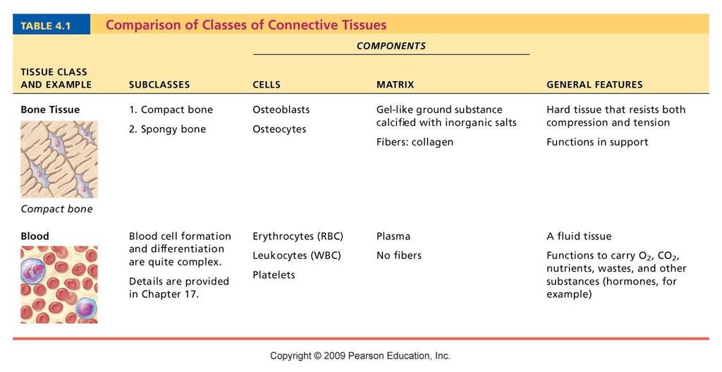
4 Table 4.1
5 Major Functions of Connective Tissue Binding and support Protection Insulation Transportation (blood)
6 Characteristics of Connective Tissue Connective tissues have: Mesenchyme as their common tissue of origin Varying degrees of vascularity Cells separated by nonliving extracellular matrix (ground substance and fibers)
7 Structural Elements of Connective Tissue Ground substance Medium through which solutes diffuse between blood capillaries and cells Components: Interstitial fluid Adhesion proteins ( glue ) Proteoglycans Protein core + large polysaccharides (chrondroitin sulfate and hyaluronic acid) Trap water in varying amounts, affecting the viscosity of the ground substance
8 Structural Elements of Connective Tissue Three types of fibers Collagen (white fibers) Strongest and most abundant type Provides high tensile strength Elastic Networks of long, thin, elastin fibers that allow for stretch Reticular Short, fine, highly branched collagenous fibers
9 Structural Elements of Connective Tissue Cells Mitotically active and secretory cells = blasts Mature cells = cytes Fibroblasts in connective tissue proper Chondroblasts and chondrocytes in cartilage Osteoblasts and osteocytes in bone Hematopoietic stem cells in bone marrow Fat cells, white blood cells, mast cells, and macrophages

10 Cell types Macrophage Extracellular matrix Ground substance Fibers Collagen fiber Elastic fiber Reticular fiber Fibroblast Lymphocyte Fat cell Capillary Mast cell Neutrophil Figure 4.7
11 Connective Tissue: Embryonic Mesenchyme embryonic connective tissue Gives rise to all other connective tissues Gel-like ground substance with fibers and star-shaped mesenchymal cells
12 Overview of Connective Tissues For each of the following examples of connective tissue, note: Description Function Location
13 Connective Tissue Proper Types: Loose connective tissue Dense connective tissue Areolar Dense regular Adipose Dense irregular Reticular Elastic
14 Connective tissue proper; loose - AREOLAR (a) Connective tissue proper: loose connective tissue, areolar Description: Gel-like matrix with all three fiber types; cells: fibroblasts, macrophages, mast cells, and some white blood cells. Elasti c fibers Function: Wraps and cushions organs; its macrophages phagocytize bacteria; plays important role in inflammation; holds and conveys tissue fluid.widely distributed under Location: Collagen fibers epithelia of body, e.g., forms lamina propria of mucous membranes; packages organs; surrounds capillaries. Fibroblast nuclei Epithelium Lamin a propria Photomicrograph: Areolar connective tissue, a soft packaging tissue of the body (300x). Figure 4.8a

15 Connective tissue proper; loose - ADIPOSE (b) Connective tissue proper: loose connective tissue, adipose Description: Matrix as in areolar, but very sparse; closely packed adipocytes, or fat cells, have nucleus pushed to the side by large fat droplet. Function: Provides reserve food fuel; insulates against heat loss; supports and protects organs. Location: Under skin in the hypodermis; around kidneys and eyeballs; within abdomen; in breasts. Nucleus of fat cell Vacuole containing fat droplet Adipose tissue Mammar y glands Photomicrograph: Adipose tissue from the subcutaneous layer under the skin (350x). Figure 4.8b
16 Connective tissue proper; loose - RETICULAR (c) Connective tissue proper: loose connective tissue, reticular Description: Network of reticular fibers in a typical loose ground substance; reticular cells lie on the network. Function: Fibers form a soft internal skeleton (stroma) that supports other cell types including white blood cells, mast cells, and macrophages. Location: Lymphoid organs (lymph nodes, bone marrow, and spleen). White blood cell (lymphocyte ) Reticula r fibers Spleen Photomicrograph: Dark-staining network of reticular connective tissue fibers forming the internal skeleton of the spleen (350x). Figure 4.8c
17 Connective tissue proper; dense - DENSE REGULAR (d) Connective tissue proper: dense connective tissue, dense regular Description: Primarily parallel collagen fibers; a few elastic fibers; major cell type is the fibroblast. Collagen fibers Function: Attaches muscles to bones or to muscles; attaches bones to bones; withstands great tensile stress when pulling force is applied in one direction. Location: Tendons, most ligaments, aponeuroses. Nuclei of fibroblasts Shoulder joint Ligament Tendon Photomicrograph: Dense regular connective tissue from a tendon (500x). Figure 4.8d

18 Connective tissue proper; dense - DENSE IRREGULAR (e) Connective tissue proper: dense connective tissue, dense irregular Description: Primarily irregularly arranged collagen fibers; some elastic fibers; major cell type is the fibroblast. Function: Able to withstand Nuclei of fibroblast s tension exerted in many directions; provides structural strength. Location: Fibrous capsules of organs and of joints; dermis of the skin; submucosa of digestive tract. Fibrous joint capsul e Collage n fibers Photomicrograph: Dense irregular connective tissue from the dermis of the skin (400x). Figure 4.8e

19 Connective tissue proper; dense - ELASTIC (f) Connective tissue proper: dense connective tissue, elastic Description: Dense regular connective tissue containing a high proportion of elastic fibers. Function: Allows recoil of tissue following stretching; maintains pulsatile flow of blood through arteries; aids passive recoil of lungs following inspiration. Location: Walls of large arteries; within certain ligaments associated with the vertebral column; within the walls of the bronchial tubes. Elastic fibers Aort a Hear t Photomicrograph: Elastic connective tissue in the wall of the aorta (250x). Figure 4.8f
20 Connective Tissue: Cartilage Three types of cartilage: Hyaline cartilage Elastic cartilage Fibrocartilage
21 Cartilage - HYALINE (g) Cartilage: hyaline Description: Amorphous but firm matrix; collagen fibers form an imperceptible network; chondroblasts produce the matrix and when mature (chondrocytes) lie in lacunae. Function: Supports and reinforces; has resilient cushioning properties; resists compressive stress. Location: Forms most of the embryonic skeleton; covers the ends of long bones in joint cavities; forms costal cartilages of the ribs; cartilages of the nose, trachea, and larynx. Chondrocyt e in lacuna Matri x Costal cartilage s Photomicrograph: Hyaline cartilage from the trachea (750x). Figure 4.8g
22 Cartilage - ELASTIC (h) Cartilage: elastic Description: Similar to hyaline cartilage, but more elastic fibers in matrix. Function: Maintains the shape of a structure while allowing great flexibility. Chondrocyt e in lacuna Location: Supports the external ear (pinna); epiglottis. Matri x Photomicrograph: Elastic cartilage from the human ear pinna; forms the flexible skeleton of the ear (800x). Figure 4.8h
23 Cartilage - FIBROCARTILAGE (i) Cartilage: fibrocartilage Description: Matrix similar to but less firm than that in hyaline cartilage; thick collagen fibers predominate. Function: Tensile strength with the ability to absorb compressive shock. Location: Intervertebral discs; pubic symphysis; discs of knee joint. Intervertebra l discs Chondrocyte s in lacunae Collage n fiber Photomicrograph: Fibrocartilage of an intervertebral disc (125x). Special staining produced the blue color seen. Figure 4.8i
24 Others - BONE (j) Others: bone (osseous tissue) Description: Hard, calcified matrix containing many collagen fibers; osteocytes lie in lacunae. Very well vascularized. Function: Bone supports and protects (by enclosing); provides levers for the muscles to act on; stores calcium and other minerals and fat; marrow inside bones is the site for blood cell formation (hematopoiesis). Location: Bones Centra l canal Lacuna e Lamell a Photomicrograph: Cross-sectional view of bone (125x). Figure 4.8j
25 Others - BLOOD (k) Others: blood Description: Red and white blood cells in a fluid matrix (plasma). Function: Transport of respiratory gases, nutrients, wastes, and other substances. Location: Contained within blood vessels. Plasm a Neutrophi l Red blood cells Lymphocyt e Photomicrograph: Smear of human blood (1860x); two white blood cells (neutrophil in upper left and lymphocyte in lower right) are seen surrounded by red blood cells. Figure 4.8k
26 Quick Review!!
27 Tissue Lab Part 2 Connective Tissue Day! (Slides 6-11) (Questions 9-15) SKIP #13 Share pics of your slides on social media! *If you need to makeup and Epithelial slides from last lab... now s the time! PRACTICE focusing the microscope!!!
28 When you re finished Return all slides, microscopes, and class copies You should be finished with questions 1-15 as of today (for pacing) You should have labeled and colored pictures of slides 1-11 as of today! After you ve done this - you may start tissue note cards Set 2 Running Packet Checklist 1. Body Map 2. Tissue Introduction 3. Unit 2 Guide 4. 4A Notes 5. Microscope Intro Lab 6. Nose Stem Cell Article and Questions 7. Modeling Epithelial Tissues Activity/?s 8. Tissue Notecards 9. Cancer Article Exchange 10. Tissue Lab (Parts 1-4)
29
30 Warm Up 9/28/17 What are the 4 major types of connective tissue? List any subdivisions of these categories Create a mind map or chart to represent the different classifications of connective tissue. At first, try not to use your notes remember what you can! Then feel free to use your 4B notes.
31 Connective Tissue Proper Loose: Areolar, Adipose, Reticular Dense: Dense Regular, Dense Irregular, Elastic Cartilage Hyaline, Elastic, Fibrocartilage Bone - Osseous Tissue Blood
32 Four Corners Front Left I feel AWESOME about this! I should be teaching this class Back Left I feel okay with this need to study a little more! Front Right I need some help, but I m starting to get it Back Right I don t understand this AT ALL (yet;))
33 Nervous Tissue Nervous system (more detail with the Nervous System, Chapter 11)
.")
34 Nervous tissue Description: Neurons are branching cells; cell processes that may be quite long extend from the nucleus-containing cell body; also contributing to nervous tissue are nonirritable supporting cells (not illustrated). Neuron processes Axon Dendrite s Nuclei of supporting cells Cell body Cell body of a neuron Function: Transmit electrical signals from sensory receptors and to effectors (muscles and glands) which control their activity. Location: Brain, spinal cord, and nerves. Neuron processe s Photomicrograph: Neurons (350x) Figure 4.9
35 Muscle Tissue Skeletal muscle (more detail with the Muscular System, Chapter 10)
36 (a) Skeletal muscle Description: Long, cylindrical, multinucleate cells; obvious striations. Striation s Function: Voluntary movement; locomotion; manipulation of the environment; facial expression; voluntary Location:control. In skeletal Nucle i muscles attached to bones or occasionally to skin. Part of muscle fiber (cell) Photomicrograph: Skeletal muscle (approx. 460x). Notice the obvious banding pattern and the fact that these large cells are multinucleate. Figure 4.10a
37 Muscle Tissue Cardiac muscle (more detail with the Cardiovascular System, Chapters 18 and 19)
38 (b) Cardiac muscle Description: Branching, striated, generally uninucleate cells that interdigitate at specialized junctions (intercalated discs). Striation s Intercalate d discs Function: As it contracts, it propels blood into the circulation; involuntary control. Location: The walls of the heart. Nucleu s Photomicrograph: Cardiac muscle (500X); notice the striations, branching of cells, and the intercalated discs. Figure 4.10b
39 Muscle Tissue Smooth muscle
40 (c) Smooth muscle Description: Spindle-shaped cells with central nuclei; no striations; cells arranged closely to form sheets. Smoot h muscle cell Function: Propels substances or objects (foodstuffs, urine, a baby) along internal passageways; involuntary Location: Mostly control. in the walls of hollow organs. Nucle i Photomicrograph: Sheet of smooth muscle (200x). Figure 4.10c
41 Epithelial Membranes Cutaneous membrane (skin) (More detail with the Integumentary System, Chapter 5)
 covers the body surface. Figure 4.11a")
42 Cutaneous membrane (skin) (a) Cutaneous membrane (the skin) covers the body surface. Figure 4.11a
43 Epithelial Membranes Mucous membranes Mucosae Line body cavities open to the exterior (e.g., digestive and respiratory tracts)
 Mucous membranes line body cavities open to the exterior.")
44 Mucosa of nasal cavity Mucosa of mouth Esophagu s lining Mucosa of lung bronchi (b) Mucous membranes line body cavities open to the exterior. Figure 4.11b
45 Epithelial Membranes Serous Membranes Serosae membranes (mesothelium + areolar tissue) in a closed ventral body cavity Parietal serosae line internal body walls Visceral serosae cover internal organs

46 Parietal peritoneu m Visceral peritoneu m Parietal pericardiu m (c) Serous membranes line cavities closed to the exterior. Parieta l pleura Viscera l pleura Visceral pericardiu m body Figure 4.11c
47 Steps in Tissue Repair Inflammation Release of inflammatory chemicals Dilation of blood vessels Increase in vessel permeability Clotting occurs

48 Sca b Epidermi s Blood clot in incised wound Inflammator y chemicals 1 Inflammation sets the stage: Vein Migrating white blood cell Arter y Severed blood vessels bleed and inflammatory chemicals are released. Local blood vessels become more permeable, allowing white blood cells, fluid, clotting proteins and other plasma proteins to seep into the injured area. Clotting occurs; surface dries and forms a scab. Figure 4.12, step 1
49 Steps in Tissue Repair Organization and restored blood supply The blood clot is replaced with granulation tissue Epithelium begins to regenerate Fibroblasts produce collagen fibers to bridge the gap Debris is phagocytized

50 Regenerating epithelium Area of granulation tissue ingrowth Fibroblas tmacrophage 2 Organization restores the blood supply: The clot is replaced by granulation tissue, which restores the vascular supply. Fibroblasts produce collagen fibers that bridge the gap. Macrophages phagocytize cell debris. Surface epithelial cells multiply and migrate over the granulation tissue. Figure 4.12, step 2
51 Steps in Tissue Repair Regeneration and fibrosis The scab detaches Fibrous tissue matures; epithelium thickens and begins to resemble adjacent tissue Results in a fully regenerated epithelium with underlying scar tissue
52 Regenerated epithelium Fibrosed area 3 Regeneration and fibrosis effect permanent repair: The fibrosed area matures and contracts; the epithelium thickens. A fully regenerated epithelium with an underlying area of Figure 4.12, step 3
53 Developmental Aspects Primary germ layers: ectoderm, mesoderm, and endoderm Formed early in embryonic development Specialize to form the four primary tissues Nerve tissue arises from ectoderm Muscle and connective tissues arise from mesoderm Epithelial tissues arise from all three germ layers
 Nervous tissue (from ectoderm)")
54 16-day-old embryo (dorsal surface view) Ectoder m Mesoder m Endoder m Epitheliu m Muscle and connective tissue (mostly from mesoderm) Nervous tissue (from ectoderm) Figure 4.13
10/3/2012. Tissue: The Living Fabric: Part B. Extracellular matrix Ground substance Fibers Collagen fiber Elastic fiber Reticular fiber.
 PowerPoint Lecture Slides prepared by Janice Meeking, Mount Royal College C H A P T E R 4 Tissue: The Living Fabric: Part B Copyright 2010 Pearson Education, Inc. Copyright 2010 Pearson Education, Inc.
Function: Provides reserve food fuel; Copyright 2011 Pearson Education, Inc. Copyright 2011 Pearson Education, Inc. White blood cell (lymphocyte)
") Adipose Tissue Closely packed adipocytes Have nucleus pushed to one side by fat droplet Richly vascularized Provides reserve food fuel Insulates against heat loss Supports and protects organs Under skin
Adipose Tissue Closely packed adipocytes Have nucleus pushed to one side by fat droplet Richly vascularized Provides reserve food fuel Insulates against heat loss Supports and protects organs Under skin
Connec<ve Tissue. Major Func<ons of Connec<ve Tissue 9/8/14. Most and widely distributed <ssue type Four classes. Tissue: The Living Fabric: Part B
 Connec
Connec<ve Tissue 9/8/14. Major Func<ons of Connec<ve Tissue. of Connec<ve Tissue. Characteris<cs of Connec<ve Tissue
 Connec
Connective Tissue. Found everywhere in the body. Most abundant and widely distributed. Never exposed to the outside environment.
 Connective Tissue Found everywhere in the body. Most abundant and widely distributed. Never exposed to the outside environment. Connective Tissue Functions Binding and support Protection Insulation Transportation
8/30/2017. Tissue: The Living Fabric. 4.3 Connective Tissue
 Chapter 4 Part B Tissue: The Living Fabric Annie Leibovitz/Contact Press Images PowerPoint Lecture Slides prepared by Karen Dunbar Kareiva Ivy Tech Community College 4.3 Connective Tissue Connective tissue
Most abundant and widely distributed tissues in the body Binds, support, and strengthen body tissues, protect and insulate internal organ, serve as
 Connective tissue Most abundant and widely distributed tissues in the body Binds, support, and strengthen body tissues, protect and insulate internal organ, serve as major transport system, compartmentalizes
Cells and Tissues 3PART D. PowerPoint Lecture Slide Presentation by Patty Bostwick-Taylor, Florence-Darlington Technical College
 PowerPoint Lecture Slide Presentation by Patty Bostwick-Taylor, Florence-Darlington Technical College Cells and Tissues 3PART D Connective Tissue Found everywhere in the body Includes the most abundant
Bio& 241 Unit 1 / Lecture 4
 Bio& 241 Unit 1 / Lecture 4 Connective Tissue Consists of two basic elements: Cells and Extra-cellular matrix 1 True Connective Tissue Cells Fibroblasts: Secrete both fibers and ground substance of the
Connective Tissue. Consists of two basic elements: Cells and Extra-cellular matrix
 Connective Tissue Consists of two basic elements: Cells and Extra-cellular matrix True Connective Tissue Cells Fibroblasts: Secrete both fibers and ground substance of the matrix (wandering) Macrophages:
Body Tissues Pearson Education, Inc.
 Body Tissues Tissues Groups of cells with similar structure and function Four primary types: Epithelial tissue (epithelium).1 Connective tissue.2 Muscle tissue.3 Nervous tissue.4 Epithelial Tissues Locations:
HISTOLOGY. Simple squamal lungs
 HISTOLOGY Lab Objectives: Students should be able to... 1. Visually identify each class of tissue and examples within each class 2. Indicate the location (in the human body and/or organ) and function of
Histology. The study of tissues.
 Histology The study of tissues. Body Tissues Cells are specialized for particular functions Tissues Groups of cells with similar structure and function Four primary types Epithelium Connective tissue Nervous
Lecture Overview. Connective Tissues. Marieb s Human Anatomy and Physiology. Chapter 4 Tissues: The Living Fabric Connective Tissues Lecture 10
 Marieb s Human Anatomy and Physiology Marieb Hoehn Chapter 4 Tissues: The Living Fabric Connective Tissues Lecture 10 Lecture Overview General composition and function of connective tissue Components of
A. cells that perform related functions and are similar in structure. B. extracellular material - made by cells and secreted into interstitial space
 I. tissue components A. cells that perform related functions and are similar in structure B. extracellular material - made by cells and secreted into interstitial space II. tissue types A. epithelium (e.)
HOLE S ANATOMY CHAPTER 5, PART II Lecture notes
 HOLE S ANATOMY CHAPTER 5, PART II Lecture notes I. Connective Tissue A. Structure 1. have few cells that are spaced apart and can divide; two categories: a. fixed cells cells that are present in tissue
TISSUES. Dr. Gary Mumaugh
 TISSUES Dr. Gary Mumaugh Tissues Tissues - Groups of cells similar in structure and function and perform a common function Histology The study of tissues The four types of tissues Epithelial Connective
Tissues- of cells with similar and
 Tissues- of cells with similar and. Four types of tissues 1. 2. 3. 4. Characteristics of Epithelial Tissue -Highly Cellular -Special contacts -Polar (apical and basal surfaces) -Supported by connective
Tissues Description Function(s) Locations Miscellaneous. avascular -thelium = covering
 Locations Miscellaneous. avascular -thelium = covering") Epithelial Tissue Simple Squamous flattened cells diffusion and Kidney glomeruli disc-shaped central filtration air sacs of lung Simple = Single layer nuclei secretes lubricating lining of heart, blood
Epithelial Tissue Simple Squamous flattened cells diffusion and Kidney glomeruli disc-shaped central filtration air sacs of lung Simple = Single layer nuclei secretes lubricating lining of heart, blood
The Tissue Level of Organization
 The Tissue Level of Organization 4.5-4.11 August 31, 2012 4.5 Connective Tissues Describe the general features of connective Describe the structure, location, and function of the various types of connective
Tissues. Cells work together in functionally related groups called tissues Types of tissues: 1. Epithelial lining and covering. 2. Connective support
 Histology Tissues Cells work together in functionally related groups called tissues Types of tissues: 1. Epithelial lining and covering 2. Connective support 3. Muscle movement 4. Nervous control Epithelial
BIOL 2457 CHAPTER 4 Part 2 SI All connective tissues arise from, an embryonic tissue.
 BIOL 2457 CHAPTER 4 Part 2 SI 1 1. All connective tissues arise from, an embryonic tissue. 2. Describe the vascularity of connective tissues, which are very diverse. 3. Describe the innervation of connective
Tissues 10/21/2016. Epithelial Tissue
 Tissues This is a generalized cell diagram. It shows the anatomy of a cell, but most cells do not actually look like this. Cells can have a wide variety of shapes and sizes, depending on their function.
Histology 101! !! Name:! Block: Identify and describe the functions of major tissue types including their subclasses and varieties!
 Histology 101 Identify and describe the functions of major tissue types including their subclasses and varieties Name: Block: "1 Introduction to Tissues Histology Notes Tissue (living fabric) : groups
Lab 1 ANIMAL TISSUES
 Lab 1 ANIMAL TISSUES Levels of Organization Animals are multicellular heterotrophs whose cells lack cell walls. Most animals exhibit a hierarchical level of organization: Cells are organized into tissues
Human anatomy Unit III. Tissue
 Human anatomy Unit III Tissue Definition of Tissues Biological tissue is a collection of interconnected cells that perform a similar function within an organism. In other words, it is a group of cells
Body Tissues. Cells are specialized for particular functions Tissues - groups of cells with similar structure. and function Four primary tissue types:
 Chapter 3 Tissues Body Tissues Cells are specialized for particular functions Tissues - groups of cells with similar structure and function Four primary tissue types: Epithelium Connective tissue Nervous
1/26/2018. Tissues - Learn and Understand. Tissue: The Living Fabric. Individual body cells specialized. Tissues. Histology. Tissues and Histology
 Tissues - Learn and Understand The composition of a tissue helps us understand what functions it is capable of. Concepts of cell surface area, attachment of tissues to others, mucus production, role of
Study of different tissues Abnormal cells and tissues can be compared to normal tissues to identify disease, such as cancer Being able to know and
 CHAPTER 4 Study of different tissues Abnormal cells and tissues can be compared to normal tissues to identify disease, such as cancer Being able to know and recognize normal tissues under the microscope
Epithelial Tissue. Simple Cuboidal Function: secretion and absorption. Simple Squamous
 Epithelial Tissue General Functions: Lines and covers organs Absorbs / secretes substances Gas exchange Protection Special Characteristics: - have an apical surface on top - have a basement membrane below
Classification of Tissues
 6 R e v i e w S h e e t Exercise Classification of Tissues NAME LAB TIME/DATE Tissue Structure and Function General Review 1. Define tissue. A group of cells similar to one another in structure that perform
Anatomy and Physiology Tissue Review
 Anatomy and Physiology Tissue Review OVERVIEW Histology practicals can be rough, especially when access to slides is limited to the lab period. This resource provides an opportunity to learn or review
Chapter 5. Tissues. 4 Types of Body Tissues. Tissues
 Chapter 5 Tissues Tissues Tissues - groups of cells that are similar in structure & function RBC, WBC, & platelets are a group of cells working together to form BLOOD tissue Histology Pathohistology study
Introduction to Types of Body Tissue Putting it All Together. Packet #12
 Introduction to Types of Body Tissue Putting it All Together Packet #12 Introduction Body Tissues Tissues Groups of cells with similar structure and function Four primary types Epithelial tissue (epithelium)
Classification of Tissues
 M06_MARI0000_00_SE_CH06.qxd 3/28/11 4:37 PM Page 35 NAME LAB TIME/DATE R E V I E W S H E E T EXERCISE 6 Classification of Tissues Tissue Structure and Function General Review 1. Define tissue. A group
Tissue = groups of cells that are similar in structure and function
 Tissue = groups of cells that are similar in structure and function Types Epithelial - covering Connective - support Muscle - movement Nervous - control Membranes line body cavities and hold organs together
The Tissue Level of Organization
 Tissue The Tissue Level of Organization Chapter 3 Definition an aggregation of cells in which each cooperates with all others in the performance of a given function Examples of general functions Movement
Chapter 1: Cells and Tissues
 Chapter 1: Cells and Tissues Cells and Tissues Carry out all chemical activities needed to sustain life Cells are the building blocks of all living things Tissues are groups of cells that are similar in
Histology. Histology. Tissue - Four main tissues in body. 1. Epithelial tissue an epithelium; plural: epithelia. Function. Location.
 Histology Histology Tissue Four main tissues in body 1. Epithelial tissue an epithelium; plural: epithelia Function Location Characteristics Example 2. Connective tissue Function Location Characteristics
Histology. Study of body tissues
 Histology Study of body tissues 2 Introduction to Body Tissues 1. Composed of specialized cells of similar structure and perform a common function 2. Four major types (4 Cs) a. Epithelial - Cover b. Connective
Tissues. groups of cells similar in structure and function 4 types. epithelium connective muscle nervous
 Tissues groups of cells similar in structure and function 4 types epithelium connective muscle nervous Epithelial Tissue lining covering glandular Functions protection absorption filtration secretion Epithelium
Chapter 3. Cells and Tissues. Lecture Presentation by Patty Bostwick-Taylor Florence-Darlington Technical College Pearson Education, Inc.
 Chapter 3 Cells and Tissues Lecture Presentation by Patty Bostwick-Taylor Florence-Darlington Technical College Body Tissues Tissues Groups of cells with similar structure and function Four primary types:
Basic Histology. By Mrs. Bailey
 Basic Histology By Mrs. Bailey Primary Tissues 1. Epithelial Tissue 2. Connective Tissue 3. Muscle Tissue 4. Nervous Tissue Very cellular Supported by underlying connective tissue Epithelial & connective
4 Types of Tissue. Epithelial Connective Muscle Neural
 Connective Tissue 4 Types of Tissue Epithelial Connective Muscle Neural Connective Tissue Fills internal spaces Supports & binds other tissues Transports materials Stores energy Classification of Connective
Cells are specialized for particular functions Tissues
 Histology Body Tissues Cells are specialized for particular functions Tissues Groups of cells with similar structure and function Extracellular Matrix cell glue between cells Histology study of tissue
Epithelia of Coverings and Linings. Tissues. Tissue
 Tissue Tissues Chapter 3 Definition an aggregation of cells in which each cooperates with all others in the performance of a given function Examples of general functions Movement Protection Support Production
Basic Tissue Types and Functions
 Tissues Histology Basic Tissue Types and Functions 1) Epithelial tissue covering 2) Connective tissue support 3) Muscle tissue movement 4) Nervous tissue control Epithelial Tissue 1) Covers a body surface
Hole s Human Anatomy and Physiology
 Hole s Human Anatomy and Physiology 1 Chapter 5 Tissues Four major tissue types 1. Epithelial 2. Connective 3. Muscle 4. Nervous 2 Epithelial Tissues General characteristics - cover organs and the body
Tissues. Group of cells that are similar in structure and function. 4 primary types. Epithelium (covering) Connective (support) Nervous(control)
 Connective (support) Nervous(control)") Tissues Tissues Group of cells that are similar in structure and function 4 primary types Epithelium (covering) Connective (support) Nervous(control) Epithelial tissue (epithelium) Lining, covering, and
Tissues Tissues Group of cells that are similar in structure and function 4 primary types Epithelium (covering) Connective (support) Nervous(control) Epithelial tissue (epithelium) Lining, covering, and
Air sacs of lungs and the lining of the heart, blood vessels, and lymphatic vessels
 Cells Location Function Simple squamous epithelium Air sacs of lungs and the lining of the heart, blood vessels, and lymphatic vessels Allows materials to pass through by diffusion and filtration, and
4 Types of Tissue. Epithelial Connective Muscle Neural
 Connective Tissue 4 Types of Tissue Epithelial Connective Muscle Neural Connective Tissue Fills internal spaces Supports & binds other tissues Transports materials Stores energy Classification of Connective
Anatomy &- Physiology Histology Worksheet
 Anatomy &- Physiology Histology Worksheet 1. The four primary tissue types found in the human body are a) squamous, cuboidal, columnar, glandular b) adipose, elastic, reticular, cartilage c) skeletal,
Outline. Bio 105: Tissues Laboratory. Organization of the Human Body. Tissue - Epithelium. Tissues 3/2/ Copyright 2009 Pearson Education, Inc
 Outline Bio 105: Tissues Laboratory Laboratory 5 Reading: Chapter 4 I. Cell to cell contact II. Body Cavities III. Membranes IV. Homeostasis V. Integumentary System I. Includes skin, hair and nails 1 2
Section B: Epithelial Tissue 1. Where are epithelial tissues found within the body? 2. What are the functions of the epithelial tissues?
 Tissue worksheet Name Section A: Intro to Histology Cells are the smallest units of life. In complex organisms, cells group together with one another based on similar structure and function to form tissues.
Connective Tissues. Copyright 2009 Pearson Education, Inc., publishing as Pearson Benjamin Cummings
 C.T. are found in all parts of the body & diverse in structure & function. C.T. Functions: -connect structures -provide support -protect vital organs -fill space b/w structures -stores fat -defends body
Epithelial Tissue lining, covering, glandular tissue > Function protect, absorption, filtration, secretion, excretion
 Chapter 4: TISSUES IX. Tissues Intro Epithelial Tissue lining, covering, glandular tissue > Function protect, absorption, filtration, secretion, excretion Connective Tissue most widespread tissue type
B. Classification of epithelium: by number of cell layers present and by shape of the superficial cell layers.
 I. Introduction - tissue: group of cells that are closely associated, similar in structure and function, and perform a common or related function. - four primary tissues: epithelial tissue, connective
Practical Histology. Lab 3: Connective tissue
 Practical Histology Lab 3: Connective tissue Connective tissues Connective tissue provides structural support for the body by binding cells and tissues together to form organs. It also provides metabolic
Study of Tissues Dr. A. Ebneshahidi
 Study of Tissues Dr. A. Ebneshahidi Tissues Tissues are composed of cells similar in structure and specialized to perform a specific function for the body. The human body is made of four general types
CONNECTIVE TISSUE (Refer to pp for specific characteristics of each) VAN (**Be familiar with exceptions**)
 VAN (**Be familiar with exceptions**)") CONNECTIVE TISSUE CHARACTERISTICS: *Most abundant tissue type; Composed of ECM (GS & Protein Fibers) + Cells (Refer to pp.129-131 for specific characteristics of each) *Highly equipped with VAN assists
CONNECTIVE TISSUE CHARACTERISTICS: *Most abundant tissue type; Composed of ECM (GS & Protein Fibers) + Cells (Refer to pp.129-131 for specific characteristics of each) *Highly equipped with VAN assists
Collin College. BIOL Chapter 4. Tissue Levels CONNECTIVE TISSUE. C.T. derives from Mesenchyme embryonic tissue.
 Collin College BIOL. 2401 Chapter 4 Tissue Levels. CONNECTIVE TISSUE C.T. derives from Mesenchyme embryonic tissue. Depending on the stimuli, mesenchyme develops into specific cells that give rise to the
Body Tissues PART C. PowerPoint Lecture Slide Presentation by Patty Bostwick-Taylor, Florence-Darlington Technical College
 PowerPoint Lecture Slide Presentation by Patty Bostwick-Taylor, Florence-Darlington Technical College Body Tissues 3 PART C I. Body Tissues A. Tissues (tissue = woven) Histology the study of tissues Groups
UNIT 4 T I S S U E S
 UNIT 4 T I S S U E S WHAT IS A TISSUE Group of cells that work together to do a function Cells are similar Extracellular fluid around them is similar Histology EPITHELIAL TISSUE Also called epithelium
2/21/2012. Components Connective Tissue. Connective Tissue??? Connective Tissue What is it?
 Connective Tissue??? Connective Tissue What is it? It Binds, It Supports, It Strengthens, It Protects, It Insulates, It Compartmentalizes, It helps us move, It helps transport stuff, It is a site for storing
Tissues Chapter 5...Tissue - a group or mass of similar cells working together to perform certain common functions
 Tissues Chapter 5...Tissue - a group or mass of similar cells working together to perform certain common functions There are 4 major types of tissue Epithelial Connective Muscle Nervous 1. Epithelial Tissue
Lab Animal Tissue. LEARNING OBJECTIVES: To understand the relationship between the structure and function of different animal tissues
 Name: Bio A.P. PURPOSE: HYPOTHESIS: NONE Lab Animal Tissue BACKGROUND: In animals, groups of closely related cells specialized to perform the same function are called tissues. There are four general classes
A. Incorrect! Axons covey messages from the cell body of the neuron. D. Correct! Dendrites convey messages to the cell body of the neuron.
 CLEP Biology - Problem Drill 14: Animal Form No. 1 of 10 1. The branches of a neuron receiving information from another cell and which transmit the message to the cell body are called? (A) (B) (C) (D)
Connexons: hollow connective tubes
 Chapter 3 1. tight junctions: like a zipper, these junctions hold the cells tightly together making them impermeable to the extracellular fluid that surrounds them. 2. desmosomes: like buttons, these
Tissue Outline. Chapter 4. Tissue. Cellular Connections. I. Definitions II. Cellular Connections III. Tissue Types IV. Membranes V.
 Tissue Outline Chapter 4 The Tissue Level of Organization I. Definitions II. Cellular Connections III. Tissue Types IV. Membranes V. Tissue Repair 1 2 Tissue Cellular Connections Tissue Groups of cells
Chapter 4. The Tissue Level of Organization
 Chapter 4 The Tissue Level of Organization 1 Tissue Outline I. Definitions II. Cellular Connections III.Tissue Types IV. Membranes V. Tissue Repair 2 Tissue Tissue Groups of cells that are similar in structure
ACTIVITY 2: HISTOLOGY AND INTEGUMENT
 ACTIVITY 2: HISTOLOGY AND INTEGUMENT Objectives: 1) How to get ready: Read Chapter 4 and 5, McKinley et al., Human Anatomy, 4e. All text references are for this textbook. 2) Identify each tissue (26 tissues)
THE TISSUE LEVEL OF ORGANIZATION PART I: EPITHELIAL TISSUE
 THE TISSUE LEVEL OF ORGANIZATION PART I: EPITHELIAL TISSUE 4 Main Tissue Types Epithelium Covers surfaces, lines cavities, forms glands Connective Tissue Support and protects body Muscular Tissue Movement
Name: Test Date: Chapter 4- Tissues. Use the choices to identify the major tissue types found below:
 Name: Test Date: Chapter 4- Tissues Use the choices to identify the major tissue types found below: A. Connective B. Epithelium C. Muscle D. Nervous 1. B Lines body cavities and covers the body s external
Lab Exercise 6a-2. Classification of connective tissues. Connective Tissue. Connective tissues. Areolar. Areolar tissue
 Classification of connective tissues Lab Exercise 6a-2 Connective Tissue Nervous Muscle Connective Tissue Connective tissues Connective tissue proper Fluid connective tissue Supportive connecting tissue
Sheet #9. Dr. Heba Kalbouneh. Dr. Heba Kalbouneh. Dr. Heba Kalbouneh
 Sheet #9 Dr. Heba Kalbouneh Dr. Heba Kalbouneh Dr. Heba Kalbouneh Elastic fibers The main function of elastic fibers is to provide elasticity. In other words these fibers are able to restore the original
They cells can not function death.
 Jenna Hellack Jan 2001 Tissues What do you think happens when the cells use up their food and oxygen before there is time to replenish it? They cells can not function death. Blood Cell Cancer cell Plant
Tissues and Membranes
 I. In the Beginning a. Egg + sperm! Tissues and Membranes b. 1 cell divides to make 2, 2 divide to make 4, 4 divide to make 8, and then? c. d. e. Totipotent: f. Pluripotent: II. III. Tissues a. Tissues
Use for reference if needed:
 A- 2.5 Describe how structure and function are related in terms of cell and tissue types. I can recognize different types of body tissue. I can explain how different tissue structures affect their functions.
Tissues are: group of similar or identical cells that share a common function. used to build organs
 Tissues: Four classes Epithelium Connective Muscle Nervous Tissues are: group of similar or identical cells that share a common function. used to build organs Overview: Epithelial o Line body cavities
Connective Tissue Nervous Muscle. Classification of connective tissues
 Connective Tissue Nervous Muscle Lab Exercise 6a-2 Classification of connective tissues 1 Connective Tissue Connective tissue proper Fluid connective tissue Supportive connecting tissue Connective tissues
Tissue Outline (chapter 4) Tissues group of cells that perform structural and roles. List the 4 types:
 Tissues group of cells that perform structural and roles. List the 4 types:") Tissue Outline (chapter 4) Tissues group of cells that perform structural and roles. List the 4 types: 1. 2. 3. 4. I. Epithelial Tissue covers all the surfaces, inside & out. Are the major tissues of,
Tissue Outline (chapter 4) Tissues group of cells that perform structural and roles. List the 4 types: 1. 2. 3. 4. I. Epithelial Tissue covers all the surfaces, inside & out. Are the major tissues of,
Unit II: Tissues and Integumentary System
 Unit II: Tissues and Integumentary System 2.1 - Tissues Chapter 4 Written Response #1 1. What is a tissue? 2. What are four major types of tissues? Tissue Definition: a group or mass of similar cells working
Tejido Conectivo Parte B. Informe #3 Laboratorio Biología # 240 Profesor: Javier Cabello
 Tejido Conectivo Parte B Informe #3 Laboratorio Biología # 240 Profesor: Javier Cabello Figure 4-8 The Cells and Fibers of Connective Tissue Proper Areolar Elastic fibers Collagen fibers Fibroblast Free
Tissues, Glands, and Membranes. Chapter Five Mrs. Hornacek
 Tissues, Glands, and Membranes Chapter Five Mrs. Hornacek Objectives 1. Name the four main groups of tissues and give the location and general characteristics of each. 2. Differentiate between voluntary
SHORT ANSWER. Write the word or phrase that best completes each statement or answers the question.
 Exam Name SHORT ANSWER. Write the word or phrase that best completes each statement or answers the question. Figure 4.2 Using Figure 4.2, match the following: 1) Simple cuboidal epithelium. 2) Cardiac
TISSUE. A group of cells that perform a similar function within an organism. Epithelium Connective Muscle Nervous CREDITS
 TISSUE A group of cells that perform a similar function within an organism. Epithelium Connective Muscle Nervous CREDITS Epithelium Connective Muscle Nervous Epithelium Composed of a layer of cells. Lines
Tissues (Histology) Ch. 3 Human Anatomy lecture
 Ch. 3 Human Anatomy lecture") I. Histology the study of tissues A. 4 basic tissue types epithelial connective muscle nervous Tissues (Histology) Ch. 3 Human Anatomy lecture B. Usually found in combinations to form organs. C. As you
I. Histology the study of tissues A. 4 basic tissue types epithelial connective muscle nervous Tissues (Histology) Ch. 3 Human Anatomy lecture B. Usually found in combinations to form organs. C. As you
Anatomy Chapter 4 Tissues
 4 Principle Tissue Types Epithelial tissue Covering and lining Glandular Connective tissue Highly variable Most abundant tissue type Muscular tissue 3 major types Produce force through contraction Nervous
ACTIVITY 2: HISTOLOGY AND INTEGUMENT
 ACTIVITY 2: HISTOLOGY AND INTEGUMENT Objectives: 1) How to get ready: Read Chapter 4 and 5, McKinley et al., Human Anatomy, 5e. All text references are for this textbook. 2) Identify each tissue (26 tissues)
contains an antiangiogenesis factor
 CARTILAGE & BONE Cartilage and Bone objectives Student must learn :. What is the meaning of cartilage, and their function, location in human body.. To distinguish the 3 types of cartilage. And their cells,
Unit I Problem 9 Histology: Basic Tissues of The Body
 Unit I Problem 9 Histology: Basic Tissues of The Body - What is the difference between cytology and histology? Cytology: it is the study of the structure and functions of cells and their contents. Histology:
Chapter 4 :Organization & Regulation of Body Systems
 Chapter 4 :Organization & Regulation of Body Systems 4.1 Types of tissues What is a tissue? A collection of cells of the same type that perform a common function There are 4 major tissue types in the body:
Tissues. Tissues. Four basic tissues. A collection of cells with a common function. 1. Epithelial 2. Connective 3. Muscular 4.
 Tissues Tissues A collection of cells with a common function Four basic tissues 1. Epithelial 2. Connective 3. Muscular 4. Nervous Epithelia: cells in layers Types of epithelia 1) lining Layers of cells
Tissues organs system organism. pg151
 Histology is the study of tissues A TISSUE is a group of cells, usually of one kind, & their intercellular substance (e.g. intercellular matrix in animal) which are linked together & perform a particular
Connective Tissue. Practice Identifying Your Tissue With This Slide Show! Then try the quiz at the bottom of this page!
 Connective Tissue scientistcindy.com /connective-tissue.html Practice Identifying Your Tissue With This Slide Show! Then try the quiz at the bottom of this page! The 4 Types of Connective Tissues Connective
Dr. heba kalbouneh. Dr. heba kalbouneh
 7 Dr. heba kalbouneh Dr. heba kalbouneh Clinical applications: In surgical incision site, the site of injury is filled by collagen fibers synthesized by fibroblasts (the fibrocytes in the connective tissue
Chapter 05. Review. Copyright The McGraw-Hill Companies, Inc. Permission required for reproduction or display.
 Chapter 05 Review 5.1: Introduction Similar cells with a common function are called tissues. The study of tissues is called histology. There are four (4) primary or major tissue types: 1. Epithelial Tissue
Tissues and Structures to Know for the Lab Practical
 Ch. 3 - Cells and Tissues Tissues and Structures to Know for the Lab Practical Miss School, Miss Out! Simple squamous epithelium line and cover; site of diffusion Simple squamous epithelium apical surface
TISSUES. Objectives. Tissues
 TISSUES Objectives Introduce the four major types of tissues Describe the general characteristics and functions of epithelial & connective tissue Name the major types of epithelial & connective tissues