Uveitis Review for the OKAPS
|
|
|
- Peter Bridges
- 6 years ago
- Views:
Transcription
1 Uveitis Review for the OKAPS What you should and should not know! By: Armando L. Oliver, MD Ocular Immunology and Uveitis Specialist Vitreoretinal Surgeon Assistant Professor, UPR
2 Disclaimer... The information you are about to whiteness has been oversimplified for the purpose of passing the OKAPs, boards, etc Some of this information sounds OK, and you may have been brought up to believe is true, but it isn t! Using this information for real life patient management may have deleterious consequences. Contrary to popular believe, I m not insane!
3 Anterior Uveitis
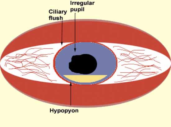
4 Acute Anterior Uveitis AKA: Acute Recurrent Alternating Anterior Uveitis Pain, Redness & Photophobia. HLA-B27 Associated 50-86% cases. Ankilosing Spondilitis, Reiter s, Psoriatic Arthritis, Inflammatory Bowel Disease Spondiloarthropathy. Most common cause of hypopion uveitis on an angry (red) eye. Hypopion Uveitis with white eye, mouth and genital ulcers = Becet s.
5
6 Fuch s Heterochromic Iridocyclitis The name holds true! Unilateral chronic uveitis with stellate KPs Needs no steroid treatment for the uveitis. Needs no cycloplegia, does not form synechiae. Increased incidence of POAG and cataracts! Amsler sign: Patogneumonic. Filiform hemorrhage on the site opposite to the paracenthesis during cataract surgery.

7 Heterochromia, Cataract, Good Dilation

8 Stellate KPs 3/3 of endothelium
9 Juvenile Idiopathic (Rheumatoid) Arthritis Iridocyclitis most common on patients with: Pauciarticular presentation (4 or less joints) ANA+ Patients with above criteria must be screened q3 months x 7 years. Associated Findings: Asymptomatic, Band Keratopathy, White Cataract, Posterior Synechia. If not adequately suppressed patients may go pthysical in a short period of time.

10 Note: White eye, White cataract, Band Keratopathy, Posterior Synechiae Personal Note: Mostly avoidable with early aggressive treatment!
11 TINU Tubuloinsterstitial Nephritis & Uveitis Syndrome. Guess What?! First, you get a tubuloinstersttial nephritis. Microhematuria, malaise, fever, etc... Then you get uveitis. Bilateral, Acute, Non-Granulomatous Responds well to Topical Steroids. Common in children!
12 Granulomatous Uveitis Non-pathological term used to describe uveitis that has: Mutton Fat KPs Koeppe or Busacca Nodules Giant Cell Deposits These uveitides are generally of chronic or infectious nature. Tend to present insidiously with decreased vision more common than with pain, redness and photophobia. DDx Includes: Sarcoidosis, Tb, Syphilis, Toxoplasma, HSV, VZV, VKH, Sympathetic Ophthalmia and idiopathic.


13 Koeppe Nodules Busacca Nodules Posterior Synechiae & Iris Bombè
14 Herpes Simplex Virus Uveitis Unilateral Granulomatous Uveitis with Iris transillumination defects in a patient that suffers recurrent cold sores who has corneal aesthesia and possibly, high IOP and a corneal stromal haze. Treatment: Acyclovir 400mg 5times/d, Pred Forte and Atropine. Note: Acute if treated very early otherwise may become chronic.

15 HSV Uveitis Iris Atrophy 360
16 Herpes Zoster Virus Uveitis Unilateral Granulomatous Uveitis with Iris transillumination defects in a patient that suffered V1 shingles that likely affected the nasocilliary nerve, who may have corneal aesthesia and possibly high IOP and possibly a corneal stromal haze. Treatment: Acyclovir 800mg 5times/d, Pred Forte and Atropine. Note: Look at the fundus and R/O Acute Retinal Necrosis.

17 Zoster Uveitis Note: Segmental Iris Atrophy
18 Uveitis that may have high IOP upon presentation. DDX for the OKAPS: HSV & VZV CMV Toxoplasma Posner-Schlossman Syndrome (Dx of Exclusion) Rare in real life, common in the OKAP! After the initial presentation, the differential of glaucoma in uveitis patients includes: anterior synechiae, bombè, steroid response, and pigment deposition (HSV/VZV).
19 Intermediate Uveitis
20 Intermediate Uveitis AKA: Pars Planitis Findings: Bilateral, Snow Banking, Snow Balls, (Band Keratopathy in children). Systemic Associations: Multiple Sclerosis Ophtalmic Associations: Optic Neuritis Does not need treatment if VA > 20/40 Complications: CME: Most common cause of visual loss Posterior (and anterior) Sub-Capsular Cataracts Tx: Steroids (sub-tenon s > Oral > Topical)

21
22 Posterior Uveitis
23 Retinitis: Definitions Fluffy white retina with diffuse borders and lots of vitritis. Choroiditis: Yellow or grey retinal elevation with demarcated borders and no vitritis. Chorioretinitis: Choroiditis with a little vitritis. Old Choroiditis: Punched Out Scars, may vary in size & shape.
24 Definitions Plaque: A lesion approximately 1DA. Spots & Dots: Lesions approximately microns.
25 Definitions Focal: Single lesion, may be localized like in Toxoplasma or spread like CMV retinitis. Multifocal: Multiple lesions, I.e: MEWDS. Diffuse: Sympathetic Ophthalmia.
26 Choroiditis and Chorioretinitis
27 Which is the unilateral one? MEWDS: Multiple Evanescent White Dot Syndrome. Multiple dots that subsequently disappear are required for the diagnosis! Has a good prognosis, without treatment! Late wreath pattern fluorescein.
28 Multiple Evanescent White Dot Syndrome

29 Multiple Evanescent White Dot Syndrome
30 Multiple Evanescent White Dot Syndrome
31 OK lets talk about choroiditis!
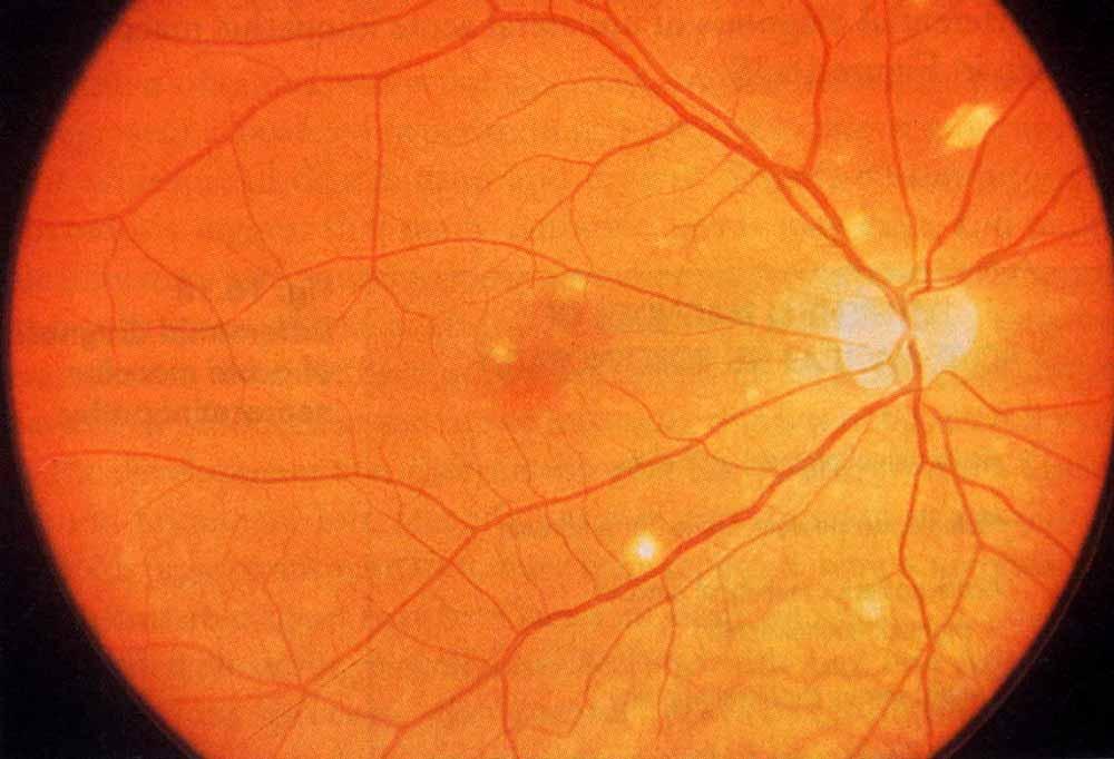
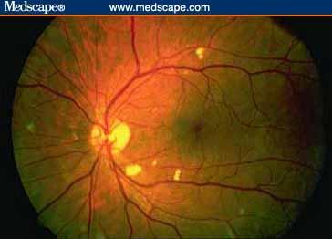
32 Which is the purest form of choroiditis? OHS: Ocular Histoplasmosis Syndrome Classic triad of: Peripapilary atrophy Punched Out Chorioretinal Lesions Macular CNVM By definition: No vitritis. For more info ask the expert: Dr. Feinberg

33 Ocular Histoplasmosis Note: PPA, Choroiditis in different stages!
34 MFC PU Multifocal choroiditis with panuveitis syndrome. OHS + Vitritis = MFC PU The lesions are somewhat smaller and more peripheral than OHS, if untreated may look like large dark plaques. Needs chronic systemic treatment or may loose peripheral vision over the years. Sub-retinal fibrosis variant 2ry to CNVM.

35 Multifocal Choroiditis with Panuveitis Syndrome Multifocal Choroiditis
36 PIC Punctate Inner Choroiditis As the name (choroiditis) implies, has no vitritis. Bilateral, macular dots, (puncta = dot). High risk for CNVM. May follow a uniphasic course which is better off if treated with systemic steroids. Note: Whether PIC exists or not is controversial among uveitis specialists, I doubt they would ask this one. I have seen it, I understand it; therefore, I am a believer!
37
38 Now Two syndromes with early blockage and late leakage...
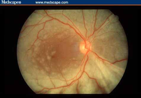
39 APMPPE Acute Posterior Multifocal Placoid Pigment Epitheliopathy Bilateral, confluent plaques deep to the retina. Early blockage, late staining of the entire lesion. In the OKAPs patients may get a cold or flu! Good prognosis, no treatment needed!

40 Acute Posterior Multifocal Placoid Pigment Epitheliopathy Can sometimes look like Serpiginous but...


41 Acute posterior multifocal placoid pigment epitheliopathy Early phase hypofluorescence Late phase hyperfluorescence of the entire lesion! Note: These are two different eyes.


42 APMPPE Placoid Lesions Early Hypo-fluorescence Late Hyper-fluorescence Late hyper-fluorescence of the entire lesion!
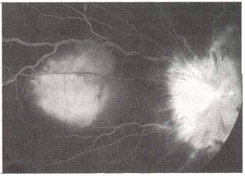
43 Serpiginous Choroiditis Bilateral Serpentine/Ameboid choroidal lesions that most of the time emerge from the optic nerve. Follows a relapsing and remitting course. If the fovea becomes involve the prognosis is poor. No kidding! Early blockage, late staining at the border of the lesion! Tx: Cyclophosphamide x 1yr with oral prednisone for acute suppression of the lesions.

44 Serpiginous Choroiditis


45 Serpiginous Choroiditis Early Blockage Late staining at the border of the lesion.
46 Now 2 very similar panuveitis syndromes which differ on the systemic manifestations!
47 Vogt Koyanagi Harada Bilateral Granulomatous Panuveitis with serous RD Latinos, asians, arabs and american indians. IVFA: Stars in the night appaerance Alopecia, Poliosis, Vitiligo, Meningismus, Tinitus, Sensorineural Hearing loss Tx: Systemic prednisone.

48 VKH

49 VKH Note: Multifocal pinpoint leakage

50 VKH Note: Poliosis
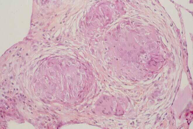
51 Sympathetic Ophthalmia Bilateral Panuveitis with Dalen-Fuchs Nodules, in a patient with history of eye surgery or trauma to one eye. Pathology: Choriocapillaris is spared Ophthalmic findings virtually indistinguishable from VKH May have serous RDs just like VKH Fundus findings indistinguishable from VKH in the late phases Follows a chronic course, needs prednisone and immunosuppression.


52 Sympathetic Ophthalmia Dalen-Fuchs Nodules
53 Finally a retinochoroiditis...
54 Birdshot Retinochoroidopathy Bilateral Retinochoroidopathy: A choroiditis with more vitritis and uveitis than it should really have (may have some cells & flare)! Initially may loose VA due to CME. Subsequently, (after many years) they suffer diffuse outer retinal dysfunction with centripetal (peripheral to posterior) loss of visual field. Tx: Oral Prednisone & Steroid Sparing if needed. F/U: Annual HVF, GVF & ERG
55 Birdshot Retinochoroidopathy Bilateral Retinochoroidopathy: A chronic posterior uveitis characterized by vitritis and multiple ovoid, orange to cream colored, hypopigmented spots occurring in the posterior pole and mid-periphery of the retina. CME is a mayor cause of VA loss: ~20% prevalence upon presentation ~50% cumulative incidence at 5 years. Subsequently, (after many years) they suffer diffuse outer retinal dysfunction with centripetal (peripheral to posterior) loss of visual field.

56
57 Birdshot Retinochoroidopathy HLA-A29 gene has been reported to have a relative risk of for this disease!
58 Birdshot Retinochoroidopathy Treatment: Immunosupressive therapy reduces the risk of CME, which is an important cause of visual acuity loss. Low dose oral corticosteroids (10mg or less), does not seem to prevent the occurrence of CME. Immunosupressive therapy may reduce the risk progressive diffuse retinal dysfuntion. JE Thorne, DA Jabs, et al. AJO 2005;140:45-51.
59 Now, two focal retinitis that differ on the clinical scenario...
60 Toxoplasma Retinitis Unilateral Focal localized retinitis with lots of vitritis, light at the end of the fog, in an ambulatory patient. Granulomatous A/C findings. Tx: Pyrimethamine/Sulfadiazine Clindamycin Zithromax Bactrim (1-2) DS qid. Please do not forget Pred Forte and Atropine (nothing personal)!

61 Toxoplasma Light at the end of the fog...

62 Focal Retinitis Toxoplasma until proven otherwise!
63 Candida Retinochoroiditis In the OKAPS: Focal Retinochoroiditis. Unilateral Focal localized retinitis with some vitritis, in a bedridden patient on TPN. In real life: Bilateral Large Multifocal Choroiditis & Chorioretinitis.

64 Candida Chorioretinitis In the OKAPS it will likely have more fluffy stuff in the vitreous!
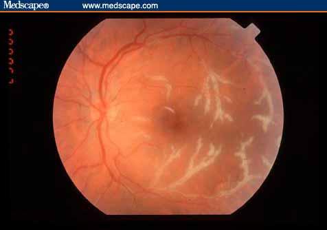
65 Sarcoidosis Systemic Disease Lungs, Skin, CNS, Uveitis, Optic Nerve, Lacrimal Gland, etc... Non-Caseating Granulomata In the OKAPs: Candle Wax Drippings! Severe Venous Sheathing! In real life: Granulomatous uveitis, Panuveitis, Multifocal Choroiditis & Intermediate Uveitis with snowballs but without Snowbanking. In theory can mimic many forms of uveitis Most common eye manifestation: Keratoconjunctivitis sicca!
66 Sarcoidosis Chest X-Ray 90% sensitive! Other tests, neither sensitive nor specific, but otherwise good tests! ACE & Lisozyme!
67


68 Sarcoidosis Atypical Optic Neuropathy CXR Diagnose with... Without the (typical) symptoms of Optic Neuritis Rx Prednisone

69 Sarcoidosis Candle Wax Drippings
70 Syphilis A treponema that can mimic any form of: Scleritis Uveitis: Retinitis Choroiditis: Think, all of the white dot Syndromes! Therefore all of the above must always get an FTA-Abs (positive for life)! Nothing Personal!
71 Syphilis Most Uveitis: Secondary Stage Most Retinitis: Latent Secondary Stage Treatment needed: IV Penicillin: As in tertiary stage!
72 Syphilis Can also Cause: Interstitial Keratitis (congenital syphilis) Argyll-Robertson Pupil (tertiary syphilis) Accommodates but does not react! Optic Neuropathy Mostly Atypical CN III & CN VI palsies Visual Field Defects Gummae in the brain.

73 Syphilis Your neighbor can have it!

74 Syphilis Retinitis: Pseudo Retinitis Pigmentosa common in BUMC.

75 Syphilis Iris Roseolae: Rare in real life!
76 Lyme Disease Close relative of Syphilis, a spirochette! Borrelia Burdoferi Your neighbor can also have it! Can cause: Interstitial Keratitis, just like Syphilis! Non-Congenital, this is different! Granulomatous Uveitis: Just like Syphilis! Intermediate Uveitis: Guess what?, just like syphilis!

77 Lyme Disease Erythema Chronicum Migrans Your neighbor!
78 Tuberculosis Rare cause of Uveitis in the USA Overstated historically as a cause of uveitis! Likely ocular manifestations: A Multifocal Choroiditis or Granulomatous Uveitis on someone with Active Pulmonary Involvement. Focal Retinal Tuberculoma (possible & rare). Scleritis: Rare cause of scleritis, does not need lung involvement because it occurs due to direct contact trough the eye surface, can be caused by atypical mycobacteria.
79 Choroidal Tuberculoma
80 A few more things...
81 Acute Retinal Necrosis HSV/VZV associated focal, rapidly spreading retinitis that usually starts in the retinal periphery and may turn bilateral. Tx: Acyclovir, Valacyclovir, Foscarnet. May become complicated with RRD OKAP Tx: PPV & Silicone Oil.

82 Acute Retinal Necrosis Occluded Arteriole Active border of retinitis Non-Granular, unlike CNV This is an emergency, needs treatment STAT!
83 Toxocara Canis Answer to the OKAP question where you see a granuloma possibly in the periphery connected to the optic disk by a vitreous traction band. Etiology: Dog roundworm ingested by children who eat dirt (pica). Average age 7.5 years. Typically no eosinophilia, unlike Visceral Larva Migrans, which is the systemic version of this. Tx: Ocular disease: Prednisone if active.

84 Toxocara canis Note the connection to the optic nerve!
85 Briefly HIV eye disease...
86 CMV Retinitis Common with CD4 < 50. Rare CD4 > 100 Treatment: Zone 1: Ganciclovir Implant Zone 2 & 3: Valgancyclovir Three principal patterns: Fulminant/Hemorrhagic/Edematous/Posterior Indolent/Granular/Peripheral Severe Vascular Sheeting (Frosted Angitis) Spreads slowly (1/2 DD in 3 weeks).
87

88
89
90 Background HIV Retinopathy Common on patients with low CD4 counts (<50cells/mcL). If the diagnosis of CMVR is being considered, the patient should be observed in 2-3 weeks as CMVR may be expected to slowly progress in the absence of treatment.
91
92 Progressive Outer Retinal Necrosis (PORN) AKA: Herpetic Necrotizing Retinitis Etiology: HSV & HZV The ARN of the immunosuppressed. It represents a clinical emergency!
93 Progressive Outer Retinal Necrosis (PORN) Treatment Alternatives: Foscarnet I.V. Valtrex 1gm po tid. Intravitreal Foscarnet. This is a very rapidly progressing retinitis! 67% of eyes are completely blind in 1 month!
94
95 Infectious Choroiditis of Systemic Origin They can all look the same! Multifocal Choroiditis! As a general rule whatever the patient has at the moment of the active choroiditis phase is the diagnosis! I.e. Candida, Criptococcus, Tb, PCP, etc No need to order CH50, Rheumatoid Factor, etc You get the idea!
96 Anecdote It was my first weekend of call as the Uveitis Fellow! The second year resident calls me! Hey doc, there is a patient with CMVR! Guess what?

97 Fundus appearance in the right eye (A) and left eye (B) after 2 days of therapy showing multiple choroidal lesions approximately 0.5 to 1.5 disc diameters in size as well as intraretinal hemorrhage within the arcades of both eyes Fine, H. F. et al. Arch Ophthalmol 2004;122: Copyright restrictions may apply.

98 Cutaneous cryptococcal rash consisting of multiple papules, some with an umbilicated appearance, which can masquerade as molluscum contagiosum Fine, H. F. et al. Arch Ophthalmol 2004;122: Copyright restrictions may apply.
 Fine, H. F. et al. Arch Ophthalmol 2004;122:1726-1727.")
99 Dermatopathologic biopsy specimen from a facial lesion demonstrating dense Cryptococcus neoformans with calcofluor white stain (original magnification x 340) Fine, H. F. et al. Arch Ophthalmol 2004;122: Copyright restrictions may apply.
![Fundus appearance (right eye [A], left eye [B]) following treatment with antifungal therapy for 3 weeks, showing partial](/docs-images/77/75975474/images/100-0.jpg "resolution of choroidal lesions and hemorrhage Fine, H. F. et al. Arch Ophthalmol 2004;122:1726-1727.")
100 Fundus appearance (right eye [A], left eye [B]) following treatment with antifungal therapy for 3 weeks, showing partial resolution of choroidal lesions and hemorrhage Fine, H. F. et al. Arch Ophthalmol 2004;122: Copyright restrictions may apply.
101 Scleritis
102 Scleritis 50 % Identified Etiology 10 % Infectious Zoster (most common) HSV, Lyme, Syphilis 40% Systemic Disease Rheumatoid Arthritis (most common) Vasculitis: Wegener s & PAN IBD, Lupus & Relapsing Polychondritis


103 Anterior Scleritis Two Sub-types: Nodular Diffuse
104 Necrotizing Scleritis In a red eye, AKA (necrotizing with inflammation): Poor life prognosis: A Systemic Vasculitis is likely. Sleromalacia Perforans: White eye necrotizing scleritis Rheumatoid Arthritis Associated


105 Necrotizing Scleritis Scleral Necrosis Scleromalacia Perforans Associated with poor life prognosis in the older literature. Common in patients with RA

106 Posterior Scleritis Patients typically present with proptosis, retrobulbar pain, gaze restriction and a visual field loss (from serous RD).

107 Posterior Scleritis T-Sign
108 Scleritis Treatment: Non Complicated Scleritis NSAIDs (Indomethacin of Flurbiprophen), 33% response. Systemic Steroids Steroid Sparing Agents: CellCept, MTX, CSA. Eye Saving and Life Saving Agents: Cyclophosphamide & Chlorambucil Never: Topical Steroids, Periocular/Sub-Tenon s Steroids! Posterior Scleritis: Always start with prednisone!
109 The last one. I promise!...
110 Neuroretinitis Practical Definition: Disk Swelling with Full Macular Scar First: Check BP if very high you have a diagnosis. Then, think infectious: Borrelia (Lyme), Bartonella (Cat Scratch) Syphilis (FTA-Abs), etc Then, call Dr. Serrano, STAT!!!!!
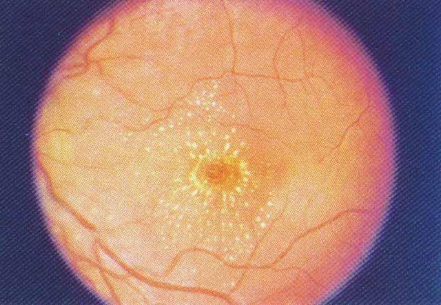
111 Neuroretinitis Macular Scar
Diagnosis of uveitis, how to proceed?
 EOS meeting Cairo, 2018 Diagnosis of uveitis, how to proceed? Mohamed G.A Saleh Lecturer of Ophthalmology Assiut University Size of the problem 15/100000 in US every year. 10% of blindness Prevalence varies
Differential diagnosis of posterior uveitis
 Differential diagnosis of posterior uveitis Diagnostic approach 45-year old male. Floaters and decreased vision since 1 week Fever, lymphadenopathy, myalgias, night sweats, two months ago Oral ulcer sporadically
Head prof. MUDr. E. Vlková, CSc.
 MUDr. Karkanová Michala, Oční klinika LF MU a FN Brno Head prof. MUDr. E. Vlková, CSc. 3 parts: iris (iris) ciliary body (corpus ciliare) choroid (choroidea) Function: regulating the entry of light into
Glaucoma & Inflammation. Jorge L. Fernandez Bahamonde, MD.
 Glaucoma & Inflammation. Jorge L. Fernandez Bahamonde, MD. Definition. Inflammatory ocular conditions compromise outflow of aqueous humor. Keratitis Episcleritis. Scleritis. Uveitis Glaucoma & Keratitis.
Moncef Khairallah, MD
 Moncef Khairallah, MD Department of Ophthalmology, Fattouma Bourguiba University Hospital Faculty of Medicine, University of Monastir Monastir, Tunisia INTRODUCTION IU: anatomic form of uveitis involving
Approach to Pediatric Uveitis. Paris Tranos PhD,ICO,FRCS OPHTHALMICA Vitreoretinal & Uveitis Service
 Approach to Pediatric Uveitis Paris Tranos PhD,ICO,FRCS OPHTHALMICA Vitreoretinal & Uveitis Service Epidemiology Uveitis is the 3 rd leading cause of blindness in USA 5-10% of uveitis cases involve children
Management of uveitis
 Management of uveitis DR. ANUPAMA KARANTH Anti-inflammatory agents -itis = inflammation Treatment : stop inflammation Use anti-inflammatory drugs Most potent of such agents : Corticosteroids Corticosteroids
Rare Presentation of Ocular Toxoplasmosis
 Case Report Rare Presentation of Ocular Toxoplasmosis Rakhshandeh Alipanahi MD From Department of Ophthalmology, Nikookari Eye Hospital, Tabriz University of Medical Sciences, Tabriz, Iran. Correspondence:
UVEITIS. Dr. Yılmaz ÖZYAZGAN
 UVEITIS Dr. Yılmaz ÖZYAZGAN UVEITIS DEFINITION BY STRICT DEFINITION, UVEITIS IS AN INFLAMMATION OF UVEAL TRACT. BUT IN PRACTICAL, IT IS GENERALLY NOT RESTRICTED TO THE UVEA AND INVOLVES OTHER ADJACENT
Acute Retinal Necrosis Secondary to Varicella Zoster Virus in an Immunosuppressed Post-Kidney Transplant Patient
 CM&R Rapid Release. Published online ahead of print September 20, 2012 as Aperture Acute Retinal Necrosis Secondary to Varicella Zoster Virus in an Immunosuppressed Post-Kidney Transplant Patient Elizabeth
Cases CFEH. CFEH Facebook Case #4
 CFEH Cases CFEH Facebook Case #4 A 42 year old female has noticed a floater in her left eye for many years but no flashes. She also reports hazy vision in this eye that has been present all her life. She
Misdiagnosed Vogt-Koyanagi-Harada (VKH) disease and atypical central serous chorioretinopathy (CSC)
 disease and atypical central serous chorioretinopathy (CSC)") HPTER 12 Misdiagnosed Vogt-Koyanagi-Harada (VKH) disease and atypical central serous chorioretinopathy (S) linical Features VKH disease is a bilateral granulomatous panuveitis often associated with exudative
HPTER 12 Misdiagnosed Vogt-Koyanagi-Harada (VKH) disease and atypical central serous chorioretinopathy (S) linical Features VKH disease is a bilateral granulomatous panuveitis often associated with exudative
White-Spot Syndromes of the Retina Lee Jampol, M.D. Chicago, IL
 Objectives At the conclusion of the program, the attendees will be able to: 1. recognize the various white-spot syndromes of the retina 2. initiate appropriate diagnostic tests of patients with the white-spot
Choroidal Neovascularization in Sympathetic Ophthalmia
 Choroidal Neovascularization in Sympathetic Ophthalmia Lucia Sobrin, Miguel Cordero Coma, C. Stephen Foster Case Report A 49-year-old man presented after a ruptured globe repair of his left eye status
Retinal Manifestations of Systemic Disease Part 1
 The Retina and Systemic diseases Retinal Manifestations of Systemic Disease Part 1 Sundeep Dev, MD VRSF Retinal Update 2019 VitreoRetinal Surgery, PA 1 Retinitis/Vasculitis Vitreous cells Serous detachments
REFRESHER: ANTERIOR UVEITIS
 REFRESHER: ANTERIOR UVEITIS 2. SAoO Kongress 28.2.2018 Messe Luzern Dr. med. Christian Böni Augenklinik Universitätsspital Zürich Christian Böni Seite 1 Anterior Uveitis: Clinical Issues Diagnostics: yes
WHAT IS YOUR DIAGNOSIS? By ADREA R. BENKOFF M.D.
 WHAT IS YOUR DIAGNOSIS? By ADREA R. BENKOFF M.D. Anterior Chamber Inflammation and Iris Depigmentation Noted 25 Years After Cataract Extraction Decreasing Vision Over a 5- Year Period 64 year old white
Nausheen Khuddus, MD Melissa Elder, MD, PhD
 Nausheen Khuddus, MD Melissa Elder, MD, PhD Nausheen Khuddus, MD Pediatric Ophthalmologist and Strabismus Specialist Accent Physicians Gainesville, Florida What Is Uveitis? Uveitis is caused by inflammatory
2/16/17. 3 main underlying causes are:
 Definition Etiology Signs/Symptoms Nathan Lighthizer, O.D., F.A.A.O. Assistant Professor Chief of Specialty Care Clinics Chief of Electrodiagnostics Clinic Northeastern State University Oklahoma College
Anthony DeWilde, O.D Linwood Blvd. Kansas City, MO x
 Anthony DeWilde, O.D. 4801 Linwood Blvd. Kansas City, MO 64128 816-861-4700 x 57411 anthony.dewilde@va.gov Uveitis and Glaucoma: The Seven Reasons Why IOP Can Increase in Uveitis (and What to do About
3 main underlying causes are:
 Nathan Lighthizer, O.D., F.A.A.O. Assistant Professor Chief of Specialty Care Clinics Chief of Electrodiagnostics Clinic Northeastern State University Oklahoma College of Optometry Tahlequah, OK lighthiz@nsuok.edu
Uveitis. What is Uveitis?
 Uveitis What is Uveitis? Uveitis [u-vee-i-tis] is a term for inflammation of the eye. It can occur in one eye or both eyes and affects the layer of the eye called the uvea [u-vee-uh]. It also can be associated
!! Definition. !! Etiology. !! Signs/Symptoms. !! Classification/Diagnosis. !! Systemic Associations. !! Lab Testing. !! Treatment. !!
 Nathan Lighthizer, O.D., F.A.A.O. Assistant Professor Chief of Specialty Care Clinics Chief of Electrodiagnostics Clinic Northeastern State University Oklahoma College of Optometry Tahlequah, OK lighthiz@nsuok.edu!!
White Dot Syndromes Noninfectious Chorioretinopathies Update 2019
 White Dot Syndromes Noninfectious Chorioretinopathies Update 2019 Kelly T. Mitchell, MD Retina Service TTUHSC Definition Noninfectious disease Inflammation of choroid, choriocapillaris, RPE, and Retina
A Tailored Approach to Uveitis and Associated Systemic Conditions Anthony DeWilde O.D.
 A Tailored Approach to Uveitis and Associated Systemic Conditions Anthony DeWilde O.D. I. Introduction II. III. IV. A. Why I am giving this talk B. What to take from lecture Diagnosis 1. Better understanding
What do you need to know about posterior uveitis
 What do you need to know about posterior uveitis Dr. Anthony Hall MD FRANZCO Director of Ophthalmology Alfred Hospital, Melbourne, Australia Alfred Hospital Disclosures Off label treatments Paid advisory
Ophthalmology. Juliette Stenz, MD
 Ophthalmology Juliette Stenz, MD Required Slide Disclosures NO SIGNIFICANT FINANCIAL, GENERAL, OR OBLIGATION INTERESTS TO REPORT Required Slide At the end of this session, students will be able to: 1.
Bilateral acute retinal necrosis in a patient with multiple sclerosis on natalizumab
 Bilateral acute retinal necrosis in a patient with multiple sclerosis on natalizumab Arjun B. Sood, Emory University Gokul Kumar, Emory University Joshua Robinson, Emory University Journal Title: Journal
Retina Grand Rounds. Stephen Huddleston MD Charles Retina Institute University of Tennessee Hamilton Eye Institute
 Retina Grand Rounds Stephen Huddleston MD Charles Retina Institute University of Tennessee Hamilton Eye Institute Fundus Autoflourescence 2013 2016 Plaquenil Toxicity Excellent treatment for a variety
OCCASIONAL COMMUNICATIONS
 UVEITIS: DIAGNOSIS AND MANAGEMENT T. Akerele and S. Lightman, Department of Clinical Ophthalmology, Institute of Ophthalmology, London and Moorfields Eye Hospital, London Uveitis is a generic term which
10/18/2018. Unraveling Uveitis
 Unraveling Uveitis Trenton Cleghern, OD, FAAO VisionAmerica UAB School of Optometry 11/10/2018 1 Disclosure Statement: Nothing to disclose 2 Objectives Classify uveitis Current and new therapeutic options
DIAGNOSIS AND MANAGEMENT OF ANTERIOR UVEITIS POA SPRING CONFERENCE BERNARD H. BLAUSTEIN, O.D., F.A.A.O.
 DIAGNOSIS AND MANAGEMENT OF ANTERIOR UVEITIS POA SPRING CONFERENCE - 2009 Classification of Anterior Uveitis BERNARD H. BLAUSTEIN, O.D., F.A.A.O. A. Anatomic location 1. Iritis Cells and flare mostly in
Scleritis LEN V KOH OD
 Scleritis LEN V KOH OD 2014 PUCO 1 Introduction A painful, destructive, and potentially blinding disorder Highly symptomatic High association with systemic disease Immunosuppresssive agents 2014 PUCO 2
o White dot syndromes pattern recognition o Activity and damage o Quality of life o Key points o Idiopathic o Sarcoidosis o Multiple sclerosis
 Introduction Clinical Assessment of Posterior Uveitis Philip I. Murray Centre for Translational Inflammation Research University of Birmingham Birmingham and Midland Eye Centre o Classification of uveitis
Slide 4. Slide 5. Slide 6
 Slide 1 Slide 4 Demographics El Paso Eye Care Border Healthcare-Based Grand Rounds Derek N. Cunningham, O.D. 80-90% Mexican-Americans Diabetes Hypertension Hyperlipidemia Obesity 70% uninsured High poverty
Various presentations of herpes simplex retinochoroiditis A case series
 Various presentations of herpes simplex retinochoroidits 47 Various presentations of herpes simplex retinochoroiditis A case series M. T. K. Perera 1, T. S. Keragala 1, M. Gamage 2 The Journal of the College
Imaging in uveitis. Anthony Hall
 Imaging in uveitis Anthony Hall Causes of Vision Loss in Uveitis 1. Cystoid macular oedema 26% 2. Cataract 19% 3. Glaucoma 11% 4. Permanent macular damage 5% Rothova et al BJO 1996; 80: 332-336 Macular
ISPUB.COM. Photopsia post flu: A case of MEWDS. S Baisakhiya, S Dulani, S Lele INTRODUCTION CASE HISTORY
 ISPUB.COM The Internet Journal of Ophthalmology and Visual Science Volume 8 Number 1 Photopsia post flu: A case of MEWDS S Baisakhiya, S Dulani, S Lele Citation S Baisakhiya, S Dulani, S Lele. Photopsia
Ocular Pathology. I. Congenital and/or developmental. A. Trisomy 21. Hypertelorism (widely spaced eyes) Keratoconus (cone shaped cornea)
 Keratoconus (cone shaped cornea)") I. Congenital and/or developmental Robbins Pathologic Basis of Disease, 6 th Ed. A. Trisomy 21 Hypertelorism (widely spaced eyes) Keratoconus (cone shaped cornea) Focal hypoplasia of iris Cataracts frequently
I. Congenital and/or developmental Robbins Pathologic Basis of Disease, 6 th Ed. A. Trisomy 21 Hypertelorism (widely spaced eyes) Keratoconus (cone shaped cornea) Focal hypoplasia of iris Cataracts frequently
Retina Conference. Janelle Fassbender, MD, PhD University of Louisville Department of Ophthalmology and Visual Sciences 09/04/2014
 Retina Conference Janelle Fassbender, MD, PhD University of Louisville Department of Ophthalmology and Visual Sciences 09/04/2014 Subjective CC/HPI: 64 year old Caucasian female referred by outside ophthalmologist
ISPUB.COM. An Atypical Presentation of Posterior Scleritis. A Ramanathan, A Gaur CASE REPORT
 ISPUB.COM The Internet Journal of Ophthalmology and Visual Science Volume 8 Number 2 A Ramanathan, A Gaur Citation A Ramanathan, A Gaur.. The Internet Journal of Ophthalmology and Visual Science. 2009
Pattern of Uveitis in Saudi Female Patients in Western Region of Saudi Arabia
 JKAU: Med. Sci., Vol. 19 No. 3, pp: 73-83 (2012 A.D. / 1433 A.H.) DOI: 10.4197/Med. 19-3.6 Pattern of Uveitis in Saudi Female Patients in Western Region of Saudi Arabia Nizamuddin Shaik Hakim Mohammad
Uveitis. Pt Info Brochure. Q: What is Uvea?
 Pt Info Brochure Uveitis Q: What is Uvea? A: Uvea is the middle layer of the eye. It is the most vascular structure of the eye. It provides nutrition to the other parts of the eye. The uvea is made up
Ophthalmology Technician conference 2018 UVEITIS. Middle pigmented, vascular layer: iris, ciliary body and choroid (the layer between retina & Sclera)
") Ophthalmology Technician conference 2018 UVEITIS Kelly Mitchell, MD Uveitis Definitions: Uvea (Latin = grape) Middle pigmented, vascular layer: iris, ciliary body and choroid (the layer between retina
Ophthalmology Technician conference 2018 UVEITIS Kelly Mitchell, MD Uveitis Definitions: Uvea (Latin = grape) Middle pigmented, vascular layer: iris, ciliary body and choroid (the layer between retina
Necrotizing retinitis of multifactorial etiology
 Romanian Journal of Ophthalmology, Volume 61, Issue 1, January-March 2017. pp:49-53 CASE REPORT Necrotizing retinitis of multifactorial etiology Pirvulescu Ruxandra Angela* **, Popa Cherecheanu Alina*
Uveitis: Classification, Etiologies and Clinical Signs
 E-ISSN 2454-2784 Recent Advances Uveitis: Classification, Etiologies and Clinical Signs Aditya Shreekant Kelkar 1, Ekta Rakesh Arora 1, B.Sowkath 2, Jyotirmay Biswas 3 1 National Institute of Ophthalmology,Pune
Regional vs. Systemic Therapy. Corticosteroids. Regional vs. Systemic Therapy for Uveitis. Considerations
 Regional vs. Systemic Therapy for Uveitis Nisha Acharya,, M.D., M.S. Director, Uveitis Service F.I. Proctor Foundation University of California, San Francisco December 4, 2010 No financial disclosures
Optical coherence tomography findings in a child with posterior scleritis
 European Journal of Ophthalmology / Vol. 18 no. 6, 2008 / pp. 1007-1010 SHORT OMMUNITIONS & SE REPORTS Optical coherence tomography findings in a child with posterior scleritis H. ERDÖL, M. KOL,. TÜRK
Uveitis in patients with Multiple Sclerosis
 Uveitis in patients with Multiple Sclerosis An observational descriptive clinical study submitted in partial fulfillment of Master Degree in Ophthalmology By Dr. Ahmed Mohamed Karara M.B, B Ch. Supervised
The White Re)na. Joseph Alsberge, MD January 20, 2018
na. Joseph Alsberge, MD January 20, 2018") The White Re)na Joseph Alsberge, MD January 20, 2018 58 y/o man with floaters and pain OD x 2 weeks PMH: oral and genital herpes Va OD 20/50 Anterior OD: KP and 3+ AC cell Posterior: Vitri)s, occlusive
The White Re)na Joseph Alsberge, MD January 20, 2018 58 y/o man with floaters and pain OD x 2 weeks PMH: oral and genital herpes Va OD 20/50 Anterior OD: KP and 3+ AC cell Posterior: Vitri)s, occlusive
The Royal College of Ophthalmologists
 Clinical Dataset Uveitis Dataset The Royal College of Ophthalmologists Authorship Group: Alastair K. Denniston, Richard W. Lee, Carlos Pavesio, Miles R Stanford, Philip I Murray, Annabelle Okada, H. Nida
Patterns of Uveitis at a Tertiary Referral Center in Southern Iran
 Original Article Patterns of Uveitis at a Tertiary Referral Center in Southern Iran Mansour Rahimi, MD; Ghazaleh Mirmansouri, MD Poustchi Eye Research Center and Ophthalmology Department, Shiraz University
Surgery in patients with uveitis. Lyndell Lim and Anthony Hall
 Surgery in patients with uveitis Lyndell Lim and Anthony Hall Disclosures Off label treatments Paid advisory board Bayer Paid research support Allergan (makers of Ozurdex) Paid research support B and L
Clinical Practice Guide for the Diagnosis, Treatment and Management of Anterior Eye Conditions. April 2018
 Clinical Practice Guide for the Diagnosis, Treatment and Management of Anterior Eye Conditions This Clinical Practice Guide provides evidence-based information about current best practice in the management
Evolving therapies for posterior uveitis. Infliximab (Remicade) Infliximab: pharmacology. FDA-approved monoclonal antibody therapy Target
 Infliximab: pharmacology. FDA-approved monoclonal antibody therapy Target") Evolving therapies for posterior uveitis Sam Dahr, M.D. September 17, 2005 Midwest Ophthalmology Conference Infliximab (Remicade) FDA approved for Crohn s disease, rheumatoid arthritis, and psoriatic arthritis
Evolving therapies for posterior uveitis Sam Dahr, M.D. September 17, 2005 Midwest Ophthalmology Conference Infliximab (Remicade) FDA approved for Crohn s disease, rheumatoid arthritis, and psoriatic arthritis
COVER FOCUS AT A GLANCE. BY LISA J. FAIA, MD, and KIMBERLY A. DRENSER, MD, PhD
 PEDIATRIC UVEITIS: CHALLENGING FOR OPHTHALMOLOGISTS, PATIENTS, AND PARENTS Management of these complicated diseases differs between pediatric and adult patient populations. BY LISA J. FAIA, MD, and KIMBERLY
Patterns of uveitis in a Philippine eye clinic
 VOL. NO. PHILIPPINE JOURNAL OF Ophthalmology JANUARY ORIGINAL ARTICLE - MARCH 5 Harvey S. Uy, MD Irene W. Tam, OD Asian Eye Institute Makati, Philippines Patterns of uveitis in a Philippine eye clinic
Learning Objectives. Introduction. Reading List. Significance. Introduction 10/16/2008
 Learning Objectives Systemic Diseases with Ocular Manifestations Dr Nathan Kerr 1. To describe the ocular symptoms and signs associated with common systemic diseases 2. To be familiar with the non-ophthalmic
D JO. Bilateral Shallow Anterior Chamber And Transient Myopia As A Presenting Feature Of Vogt Koyanagi Harada Syndrome
 46 Bilateral Shallow Anterior Chamber And Transient Myopia As A Presenting Feature Of Vogt Koyanagi Harada Syndrome Abstract Rahul Kumar Sharma, Abhishek Dagar, Vivek Kumar Vitreo-Retina Department, Venu
CLINICALCASE PROVOST J, SEKFALI R, AMOROSO F, ZAMBROWSKI O, MIERE A
 CLINICALCASE PROVOST J, SEKFALI R, AMOROSO F, ZAMBROWSKI O, MIERE A Department of ophthalmology, Souied E. (MD,PhD) Centre Hospitalier Intercommunal de Créteil Université Paris Est HISTORY 13 years old
Learning Objectives:
 Viral keratitis and antivirals Learning Objectives: Recognise and distinguish different types of viral keratitis HSV HZO Adenovirus Discuss the use of antiviral agents in the treatment of herpetic infections
Re-emerging infections: Syphilis & Tuberculosis
 Re-emerging infections: Syphilis & Tuberculosis Nicholas Jones Manchester Royal Eye Hospital Syphilis and TB - historical plagues? Syphilis incidence over 40yrs Manchester: Manchester: The Syphilis Capital
Convergence in. Introduction. Case Report: Dr. Piyali SenM.B.B.S, Dr. Abhipsha Saha M.B.B.S, Dr. Anuradha Chandra M.S,FAICO
 Convergence in Dr. Piyali SenM.B.B.S, Dr. Abhipsha Saha M.B.B.S, Dr. Anuradha Chandra M.S,FAICO Introduction non-progressive ophthalmoplegia with or without ptosis affecting part or all of the occulomotor
Sarcoidosis and Uveitis
 Sarcoidosis and Uveitis Nicholas Jones Royal Eye Hospital Manchester, UK Sarcoidosis a multisystem chronic inflammation causing multifocal non-caseating granulomas BUT Diagnosis often made indirectly (without
Page 1 RED EYES. conjunctivitis keratitis episcleritis / scleritis. Frank Larkin Moorfields Eye Hospital. acute glaucoma anterior uveitis
 The RED EYE and ALLERGIC EYE DISEASE DIAGNOSIS & MANAGEMENT Frank Larkin Moorfields Eye Hospital RED EYES conjunctivitis keratitis episcleritis / scleritis acute glaucoma anterior uveitis post-op. / trauma
1/5/11. Management of Posterior Segment Uveitis. Posterior uveitis classification. Presentation goes beyond classification
 Posterior uveitis classification Management of Posterior Segment Uveitis Focal or Multifocal Choroiditis or Chorioretinitis Retinitis or Retinochoroiditis Neuroretinitis SUN = Standardization of uveitis
Deep Trouble. Thomas Stone, MD Retina Associates of Kentucky River City Retina Conference May 15, 2014
 Deep Trouble Thomas Stone, MD Retina Associates of Kentucky River City Retina Conference May 15, 2014 History 20 yo WM Decreased vision OU, OD>OS Sudden onset blurred central vision 12 days prior 4 days
Case History. The SEVEN HABITS of Highly Effective Anterior Uveitis Management. SLEx findings: SLEx corneal findings: y.o.
 The SEVEN HABITS of Highly Effective Anterior Uveitis Management Case History! 68 y.o. Caucasian female of photophobia and blurred vision! As well as a headache over right eye for 2 days! Complains Paul
Etiologies 85 causes 50% idiopathic conditions 20% trauma 20% systemic 10% local (H Zoster, Toxoplasmosis, etc)
") 1 Doc I have eyeritis Uveitis front to back Brian E. Mathie. OD, FAAO 2 Demographics Most commonly in patients 20-59 years old 5-10% in pts
1 Doc I have eyeritis Uveitis front to back Brian E. Mathie. OD, FAAO 2 Demographics Most commonly in patients 20-59 years old 5-10% in pts
Dr Jo-Anne Pon. Dr Sean Every. 8:30-9:25 WS #70: Eye Essentials for GPs 9:35-10:30 WS #80: Eye Essentials for GPs (Repeated)
") Dr Sean Every Ophthalmologist Southern Eye Specialists Christchurch Dr Jo-Anne Pon Ophthalmologist Southern Eye Specialists, Christchurch Hospital, Christchurch 8:30-9:25 WS #70: Eye Essentials for GPs
Dr Sean Every Ophthalmologist Southern Eye Specialists Christchurch Dr Jo-Anne Pon Ophthalmologist Southern Eye Specialists, Christchurch Hospital, Christchurch 8:30-9:25 WS #70: Eye Essentials for GPs
Interesting, unusual and eclectic cases from 2017 Robert A. Mittra, MD VitreoRetinal Surgery, P.A. Minneapolis, MN
 Fundus, SG Interesting, unusual and eclectic cases from 2017 Robert A. Mittra, MD VitreoRetinal Surgery, P.A. Minneapolis, MN Which is most likely? A) Age > 65, history of HTN B) Age 40 65, history of
OPTIC NERVE DISORDERS
 OPTIC NERVE DISORDERS OPTIC NEUROPATHIES INFLAMMATORY OPTIC NEUROPATHIES Cat scratch disease. Lyme disease. Viral infections of childhood (measles, mumps, chicken pox) with or without encephalitis Immun-
Doc, I See a Donut in My Vision : An Optometrist s Guide to a Rare Cause of Choroidal Neovascular Membrane
 Doc, I See a Donut in My Vision : An Optometrist s Guide to a Rare Cause of Choroidal Neovascular Membrane Linda Pham, OD, Tobin Ansel, OD, Nancy Shenouda-Awad, OD, FAAO, West Haven VA Medical Center Abstract
HLA-B27-related anterior Uveitis
 HLA-B27-related anterior Uveitis Nicholas Jones Manchester Uveitis Clinic The Royal Eye Hospital Manchester Anterior means anterior only IUSG classification: Anterior uveitis = Iris & pars plicata AU
Condition: Herpes Zoster Ophthalmicus (HZO)
") Condition: Herpes Zoster Ophthalmicus (HZO) Description: Herpes zoster represents a reactivation of the varicella zoster virus (VZV) which leads to characteristic skin lesions and, in many cases, ocular
Condition: Herpes Zoster Ophthalmicus (HZO) Description: Herpes zoster represents a reactivation of the varicella zoster virus (VZV) which leads to characteristic skin lesions and, in many cases, ocular
Management of Immune Reconstitution Inflammatory Syndrome (IRIS)
") Management of Immune Reconstitution Inflammatory Syndrome (IRIS) Adult Clinical Guideline from the New York State Department of Health AIDS Institute www.hivguidelines.org Purpose of the IRIS Guideline
Management of Immune Reconstitution Inflammatory Syndrome (IRIS) Adult Clinical Guideline from the New York State Department of Health AIDS Institute www.hivguidelines.org Purpose of the IRIS Guideline
UNDERSTAND MORE ABOUT UVEITIS UVEITIS
 UNDERSTAND MORE ABOUT UVEITIS UVEITIS Uveitis What is uveitis? Uveitis is inflammation of the uvea, the middle layer of your eye. The eye is shaped much like a tennis ball, with three different layers
Herpetic Eye Disease Jason Duncan, OD, FAAO Diplomate, American Board of Optometry Associate Professor, Southern College of Optometry
 Herpetic Eye Disease Jason Duncan, OD, FAAO Diplomate, American Board of Optometry Associate Professor, Southern College of Optometry I have what?! How to break the news Meet the Herpes Quick virology
Interesting, unusual, eclectic cases from 2017 Robert A. Mittra, MD VitreoRetinal Surgery, P.A. Minneapolis, MN
 56 yo female, EW Presented to outside Ophthalmologist Diagnosed with viral conjunctivitis, but viral testing was negative. Also had pain around the eye and on the right side of her face Interesting, unusual,
UVEITIS IN GENERAL. Information for patients UVEITIS CLINIC WHAT IS UVEITIS? MAIN CATEGORIES OF UVEITIS
 Information for patients UVEITIS CLINIC UVEITIS IN GENERAL WHAT IS UVEITIS? The uvea is a name given to the pigmented layer of tissue inside the eye. When all or part of the uvea becomes inflamed, the
Solitary idiopathic choroiditis
 Optometry (2007) 78, 176-180 Solitary idiopathic choroiditis Kimberly D. Kohne, O.D., a Victor E. Malinovsky, O.D., a and Hua Gao, M.D., Ph.D. b a School of Optometry and b Department of Ophthalmology,
Chapter 2 Indocyanine Green Angiography in Uveitis
 Chapter 2 Indocyanine Green Angiography in Uveitis Shilpa Kodati, Samuel P. Burke, and Thomas A. Albini Introduction Indocyanine green angiography (ICGA) became available in the early 1990s and has since
Neuro-Ocular Grand Rounds
 Neuro-Ocular Grand Rounds Anthony B. Litwak,OD, FAAO VA Medical Center Baltimore, Maryland Dr. Litwak is on the speaker and advisory boards for Alcon and Zeiss Meditek COMMON OPTIC NEUROPATHIES THAT CAN
2009 REIMBURSEMENT GUIDE, VISUCAM and VISUCAM NM/FA
 2009 REIMBURSEMENT GUIDE FF 450 PLUS PRO NM, VISUCAM and VISUCAM NM/FA Zeiss Fundus Cameras INTRODUCTION The following guide provides an overview of billing and reimbursement for procedures performed with
Aging & Ophthalmology
 Aging & Ophthalmology Pr Jean-Marie Rakic Dr Denis Malaise January 2018 Major ocular diseases 1. Cataract 2. Age-related macular degeneration 3. Ischemic optic neuropathy 4. Horton arteritis 5. Glaucoma
Mycobacterial Ocular Inflammation. Akbar Shakoor, M.D. John A. Moran Eye Center, University of Utah
 Mycobacterial Ocular Inflammation Akbar Shakoor, M.D. John A. Moran Eye Center, University of Utah Financial Disclosure I have no financial interests or relationships to disclose. Applied anatomy What
Q: (picture of typical dendrite) What is the differential diagnosis and describe this entity? How would you treat and why?
 What is the differential diagnosis and describe this entity? How would you treat and why?") Q: (picture of typical dendrite) What is the differential diagnosis and describe this entity? How would you treat and why? Etiology/Risks: Critical symptoms: HSV is transmitted by direct contact of epidermis
Q: (picture of typical dendrite) What is the differential diagnosis and describe this entity? How would you treat and why? Etiology/Risks: Critical symptoms: HSV is transmitted by direct contact of epidermis
Rhegmatogenous retinal detachment in uveitis
 De Hoog et al. Journal of Ophthalmic Inflammation and Infection (2017) 7:22 DOI 10.1186/s12348-017-0140-5 Journal of Ophthalmic Inflammation and Infection ORIGINAL ARTICLE Open Access Rhegmatogenous retinal
Sudden Vision Loss. Brendan Girschek, MD, FRCSC, FACS Vitreoretinal Surgery Cedar Valley Medical Specialists
 Sudden Vision Loss Brendan Girschek, MD, FRCSC, FACS Vitreoretinal Surgery Cedar Valley Medical Specialists My Credentials -Residency in Ophthalmology at the LSU Eye Center in New Orleans, LA -Fellowship
11/29/2016 MACULAR MALADIES: TYPICAL & ATYPICAL CASES
 MACULAR MALADIES: TYPICAL & ATYPICAL CASES Dawn Pewitt, OD, FAAO Triad Eye Institute, Grove, OK Dpewitt@triadeye.com Disclosure Statement: No financial disclosures COPE 51218-PS Please silence all mobile
Table of Contents 1 Orbit 3 2 Eyelids 7
 Table of Contents Preface, x List of abbreviations xi Glossary xii Section I Atlas 1 1 Orbit 3 Clinical signs associated with orbital neoplasia 3 Clinical signs associated with orbital cellulitis 3 Enophthalmos
OCCLUSIVE VASCULAR DISORDERS OF THE RETINA
 OCCLUSIVE VASCULAR DISORDERS OF THE RETINA Learning outcomes By the end of this lecture the students would be able to Classify occlusive vascular disorders (OVD) of the retina. Correlate the clinical features
Double trouble: a patient with both HLA-B27 anterior uveitis and HLA-A29 birdshot chorioretinitis
 Haddad and Reddy Journal of Ophthalmic Inflammation and Infection 2014, 4:28 BRIEF REPORT Open Access Double trouble: a patient with both HLA-B27 anterior uveitis and HLA-A29 birdshot chorioretinitis Zeina
Infectious Retina Raman Bhakhri, OD, FAAO Assistant Professor SCCO/MBKU
 Infectious Retina Raman Bhakhri, OD, FAAO Assistant Professor SCCO/MBKU rbhakhri@ketchum.edu Please silence all mobile devices and remove items from chairs so others can sit. Unauthorized recording of
The Diagnosis & Treatment of Uveitis in Optometric Practice. Megan A. Hunter, OD, FAAO Michelle M. Marciniak, OD, FAAO
 The Diagnosis & Treatment of Uveitis in Optometric Practice Megan A. Hunter, OD, FAAO Michelle M. Marciniak, OD, FAAO Definition! Inflammation of the uveal tract! Iris! Ciliary body! Choroid! Uvea derives
JOURNAL OF OPHTHALMOLOGY AND RELATED SCIENCES
 JOURNAL OF OPHTHALMOLOGY AND RELATED SCIENCES BILATERAL ACUTE TRANSILLUMINATION OF THE IRIS Kavitha Avadhani 1, MD, MS, Jay Kalliath 1, MS, FRCS 1 Department of Ophthalmology, NMC Speciality Hospital,
Neuro-Ocular Grand Rounds Anthony B. Litwak,OD, FAAO VA Medical Center Baltimore, Maryland
 Neuro-Ocular Grand Rounds Anthony B. Litwak,OD, FAAO VA Medical Center Baltimore, Maryland Dr. Litwak is on the speaker and advisory boards for Alcon and Zeiss Meditek COMMON OPTIC NEUROPATHIES THAT CAN
Infectious Retina. The Fight Against HIV/AIDS?? HAART. Common Posterior Segment Manifestations 1/8/2019. Raman Bhakhri, OD, FAAO Assistant Professor
 Infectious Retina Raman Bhakhri, OD, FAAO Assistant Professor No Disclosures Disclosure Statement: Nothing to disclose Common Posterior Segment Manifestations o HIV Retinopathy o Cytomegalovirus (CMV)
Course # Flashes and Floaters and Curtains, Oh My!
 Course # 132 Flashes and Floaters and Curtains, Oh My! FLASHES and FLOATERS and CURTAINS, OH MY!!! FLASHES OF LIGHT Vitreous is the villain Retinal traction Retinal hole Retinal tear Migraine Classic migraine
Course # Flashes and Floaters and Curtains, Oh My!
 Course # 132 Flashes and Floaters and Curtains, Oh My! FLASHES and FLOATERS and CURTAINS, OH MY!!! FLASHES OF LIGHT Vitreous is the villain Retinal traction Retinal hole Retinal tear Migraine Classic migraine