Physical therapy of the wrist and hand
|
|
|
- Dennis Casey
- 6 years ago
- Views:
Transcription
1 Physical therapy of the wrist and hand
2 Functional anatomy wrist and hand The wrist includes distal radius, scaphoid, lunate, triquetrum, pisiform, trapezium, trapezoid, capitate, and hamate. The hand includes five metacarpals and 14 phalanges make up the hand and five digits The wrist is allowed for FL, EXT, radial deviation and ulnar deviation


3


4
5 Functional anatomy: the radiocarpal joint The radiocarpal joint: The concave distal end of the radius and radioulnar disc connects the convex proximal row of the scaphoid, lunate, and triquetrum. The triquetrum is mainly articulated with the disc
6
7 Functional anatomy: the radiocarpal joint Physiological motion of wrist Flexion Extension Radial deviation Ulnar deviation Carpal glide of the proximal row Dorsal Volar Ulnar Radial
8 Functional anatomy: the midcarpal joint The proximal row of the scaphoid, lunate, and triquetrum articulates with the distal row the trapezium, trapezoid, capitate, and hamate. The capitate and hamate are convex and glide on the concave surface of the scaphoid, lunate, and triquetrum The concave trapezium and trapezoid glide on the convex distal surface of the scaphoid
9 Functional anatomy: the carpometacarpal (CMC) joint of digit 2 through 5 Between each metacarpal and the distal row of carpals and the articulations between the base of each metacarpal The flexion of the metacarpals and additional adduction of the fifth contribute to the cupping of the hand, improving prehension.
10 Functional anatomy: the carpometacarpal (CMC) joint of the thumb Saddle-shaped joint between the trapezium and base of the first metacarpal. For flexion-extension of the thumb occurring in the frontal plane, the trapezium is convex and the base of the metacarpal is concave. For abduction-adduction occurring in the sagittal plane, the trapezium is concave and the metacarpal is convex
11 Functional anatomy: the carpometacarpal (CMC) joint of the thumb The first metacarpal of the thumb Flexion Extension Abduction Adduction Direction of glide of base of metacarpal Ulnar Radial Dorsal Volar
12 Functional anatomy: the metacarpophalangeal (MCP) joint of the thumb Convex distal end of each metacarpal and concave proximal phalanx Supported by a volar and two collateral ligaments
13 Functional anatomy: the interphalangeal (IP) joint PIP and DIP for digit 2 to 5; the thumb has only one IP The articulating surface at the distal end of each phalanx is convex, the articulating surface at the proximal end of each phalanx is concave
14 Hand function: length-tension relationships As the fingers or thumb flex, the wrist must be stabilized by the wrist extensors from simultaneously flexing the wrist. For strong fingers or thumb extension, the wrist flexors stabilize or flex the wrist so the extensors can function more efficiently.
15 Hand function: cupping and flattening Cupping of the hand occurs with finger flexion, and flattening of the hand occurs with extension.
16 Hand function: extensor mechanism Isolated contraction of the extensor digitorum produces clawing of he fingers (MCP hyperextension with IP flexion from passive pull of the extrinsic flexor tendons) PIP and DIP extensions occur concurrently and can be caused by the interossei or lumbrical muscles There must be tension in the extensor digitorum communis tendon for there to the interphalangeal extension
17 Hand function: grips and prehension patterns Grips invlove clamping an object with partially flexed fingers against the palm of the hand, with counterpressure from the adducted thumb. Varieties include cylindrical grip, spherical grip, hook grip, and lateral prehension. Precisions involve manipulating an object that is not in contact with the palm of the hand between the opposing abducted thumb and fingers varieties include pad-to-pad, tip-to-tip, and pad-to-side prehensions
18 Hand function: grips and prehension patterns Combined grips involve digit 1 and 2 performing precision activities, whereas digit 3-5 supplement with power
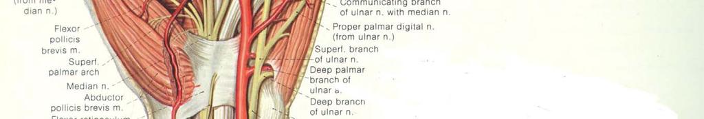
19 Median nerve subject to pressure and trauma around the wrist and hand Median nerve passes through the carpal tunnel at the wrist with the flexor tendons. Nerve entrapment in the tunnel may occur. Sensory changes: over the radial 2/3 of the palm, the aplmar surfaces of the first three and ½ digits, and the dorsum of the distal phalanges. Weakness distal to the wrist: opponens pollicis, abductor pollicis brevis, superficial head of the flexor pollicis brevis, and lumbricals I and II Ape-hand deformity

20
21 Ulnar nerve subject to pressure and trauma around the wrist and hand Ulnar nerve enters the hand through a tunnel formed by the pisiform and hook of hamate and is covered by the volar carpal ligament and palmaris brevis muscle Sensroy chnge: ulnar 1/3 of the hand, 5 th digit and ulnar side of the 4 th digit Weakness to muscle distal to the site: palmaris brevis, muscles of the hypothenar eminence, lumbricals III and IV, interossei, adductor pollicis, and deep head of the flexor pollicis brevis Claw-hand deformity
22 Radial nerve subject to pressure and trauma around the wrist and hand Radial nerve enters the hand on the dorsal surface as the superficial radial nerve, only sensory. Sensory change: over the radial 2/3 of the dorsum of the hand and thumb and the proximal phalanx of the 2 nd, 3th, the half of the 4 th digit Muscles innervated by radial nerve are proximal to the wrist

23
24 Common wrist and hand deformities: Colles fracture Disturbance of the inferior radio-ulnar joint Residual deformities: radial deviation of the hand and prominence of the ulna Loss of movement of supination and wrist extension Two complications after Colles fracture: Delayed rupture of extensor pollicis longus may occur some months after injury and is due to ischaemia or attrition of the tendon
25 Common wrist and hand deformities: Colles fracture Sudeck s atrophy: marked swelling of the wrist, hand and fingers, gross stiffness of the fingers, and carpal decalcification
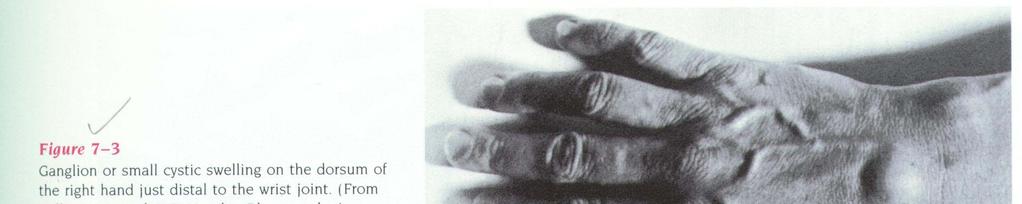
26 Common wrist and hand deformities: ganglions In the carpal joint or tendon sheath or in the fingers Size fluctuated local swelling and tenderness may only obvious when the wrist is flexed.
27
28 Common wrist and hand deformities: De Quervain s disease Tenosynovitis involving abductor pollicis longus and extensor pollicis brevis P t may complain of pain on certain movement of the wrist, and weakness of grip Splitting the lateral wall of the sheath is the choice of treatment


29
30 Common wrist and hand deformities: carpal tunnel syndrome Compression of the median nerve leads to symptoms related to its distribution Premenstrual fluid retention, early RA with synovial tendon sheath thickening, and old colles or carpal fractures may be responsible by restricting the space P t complains paresthesia in the hand except little finger; symptoms may become obvious in midnight; shaking the hand to release the symptoms is usually needed P t may have both signs of the cervical spondylosis and carpal tunnel syndrome
31
32 Common wrist and hand deformities: ulnar tunnel syndrome Ulnar nerve is compressed as passing through the ulnar canal between the pisiform and the hook of the hamate Symptoms include small muscle wasting and weakness in the hand with sensory disturbance on the volar aspect of the little finger Causes of nerve involvement are ganglion, occupational trauma, old carpal or metacarpal fractures
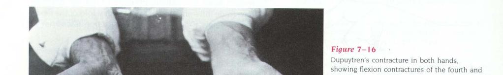
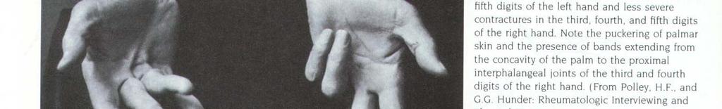
33 Common wrist and hand deformities: Dupuytren s contracture Nodular thickening and contracture of the palmar fascia The palm of the hand is affected first, and later the 4 th finger, followed by the little and middle fingers The progressive flexion of the affected fingers interferes with the function of the hand affect men more than women over 40 May be hereditary tendency, or associated with epilepsy, diabetes or alcoholic cirrhosis
34
35 Common wrist and hand deformities: tendon and tendon sheath lesions Mallet finger: DIP is held in a permanent position of flexion; unable to extend the distal joint The extensor tendon either ruptures close to insertion in the distal phalanx, or it avulses its bony attachment Healing may occur over 6 to 12 month period Use of light splint holding the DIP in hyperextension for 6 weeks is practical
36
37 Common wrist and hand deformities: tendon and tendon sheath lesions Mallet thumb: Delayed rupture of the extensor pollicis longus tendon may follow Colles fracture or rheumatic arthritis, and repair by tendon transfer is advised
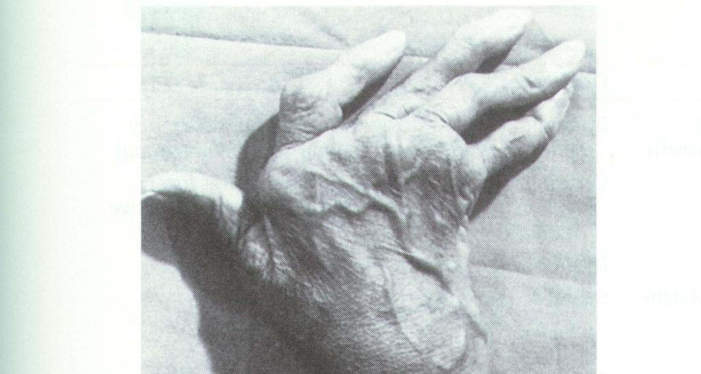
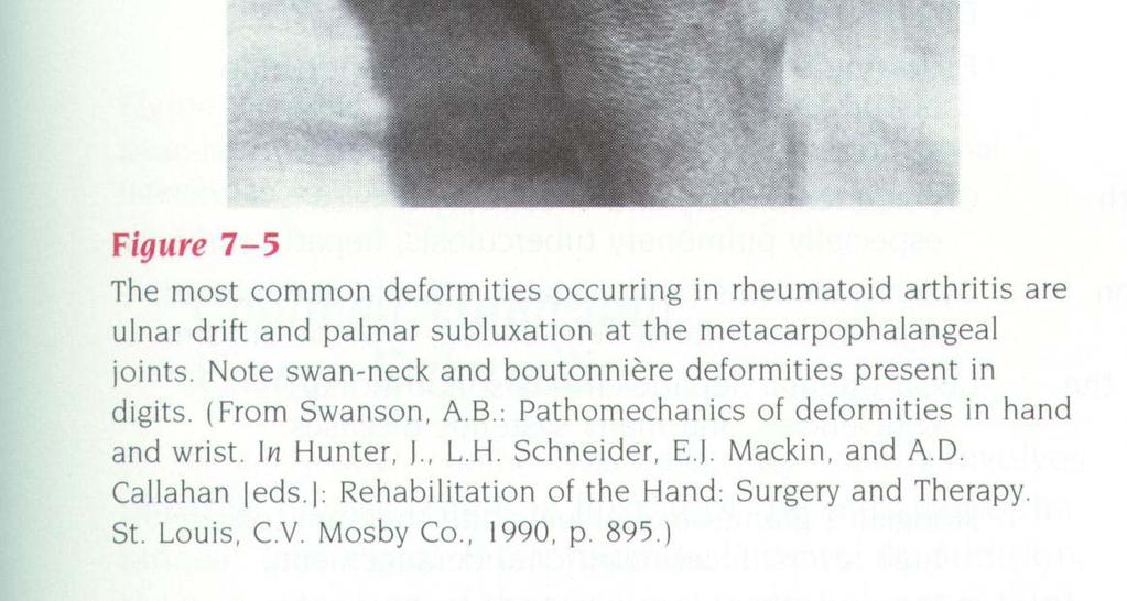
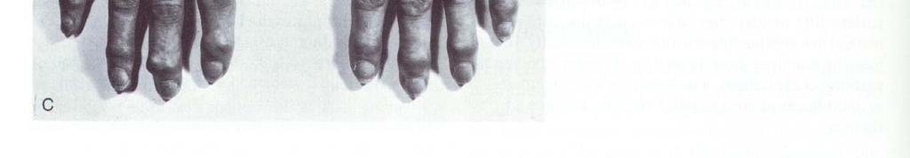
38 Common wrist and hand deformities: tendon and tendon sheath lesions Swan-neck deformity: Flexion of the metacarpophalangeal and distal interphalangeal joints in addition to extension of the proximal interphalangel joint A result of contracture of the intrinsic muscles and often seen in rheumatoid arthritis
39
40 Common wrist and hand deformities: tendon and tendon sheath lesions Boutonniere deformity: Flexion of the interphalangeal joint of a finger with extension of the distal interphalangeal joint Due to rupture of the central slip of the extensor tendon attached to the base of the middle phalanx Due to incised wounds on the dorsum of the finger and avulsion injuries; Commonly seen in rheumatoid arthritis Surgical repair of the extensor band is often undertaken
41
42 Common wrist and hand deformities: trigger finger and thumb Thickening of a fibrous tendon sheath or nodular thickening in a flexor tendon When the fingers are extended, the affected finger lag behind and then quite suddenly straightens. The middle, ring finger or thumb are most frequently involved. Nodular thickening at the level of the MP joint may also be palpable.
43 Common wrist and hand deformities: Z deformity of the thumb The thumb is flexed at the metacarpophalangeal joint and hyperextended at the interphalangeal joint May related to heredity, or rheumatic arthritis.
44
45 Common wrist and hand deformities: rheumatic arthritis In the early stage the hands are warm and moist Later the joints become obviously swollen and tender. Synovial tendon sheath and joint thickening with effusion, muscle wasting, and deformity then become apparent Tendon rupture and joint subluxation are the main factors leading to the more severe deformities
46
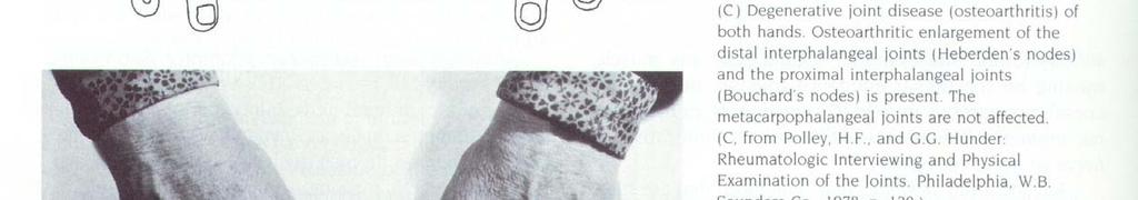
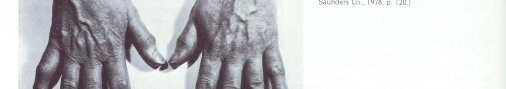
47 Common wrist and hand deformities: osteoarthritis of the interphalangeal joint Heberden s nodes: nodular swellings situated dorsally over the bases of the distal phalanges Bouchard s nodes: nodular over the bases of the middle phalanges; less common Occur most in women after the menopause; not related to osteoarthritis elsewhere Most are symptom-free, may be related to joint damage and consequent pain
48
49 Common wrist and hand deformities: osteoarthritis of the carpo-metacarpal joint of the thumb common between the thumb metacarpal and trapezium and may give rise to disabling pain and dysfunction May related to Bennett s fracture or occupational overuse
Kinesiology of The Wrist and Hand. Cuneyt Mirzanli Istanbul Gelisim University
 Kinesiology of The Wrist and Hand Cuneyt Mirzanli Istanbul Gelisim University Bones The wrist and hand contain 29 bones including the radius and ulna. There are eight carpal bones in two rows of four to
10/10/2014. Structure and Function of the Hand. The Hand. Osteology of the Hand
 Structure and Function of the Hand 19 bones and 19 joints are necessary to produce all the motions of the hand The Hand Dorsal aspect Palmar aspect The digits are numbered 1-5 Thumb = #1 Little finger
Trapezium is by the thumb, Trapezoid is inside
 Trapezium is by the thumb, Trapezoid is inside Intercarpal Jt Radiocarpal Jt Distal Middle Proximal DIP PIP Interphalangeal Jts Metacarpalphalangeal (MCP) Jt Metacarpal Carpometacarpal (CMC) Jt Trapezium
Main Menu. Wrist and Hand Joints click here. The Power is in Your Hands
 1 The Wrist and Hand Joints click here Main Menu K.5 http://www.handsonlineeducation.com/classes/k5/k5entry.htm[3/23/18, 1:40:40 PM] Bones 29 bones, including radius and ulna 8 carpal bones in 2 rows of
Wrist and Hand Anatomy/Biomechanics
 Wrist and Hand Anatomy/Biomechanics Kristin Kelley, DPT, OCS, FAAOMPT Orthopaedic Manual Physical Therapy Series Charlottesville 2017-2018 Orthopaedic Manual Physical Therapy Series 2017-2018 Anatomy -
Anatomy - Hand. Wrist and Hand Anatomy/Biomechanics. Osteology. Carpal Arch. Property of VOMPTI, LLC
 Wrist and Hand Anatomy/Biomechanics Kristin Kelley, DPT, OCS, FAAOMPT The wrist The metacarpals The Phalanges Digit 1 thumb Digit 5 digiti minimi Anatomy - Hand Orthopaedic Manual Physical Therapy Series
Wrist and Hand Anatomy
 Wrist and Hand Anatomy Bone Anatomy Scapoid Lunate Triquetrium Pisiform Trapeziod Trapezium Capitate Hamate Wrist Articulations Radiocarpal Joint Proximal portion Distal portion Most surface contact found
Muscles of the hand Prof. Abdulameer Al-Nuaimi
 Muscles of the hand Prof. Abdulameer Al-Nuaimi a.alnuaimi@sheffield.ac.uk abdulameerh@yahoo.com Thenar Muscles Thenar muscles are three short muscles located at base of the thumb. All are innervated by
Wrist & Hand Assessment and General View
 Wrist & Hand Assessment and General View Done by; Mshari S. Alghadier BSc Physical Therapy RHPT 366 m.alghadier@sau.edu.sa http://faculty.sau.edu.sa/m.alghadier/ Functional anatomy The hand can be divided
Hand and Wrist Editing file. Color Code Important Doctors Notes Notes/Extra explanation
 Hand and Wrist Editing file Color Code Important Doctors Notes Notes/Extra explanation Objectives Describe the anatomy of the deep fascia of the wrist & hand (flexor & extensor retinacula & palmar aponeurosis).
Introduction. The wrist contains eight small carpal bones, which as a group act as a flexible spacer between the forearm and hand.
 Wrist Introduction The wrist contains eight small carpal bones, which as a group act as a flexible spacer between the forearm and hand. Distal forearm Distal forearm 4 Distal end of the radius A. anterior
Elbow, Wrist & Hand Evaluation.
 Elbow, Wrist & Hand Evaluation www.fisiokinesiterapia.biz Common Injuries to the Elbow, Wrist, Hand & Fingers Lateral epicondylitis tennis elbow Medial epicondylitis golfer s s elbow, little league elbow
Hand Anatomy A Patient's Guide to Hand Anatomy
 Hand Anatomy A Patient's Guide to Hand Anatomy Introduction Few structures of the human anatomy are as unique as the hand. The hand needs to be mobile in order to position the fingers and thumb. Adequate
Nerves of Upper limb. Dr. Brijendra Singh Professor & Head Department of Anatomy AIIMS Rishikesh
 Nerves of Upper limb Dr. Brijendra Singh Professor & Head Department of Anatomy AIIMS Rishikesh 1 Objectives Origin, course & relation of median & ulnar nerves. Motor & sensory distribution Carpal tunnel
Clinical examination of the wrist, thumb and hand
 Clinical examination of the wrist, thumb and hand 20 CHAPTER CONTENTS Referred pain 319 History 319 Inspection 320 Functional examination 320 The distal radioulnar joint.............. 320 The wrist.......................
8/25/2014. Radiocarpal Joint. Midcarpal Joint. Osteology of the Wrist
 Structure and Function of the Wrist 2 joints and 10 different bones Combine to create wrist motion Anatomical Terms: Wrist/Hand Palmar = anterior aspect of the wrist and hand Dorsal = posterior aspect
Forearm and Wrist Regions Neumann Chapter 7
 Forearm and Wrist Regions Neumann Chapter 7 REVIEW AND HIGHLIGHTS OF OSTEOLOGY & ARTHROLOGY Radius dorsal radial tubercle radial styloid process Ulna ulnar styloid process ulnar head Carpals Proximal Row
Ligaments of Elbow hinge: sagittal plane so need lateral and medial ligaments
 Ligaments of Elbow hinge: sagittal plane so need lateral and medial ligaments Ulnar Collateral ligament on medial side; arising from medial epicondyle and stops excess valgus movement (lateral movement)
10/15/2014. Wrist. Clarification of Terms. Clarification of Terms cont
 Wrist Clarification of Terms Palmar is synonymous with anterior aspect of the wrist and hand Ventral is also synonymous with anterior aspect of the wrist and hand Dorsal refers to the posterior aspect
LECTURE 8 HANDS: BONES AND MUSCLES
 LECTURE 8 HANDS: BONES AND MUSCLES WRIST AND HAND - Human hand can do power grip and precision grip - Thumb is 90 to the rest of the hand can do fine actions - Often able to do power actions o Take tools
Structure and Function of the Hand
 Structure and Function of the Hand Some say it takes a village to raise a child, but it takes 19 bones and 19 joints in the hand for it to function smoothly. The Hand Dorsal aspect 2 3 4 The digits are
ARM Brachium Musculature
 ARM Brachium Musculature Coracobrachialis coracoid process of the scapula medial shaft of the humerus at about its middle 1. flexes the humerus 2. assists to adduct the humerus Blood: muscular branches
Clinical Examination of the Hand and Wrist
 Clinical Examination of the Hand and Wrist OBJECTIVES Review the clinical anatomy and physical exam of the wrist and hand Formulate a pathoanatomic diagnosis in the clinical setting Discuss common clinical
Upper Limb- Sports Medicine II
 Upper Limb- Sports Medicine II I. Palpation A. With patient sitting, supine, & prone, palpate for pain, specific tenderness, swelling, effusion, local hyperthermia B. Bony Palpation 1. Carpal Bones (8)
This presentation is the intellectual property of the author. Contact them for permission to reprint and/or distribute.
 The Stiff Hand: Manual Therapy Sylvia Dávila, PT, CHT San Antonio, Texas Orthopedic Manual Therapy Common Applications Passive stretch Tensile force to tissue to increase extensibility of length & ROM
Common. Common Hand Problems in Elite Athletes
 Common Hand Problems in Elite Athletes Fred Corley M.D. Dept. of Orthopaedic Surgery UTHSCSA I have no disclosures concerning this talk. The University of Texas Health Science Center @ San Antonio - Orthopaedics
Small muscles of the hand
 By the name of Allah Small muscles of the hand Revision: The palmar aponeurosis is triangular in shape with apex and base. It is divided into 4 bands that radiate to the medial four fingers. Dupuytren
The Rheumatoid Hand Deformities & Management. Dr. Anirudh Sharma Resident Department of Orthopedics
 + The Rheumatoid Hand Deformities & Management Dr. Anirudh Sharma Resident Department of Orthopedics + Why is Rheumatoid Arthritis important? + RA is a very debilitating disease median life expectancy
Kristin Kelley, DPT, OCS, FAAOMPT Orthopaedic Manual Physical Therapy Series Charlottesville Trauma/Fractures
 WRIST/HAND PATHOLOGY Kristin Kelley, DPT, OCS, FAAOMPT Orthopaedic Manual Physical Therapy Series Charlottesville 2017-2018 Trauma/Fractures Hook of Hamate Fractures Triangular Fibrocartilage Complex (TFCC)
Trauma/Fractures WRIST/HAND PATHOLOGY. TFCC Injury. Hook of Hamate Fracture. Property of VOMPTI, LLC
 WRIST/HAND PATHOLOGY Kristin Kelley, DPT, OCS, FAAOMPT Orthopaedic Manual Physical Therapy Series Charlottesville 2017-2018 Trauma/Fractures Hook of Hamate Fractures Triangular Fibrocartilage Complex (TFCC)
MCQWeek2. All arise from the common flexor origin. The posterior aspect of the medial epicondyle is the common flexor origin.
 MCQWeek2. 1. Regarding superficial muscles of anterior compartment of the forearm: All arise from the common flexor origin. The posterior aspect of the medial epicondyle is the common flexor origin. Flexor
FINGER INJURIES. Chapter 24, pgs ,
 FINGER INJURIES Chapter 24, pgs 727 730, 741 743 1. Demonstrate mastery of anatomical references to the hand and fingers. 2. Compare and contrast Mallet Finger, Swan Neck Deformity and Boutonnière Deformity.
Dr Nabil khouri MD. MSc. Ph.D
 Dr Nabil khouri MD. MSc. Ph.D Foot Anatomy The foot consists of 26 bones: 14 phalangeal, 5 metatarsal, and 7 tarsal. Toes are used to balance the body. Metatarsal Bones gives elasticity to the foot in
Wrist and Hand Complaints
 Wrist and Hand Complaints Charles S. Day, M.D., M.B.A. Chief, Hand & Upper Extremity Surgery St. Elizabeth s Medical Center Tufts University School of Medicine Primary Care Internal Medicine 2018 Outline
[[Sally Leaning Towards Peter To Take Cold Hand]]
![[[Sally Leaning Towards Peter To Take Cold Hand]]](/thumbs/84/91174469.jpg "[[Sally Leaning Towards Peter To Take Cold Hand]]") In this lecture we will talk about the bones of the hand, and the muscles and contents of the forearm. *The hand bones are: - Carpal bones. -Metacarpals. -Phalanges. *The carpal bones (wrist bones): They
In this lecture we will talk about the bones of the hand, and the muscles and contents of the forearm. *The hand bones are: - Carpal bones. -Metacarpals. -Phalanges. *The carpal bones (wrist bones): They
Biceps Brachii. Muscles of the Arm and Hand 4/4/2017 MR. S. KELLY
 Muscles of the Arm and Hand PSK 4U MR. S. KELLY NORTH GRENVILLE DHS Biceps Brachii Origin: scapula Insertion: radius, fascia of forearm (bicipital aponeurosis) Action: supination and elbow flexion Innervation:
CHAPTER 6: THE UPPER EXTREMITY: THE ELBOW, FOREARM, WRIST, AND HAND
 CHAPTER 6: THE UPPER EXTREMITY: THE ELBOW, FOREARM, WRIST, AND HAND KINESIOLOGY Scientific Basis of Human Motion, 12 th edition Hamilton, Weimar & Luttgens Presentation Created by TK Koesterer, Ph.D.,
In the name of Allah, Most gracious, Most merciful
 In the name of Allah, Most gracious, Most merciful This lecture includes the following: The Palmer Oponeurosis. The Carpel tunnel. The palmaris brevis muscle. The anatomical snuffbox. The Fibrous flexor
EXAMINATION OF THE WRIST BEYOND THE BASICS OMA SPORT MED Janice Harvey MD CCFP CFFP Dip. Sp Med.
 EXAMINATION OF THE WRIST BEYOND THE BASICS OMA SPORT MED 2019 Janice Harvey MD CCFP CFFP Dip. Sp Med. CFPC CoI Templates: Slide 1 used in Faculty presentation only. FACULTY/PRESENTER DISCLOSURE Faculty:
The Painful Elbow, Wrist, and Hand. Jennifer R Marks, MD
 The Painful Elbow, Wrist, and Hand Jennifer R Marks, MD The Painful Elbow A 44 yo M presents to clinic complaining of a sore elbow What further questions do you have for this patient? What is on your differential
The Forearm, Wrist, Hand and Fingers. Contusion Injuries to the Forearm. Forearm Fractures 12/11/2017. Oak Ridge High School Conroe, Texas
 The Forearm, Wrist, Hand and Fingers Oak Ridge High School Conroe, Texas Contusion Injuries to the Forearm The forearm is constantly exposed to bruising and contusions in contact sports. The ulna receives
The hand. it's the most important subject of the upper limb because it has a clinical importance. the palm of the hand**
 Today at 12:48 AM The hand it's the most important subject of the upper limb because it has a clinical importance. the palm of the hand** -the palmar aponeurosis located in the palm of the hand which is
13 13/3/2012. Adel Muhanna
 13 13/3/2012 Adel Muhanna بسم هللا الرحمن الرحيم The Hand Extensor retinaculum: Deep fascia of anterior compartment of the wrist is thickened to form flexor retinaculum : a bridge that have 6 structures
Anatomy of the Hand and Nomenclature. R K Kankate Specialist Registrar St.George s Hospital
 Anatomy of the Hand and Nomenclature R K Kankate Specialist Registrar St.George s Hospital Bony skeleton muscles and ligaments nervous, arterial and venous system Ossification of bones: carpus Most carpal
8 Recovering From HAND FRACTURE SURGERY
 8 Recovering From HAND FRACTURE SURGERY Hand fractures are caused by trauma and result in breaking (fracturing) the phalanges or metacarpals. Surgery involves achieving acceptable alignment and providing
Hand & Wrist Injuries. DR MA Manjra
 Hand & Wrist Injuries DR MA Manjra 1 Background Up to 25% of all athletic injuries General population Sport people Sport specific Position specific Multifaceted Time of season Level of athlete Parents
The hand is full with sweat glands, activated at times of stress. In Slide #2 there was a mistake where the doctor mentioned lateral septum twice.
 We should only know: Name, action & nerve supply Layers - Skin - Superficial fascia - Deep fascia The hand is full with sweat glands, activated at times of stress. Deep fascia In Slide #2 there was a mistake
medial half of clavicle; Sternum; upper six costal cartilages External surfaces of ribs 3-5
 MUSCLE ORIGIN INSERTION ACTION NERVE Pectoralis Major medial half of clavicle; Sternum; upper six costal cartilages Lateral lip of intertubercular groove of horizontal adduction Medial and lateral pectoral
Accreditation of this course does not necessarily imply the FPTA supports the views of the presenter or the sponsors.
 SCS Continuing Education presents: Basic Structure and Function of the Wrist and Hand Copyright 2004 Introduction: Hello and welcome to this program from SCS Continuing Education! Knowledge is the key
RHEUMATOID HAND. History Pain Loss of function Neck pain. Diminished ADL assessment:
 RHEUMATOID HAND History Pain Loss of function Neck pain Diminished ADL assessment: Using toothbrush, hairbrush, knife, fork Dressing bra, Pulling up trousers / stockings Operating remote control Hobbies
Metacarpophalangeal Joint Implant Arthroplasty REHABILITATION PROTOCOL
 Andrew McNamara, MD The Orthopaedic and Fracture Clinic 1431 Premier Drive Mankato, MN 56001 507-386-6600 Metacarpophalangeal Joint Implant Arthroplasty REHABILITATION PROTOCOL Patient Name: Date: Diagnosis:
Supplied in part by the musculocutaneous nerve. Forms the axis of rotation in movements of pronation and supination
 Anatomy: Upper limb (15 questions) 1. Latissimus Dorsi: Is innervated by the dorsal scapular nerve Lies above feres major muscle Medially rotates the humerus All of the above 2. Supinator muscle is: Deep
The Muscular System. Chapter 10 Part C. PowerPoint Lecture Slides prepared by Karen Dunbar Kareiva Ivy Tech Community College
 Chapter 10 Part C The Muscular System Annie Leibovitz/Contact Press Images PowerPoint Lecture Slides prepared by Karen Dunbar Kareiva Ivy Tech Community College Table 10.9: Muscles Crossing the Shoulder
The Forearm 2. Extensor & lateral Compartments of the Forearm
 The Forearm 2 Extensor & lateral Compartments of the Forearm 1-Lateral Fascial Compartment (at the lateral side of the forearm ) *Some books mention the lateral compartment contain just the Brachioradialis
Dupuytren's Contracture Assessment
 Dupuytren's Contracture Assessment Link to guidance: http://www.enhertsccg.nhs.uk/ bedfordshire-and-hertfordshire-priorities-forum Dupuytren's contracture - clinical presentation for patients History Examination
Netter's Anatomy Flash Cards Section 6 List 4 th Edition
 Netter's Anatomy Flash Cards Section 6 List 4 th Edition https://www.memrise.com/course/1577581/ Section 6 Upper Limb (66 cards) Plate 6-1 Humerus and Scapula: Anterior View 1.1 Acromion 1.2 Greater tubercle
Objectives. How your brain sees your hand. Surface anatomy Bones Joints Muscles Tendons Nerves Arteries
 Hand Therapy Review Course Washington University St. Louis, MO April 7 9, 2017 Over eighty percent of activities of daily living involve grasping or seizing objects with the hand. (Katz et al., 1970) Anatomy
Clinical Orthopaedic Rehabilitation Volume 1 and 2
 Clinical Orthopaedic Rehabilitation Volume 1 and 2 COURSE DESCRIPTION This program is a practical, clinical guide that provides guidance on the evaluation, differential diagnosis, treatment, and rehabilitation
SPORTS INJURIES IN HAND
 Grundkurs SGSM-SSMS Sion 2015 SPORTS INJURIES IN HAND Dr S. KŠmpfen EPIDEMIOLOGY Incidence of hand, finger and wrist injuries in sports : 3% Ð 9 % RADIAL-SIDED WRIST PAIN 1)! Distal Radius Fractures 2)!
MR: Finger and Thumb Injuries
 MR: Finger and Thumb Injuries Laura W. Bancroft, M.D. Professor of Radiology University of Central Florida Florida State University Outline Normal anatomy of the fingers and thumb MR imaging protocols
compartments of the forearm
 " forearm posterior compartment " compartments of the forearm Posterior Fascial compartment Muscles: ** The superficial group 1. Extensor carpi radialis brevis 2. Ex. digitorum 3. Ex. digiti minimi 4.
Viorel Nacu. The clinical anatomy of the Hand
 Viorel Nacu The clinical anatomy of the Hand The distal part of the upper limb is divided in to three regions: 1. The wrist (carpus) 2. The hand (metacarpus) 3. The digits (fingers) The landmarks of this
Swan-Neck Deformity. Introduction. Anatomy
 Swan-Neck Deformity Introduction Normal finger position and movement occur from the balanced actions of many important structures. Ligaments support the finger joints. Muscles hold and move the fingers.
Wrist & Hand Ultrasonography 대구가톨릭대학교병원재활의학과 권동락
 Wrist & Hand Ultrasonography 대구가톨릭대학교병원재활의학과 권동락 Dorsal Wrist Evaluation (1 st Compartment) EPB APL Transverse View APL, abductor pollicis longus; EPB, extensor pollicis brevis Dorsal Wrist Evaluation
SUSPECTS THE UNUSUAL. Often-Overlooked Muscles. Sternohyoid, longus colli, and longus capitis. Flexor pollicis longus.
 THE UNUSUAL SUSPECTS Often-Overlooked Muscles By Joseph E. Muscolino, DC Sternohyoid, longus colli, and longus capitis Flexor pollicis longus Quadratus femoris 52 massage & bodywork november/december 2016
divided by the bones ( redius and ulna ) and interosseous membrane into :
 and interosseous membrane into :") fossa Cubital Has: * floor. * roof : - Skin - superficial fasica - deep fascia ( include bicipital aponeurosis ) Structures within the roof : -cephalic and basilic veins -and between them median cubital
fossa Cubital Has: * floor. * roof : - Skin - superficial fasica - deep fascia ( include bicipital aponeurosis ) Structures within the roof : -cephalic and basilic veins -and between them median cubital
Hand injuries. The metacarpal bones may fracture through the base, shaft or the neck.
 Hand injuries Metacarpal injuries The metacarpal bones may fracture through the base, shaft or the neck. Shaft fractures; these are caused by direct trauma which may cause transverse # of one or more metacarpal
Acute Wrist Injuries OUCH!
 Acute Wrist Injuries OUCH! Case the athlete FOOSH from sporting event 2 days ago C/O wrist swelling, pain, worse with movement Hmmm Wrist pain Exam of the wrist - basics Appearance Swelling, bruising,
Trigger Digits, Mallet Finger & Metacarpal Injuries. Joseph P. McCormick, M.D. Affinity Orthopaedics & Sports Medicine 2013
 Trigger Digits, Mallet Finger & Metacarpal Injuries Joseph P. McCormick, M.D. Affinity Orthopaedics & Sports Medicine 2013 Overview Trigger Digits: diagnosis and treatment Bonus: approach in children Mallet
Dr. Mahir Alhadidi Anatomy Lecture #9 Feb,28 th 2012
 Quick Revision: Upper arm is divided into two compartments: 1. Anterior Compartment: Contains three muscles (Biceps brachii, Coracobrachialis, Brachialis). Innervated by Musculocutaneous nerve. 2. Posterior
This presentation is the intellectual property of the author. Contact them for permission to reprint and/or distribute.
 The Stiff Hand: Boutonniere & Sylvia Dávila, PT, CHT San Antonio, Texas Extensor Mechanism Central slip inserts into base of the middle phalanx Lateral bands lie dorsal to the PIP joint center of rotation
Wrist movements, apart from the distal radioulnar joint, take place in two planes:
 The wrist consists of eight bones in two rows: the proximal and distal. The proximal row includes (starting from the radial bone): the scaphoid bone, the lunate bone, the triangular bone and the postulnar
Nerve Injury. 1) Upper Lesions of the Brachial Plexus called Erb- Duchene Palsy or syndrome.
 Upper Lesions of the Brachial Plexus called Erb- Duchene Palsy or syndrome.") Nerve Injury - Every nerve goes to muscle or skin so if the nerve is injured this will cause paralysis in the muscle supplied from that nerve (paralysis means loss of function) then other muscles and other
Nerve Injury - Every nerve goes to muscle or skin so if the nerve is injured this will cause paralysis in the muscle supplied from that nerve (paralysis means loss of function) then other muscles and other
Intrinsic muscles palsies of the hand Management of Thumb Opposition with BURKHALTER s Procedure
 Intrinsic muscles palsies of the hand Management of Thumb Opposition with BURKHALTER s Procedure TRUONG LE DAO, MD, IFAAD 1 Burkhalter W.E, Cristhensen R.C, Brown P.W, Extensor Indicis Proprius opponensplasty
STRUCTURAL BASIS OF MEDICAL PRACTICE EXAMINATION 5 October 6, 2006
 STRUCTURAL BASIS OF MEDICAL PRACTICE EXAMINATION 5 October 6, 2006 PART l. Answer in the space provided. (8 pts) 1. Identify the structures. (2 pts) B C A. _pisiform B. _ulnar artery A C. _flexor carpi
Difference Between Angle You Can Bend Your Left Wrist Back vs Your Right Wrist Jenna Priest Science Department Altoona High School January 25, 2017
 Difference Between Angle You Can Bend Your Left Wrist Back vs Your Right Wrist Jenna Priest Science Department Altoona High School January 25, 2017 Background 1- The wrist joint (also known as the radiocarpal
Anatomy of the Upper Limb
 Anatomy of the Upper Limb Figure 53: The thenar & midpalmar spaces. The synovial (tendon) sheaths of the long flexors [Figure.54] These sheaths surround the tendons of the long flexors; flexor digitorum
Lab Activity 11: Group II
 Lab Activity 11: Group II Muscles Martini Chapter 11 Portland Community College BI 231 Origin and Insertion Origin: The place where the fixed end attaches to a bone, cartilage, or connective tissue. Insertion:
PIP Joint Injuries of the Finger A Patient's Guide to PIP Joint Injuries of the Finger
 PIP Joint Injuries of the Finger A Patient's Guide to PIP Joint Injuries of the Finger Introduction We use our hands constantly, placing them in harm's way continuously. Injuries to the finger joints are
Key Points for Success:
 SELF WRIST & HAND 1 2 All of the stretches described in this chapter are detailed to stretch the right side. Key Points for Success: Sit comfortably in a position where you can straighten or fully extend
HAND EXAMINATION & COMMON INJURIES OF THE HAND. Majoring in Minors Conference th January 2013 Derriford Hospital
 HAND EXAMINATION & COMMON INJURIES OF THE HAND Majoring in Minors Conference 16-17 th January 2013 Derriford Hospital Objectives Anatomy & Terminology History Examination Look Feel Move Investigations
Anatomy and Physiology II. Review Shoulder Girdle New Material Upper Extremities - Bones
 Anatomy and Physiology II Review Shoulder Girdle New Material Upper Extremities - Bones Anatomy and Physiology II Shoulder Girdle Review Questions From Last Lecture Can you identify the following muscles?
COURSE TITLE: Skeletal Anatomy and Fractures of the Lower Arm, Wrist, and Hand
 COURSE DESCRIPTION Few parts of the human body are required to pivot, rotate, abduct, and adduct like the wrist and hand. The intricate and complicated movements of the arm, wrist, and hand exist partly
An Investigation of Finger Motion and Hand Posture during Clarinet Performance. Stefanie Harger
 An Investigation of Finger Motion and Hand Posture during Clarinet Performance by Stefanie Harger A Research Paper Presented in Partial Fulfillment of the Requirements for the Degree Doctor of Musical
MLT Muscle(s) Patient Position Therapist position Stabilization Limb Position Picture Put biceps on slack by bending elbow.
 Patient Position Therapist position Stabilization Limb Position Picture Put biceps on slack by bending elbow.") MLT Muscle(s) Patient Position Therapist position Stabilization Limb Position Picture Put biceps on slack by bending elbow. Pectoralis Minor Supine, arm at side, elbows extended, supinated Head of Table
MLT Muscle(s) Patient Position Therapist position Stabilization Limb Position Picture Put biceps on slack by bending elbow. Pectoralis Minor Supine, arm at side, elbows extended, supinated Head of Table
LIST OF STRUCTURES TO BE IDENTIFIED IN LAB: UPPER EXTREMITY REVIEW 2016
 LIST OF STRUCTURES TO BE IDENTIFIED IN LAB: UPPER EXTREMITY REVIEW 2016 BONES Ribs, sternum, clavicle Humerus: Head, greater tubercle, lesser tubercle, intertubercular sulcus, surgical neck, anatomical
Revisiting the Curtis Procedure for Boutonniere Deformity Correction
 180 Letter to Editor Revisiting the Curtis Procedure for Boutonniere Deformity Correction Lee Seng Khoo*, Vasco Senna-Fernandes Ivo Pitanguy Institute, Rua Dona Mariana 65, Botafogo, Rio De Janeiro, Brazil
Hand Fractures: When is closed treatment OK? Epidemiology in USA: Metacarpal fractures: Page 1
 Hand Fractures: When is closed treatment OK? Robert J Strauch MD Professor of Orthopaedic Surgery Columbia University New York City Epidemiology in USA: 2009 Distal radius fx s: 16/10,000 Phalangeal fx
One central fixed unit for stability Three mobile units for dexterity and power. Eight carpal bones tightly bound to the second and third metacarpals
 Wrist and Hand Orthopedics and Neurology Osteology of the Hand and Wrist James J. Lehman, DC, MBA, DABCO University of Bridgeport College of Chiropractic Four Units of Bone in Hand One central fixed unit
Mallet Baseball Finger
 Mallet Baseball Finger Introduction When you think about how much we use our hands, it's not hard to understand why injuries to the fingers are common. Most of these injuries heal without significant problems.
# Anatomy. Upper Extremities Muscles and anatomy of axilla. Tiba Al-Ani 9/10/2015 Nabil. Page 0 of 16
 #10 25 Anatomy Upper Extremities Muscles and anatomy of axilla Tiba Al-Ani 9/10/2015 Nabil Page 0 of 16 Salam AWN Today s lecture is divided into two parts, the first part is the continuation of the upper
The hand that hurts. Hand. Differential diagnosis
 Hand 511 The hand that hurts Tis article looks at the common and important causes of pain in the hand relevant to a population over 50 years of age and suggests a structure to approach the clinical assessment.
3. Ulno lunate, Ulno triquetral ligament. Poirier: Between RSC &LRL. 5. Dorsal intercarpal ligament
 CARPAL INSTABILITY Ligaments Intrinsic Scapho lunate ligament: Dorsal component stronger than volar ligament Luno triquetral ligament: Volar component stronger than dorsal ligament Extrinsic Palmar 1 Radio
Lecture 04 Osteology Of Hand
 Lecture 04 Osteology Of Hand By: A. Prof. Dr Farooq A. Khan PMC Date: 09 th Jan. 2018 HAND Distal to wrist joint Divided into three parts: Carpus Metacarpus Digits Bones Carpal bones o Prox row S L Tq
THE EPIDEMIOLOGY OF HAND EMERGENCIES
 THE EPIDEMIOLOGY OF HAND EMERGENCIES Dr. Adel Abdel Aziz Senior Emergency Physician Honorary Senior Clinical Lecturer, University of Southampton Training Program Director Emergency Medicine/ Health Education
 P R E S E N T S Dr. Mufa T. Ghadiali is skilled in all aspects of General Surgery. His General Surgery Services include: General Surgery Advanced Laparoscopic Surgery Surgical Oncology Gastrointestinal
Musculoskeletal Assessment ANATOMY AND PHYSIOLOGY
 Health Assessment - 1 - Lab 8: Musculoskeletal Assessment University of Kerbala / College of Nursing Adult Nursing Department Health Assessment Musculoskeletal Assessment ANATOMY AND PHYSIOLOGY The musculoskeletal
Ulnar Neuropathy in the Distal Ulnar Tunnel
 Ulnar Neuropathy in the Distal Ulnar Tunnel DAVID W. SHUPE, PT, ATC' Journal of Orthopaedic & Sports Physical Therapy A brief anatomical review of the ulnar nerve and areas of ulnar nerve entrapment is
Anatomy of the Forearm
 Anatomy of the Forearm Musculoskeletal block- Anatomy-lecture 8 Editing file Objectives List the names of the Flexors Group of Forearm (superficial & deep muscles). Identify the common flexor origin of