CARTILAGE. Dr. Emad I Shaqoura M.D, M.Sc. Anatomy Faculty of Medicine, Islamic University-Gaza October, 2015
|
|
|
- Tyrone Long
- 5 years ago
- Views:
Transcription
1 CARTILAGE Dr. Emad I Shaqoura M.D, M.Sc. Anatomy Faculty of Medicine, Islamic University-Gaza October, 2015
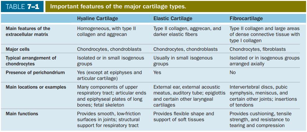
2 Introduction Hyaline Cartilage Elastic Cartilage Fibrocartilage Cartilage Formation, Growth, & Repair
3
4 Cartilage is a tough, flexible form of connective tissue. It is composed of: 1. Chondrocytes: that synthesize and maintain ECM components and are located in matrix cavities called lacunae. 2. An extracellular matrix (ECM) rich in GAGs and proteoglycans, which interact with collagen and elastic fibers.
5 The physical properties of cartilage depend on electrostatic bonds between the collagen and elastin fibers and the GAGs. Its semi-rigid consistency is attributable to water bound to the negatively charged sulfated GAGs. Cartilage is avascular and receives nutrients by diffusion from capillaries in adjacent connective tissue (perichondrium), so, chondrocytes exhibit low metabolic activity. Cartilage also lacks lymphatic vessels and nerves.

6 FIGURE 7-1 Copyright McGraw-Hill Companies
7 Support of soft tissue e.g., respiratory tract, ear & nose. Cartilage provides shock absorbing and sliding regions within joints and facilitates bone movements. Cartilage also guides development and growth of long bones, both before and after birth.


8 Hyaline Cartilage Elastic Cartilage Fibrocartilage
9
10 Hyaline (Gr. hyalos, glassy) cartilage, is the most common of the three forms. It is homogeneous and semitransparent in the fresh state.
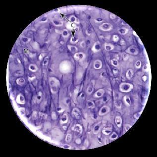
11 Sites 1. Articular surfaces of movable joints. 2. Walls of larger respiratory passages (nose, larynx, trachea, bronchi). 3. Costal cartilages. 4. Epiphyseal plates of long bones. 4. Temporary skeleton of the embryo.
12 Osteoarthritis, a chronic condition that commonly occurs during aging, involves the gradual loss or changed physical properties of the hyaline cartilage that lines the articular ends of bones in joints. Joints that are weight bearing (knees, hips) or heavily used (wrist, fingers) are most prone to cartilage degeneration. Fragments released by wear-and-tear to the articular cartilage trigger secretion of matrix metalloproteinases and other factors from macrophages in adjacent tissues, which exacerbate damage and cause pain and inflammation within the joint.
13 The dry weight of hyaline cartilage is 40% collagen (mainly type II) embedded in a firm, hydrated gel of proteoglycans and structural glycoproteins. In routine histology preparations, the proteoglycans cause the matrix to be generally basophilic and the thin collagen fibrils are barely discernible. Aggrecan (250 kd), with approximately 150 GAG side chains of chondroitin sulfate and keratan sulfate, is the most abundant proteoglycan of hyaline cartilage.
14 Hundreds of aggrecan proteoglycans are bound noncovalently by link proteins to long polymers of hyaluronic acid. These proteoglycan complexes bind further to the surface of type II collagen fibrils. Water bound to GAGs in the proteoglycans constitutes up 60%-80% of the weight of fresh hyaline cartilage.

15 FIGURE 7-2 Copyright McGraw-Hill Companies
16 Chondronectin is the main glycoprotein of cartilage that binds specifically to GAGs, collagen type II, and integrins, mediating the adherence of chondrocytes to the ECM. Staining variations within the matrix reflect local differences in its molecular composition. Immediately surrounding each chondrocyte, the territorial matrix (richer in GAGs) stains differently from the intervening areas of interterritorial matrix (more collagen).
17 FIGURE 7-2 Copyright McGraw-Hill Companies
18 Cells occupy relatively little of the hyaline cartilage mass. Two cell types are present: 1. Young chondrocytes (chondroblasts): Present at the periphery of the cartilage. Have an elliptic shape, with the long axis parallel to the surface. 2. Mature Chondrocytes: Deeper in the cartilage. They are round. They may appear in groups of up to eight cells that originate from mitotic divisions of a single chondrocyte and are called isogenous aggregates.
19 FIGURE 7-2 Copyright McGraw-Hill Companies

20 FIGURE 7-3 Copyright McGraw-Hill Companies
21 As the chondrocytes become more active in secreting collagens and other ECM components, the aggregated cells are pushed apart and occupy separate lacunae. Cartilage cells and the matrix often shrink during routine histologic preparation, resulting in both the irregular shape of the chondrocytes and their retraction from the matrix. In living tissue, and in properly prepared sections, the chondrocytes fill the lacunae completely.
22 Because cartilage is devoid of blood capillaries, chondrocytes respire under low-oxygen tension. Hyaline cartilage cells metabolize glucose mainly by anaerobic glycolysis to produce lactic acid as the end product. Chondrocyte synthesis of sulfated GAGs and secretion of proteoglycans are accelerated by many hormones and growth factors e.g., GH.
23 Cells of cartilage can give rise to either benign (chondroma) or slow-growing, malignant (chondrosarcoma) tumors in which cells produce normal matrix components. Chondrosarcomas seldom metastasize and are generally removed surgically.
24 Except in the articular cartilage of joints, all hyaline cartilage is covered by a layer of dense connective tissue, the perichondrium, which is essential for the growth and maintenance of cartilage. The perichondrium consists largely of collagen type I fibers and fibroblasts. Among these fibroblasts in the inner layer of the perichondrium are progenitor cells for chondroblasts that divide and differentiate into chondrocytes.

25 FIGURE 7-2 Copyright McGraw-Hill Companies

26 FIGURE 7-3 Copyright McGraw-Hill Companies
27 Nutrients from the blood diffuse from the perichondrium to reach the deeper chondrocytes. Transport of water and solutes in the matrix is promoted by the pumping action of intermittent cartilage compression and decompression. Because of the limits of diffusion, the maximum thickness of the hyaline cartilage is limited and it usually exists as small, thin plates.
28 The inability of cartilage to regenerate or to be repaired fully may be attributed to the chondrocytes immobility, low metabolic and mitotic rates, and avascularity. If a cartilage injury involves the perichondrium, new chondroblasts and fibroblasts may be mobilized and limited repair can occur, but most of the new tissue produced is dense connective tissue and normal function of the cartilage is often impaired.
29
30 Elastic cartilage is similar to hyaline cartilage except that it contains an abundant network of elastic fibers in addition to collagen type II, which give fresh elastic cartilage a yellowish color. Demonstration of the elastic fibers usually requires stains such as orcein or resorcin fuchsin.

31 FIGURE 7-4 Copyright McGraw-Hill Companies
32 Elastic cartilage is found in: 1. The auricle of the ear. 2. The walls of the external auditory canals. 3. The auditory (eustachian) tubes. 4. The epiglottis, and the cuneiform cartilage in the larynx. Elastic cartilage in these locations includes a perichondrium similar to that of most hyaline cartilage.
33
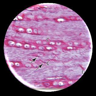
34 Fibrocartilage is essentially a combination of hyaline cartilage and dense connective tissue with gradual transitions between these tissues. It is found in intervertebral discs, in attachments of certain ligaments, and in the pubic symphysis. Chondrocytes of fibrocartilage occur singly and in aligned isogenous aggregates and produce matrix containing type II collagen.

35 FIGURE 7-5 Copyright McGraw-Hill Companies
36 Regions with chondrocytes and hyaline matrix are separated by other regions containing bundles of type I collagen and scattered fibroblasts. The relative scarcity of proteoglycans makes the matrix of fibrocartilage more acidophilic than that of hyaline or elastic cartilage. There is no distinct surrounding perichondrium in fibrocartilage.
37
38 All cartilage forms from embryonic mesenchyme in the process of chondrogenesis. The first indication of cell differentiation is the rounding up of the mesenchymal cells, which retract their extensions, multiply rapidly, and become more densely packed together. The dividing cells are typically called chondroblasts and chondrocytes when proliferation has ceased; both have basophilic cytoplasm rich in RER for collagen synthesis.

39 FIGURE 7-6 Copyright McGraw-Hill Companies

40 FIGURE 7-7 Copyright McGraw-Hill Companies
41 Production of the ECM encloses the cells in their lacunae and then gradually separates chondroblasts from one another. During embryonic development, the differentiation of cartilage takes place primarily from the center outward; therefore the more central cells have the characteristics of chondrocytes, whereas the peripheral cells are typical chondroblasts. The superficial mesenchyme forms the perichondrium.
42 The cartilage tissue enlarges by: 1. Interstitial growth, resulting from the mitotic division of preexisting chondroblasts. 2. Appositional growth, which involves differentiation of new chondroblasts from the perichondrium. In both cases, the synthesis of matrix contributes greatly to the growth of the cartilage.
43 Appositional growth of cartilage is more important during postnatal development, although interstitial growth in the articular cartilage and epiphyseal plates of long bones is important in increasing the length of long bones. In articular cartilage, cells and matrix near the articulating surface are gradually worn away and must be replaced from within, because there is no perichondrium to add cells by appositional growth.
44 Damaged cartilage undergoes slow and often incomplete repair, except in young children. It occurs primarily by activity of cells in the perichondrium, which invade the injured area and produce new cartilage. In extensively damaged areas the perichondrium produces a scar of dense connective tissue instead of forming new cartilage. The poor capacity of cartilage for repair or regeneration is due in part to the avascularity of this tissue.
45 In contrast to other forms of cartilage and other tissues, hyaline cartilage is susceptible to calcification during aging. Calcification of the hyaline matrix, accompanied by degenerative changes in the chondrocytes, is a common part of the aging process. It resembles endochondral ossification by which bone is formed in many respects.
46

47
Cartilage. Dr. Heba Kalbouneh Associate Professor of Anatomy and Histology
 Cartilage Dr. Heba Kalbouneh Associate Professor of Anatomy and Histology 1 Cartilage is a specialized type of connective tissue designed to give support, bear weight and withstand tension, torsion and
Biology. Dr. Khalida Ibrahim
 Biology Dr. Khalida Ibrahim The cartilage General characteristics: 1. Cartilage is a specialized type of connective tissue (supporting connective tissue). 2. Consists, like other connective tissues, of
Dr. Heba Kalbouneh. Dr. Heba Kalbouneh. Dr. Heba Kalbouneh
 Dr. Heba Kalbouneh Dr. Heba Kalbouneh Dr. Heba Kalbouneh Cartilage Cartilage is a special form of connective tissue; it has the same origin of connective tissue (embryonic mesenchyme). It contains cells
Dr.Heba Kalbouneh. Ragad Alhawi. Dr.Heba Kalbouneh
 9 Dr.Heba Kalbouneh Ragad Alhawi Dr.Heba Kalbouneh "Cartilage" Cartilage is a special form of connective tissue; it has the same origin of connective tissue (embryonic mesenchyme). It contains cells and
contains an antiangiogenesis factor
 CARTILAGE & BONE Cartilage and Bone objectives Student must learn :. What is the meaning of cartilage, and their function, location in human body.. To distinguish the 3 types of cartilage. And their cells,
Cartilage. - Cartilage together with long bone form the skeleton and support the body.
 Cartilage - Cartilage is a special type of CT has a firm pliable matrix that can resist mechanical stress, act as a shock absorber. - Cartilage together with long bone form the skeleton and support the
physical properties depend on: electrostatic bonds between collagen/elastic fibers and GAGs water bound to negatively charged sulfated GAG chains
 connective/supporting tissue bears mechanical stress without distortion -> shock absorption smooth surface -> facilitates movements of joints guides development of bones chondrocytes extracellular matrix
Growth and repair: Cartilage is a vascular tissues that receives nutrients by diffusion through its matrix, cartilage grow by 2 mechanisms:
 Skeletal connective tissues: (cartilage and bone): Cartilage and bone are specialized connective tissues both adapted to serve as skeletal framework in most vertebrates the presence of solid inter cellular
Most abundant and widely distributed tissues in the body Binds, support, and strengthen body tissues, protect and insulate internal organ, serve as
 Connective tissue Most abundant and widely distributed tissues in the body Binds, support, and strengthen body tissues, protect and insulate internal organ, serve as major transport system, compartmentalizes
Lecture Overview. Connective Tissues. Marieb s Human Anatomy and Physiology. Chapter 4 Tissues: The Living Fabric Connective Tissues Lecture 10
 Marieb s Human Anatomy and Physiology Marieb Hoehn Chapter 4 Tissues: The Living Fabric Connective Tissues Lecture 10 Lecture Overview General composition and function of connective tissue Components of
Connective Tissue. Found everywhere in the body. Most abundant and widely distributed. Never exposed to the outside environment.
 Connective Tissue Found everywhere in the body. Most abundant and widely distributed. Never exposed to the outside environment. Connective Tissue Functions Binding and support Protection Insulation Transportation
Chapter 4. Cartilage and Bone. Li Shu-Lei instructor. Dept. Histology and Embryology, School of Basic Medical Sciences, Jilin University
 Chapter 4 Cartilage and Bone Li Shu-Lei instructor Dept. Histology and Embryology, School of Basic Medical Sciences, Jilin University I Cartilage a specialized connective tissue Characterizers: Cartilage
SKELETAL TISSUES CHAPTER 7 INTRODUCTION TO THE SKELETAL SYSTEM TYPES OF BONES
 SKELETAL TISSUES CHAPTER 7 By John McGill Supplement Outlines: Beth Wyatt Original PowerPoint: Jack Bagwell INTRODUCTION TO THE SKELETAL SYSTEM STRUCTURE Organs: Bones Related Tissues: Cartilage and Ligaments
Quiz 6. Cartilage and Bone
 Quiz 6 Cartilage and Bone MCQs X type (true or false): 1. Cartilage tissue: a. Has a rich blood supply. b. Develops from mesenchyme. c. Has ability for a quick regeneration. d. Has chondrocytes as precursor
HOLE S ANATOMY CHAPTER 5, PART II Lecture notes
 HOLE S ANATOMY CHAPTER 5, PART II Lecture notes I. Connective Tissue A. Structure 1. have few cells that are spaced apart and can divide; two categories: a. fixed cells cells that are present in tissue
Blood. Hematopoietic Tissue
 Blood Hematopoietic Tissue Is a type of connective tissue in which its cells are suspended in a circulating fluid. Erythrocytes+ leukocytes + platelets (thrombocytes) =formed elements of blood. These formed
Epithelia of Coverings and Linings. Tissues. Tissue
 Tissue Tissues Chapter 3 Definition an aggregation of cells in which each cooperates with all others in the performance of a given function Examples of general functions Movement Protection Support Production
The Tissue Level of Organization
 The Tissue Level of Organization 4.5-4.11 August 31, 2012 4.5 Connective Tissues Describe the general features of connective Describe the structure, location, and function of the various types of connective
Connective Tissue. Consists of two basic elements: Cells and Extra-cellular matrix
 Connective Tissue Consists of two basic elements: Cells and Extra-cellular matrix True Connective Tissue Cells Fibroblasts: Secrete both fibers and ground substance of the matrix (wandering) Macrophages:
The Tissue Level of Organization
 Tissue The Tissue Level of Organization Chapter 3 Definition an aggregation of cells in which each cooperates with all others in the performance of a given function Examples of general functions Movement
Bio& 241 Unit 1 / Lecture 4
 Bio& 241 Unit 1 / Lecture 4 Connective Tissue Consists of two basic elements: Cells and Extra-cellular matrix 1 True Connective Tissue Cells Fibroblasts: Secrete both fibers and ground substance of the
Tissue engineering of cartilage
 Tissue engineering of cartilage Cartilage responds to mechanical forces and is able to remodel in response to the prevailing stress Cartilage, like bone, may respond to mechanical stimulation by increasing
BONE AND CARTILAGE LIA DAMAYANTI. Department of Histology - FMUI
 1 BONE AND CARTILAGE LIA DAMAYANTI Department of Histology - FMUI Agenda 2 Introduction Cartilage Development of cartilage Growth and repair Components of cartilage Type of Cartilage Agenda 3 Bone Components
8/30/2017. Tissue: The Living Fabric. 4.3 Connective Tissue
 Chapter 4 Part B Tissue: The Living Fabric Annie Leibovitz/Contact Press Images PowerPoint Lecture Slides prepared by Karen Dunbar Kareiva Ivy Tech Community College 4.3 Connective Tissue Connective tissue
Mast Cell. Mast Cells. James W. Truman, Ph.D. Howard Hughes Medical Institute Chevy Chase, Maryland
 5 th ANNUAL SINAUER ASSOCIATES DISTINGUISHED SCIENTIST LECTURE James W. Truman, Ph.D. Howard Hughes Medical Institute Chevy Chase, Maryland Neuronal Lineages in the CNS of Drosophila: Units of Development,
Cartilage & bone. Red: important. Black: in male female slides. Gray: notes extra. Editing File
 Cartilage & bone Red: important. Black: in male female slides. Gray: notes extra. Editing File OBJECTIVES describe the microscopic structure, distribution and growth of the different types of Cartilage
Chapter 6: Skeletal System: Bones and Bone Tissue
 Chapter 6: Skeletal System: Bones and Bone Tissue I. Functions A. List and describe the five major functions of the skeletal system: 1. 2. 3.. 4. 5.. II. Cartilage A. What do chondroblasts do? B. When
Which compound is reponsible for the viscous character of the ground substance?
 1 2 Which type of collagen forms the coarse collagen fibres in dense regular and irregular connective tissues? Which compound is reponsible for the viscous character of the ground substance? 3 Which class
Connective Tissues. Copyright 2009 Pearson Education, Inc., publishing as Pearson Benjamin Cummings
 C.T. are found in all parts of the body & diverse in structure & function. C.T. Functions: -connect structures -provide support -protect vital organs -fill space b/w structures -stores fat -defends body
Study of different tissues Abnormal cells and tissues can be compared to normal tissues to identify disease, such as cancer Being able to know and
 CHAPTER 4 Study of different tissues Abnormal cells and tissues can be compared to normal tissues to identify disease, such as cancer Being able to know and recognize normal tissues under the microscope
5.3. The Nature of Cartilage Matrix The components of cartilage matrix include a high component of fibers, and proteoglycans. Proteoglycans are a
 Chapter 5 Supportive Tissues Support in Animals is carried out by Cartilage and Bone. A. Cartilage 5.1. Nature of Cartilage Cartilage is a highly resilient c..t that provides strength and support in areas
Hole s Human Anatomy and Physiology
 Hole s Human Anatomy and Physiology 1 Chapter 5 Tissues Four major tissue types 1. Epithelial 2. Connective 3. Muscle 4. Nervous 2 Epithelial Tissues General characteristics - cover organs and the body
CONNECTIVE TISSUE (Refer to pp for specific characteristics of each) VAN (**Be familiar with exceptions**)
 VAN (**Be familiar with exceptions**)") CONNECTIVE TISSUE CHARACTERISTICS: *Most abundant tissue type; Composed of ECM (GS & Protein Fibers) + Cells (Refer to pp.129-131 for specific characteristics of each) *Highly equipped with VAN assists
CONNECTIVE TISSUE CHARACTERISTICS: *Most abundant tissue type; Composed of ECM (GS & Protein Fibers) + Cells (Refer to pp.129-131 for specific characteristics of each) *Highly equipped with VAN assists
Tissues organs system organism. pg151
 Histology is the study of tissues A TISSUE is a group of cells, usually of one kind, & their intercellular substance (e.g. intercellular matrix in animal) which are linked together & perform a particular
4 Types of Tissue. Epithelial Connective Muscle Neural
 Connective Tissue 4 Types of Tissue Epithelial Connective Muscle Neural Connective Tissue Fills internal spaces Supports & binds other tissues Transports materials Stores energy Classification of Connective
4 Types of Tissue. Epithelial Connective Muscle Neural
 Connective Tissue 4 Types of Tissue Epithelial Connective Muscle Neural Connective Tissue Fills internal spaces Supports & binds other tissues Transports materials Stores energy Classification of Connective
BONE TISSUE. Dr. Heba Kalbouneh Associate Professor of Anatomy and Histology
 BONE TISSUE Dr. Heba Kalbouneh Associate Professor of Anatomy and Histology BONE FUNCTION Support Protection (protect internal organs) Movement (provide leverage system for skeletal muscles, tendons, ligaments
Compact bone; Many parallel Haversian canals contain: small blood vessels. very small nerve. Interconnected by Volkmann s canals.
 Special characteristics of COMPACT BONE (dense bone) Thick; well vascularized Osteocytes and lamellae Concentric rings around blood vessels Most bones: outer compact bone inner spongy bone Marrow cavity
Classification of Tissues
 6 R e v i e w S h e e t Exercise Classification of Tissues NAME LAB TIME/DATE Tissue Structure and Function General Review 1. Define tissue. A group of cells similar to one another in structure that perform
Classification of Tissues
 M06_MARI0000_00_SE_CH06.qxd 3/28/11 4:37 PM Page 35 NAME LAB TIME/DATE R E V I E W S H E E T EXERCISE 6 Classification of Tissues Tissue Structure and Function General Review 1. Define tissue. A group
The Skeletal System:Bone Tissue
 The Skeletal System:Bone Tissue Dynamic and ever-changing throughout life Skeleton composed of many different tissues cartilage, bone tissue, epithelium, nerve, blood forming tissue, adipose, and dense
Histology. There are four basic tissue types in the body are :-
 Histology Lab.I There are four basic tissue types in the body are :- 1- Epithelial tissues (Epithelium) 2- Connective tissues 3- Muscular tissues 4- Nervous tissues 1-Epithelial tissues epithelial tissues
Tejido Conectivo Parte B. Informe #3 Laboratorio Biología # 240 Profesor: Javier Cabello
 Tejido Conectivo Parte B Informe #3 Laboratorio Biología # 240 Profesor: Javier Cabello Figure 4-8 The Cells and Fibers of Connective Tissue Proper Areolar Elastic fibers Collagen fibers Fibroblast Free
OSSEOUS TISSUE & BONE STRUCTURE PART I: OVERVIEW & COMPONENTS
 OSSEOUS TISSUE & BONE STRUCTURE PART I: OVERVIEW & COMPONENTS The Skeletal System Skeletal system includes: bones of the skeleton, cartilages, ligaments, and connective tissues What are the functions of
組織學 Historlogy 台北醫學大學 / 解剖學科教授 : 邱瑞珍分機號碼 :3261. 電子郵件信箱
 組織學 Historlogy 台北醫學大學 / 解剖學科教授 : 邱瑞珍分機號碼 :3261 電子郵件信箱 :rueijen@tmu.edu.tw 1 Cartilage & Bone 台北醫學大學 / 解剖學科教授 : 邱瑞珍分機號碼 :3261 電子郵件信箱 :rueijen@tmu.edu.tw 2 學習目的 (Learning objectives) chondrocytes ( 軟骨細胞
Tissues Chapter 5...Tissue - a group or mass of similar cells working together to perform certain common functions
 Tissues Chapter 5...Tissue - a group or mass of similar cells working together to perform certain common functions There are 4 major types of tissue Epithelial Connective Muscle Nervous 1. Epithelial Tissue
Functions of the Skeletal System. Chapter 6: Osseous Tissue and Bone Structure. Classification of Bones. Bone Shapes
 Chapter 6: Osseous Tissue and Bone Structure Functions of the Skeletal System 1. Support 2. Storage of minerals (calcium) 3. Storage of lipids (yellow marrow) 4. Blood cell production (red marrow) 5. Protection
KEY CONCEPTS Unit 6 THE SKELETAL SYSTEM
 ANATOMY & PHYSIOLOGY 1 (101-805 - AB) PAUL ANDERSON 2011 KEY CONCEPTS Unit 6 THE SKELETAL SYSTEM A Overview of The Skeletal System 1. Definition: Anatomically the SKELETAL SYSTEM consists of bones, cartilages,
Human Anatomy and Physiology I Laboratory
 Human Anatomy and Physiology I Laboratory Skeletal Tissue: Cartilage and Bone This lab involves study of the laboratory exercise Overview of the Skeleton, Classification and Structure of Bones and Cartilages,
Anatomy and Physiology Tissue Review
 Anatomy and Physiology Tissue Review OVERVIEW Histology practicals can be rough, especially when access to slides is limited to the lab period. This resource provides an opportunity to learn or review
TISSUE. A group of cells that perform a similar function within an organism. Epithelium Connective Muscle Nervous CREDITS
 TISSUE A group of cells that perform a similar function within an organism. Epithelium Connective Muscle Nervous CREDITS Epithelium Connective Muscle Nervous Epithelium Composed of a layer of cells. Lines
Connective Tissue Part-2. Dr. Heba Kalbouneh Assistant Professor of Anatomy and Histology
 Connective Tissue Part-2 Dr. Heba Kalbouneh Assistant Professor of Anatomy and Histology 1 Features Composed of cells, fibers and extracellular matrix. Highly vascular Variable regenerative power Originates
FORMATION OF BONE. Intramembranous Ossification. Bone-Lec-10-Prof.Dr.Adnan Albideri
 FORMATION OF BONE All bones are of mesodermal origin. The process of bone formation is called ossification. We have seen that formation of most bones is preceded by the formation of a cartilaginous model,
A. cells that perform related functions and are similar in structure. B. extracellular material - made by cells and secreted into interstitial space
 I. tissue components A. cells that perform related functions and are similar in structure B. extracellular material - made by cells and secreted into interstitial space II. tissue types A. epithelium (e.)
Basic Histology. By Mrs. Bailey
 Basic Histology By Mrs. Bailey Primary Tissues 1. Epithelial Tissue 2. Connective Tissue 3. Muscle Tissue 4. Nervous Tissue Very cellular Supported by underlying connective tissue Epithelial & connective
What are the parts of the skeletal system? Chapter 6- Part I Bones and Skeletal Tissues. Growth of Cartilage. Bones come in many shapes
 Chapter 6- Part I Bones and Skeletal Tissues Components of the skeletal system Classification of Bone (bone shapes) Functions of bone Bone structure Microscopic structure of bone and bone cells What are
Sheets 16&17. Dr. Heba Kalbouneh. Dr. Heba Kalbouneh. Dr. Heba Kalbouneh
 Sheets 16&17 Dr. Heba Kalbouneh Dr. Heba Kalbouneh Dr. Heba Kalbouneh Ossification (formation of bone) - Osteoblasts are responsible for producing the extracellular matrix of the bone and these osteoblasts
Tissues are: group of similar or identical cells that share a common function. used to build organs
 Tissues: Four classes Epithelium Connective Muscle Nervous Tissues are: group of similar or identical cells that share a common function. used to build organs Overview: Epithelial o Line body cavities
Unit 5: SKELETAL SYSTEM
 Unit 5: SKELETAL SYSTEM (a) NRSG231 Dr. Moattar Raza Rizvi Skeletal System: Contents Functions of the Skeletal System Fracture Structure of Bone Tissue Articulations & Joint Bone Development and Growth
Dr. Heba Kalbouneh. Saba Alfayoumi. Heba Kalbouneh
 11 Dr. Heba Kalbouneh Saba Alfayoumi Heba Kalbouneh 2- Bone Bone tissue is also classified into primary bone and secondary bone. In the beginning, the first bone that is deposited by the osteoblasts is
Histology. The study of tissues.
 Histology The study of tissues. Body Tissues Cells are specialized for particular functions Tissues Groups of cells with similar structure and function Four primary types Epithelium Connective tissue Nervous
Tissues. Tissues. Four basic tissues. A collection of cells with a common function. 1. Epithelial 2. Connective 3. Muscular 4.
 Tissues Tissues A collection of cells with a common function Four basic tissues 1. Epithelial 2. Connective 3. Muscular 4. Nervous Epithelia: cells in layers Types of epithelia 1) lining Layers of cells
The Skeletal System:Bone Tissue
 The Skeletal System:Bone Tissue Dynamic and ever-changing throughout life Skeleton composed of many different tissues cartilage, bone tissue, epithelium, nerve, blood forming tissue, adipose, and dense
Tissue Outline (chapter 4) Tissues group of cells that perform structural and roles. List the 4 types:
 Tissues group of cells that perform structural and roles. List the 4 types:") Tissue Outline (chapter 4) Tissues group of cells that perform structural and roles. List the 4 types: 1. 2. 3. 4. I. Epithelial Tissue covers all the surfaces, inside & out. Are the major tissues of,
Tissue Outline (chapter 4) Tissues group of cells that perform structural and roles. List the 4 types: 1. 2. 3. 4. I. Epithelial Tissue covers all the surfaces, inside & out. Are the major tissues of,
Practical Histology. Lab 3: Connective tissue
 Practical Histology Lab 3: Connective tissue Connective tissues Connective tissue provides structural support for the body by binding cells and tissues together to form organs. It also provides metabolic
Tissue Outline. Chapter 4. Tissue. Cellular Connections. I. Definitions II. Cellular Connections III. Tissue Types IV. Membranes V.
 Tissue Outline Chapter 4 The Tissue Level of Organization I. Definitions II. Cellular Connections III. Tissue Types IV. Membranes V. Tissue Repair 1 2 Tissue Cellular Connections Tissue Groups of cells
Elastic tissue textus connectivus elasticus:
 Connective tissue 2 1. Connective tissue with special properties: elastic and reticular tissues embryonic connective tissues adipose tissue 2. Supporting connective tissues: cartilage hyaline, elastic
Sheet #9. Dr. Heba Kalbouneh. Dr. Heba Kalbouneh. Dr. Heba Kalbouneh
 Sheet #9 Dr. Heba Kalbouneh Dr. Heba Kalbouneh Dr. Heba Kalbouneh Elastic fibers The main function of elastic fibers is to provide elasticity. In other words these fibers are able to restore the original
Chapter 4. The Tissue Level of Organization
 Chapter 4 The Tissue Level of Organization 1 Tissue Outline I. Definitions II. Cellular Connections III.Tissue Types IV. Membranes V. Tissue Repair 2 Tissue Tissue Groups of cells that are similar in structure
10/3/2012. Tissue: The Living Fabric: Part B. Extracellular matrix Ground substance Fibers Collagen fiber Elastic fiber Reticular fiber.
 PowerPoint Lecture Slides prepared by Janice Meeking, Mount Royal College C H A P T E R 4 Tissue: The Living Fabric: Part B Copyright 2010 Pearson Education, Inc. Copyright 2010 Pearson Education, Inc.
الكيمياء احليوية لألنسجة املتخصصة. Biochemistry of Specialized Tissues
 الكيمياء احليوية لألنسجة املتخصصة Biochemistry of Specialized Tissues BCH 443 Biochemistry of specialized Tissues Course Symbol & No. : BCH 443 Credit Hours : 2 (2+0) Prerequisite : BCH 347 Class schedule
Joints. Articulations Arthroses
 Joints Articulations Arthroses 1 Joints, defined Points of contact between Two bones Bone and teeth Joint classification: 2 schemes Functional classification degree of movement permitted Structural classification
HISTOLOGY Lecture 2 Connective tissue, muscle and bone tissue PCL Prof. P. Kyamanywa DoS-FACMED NUR
 HISTOLOGY Lecture 2 Connective tissue, muscle and bone tissue PCL1 2012 Prof. P. Kyamanywa DoS-FACMED NUR 4: Connective Tissue nur General function and composition providing structural support for the
Connective Tissue. Practice Identifying Your Tissue With This Slide Show! Then try the quiz at the bottom of this page!
 Connective Tissue scientistcindy.com /connective-tissue.html Practice Identifying Your Tissue With This Slide Show! Then try the quiz at the bottom of this page! The 4 Types of Connective Tissues Connective
SKELETAL SYSTEM I NOTE: LAB ASSIGNMENTS for this topic will run over 3 Weeks. A SEPARATE WORKSHEET WILL BE PROVIDED.
 BIO 211; Anatomy and Physiology I REFERENCE: CHAPTER 07 1 Dr. Lawrence Altman Naugatuck Valley Community College LECTURE TOPICS OUTLINE SKELETAL SYSTEM I NOTE: LAB ASSIGNMENTS for this topic will run over
Chapter 05. Review. Copyright The McGraw-Hill Companies, Inc. Permission required for reproduction or display.
 Chapter 05 Review 5.1: Introduction Similar cells with a common function are called tissues. The study of tissues is called histology. There are four (4) primary or major tissue types: 1. Epithelial Tissue
Chapter 5. Tissues. 4 Types of Body Tissues. Tissues
 Chapter 5 Tissues Tissues Tissues - groups of cells that are similar in structure & function RBC, WBC, & platelets are a group of cells working together to form BLOOD tissue Histology Pathohistology study
Skeletal System Functions
 Chapter 6 Skeletal System: Bones and Bone Tissue 6-1 Skeletal System Functions Support. Bone is hard and rigid; cartilage is flexible yet strong. Cartilage in nose, external ear, thoracic cage and trachea.
2 PROCESSES OF BONE OSSIFICATION
 2 PROCESSES OF BONE OSSIFICATION ENDOCHONDRAL OSSIFICATION 6 STEPS 1. CARTILAGE ENLARGES, BY APPOSITIONAL GROWTH; CHONDROCYTES AT CENTER OF CARTILAGE GROW IN SIZE; MATRIX REDUCES IN SIZE & SPICULES CALCIFY;
Epithelial Tissue. Simple Cuboidal Function: secretion and absorption. Simple Squamous
 Epithelial Tissue General Functions: Lines and covers organs Absorbs / secretes substances Gas exchange Protection Special Characteristics: - have an apical surface on top - have a basement membrane below
UNIT 4 T I S S U E S
 UNIT 4 T I S S U E S WHAT IS A TISSUE Group of cells that work together to do a function Cells are similar Extracellular fluid around them is similar Histology EPITHELIAL TISSUE Also called epithelium
An Introduction to the Skeletal System Skeletal system includes Bones of the skeleton Cartilages, ligaments, and connective tissues
 An Introduction to the Skeletal System Skeletal system includes Bones of the skeleton Cartilages, ligaments, and connective tissues Functions of the Skeletal System Support Storage of minerals (calcium)
Function: Provides reserve food fuel; Copyright 2011 Pearson Education, Inc. Copyright 2011 Pearson Education, Inc. White blood cell (lymphocyte)
") Adipose Tissue Closely packed adipocytes Have nucleus pushed to one side by fat droplet Richly vascularized Provides reserve food fuel Insulates against heat loss Supports and protects organs Under skin
Adipose Tissue Closely packed adipocytes Have nucleus pushed to one side by fat droplet Richly vascularized Provides reserve food fuel Insulates against heat loss Supports and protects organs Under skin
-the emphasis on this section is the structure and function of bone tissue and on the dynamics of its formation and remodeling throughout life.
 Biology 325 Fall 2004 BONES AND SKELETAL TISSUES Introduction -skeleton contains cartilage and bones -the emphasis on this section is the structure and function of bone tissue and on the dynamics of its
Connective Tissues. 2. Describe the function of fibroblasts. 3. What is ground substance? What is its function?
 Connective Tissues Directions: Insert and install your Interactions: Foundations CD. a. Click the "Contents" button. b. Open the Tissue Level of Organization file. c. Click on Anatomy Overviews. d. Work
What is histology? HISTOLOGY
 Introduction to Histology What is histology? HISTOLOGY histo = tissue ogy = study So HISTOLOGY = the study of tissues! What is a TISSUE? Tissues are groups of cells with specialized structural and functional
2/21/2012. Components Connective Tissue. Connective Tissue??? Connective Tissue What is it?
 Connective Tissue??? Connective Tissue What is it? It Binds, It Supports, It Strengthens, It Protects, It Insulates, It Compartmentalizes, It helps us move, It helps transport stuff, It is a site for storing
Connective Tissue. Answer Choices(In CAPITAL BOLD): RETICULAR ELASTIC. IRREGULAR Spongy bone ELASTIC BLOOD
: RETICULAR ELASTIC. IRREGULAR Spongy bone ELASTIC BLOOD") Connective Tissue Answer Choices(In CAPITAL BOLD): Proper: Specialized: Loose- Cartilage- AREOLAR HYALINE ADIPOSE FIBROCARTILAGE RETICULAR ELASTIC Dense- Bone- REGULAR COMPACT BONE IRREGULAR Spongy bone
Connective Tissue Answer Choices(In CAPITAL BOLD): Proper: Specialized: Loose- Cartilage- AREOLAR HYALINE ADIPOSE FIBROCARTILAGE RETICULAR ELASTIC Dense- Bone- REGULAR COMPACT BONE IRREGULAR Spongy bone
The Skeletal System PART A. PowerPoint Lecture Slide Presentation by Patty Bostwick-Taylor, Florence-Darlington Technical College
 PowerPoint Lecture Slide Presentation by Patty Bostwick-Taylor, Florence-Darlington Technical College The Skeletal System 5 PART A The Skeletal System Parts of the skeletal system Bones (skeleton) Joints
Tissues and Membranes
 I. In the Beginning a. Egg + sperm! Tissues and Membranes b. 1 cell divides to make 2, 2 divide to make 4, 4 divide to make 8, and then? c. d. e. Totipotent: f. Pluripotent: II. III. Tissues a. Tissues
Module 2:! Functional Musculoskeletal Anatomy A! Semester 1! !!! !!!! Hard Tissues, Distal Upper Limb & Neurovascular Supply of Upper Limb!
 Functional Musculoskeletal Anatomy A Module 2: Hard Tissues, Distal Upper Limb & Neurovascular Supply of Upper Limb Semester 1 1 18. Bone Tissue & Growth of Bones 18.1 Describe the structure of bone tissue
BIOH111. o Cell Module o Tissue Module o Integumentary system o Skeletal system o Muscle system o Nervous system o Endocrine system
 BIOH111 o Cell Module o Tissue Module o Integumentary system o Skeletal system o Muscle system o Nervous system o Endocrine system Endeavour College of Natural Health endeavour.edu.au 1 TEXTBOOK AND REQUIRED/RECOMMENDED
Unit II: Tissues and Integumentary System
 Unit II: Tissues and Integumentary System 2.1 - Tissues Chapter 4 Written Response #1 1. What is a tissue? 2. What are four major types of tissues? Tissue Definition: a group or mass of similar cells working
TISSUES. Objectives. Tissues
 TISSUES Objectives Introduce the four major types of tissues Describe the general characteristics and functions of epithelial & connective tissue Name the major types of epithelial & connective tissues
Tissues. How do cells form tissues?
 Tissues How do cells form tissues? Using cell junctions Tissues Epithelial tissue Connective tissue Muscle tissue Nervous tissue Epithelial Tissue Closely packed cells in continuous sheets connected by
Car$lage and Bone. Kris$ne Kra0s, M.D.
 Car$lage and Bone Kris$ne Kra0s, M.D. Car$lage and Bone Lecture Objec$ves Describe the general func$ons of car$lage and bone. Compare the func$on and composi$on of the three types of car$lage. Describe
Introduction to Biomedical Engineering
 Introduction to Biomedical Engineering FW 16/17, AUT Biomechanics of tendons and ligaments G. Rouhi Biomechanics of tendons and ligaments Biomechanics of soft tissues The major soft tissues in musculoskeletal
Tissues 10/21/2016. Epithelial Tissue
 Tissues This is a generalized cell diagram. It shows the anatomy of a cell, but most cells do not actually look like this. Cells can have a wide variety of shapes and sizes, depending on their function.
Tissues- of cells with similar and
 Tissues- of cells with similar and. Four types of tissues 1. 2. 3. 4. Characteristics of Epithelial Tissue -Highly Cellular -Special contacts -Polar (apical and basal surfaces) -Supported by connective
Anatomy & Physiology Skeletal System
 I. Functions of the Skeletal System A. the body Anatomy & Physiology Skeletal System B. of vital organs C. Provide for movement D. storage (calcium & phosphate) E. cell formation II. Bone Structure A.