Connective tissues. All forms of connective tissue share some common structural features and a common embryonic origin.
|
|
|
- Elisabeth Bates
- 5 years ago
- Views:
Transcription
1 Connective tissues Connective tissue forms a framework upon which epithelial tissue rests and within which nerve tissue and muscle tissue are embedded. Blood vessels and nerves travel through connective tissue. Functions of connective tissue include: transport, immunological defense, {mechanical support, growth and repair, energy reserve, haemopoiesis, and inflammation. All forms of connective tissue share some common structural features and a common embryonic origin. Connective tissue consists of individual cells scattered within a matrix. Cells of connective tissue are not directly attached to one another. Connective tissue is derived from mesoderm (unlike most epithelial tissue which is I derived from ectoderm and endoderm). Components of Connective Tissue: Connective tissue consists of cells embedded in an extracellular matrix.the matrix, in turn, consists of fibers and ground substance. Cells of the connective tissue include: 1. Fibroblasts: are the most common cells in proper connective tissue. Fibroblasts are responsible for secreting collagen and other elements of the extracellular matrix of connective tissue. Resting fibroblasts typically have so little pale cytoplasm with branched processes that the cells appear, by light microscopy, as "naked" unclei. Fibroblast nuclei appear dense (heterochromatic) and are usually flattened or spindleshaped, with one or two nucleoli. Closely related to fibroblasts are the chondroblasts which produce the matrix of cartilage and the osteoblasts which produce the matrix of bone. 1
2 2. Mast cells are secretory cells. Upon the slightest disturbance, they release chemical signals which diffuse through the surrounding ground substance and trigger the process of inflammation. Mast cells occur as small individual cells, scattered rather widely in proper connective tissue. The cytoplasm of mast cells is packed with secretory vesicles, which can be fairly obvious in high-quality light microscope preparations. The granules contain histamine, heparin, and various other chemical mediators whose release signals for a number of physiological defense responses. 3. Macrophages (histocytes): remove and digest the by-products of both bacteria and normal growth and degeneration. They are irregular cells larger than fibroblasts, with more cytoplasm, the nucleus is smaller and darker than fibroblast nucleus, the nucleus eccentric in position. Macrophages contain numerous lysosomes which are used for.breaking down ingested material. These lysosomes are usually not easily seen by light microscopy but easily visible by electron microscopy. In macrophages which have been active and have accumulated indigestible residue, the lysosomes may be visible by light microscopy as dark intracellular granules, as in the lung contain carbon particles 4. Plasma cells: small round cells with large eccentric nucleus, the nucleus characterized by the big dark masses of chromatin which arranged radically. 5. Fat cells: round cell with packed nucleus in the side of the cell and large droplet of fat, this droplet looks as large cavity occupied the cell. 6. Pigment cells (or melanocytes): star shaped cells filled with dark granules. 7. Mesenchymal cells: star shaped cells with branched processes, like the fibroblast but smaller than it. 8. Reticular cells: the cell has large nucleus and processes attached with the processes of the adjacent cells. 2
3 9. Leucocytes: including the eosinophils, neutrophils, and lymphocytes, this found in the connective tissue of the intestine. Connective tissue fibers: 1- White or collagenous fibers: they form wave bundles of fibers, also they may found as branched bundles, Densely packed collagen fibers (in dense connective tissues such as dermis and tendon) provide main strength with resistance to tearing and stretching. Loosely packed collagen fibers (in loose connective tissues, such as hypodermis or the submucosa of internal organs) allow free movement within definite limits. 2- Yellow or elastic fibers: they are single branched fibers may be thick as in the aorta, or thin as in the areolar connective tissue, this type of fibers confer elasticity. 3- Reticular fibers: they found as a network of dark branched fibers. Reticular fibers do not show up in routine H&E stained specimens, but they can be demonstrated with silver salts. Ground substance is the background material within which all other connective tissue elements are embedded. In proper connective tissue, the ground substance consists mainly of water whose major role is to provide a route for communication and transport (by diffusion) between tissues. This water is stabilized by a complex of glycosaminoglycans (GAGs), proteoglycans, and glycoproteins, all of which comprise only a small fraction of the weight of the ground substance. Ground substance may be highly modified in the special forms of connective tissue. In blood, the ground substance lacks stabilizing macromolecules. We call this free- flowing ground substance plasma. 3
4 In skeletal tissue, the ground substance may become mineralized by deposition of calcium salts. We call this rigid ground substance bone. In cartilage, the ground substance is much more solid than in proper connective tissue but still retains more resiliency than bone. Classification of connnective tissues: Connective tissues classified to proper and specialized connective tissues. Proper connective tissues include loose and dense connective tissues. Loose connective tissues include: 1. Areolar con. T. hypodermis 2. Mucoid con. T. (umbilical cord) 3. Reticular con T. (lymph node) 4. Adipose con. T. (under the skin) 5. Mesenchymal con. T. (emberyo) Dense connective tissue A. Irregular con. T. (dermis) B. Regular con. T. include: 1. White fibrous con. T. (tendon) 2. Elastic con. T. (legament of the vertebral column). 4
5 Skeletal Connective Tissues Bone and cartilage, like all other connective tissues, consist of cells and extracellular matrix. It is the ground substance of the matrix which is most responsible for the conspicuous differences between bone and cartilage. The ground substance of bone is mineralized, making the bone rigid and strong, but brittle. The ground substance of cartilage is not mineralized but is more like very firm Jell, making cartilage stiff and incompressible but more flexible and resilient than bone. In spite of their solidity, both bone and cartilage are capable of growth. In the case of bone, internal remodeling (essentially, ongoing destruction and renewal is an active process throughout life. The ground substance of cartilage is characterized by a firm solid gel-like matrix reinforced by collagen. Cartilage is relatively stiff and incompressible. However, since cartilage is not mineralized, it is more flexible and resilient than bone. The characteristic microscopic texture of cartilage, with cells enclosed in individual lacunae (small chambers), reflects the nature of cartilage matrix and its mode of formation. While cartilage is growing, new cartilage matrix is secreted by chondroblasts, which become isolated from one another as the matrix is formed. As more matrix is secreted, these chondroblasts are pushed farther apart. Chondroblasts divide within their lacunae to form clusters of cells called cell nest, which then form more matrix and become separated from one another. Once growth ceases, the resting cells are called chondrocytes. Since cartilage is avascular, as the cartilage increases in size, its cells also grow farther from their source nutrition. As a direct result, mature cartilage is a relatively inactive tissue with minimal ability to respond to injury There are three types of cartilages: 1. Mature Hyaline cartilage found the Slides of "trachea" display the C- shaped rings of cartilage that reinforce the trachea, in this tissue you can see the single 5
6 chondrocytes and the aggregations of cells (cell nests) surrounded by capsule found in lacunae in the matrix. the matrix looks homogenous, the hyaline cartilage surrounded by vesicular fibrous. membrane contain bundles of white fibers and fibroblasts and blood vessels, this membrane called the perichondrium, The cells under this membrane are flattened lie in a parallel level to the surface called chondroblasts. Function = flexible, provides support, allows movement at joints The fine collagen fibers throughout the matrix of hyaline cartilage do not stain with routine H&E 6

7 Fetal Hyaline Cartilage: illustrates a cartilage model of a bone in an early stage of development. Most of the model consists of young chondroblasts that still resemble mesenchymal cells, having spherical nuclei and cytoplasmic processes. Lacunae have not developed at this stage. The chondroblasts are numerous, and randomly distributed in the cartilage without forming isogenous groups. At this stage of development, cartilage matrix is secreted. On the periphery of the cartilage model (left side), mesenchymal cells and concentrated and exhibit a parallel arrangement. The nuclei of these cells are elongated flattened, and the cell membranes are not distinct. This peripheral area of the cartilage develops into perichondrium, a sheath of dense connective tissue that surrounds hyaline and elastic cartilage. 7


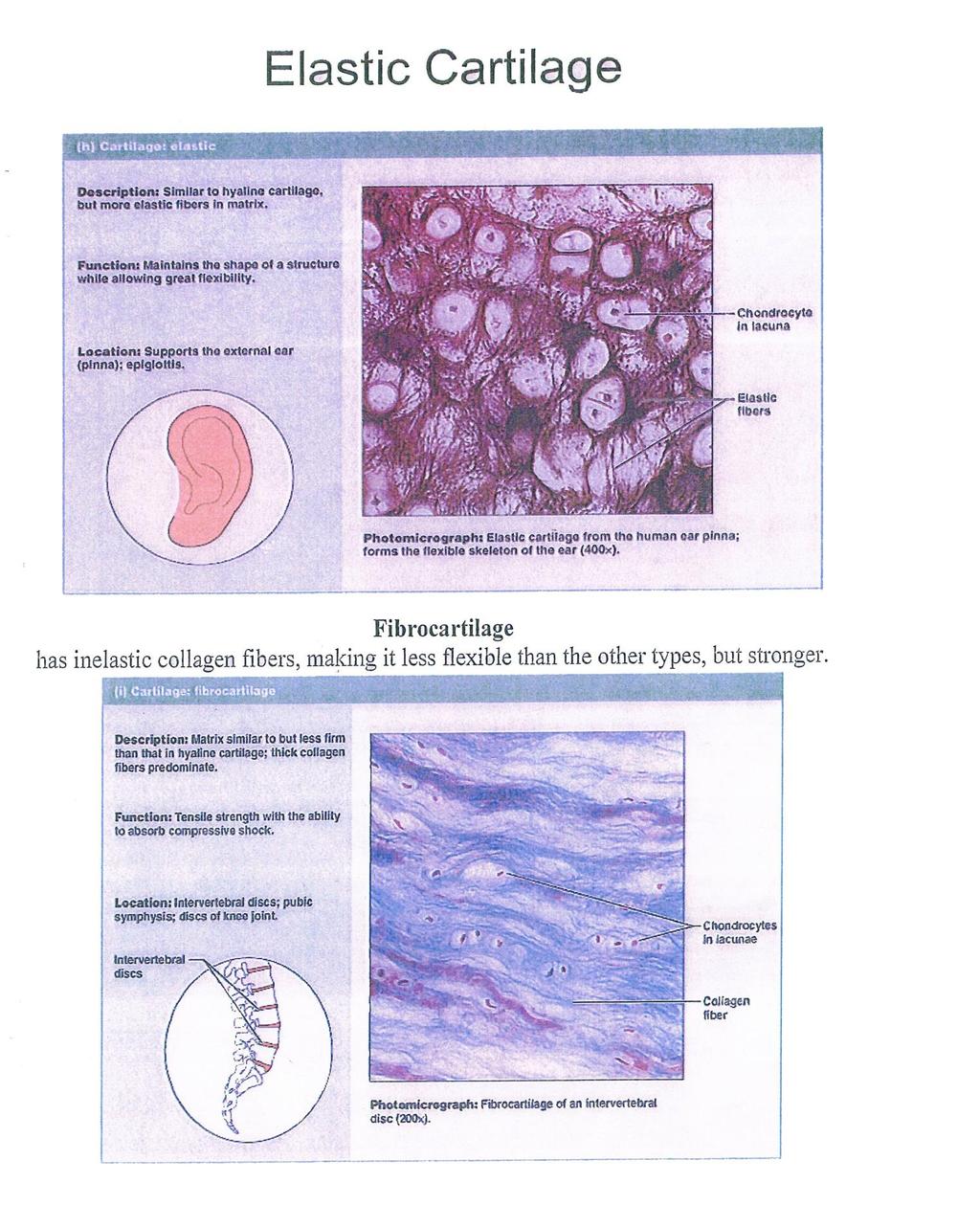
8 Elastic cartilage : Elastic cartilage differs from hyaline cartilage principally by the presence of elastic fibers in its matrix After staining the cartilage with orcein, these are visible as deep purple fibers. this cartilage surrounded by perichondrium. The fibers enter the cartilagenous matrix from the perichondrium. found in the external ear and the auditory tubes, epiglottis. Function gives support, maintains shape, allows flexibility. 2. White fibro cartilage found in the intervertebral disc, pubic symphysis. and menisci of the knee. the matrix contain bundles of white fibers parallel to each other with little ground substance, this type of cartilage is not surrounded by perichondrium. Function = support and fusion, and absorbs shocks. 8
9 Most mature bones are more-or-less hollow. The outer bone is called cortical or compact bone, while the inner marrow cavity may be crisscrossed by thin strands of bone called trabeculae or spongy bone. All mature bone is formed in layers called lamellae. Within the lamellae are small s or lacunae, in which osteocytes reside. Neighboring lacunae are interconnected by thin channels, or canaliculi. In compact bone, the lamellae are organized into sets of concentric rings called osteons or Haversian systems. Each osteon includes a central channel, the Haversian canal, which contains a blood vessel. Volkmann's canal is a cross canal connects between the haversian canals, Volkmann's canal not surrounded by lamellae. In spongy bone the bone matter found as irregular trabeculae not contain the osteons, but they branched and meet forming spaces filled with the bone marrow. These trabeculae surrounded by osteoblasts, the matrix contain oste and osteoclasts. osteocytes, osteoblasts and osteoclasts can be identified not only by their appearance as cells but also by their position in relation to the adjacent bone lamellae. Osteocytes are isolated within lacunae, separated from other cells by bone matrix. Osteoblasts (bone-forming cells) are small cuboidal cells, usually found lying adjacent to one another upon lamellae they have just secreted. Osteoclasts (bone removing cells) are large cells with multiple nuclei, occur in small. spaces (called Howship's lacunae) which they have eroded into the surface. Osteoclasts remove preexisting bone. They are active in bone development and also in bone remodeling. 9
10 BONE Bone is the main constituent ofthe adult skeleton. It is a skeletal connective tissue specialized for support and protection. Bone tissue is rich in blood supply, i.e. highly vascular tissue. Moreover, bone is a type of connective tissue but with calcified matrix. Types of Bone Tissue: Regular (long) or compact bone. Irregular (flat) or spongy bone. Structure of bone: As other connective tissues, bone is formed of :- - Cells: steogeneic cells, Osteoblasts, osteocytes and osteoclasts. - Matrix: of organic and non-organic parts. - Fibers: collagen type I fibers. Bone cells 1- Osteogenic (osteoprogenitor) cells They are mesenchymal stem cells found in the periosteum and endosteum. They are small spindle-shaped cells with pale cytoplasm and ovoid nuclei. Two types are distinguishable by the electron microscope: one can differentiate into osteoblasts, and the other into osteoclasts. Osteoblast precursors derive from embryonic mesenchyme and have sparse RER and Golgi complexes. Osteoclast precursors derive from blood monoctyes and have abundant free ribosomes and mitochondria. 10
11 2- osteoblasts: - They are bone forming cells, found in the growing surface of the bone (in the periosteum and endosteum) they form one-cell-thick sheets. - They are dividing cells that synthesize the organic components of bone matrix. - They are large rounded or cuboidal cells, with deep basophilic cytoplasm, well- developed RER and Golgi and eccentric nucleus - They synthesize and secrete all the organic components of bone matrix and may be involved in bone mineralization. - Once surrounded by matrix, osteoblasts are considered mature and called osteocytes. 3- Osteocytes - The cells are branched, smaller than osteoblasts, and not divide. - They are terminally differentiated bone (mature) cells found in cavities in the bone matrix called lacunae, and their processes (branches) extend into canaliculi in the classified matrix. - Osteocytes are isolated from one another by an impermeable bone matrix and contact one another at the tips of their filopodia, often through gap junctions. - Osteocytes recently derived from osteoblasts are located near bone surfaces in rounded lacunae; older cells are found farther from the surface in flattened lacunae. - They maintain bone matrix. - The death of osteocytes results in bone breakdown, or resorption. 4- Osteoclasts - Osteoclasts are bone-resorbing cells that lie on bony surfaces in shallow depressions called Hawship's lacunae. - Osteoclasts are large multinucleated cells (~50 nuclei), with acidophilic cytoplasm containing abundant lysosomes and mitochondria and well 11

12 developed Golgi complex; and brush border (Ruffled border) of plasmamembrane facing the bone marrow. - They are derive from the fusion of blood monocyte derivatives and are considered components of the mononuclear phagocyte system. - The cells release acid, collagenase, and other lytic enzymes into the compartments; these break down bone matrix and release minerals, a process called bone resorption. Histology Bone matrix: Bone matrix contains organic components (osteoid), and inorganic components (bone mineral). The organic components constitutes about 50% of bone volume and 25% of bone weight. It is composed of type I collagen fibers and unmineralized ground substance, which is composed of proteins. Carbohydrates, and small amounts of proteoglycans and lipids. The inorganic components (bone 12


13 mineral) makes up about 50% of bone volume and 75% of bone weight. It is composed of calcium and phosphate, with some bicarbonate, citrate, magnesium and potassium and trace amounts of other metals. Compact Bone is solid in appearance more complex arrangement of blood vessels cells and matrix surround blood vessels in long bones 1- Periosteum: covering the long bone, and formed of two layers:- - Outer fibrous layer of collagen fibers. - Inner cellular layer of osteogenic cells and cells and osteoblasts. 2- Endosteum: a cellular layer lining the bone cavities, and formed of osteogenic cells and osteoblasts. 3- Haversian system (osteon):- Bone lamellae are arranged concentrically around the blood vessels. The bone lamellae are formed of osteocytes inside lacunae and canaliculi embedded in calcified matrix. 4- Volkmann's Canals :- They are transverse canals connecting blood vessels in the Haversian canals to each other and to those in the periosteum and in marrow cavities. 5- External circumferential lamellae. 6- Internal circumferential lamellae. 7- Interstitial lamellae. 13

14 Spongy or cancellous bone bone looks spongy in appearance no complex arrangement for blood vessels fills heads of long bones called marrow always covered by compact bone. - Spongy bone forms a fine 3-dimensional lattice with many bony cavities, with branching and anastomosing slips of bone between the cavities, called trabeculae. - Spongy bone is found at the core of the epiphyses of mature long bones, at the core of short bones, and between the thick plates, or tables, of the flat bones of the skull, where it is called the diploii. 14

15 Cartilage Cartilage differs from the "connective tissues proper" which we studied in the last lab in that the matrix is a gel-like substance which gives the cartilage shape along with flexibility. Unlike other CT, cartilage has NO blood vessels or nerves except in the perichondrium.combining various matrix components including one or more type of fibers produces a variety of cartilage types. Type of cartilage. 1-hyaline 2-fibrocartilage 3-elastic Hyaline The most common type of cartilage is hyaline. Hyaline cartilage has both elastic and inelastic fibers, but they are so finely divided that they cannot be seen under the light microscope. In cartilage the cells wall themselves off from the matrix inside lacunae or "lakes". Cartilage will usually have a fibrous covering known as the perichondrium. Hyaline Cartilage Elastic cartilage Ls very rare, found only in the eoiglottis and aura of the ear. The elastic fibers are found in dense bundles. 15
16 16
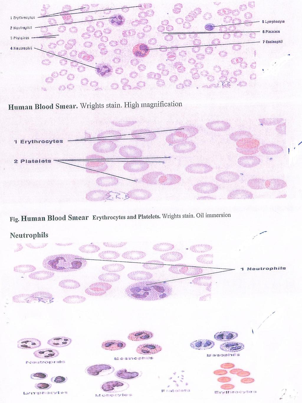
17 Vesicular Connective Tissues The Blood Blood is classified as a specialized form of connective tissue. All connective tissue consists of cells embedded in a matrix which consists of ground substance and fibers. Blood may thus be described as connective tissue whose matrix consists of free-flowing ground substance (plasma) with no fibers. Red blood cells (RBCs, also called erythrocytes) are continually produced in bone marrow and recycled in spleen. In mature form the lack nuclei and most cytoplasmic structures they are flexible bags of hemoglobin, have biconcave disc shape. White blood cells (WBCs, also called leukocytes ) include several distinct cell types, neutrophils, eosinophil, basophils, lymphocytes and monocytes. Certain developmental and morphological similarities permit the first three these cells to grouped together as granulocytes. The latter two types are then categorized as a granulocytes or mononuclear leukocytes. Granulocytes, also called Dolymorphonuclear leukocytes, have grainy,cytoplasm and elongated or lobed nuclei. They arise in bone marrow from cells called myeloblasts and pass through stages called myelocytes and metamyelocytes. The granulocytes include neutrophils, eosinophils, and basophils. Neutrophils are the most numerous of the leukocytes, about 60% of the white blood cell count. Neutrophils take their name from the staining properties of their cytoplasmic lysosomal granules, these granules are neutrophilic, meaning they show no special affinity for either acidic or basic stains but are stained mildly by both. (This is in contrast to the specific granules of eosinophils, which stain red with acidic stains such as eosin, and those of basophils, which stain 17
18 with basic stains), the nuclei of mature neutrophils are elongated and inched into several distinct lobes. (Neutrophils are anti-bacterial cells which lyse (break down) bacterial cells). Eosinophils or acidophils (eosinophilic granulocytes) normally comprise less than 2-4 of the per era Their specific granules are intense eosinophilic (stained by eosin) Eosinophils are about the same size as neutrophils. Their nuclei are typically band shape (elongated) or two-lobed. The function of eosinophils remains unclear, although they are known to proliferate in association with allergies and parasites. Basophils (basophilic granulocytes) normally comprise less than 1% of the peripheral leukocytes. Their specific granules are intense basophilic. Like eosinophils, Basophils are. similar in size to neutrophils. Their nuclei may be band shaped or segmented. Basophils seem to be functionally similar to tissue mast cells, involved in triggering inflammation. Mononuclear leukocytes comprise both lymphocytes and Monocytes. Both cell types work together in immune responses. Monocytes are the largest of the leukocytes, and constitute about 5-10 of the WBC population in peripheral blood. In blood smears, their nuclei have kidney or bean shape. Monocytes belong to the same functional population as tissue macrophages Monocytes/macrophages engulf and digest foreign microorganisms, dead or worn-out cells and other tissue debris. They interact closely with lymphocytes to recognize and destroy foreign substances. Lymphocytes are small cells, and they are the second most common white blood cell type, comprising about 30% of the leukocyte population in peripheral 18
19 blood. Lymphocytes have a round heterochromatic (deeply staining) nucleus surrounded by a relatively thin rim of cytoplasm. Lymphocytes also emigrate from blood in response to inflammation, but they accumulate somewhat later during the inflammatory process than neutrophils. Their presence in large numbers indicates the continuing presence of antigen. Lymphocytes produce the antibody molecules (one specific type of antibody per lymphocyte) which provide the mechanism for chemical recognition of foreign materials. Lymphocytes are most easily recognized in histological sections as small "naked" nuclei (the cytoplasm is usually inconspicuous) which occur here and there in most connective tissues, especially commonly near mucous membranes. Lymphocytes are found densely packed in lymphoid tissue-spleen, lymph nodes, and lymph nodules in mucous membranes (e.g. tonsils, appendix), where they proliferate. Platelets are small fragments of cytoplasm derived from bone marrow cells called Megakaryocytes Megakaryocytes are easy to recognize by their huge size and large lobulated nuclei. Small bits of megakaryocyte cytoplasm are continually pinched off to enter circulation as platelets, where they are important for blood clotting. Hemopoietic Bone Marrow: Blood cells are made in the bone marrow. The bone marrow is the spongy material in the center of the bones that produces about 95 percent of the body's blood cells. There are other organs and systems in our bodies help regulate blood cells. The lymph spleen, and liver help the production, destruction, and differentiation. 19
20 (developing a specific function) of cells. The production and development of new cells is a process called hematopoiesis. Blood cells formed in the bone marrow start out as a stem cell. A "stem cell" (or hematopoietic cell) is the initial phase of all blood cells. As the stem cell matures, several distinct cells evolve such as the red blood cells, white blood cells, and platelets. Immature blood cells are also called blasts. Some blasts stay in the marrow to mature and others travel to other parts of the body to develop into mature, functioning blood cells. Blood is a specialized body fluid. It has four main components: plasma, red blood cells, white blood cells, and platelets. Blood has many different functions, including: transporting. oxygen and nutrients to the lungs and tissues. forming blood clots to prevent excess blood loss. carrying cells and antibodies that fight infection. bringing waste products to the kidneys and liver, which filter and clean the blood. regulating body temperature. Regulation of body ph. The blood that runs through the veins, arteries, and capillaries is known as whole blood, a mixture of about 5 percent plasma and 45 percent blood cells. About 7 to 8 percent of total body weight is blood. 20
21 21
22 22
Practical Histology. Lab 3: Connective tissue
 Practical Histology Lab 3: Connective tissue Connective tissues Connective tissue provides structural support for the body by binding cells and tissues together to form organs. It also provides metabolic
BONE TISSUE. Dr. Heba Kalbouneh Associate Professor of Anatomy and Histology
 BONE TISSUE Dr. Heba Kalbouneh Associate Professor of Anatomy and Histology BONE FUNCTION Support Protection (protect internal organs) Movement (provide leverage system for skeletal muscles, tendons, ligaments
Connective Tissue. Found everywhere in the body. Most abundant and widely distributed. Never exposed to the outside environment.
 Connective Tissue Found everywhere in the body. Most abundant and widely distributed. Never exposed to the outside environment. Connective Tissue Functions Binding and support Protection Insulation Transportation
Blood. Hematopoietic Tissue
 Blood Hematopoietic Tissue Is a type of connective tissue in which its cells are suspended in a circulating fluid. Erythrocytes+ leukocytes + platelets (thrombocytes) =formed elements of blood. These formed
Biology. Dr. Khalida Ibrahim
 Biology Dr. Khalida Ibrahim BONE TISSUE Bone tissue is a specialized form of connective tissue and is the main element of the skeletal tissues. It is composed of cells and an extracellular matrix in which
Chapter 4. Cartilage and Bone. Li Shu-Lei instructor. Dept. Histology and Embryology, School of Basic Medical Sciences, Jilin University
 Chapter 4 Cartilage and Bone Li Shu-Lei instructor Dept. Histology and Embryology, School of Basic Medical Sciences, Jilin University I Cartilage a specialized connective tissue Characterizers: Cartilage
Most abundant and widely distributed tissues in the body Binds, support, and strengthen body tissues, protect and insulate internal organ, serve as
 Connective tissue Most abundant and widely distributed tissues in the body Binds, support, and strengthen body tissues, protect and insulate internal organ, serve as major transport system, compartmentalizes
Cartilage & bone. Red: important. Black: in male female slides. Gray: notes extra. Editing File
 Cartilage & bone Red: important. Black: in male female slides. Gray: notes extra. Editing File OBJECTIVES describe the microscopic structure, distribution and growth of the different types of Cartilage
Lecture Overview. Connective Tissues. Marieb s Human Anatomy and Physiology. Chapter 4 Tissues: The Living Fabric Connective Tissues Lecture 10
 Marieb s Human Anatomy and Physiology Marieb Hoehn Chapter 4 Tissues: The Living Fabric Connective Tissues Lecture 10 Lecture Overview General composition and function of connective tissue Components of
The Tissue Level of Organization
 Tissue The Tissue Level of Organization Chapter 3 Definition an aggregation of cells in which each cooperates with all others in the performance of a given function Examples of general functions Movement
What are the parts of the skeletal system? Chapter 6- Part I Bones and Skeletal Tissues. Growth of Cartilage. Bones come in many shapes
 Chapter 6- Part I Bones and Skeletal Tissues Components of the skeletal system Classification of Bone (bone shapes) Functions of bone Bone structure Microscopic structure of bone and bone cells What are
Which compound is reponsible for the viscous character of the ground substance?
 1 2 Which type of collagen forms the coarse collagen fibres in dense regular and irregular connective tissues? Which compound is reponsible for the viscous character of the ground substance? 3 Which class
Quiz 6. Cartilage and Bone
 Quiz 6 Cartilage and Bone MCQs X type (true or false): 1. Cartilage tissue: a. Has a rich blood supply. b. Develops from mesenchyme. c. Has ability for a quick regeneration. d. Has chondrocytes as precursor
CONNECTIVE TISSUE (Refer to pp for specific characteristics of each) VAN (**Be familiar with exceptions**)
 VAN (**Be familiar with exceptions**)") CONNECTIVE TISSUE CHARACTERISTICS: *Most abundant tissue type; Composed of ECM (GS & Protein Fibers) + Cells (Refer to pp.129-131 for specific characteristics of each) *Highly equipped with VAN assists
CONNECTIVE TISSUE CHARACTERISTICS: *Most abundant tissue type; Composed of ECM (GS & Protein Fibers) + Cells (Refer to pp.129-131 for specific characteristics of each) *Highly equipped with VAN assists
CONNECTIVE TISSUE (C.T.)
") CONNECTIVE TISSUE (C.T.) Objectives: By the end of this lecture, the student should be able to: 1. Enumerate the general characteristics of C.T. 2. Classify C.T into C.T. proper and special types of C.T.
CONNECTIVE TISSUE (C.T.) Objectives: By the end of this lecture, the student should be able to: 1. Enumerate the general characteristics of C.T. 2. Classify C.T into C.T. proper and special types of C.T.
Human Anatomy and Physiology I Laboratory
 Human Anatomy and Physiology I Laboratory Skeletal Tissue: Cartilage and Bone This lab involves study of the laboratory exercise Overview of the Skeleton, Classification and Structure of Bones and Cartilages,
8/30/2017. Tissue: The Living Fabric. 4.3 Connective Tissue
 Chapter 4 Part B Tissue: The Living Fabric Annie Leibovitz/Contact Press Images PowerPoint Lecture Slides prepared by Karen Dunbar Kareiva Ivy Tech Community College 4.3 Connective Tissue Connective tissue
HOLE S ANATOMY CHAPTER 5, PART II Lecture notes
 HOLE S ANATOMY CHAPTER 5, PART II Lecture notes I. Connective Tissue A. Structure 1. have few cells that are spaced apart and can divide; two categories: a. fixed cells cells that are present in tissue
Connective Tissue. Consists of two basic elements: Cells and Extra-cellular matrix
 Connective Tissue Consists of two basic elements: Cells and Extra-cellular matrix True Connective Tissue Cells Fibroblasts: Secrete both fibers and ground substance of the matrix (wandering) Macrophages:
SKELETAL TISSUES CHAPTER 7 INTRODUCTION TO THE SKELETAL SYSTEM TYPES OF BONES
 SKELETAL TISSUES CHAPTER 7 By John McGill Supplement Outlines: Beth Wyatt Original PowerPoint: Jack Bagwell INTRODUCTION TO THE SKELETAL SYSTEM STRUCTURE Organs: Bones Related Tissues: Cartilage and Ligaments
Bio& 241 Unit 1 / Lecture 4
 Bio& 241 Unit 1 / Lecture 4 Connective Tissue Consists of two basic elements: Cells and Extra-cellular matrix 1 True Connective Tissue Cells Fibroblasts: Secrete both fibers and ground substance of the
Connective Tissues. Copyright 2009 Pearson Education, Inc., publishing as Pearson Benjamin Cummings
 C.T. are found in all parts of the body & diverse in structure & function. C.T. Functions: -connect structures -provide support -protect vital organs -fill space b/w structures -stores fat -defends body
Compact bone; Many parallel Haversian canals contain: small blood vessels. very small nerve. Interconnected by Volkmann s canals.
 Special characteristics of COMPACT BONE (dense bone) Thick; well vascularized Osteocytes and lamellae Concentric rings around blood vessels Most bones: outer compact bone inner spongy bone Marrow cavity
Lab Exercise 6a-2. Classification of connective tissues. Connective Tissue. Connective tissues. Areolar. Areolar tissue
 Classification of connective tissues Lab Exercise 6a-2 Connective Tissue Nervous Muscle Connective Tissue Connective tissues Connective tissue proper Fluid connective tissue Supportive connecting tissue
Cartilage. - Cartilage together with long bone form the skeleton and support the body.
 Cartilage - Cartilage is a special type of CT has a firm pliable matrix that can resist mechanical stress, act as a shock absorber. - Cartilage together with long bone form the skeleton and support the
Chapter 5. Tissues. 4 Types of Body Tissues. Tissues
 Chapter 5 Tissues Tissues Tissues - groups of cells that are similar in structure & function RBC, WBC, & platelets are a group of cells working together to form BLOOD tissue Histology Pathohistology study
Connective Tissue Nervous Muscle. Classification of connective tissues
 Connective Tissue Nervous Muscle Lab Exercise 6a-2 Classification of connective tissues 1 Connective Tissue Connective tissue proper Fluid connective tissue Supportive connecting tissue Connective tissues
Basic Histology. By Mrs. Bailey
 Basic Histology By Mrs. Bailey Primary Tissues 1. Epithelial Tissue 2. Connective Tissue 3. Muscle Tissue 4. Nervous Tissue Very cellular Supported by underlying connective tissue Epithelial & connective
Lab Animal Tissue. LEARNING OBJECTIVES: To understand the relationship between the structure and function of different animal tissues
 Name: Bio A.P. PURPOSE: HYPOTHESIS: NONE Lab Animal Tissue BACKGROUND: In animals, groups of closely related cells specialized to perform the same function are called tissues. There are four general classes
Histology. There are four basic tissue types in the body are :-
 Histology Lab.I There are four basic tissue types in the body are :- 1- Epithelial tissues (Epithelium) 2- Connective tissues 3- Muscular tissues 4- Nervous tissues 1-Epithelial tissues epithelial tissues
Growth and repair: Cartilage is a vascular tissues that receives nutrients by diffusion through its matrix, cartilage grow by 2 mechanisms:
 Skeletal connective tissues: (cartilage and bone): Cartilage and bone are specialized connective tissues both adapted to serve as skeletal framework in most vertebrates the presence of solid inter cellular
The Skeletal System:Bone Tissue
 The Skeletal System:Bone Tissue Dynamic and ever-changing throughout life Skeleton composed of many different tissues cartilage, bone tissue, epithelium, nerve, blood forming tissue, adipose, and dense
Tissues organs system organism. pg151
 Histology is the study of tissues A TISSUE is a group of cells, usually of one kind, & their intercellular substance (e.g. intercellular matrix in animal) which are linked together & perform a particular
Epithelia of Coverings and Linings. Tissues. Tissue
 Tissue Tissues Chapter 3 Definition an aggregation of cells in which each cooperates with all others in the performance of a given function Examples of general functions Movement Protection Support Production
Cartilage. Dr. Heba Kalbouneh Associate Professor of Anatomy and Histology
 Cartilage Dr. Heba Kalbouneh Associate Professor of Anatomy and Histology 1 Cartilage is a specialized type of connective tissue designed to give support, bear weight and withstand tension, torsion and
The Tissue Level of Organization
 The Tissue Level of Organization 4.5-4.11 August 31, 2012 4.5 Connective Tissues Describe the general features of connective Describe the structure, location, and function of the various types of connective
Tejido Conectivo Parte B. Informe #3 Laboratorio Biología # 240 Profesor: Javier Cabello
 Tejido Conectivo Parte B Informe #3 Laboratorio Biología # 240 Profesor: Javier Cabello Figure 4-8 The Cells and Fibers of Connective Tissue Proper Areolar Elastic fibers Collagen fibers Fibroblast Free
Sheet #7. Dr. Heba Kalbouneh. Dr. Heba Kalbouneh. Dr. Heba Kalbouneh
 Sheet #7 Dr. Heba Kalbouneh Dr. Heba Kalbouneh Dr. Heba Kalbouneh Connective tissue The differences between epithelial and connective tissue - Epithelial cells are tightly packed (no or minimal spaces
Connective tissues. Dr. Hersh Abdul Ham-Karim BVM&S, PG Dip, MSc and PhD
 Connective tissues Dr. Hersh Abdul Ham-Karim BVM&S, PG Dip, MSc and PhD Connective tissue (CT) Connective tissue is a term applied to a basic type of tissue of mesodermal origin, which is sparsely populated
The Skeletal System:Bone Tissue
 The Skeletal System:Bone Tissue Dynamic and ever-changing throughout life Skeleton composed of many different tissues cartilage, bone tissue, epithelium, nerve, blood forming tissue, adipose, and dense
Body Tissues Pearson Education, Inc.
 Body Tissues Tissues Groups of cells with similar structure and function Four primary types: Epithelial tissue (epithelium).1 Connective tissue.2 Muscle tissue.3 Nervous tissue.4 Epithelial Tissues Locations:
10/3/2012. Tissue: The Living Fabric: Part B. Extracellular matrix Ground substance Fibers Collagen fiber Elastic fiber Reticular fiber.
 PowerPoint Lecture Slides prepared by Janice Meeking, Mount Royal College C H A P T E R 4 Tissue: The Living Fabric: Part B Copyright 2010 Pearson Education, Inc. Copyright 2010 Pearson Education, Inc.
Functions of the Skeletal System. Chapter 6: Osseous Tissue and Bone Structure. Classification of Bones. Bone Shapes
 Chapter 6: Osseous Tissue and Bone Structure Functions of the Skeletal System 1. Support 2. Storage of minerals (calcium) 3. Storage of lipids (yellow marrow) 4. Blood cell production (red marrow) 5. Protection
Tissues are groups of cells with a common structure (form) and function (job).
 and function (job).") Dr Narmeen S. Ahmad Tissues are groups of cells with a common structure (form) and function (job). There are (4) types of tissue: 1. Epithelial 2. Connective 3. Muscle 4. Nervous Epithelial cells Epithelium
Dr Narmeen S. Ahmad Tissues are groups of cells with a common structure (form) and function (job). There are (4) types of tissue: 1. Epithelial 2. Connective 3. Muscle 4. Nervous Epithelial cells Epithelium
Connective tissue CONNECTIVE TISSUE Part I
 Connective tissue CONNECTIVE TISSUE Part I Part 1 Connective Tissue Found everywhere in the body (app. 50% of body weight) Includes the most abundant and widely distributed tissues General features of
Chapter 7. Skeletal System
 Chapter 7 Skeletal System 1 Introduction: A. Bones are very active, living tissues B. Each bone is made up of several types of tissues and so is an organ. C. Bone functions include: muscle attachment,
Due in Lab. Due next week in lab - Scientific America Article Select one article to read and complete article summary
 Due in Lab 1. Skeletal System 33-34 2. Skeletal System 26 3. PreLab 6 Due next week in lab - Scientific America Article Select one article to read and complete article summary Cell Defenses and the Sunshine
contains an antiangiogenesis factor
 CARTILAGE & BONE Cartilage and Bone objectives Student must learn :. What is the meaning of cartilage, and their function, location in human body.. To distinguish the 3 types of cartilage. And their cells,
HISTOLOGY. Simple squamal lungs
 HISTOLOGY Lab Objectives: Students should be able to... 1. Visually identify each class of tissue and examples within each class 2. Indicate the location (in the human body and/or organ) and function of
Sheet #9. Dr. Heba Kalbouneh. Dr. Heba Kalbouneh. Dr. Heba Kalbouneh
 Sheet #9 Dr. Heba Kalbouneh Dr. Heba Kalbouneh Dr. Heba Kalbouneh Elastic fibers The main function of elastic fibers is to provide elasticity. In other words these fibers are able to restore the original
OSSEOUS TISSUE & BONE STRUCTURE PART I: OVERVIEW & COMPONENTS
 OSSEOUS TISSUE & BONE STRUCTURE PART I: OVERVIEW & COMPONENTS The Skeletal System Skeletal system includes: bones of the skeleton, cartilages, ligaments, and connective tissues What are the functions of
4 Types of Tissue. Epithelial Connective Muscle Neural
 Connective Tissue 4 Types of Tissue Epithelial Connective Muscle Neural Connective Tissue Fills internal spaces Supports & binds other tissues Transports materials Stores energy Classification of Connective
Function: Provides reserve food fuel; Copyright 2011 Pearson Education, Inc. Copyright 2011 Pearson Education, Inc. White blood cell (lymphocyte)
") Adipose Tissue Closely packed adipocytes Have nucleus pushed to one side by fat droplet Richly vascularized Provides reserve food fuel Insulates against heat loss Supports and protects organs Under skin
Adipose Tissue Closely packed adipocytes Have nucleus pushed to one side by fat droplet Richly vascularized Provides reserve food fuel Insulates against heat loss Supports and protects organs Under skin
Individual cells Extracellular matrix
 Connective Tissue Connective Tissue Elements Individual cells Extracellular matrix»fibers» Collagen» Elastic» Reticular»Ground Substance» PG (proteoglycans)» GAG (glycosaminoglycan)» GP (glycoprotein)
5.3. The Nature of Cartilage Matrix The components of cartilage matrix include a high component of fibers, and proteoglycans. Proteoglycans are a
 Chapter 5 Supportive Tissues Support in Animals is carried out by Cartilage and Bone. A. Cartilage 5.1. Nature of Cartilage Cartilage is a highly resilient c..t that provides strength and support in areas
Tissue Outline (chapter 4) Tissues group of cells that perform structural and roles. List the 4 types:
 Tissues group of cells that perform structural and roles. List the 4 types:") Tissue Outline (chapter 4) Tissues group of cells that perform structural and roles. List the 4 types: 1. 2. 3. 4. I. Epithelial Tissue covers all the surfaces, inside & out. Are the major tissues of,
Tissue Outline (chapter 4) Tissues group of cells that perform structural and roles. List the 4 types: 1. 2. 3. 4. I. Epithelial Tissue covers all the surfaces, inside & out. Are the major tissues of,
Module 2:! Functional Musculoskeletal Anatomy A! Semester 1! !!! !!!! Hard Tissues, Distal Upper Limb & Neurovascular Supply of Upper Limb!
 Functional Musculoskeletal Anatomy A Module 2: Hard Tissues, Distal Upper Limb & Neurovascular Supply of Upper Limb Semester 1 1 18. Bone Tissue & Growth of Bones 18.1 Describe the structure of bone tissue
Connective Tissues. 2. Describe the function of fibroblasts. 3. What is ground substance? What is its function?
 Connective Tissues Directions: Insert and install your Interactions: Foundations CD. a. Click the "Contents" button. b. Open the Tissue Level of Organization file. c. Click on Anatomy Overviews. d. Work
Mast Cell. Mast Cells. James W. Truman, Ph.D. Howard Hughes Medical Institute Chevy Chase, Maryland
 5 th ANNUAL SINAUER ASSOCIATES DISTINGUISHED SCIENTIST LECTURE James W. Truman, Ph.D. Howard Hughes Medical Institute Chevy Chase, Maryland Neuronal Lineages in the CNS of Drosophila: Units of Development,
BIOH111. o Cell Module o Tissue Module o Integumentary system o Skeletal system o Muscle system o Nervous system o Endocrine system
 BIOH111 o Cell Module o Tissue Module o Integumentary system o Skeletal system o Muscle system o Nervous system o Endocrine system Endeavour College of Natural Health endeavour.edu.au 1 TEXTBOOK AND REQUIRED/RECOMMENDED
4 Types of Tissue. Epithelial Connective Muscle Neural
 Connective Tissue 4 Types of Tissue Epithelial Connective Muscle Neural Connective Tissue Fills internal spaces Supports & binds other tissues Transports materials Stores energy Classification of Connective
physical properties depend on: electrostatic bonds between collagen/elastic fibers and GAGs water bound to negatively charged sulfated GAG chains
 connective/supporting tissue bears mechanical stress without distortion -> shock absorption smooth surface -> facilitates movements of joints guides development of bones chondrocytes extracellular matrix
A. cells that perform related functions and are similar in structure. B. extracellular material - made by cells and secreted into interstitial space
 I. tissue components A. cells that perform related functions and are similar in structure B. extracellular material - made by cells and secreted into interstitial space II. tissue types A. epithelium (e.)
BIOL 2457 CHAPTER 4 Part 2 SI All connective tissues arise from, an embryonic tissue.
 BIOL 2457 CHAPTER 4 Part 2 SI 1 1. All connective tissues arise from, an embryonic tissue. 2. Describe the vascularity of connective tissues, which are very diverse. 3. Describe the innervation of connective
What is histology? HISTOLOGY
 Introduction to Histology What is histology? HISTOLOGY histo = tissue ogy = study So HISTOLOGY = the study of tissues! What is a TISSUE? Tissues are groups of cells with specialized structural and functional
HISTOLOGY Lecture TWO DR. ASHRAF SAID
 HISTOLOGY Lecture TWO DR. ASHRAF SAID Start Of this lecture TISSUES TISSUE: A DEFINITION A group of connected and interdependent cells that cooperate to perform a specific function CONNECTIVE TISSUE The
Histology review. Histology. Slides. Epithelial tissue. Another example - kidney. Simple cuboidal epithelium. What to look for
 Histology review Histology What to look for Histology Practical = 50 pts Some slides set up on scopes (~10) Some Powerpoint pictures on the projector Questions I will ask: What kind of tissue? General
Tissues 10/21/2016. Epithelial Tissue
 Tissues This is a generalized cell diagram. It shows the anatomy of a cell, but most cells do not actually look like this. Cells can have a wide variety of shapes and sizes, depending on their function.
Chapter 6: Skeletal System: Bones and Bone Tissue
 Chapter 6: Skeletal System: Bones and Bone Tissue I. Functions A. List and describe the five major functions of the skeletal system: 1. 2. 3.. 4. 5.. II. Cartilage A. What do chondroblasts do? B. When
Skeletal Tissues. Skeletal tissues. Frame; muscles, organs and CT attach. Brain, spinal cord, thoracic organs; heart and lungs.
 Skeletal Tissues Functions 1) support 2) protection 3) movement Skeletal tissues Frame; muscles, organs and CT attach. Brain, spinal cord, thoracic organs; heart and lungs. Aids muscle contraction; generate
Connective Tissue. Red: important. Black: in male female slides. Gray: notes. Editing File
 Connective Tissue Red: important. Black: in male female slides. Gray: notes. Editing File OBJECTIVES Enumerate the general characteristics of C.T Classify C.T into C.T. proper and special types of C.T
Tissues. Tissues - Overview. Bio211 Laboratory 2. Epithelial and Connective Tissues
 Bio211 Laboratory 2 Epithelial and Connective Tissues 1 Tissues Tissues to be examined under the microscope Epithelial Tissue (p. 79 Lab Manual) [TODAY] Connective Tissue (p. 93 Lab Manual) [TODAY] Muscle/Nervous
KEY CONCEPTS Unit 6 THE SKELETAL SYSTEM
 ANATOMY & PHYSIOLOGY 1 (101-805 - AB) PAUL ANDERSON 2011 KEY CONCEPTS Unit 6 THE SKELETAL SYSTEM A Overview of The Skeletal System 1. Definition: Anatomically the SKELETAL SYSTEM consists of bones, cartilages,
Connective Tissue (CT)
") Connective Tissue (CT) YONG-MEI CHEN ( 陈咏梅 ) Dept. of Anatomy, Histology & Embryology Peking Union Medical College Tel:69156461 E-mail address: pumc_he@126.com Content Introduction of CT 1. Origin 2. Compositions
Connective Tissue (CT) YONG-MEI CHEN ( 陈咏梅 ) Dept. of Anatomy, Histology & Embryology Peking Union Medical College Tel:69156461 E-mail address: pumc_he@126.com Content Introduction of CT 1. Origin 2. Compositions
ANATOMY & PHYSIOLOGY - CLUTCH CH. 8 - BONE AND CARTILAGE.
 !! www.clutchprep.com CONCEPT: BONE CLASSIFICATIONS There are four classifications of bones based on their 1. Long bones are greater in length than in width - Found in the upper and lower limbs (ex: arm,
Histology. Study of body tissues
 Histology Study of body tissues 2 Introduction to Body Tissues 1. Composed of specialized cells of similar structure and perform a common function 2. Four major types (4 Cs) a. Epithelial - Cover b. Connective
Car$lage and Bone. Kris$ne Kra0s, M.D.
 Car$lage and Bone Kris$ne Kra0s, M.D. Car$lage and Bone Lecture Objec$ves Describe the general func$ons of car$lage and bone. Compare the func$on and composi$on of the three types of car$lage. Describe
2/21/2012. Components Connective Tissue. Connective Tissue??? Connective Tissue What is it?
 Connective Tissue??? Connective Tissue What is it? It Binds, It Supports, It Strengthens, It Protects, It Insulates, It Compartmentalizes, It helps us move, It helps transport stuff, It is a site for storing
Histology. The study of tissues.
 Histology The study of tissues. Body Tissues Cells are specialized for particular functions Tissues Groups of cells with similar structure and function Four primary types Epithelium Connective tissue Nervous
Tissues Chapter 5...Tissue - a group or mass of similar cells working together to perform certain common functions
 Tissues Chapter 5...Tissue - a group or mass of similar cells working together to perform certain common functions There are 4 major types of tissue Epithelial Connective Muscle Nervous 1. Epithelial Tissue
BONE LABORATORY DEMONSTRATIONS. These demonstrations are found on the bulletin boards outside the MCO Bookstore.
 BONE LABORATORY DEMONSTRATIONS These demonstrations are found on the bulletin boards outside the MCO Bookstore. COMPACT & TRABECULAR BONE - LM When viewed under the polarizing light microscope, the layering
SKELETAL SYSTEM I NOTE: LAB ASSIGNMENTS for this topic will run over 3 Weeks. A SEPARATE WORKSHEET WILL BE PROVIDED.
 BIO 211; Anatomy and Physiology I REFERENCE: CHAPTER 07 1 Dr. Lawrence Altman Naugatuck Valley Community College LECTURE TOPICS OUTLINE SKELETAL SYSTEM I NOTE: LAB ASSIGNMENTS for this topic will run over
The purpose of this practical session is to demonstrate cartilage and bone as specialized connective tissues to the student.
 1 CARTILAGE AND BONE The purpose of this practical session is to demonstrate cartilage and bone as specialized connective tissues to the student. 1. Hyaline cartilage Slide 73 This is a cross section through
Derived copy of Bone *
 OpenStax-CNX module: m57739 1 Derived copy of Bone * Shannon McDermott Based on Bone by OpenStax This work is produced by OpenStax-CNX and licensed under the Creative Commons Attribution License 4.0 By
Skeletal System. The skeletal System... Components
 Skeletal System The skeletal System... What are the general components of the skeletal system? What does the skeletal system do for you & how does it achieve these functions? Components The skeletal system
-the emphasis on this section is the structure and function of bone tissue and on the dynamics of its formation and remodeling throughout life.
 Biology 325 Fall 2004 BONES AND SKELETAL TISSUES Introduction -skeleton contains cartilage and bones -the emphasis on this section is the structure and function of bone tissue and on the dynamics of its
CARTILAGE. Dr. Emad I Shaqoura M.D, M.Sc. Anatomy Faculty of Medicine, Islamic University-Gaza October, 2015
 CARTILAGE Dr. Emad I Shaqoura M.D, M.Sc. Anatomy Faculty of Medicine, Islamic University-Gaza October, 2015 Introduction Hyaline Cartilage Elastic Cartilage Fibrocartilage Cartilage Formation, Growth,
Basic Tissue Types and Functions
 Tissues Histology Basic Tissue Types and Functions 1) Epithelial tissue covering 2) Connective tissue support 3) Muscle tissue movement 4) Nervous tissue control Epithelial Tissue 1) Covers a body surface
Study of different tissues Abnormal cells and tissues can be compared to normal tissues to identify disease, such as cancer Being able to know and
 CHAPTER 4 Study of different tissues Abnormal cells and tissues can be compared to normal tissues to identify disease, such as cancer Being able to know and recognize normal tissues under the microscope
Fig Articular cartilage. Epiphysis. Red bone marrow Epiphyseal line. Marrow cavity. Yellow bone marrow. Periosteum. Nutrient foramen Diaphysis
 Fig. 7.1 Articular cartilage Epiphysis Red bone marrow Epiphyseal line Marrow cavity Yellow bone marrow Nutrient foramen Diaphysis Site of endosteum Compact bone Spongy bone Epiphyseal line Epiphysis Articular
Connective Tissue. Practice Identifying Your Tissue With This Slide Show! Then try the quiz at the bottom of this page!
 Connective Tissue scientistcindy.com /connective-tissue.html Practice Identifying Your Tissue With This Slide Show! Then try the quiz at the bottom of this page! The 4 Types of Connective Tissues Connective
Biology. Dr. Khalida Ibrahim
 Biology Dr. Khalida Ibrahim The cartilage General characteristics: 1. Cartilage is a specialized type of connective tissue (supporting connective tissue). 2. Consists, like other connective tissues, of
Cells and Tissues 3PART D. PowerPoint Lecture Slide Presentation by Patty Bostwick-Taylor, Florence-Darlington Technical College
 PowerPoint Lecture Slide Presentation by Patty Bostwick-Taylor, Florence-Darlington Technical College Cells and Tissues 3PART D Connective Tissue Found everywhere in the body Includes the most abundant
Blood: Functions. Liquid connective tissue 3 general functions 1. Transportation. 2. Regulation. 3. Protection
 Blood Elements Lecture Objectives List blood components. Classify formed elements of blood. Discuss the scientific basis of the above classification. Describe the basic structure of erythrocytes and criteria
Anatomy and Physiology Tissue Review
 Anatomy and Physiology Tissue Review OVERVIEW Histology practicals can be rough, especially when access to slides is limited to the lab period. This resource provides an opportunity to learn or review
BIOLOGY. Chapter 33 Animal Body: Histology Portion Pearson Education, Inc.
 BIOLOGY Chapter 33 Animal Body: Histology Portion Tissues: groups of cells with common function Tissue Category Epithelial (covers & lines) Simple squamous Simple cuboidal Simple columnar Tissues to know:
An Introduction to the Skeletal System Skeletal system includes Bones of the skeleton Cartilages, ligaments, and connective tissues
 An Introduction to the Skeletal System Skeletal system includes Bones of the skeleton Cartilages, ligaments, and connective tissues Functions of the Skeletal System Support Storage of minerals (calcium)
Tissue Outline. Chapter 4. Tissue. Cellular Connections. I. Definitions II. Cellular Connections III. Tissue Types IV. Membranes V.
 Tissue Outline Chapter 4 The Tissue Level of Organization I. Definitions II. Cellular Connections III. Tissue Types IV. Membranes V. Tissue Repair 1 2 Tissue Cellular Connections Tissue Groups of cells
Skeletal System Functions
 Chapter 6 Skeletal System: Bones and Bone Tissue 6-1 Skeletal System Functions Support. Bone is hard and rigid; cartilage is flexible yet strong. Cartilage in nose, external ear, thoracic cage and trachea.
LECTURE OUTLINE: CTP (Connective Tissues Proper) (Ordinary Connective Tissues)
 (Ordinary Connective Tissues)") LECTURE OUTLINE: CTP (Connective Tissues Proper) (Ordinary Connective Tissues) General Definition: Tissues composed of cells embedded in an extracellular (intercellular) matrix, consisting of ground substance
LECTURE OUTLINE: CTP (Connective Tissues Proper) (Ordinary Connective Tissues) General Definition: Tissues composed of cells embedded in an extracellular (intercellular) matrix, consisting of ground substance