Metabolism of Herpes Simplex Virus 1 infected RAW macrophages
|
|
|
- Anna Perkins
- 6 years ago
- Views:
Transcription
1 Metabolism of Herpes Simplex Virus 1 infected RAW macrophages A thesis submitted in partial fulfillment of the requirements for the degree of Master of Science By Mary Kathryn Jenkins B.S., Bowling Green State University, Wright State University
2 WRIGHT STATE UNIVERSITY GRADUATE STUDIES August 22, 2016 I HEREBY RECOMMEND THAT THE THESIS PREPARED UNDER MY SUPERVISION BY Mary K. Jenkins ENTITLED Metabolism of HSV-1 Infected RAW Macrophages BE ACCEPTED IN PARTIAL FULFILLMENT OF THE REQUIREMENTS FOR THE DEGREE OF Master of Science Nancy J. Bigley, Ph.D. Thesis Director Committee on Final Examination Nancy J. Bigley, Ph.D. Professor of Microbiology and Immunology Barbara E. Hull, Ph.D. Director of Microbiology and Immunology Program, College of Science and Mathematics Debra A. Mayes, Ph.D. Assistant Professor, Department of Neuroscience, Cell Biology and Physiology Courtney Sulentic, Ph.D. Associate Professor, Department of Pharmacology and Toxicology Robert E.W. Fyffe, Ph.D. Vice President for Research and Dean of the Graduate School
3 ABSTRACT Jenkins, Mary Kathryn. M.S. Department of Microbiology and Immunology, Wright State University, Metabolism of Herpes Simplex Virus 1 infected RAW macrophages. Macrophages are immune cells that phagocytize pathogens and act as antigen presenting cells. Macrophages are critical in regulating the adaptive immune response and play a key role in neutralizing infection. Macrophages exhibit a variety of phenotypes which have different energy requirements. In the case of HSV-1 infection the virus has been shown to alter metabolic processes of its host. Utilizing a XF24 Extracellular Flux Analyzer the metabolic activity of M0, M1, and (IL-4, IL-13, and IL-10) RAW macrophages was quantified for both uninfected cells and in response to HSV-1 infection. The analysis showed uninfected M0 and (IL-4, IL-13, IL-10) macrophages undergo glycolysis and mitochondrial respiration for energy metabolism while M1 macrophages only undergo glycolysis. HSV-1 showed the ability to alter metabolism in macrophages by inhibiting oxidative phosphorylation. This inhibition leads to increased rates of glycolysis with variability shown among the macrophage phenotypes. As HSV-1 is dependent upon its host cell for energy production the degree of response can be correlated to viral replication. iii
4 Table of Contents Introduction..1 Literature Review.6 Macrophage Function and Polarization...6 Herpes Simplex Virus Metabolism Metabolism of Macrophages..11 Herpes Simplex Virus-1 and Metabolism..13 Materials and Methods...15 Cell Line.15 Polarization Treatment...15 Virus Treatment.16 Laminin Treatment 16 Cell Viability...16 Metabolism Assays...17 Statistical Significance Results Discussion..30 Figures References..56 iv
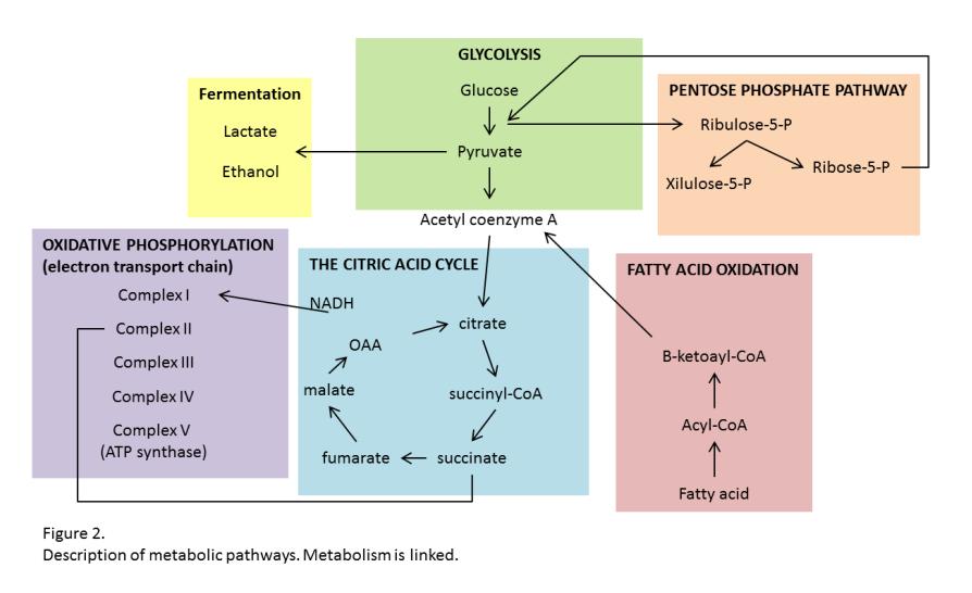
5 List of Figures Figure Page Number: 1: Macrophages are differentiated from monocytes and their functions are based on their polarization 38 2: Description of metabolic pathways : Glycolytic Function of uninfected and infected RAW macrophages : Mitochondrial Respiration of uninfected and infected RAW macrophages : OCR versus ECAR in uninfected and infected RAW macrophages : Glycolysis of uninfected and infected RAW macrophages : Glycolytic Capacity of uninfected and infected RAW macrophages, a measurement of glycolytic Function : Glycolytic Reserve of uninfected and infected RAW macrophages, a measurement of glycolytic Function : Non-glucose derived ECAR of uninfected and infected RAW macrophages, a measurement of glycolytic Function : Glycolytic Reserve Capacity (as fold-increase) of uninfected and infected RAW macrophages, a measurement of glycolytic Function : Basal Respiration of uninfected and infected RAW macrophages, a measurement of mitochondrial respiration : Proton Leak of uninfected and infected RAW macrophages, a measurement of mitochondrial respiration : ATP Production of uninfected and infected RAW macrophages, a measurement of mitochondrial respiration : Maximal Respiration of uninfected and infected RAW macrophages, a measurement of mitochondrial respiration : Spare Respiratory Capacity of uninfected and infected RAW macrophages, a measurement of mitochondrial respiration 52 16: Non-mitochondria derived OCR of uninfected and infected RAW macrophages, a measurement of mitochondrial respiration : Coupling efficiency of uninfected and infected RAW macrophages, a measurement of mitochondrial respiration 54 v
6 18: Spare Respiratory Capacity (as fold-increase) of uninfected and infected RAW macrophages, a measurement of mitochondrial respiration..55 vi
7 List of Abbreviations 2-DG 2-deoxy-glucose ATP adenosine triphosphate ECAR extracellular acidification rate FCCP carbonyl cyanide-4 (trifluoromthoxy) phenylhydrazone LPS lipopolysaccharide IL interleukin MHC major histocompatibility complex NO nitric oxide OCR oxygen consumption rate PFK-1 6-phosphofructo-1-kinase ROS reactive oxygen species vii
8 Acknowledgements I would like to thank Dr. Nancy Bigley for all the support and guidance she has provided through my graduate studies. I would like to thank Dr. Debra Mayes for her support, guidance, and the use of the Seahorse XF flux analyzer. Also, I would like to thank Dr. Courtney Sulentic for her support and guidance. I would also like to acknowledge my lab associates, who expanded my knowledge and provided guidance in my experimentation. viii
9 Dedication I would like to dedicate my thesis research to my husband for his immeasurable support and love. Achieving my goals is made easier with him by my side. Also, to my mother who always expressed the significance of education and work hard. ix
10 Introduction Herpes simplex virus 1 and humans evolved together creating a host environment for the double stranded DNA virus to thrive in (7). HSV-1 is endemic with 50-90% of the world s population carrying the lifelong latent virus, which can become serious if the virus disseminates (20). There is no cure and the only treatment for HSV-1 infection is administration of antivirals. Virus metabolism via cellular components relies on glycolysis and the pentose phosphate pathway (PPP). Glycolysis provides a quick source of energy while PPP helps in de novo nucleotide biosynthesis to create an abundance of viral DNA. HSV-1 forces cells to commit to glycolysis by catalyzing fructose 6- phosphate and ATP to fructose 1,6-bisphosphate and ADP (1). The virus depletes mitochondrial DNA to impair oxidative phosphorylation and inhibits the citric acid cycle, but when mitochondrial DNA depletion occurs the innate immune response is primed (24). Macrophages compose an important part of the innate immune response due to their ability to phagocytize pathogens as well as being involved in homeostasis, tissue repair, and development (26). Macrophages are able to respond to changes within a host environment directed by chemokines, fatty acids, cytokines and toll-like receptors (19). Macrophages exist in two phenotypic states denoted as M1 and, which are examined in this study. M1 or classically activated macrophages are a pro-inflammatory response induced by LPS, the production of IFN-γ, or both. These macrophages are involved in type I inflammation and help with the clearance of pathogens. M1 macrophages also Page 1
11 produce nitric oxide via nitric oxide synthase in order to produce a cytotoxic atmosphere. or alternatively activated macrophages are an anti-inflammatory response induced by IL-4, IL-13, or IL-10. macrophages are involved in type II inflammation, allergies, tissue repair and remodeling, immunoregulation, and encapsulation of parasites (13). Macrophages produce energy based on their phenotype (10). M1 macrophages typically undergo glycolysis and utilize the pentose phosphate pathway while macrophages undergo oxidative phosphorylation and fatty acid oxidation (FAO). M1 macrophages need a fast source of energy in order to phagocytosis pathogens within hours to days. Glycolysis has the ability to provide the rapid energy requirements by M1 macrophages. M1 macrophages enhance the uptake of glucose and found different methods to use the ATP generated by mitochondria more efficiently. The PPP is necessary for the production of NO by providing NADPH. As NO is an inhibitor of cytochrome c oxidase subsequently oxidative phosphorylation is reduced. Huang et al. showed M0 and (IL-4) macrophages undergo mitochondrial respiration, but macrophages depend on fatty acid oxidation (FAO) as well (10). Mitochondrial respiration and FAO provide energy over a longer period of time which is beneficial for macrophages due to their functions of type II inflammation, tissue repair and remodeling, and immunoregulation. M0 and macrophages have the ability to proliferate as well as retain functional plasticity. Utilizing new technology of a XF24 Extracellular Flux Analyzer the oxygen consumption and extracellular ph of RAW macrophages were examined here Page 2
12 under different phenotypes and conditions. Specifically, glycolysis and mitochondrial respiration were examined in inactivated (M0), M1, and induced by IL-4, IL-13, or IL-10 phenotypes to capture baseline metabolic rates for the polarized and non-polarized RAW macrophages. After completing the baseline groups, M0, M1, and (IL-4) macrophages were challenged with HSV-1 and the oxygen consumption and extracellular ph examined to study metabolic changes. Oxygen consumption shows the how much cells are utilizing oxygen to generate ATP which is an indicator of mitochondrial respiration. Mitochondrial respiration must have an available source of oxygen while glycolysis does not. Extracellular ph is a measurement of free protons in the media due to pyruvate being converted to lactic acid, independent of oxygen. The XF24 Extracellular Flux analyzer measures fluctuations in the media due to metabolic output of the macrophages. The XF24 Extracellular Flux analyzer utilizes reagents to block glycolytic or mitochondrial respiration metabolic processes in order to analyze step of macrophage metabolism in live cells. Three reagents are provided within the glycolysis/mito stress test kit to help discover glycolytic or mitochondrial respiration variations. The glycolysis stress test reagents are glucose (to promote conversion of glucose to pyruvate), oligomycin (to inhibit ATP synthase), and 2-deoxyglucose (to inhibit glycolysis). Glycolysis is indirectly measured by extracellular acidification rate (ECAR), the rate of conversion of pyruvate to lactic acid, which is the outcome of pyruvate under anaerobic conditions. The mito stress test reagents are oligomycin (to inhibit respiration from ATP synthase), FCCP (to uncouple oxygen consumption from Page 3
13 ATP production), and rotenone/antimycin A (inhibits complex I and complex III, respectively). Mitochondrial respiration is measured by oxygen consumption rate (OCR), which is the rate of oxygen consumption by the macrophages. Page 4
14 Hypotheses: 1. (IL-4, IL-13, IL-10) macrophages utilize oxidative respiration while M1 macrophages do not and M1 macrophages perform glycolysis. 2. HSV-1 drives macrophages to perform glycolysis. Page 5
15 Literature Review Macrophage Function and Polarization Macrophages are important immune cells derived from myeloid precursors in bone marrow, spleen, and fetal liver. Macrophages are differentiated from monocytes, which can develop into dendritic cells in a host immune stimulus-dependent fashion. Macrophages are located throughout an organism in all tissues and are considered the first line of host defense due to their ability to phagocytose pathogens (viral, bacterial, or parasitic) as well as dying and dead cells. Macrophage response goes beyond immunity; cells are also involved in homeostasis, tissue repair, and development (26). With the ability to recruit other phagocytic cells, such as neutrophils from the blood, activated macrophages also signal the adaptive immune system via chemokines and cytokines. While the importance of macrophages certainly is not completely understood the ability of a macrophage to respond to changes within the host environment is crucial for the survival of the organism. Macrophages serve as scavenger cells and are induced by phagocytizing pathogens to present foreign antigens to naïve T cells. Macrophage responses can be directed by chemokines, fatty acids, cytokines and toll-like receptors (19). With a variety of receptors that recognize microbial surface components, macrophages are able to polarize into pro-inflammatory and anti-inflammatory phenotypes. Macrophages can shift between types within the same population, and once the phenotype is no longer needed cells can revert back to their original state (19). Macrophages are considered to be Page 6
16 inactivated or resting (M0) until a stimulus activates the cells via one of two routes: classically activated (pro-inflammatory, M1), and alternatively activated (antiinflammatory, ) (Figure 1). Cytokines (IFN-γ, IL-4, IL-13, or IL-10) or foreign materials (LPS) can act in a paracrine or autocrine way to polarize macrophages and are highly potent at picomolar concentrations (5). Macrophage activation can occur from a wide variety of signals, the state of macrophage activation could be considered a continuum. M1 and macrophages both respond to the JAK-STAT pathway (13). Classically activated M1 macrophages are a pro-inflammatory response to LPS or the production of IFN-γ or tumor necrosis factor. M1 macrophages are involved in type I inflammation and provide host protection against intracellular bacteria or viruses (13). IFN-γ and LPS can polarize a macrophage into M1 independently or together having different gene expression profiles (13). IFN-γ is produced by natural killer and natural killer T cells to activate macrophages via MHCII, which directly inhibits viral replication. IFN-γ is critical for innate and adaptive immunity against foreign pathogens. Once the macrophage is activated, the adaptive immune response may be required to help in clearance of pathogens. The macrophage will polarize a naïve T helper cell into a CD4 T helper 1 cell or a CD8 cytotoxic T lymphocyte, which in turn will start to produce IFN-γ in a positive feedback loop (11). LPS is found on the outer membrane of gram-negative bacteria and when a macrophage comes into contact with the outer membrane, toll-like receptor two and four on the surface of macrophages is signaled to begin an immune response. M1 phenotype, through the stimulation of IFN-γ, produces nitric oxide (NO) or Page 7
17 reactive oxygen species (ROS) via nitric oxide synthase (inos) to create a cytotoxic environment helping to rid the organism of pathogens. Due to the cytotoxicity of NO or ROS, after prolonged exposure the host organism may need tissue repair and restoration by the phenotype. macrophages are considered to be alternatively activated and antiinflammatory. They are involved in type II inflammation, allergies, tissue repair and remodeling, immunoregulation, and encapsulation of parasites (13). The phenotype can be induced by IL-4, IL-13, or IL-10. IL-4 and IL-13 induce polarization of naïve helper T cells to T helper 2 cells in a positive feedback loop but the gene expression profiles are different (11). IL-4 and IL-13 are known to increase endocytosis of receptormediated processes, down regulate phagocytosis, and inhibit the production of NO or ROS (12). IL-13 induces IgE secretion from B-cells (11). IL-10 is produced by monocytes and T helper 2 cells to enhance B-cell survival, proliferation, and antibody production but inhibit T helper 1 cells and MHCII antigens. Whether polarization occurs or not each pathway requires different energy demands and can have different metabolic patterns (18). Herpes Simplex Virus-1 HSV-1 is considered to be an ancient virus, which evolved to replicate and exist within human hosts (7). HSV-1 is an alphaherpesvirus with a double-stranded DNA genome of 152kb which encodes 84 polypeptides. The host cells begin to copy and shed Page 8
18 live virus 8 to 12 hours after entering the host (5). The virus contains an icosahedral nucleocapsid, enveloped with ten different glycoproteins in the membrane. The glycoproteins, gb or gc, binds to herparan sulfate (found on the membrane of host cells), while gd binds to the entry receptor and invades cells (mainly neurons) (7). The viral/cellular components and precise method of attachment and fusion are still unclear. Early host control of HSV-1 is essential to host defense, and macrophage activation is induced early in response to HSV-1 (7). The advancement of infection depends on little to no exposure to extracellular substances. Infecting adjacent cells directly through cellular junctions helps to increase the spread of infection to guarantee escape from the adaptive immune response. HSV-1 is endemic with 50-90% (higher among developing countries) of the world s population carrying the virus (20). Neonates still have a 30% mortality rate if the infection becomes disseminated (7). Reviewed in Macrophages and Cytokines in the Early Defense against Herpes Simplex Virus, once contracted HSV-1 becomes a lifelong latent infection in neurons (7). This causes a persistent viral infection which can lie dormant for years. The virus is transmitted oral to oral through saliva or contact with an open sore but can also be transmitted oral to genital. HSV-1 is generally asymptomatic, but can cause painful blisters or open sores as well as tingling, itching, and burning. A small fraction of infected individuals can develop central nervous system encephalitis when HSV-1 localizes to the frontal and temporal lobes of the brain. There is no cure for Page 9
19 herpes but it can be treated with antiviral Acyclovir. Acyclovir reduces localized immune response within the ganglion to decrease severity and frequency of symptoms. Metabolism Metabolism is the process of producing energy by way of physical and chemical reactions. Individual cells need to metabolize resources to produce energy in order to function. Metabolism is linked and each cell type may use one process or many processes to complete the task (Figure 2). Cells produce energy through multiple processes, including: glycolysis, pentose phosphate pathway, the citric acid cycle, mitochondrial respiration (oxidative phosphorylation), and fatty acid oxidation (9). Glycolysis is the process of breaking down ATP and glucose to create ATP and pyruvate. Glycolysis is not an effective way to maintain energy over time but can create energy in the form of ATP very quickly and is often utilized even when oxygen isn t limiting (17). Once pyruvate is synthesized it is either broken down into lactate or ethanol (in microorganisms) under anaerobic conditions or broken down into acetyl-coa for fuel for the citric acid cycle under aerobic conditions. Both glycolysis and the citric acid cycle generate NADH and FADH 2 and in order to produce ATP from these molecules they are oxidized via mitochondrial respiration. Mitochondrial respiration is an efficient way to generate energy for long term use. A proton gradient forms to drive ATP synthesis (complex V of the electron transport chain). Page 10
20 Another way for cells to generate energy and pyruvate (indirectly) for the citric acid cycle is the pentose phosphate pathway (PPP). The PPP provides NADPH as well as ribose-5-phosphate essential for nucleic acid synthesis (9). NADPH is used in reductive biosynthesis reactions, such as fatty acid synthesis. Once fatty acids are created they will need to be broken down eventually for energy. Fatty acid oxidation (FAO) breaks down fatty acids into acetyl-coa, which is used to generate energy via the citric acid cycle. FAO is used by cells for a long term energy source (9). Metabolism of Macrophages Macrophages can create energy utilizing glycolysis, PPP, mitochondrial respiration, and fatty acid oxidation, which metabolic process is used depends on the activation state of the macrophage (10). Glycolysis and mitochondrial respiration were examined in the current studies using RAW cells which are considered to have higher metabolic fluxes than peritoneal macrophages (18). Macrophages require energy for activation. Early in the polarization process (approximately one hour after the addition of cytokines) metabolism can adapt (15). M1 macrophages typically utilize a higher amount of glycolysis and PPP to produce while macrophages undergo oxidative phosphorylation and FAO. 90% of macrophages perform glycolysis regardless of phenotype (4). The function of M1 macrophages (especially phagocytosis of pathogens) requires rapid energy, hours compared to days. Anaerobic or aerobic glycolysis has the ability to Page 11
21 produce energy quickly for short term use (18). Overexpression of glucose transporter 1 (GLUT-1) occurs with enhanced uptake of glucose allowing for faster transport of glucose across the plasma membrane. Along with GLUT-1, hexokinase 2 is highly expressed. Hexokinse 2 and adenine nt translocator (ANT) helps M1 macrophages to use ATP generated by the mitochondria more efficiently (4). Pentose phosphate pathway is also essential for M1 to generate NADPH to create reactive oxygen species (ROS) for host defense (16). This reduces mitochondrial respiration due to NO acting as an inhibitor of cytochrome c oxidase in the oxidative phosphorylation process. macrophages are involved in tissue repair, wound healing, and parasite clearance, which require a source of energy for a longer period of time (19). macrophages have a higher level of mitochondrial oxygen consumption when compared to resting or M1 macrophages but depends on fatty acid oxidation (FAO) (10). Even though macrophages rely on mitochondrial oxygen consumption and FAO, they still retain a modest dependence on glycolysis similar to basal macrophages. Resting macrophages depend on glycolysis, but convert 95% of glucose to lactate (24). The reliance on mitochondrial oxygen consumption and FAO can be correlated to the ability of macrophages to proliferate (under certain conditions), while M1 macrophages cannot proliferate (16). Proliferating macrophages require different energy needs (24). Page 12
22 Herpes Simplex Virus 1 and Metabolism Viruses have to reprogram the metabolism of an infected cell due to not having the mechanisms for channeling energy (6). Generally viruses will decrease mitochondrial oxygen consumption in cells and compensate by increasing glycolysis. HSV-1 is no exception and localizes in the mitochondria to rapidly deplete mitochondrial DNA, which impairs oxidative phosphorylation and can inhibit the citric acid cycle (25). Mitochondrial DNA stress is a necessary part of engaging the antiviral response from the host and prime the innate immune response, which is a drawback for the virus. In order to effectively counteract the positive host response HSV-1 requires a quick source of energy. For HSV-1 to alter a cell s predisposition towards to glycolysis, the virus forces 6-phosphofructo-1-kinase (a key rate limiting step in glycolysis due to the utilization of ATP) (PFK-1) to catalyze the conversion of fructose 6-phosphate (an intermediate in glycolysis) and ATP to fructose 1,6-bisphosphate and ADP (1). HSV-1 increases translation and transcription of PFK-1, glucose uptake, lactate efflux, drives ATP syntheses and diverges glucose carbons for PPP. PPP is essential for HSV-1 to amass DNA quickly via de novo nucleotide biosynthesis. Eventually HSV-1 will decrease the uptake of glucose and lactate to infect adjacent cells (22). When an infected cell has replicated so much viral protein that it is about to go through apoptosis, then the artificially upregulated glucose and lactate well be brought back to normal levels. Page 13
23 Macrophages are primary defense against pathogens and are the first to interact with HSV-1 virus. Understanding basic metabolism of macrophages will help in furthering how they will respond when an infection arises. Learning the basic metabolism of a macrophage allows for further understanding of metabolism when an infection occurs and how the metabolism is altered for the benefit of the virus. An HSV-1 infection occurs and the rate of glycolysis is increased while mitochondrial respiration is inhibited. The macrophages metabolism is transformed to suit the needs of the virus. Using new technology to measure glycolysis and mitochondrial respiration will give a more refined view of the metabolic processes. Page 14
24 Materials and Methods Cell Line Purchase from American Type Culture Collection (ATCC), RAW macrophage cell line is created from a tumor induced by Abelson murine leukemia virus derived from an adult male BALB/c mouse. RAW cells are grown in Biolife 25 cm 2 vented cap cell culture flasks in Dulbecco s Modified Eagle s Medium (DMEM) with 10% donor bovine serum (DBS) and 1% penicillin/streptomycin. Cells are incubated in a water-jacketed incubator at 37 C and 5% CO 2. To split cells a cell scraper is used to remove cells from the bottom of the flasks and poured into a 15mL centrifuge tube. Cells are typically cultured in a 1:10 ratio and split two to three times weekly. All materials (culture flasks, growth medium, and DBS) were purchased from Fisher Scientific. Polarization Treatment RAW cells are grown 24 hours (~70-80% confluency) prior to adding cytokines and LPS to the media cells are grown in to achieve a desired phenotype. To achieve M1 phenotype cells are treated with IFN-γ (20 ng/ml) and LPS (100 ng/ml) for 24 hours. The phenotype can be achieved by adding IL-4 (20 ng/ml), IL-13 (20 ng/ml), or IL-10 (20 ng/ml) for 24 hours. Cytokines are purchased from Peprotech and LPS is purchased from Chondrex. Page 15
25 Virus Treatment Herpes Simplex Virus one strain 17+ was obtained from the University of Cincinnati Children s Hospital. A plaque assay was performed to find the viral titer in order to find the correct multiplicity of infection (MOI). If the titer was higher than the MOI, the virus needs to be serially diluted to achieve the correct MOI. The MOI of 0.1 was utilized, so for every 100,000 cells, 10,000 cells should be infected. The virus titer utilized was 1.75x10 7 therefore 0.4 µl was added to 200 µl to infect 60,000 cells. Laminin Treatment The laminin (1 milligram, at 1.96 milligram per milliliter) was thawed on ice and diluted with 1mL of 1xphosphate-buffered saline (PBS) (Ca++/Mg++). The laminin was aliquoted into sterile plastic tubes at 1.96 µl per tube. When needed the aliquoted laminin should be thawed and added to 10 ml of sterile, serum-free media. Once the stock solution was created, it was added to the culture surface and incubated at room temperature for 1 hour. After the hour the stock solution was vacuumed out and the culture surface was washed one time with 1xPBS. The 1xPBS was removed by vacuum. Cell Viability In 6-well plates, RAW cells were grown to ~70-80% confluency. The cells were either not polarized (control) or polarized (see Polarization Treatment). After 4, 8, 12, and 24 hours cells were removed from the bottom of the wells and collected. The cells were centrifuged; the supernatant was aspirated, and then resuspended in 1mL of Page 16
26 media. Trypan Blue was added and both live (clear) and dead (blue) cells were counted using a hemocytometer to determine viability. Metabolism Assays Mito Stress Test (measuring OCR) and Glycolysis Stress Test (measuring ECAR) is a three day process. On day one XF24 cell culture microplates (24 wells) were treated with 200µL of Laminin (1.96 mg/ml) in each well for 1 hour, and then the media was vacuumed out of each well. The wells are washed once with 1xPBS and then the 1xPBS was vacuumed out (see Laminin Treatment). After the wells are washed, 200 µl of RAW cells, seeded at a density of 6 x 10 5 were added to 20 wells leaving 4 wells (A1, B4, C3, D6) filled with growth media (for background control for the XF24 Extracellular Flux Analyzer (Seahorse Bioscience, MA, USA)) and incubated for 24 hours at 37 C and 5% CO 2. On day two the cells were treated with the appropriate cytokine and LPS for 24 hours at 37 C and 5% CO 2 (see polarization treatment). When utilizing HSV-1, the virus was added at the same time as the polarization treatment to both polarized and nonpolarized cells for 24 hours at 37 C and 5% CO 2 (see Virus Treatment). Of the 20 wells used to seed cells, 10 wells were used for one non-polarization or polarization treatment. In addition to treating the cells, the XF24 extracellular sensor cartridge was hydrated with1 ml of Seahorse XF Calibrant. After adding the calibrant to the plate, the cartridge lid was replaced. Confirmation the cartridge was fully submerged occurred before and the Page 17
27 XF24 extracellular sensor cartridge was wrapped in plastic wrap. The cartridge was incubated in a non-co 2 incubator at 37 C for 24 hours. On day three XF base medium was prepared. When running a Mito Stress Test, 1mM sodium pyruvate (2mL), 2mM glutamine (2mL), and 10mM D-glucose (0.45g) were added to the XF base medium per 96mL (creates 100mL). When running a Glycolysis Stress Test, 2mM glutamine (2mL) was added to the XF base medium per 98mL (creates 100mL). After the media had been prepared the ph and temperature were adjusted utilizing a magnetic stirrer with hot plate. The media was adjusted to a ph of 7.4 with 0.1 NaOH at 37 C before being filtered with a disposable 250mL vacuum filter system. After the prepared media was filtered, the wells were washed. 150 µl of the 200 µl of the current growth media (DMEM+DBS+ penicillin/streptomycin ) were removed from all 24 wells in the microplate. Once removed, the wells were carefully washed twice with 1mL of prepared media; taking caution not to remove any cells. 475 ml of prepared media was added to each well for a total of 525 ml. After the wells were washed and the prepared media was added, the microplate was placed in a non-co 2 incubator at 37 C for 1 hour. During the 1 hour, reagents from the stress test kits were created to measure mitochondrial function or glycolysis. The reagents were created for constant volume by re-suspending them in the prepared media (Prepared in accordance to the Mito Stress Test Kit and Glycolysis Stress Test Kit User Guides provided by Seahorse Bioscience): Page 18
28 Mitochondrial Respiration: 1.0µm Oligomycin (Port A), 1.0µm FCCP (carbonyl cyanide-p-trifluoromethoxyphenylhydrazone) (Port B), 5µm Rotenone/Antimycin A (Port C) Glycolysis: 10mM Glucose (Port A), 1.0µm Oligomycin (Port B), 50mM 2- Deoxyglucose (Port C) Once the compounds (3 for each stress test kit) were created they were loaded into the ports on the hydrated sensor cartridge. Utilizing a multi-channel pipette, 75 µl of each compound was injected into the correct ports. After the compounds were loaded into the correct port for each column of the sensor cartridge; the sensor cartridge was inserted into the XF24 Extracellular Flux Analyzer, which runs a calibration test for 20 minutes. After an hour of incubation, the test plate was inserted into the XF24 Extracellular Flux Analyzer and the stress test ran for 2 hours. Each well was measured three times before a reagent was injected into the well. Real-time analysis of OCR and ECAR rates were performed with the XF24 Extracellular Flux Analyzer. Statistical Significance Statistical analysis was performed in Microsoft Excel utilizing one way ANOVA and t-test: Two-Sample for Means. This analysis calculates a P-value to show the significance of P <.05, P <.01, and P <.001. All experiments were performed at least 3 times. Page 19
29 Results Inactivated and activated macrophages metabolize energy via glycolysis Measuring glycolysis by ECAR demonstrates all macrophage phenotypes produce energy under glycolytic conditions (Figure 3). When exposing RAW macrophages to glucose they respond by utilizing and consuming the glucose to create ATP, NADH, water, and protons. The influx of protons creates an increase of ECAR among all macrophage phenotypes. After the injection of oligomycin the glycolytic capacity increases for M0 and phenotypes to meet the metabolic stress of the cell, but a decrease occurs under M1 phenotype conditions. With an injection of 2-DG all macrophage phenotypes drastically reduces ECAR showing the ECAR produced during the experiment is due to only glycolysis. A higher non-glycolytic acidification rate has been shown in IL-4 and IL-13 induced macrophages that the remaining ECAR is not traceable to glucose metabolism. With glycolytic reserve showing an increase in glycolysis due to shut down of any ATP production via mitochondrial respiration; M0 and phenotypes are similar with IL-13 induced macrophage having the highest rate, but glycolytic reserve for M1 phenotype is negative. Inactivated and macrophages metabolize energy via oxidative phosphorylation Measuring oxidative phosphorylation by OCR demonstrates only M0 and macrophages produce energy under mitochondrial stress conditions (Figure 4). Basal Page 20
30 respiration is highest among IL-4 and IL-13 induced macrophages, while M0 and IL-10 induced macrophages have a similar basal respiration, and M1 having the lowest basal respiration. Exposing RAW macrophages to oligomycin decreases OCR among all phenotypes (slight decrease in M1 phenotype) showing proton leak (basal respiration that is not coupled to ATP production). ATP production by the mitochondria is highest among IL-4 and IL-13 induced phenotypes and lowest in the M1 phenotype. After collapse of the proton gradient via FCCP, OCR dramatically increases among M0 and macrophages and continues to utilize oxygen at maximum OCR. M1 phenotype conditions slightly decrease. With an injection of rotenone/antimycin A M0 and phenotypes drastically reduce OCR with higher non-mitochondrial respiration rates among M0 and phenotypes. Spare respiratory capacity is the theoretical maximum. phenotypes are similar with IL-13 having the highest spare respiratory capacity. M0 and IL-10 induced macrophages have lower spare respiratory capacity. The coupling efficiency shows all phenotypes 80% of basal respiration used to drive ATP synthesis, but only <20% of basal respiration is used for M1 phenotype. The average spare respiratory capacity (as fold-increase) shows how closely mitochondrial respiration is working at maximum. IL-4 induced phenotype has the highest spare respiratory capacity (as fold-increase) compared to M0, IL-13, and IL-10 induced macrophages with similar rates working close to max, but M1 has the lowest rate. Page 21
31 HSV-1 diminishes glycolysis and shuts down oxidative phosphorylation When RAW macrophages are infected with HSV-1 the effects on glycolysis are diminished, but still apparent (Figure 3). Infected RAW ECAR increases with an injection of glucose. There is an increase in glycolytic capacity among M0 and IL-4 induced macrophages, but a slight decrease in M1 phenotype. 2-DG drastically reduces ECAR in all phenotypes with a higher non-glycolytic acidification rate in the IL-4 induced phenotype and M1 having half the non-glycolytic acidification rate than M0 macrophages. Glycolytic reserve is highest in IL-4 induced macrophages. Under mitochondrial stress conditions infected macrophages basal respiration is recorded for all phenotype, but higher for IL-4 induced macrophages than M0 and M1 (Figure 4). Once oligomycin is injected all phenotypes OCR decrease. ATP production is highest in IL-4 induced phenotype. After the injection of FCCP, OCR decreases for all phenotypes. The last injection of rotenone/antimycin A show a decrease in the IL-4 induced phenotype, but M0 and M1 phenotypes seem to level off (no increase or decrease). Non-mitochondria-derived OCR shows no difference between phenotypes. The spare respiratory capacity is negative among all phenotypes with the highest spare respiratory capacity in the M1 phenotype. Coupling efficiency shows >60% of basal respiration is used to drive ATP synthesis. Comparing ECAR to OCR in uninfected macrophages shows higher OCR and ECAR in IL-4 and IL-13 induced phenotypes, while M0 and IL-10 induced phenotype have similar rates between each other (Figure 5). M1 macrophages have higher ECAR than M0 and IL-10 induced macrophages, but lower Page 22
32 OCR. When comparing infected to uninfected, infected macrophages have a lower OCR than their counterparts, and a slightly lower ECAR in M1 and IL-4 induced macrophages, but higher ECAR in M0. Measurements of Glycolysis comparing non-virus to virus Glycolysis is a metabolic process of converting glucose to pyruvate and measured by ECAR. The average glycolysis is significantly different among (IL-4 and IL-13) macrophages than M1 or M0 macrophages (Figure 6). When comparing the statistical significance of all five conditions; M1 is significantly different M0 (p<.001), IL-4 (p=.003), IL-13 (p=.004), and IL-10 (p<.001) induced macrophages. M0 is significant when compared to IL-4 (p=.02), but not IL-13 or IL-10 induced macrophages. IL-10 induced macrophages are significant when compared to IL-4 (p<.001) and IL-13 (p=.002) induced macrophages. Comparing non-virus to HSV-1 treatment, IL-4 induced phenotype shows a slight decrease, but M0 and M1 are diminished. All macrophage phenotypes utilize glycolysis but infected macrophages at a diminished capacity. Glycolytic Capacity is the maximum ECAR macrophages reach. The average glycolytic capacity is significantly different among (IL-4 and IL-13) macrophages than M1 or M0 macrophages (Figure 7). When comparing the statistical significance of all five conditions; M1 is significantly different M0 (p<.001), IL-4 (p<.001), IL-13 (p<.001), and IL-10 (p<.001) induced macrophages. M0 is significant when compared to IL-4 (p=.048) induced macrophages, but not IL-13 or IL-10 induced macrophages. IL-10 Page 23
33 induced phenotype is significant when compared to IL-4 (p=.012), but not IL-13 induced macrophages. Comparing non-virus to HSV-1 treatment, IL-4 induced phenotype shows a slight decrease, but M0 and M1 are diminished. Maximum ECAR is highest in M0 and uninfected phenotypes, but diminished in infected phenotypes. Glycolytic reserve is the ability of macrophages to respond to energetic demands. The average glycolytic reserve is significantly different among (IL-4 and IL-13) macrophages than M1 or M0 macrophages (Figure 8). When comparing the statistical significance of all five conditions; M1 is significantly different than M0 (p<.001), IL-4 (p<.001), IL-13 (p<.001), and IL-10 (p<.001) induced phenotypes. M0 is significant when compared to IL-13 (p=.001) and IL-10 (p=.022) induced macrophages, but not IL-4 induced macrophages. IL-4 and IL-13 induced phenotypes are significant (p<.001) when compared to each other. IL-10 induced macrophages are significant when compared to IL-4 (p<.001), but not IL-13 induced macrophages. Comparing non-virus to HSV-1 treatment, IL-4 induced macrophages shows a slight decrease, but M0 and M1 are diminished. M0 and macrophages in both uninfected and infected respond to cellular energy demands but infected macrophages at a diminished rate. Non-glucose-derived ECAR is all ECAR that is not attributed to glycolysis. The average non-glucose-derived ECAR is significantly different among (IL-4 and IL-13) macrophages than M1 or M0 macrophages (Figure 9). When comparing the statistical significance of all five conditions; M1 is significantly different than M0 (p<.001), IL-4 (p<.001), IL-13 (p<.001), and IL-10 (p<.001) induced macrophages. M0 is significant Page 24
34 when compared to IL-4 (p<.015) and IL-10 (p<.04)induced macrophages, but not IL-13 induced macrophages. IL-10 induced phenotype is not significant when compared to IL-4 and IL-13 induced macrophages. Comparing non-virus to HSV-1 treatment, IL-4 induced macrophages and M1 shows a slight decrease, but M0 is diminished. All phenotypes have ECAR that is not attributed to glycolysis. Glycolytic Reserve Capacity (as fold-increase) is how closely glycolysis is working at maximum. The average glycolytic reserve capacity (as fold-increase) is significantly different than (IL-4, IL-13, or IL-10) macrophages than M1 macrophages (Figure 10). When comparing the statistical significance of all five conditions; M1 is significant different than all other phenotypes (p<.001). M0 is not significant when compared to IL-4, IL-13, or IL-10 induced macrophages. IL-10 induced phenotype is significant when compared to IL-4 (p=.003), but not IL-13 induced macrophages. Comparing non-virus to HSV-1 treatment, M1 shows a slight increase, but M0 and IL-4 induced macrophages are slightly decreased. All phenotypes are working close to maximum glycolysis. Measurements of Mitochondrial Respiration comparing non-virus to virus Basal respiration is the baseline respiration used to meet ATP demand and drive proton leak. The average basal respiration is significantly different among (IL-4 and IL-13) macrophages than M1 or M0 macrophages (Figure 11). When comparing the statistical significance of all five conditions; M1 is significant different than M0 (p<.001), Page 25
35 IL-4 (p<.001), IL-13 (p<.001), and IL-10 (p<.001) induced macrophages. M0 is significant when compared to IL-4 (p<.001) and IL-13 (p=.005) induced phenotypes, but not IL-10 induced macrophage. IL-10 induced phenotype is significant when compared to IL-4 (p=.005) and IL-13 (p=.014) induced macrophages. Comparing non-virus to HSV-1 treatment, M1 shows no significant difference, but M0 and IL-4 induced macrophages are greatly reduced. Basal respiration is the highest among M0 and uninfected macrophages but severely reduced in M1 uninfected and all infected phenotypes. The proton leak is the portion of basal respiration being leaked across the mitochondrial inner membrane and not coupled to ATP production. The average proton leak is significantly different among (IL-4 and IL-13) macrophages than M1 or M0 macrophages (Figure 12). When comparing the statistical significance of all five conditions; M1 is significantly different than M0 (p<.001), IL-4 (p<.001), IL-13 (p<.001), and IL-10 (p<.001) induced macrophages. M0 is significant when compared to IL-4 (p=.002), IL-13 (p=.002), and IL-10 (p=.004) induced phenotypes. IL-10 induced phenotype is significant when compared to IL-4 (p<.001) and IL-13 (p<.001) induced phenotypes. Comparing non-virus to HSV-1 treatment, M1 shows a slight decrease, but M0 and IL-4 induced phenotype are decreased. All phenotypes have some proton leak with the highest among M0 and uninfected macrophages. The ATP production is the portion of basal respiration used to produce ATP. The average ATP production is significantly different among (IL-4 and IL-13) Page 26
36 macrophages than M1 or M0 macrophages (Figure 13). When comparing the statistical significance of all five conditions; M1 is significantly different than M0 (p<.001), IL-4 (p<.001), IL-13 (p<.001), and IL-10 (p<.001) induced phenotypes. M0 is significant when compared to IL-4 (p<.001) and IL-13 (p=.008) induced macrophages, but not IL-10 induced macrophages. IL-10 induced phenotype is significant when compared to IL-4 (p=.026) induced macrophages. Comparing non-virus to HSV-1 treatment, M1 shows a slight increase, but M0 and IL-4 induced phenotype are decreased. ATP production is the highest among M0 and uninfected macrophages but severely reduced in M1 uninfected macrophages and all infected phenotypes. The maximal respiration is the maximum rate of oxygen consumption. The average maximal respiration is significantly different among (IL-4 and IL-13) macrophages than M1 or M0 macrophages (Figure 14). When comparing the statistical significance of all five conditions; M1 is significantly different than M0 (p<.001), IL-4 (p<.001), IL-13 (p<.001), and IL-10 (p<.001) induced macrophages. M0 is significant when compared to IL-4 (p<.001), IL-13 (p<.001), and IL-10 (p=.019) induced phenotypes. IL-10 induced phenotype is significant when compared to IL-4 (p<.001) and IL-13 (p<.001) induced phenotypes. Comparing non-virus to HSV-1 treatment, M1 shows a slight decrease, but M0 and IL-4 induced phenotype are decreased. Maximal respiration is the highest among M0 and uninfected macrophages but severely reduced in M1 uninfected macrophages and all infected phenotypes. Page 27
37 Spare respiratory capacity is the ability of the macrophage to respond to energy demands. The average spare respiratory capacity is significantly different among (IL- 4 and IL-13) macrophages than M1 or M0 macrophages (Figure 15). When comparing the statistical significance of all five conditions; M1 is significantly different than IL-4 (p<.001), IL-13 (p<.001), and IL-10 (p=.03) induced phenotypes. M0 is significant when compared to IL-4 (p=.002) and IL-13 (p=.002), but not to IL-10 induced phenotypes. IL- 10 is significant when compared to IL-4 (p=.013) and IL-13 (p=.013) induced phenotypes. Comparing non-virus to HSV-1 treatment, M1 shows a slight decrease, but M0 and IL-4 induced macrophages are decreased. M0 and m2 uninfected macrophages are able to respond to cellular demands. Non-mitochondrial respiration is all oxygen consumption not attributed to mitochondrial respiration. The average non-mitochondrial respiration is significantly different among (IL-4 and IL-13) macrophages than M1 or M0 macrophages (Figure 16). When comparing the statistical significance of all five conditions; M1 is significantly different than M0 (p<.001), IL-4 (p<.001), IL-13 (p<.001), and IL-10 (p=.001) induced macrophages. IL-10 induced phenotype is significant when compared to IL-4 (p=.002) induced phenotype. Comparing non-virus to HSV-1 treatment, M1 shows a slight decrease, but M0 and IL-4 induced macrophages are decreased. All phenotypes have OCR not attributed to mitochondrial respiration. The coupling efficiency shows the portion of basal respiration that drives ATP synthesis rather than the proton leak. The average coupling efficiency is significantly Page 28
38 different among (IL-4 and IL-13) macrophages than M1 or M0 macrophages (Figure 17). When comparing the statistical significance of all five conditions; M1 is significantly different than M0 (p<.001), IL-4 (p<.001), IL-13 (p<.001), and IL-10 (p<.001) induced macrophages. M0 is significant when compared to IL-10 (p=.002) induced phenotype but not to IL-4 or IL-13 induced phenotypes. IL-10 induced phenotype is significant when compared to IL-4 (p=.004) and IL-13 (p<.001) induced phenotypes. Comparing non-virus to HSV-1 treatment, M1 shows a massive increase, but M0 and IL-4 induced macrophages are decreased. The coupling efficiency of all phenotypes but M1 uninfected is >60%, which drives ATP synthesis. The spare respiratory capacity (as fold-increase) is how close the macrophage is working at maximum capacity during basal respiration. The average spare respiratory capacity (as fold-increase) is significantly different among (IL-13) macrophages than M1 or M0 macrophages (Figure 18). When comparing the statistical significance of all five conditions; M1 is significantly different than IL-4 (p<.001) and IL-13 (p<.001) induced phenotype. M0 is significant when compared to IL-13 (p<.001) induced phenotype but not to IL-4 or IL-10 induced phenotypes. IL-10 induced phenotype is significant when compared to IL-13 (p<.02) induced phenotype, but not IL-4 induced phenotype. Comparing non-virus to HSV-1 treatment, M1 shows a slight increase, but M0 and IL-4 induced macrophages are decreased. IL-13 induced phenotype is working close to max but the other phenotypes are not. Page 29
39 Discussion In our study we discovered inactivated and (IL-4, IL-13, IL-10) macrophages metabolize energy via glycolysis and oxidative respiration, while M1 macrophages metabolize energy by glycolysis. An analysis of the literature did not show that cells utilize glycolysis. In this study we conclusively demonstrated that cells do undergo glycolysis and at a higher rate than M1 cells. Interestingly M1 and cells responded to HSV-1 in a similar fashion with both phenotypes showing a relative decrease in glycolytic activity. HSV-1 has an anti-apoptotic effect in RAW macrophages (14). Due to the anti-apoptotic effect of HSV-1, macrophages continue to metabolize via cellular machinery. Although glycolysis has been diminished when the virus is added, macrophages still utilize glucose for the production of energy. The use of glycolysis by M0, M1 and (IL-4, IL-13, IL-10) macrophages with and without the addition of HSV-1 is prospectively due to a need for a faster generation of ATP necessary for formation of intermediates required by other metabolic processes (oxidative phosphorylation, PPP, etc.). The XF24 Extracellular Flux Analyzer adds glucose to saturate the macrophages and promotes the conversion of glucose to pyruvate measured against extracellular acidification rate. After the addition of glucose M0 macrophages convert glucose faster than M1 or (IL-4, IL-13, IL-10) macrophages. M1 macrophages convert glucose at a slower rate than M0 and phenotypes. HSV-1 addition shows IL-4 coverts glucose at a higher rate than M0 or M1 (lowest rate). As M0 macrophages are not active when HSV-1 Page 30
40 infection occurs their lower energy requirements could explain the decreased acidification rate relative to IL-4. The faster glucose is converted the faster pyruvate can be used in other sources of metabolism. This could mean M0 and phenotypes need to convert pyruvate at a faster rate because the macrophages also utilize other sources of energy, such as oxidative phosphorylation, which require pyruvate as an intermediate. When oligomycin is added oxidative phosphorylation shuts down leading to an increase in glycolysis due to the Macrophage no longer receiving energy from ATP synthase. Macrophage s glycolytic capacity is higher in the M0 phenotype, but again phenotypes are not far behind. M1 has a lower glycolytic capacity than M0 and phenotypes. With the addition of HSV-1 the glycolytic capacity is higher in IL-4, than M0 or M1 (lowest rate). The maximum conservation of glucose for M0 and could mean their cellular processes require more energy to continue function, whereas M1 macrophages do not perform the same cellular processes. An interesting finding in the M1 phenotype in non-virus and virus is after the addition of oligomycin, which blocks mitochondrial ATP synthesis, there is a decrease in glycolytic capacity versus the increase in inactivated and activated phenotype response. More research will need to be executed to conclude the exact cause of the decrease. Potential explanations for this observation include some dependence on ATP synthesis for energy, the higher increase in hexokinase, or an unknown interaction between oligomycin and macrophages/cytokines/products of activation. Page 31
41 Glycolytic reserve helps to understand the macrophage s response to the demand for energy as well as determining if the macrophage is working at the glycolytic theoretical maximum. (IL-4, IL-10) have higher glycolytic reserve than M0, IL-13, and M1. With the addition of HSV-1 IL-4 has a higher glycolytic reserve than M0 or M1 (lowest rate). (IL-4, IL-10) responds to energy demands faster than M0, IL-13, and M1 due to the involvement in endocytosis, antibody production along with inhibiting M1 responses (T helper 1 cells, production of NO, etc.), which could explain the need for a quicker response. M1 macrophages with and without the addition of virus have a negative glycolytic reserve, which could be attributed to the inability of the cell to respond at a faster rate or to the decrease after the addition of oligomycin. When comparing theoretical max (glycolytic reserve) to max (glycolytic capacity) none of the phenotypes with or without the addition of virus are working at theoretical max meaning there is a possibility for more glycolytic potential within the macrophages. After the addition of 2-DG (an inhibitor of hexokinase) the ability to discover all other contributions to the ECAR is possible. When comparing phenotypes M0 has the highest ECAR contributions from other sources followed by phenotypes with M1 showing the lowest ECAR. When virus is added IL-4 has the highest ECAR contributions than M0 or M1 (lowest). A potential source of ECAR can be contributed to the production of CO 2 during substrate oxidation where CO 2 become H 2 CO 3 to dissociate to HCO 3 +H. M0 and phenotypes utilize oxidative phosphorylation and the electron transport chain whereas M1 macrophages do not, leading to an increase in other sources Page 32
42 of ECAR. This would indicate that when glycolysis is shut down there are other potential sources of metabolizing energy. When measuring mitochondrial respiration with the XF24 Extracellular Flux Analyzer we have found that uninfected M0 and (IL-4, IL-13, IL-10) phenotypes have significantly more potential for mitochondrial respiration than M1 or infected macrophages (M0, M1, IL-4). Potential explanations for mitochondrial respiration being utilized by M0 and macrophages are M0 and having the ability to proliferate, the functional plasticity of M0 and macrophages, and macrophages need to have a longer source of energy. When considering the differences between M0, M1, and phenotypes the ability of M0 and macrophages to switch between phenotypes and to proliferate has a significant impact on mitochondrial respiration. M1 macrophages typically do not revert back to M0 or even to go as far as becoming an macrophage and do not proliferate. M1 macrophages will typically signal apoptosis after phagocytizing pathogens or the creation of a cytotoxic environment due to NO being produced. macrophages need energy stores for a longer period to execute their functions because their functions take longer than hours to days versus clearance of pathogens, which needs a quicker response. The variable energetic demands of the different phenotypes therefore correspond to variation in metabolic response to HSV-1 infection as it decreases the macrophages ability to perform mitochondrial oxidation. As the HSV-1 infection depletes mitochondrial DNA the resulting change in cellular metabolism trends toward glycolysis and increased available energy compounds. This Page 33
43 response could allow for HSV-1 to generate energy quickly in order to facilitate the creation of large quantities of viral DNA via de novo nucleotide biosynthesis. Baseline conditions are measured to determine macrophage ATP demand via oxygen consumption as well as mitochondrial proton leak contributions. When comparing phenotypes, (IL-4, IL-13) consumes oxygen at a higher rate than M0, (IL-10), and M1. With the addition of HSV-1 IL-4 consumes oxygen at a higher rate than M0 or M1 (lowest). Energy demands are higher among M0 and (IL-4, IL-13, IL-10) macrophages due to the ability to proliferate or their plasticity. HSV-1 basal respiration is lower than non-virus due to mitochondrial DNA being depleted by the virus. Once oligomycin is added successfully inhibiting ATP synthase ATP production and the proton leak can be measured. The oxygen consumption coupled to ATP production of the basal respiration is higher in phenotypes than M0 or M1 (lowest). With the addition of virus IL-4 is higher than M0 or M1 (lower) but is significantly lower than non-virus ATP production. Proton leak shows the basal respiration not coupled to ATP production and can show mitochondrial damage. When looking at the proton leak (IL-4 and IL-13) is higher than M0, (IL-10), and M1 (lowest). With the addition of virus IL-4 is higher than M0 or M1 (lower), but is significantly lower than non-virus proton leak. Comparing ATP production to proton leak ATP production contributes the majority of basal respiration with or without the addition of virus. The majority of energy macrophages require is due to ATP detonating oxidative phosphorylation is an important metabolic process for M0 and phenotypes. Page 34
44 FCCP is adding to mimic a demand for energy and can show the maximal respiration macrophages are able to achieve. After the collapse of the proton gradient via FCCP, (IL-13, IL-4) macrophages have a higher rate of oxygen consumption than M0, IL-10, or M1 (lowest). With the addition of virus IL-4 is higher than M0 or M1 (lower), but is significantly lower than non-virus maximal respiration. Knowing the maximal respiration can tell how often or how long the macrophages are working at max oxidative phosphorylation. M0 and phenotypes are able to meet a metabolic challenge by oxidizing substrates at a higher rate for their cellular processes. Spare respiratory capacity shows the ability of the macrophage to respond to a demand of energy as well as the theoretical max. phenotypes have a higher response than M0 or M1 (lowest). With the addition of virus M1 is higher than M0 or IL-4 (lower), but is significantly lower than non-virus maximal respiration. macrophages are involved in tissue repair and remodeling meaning that they require higher energy demands than M0 macrophages. Comparing theoretical max to the maximal respiration M0 and phenotypes are working very close to their maximum energy output. When rotenone and antimycin A are added oxidative phosphorylation is shut off and the remaining oxygen consumption rate can be contributed to other sources. (IL- 4, IL-13) are slightly higher than M0 and IL-10, but M1 is the lowest. With the addition of virus IL-4 is slightly higher, while M0 and M1 are very similar. A possible source for the oxygen consumption is glycolysis via NADH. NADH is oxidized to NAD+ and the H+ consumes oxygen to become water (H 2 O). Page 35
45 We found (IL-4, IL-13, IL-10) macrophages utilize oxidative respiration while M1 macrophages do not but glycolysis is performed by all macrophages not just M1 macrophages. HSV-1 drives macrophages to perform glycolysis and shuts down oxidative phosphorylation. Metabolic changes can occur within macrophages based on their phenotype. Using the XF24 Extracellular Flux analyzer we found M0 macrophages along with all three induced phenotypes (IL-4, IL-13, and IL-10) do utilize oxidative respiration but discovered the utilization glycolysis by these macrophage phenotypes to generate energy. M1 macrophages perform glycolysis and inhibit oxidative phosphorylation. Viruses do not have the ability to metabolize energy and requires cellular machinery to generate a source of energy. HSV-1 does replicate and infect macrophages while causing metabolic changes within the macrophages. The macrophages metabolism is altered to perform glycolysis, while mitochondrial respiration is inhibited in all three infected phenotypes. Since the virus utilizes the cellular machinery, a way to prevent or slow down infection by blocking or shutting down glycolysis in the cell would be beneficial in combating HSV-1. The infectivity of the macrophages will be decreased and prevent further infection among macrophages as well as in neurons. In future studies it would be beneficial to explore HSV-1 virus treatment, M1 possibilities for the decrease in glycolysis, and macrophages utilization of glycolysis. With the addition of HSV-1 we discovered inactivated and activated macrophages perform glycolysis at a diminished capacity but do not undergo oxidative Page 36
46 phosphorylation. The HSV-1 was added 24 hours prior to running the experiment which could make time intervals a reason for diminished glycolysis. While previous research has shown that the uptake of glucose eventually decreases and the reasons for this decrease are not completely understood. I propose running glycolysis at four, eight, twelve, and 48 hours along with 24 hours again to see if there is a difference among time treatments. Another future study is to explore the decrease in glycolysis for M1 macrophages and why a decrease occurs when observing the maximum in glycolytic activity versus in increase to max. Exploring macrophages utilization of glycolysis can be beneficial to discover the reasons behind utilizing glucose for energy. Page 37
47 Page 38
48 Page 39
Performing Bioenergetic Analysis: Seahorse XFe96 Analyzer
 icell Cardiomyocytes 2 Application Protocol Performing Bioenergetic Analysis: Seahorse XFe96 Analyzer Introduction The myocardium is the most metabolically active tissue in the body and is highly sensitive
Application Note. Introduction
 Simultaneously Measuring Oxidation of Exogenous and Endogenous Fatty Acids Using the XF Palmitate-BSA FAO Substrate with the Agilent Seahorse XF Cell Mito Stress Test Application Note Introduction The
Cellular Respiration. Cellular Respiration. C 6 H 12 O 6 + 6O > 6CO 2 + 6H energy. Heat + ATP. You need to know this!
 Cellular Respiration LISA Biology Cellular Respiration C 6 H 12 O 6 + 6O 2 - - - - - > 6CO 2 + 6H 2 0 + energy You need to know this! Heat + ATP 1 Did that equation look familiar? * The equation for cellular
Cell Metabolism Assays. for. Immunology. Research
 the immunity-metabolism link Cell Metabolism Assays for Immunology Research GOLD STANDARD ASSAYS FOR MEASURING METABOLIC RePROGRAMMING Metabolic Signatures Cell activation, amplification, and effector
4. Which step shows a split of one molecule into two smaller molecules? a. 2. d. 5
 1. Which of the following statements about NAD + is false? a. NAD + is reduced to NADH during both glycolysis and the citric acid cycle. b. NAD + has more chemical energy than NADH. c. NAD + is reduced
Defense mechanism against pathogens
 Defense mechanism against pathogens Immune System What is immune system? Cells and organs within an animal s body that contribute to immune defenses against pathogens ( ) Bacteria -Major entry points ;open
Cellular Respiration: Harvesting Chemical Energy CHAPTER 9
 Cellular Respiration: Harvesting Chemical Energy CHAPTER 9 9.1 Metabolic pathways that release energy are exergonic and considered catabolic pathways. Fermentation: partial degradation of sugars that occurs
Identifying Metabolic Phenotype Switches in Cancer Cells Using the Agilent Seahorse XF Analyzer in an Hypoxic Environment
 Identifying Metabolic Phenotype Switches in Cancer Cells Using the Agilent Seahorse XF Analyzer in an Hypoxic Environment Application Note Introduction Decreased oxygen levels, or hypoxia, and hypoxic-mediated
Cellular Respiration Harvesting Chemical Energy ATP
 Cellular Respiration Harvesting Chemical Energy ATP 2009-2010 Ch.8.3 Section Objectives: Compare and contrast cellular respiration and fermentation. Explain how cells obtain energy from cellular respiration.
RESPIRATION Worksheet
 A.P. Bio L.C. RESPIRATION Worksheet 1. In the conversion of glucose and oxygen to carbon dioxide and water a) which molecule becomes reduced? b) which molecule becomes oxidized? c) what happens to the
In glycolysis, glucose is converted to pyruvate. If the pyruvate is reduced to lactate, the pathway does not require O 2 and is called anaerobic
 Glycolysis 1 In glycolysis, glucose is converted to pyruvate. If the pyruvate is reduced to lactate, the pathway does not require O 2 and is called anaerobic glycolysis. If this pyruvate is converted instead
Harvesting energy: photosynthesis & cellular respiration
 Harvesting energy: photosynthesis & cellular respiration Learning Objectives Know the relationship between photosynthesis & cellular respiration Know the formulae of the chemical reactions for photosynthesis
3.7.1 Define cell respiration [Cell respiration is the controlled release of energy from organic compounds in cells to form ATP]
![3.7.1 Define cell respiration [Cell respiration is the controlled release of energy from organic compounds in cells to form ATP]](/thumbs/72/66350985.jpg "3.7.1 Define cell respiration [Cell respiration is the controlled release of energy from organic compounds in cells to form ATP]") 3.7 Cell respiration ( Chapter 9 in Campbell's book) 3.7.1 Define cell respiration [Cell respiration is the controlled release of energy from organic compounds in cells to form ATP] Organic compounds store
3.7 Cell respiration ( Chapter 9 in Campbell's book) 3.7.1 Define cell respiration [Cell respiration is the controlled release of energy from organic compounds in cells to form ATP] Organic compounds store
Chemical Energy. Valencia College
 9 Pathways that Harvest Chemical Energy Valencia College 9 Pathways that Harvest Chemical Energy Chapter objectives: How Does Glucose Oxidation Release Chemical Energy? What Are the Aerobic Pathways of
Metabolism. Metabolic pathways. BIO 5099: Molecular Biology for Computer Scientists (et al) Lecture 11: Metabolic Pathways
 Lecture 11: Metabolic Pathways") BIO 5099: Molecular Biology for Computer Scientists (et al) Lecture 11: Metabolic Pathways http://compbio.uchsc.edu/hunter/bio5099 Larry.Hunter@uchsc.edu Metabolism Metabolism is the chemical change of
BIO 5099: Molecular Biology for Computer Scientists (et al) Lecture 11: Metabolic Pathways http://compbio.uchsc.edu/hunter/bio5099 Larry.Hunter@uchsc.edu Metabolism Metabolism is the chemical change of
Cell Respiration. Anaerobic & Aerobic Respiration
 Cell Respiration Anaerobic & Aerobic Respiration Understandings/Objectives 2.8.U1: Cell respiration is the controlled release of energy from organic compounds to produce ATP. Define cell respiration State
Class XI Chapter 14 Respiration in Plants Biology. 1. It is a biochemical process. 1. It is a physiochemical process.
 Question 1: Differentiate between (a) Respiration and Combustion (b) Glycolysis and Krebs cycle (c) Aerobic respiration and Fermentation (a) Respiration and combustion Respiration Combustion 1. It is a
Islet Oxygen Consumption Assay using the XF24 Islet Capture Microplate Shirihai Lab and Seahorse Bioscience Aug
 1 Islet Oxygen Consumption Assay using the XF24 Islet Capture Microplate Shirihai Lab and Seahorse Bioscience Aug 6 2009 XF Analyzers are most commonly used with an adherent monolayer of cells attached
Citric Acid Cycle and Oxidative Phosphorylation
 Citric Acid Cycle and Oxidative Phosphorylation Page by: OpenStax Summary The Citric Acid Cycle In eukaryotic cells, the pyruvate molecules produced at the end of glycolysis are transported into mitochondria,
Cellular Respiration: Harvesting Chemical Energy
 Chapter 9 Cellular Respiration: Harvesting Chemical Energy PowerPoint Lecture Presentations for Biology Eighth Edition Neil Campbell and Jane Reece Lectures by Chris Romero, updated by Erin Barley with
7 Pathways That Harvest Chemical Energy
 7 Pathways That Harvest Chemical Energy Pathways That Harvest Chemical Energy How Does Glucose Oxidation Release Chemical Energy? What Are the Aerobic Pathways of Glucose Metabolism? How Is Energy Harvested
 Question 1: Differentiate between (a) Respiration and Combustion (b) Glycolysis and Krebs cycle (c) Aerobic respiration and Fermentation (a) Respiration and combustion Respiration Combustion 1. It is a
Cellular Respiration
 Cellular Respiration 1. To perform cell work, cells require energy. a. A cell does three main kinds of work: i. Mechanical work, such as the beating of cilia, contraction of muscle cells, and movement
Cellular Pathways That Harvest Chemical Energy. Cellular Pathways That Harvest Chemical Energy. Cellular Pathways In General
 Cellular Pathways That Harvest Chemical Energy A. Obtaining Energy and Electrons from Glucose Lecture Series 12 Cellular Pathways That Harvest Chemical Energy B. An Overview: Releasing Energy from Glucose
Chapter 7 Cellular Respiration and Fermentation*
 Chapter 7 Cellular Respiration and Fermentation* *Lecture notes are to be used as a study guide only and do not represent the comprehensive information you will need to know for the exams. Life Is Work
Medical Virology Immunology. Dr. Sameer Naji, MB, BCh, PhD (UK) Head of Basic Medical Sciences Dept. Faculty of Medicine The Hashemite University
 Head of Basic Medical Sciences Dept. Faculty of Medicine The Hashemite University") Medical Virology Immunology Dr. Sameer Naji, MB, BCh, PhD (UK) Head of Basic Medical Sciences Dept. Faculty of Medicine The Hashemite University Human blood cells Phases of immune responses Microbe Naïve
Medical Virology Immunology Dr. Sameer Naji, MB, BCh, PhD (UK) Head of Basic Medical Sciences Dept. Faculty of Medicine The Hashemite University Human blood cells Phases of immune responses Microbe Naïve
Chapter 9: Cellular Respiration
 Chapter 9: Cellular Respiration Breaking down glucose a little at a time.. It s like turning a five pound bag of sugar into several tiny sugar packets worth of energy in the form of ATP. Remember the carbon
Ch 9: Cellular Respiration
 Ch 9: Cellular Respiration Cellular Respiration An overview Exergonic reactions and catabolic pathway Energy stored in bonds of food molecules is transferred to ATP Cellular respiration provides the energy
Chapter Seven (Cellular Respiration)
") Chapter Seven (Cellular Respiration) 1 SECTION ONE: GLYCOLYSIS AND FERMENTATION HARVESTING CHEMICAL ENERGY Cellular respiration is the process in which cells make adenosine triphosphate (ATP) by breaking
Chapter Seven (Cellular Respiration) 1 SECTION ONE: GLYCOLYSIS AND FERMENTATION HARVESTING CHEMICAL ENERGY Cellular respiration is the process in which cells make adenosine triphosphate (ATP) by breaking
Harvesting energy: photosynthesis & cellular respiration part 1I
 Harvesting energy: photosynthesis & cellular respiration part 1I Agenda I. Overview (Big Pictures) of Photosynthesis & Cellular Respiration II. Making Glucose - Photosynthesis III. Making ATP - Cellular
10/31/2016 CHAPTER 9 RESPIRATION I. RESPIRATION II. ENERGY FOR LIFE A. DEFINITION-THE TOTAL CHEMICAL BREAK DOWN OF GLUCOSE WITH OXYGEN
 CHAPTER 9 RESPIRATION KENNEDY BIOL. 1AB I. RESPIRATION A. DEFINITION-THE TOTAL CHEMICAL BREAK DOWN OF GLUCOSE WITH OXYGEN II. ENERGY FOR LIFE ALL THE ENERGY FOR LIFE COMES FROM THE METABOLISM OF GLUCOSE
Cellular Respiration
 Cellular Respiration C 6 H 12 O 6 + 6O 2 -----> 6CO 2 + 6H 2 0 + energy (heat and ATP) 1. Energy Capacity to move or change matter Forms of energy are important to life include Chemical, radiant (heat
Mitochondria and ATP Synthesis
 Mitochondria and ATP Synthesis Mitochondria and ATP Synthesis 1. Mitochondria are sites of ATP synthesis in cells. 2. ATP is used to do work; i.e. ATP is an energy source. 3. ATP hydrolysis releases energy
Cellular Respiration
 Cellular Respiration I. The Importance of Food A. Food provides living things with the: B. Food serves as a source of: C. Food serves as a source of: II. Chemical Energy and ATP A. Inside living cells,
BIOLOGY - CLUTCH CH.9 - RESPIRATION.
 !! www.clutchprep.com CONCEPT: REDOX REACTIONS Redox reaction a chemical reaction that involves the transfer of electrons from one atom to another Oxidation loss of electrons Reduction gain of electrons
Chapter 5. Microbial Metabolism
 Chapter 5 Microbial Metabolism Metabolism Collection of controlled biochemical reactions that take place within a microbe Ultimate function of metabolism is to reproduce the organism Metabolic Processes
XF Data Normalization by the Agilent Seahorse XF Imaging and Normalization System
 Application Note Normalization XF Data Normalization by the Agilent Seahorse XF Imaging and Normalization System Authors Yoonseok Kam Kellie Chadwick Ned Jastromb Brian P. Dranka Agilent Technologies,
2. What are the products of cellular respiration? Include all forms of energy that are products.
 Name Per Cellular Respiration An Overview Why Respire Anyhoo? Because bucko all cells need usable chemical energy to do work. The methods cells use to convert glucose into ATP vary depending on the availability
III. 6. Test. Respiració cel lular
 III. 6. Test. Respiració cel lular Chapter Questions 1) What is the term for metabolic pathways that release stored energy by breaking down complex molecules? A) anabolic pathways B) catabolic pathways
Cellular Respiration. Overview of Cellular Respiration. Lecture 8 Fall Overview of Cellular Respiration. Overview of Cellular Respiration
 Overview of Cellular Respiration 1 Cellular Respiration Lecture 8 Fall 2008 All organisms need ATP to do cellular work Cellular Respiration: The conversion of chemical energy of carbon compounds into another
What is Glycolysis? Breaking down glucose: glyco lysis (splitting sugar)
") What is Glycolysis? Breaking down glucose: glyco lysis (splitting sugar) Most ancient form of energy capture. Starting point for all cellular respiration. Inefficient: generates only 2 ATP for every 1
What is Glycolysis? Breaking down glucose: glyco lysis (splitting sugar) Most ancient form of energy capture. Starting point for all cellular respiration. Inefficient: generates only 2 ATP for every 1
Respiration. Respiration. How Cells Harvest Energy. Chapter 7
 How Cells Harvest Energy Chapter 7 Respiration Organisms can be classified based on how they obtain energy: autotrophs: are able to produce their own organic molecules through photosynthesis heterotrophs:
Metabolism Energy Pathways Biosynthesis. Catabolism Anabolism Enzymes
 Topics Microbial Metabolism Metabolism Energy Pathways Biosynthesis 2 Metabolism Catabolism Catabolism Anabolism Enzymes Breakdown of complex organic molecules in order to extract energy and dform simpler
Quantifying Cellular ATP Production Rate Using Agilent Seahorse XF Technology
 White Paper Quantifying Cellular ATP Production Rate Using Agilent Seahorse XF Technology Authors Natalia Romero George Rogers Andy Neilson Brian P. Dranka Agilent Technologies, Inc., Lexington, MA, USA
Glycolysis. Cellular Respiration
 Glucose is the preferred carbohydrate of cells. In solution, it can change from a linear chain to a ring. Energy is stored in the bonds of the carbohydrates. Breaking these bonds releases that energy.
Agilent Technologies: Seahorse XF Introduction
 Agilent Technologies: Seahorse XF Introduction Tools for investigating cell metabolism diagnostic procedures. 1 The Engines Cells Generate Energy via Two Metabolic Pathways Glycolysis [ H + ] Respiration
Citric Acid Cycle and Oxidative Phosphorylation
 Citric Acid Cycle and Oxidative Phosphorylation Bởi: OpenStaxCollege The Citric Acid Cycle In eukaryotic cells, the pyruvate molecules produced at the end of glycolysis are transported into mitochondria,
Aerobic vs Anaerobic Respiration. 1. Glycolysis 2. Oxidation of Pyruvate and Krebs Cycle
 CELLULAR RESPIRATION Student Packet SUMMARY ALL LIVING SYSTEMS REQUIRE CONSTANT INPUT OF FREE ENERGY Cellular respiration is a catabolic pathway in which glucose and other organic fuels (such as starch,
Ch. 9 Cell Respiration. Title: Oct 15 3:24 PM (1 of 53)
") Ch. 9 Cell Respiration Title: Oct 15 3:24 PM (1 of 53) Essential question: How do cells use stored chemical energy in organic molecules and to generate ATP? Title: Oct 15 3:28 PM (2 of 53) Title: Oct 19
Ch. 9 Cell Respiration Title: Oct 15 3:24 PM (1 of 53) Essential question: How do cells use stored chemical energy in organic molecules and to generate ATP? Title: Oct 15 3:28 PM (2 of 53) Title: Oct 19
Cellular Respiration. Biochemistry Part II 4/28/2014 1
 Cellular Respiration Biochemistry Part II 4/28/2014 1 4/28/2014 2 The Mitochondria The mitochondria is a double membrane organelle Two membranes Outer membrane Inter membrane space Inner membrane Location
Name Class Date. 1. Cellular respiration is the process by which the of "food"
 Name Class Date Cell Respiration Introduction Cellular respiration is the process by which the chemical energy of "food" molecules is released and partially captured in the form of ATP. Carbohydrates,
AP Biology Review: Theme 3- Energy
 AP Biology Review: Theme 3- Energy 3.1: All living systems require constant input of free energy. 3.2: Interactions between molecules affect their structure and function. 3.3: Organisms capture and store
Higher Biology. Unit 2: Metabolism and Survival Topic 2: Respiration. Page 1 of 25
 Higher Biology Unit 2: Metabolism and Survival Topic 2: Respiration Page 1 of 25 Sub Topic: Respiration I can state that: All living cells carry out respiration. ATP is the energy currency of the cell
Chapter 9 Cellular Respiration. Copyright Pearson Prentice Hall
 Chapter 9 Cellular Respiration Copyright Pearson Prentice Hall 9-1 Chemical Pathways Both plant and animal cells carry out the final stages of cellular respiration in the mitochondria. Animal Cells Animal
Chapter 7: How Cells Harvest Energy AP
 Chapter 7: How Cells Harvest Energy AP Essential Knowledge 1.B.1 distributed among organisms today. (7.1) 1.D.2 Organisms share many conserved core processes and features that evolved and are widely Scientific
BIOLOGY. Cellular Respiration and Fermentation CAMPBELL. Photosynthesis in chloroplasts. Light energy ECOSYSTEM. Organic molecules CO 2 + H 2 O
 9 Cellular Respiration and Fermentation CAMPBELL BIOLOGY TENTH EDITION Reece Urry Cain Wasserman Minorsky Jackson Lecture Presentation by Nicole Tunbridge and Kathleen Fitzpatrick Figure 9.1 Figure 9.2
Cellular Respiration Harvesting Chemical Energy ATP
 Cellular Respiration Harvesting Chemical Energy ATP 2006-2007 What s the point? The point is to make ATP! ATP 2006-2007 Harvesting stored energy Energy is stored in organic molecules carbohydrates, fats,
How Cells Harvest Chemical Energy
 How Cells Harvest Chemical Energy Global Athlete Outreach Program US CytoThesis Systems Medicine Center www.cytothesis.us US OncoTherapy Systems BioMedicine Group CytoThesis Bioengineering Research Group
Cellular Respiration Let s get energized!
 Copyrighted by Amy Brown Science Stuff Cellular Respiration Let s get energized! Amy Brown Science Food provides living things with the: chemical building blocks they need to grow and reproduce. Food serves
7 Cellular Respiration and Fermentation
 CAMPBELL BIOLOGY IN FOCUS URRY CAIN WASSERMAN MINORSKY REECE 7 Cellular Respiration and Fermentation Lecture Presentations by Kathleen Fitzpatrick and Nicole Tunbridge, Simon Fraser University SECOND EDITION
AP BIOLOGY Chapter 7 Cellular Respiration =
 1 AP BIOLOGY Chapter 7 Cellular Respiration = Day 1 p. I. Overview A. Cellular Respiration 1. Respiration breathing, exchange of O 2 for CO 2 2. Cellular respiration aerobic harvesting of energy from food
Overview of the immune system
 Overview of the immune system Immune system Innate (nonspecific) 1 st line of defense Adaptive (specific) 2 nd line of defense Cellular components Humoral components Cellular components Humoral components
How Did Energy-Releasing Pathways Evolve? (cont d.)
") How Did Energy-Releasing Pathways Evolve? (cont d.) 7.1 How Do Cells Access the Chemical Energy in Sugars? In order to use the energy stored in sugars, cells must first transfer it to ATP The energy transfer
How Did Energy-Releasing Pathways Evolve? (cont d.) 7.1 How Do Cells Access the Chemical Energy in Sugars? In order to use the energy stored in sugars, cells must first transfer it to ATP The energy transfer
MULTIPLE CHOICE. Choose the one alternative that best completes the statement or answers the question.
 Respiration Practice Name MULTIPLE CHOICE. Choose the one alternative that best completes the statement or answers the question. 1) Which of the following statements describes NAD+? A) NAD+ can donate
Energy Production In A Cell (Chapter 25 Metabolism)
") Energy Production In A Cell (Chapter 25 Metabolism) Large food molecules contain a lot of potential energy in the form of chemical bonds but it requires a lot of work to liberate the energy. Cells need
Energy Production In A Cell (Chapter 25 Metabolism) Large food molecules contain a lot of potential energy in the form of chemical bonds but it requires a lot of work to liberate the energy. Cells need
Chapter 9 Notes. Cellular Respiration and Fermentation
 Chapter 9 Notes Cellular Respiration and Fermentation Objectives Distinguish between fermentation and anaerobic respiration. Name the three stages of cellular respiration and state the region of the cell
1- Which of the following statements is TRUE in regards to eukaryotic and prokaryotic cells?
 Name: NetID: Exam 3 - Version 1 October 23, 2017 Dr. A. Pimentel Each question has a value of 4 points and there are a total of 160 points in the exam. However, the maximum score of this exam will be capped
Chap 3 Metabolism and Growth
 Chap 3 Metabolism and Growth I. Metabolism Definitions: Metabolism includes two parts: anabolism and catabolism Catabolism: Anabolism: Aerobic metabolism: catabolism anabolis m catabolis anabolis m Anaerobic
Structure of the Mitochondrion. Cell Respiration. Cellular Respiration. Catabolic Pathways. Photosynthesis vs. Cell Respiration ATP 10/14/2014
 Structure of the Mitochondrion Cellular Respiration Chapter 9 Pgs. 163 183 Enclosed by a double membrane Outer membrane is smooth Inner, or cristae, membrane is folded - this divides the mitochondrion
Cellular Respiration and Fermentation
 LECTURE PRESENTATIONS For CAMPBELL BIOLOGY, NINTH EDITION Jane B. Reece, Lisa A. Urry, Michael L. Cain, Steven A. Wasserman, Peter V. Minorsky, Robert B. Jackson Chapter 9 Cellular Respiration and Fermentation
What s the point? The point is to make ATP! ATP
 ATP Chapter 8 What s the point? The point is to make ATP! ATP Flows into an ecosystem as sunlight and leaves as heat Energy is stored in organic compounds Carbohydrates, lipids, proteins Heterotrophs eat
Islet Assay Using the Agilent Seahorse XF24 Islet Capture Microplate
 Islet Assay Using the Agilent Seahorse XF24 Islet Capture Microplate Technical Overview Introduction Agilent Seahorse XF Analyzers are most commonly used with an adherent monolayer of cells attached to
How Cells Harvest Chemical Energy
 How Cells Harvest Chemical Energy Chapter 6 Introduction: How Is a Marathoner Different from a Sprinter? Individuals inherit various percentages of the two main types of muscle fibers, slow and fast The
Introduction to Carbohydrate metabolism
 Introduction to Carbohydrate metabolism Some metabolic pathways of carbohydrates 1- Glycolysis 2- Krebs cycle 3- Glycogenesis 4- Glycogenolysis 5- Glyconeogenesis - Pentose Phosphate Pathway (PPP) - Curi
Cellular Respiration: Harvesting Chemical Energy
 Chapter 9 Cellular Respiration: Harvesting Chemical Energy You should be able to: 1. Explain how redox reactions are involved in energy exchanges. Name and describe the three stages of cellular respiration;
Rapid Analysis of Glycolytic and Oxidative Substrate Flux of Cancer Cells in a Microplate
 Rapid Analysis of Glycolytic and Oxidative Substrate Flux of Cancer Cells in a Microplate Lisa S. Pike Winer, Min Wu* Seahorse Bioscience Inc., North Billerica, Massachusetts, United States of America
Background knowledge
 Background knowledge This is the required background knowledge: State three uses of energy in living things Give an example of an energy conversion in a living organism State that fats and oils contain
Growth. Principles of Metabolism. Principles of Metabolism 1/18/2011. The role of ATP energy currency. Adenosine triphosphate
 Metabolism: Fueling Cell Growth Principles of Metabolism Cells (including your own) must: Synthesize new components (anabolism/biosynthesis) Harvest energy and convert it to a usable form (catabolism)
NAME KEY ID # EXAM 3a BIOC 460. Wednesday April 10, Please include your name and ID# on each page. Limit your answers to the space provided!
 EXAM 3a BIOC 460 Wednesday April 10, 2002 Please include your name and ID# on each page. Limit your answers to the space provided! 1 1. (5 pts.) Define the term energy charge: Energy charge refers to the
Chapter 6 : How Cells Harvest Energy (B) Dr. Chris Doumen 10/28/14 CITRIC ACID CYCLE. Acetyl CoA CoA CoA CO 2 NAD + FADH 2 NADH FAD + 3 H + ADP + ATP
 Dr. Chris Doumen 10/28/14 CITRIC ACID CYCLE. Acetyl CoA CoA CoA CO 2 NAD + FADH 2 NADH FAD + 3 H + ADP + ATP") Chapter 6 : How Cells Harvest Energy (B) Dr. Chris Doumen Acetyl CoA CoA CoA Oxaloacetate Citrate CITRIC ACID CYCLE CO FADH 3 NAD + FAD 3 NADH ADP + P + 3 1 Pyruvate oxida.on and Citric Acid Cycle Thus
Chapter 6 : How Cells Harvest Energy (B) Dr. Chris Doumen Acetyl CoA CoA CoA Oxaloacetate Citrate CITRIC ACID CYCLE CO FADH 3 NAD + FAD 3 NADH ADP + P + 3 1 Pyruvate oxida.on and Citric Acid Cycle Thus
chemical compounds
 chemical compounds Adenine 3 Phosphate groups Ribose The three phosphate groups are the key to ATP's ability to store and release energy. Storing Energy ADP has two (di) phosphate groups instead of three.
g) Cellular Respiration Higher Human Biology
 Cellular Respiration Higher Human Biology") g) Cellular Respiration Higher Human Biology What can you remember about respiration? 1. What is respiration? 2. What are the raw materials? 3. What are the products? 4. Where does it occur? 5. Why does
g) Cellular Respiration Higher Human Biology What can you remember about respiration? 1. What is respiration? 2. What are the raw materials? 3. What are the products? 4. Where does it occur? 5. Why does
Chapter 5 MITOCHONDRIA AND RESPIRATION 5-1
 Chapter 5 MITOCHONDRIA AND RESPIRATION All organisms must transform energy. This energy is required to maintain a dynamic steady state, homeostasis, and to insure continued survival. As will be discussed
Evidence for an Alternative Glycolytic Pathway in Rapidly Proliferating Cells. Matthew G. Vander Heiden, et al. Science 2010
 Evidence for an Alternative Glycolytic Pathway in Rapidly Proliferating Cells Matthew G. Vander Heiden, et al. Science 2010 Introduction The Warburg Effect Cancer cells metabolize glucose differently Primarily
How Cells Harvest Energy. Chapter 7. Respiration
 How Cells Harvest Energy Chapter 7 Respiration Organisms classified on how they obtain energy: autotrophs: produce their own organic molecules through photosynthesis heterotrophs: live on organic compounds
Cellular Respiration. 3. In the figure, which step of the citric acid cycle requires both NAD+ and ADP as reactants? a. Step 1. c. Step 3 b.
 Cellular Respiration 1. Enzymes are organic catalysts. How do they increase the rate of chemical reactions? a. By decreasing the free-energy change of the reaction b. By increasing the free-energy change
Concept 9.1: Catabolic pathways yield energy by oxidizing organic fuels Several processes are central to cellular respiration and related pathways
 Overview: Life Is Work Living cells require energy from outside sources Some animals, such as the chimpanzee, obtain energy by eating plants, and some animals feed on other organisms that eat plants Energy
Active Learning Exercise 5. Cellular Respiration
 Name Biol 211 - Group Number Active Learning Exercise 5. Cellular Respiration Reference: Chapter 9 (Biology by Campbell/Reece, 8 th ed.) 1. Give the overall balanced chemical equation for aerobic cellular
Chapter 9: Cellular Respiration
 Chapter 9: Cellular Respiration To perform their many tasks, living cells require energy from outside sources. Energy stored in food utimately comes from the sun. Photosynthesis makes the raw materials
MULTIPLE CHOICE QUESTIONS
 MULTIPLE CHOICE QUESTIONS 1. Which of the following statements concerning anabolic reactions is FALSE? A. They are generally endergonic. B. They usually require ATP. C. They are part of metabolism. D.
Plant Respiration. Exchange of Gases in Plants:
 Plant Respiration Exchange of Gases in Plants: Plants do not have great demands for gaseous exchange. The rate of respiration in plants is much lower than in animals. Large amounts of gases are exchanged
cell respiration bi Biology Junction Everything you need in Biology Cellular Respiration All Materials Cmassengale
 Biology Junction Everything you need in Biology cell respiration bi Cellular Respiration All Materials Cmassengale C6H12O6 + 6O2 > 6CO2 + 6H20 + energy (heat and ATP) Energy http://www.biologyjunction.com/cell_respiration_bi.htm
Cellular Metabolism 6/20/2015. Metabolism. Summary of Cellular Respiration. Consists of all the chemical reactions that take place in a cell!
 Cellular Metabolism Biology 105 Lecture 6 Chapter 3 (pages 56-61) Metabolism Consists of all the chemical reactions that take place in a cell! Cellular metabolism: Aerobic cellular respiration requires
Chapter 9: Cellular Respiration: Harvesting Chemical Energy
 AP Biology Reading Guide Name: Date: Period Chapter 9: Cellular Respiration: Harvesting Chemical Energy Overview: Before getting involved with the details of cellular respiration and photosynthesis, take
A. Incorrect! No, this statement is accurate so is not the correct selection to the question.
 Biochemistry - Problem Drill 14: Glycolysis No. 1 of 10 1. Which of the following statements is incorrect with respect to glycolysis? (A) It is the conversion of glucose to pyruvate. (B) In glycolysis
MIDDLETOWN HIGH SCHOOL SOUTH BIOLOGY
 MIDDLETOWN HIGH SCHOOL SOUTH BIOLOGY BOOKLET 10 NAME: CLASS: 1 S.Tagore Middletown South High School March 2013 LEARNING OUTCOMES The role and production of ATP (a) Importance, role and structure of ATP
Respiration. Respiration. Respiration. How Cells Harvest Energy. Chapter 7
 How Cells Harvest Energy Chapter 7 Organisms can be classified based on how they obtain energy: autotrophs: are able to produce their own organic molecules through photosynthesis heterotrophs: live on
Energy Systems. PSK 4U Mr. S. Kelly North Grenville DHS
 Energy Systems PSK 4U Mr. S. Kelly North Grenville DHS Review I hope Three key energy nutrients we get in our food: Carbohydrates: 4.1cal/g Protein: 4.3 cal/g Fats: 9.3 cal/g All three are used in various
3.2 Aerobic Respiration
 3.2 Aerobic Respiration Aerobic Cellular Respiration Catabolic pathways Breaks down energy-rich compounds to make ATP Requires oxygen Occurs in different parts of the cell C 6 H 12 O 6 (s) + 6O 2 (g) 6CO
Chapter 8 Mitochondria and Cellular Respiration
 Chapter 8 Mitochondria and Cellular Respiration Cellular respiration is the process of oxidizing food molecules, like glucose, to carbon dioxide and water. The energy released is trapped in the form of