Imaging of the Lung in Children
|
|
|
- Douglas Preston
- 6 years ago
- Views:
Transcription
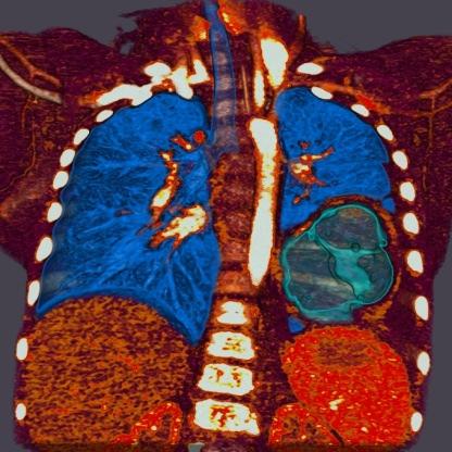
1 Imaging of the Lung in Children

 CT, HRCT MRI")
2 Imaging methods X-Ray of the Lung (Anteroposterior, ) CT, HRCT MRI USG
3 Congenital developmental defects of the lungs Agenesis, aplasia, hypoplasia Tension pulmonary anomalies (pulmonary cyst, congenital lobar emphysema, cystic adenomatoid malformation) Pulmonary sequestration Anomalous origine of the bronchus Congenital diaphragmatic herniation


4 Agenesis, aplasia, hypoplasia Bilateral agenesis or aplasia is not compatible with life X ray, CT Agenesis missing pulmonary and bronchial tissue, blood vessels are missing (unilateral compatible with life) Aplasia the bronchus is present, without pulmonary parenchyma, without blood vessels Hypoplasia rudimentary bronchi are present, pulmonary parenchyma and pulmonary blood vessels (in congenital diaphragmatic hernia)
5 Agenesis, aplasia, hypoplasia of the lung Shading caused by movement of the mediastinum to the affected side Compensatory emphysema of the preserved lung The image is the same as lobar athelectasis
6 Tension pulmonary anomalies Pulmonary cyst Congenital lobar emphysema Cystic adenomatoid pulmonary malformation
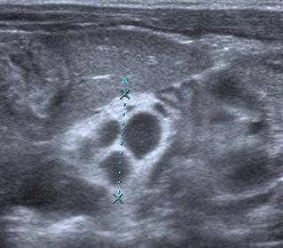
7 Tension pulmonary anomalies-- pulmonary cyst Bronchogenic cyst Congenital anomaly resulting in the development of bronchi and lung, localized centrally, or in the periphery Communication with the bronchial tree is more common in peripheral cysts Localisation of cysts extra, - intrapulmonary X-ray, CT


8 Tension pulmonary anomalies-- pulmonary cyst Non-communicating sharply defined rounded lucency Communicating circular bounded, hydro-aeric levels The thin wall (unlike abscess)
9 Tension pulmonary abnormalitiescongenital lobar emphysema Valve like obstruction of the bronchus Postnataly resulting from increased airiness of one or more pulmonary lobes in normal lung tissue Bronchomalacy with the collapse of bronchus, external pressure on the bronchus (blood vessel), idiopathic The cause of the obstruction, mucosal fold, cartilage agenesis, fibrous band, anomalous blood vessel The most commonly affected bronchus for the left upper lobe, right, middle, and upper right lobe

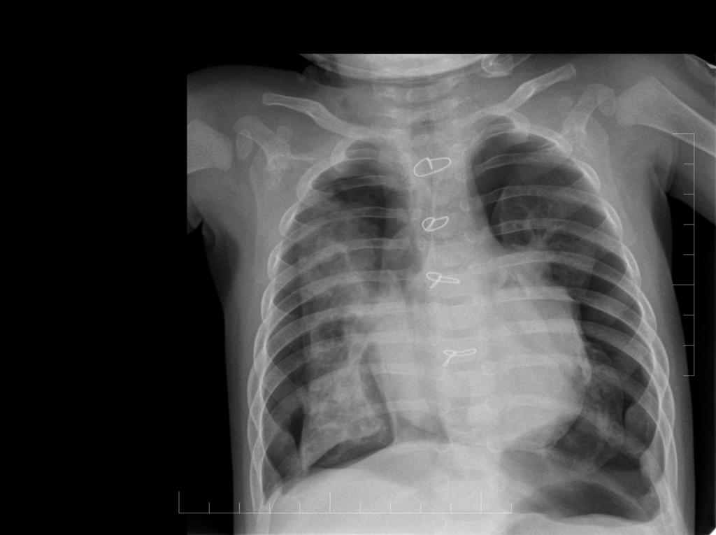
10 Tension pulmonary anomalycongenital lobar emphysema The large volume of the affected lobe of the lung with higher transparency Mediastinal shifting to the opposite side Útlak přilehléčásti plíce Compression of adjacent parts of the lungs
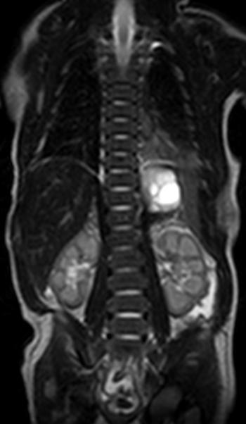
11 Tension pulmonary anomaly-congenital cystic adenomatoid malformation (CCAM) Hamartogenic proliferation of the terminal bronchi Cystoid appearance may vary according to the content of the sputum in the cysts Multicystic mass of lung tissue, with the proliferation of bronchial structures, communicates with the normal tracheobronchial tree and only rarely has the anomalous vascular supply 3 types I. even simple or multiple cysts, II. multiple small cysts, III. solid lesions without the presence of cysts Prenatal MRI/USG, can regress


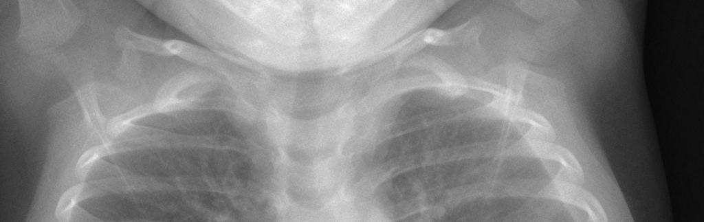
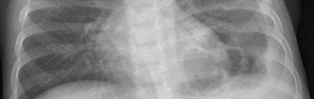
12 Tension pulmonary anomaly-congenital cystic adenomatoid malformation (CCAM) Enlargement of the affected part of the lungs, with mediastinal shifting Cystic lucency According to the quantity of the sputum in the cysts and the event. inflammatory complications and shading

13 Pulmonary sequestration Usually in the basal parts of the lung on the left Cystic or solid Cystic and solid mass of lung tissue, which does not have the normal communication with the tracheobronchial tree and is supplied of an arterial blood by anomaly arteries from the thoracic and abdominal aorta Intralobar x extralobar X-ray, USG, CT, MRI
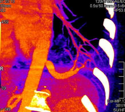
14 Pulmonary sequestration Recurrent pulmonary inflammation


15 Anomalous bronchial origine Without signs X narrowing of the bronchi Obstructive emphysema, hypoventilation, inflammatory changes The upper lobe CT x endoscopy
16 The current indications for pulmonary resection in children: -Congenital Defects- 123 patients (33%) CPS CD Bronchogenic cyst Lobar emphysema Pulmonary sequestration CPAM


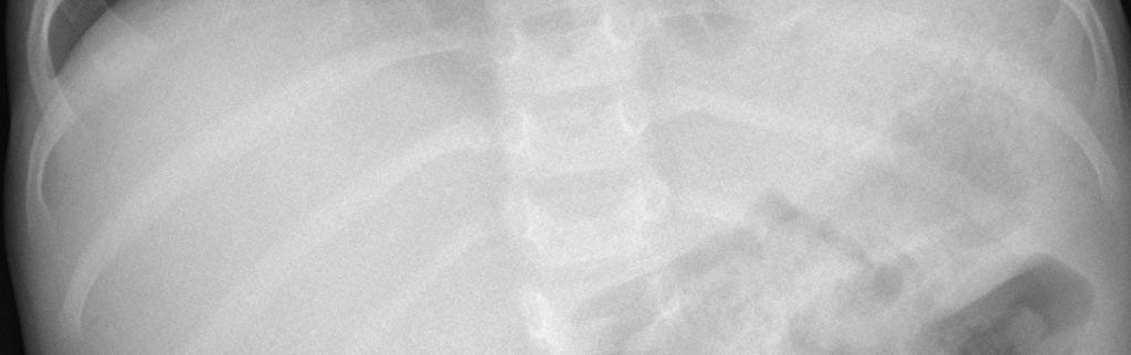
17 Congenital diaphragmatic herniation The anomaly of the diaphragm, usually to the left The defect in the membranous part The lung on the affected side is hypoplastic Poor gas content in the area of the abdomen


18 Newborn pneumopaty (pneumopaty of immature newborns) Wet lungs The respiratory distress syndrome (RDS), Hyaline Membrane Dissease Bronchopulmonary dysplasia
19 Wet lung Late foetal fluid removal from the lungs When larger quantities of fluid - White lung In healthy children, cleansing the lungs from fetal fluids (normalisation of X-ray) within 48 hours after birth Complications - lead to other pneumopathy, hyalin membrane dissease


20 Hyaline Membrane Dissease, respiratory distress syndrome, RDS Diseases of immature infants with a birth weight below 1700 g The lack of antiatelect. factorsurfactant day after birth Granular shadows to the periphery, negative bronchogram 3 degrees


21 Hyaline Membrane Dissease, respiratory distress syndrome, RDS Diseases of immature infants with a birth weight below 1700 g The lack of antiatelect. factorsurfactant day after birth Granular shadows to the periphery, negative bronchogram 3 degrees

22 Bronchopulmonary dysplasia 14 days after the artificial pulmonary ventilation
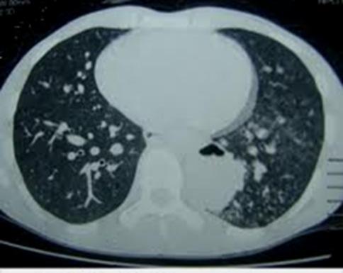
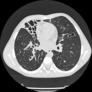
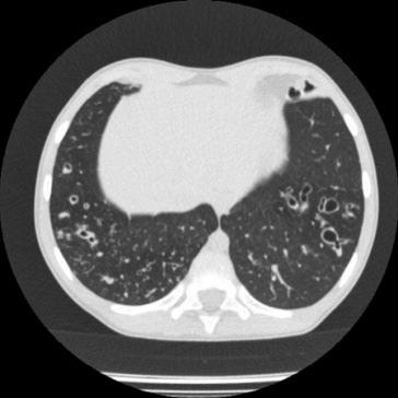
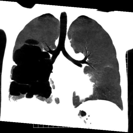
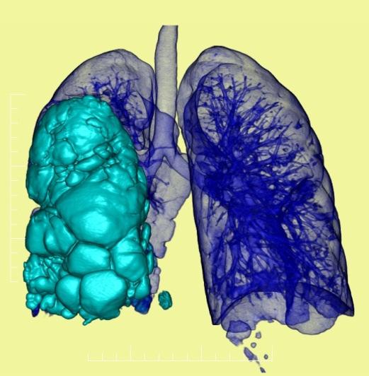
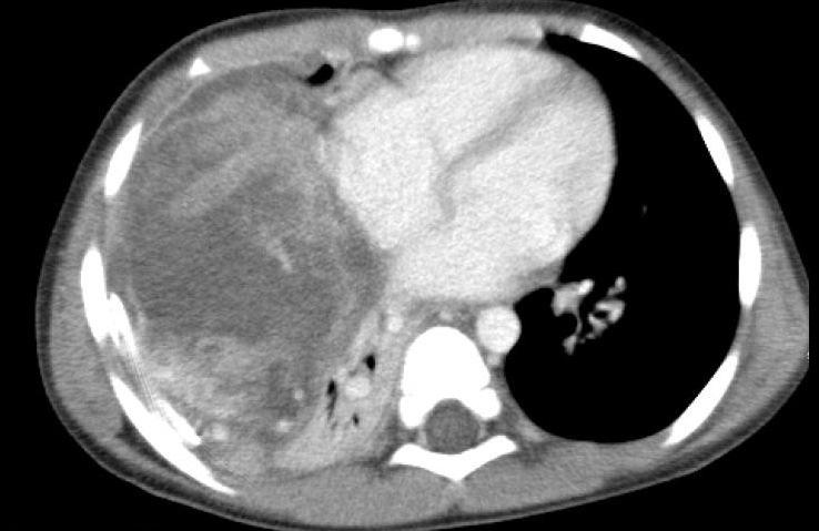
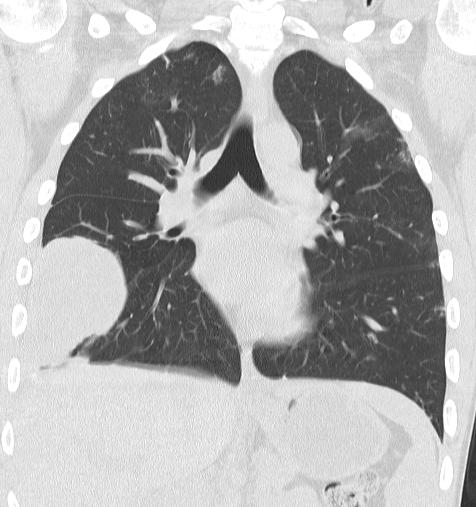
23 Pulmonary fibrosiscystic fibrosis (CF) (pulmonary changes) Obstructive changes emphysema, athelectasis Recurrent inflammation of the lungs In the later stages - pulmonary fibrosis Bronchiectasia, emphysema, pneumothorax HRCT the image of the Signet Ring sign, air trapping


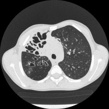
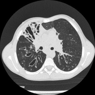
24 Chest CF The lung parenchyma lung HRCT A boy of 10 years CF-progression of bronchiectasy Helikální acquisitions in inhale = volume pro 3R AX view 1mm ST incr. 15mm


 NÁDECH")
 VÝDECH")
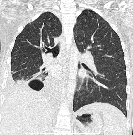
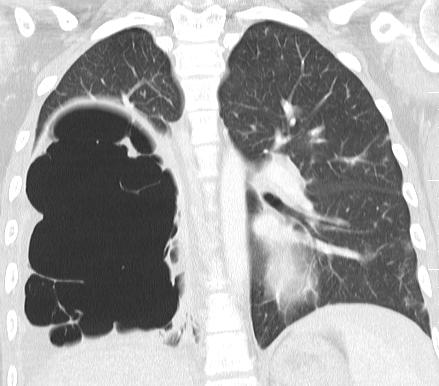
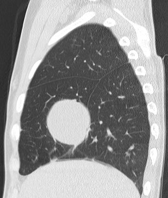
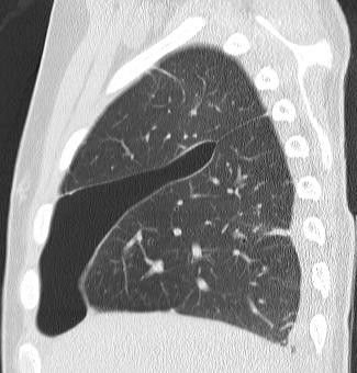
25 Chest CF The lung parenchyma lung HRCT Klasické sekvenční 1mm scany (Inc mm) NÁDECH Klasické sekvenční 1mm scany (Inc mm) VÝDECH

 VÝDECH :")

26 Hrudník Plicní parenchym HRCT plic The first examination for CF: NÁDECH : Helical (Spi) VÝDECH : Sekvenčně Control examination for CF: NÁDECH : Sekvenčně VÝDECH : Sekvenčně

27 Aspirations, foreign body Pre-school age As a general rule, valveobstructive emphysema on the affected side Athelectasis just an exceptionally A cough can lead to pneumomediastinum


28 CT of the Lung in children: inflammation

29 CT of the Lung in children: inflammation
30 CT of the Lung in Children: inflammationpulmonary wedge resection CPS in Motol Inflammatory diseases-65 patients (17%), chronic atelectasis, pulmonary abscess, gangrene, inflammatory pseudotumor, bronchiectasy patients patients patients
31 CT examination of lungs in children: tumors Primary Lung Tumors Benign Karcinoid 6 Hamartoma 2 Adenoma 1 Neuroepitelioma 1 Fibrous histiocytoma 1 Invasive fibroblastic tumor 1 Malignant Pneumoblastoma 3 Rabdomyosarkoma 2 Adenokarcinoma 1 Mukoepidermoid karcinoma 1 m.hodgkin (the main bronchus) 1
32 CT examination of lungs in children: tumors
33 Pneumotorax
34
Congenital Lung Malformations: Radiologic-Pathologic Correlation
 Acta Radiológica Portuguesa, Vol.XVIII, nº 70, pág. 51-60, Abr.-Jun., 2006 Congenital Lung Malformations: Radiologic-Pathologic Correlation Marilyn J. Siegel Mallinckrodt Institute of Radiology, Washington
Lung sequestration and Scimitar syndrome
 Lung sequestration and Scimitar syndrome Imaging approaches M. Mearadji International Foundation for Pediatric Imaging Aid Rotterdam, The Netherlands Pulmonary sequestration Pulmonary sequestration (PS)
Case Based Fetal Lung Masses
 Case Based Fetal Lung Masses Advances in Fetal and Neonatal Imaging Course Orlando, Florida, January 28, 2017 Leann E. Linam, MD Associate Professor Radiology University of Arkansas for Medical Sciences/
Surgical indications: Non-malignant pulmonary diseases. Punnarerk Thongcharoen
 Surgical indications: Non-malignant pulmonary diseases Punnarerk Thongcharoen Non-malignant Malignant as a pathological term: Cancer Non-malignant = not cancer Malignant as an adjective: Disposed to cause
ISUOG Basic Training. Assessing the Neck & Chest Gihad Chalouhi, Lebanon
 ISUOG Basic Training Assessing the Neck & Chest Gihad Chalouhi, Lebanon Learning objectives 9 & 10 At the end of the lecture you will be able to: recognise the differences between the normal & most common
Congenital anomalies of the lungs. Atelectasis. Acute lung injury
 Congenital anomalies of the lungs Atelectasis Acute lung injury Gábor Smuk M.D. Developmental lung diseases I.a. Bronchogenic cyst: abnormal budding of the tracheobronchial primordium of the primitive
Case Report Coexistent Congenital Diaphragmatic Hernia with Extrapulmonary Sequestration
 Canadian Respiratory Journal Volume 2016, Article ID 1460480, 4 pages http://dx.doi.org/10.1155/2016/1460480 Case Report Coexistent Congenital Diaphragmatic Hernia with Extrapulmonary Sequestration Nao
HOW TO IMAGE AND DESCRIBE CONGENITAL LUNG MALFORMATIONS
 HOW TO IMAGE AND DESCRIBE CONGENITAL LUNG MALFORMATIONS Paul Thacker, MD Assistant Professor Departments of Radiology and Pediatrics Medical University of South Carolina DISCLOSURES I have no relevant
Case Report Pulmonary Sequestration with Renal Aplasia and Elevated SUV Level in PET/CT
 Case Reports in Pulmonology Volume 2012, Article ID 276012, 4 pages doi:10.1155/2012/276012 Case Report Pulmonary Sequestration with Renal Aplasia and Elevated SUV Level in PET/CT Serdar Şen, 1 Nilgün
Pulmonary Sequestration
 July 26, 2004 Pulmonary Sequestration Jonathan Shaw, Harvard Medical School Year IV What do these two patients have in common? Patient 1: 50 y.o. non-smoking female with several months cough and hemoptysis;
There are four general types of congenital lung disorders:
 Pediatric Pulmonology Conditions Evaluated and Treated As a parent, watching a child suffer from a respiratory disorder can be frightening and worrisome. Our respiratory specialists provide compassionate
24. An infant with recurrent pneumonia underwent a frontal chest radiograph (Fig 24-A) followed by
 followed by") 24. An infant with recurrent pneumonia underwent a frontal chest radiograph (Fig 24-A) followed by diagnosis? ndings, what is the most likely A. Pulmonary sequestration B. Congenital pulmonary airway malformation
24. An infant with recurrent pneumonia underwent a frontal chest radiograph (Fig 24-A) followed by diagnosis? ndings, what is the most likely A. Pulmonary sequestration B. Congenital pulmonary airway malformation
Cardiopulmonary Syndromes: Conditions With Concomitant Cardiac and Pulmonary Abnormalities
 Cardiopulmonary Syndromes: Conditions With Concomitant Cardiac and Pulmonary Abnormalities Carlos S. Restrepo M.D. Professor of Radiology The University of Texas HSC at San Antonio Cardiopulmonary Syndromes
Shedding Light on Neonatal X-rays. Objectives. Indications for X-Rays 5/14/2018
 Shedding Light on Neonatal X-rays Barbara C. Mordue, MSN, NNP-BC Neonatal Nurse Practitioner LLUH Children s Hospital, NICU Objectives Utilize a systematic approach to neonatal x-ray interpretation Identify
Heart and Lungs. LUNG Coronal section demonstrates relationship of pulmonary parenchyma to heart and chest wall.
 Heart and Lungs Normal Sonographic Anatomy THORAX Axial and coronal sections demonstrate integrity of thorax, fetal breathing movements, and overall size and shape. LUNG Coronal section demonstrates relationship
Case report Esophageal lung: a rare case of communicating bronchopulmonary foregut malformation
 Case report Esophageal lung: a rare case of communicating bronchopulmonary foregut malformation 1 Dr.Varsha Rathi, 2 Dr. Saurabh Deshpande*, 3 Dr.Almas Nazim, 4 Dr.Shilpa Domkundwar 1 Professor, Department
PULMONARY VENOLOBAR SYNDROME. Dr.C.Anandhi DNB Resident, Southern Railway Headquarters Hospital.
 PULMONARY VENOLOBAR SYNDROME Dr.C.Anandhi DNB Resident, Southern Railway Headquarters Hospital. Presenting complaint: 10 yrs old girl with recurrent episodes of lower respiratory tract infection from infancy.
Chest X rays and Case Studies. No disclosures. Outline 5/31/2018. Carlo Manalo, M.D. Department of Radiology Loma Linda University Children s Hospital
 Chest X rays and Case Studies Carlo Manalo, M.D. Department of Radiology Loma Linda University Children s Hospital No disclosures. Outline Importance of history Densities delineated on radiography An approach
It s Rare So Be Aware: Pleuropulmonary Blastoma Mimicking Congenital Pulmonary Airway Malformation
 e10 Case Report: Thoracic THIEME It s Rare So Be Aware: Pleuropulmonary Blastoma Mimicking Congenital Pulmonary Airway Malformation Fayza Haider 1 Khulood Al Saad 2 Fatima Al-Hashimi 3 Hakima Al-Hashimi
Bronchial syndrome. Atelectasis Draining bronchus Bronchiectasis
 Bronchial syndrome Atelectasis Draining bronchus Bronchiectasis Etienne Leroy Terquem Pierre L Her SPI / ISP Soutien Pneumologique International / International Support for Pulmonology Atelectasis Consequence
Chest XRay interpretation INTERPRETATIONS Identifications: Name & Date Technical evaluation Basic Interpretations
 Chest XRay interpretation INTERPRETATIONS Identifications: Name & Date Technical evaluation Basic Interpretations TECHNICAL EVALUATION 1. Projection: AP/PA view To differentiate between AP & PA films,
FOREIGN BODY ASPIRATION in children. Dr. Xayyavong Bouathongthip, M.D Emergency department, children s hospital
 FOREIGN BODY ASPIRATION in children Dr. Xayyavong Bouathongthip, M.D Emergency department, children s hospital How common is choking? About 3,000 people die/year from choking Figure remained unchanged
-Tamara Wahbeh. -Razan Abu Rumman. Dr. Mohammed Al-Muhtaseb
 -2 -Tamara Wahbeh -Razan Abu Rumman Dr. Mohammed Al-Muhtaseb I tried to include everything the doctor mentioned in both the lecture and his slides in the simplest way possible, so hopefully there would
disease, bronchopulmonary dysplasia, pulmonary hypoplasia and congenital diaphragmatic hernia.
 Neonatal Chest Imaging - What the Nurse Should Know Expires Monday, April 30, 2018 Nursing Michael J. Diament, M.D. Objectives 1. Describe a good technique for positioning a neonate for the purpose of
Case Report Left Upper Lobectomy for Congenital Lobar Emphysema in a Low Weight Infant
 Case Reports in Surgery Volume 2016, Article ID 4182741, 4 pages http://dx.doi.org/10.1155/2016/4182741 Case Report Left Upper Lobectomy for Congenital Lobar Emphysema in a Low Weight Infant Meletios Kanakis,
PREMATURITY/ESTIMATED GESTATIONAL AGE
 PREMATURITY/ESTIMATED GESTATIONAL AGE 765.21 < 24 completed weeks of gestation P07.21 Extreme immaturity of newborn, gestational age less than 23 completed weeks P07.22 Extreme immaturity of newborn, gestational
HelmiLubis, RidwanMuchtarDaulay, WismanDalimunthe, Rini Savitri Daulay
 Congenital Malformation of the Lung and Airways HelmiLubis, RidwanMuchtarDaulay, WismanDalimunthe, Rini Savitri Daulay DivisiRespirologiDepartemenIlmuKesehatanAnak FakultasKedokteran Universitas Sumatera
Evaluation of the chest Part II.
 Evaluation of the chest Part II. Nagy Endre SZEGEDI TUDOMÁNYEGYETEM ÁOK, RADIOLÓGIAI KLINIKA, SZEGED ANATOMY parenchyma: alveoloacinar system, pulmonary arteries and veins interstitium: connective tissues
Imaging in pediatric lung diseases The roles of CT and pathology in diagnosing inherited and developmental lung diseases
 Imaging in pediatric lung diseases The roles of CT and pathology in diagnosing inherited and developmental lung diseases Dr Alistair D Calder Consultant Radiologist We re not so different, you and I. Invasiveness
An Image Repository for Chest CT
 An Image Repository for Chest CT Francesco Frajoli for the Chest CT in Antibody Deficiency Group An Image Repository for Chest CT he Chest CT in Antibody Deficiency Group is an international and interdisciplinary
Molla Teshome MD, Habtamu Belete MD Aurora Health Care Internal Medicine Residency Program
 Molla Teshome MD, Habtamu Belete MD Aurora Health Care Internal Medicine Residency Program History 32 year-old male who presented with a 4 days history of: Productive cough Right sided pleuritic chest
Boy 8 months TPRC. 21 Sep 06 CXR. Flat and. CLE findings. BPD findings a. Left opacity
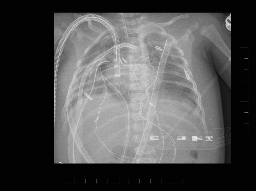
 CLE in BPD lung Boy 8 months 17 Sep 06 21 Sep 06 CXR Flat and low position of the diaphragm ICD insertion, right; ET tube slightly shift to the left RUL atelectasis RML hyperinflation, herniating across
Undergraduate Teaching
 Prof. James F Meaney Undergraduate Teaching Chest X-Ray Understanding the normal anatomical by reference to cross sectional imaging Radiology? It s FUN! Cryptic puzzle Sudoku (Minecraft?) It s completely
Congenital Diaphragmatic Hernia information for parents. David M Notrica MD FACS FAAP Pediatric Surgeons of Phoenix
 Congenital Diaphragmatic Hernia information for parents David M Notrica MD FACS FAAP Pediatric Surgeons of Phoenix CDH Congenital absence of a portion of the diaphragm allowing abdominal contents to migrate
Subject Index. Bacterial infection, see Suppurative lung disease, Tuberculosis
 Subject Index Abscess, virtual 107 Adenoidal hypertrophy, features 123 Airway bleeding, technique 49, 50 Airway stenosis, see Stenosis, airway Anaesthesia biopsy 47 complications 27, 28 flexible 23 26
X-Rays. Prepared by Prof.Dr. Magda Hassab Allah Assist.lecturer Marwa Al Hady
 X-Rays Prepared by Prof.Dr. Magda Hassab Allah Assist.lecturer Marwa Al Hady CHEST X-RAYS Normal Chest X-ray Comments on chest X ray includes examination of 1- Bony cage (ribs,clavicles &vertebral column
Thorax Lecture 2 Thoracic cavity.
 Thorax Lecture 2 Thoracic cavity. Spring 2016 Dr. Maher Hadidi, University of Jordan 1 Enclosed by the thoracic wall. Extends between (thoracic inlet) & (thoracic outlet). Thoracic inlet At root of the
4/16/2017. Learning Objectives. Interpretation of the Chest Radiograph. Components. Production of the Radiograph. Density & Appearance
 Interpretation of the Arthur Jones, EdD, RRT Learning Objectives Identify technical defects in chest radiographs Identify common radiographic abnormalities This Presentation is Approved for 1 CRCE Credit
CASE REPORTS. Idiopathic Unilateral Hyperlucent Lung
 CASE REPORTS Idiopathic Unilateral Hyperlucent Lung The Swyer-James Syndrome J. Judson McNamara, M.D., Harold C. Urschel, M.D., J. H. Arndt, M.D., Herman Ulevitch, M.D., and W. B. Kingsley, M.D. I diopathic
Thoracoscopic treatment of congenital malformation of the lung
 Jemis, 1 2013 Thoracoscopic treatment of congenital malformation of the lung Preliminary experience with preoperative 3D virtual rendering F. Destro M. Maffi T. Gargano G. Ruggeri L. Soler M. Lima Table
Pediatric High-Resolution Chest CT
 Pediatric High-Resolution Chest CT Alan S. Brody, MD Professor of Radiology and Pediatrics Chief, Thoracic Imaging Cincinnati Children s s Hospital Cincinnati, Ohio, USA Pediatric High-Resolution CT Short
CONGENITAL LUNG LESION Round Table. Objectives. Congenital Lung Lesions: Anatomy and Physiology Leah Barefoot, DNP, CPNP-PC
 CONGENITAL LUNG LESION Round Table L. Barefoot, E. Paton, C. Schultz, R. Caskey, M. O Day Objectives Review the anatomy and pathophysiology of congenital lung lesions List the preoperative evaluation of
B-I-2 CARDIAC AND VASCULAR RADIOLOGY
 (YEARS 1 3) CURRICULUM FOR RADIOLOGY 13 B-I-2 CARDIAC AND VASCULAR RADIOLOGY KNOWLEDGE To describe the normal anatomy of the heart and vessels including the lymphatic system as demonstrated by radiographs,
Congenital lung anomalies: can we postpone resection?
 Journal of Pediatric Surgery (2012) 47, 87 92 www.elsevier.com/locate/jpedsurg Congenital lung anomalies: can we postpone resection? Nadja Colon a, Cameron Schlegel a, John Pietsch a, Dai H. Chung a,b,
The Foetal Lung. Educational Exhibit Authors:
 The Foetal Lung Poster No.: C-1649 Congress: ECR 2015 Type: Educational Exhibit Authors: V. B. Pai, R. N. Chaubal, N. G. Chaubal, B. Pai ; Navi Mumbai/ 1 2 2 2 3 1 3 IN, Thane/IN, Mumbai/IN Keywords: Foetal
Review of Neonatal Respiratory Problems
 Review of Neonatal Respiratory Problems Respiratory Distress Occurs in about 7% of infants Clinical presentation includes: Apnea Cyanosis Grunting Inspiratory stridor Nasal flaring Poor feeding Tachypnea
Bronchioles. Alveoli. Type I alveolar cells are very thin simple squamous epithelial cells and form most of the lining of an alveolus.
 276 Bronchioles Bronchioles continue on to form bronchi. The primary identifying feature is the loss of hyaline cartilage. The epithelium has become simple ciliated columnar, and there is a complete ring
Rare diseases in pulmonology. Agnieszka Strzelak
 Rare diseases in pulmonology Agnieszka Strzelak Rare diseases: < 1/2000 genetic, infectious, autoimmune, rare neoplasms, of unknown origin in most cases severe, chronic and progressing present after birth,
THE GOOFY ANATOMIST QUIZZES
 THE GOOFY ANATOMIST QUIZZES 7. LUNGS Q1. Fill in the blanks: the lung has lobes and fissures. A. Right, three, two. B. Right, two, one. C. Left, three, two. D. Left, two, three. Q2. The base of the lung
CONGENITAL LOBAR EMPHYSEMA
 CONGENITAL LOBAR EMPHYSEMA BY R. H. WHITE-JONES and L. J. TEMPLE From Whiston County Hospital, Lancs., and Broadgreen Hospital, Liverpool Regional obstructive emphysema is not uncommon and is due in most
Obstetrics Content Outline Obstetrics - Fetal Abnormalities
 Obstetrics Content Outline Obstetrics - Fetal Abnormalities Effective February 2007 10 16% renal agenesis complete absence of the kidneys occurs when ureteric buds fail to develop Or degenerate before
Semiology of respiratory system in children Simple choice 1. Mark the intrauterine age of lung development onset from the gut: a) 1 week b) 24 days
 1 week b) 24 days") Semiology of respiratory system in children Simple choice 1. Mark the intrauterine age of lung development onset from the gut: a) 1 week b) 24 days c) 6 weeks d) 12 weeks e) 35 weeks 2. Stridor is not
Semiology of respiratory system in children Simple choice 1. Mark the intrauterine age of lung development onset from the gut: a) 1 week b) 24 days c) 6 weeks d) 12 weeks e) 35 weeks 2. Stridor is not
A Case of Pediatric Plasma Cell Granuloma
 August 2001 A Case of Pediatric Plasma Cell Granuloma Nii Tetteh, Harvard Medical School Year IV Our Patient 8 year old male with history of recurrent left lower lobe and lingular pneumonias since 1994.
Radiological conference. Left upper lobe collapse. Citation Hong Kong Practitioner, 1998, v. 20 n. 9, p
 Title Radiological conference. Left upper lobe collapse Author(s) Wong, LLS; Peh, WCG Citation Hong Kong Practitioner, 1998, v. 20 n. 9, p. 513-517 Issued Date 1998 URL http://hdl.handle.net/10722/44672
Respiratory Disease. Dr Amal Damrah consultant Neonatologist and Paediatrician
 Respiratory Disease Dr Amal Damrah consultant Neonatologist and Paediatrician Signs and Symptoms of Respiratory Diseases Cardinal Symptoms Cough Sputum Hemoptysis Dyspnea Wheezes Chest pain Signs and Symptoms
Eun-Young Kang, M.D., Jae Wook Lee, M.D., Ji Yung Choo, M.D., Hwan Seok Yong, M.D., Ki Yeol Lee, M.D., Yu-Whan Oh, M.D.
 Eun-Young Kang, M.D., Jae Wook Lee, M.D., Ji Yung Choo, M.D., Hwan Seok Yong, M.D., Ki Yeol Lee, M.D., Yu-Whan Oh, M.D. Department of Radiology, Korea University Guro Hospital, College of Medicine, Korea
Perinatal Imaging in Congenital Thoracic Cystic Malformations.
 Perinatal Imaging in Congenital Thoracic Cystic Malformations. Poster No.: C-0898 Congress: ECR 2013 Type: Educational Exhibit Authors: R. Llorens, G. Montoliu, A. Moreno, F. Menor; Valencia/ES Keywords:
Radiology of the respiratory disease
 Radiology of the respiratory disease [ Color index: Important Notes Extra ] [ Editing file Feedback Share your notes Shared notes ] Resources: - 435 Slides - 434 Team - 435 Notes Done by: - Mai Alageel
Basic Data. Sex:Male 31 years old Occupation: 搬家工人
 Basic Data Sex:Male 31 years old Occupation: 搬家工人 Chief Complaint Intermittent chest pain with shortness of breath for 2-3 months. Present Illness 4 months ago, he started having occasional chest pain
PRESENCE OF LOWER ACCESSORY LOBES IN THE LUNGS
 Int. J. Pharm. Med. & Bio. Sc. 2013 Hemanth Kommuru et al., 2013 Research Paper ISSN 2278 5221 www.ijpmbs.com Vol. 2, No. 3, July 2013 2013 IJPMBS. All Rights Reserved PRESENCE OF LOWER ACCESSORY LOBES
CONGENITAL LUNG MALFORMATIONS, UPDATE AND TREATMENT
 CONGENITAL LUNG MALFORMATIONS, UPDATE AND TREATMENT STEVEN ROTHENBERG MD.(1) 1. Department of Pediatrics. The Rocky Mountain Hospital For Children. Hospital for Children, Denver, Colorado, USA. steverberg@aol.com
An Introduction to Radiology for TB Nurses
 An Introduction to Radiology for TB Nurses Garold O. Minns, MD September 14, 2017 TB Nurse Case Management September 12 14, 2017 EXCELLENCE EXPERTISE INNOVATION Garold O. Minns, MD has the following disclosures
Wheeze. Dr Jo Harrison
 Wheeze Dr Jo Harrison 9.9.14 Wheeze - Physiology a continuous musical sound that lasts longer than 250 msec. can be high-pitched or low-pitched, consist of single or multiple notes, and occur during inspiration
Chronic lung diseases in children Simple choice 1. Finger clubbing is not characteristic for: a) Diffuse bronchiectasis b) Cystic fibrosis c)
 Diffuse bronchiectasis b) Cystic fibrosis c)") Chronic lung diseases in children Simple choice 1. Finger clubbing is not characteristic for: a) Diffuse bronchiectasis b) Cystic fibrosis c) Bronchiolitis obliterans d) Complicated acute pneumonia e)
Chronic lung diseases in children Simple choice 1. Finger clubbing is not characteristic for: a) Diffuse bronchiectasis b) Cystic fibrosis c) Bronchiolitis obliterans d) Complicated acute pneumonia e)
Chest and cardiovascular
 Module 1 Chest and cardiovascular A. Doss and M. J. Bull 1. Regarding the imaging modalities of the chest: High resolution computed tomography (HRCT) uses a slice thickness of 4 6 mm to identify mass lesions
TB Radiology for Nurses Garold O. Minns, MD
 TB Nurse Case Management Salina, Kansas March 31-April 1, 2010 TB Radiology for Nurses Garold O. Minns, MD April 1, 2010 TB Radiology for Nurses Highway Patrol Training Center Salina, KS April 1, 2010
Introduction to Chest Radiography
 Introduction to Chest Radiography RSTH 366: DIAGNOSTIC TECHNIQUES Alan Alipoon BS, RCP, RRT Instructor Department of Cardiopulmonary Sciences 1 Introduction Discovered in 1895 by Wilhelm Roentgen Terminology
9/8/2009 < 1 1,2 3,4 5,6 7,8 9,10 11,12 13,14 15,16 17,18 > 18. Tetralogy of Fallot. Complex Congenital Heart Disease.
 Current Indications for Pediatric CTA S Bruce Greenberg Professor of Radiology Arkansas Children s Hospital University of Arkansas for Medical Sciences greenbergsbruce@uams.edu 45 40 35 30 25 20 15 10
Clinical History. 29 yo woman with polyhydramnios Cardiac mass at fetal ultrasound At 35 weeks, newborn died 30 minutes after delivery
 CASE 1 a Clinical History 29 yo woman with polyhydramnios Cardiac mass at fetal ultrasound At 35 weeks, newborn died 30 minutes after delivery Interface between tumor and normal myocardium Smaller well-demarcated
Pulmonary Agenesis sentations picked up over a period of 4 years. These cases were suspected and then documented during life.
 Pulmonary Agenesis sentations picked up over a period of 4 years. These cases were suspected and then documented during life. B. Rajshekhar Case Reports Sunil Gomber Anurag Krishna* Five cases of pulmonary
Neonatal Chest X-Ray Interpretation
 CHAPTER 7 Neonatal Chest X-Ray Interpretation Prof. Praveen Kumar Neonatal unit, Department of Pediatrics, PGIMER, Chandigarh Learning Objectives At the end of this session, you should be able to: 1. Schematically
Pitfalls of the Pediatric Chest and Abdomen SPR 2017
 Pitfalls of the Pediatric Chest and Abdomen SPR 2017 Richard I. Markowitz, MD, FACR Children s Hospital of Philadelphia Perelman School of Medicine University of Pennsylvania No Disclosures Cognitive Perceptual
Case of the Day Chest
 Case of the Day Chest Darin White MDCM FRCPC Department of Radiology, Mayo Clinic 76 th Annual Scientific Meeting Canadian Association of Radiologists Montreal, QC April 26, 2013 2013 MFMER slide-1 Disclosures
Imaging of congenital pulmonary malformations
 Acta Biomed 2016; Vol. 87, Supplement 3: 45-50 Mattioli 1885 Review Imaging of congenital pulmonary malformations Francesco Emanuele Praticò 1, Michele Corrado 1, Giovanni Della Casa 1, Raffaele Parziale
Radiological Anatomy of Thorax. Dr. Jamila Elmedany & Prof. Saeed Abuel Makarem
 Radiological Anatomy of Thorax Dr. Jamila Elmedany & Prof. Saeed Abuel Makarem Indications for Chest x - A chest x-ray may be used to diagnose and plan treatment for various conditions, including: Diseases/Fractures
UERMMMC Department of Radiology. Basic Chest Radiology
 UERMMMC Department of Radiology Basic Chest Radiology PHYSICS DENSITIES BONE SOFT TISSUES WATER FAT AIR TELEROENTGENOGRAM Criteria for an Ideal Chest Radiograph 1. Upright 2. Posteroanterior View 3. Full
Signs in Chest Radiology
 Signs in Chest Radiology Jonathan H. Chung, MD Disclosures No pertinent disclosures Jonathan H. Chung, MD Assistant Professor Institute t of fadvanced d Biomedical Imaging National Jewish Health Denver,
Cystic adenomatoid malformation in adults: radiological findings and pathologic correlation
 Cystic adenomatoid malformation in adults: radiological findings and pathologic correlation Poster No.: C-1734 Congress: ECR 2014 Type: Educational Exhibit Authors: C. Batz Colvée, M. Vidal, M. Ruiz Tolón,
Case Report 3D Imaging in Unilateral Primary Pulmonary Hypoplasia in an Adult: A Case Report
 Case Reports in Radiology Volume 2011, Article ID 659586, 6 pages doi:10.1155/2011/659586 Case Report 3D Imaging in Unilateral Primary Pulmonary Hypoplasia in an Adult: A Case Report Aristida Georgescu,
Lung Cancer - Suspected
 Lung Cancer - Suspected Shared Decision Making Lung Cancer: http://www.enhertsccg.nhs.uk/ Patient presents with abnormal CXR Lung cancer - clinical presentation History and Examination Incidental finding
The neonatal chest X-ray
 PAEDIATRIC RESPIRATORY REVIEWS (2001) 2, 311 323 doi:10.1053/prrv.2001.0169, available online at http://www.idealibrary.com on SERIES: IMAGING The neonatal chest X-ray R. Arthur X-ray and Ultrasound Department,
The Fetal Care Center at NewYork-Presbyterian/ Weill Cornell Medicine
 The Fetal Care Center at NewYork-Presbyterian/ Weill Cornell Medicine Prompt and Personalized Care for Women with Complex Pregnancies A Team of Experts additional training in maternal and fetal complications
White hemithorax in children
 DOI 10.1007/s00247-011-2065-8 PICTORIAL ESSAY White hemithorax in children Javier Lucaya & Enrique F. Garcés-Iñigo & Pilar García-Peña & Joaquim Piqueras & Goya Enriquez Received: 28 July 2010 /Revised:
Large veins of the thorax Brachiocephalic veins
 Large veins of the thorax Brachiocephalic veins Right brachiocephalic vein: formed at the root of the neck by the union of the right subclavian & the right internal jugular veins. Left brachiocephalic
CT Findings and Temporal Course of Persistent Pulmonary Interstitial Emphysema in Neonates: A Multiinstitutional Study
 Lane F. Donnelly 1 Javier Lucaya 2 Vanildo Ozelame 3 Donald P. Frush 4 Peter J. Strouse 5 Thomas E. Sumner 6 Harriet J. Paltiel 7 Received June 21, 2002; accepted after revision ugust 12, 2002. 1 Department
Lecture 2: Clinical anatomy of thoracic cage and cavity II
 Lecture 2: Clinical anatomy of thoracic cage and cavity II Dr. Rehan Asad At the end of this session, the student should be able to: Identify and discuss clinical anatomy of mediastinum such as its deflection,
Spectrum of Pulmonary Sequestration M. Wayne Flye, M.D., Martin Conley, M.D., and Donald Silver, M.D.
 Spectrum of Pulmonary Sequestration M. Wayne Flye, M.D., Martin Conley, M.D., and Donald Silver, M.D. ABSTRACT Bronchopulmonary sequestration was diagnosed in 17 patients ranging in age from newborn to
The Adult Form of. Pulmonary Hamartoma. O.S. U. Series. A Reappraisal. Malvin Weinberger, M.D., Gerard S. Kakos, M.D., and James W. Kilman, M.D.
 The Adult Form of Pulmonary Hamartoma A Reappraisal Malvin Weinberger, M.D., Gerard S. Kakos, M.D., and James W. Kilman, M.D. ABSTRACT Pulmonary hamartoma is often an incidental, asymptomatic finding on
Chest X-ray Interpretation
 Chest X-ray Interpretation Introduction Routinely obtained Pulmonary specialist consultation Inherent physical exam limitations Chest x-ray limitations Physical exam and chest x-ray provide compliment
OSAMA A. ABDULMAJID, ABDELMOMEN M. EBEID, MOHAMED M. MOTAWEH, and IBRAHIM S. KLEIBO
 Aspirated foreign bodies in the tracheobronchial tree: report of 250 cases Thorax (1976), 31, 635. OSAMA A. ABDULMAJID, ABDELMOMEN M. EBEID, MOHAMED M. MOTAWEH, and IBRAHIM S. KLEIBO Thoracic Surgical
Bronchopulmonary foregut malformation: A pictorial review.
 Bronchopulmonary foregut malformation: A pictorial review. Poster No.: C-1676 Congress: ECR 2013 Type: Educational Exhibit Authors: N. L. Eun, C. S. Yoon, M.-J. Lee, M.-J. Kim ; Rep. of KOREA/ 1 2 2 2
Congenital Pulmonary Airways Malformation: an update
 Congenital Pulmonary Airways Malformation: an update Poster No.: P-0111 Congress: ESTI 2014 Type: Educational Poster Authors: S. M. Mak, B. Annan, S. P. G. Padley, A. G. Nicholson; London/UK Keywords:
The Respiratory System. Dr. Ali Ebneshahidi
 The Respiratory System Dr. Ali Ebneshahidi Functions of The Respiratory System To allow gases from the environment to enter the bronchial tree through inspiration by expanding the thoracic volume. To allow
WF RESPIRATORY SYSTEM. RESPIRATORY MEDICINE
 WF RESPIRATORY SYSTEM. RESPIRATORY MEDICINE 1 Societies 11 History 13 Dictionaries. Encyclopaedias. Bibliographies Use for general works only. Classify with specific aspect where possible 15 Classification.
Function of Breathing. Jeanine D Armiento, M.D., Ph.D. Respiratory Portion. Conducting Portion. Critical to the Development of the Lung
 Function of Breathing Jeanine D Armiento, M.D., Ph.D. Associate Professor Department of Medicine P&S 9-449 5-3745 jmd12@columbia.edu Air Sacs (alveoli) Ventilation-air conduction Moving gas in and out
Lung- and airway emergencies
 Lung- and airway emergencies Charlotte de Lange,MD,PhD Pediatric Radiology unit, Oslo University Hospital, Norway 5th Nordic course - Emergency Radiology Oslo 18-21.5.2015 clange@ous-hf.no How come pediatric
Approach to CXR. Terminology. 1.Identification. Greg Blecher SCH Respir Fellow. Correct patient Correct date and time Correct examination
 Approach to CXR Greg Blecher SCH Respir Fellow From Rob Posteraro http://home.earthlink.net/~rhpos/cxr_interpret.txt.html ; http://home.earthlink.net/~rhpos/cxr_main.txt.html) Approach to viewing Chest
Congenital pulmonary malformations diagnosed in adulthood, how to recognize them?
 Congenital pulmonary malformations diagnosed in adulthood, how to recognize them? Poster No.: C-1407 Congress: ECR 2015 Type: Educational Exhibit Authors: E. De la Via, S. Isarria, M. L. Domingo, S. P.