In adults: trachea is about cm long & 2.5 cm in diameter.
|
|
|
- Lawrence Montgomery
- 5 years ago
- Views:
Transcription


1
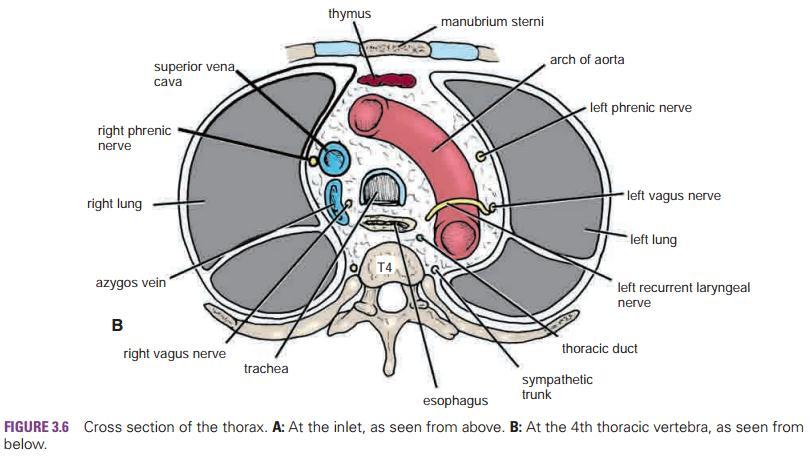
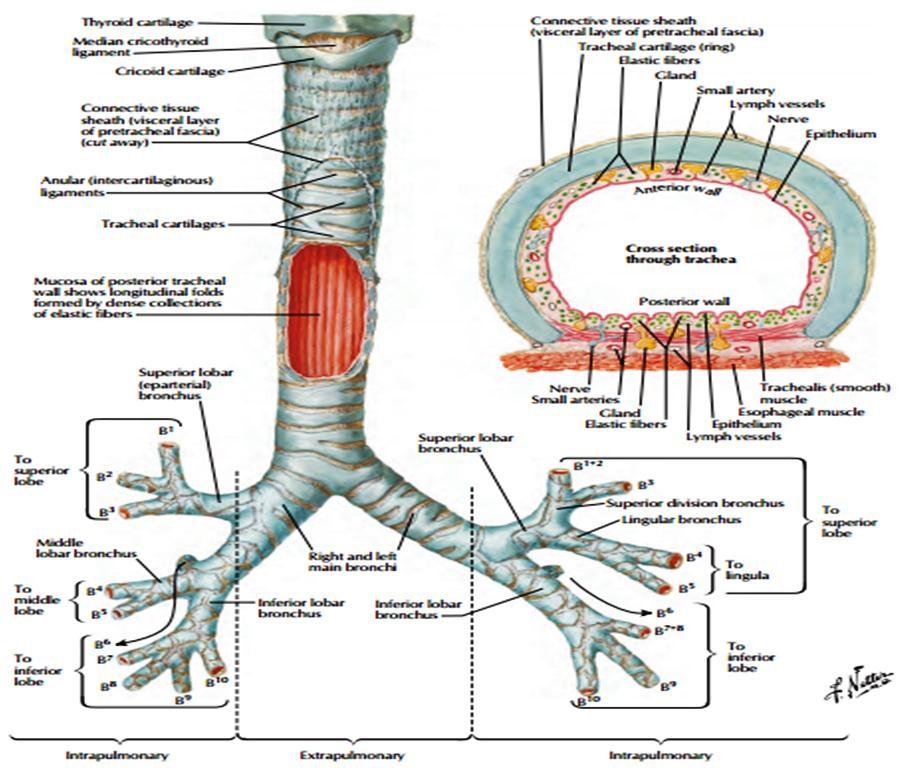
2 Trachea is a mobile cartilaginous and membranous tube. This fibroelastic tube is kept patent by U- or C- shaped bars of hyaline cartilage embedded in its wall. The posterior free ends of this cartilage are connected by smooth muscle, the trachealis muscle. In adults: trachea is about cm long & 2.5 cm in diameter. It begins: in the neck as a continuation of the larynx at the lower border of the cricoid cartilage at level of the 6th cervical vertebra. It descends in the midline of the neck. The trachea ends below at the carina by dividing into Rt. & Lt. principal (main) bronchi at the level of the sternal angle. In expiration: Bifurcation rises by one vertebral level. In deep inspiration: it may be lowered as far as T6 vertebra. 2

3 posterior anterior 3
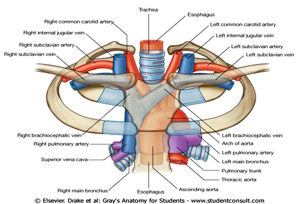
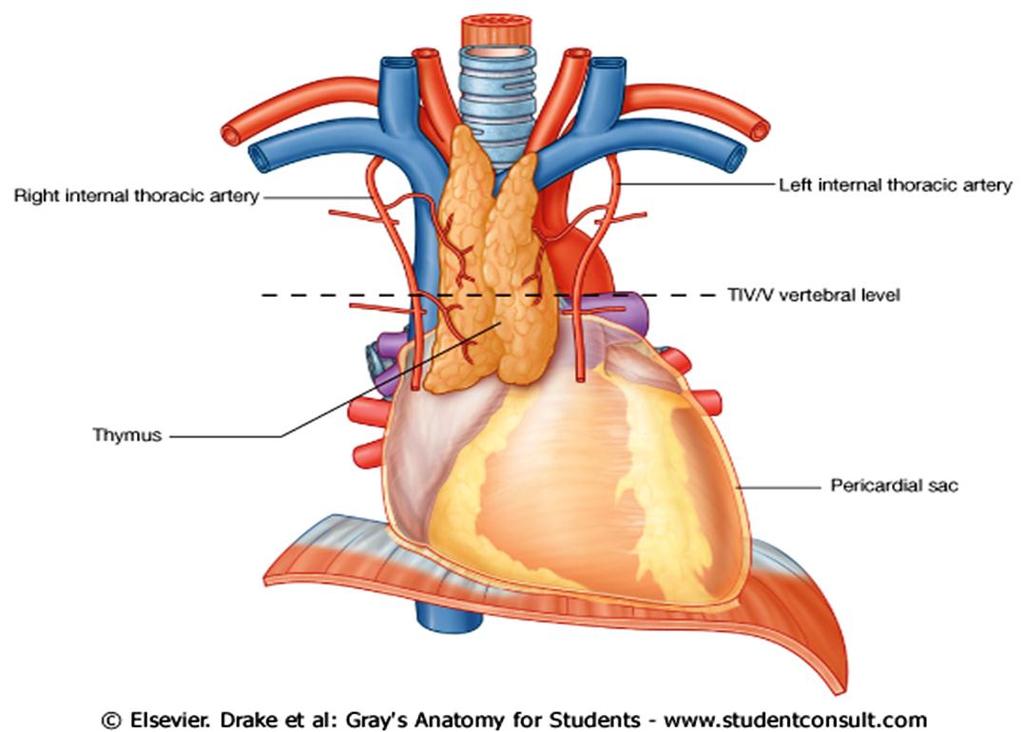
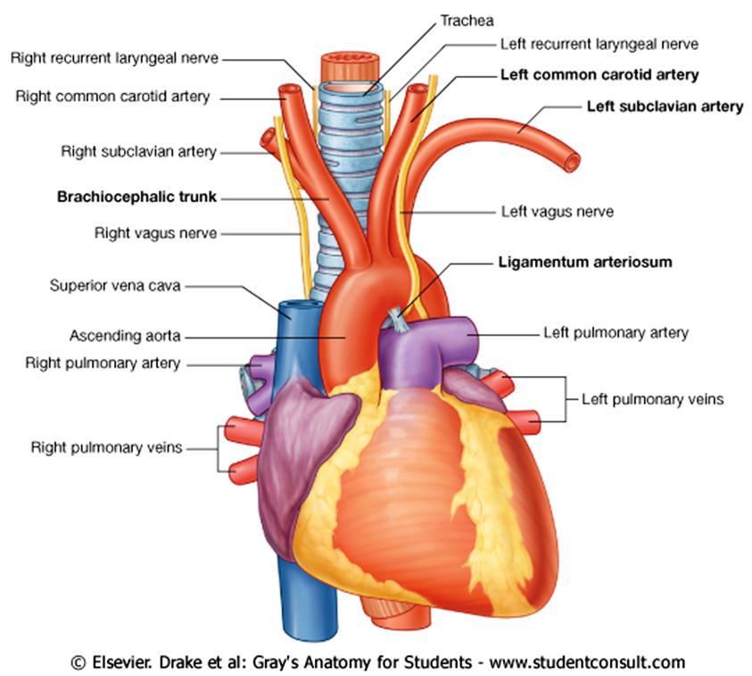
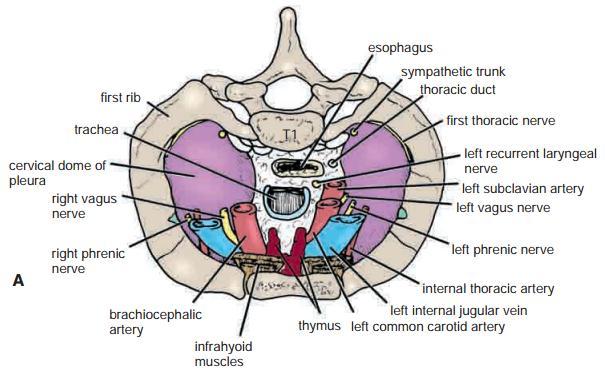
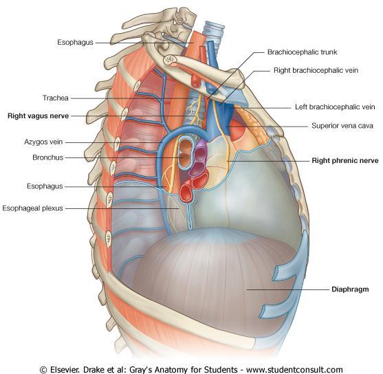
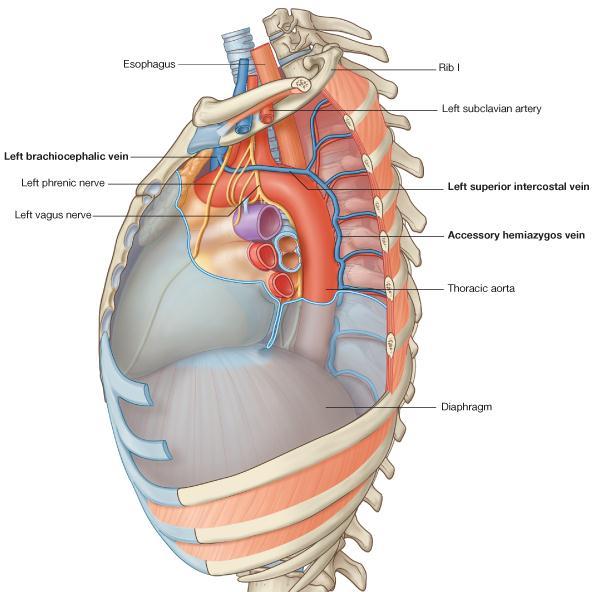
4 Anteriorly: Sternum, thymus, Lt. brachiocephalic vein, origins of brachiocephalic & Lt. common carotid arteries & the arch of aorta. Posteriorly: Esophagus & Lt recurrent laryngeal N. Right side: Azygos vein, Rt. Vagus nerve & Rt. Pleura. Left side: Arch of aorta, Lt. common carotid, Lt. subclavian arteries, Lt. pleura, Lt. vagus & Lt. phrenic nerves. 4
5 5
6 6
7 7
8 8
9 9
10 10
11 11
12 1. Arterial supply: Upper two thirds: are supplied by the inferior thyroid arteries. Lower third: is supplied by the bronchial arteries. 2. Venous drainage: Rt. Side: Azygos vein. Lt. Side: Hemiazyos vein. Also a little via bronchial and pulmonary veins. 3. Nerve supply: Branches from vagus nerve & recurrent laryngeal nerve. Sympathetic nerves supply trachealis muscle and mucus membrane. 4. Lymph drainage of the trachea The lymph drains into the pretracheal and paratracheal lymph nodes and the deep cervical nodes. 12
13 13
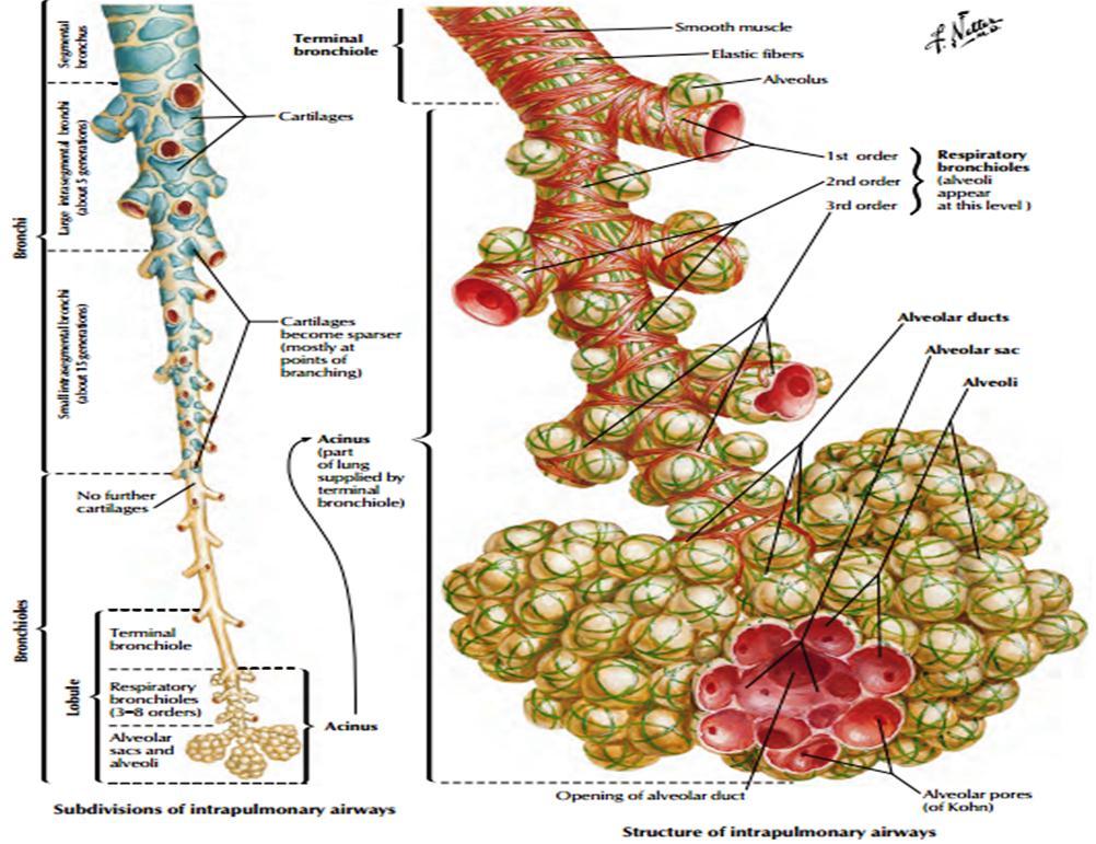
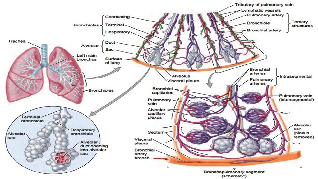
14 The trachea bifurcates behind the arch of the aorta into Rt. & Lt. principal (primary or main) bronchi. The bronchi then divide dichotomously, giving rise to millions of terminal bronchioles that terminate in 1 or 2 respiratory bronchioles. Each respiratory bronchiole divides into 2 to 11 alveolar ducts that enter the alveolar sacs. The alveoli arise from the walls of the sacs as diverticula. 14
15 15
16 16
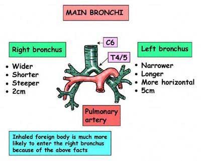
17 Rt. Main bronchus Wider, shorter (2.5 cm long). More vertical. Before entering the hilum of the right lung, the principal bronchus gives off the superior lobar bronchus. On entering the hilum, it divides into a middle and an inferior lobar bronchi. Lt. Main bronchus Narrower, longer (5 cm long). More horizontal. It passes to the left below the arch of the aorta and in front of the esophagus. On entering the hilum of the left lung, the principal bronchus divides into a superior and an inferior lobar bronchi. 17
18 18
19 During life, lungs are soft, spongy and very elastic. If the thoracic cavity were opened, the lungs would immediately shrink to one third or less in volume. In the child, they are pink, but with age, they become dark and mottled because of inhalation of dust particles (trapped in the lung phagocytes) especially seen in city dwellers, smokers and coal miners. Each lung is conical, covered with visceral pleura, and suspended free in its own pleural cavity, being attached to the mediastinum only by its root. 19
20 20
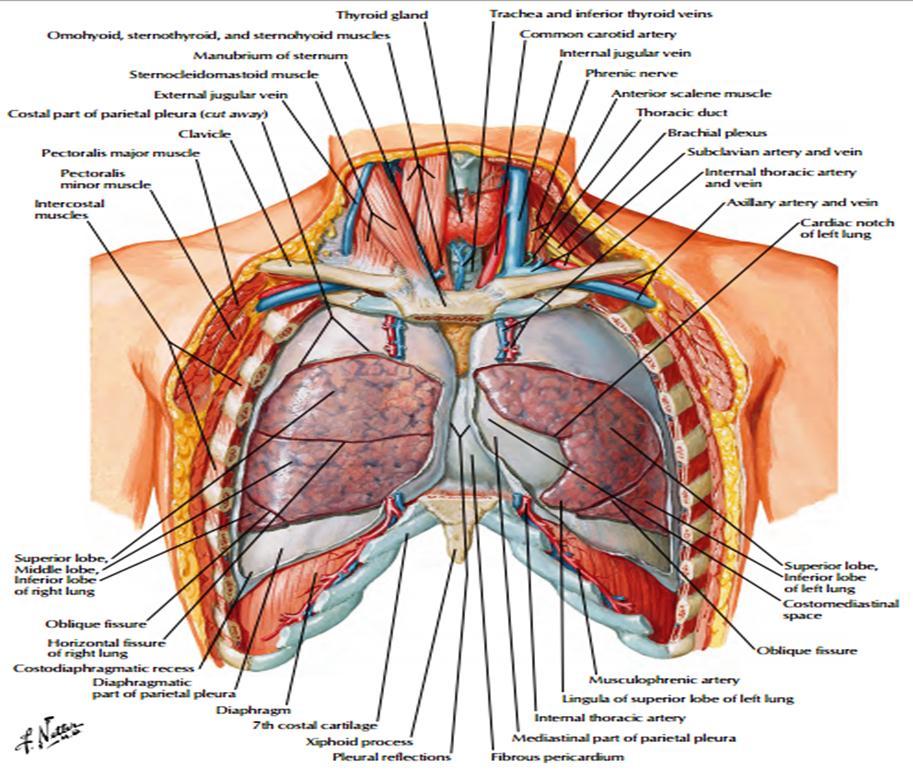
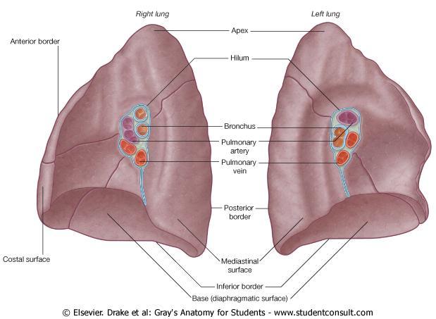
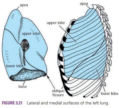
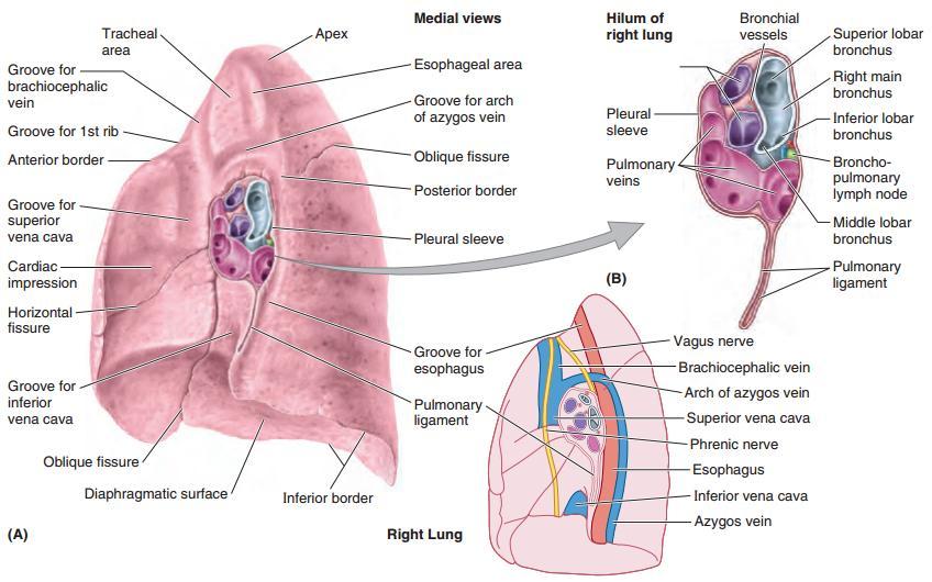
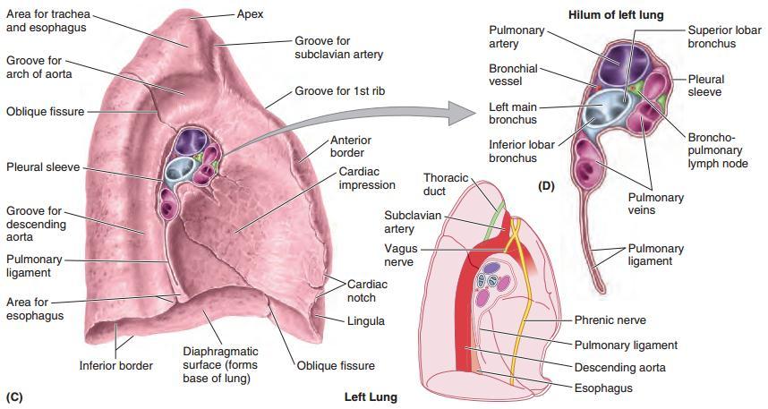
21 Each lung has: 1. A blunt apex projecting upward into the neck for about 2.5 cm above the clavicle. 2. A concave base that sits on the diaphragm. 3. A convex costal surface, which corresponds to the concave chest wall. 4. A concave mediastinal surface, which is molded to the pericardium and other mediastinal structures. In the middle of this surface is the hilum, a depression from which the bronchi, vessels, and nerves that form the lung root enter and leave the lung. 5. The anterior border is thin and overlaps the heart; it is here on the left lung that the cardiac notch is found. 6. The posterior border is thick and lies beside the vertebral column. 21
22 22
23 23
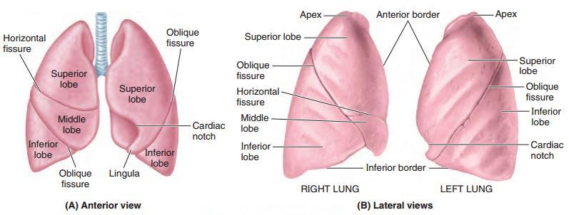
24 Right Lung: Right lung is slightly larger than the left. It is divided by oblique & horizontal fissures into upper, middle, & lower lobes. The oblique fissure runs from the inferior border upward and backward across the medial and costal surfaces until it cuts the posterior border about 6.25 cm below the apex. The horizontal fissure runs horizontally across the costal surface at the level of the 4 th costal cartilage to meet the oblique fissure in the midaxillary line. The middle lobe is thus a small triangular lobe bounded by the horizontal and oblique fissures. 24
25 Left Lung: It is divided by a similar oblique fissure into upper & lower lobes (Superior & Inf. lobes). No horizontal fissure in the left lung. 25
26 26


27 Rt. lung 4 th costal cart. Midaxillary line Lt. lung 2.5 in. 27
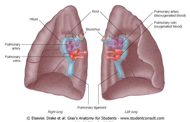
28 The root of the lung is formed by structures that are entering or leaving the lung including the bronchi, pulmonary artery and veins, lymph vessels, bronchial vessels, and nerves. The root is surrounded by a tubular sheath (Cuff) of pleura, which joins the mediastinal parietal pleura to the visceral pleura covering the lungs. 28
29 29
30 30

31 right left 31
32 32
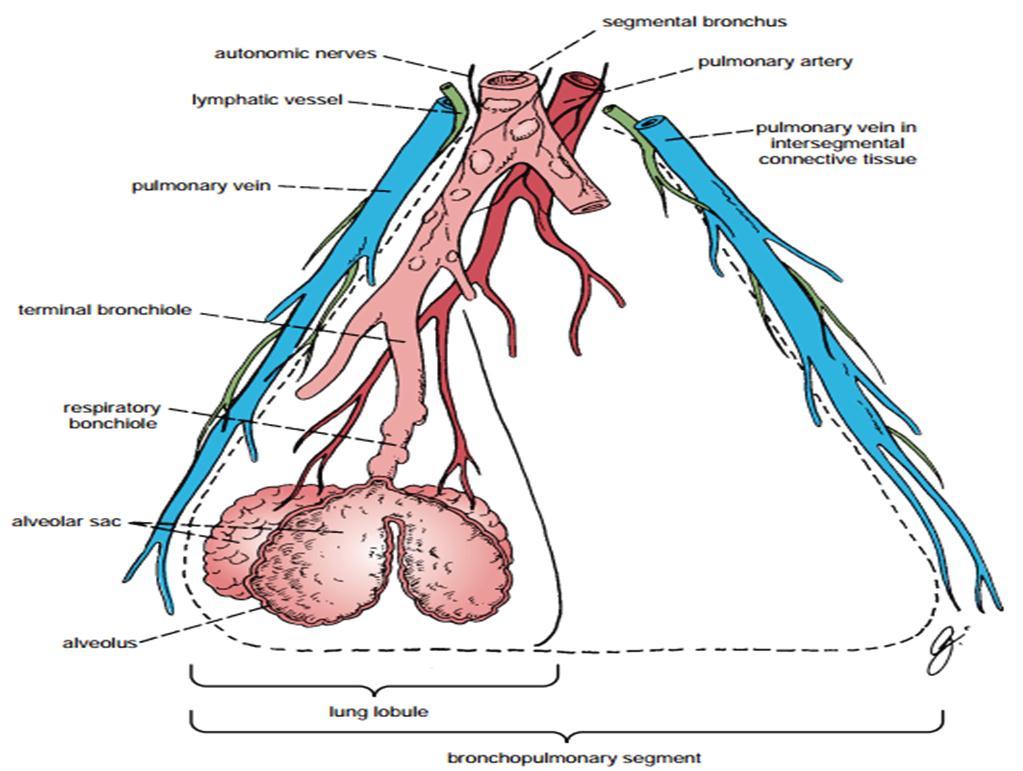
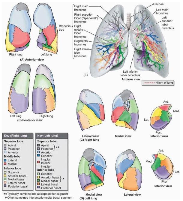
33 Definition: The bronchopulmonary segments are the anatomic, functional, and surgical units of the lungs. Each lobar (secondary) bronchus, passes to a lung lobe, & gives off branches called segmental (tertiary) bronchi. Each segmental bronchus passes to a structurally and functionally independent unit of a lung lobe called a bronchopulmonary segment, which is surrounded by connective tissue. 33
34 The segmental bronchus is accompanied by a pulmonary artery branch, but the tributaries of the pulmonary veins run in the connective tissue between adjacent bronchopulmonary segments. Each segment has its own lymphatic and autonomic nerve supply. 34
35 On entering a bronchopulmonary segment, each segmental bronchus divides repeatedly. As the bronchi become smaller, the U-shaped bars of cartilage found in the trachea are gradually replaced by irregular plates of cartilage, which become smaller and fewer in number. Smallest bronchi divide & give rise to bronchioles ( 1 mm in diameter). Bronchioles possess no cartilage in their walls and are lined with columnar ciliated epithelium. The submucosa possesses a complete layer of circularly arranged smooth muscle fibers. 35
36 The bronchioles then divide and give rise to terminal bronchioles, which are the last part of the conducting portion of the respiratory system. Each terminal bronchiole gives rise to several generations of respiratory bronchioles, characterized by scattered, thin-walled outpocketings (alveoli) that extend from their lumens. Gaseous exchange between blood and air takes place in the walls of these alveoli, which explains the name respiratory bronchiole. The diameter of a respiratory bronchiole is about 0.5 mm. 36
37 The respiratory bronchioles end by branching into alveolar ducts, which lead into tubular passages with numerous thin-walled outpouchings called alveolar sacs. The alveolar sacs consist of several alveoli opening into a single chamber. Each alveolus is surrounded by a rich network of blood capillaries. Gaseous exchange takes place between the air in the alveolar lumen through the alveolar wall into the blood within the surrounding capillaries. 37
38 38
39 39
40 1. It is a subdivision of a lung lobe. 2. It is a pyramidal-shaped structure. 3. It s apex is pointing towards the root of lung. 4. It is surrounded by connective tissue. 5. It has: segmental bronchus, artery, lymph vessel & autonomic nerves. 6. The vein lies in the connective tissue between adjacent segments. 7. Because it is a structural unit, A diseased segment can be removed surgically. 40
41 41
42 42
43 43
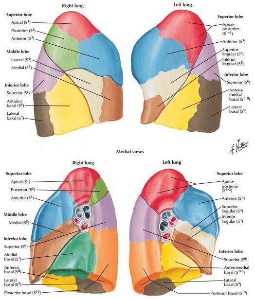
44 Rt. Lung Superior Lobe: 1. Apical. 2. Posterior. 3. Anterior. Middle Lobe: 1. Lateral. 2. Medial. Inferior Lobe: 1. Apical. 2. Medial basal. 3. Anterior basal. 4. Lateral basal. 5. Posterior basal. Lt. Lung Superior Lobe: 1. Apico-posterior. (*) 2. Anterior. 3. Superior lingular. 4. Inferior lingular. Inferior Lobe: 1. Superior. 2. Antero-medial basal. (*) 3. Lateral basal. 4. Inferior basal. 44
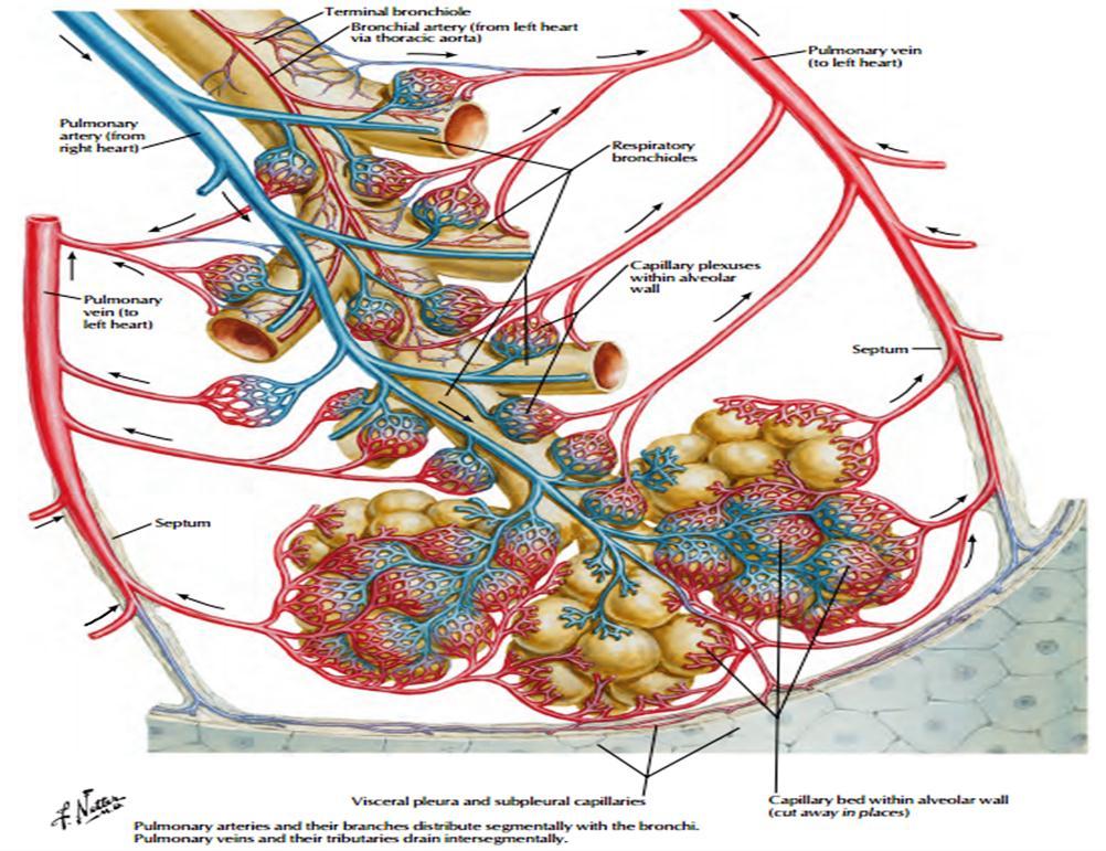
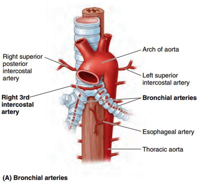
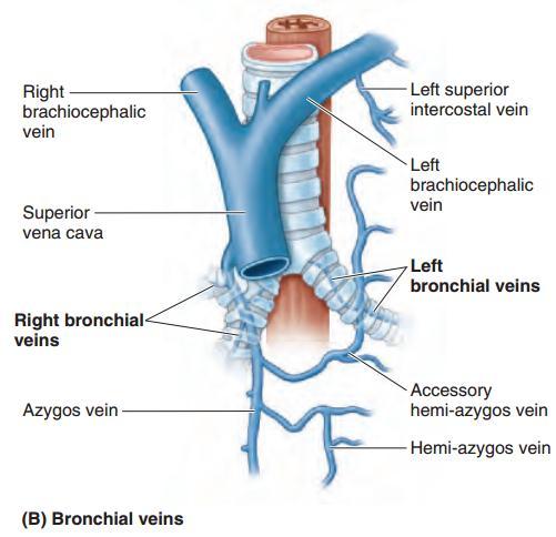
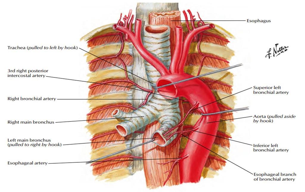
45 1. The bronchial arteries, which are branches of the descending aorta or one of its branches, supply the bronchi, the connective tissue of the lung, and the visceral pleura. A single right bronchial artery normally arises from the Rt. third posterior intercostal artery (but occasionally, it originates from the upper left bronchial artery). Two left bronchial arteries (superior & inferior) arise directly from the thoracic aorta. 2. The bronchial veins (which communicate with the pulmonary veins) drain into the azygos and hemiazygos veins. 45
46 46
47 47
48 48
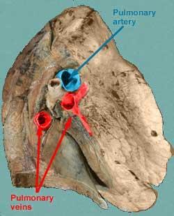
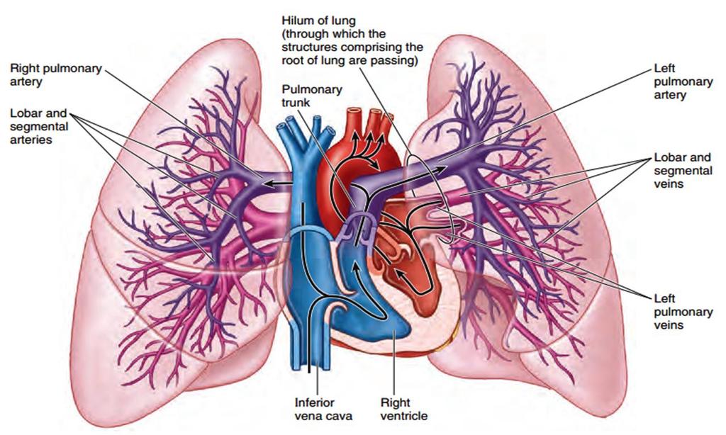
49 The alveoli receive deoxygenated blood from the terminal branches of the pulmonary arteries. The oxygenated blood leaving the alveolar capillaries drains into the tributaries of the pulmonary veins, which follow the intersegmental connective tissue septa to the lung root. Two pulmonary veins leave each lung root to empty into the left atrium of the heart. 49
50 50
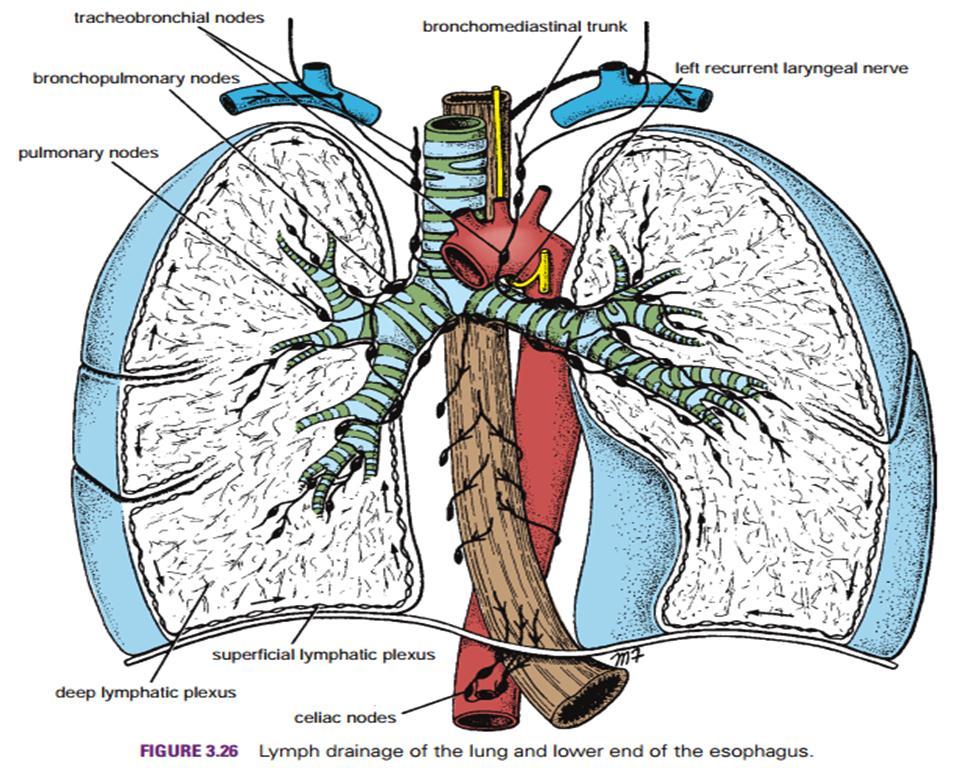
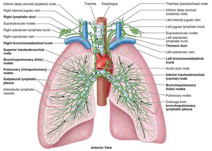
51 Subpleural (superficial) plexus drains the lung surface toward the hilum into bronchopulmonary nodes. Deep lymphatic plexus travels along pulmonary vessels toward the hilum passing through pulmonary nodes. From the hilum it drains into tracheobronchial and broncho-mediastinal nodes. 51
52 52
53 53
54 At the root of each lung is a pulmonary plexus composed of efferent & afferent autonomic nerve fibers. The plexus is formed from: 1. Branches of the sympathetic trunk. 2. Parasympathetic fibers from the vagus nerve. 54


55 55


56 It can be compressed by: 1. Enlarged thyroid gland. 2. Aortic arch aneurysm. 3. Double aortic arch. 56

57 57
58 Inflammation of these structures (Trachea or Bronchus) causes pain deep to the sternum. N.B: Mucosa of trachea is supplied by recurrent laryngeal nerve. Mucosa of bronchi is supplied by pulmonary plexus. 58
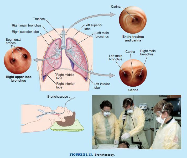
59 It is common especially in children. Loose tooth can slip to the trachea in anesthetized patient. F.B tends to enter the right bronchus more than the left. It may lodge in the larynx causing asphyxia which may necessitates emergency tracheostomy. 59
60 Allows examination of: 1. The trachea. 2. Carina. 3. Main bronchi. 4. Even the lobar bronchi. Allows biopsy. Allows removal of F.B. 60
61 61
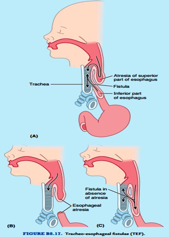
62 TE Fistula: is an opening between the trachea and esophagus. If the margins of the laryngotracheal groove fail to fuse adequately, an abnormal opening may be left between the laryngotracheal tube and the esophagus. 62
63 63
64 Atresia: is a congenital condition (birth defect) which affects the alimentary tract. It causes the esophagus to end in a blind-ended pouch rather than connecting normally to the stomach. Obstruction of the esophagus prevents the child from swallowing saliva and milk, and this leads to aspiration into the larynx and trachea, which usually results in pneumonia. Diagnosis can be made by insertion of NG tube and x- ray. The child presents with drooling of saliva, recurrent aspirations. With early diagnosis, it is often possible to correct this serious anomaly surgically. 64
65 65
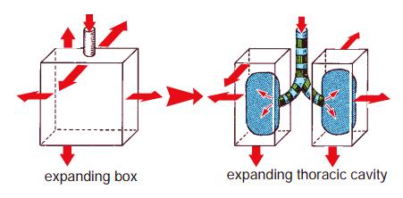
66 Respiratory cycle consists of 2 phases (inspiration & expiration) which are accomplished by the alternate increase and decrease of the capacity of the thoracic cavity. RR is /min (Resting), faster in children & slower in old. Inspiration: 1. Quiet Inspiration. Compare the thoracic cavity to a box with a single entrance at the top, which is a tube called the trachea. The capacity of the box can be increased by elongating all its diameters, and this results in air under atmospheric pressure entering the box through the tube. 66
67 67
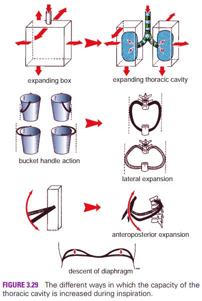
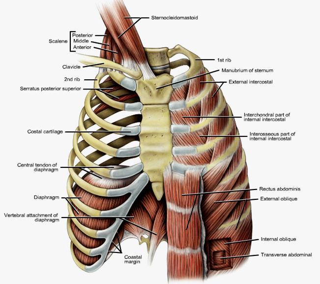
68 Now, Consider the 3 diameters of the thoracic cavity and how they may be increased. 1. Vertical Diameter Theoretically, the roof could be raised and the floor lowered. The roof is formed by suprapleural membrane and is fixed. Conversely, the floor is formed by diaphragm. When diaphragm contracts, the domes become flattened and the level is lowered. 2. Antero-posterior (AP) Diameter If we fix the 1 st rib by contraction of the scaleni muscles & intercostal muscles, meaning all the ribs are drawn together and raised toward the 1 st rib & the downward-sloping ribs are raised at their sternal ends, and the lower end of the sternum is thrusted forward. This 'pump handle' type of movement increase the AP diameter of the thorax. 68
69 3. Transverse Diameter The ribs articulate in front with the sternum via their costal cartilages and behind with the vertebral column. Because the ribs curve downward as well as forward around the chest wall, they resemble bucket handles. It therefore follows that if the ribs are raised (like bucket handles), the transverse diameter of the thoracic cavity will be increased. As described previously, this can be accomplished by fixing the 1 st rib and raising the other ribs to it by contracting the intercostal muscles. 69


70 Gray s Anatomy for Students 70

71 Moore Clinically Oriented Anatomy 7 th ed. 71
72 72

73 VERTICAL DIAMETER The Roof The Floor 73

74 AP DIAMETER Fixing First Rib Intercostal Muscles 74

75 TRANSVERSE DIAMETER Fixing First Rib Intercostal Muscles 75
76 The abdominal muscles play an important role in inspiration. Descent of the diaphragm is accompanied by abdominal Muscle relaxation. Abdominal muscle relaxation reaches its maximum, then the diaphragm is supported from below assisting the intercostal muscles in raising the ribs. 76
77 This results in maximum increase in the capacity of thoracic cavity. Accessory muscles (scalenus, sternomastoid) helps in more fixing of the first rib. Any muscle which can raise ribs is brought to action. Fixation of the scapulae also occurs. 77
78 78
79 1. The root descends as much as 2 vertebrae. 2. Bronchi elongate and dilate. 3. Alveolar capillaries dilate. 4. The air is drawn from the positive pressure to the negative one inside. 5. Expansion of the lungs opens the costodiaphragmatic recess. 79
80 Quiet Expiration (Passive Process) Lung Recoil Relaxation of Muscles Tone of Abd. Muscles 80
81 Forced Expiration (Active Process) Abd. Muscle Contraction Quadratus Lumborum Pull Of Ribs Downward 81
82 1. The lung root ascends. 2. Lung tissue recoil and thus decrease in size. 3. The costo-diaphragmatic recess close. 82
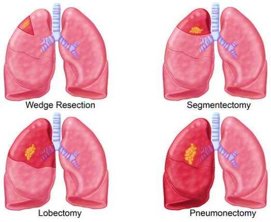
83 In babies & young children, ribs are nearly horizontal. Thus, babies have to rely mainly on the descent of the diaphragm to increase their thoracic capacity on inspiration. They are characterized by marked inward and outward excursion of the anterior abdominal wall, respiration at this age is referred to as the abdominal type of respiration. After the 2nd year of life, the ribs become more oblique, and the adult form of respiration is established. 83
84 In adults, a sexual difference exists in the type of respiratory movements. The female tends to rely mainly on the movements of the ribs rather than on the descent of the diaphragm on inspiration. This is referred to as the thoracic type of respiration. The male uses both the thoracic and abdominal forms of respiration, but mainly the abdominal form of respiration. 84
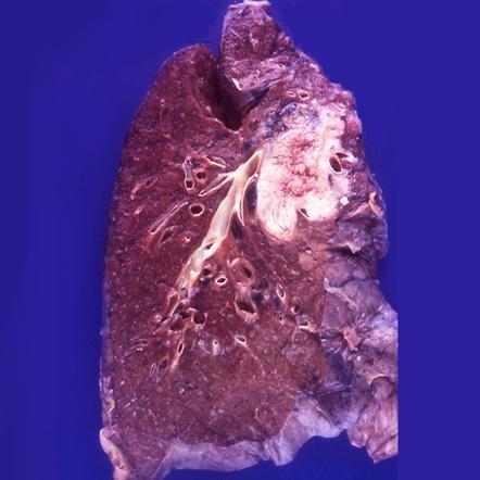
85 The upper zones are best examined from front. The lower lobes are best examined from back. The axilla enables the examination of both upper and lower lobes. 85
86 The apex of lung is liable for injury in the area above the clavicle. Rib fracture may injure the lung causing leak of air (pneumothorax) and lung collapse. Air may also reach the subcutaneous tissue (subcut. Emphysema) Depending on the phase of respiration abdominal muscles may or may not be injured in penetrating wounds of the lower chest. 86
87 Pain becomes a prominent feature if the disease involves the parietal pleura which has the characters of pleurisy pain. Pleurisy of the lower parts of the pleura may cause referred pain to the ant. abd. wall. Referred pain over the shoulder region results from irritation of the central part of diaphragmatic pleura. 87


88 Thoracotomy followed by rib retraction Bilateral anterolateral thoracotomy + transverse sternotomy = "clamshell" incision 88
89 89
90 Indicated for a localized chronic lesion such as that of tuberculosis or a benign neoplasm. Segmental resection requires that the radiologist and thoracic surgeon have a sound knowledge of the bronchopulmonary segments. 90
91 91
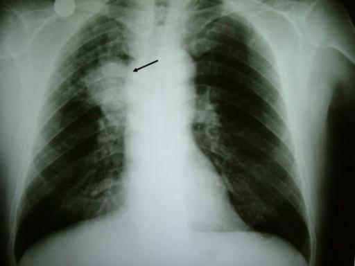
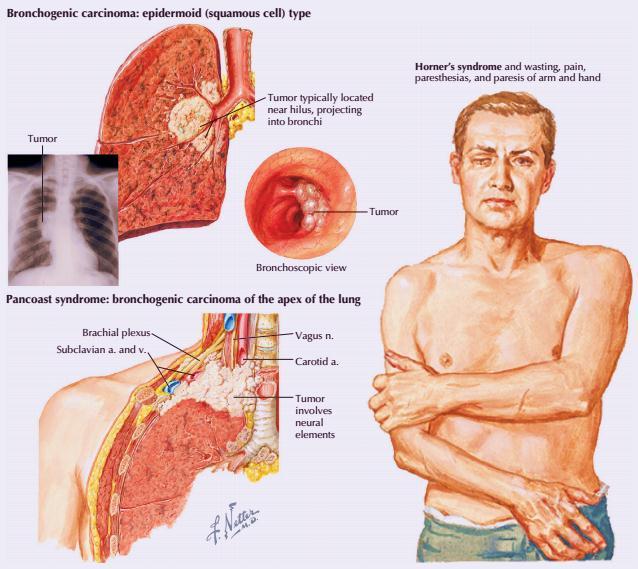
92 Accounts for about one third of all cancer deaths in men and is becoming increasingly common in women. It mostly commences in the mucous membrane lining the larger bronchi and is therefore situated close to the hilum. The neoplasm rapidly spreads to the tracheobronchial and bronchomediastinal nodes. It may involve the recurrent laryngeal nerves, leading to hoarseness of the voice. Lymphatic spread via the bronchomediastinal trunks may result in early involvement in the lower deep cervical nodes just above the clavicle (supraclavicular L.N). Hematogenous spread to bones and the brain commonly occurs. 92
93 93
94 94
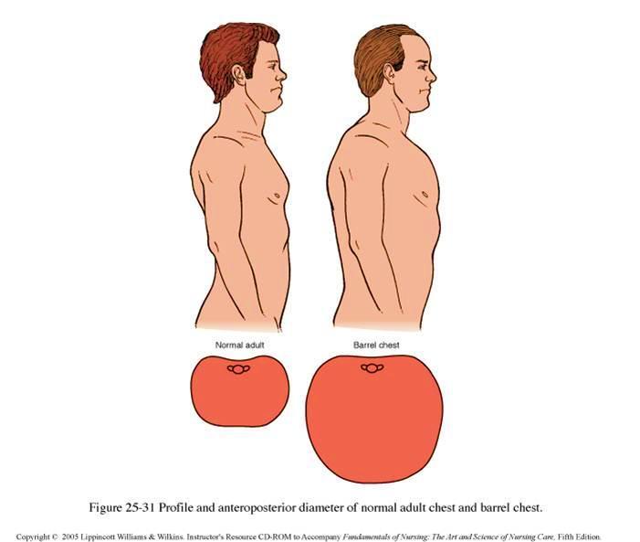
95 It is a chronic inflammatory disease of the lung causing reduction in the diameter of bronchioles by: mucus and constriction. One of the problems associated with bronchial asthma is the spasm of the smooth muscle in the wall of the bronchioles. This particularly reduces the diameter of the bronchioles during expiration, causing the asthmatic patient to experience great difficulty during expiration only. The lungs consequently become greatly distended and the thoracic cage becomes permanently enlarged ( hyper-inflated), forming the so-called barrel chest. 95
96 96
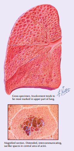
97 Many Lung diseases (Emphysema and pulmonary fibrosis) destroy the elasticity of the lungs, and makes the lungs unable to recoil causing incomplete expiration. In these patients the expiration is active process. Emphysema: Is characterized by loss of elasticity of the lung tissue, destruction of structures supporting the alveoli, and destruction of capillaries feeding the alveoli. The result is that the small airways collapse during expiration, leading to an obstructive form of lung disease (airflow is impeded and air is generally "trapped" in the lungs in obstructive lung disease. 97
98 98

99 Diseases such as silicosis, asbestosis, cancer, and pneumonia decrease the compliance of the lungs and the chest wall. Then a greater effort has to be undertaken by the inspiratory muscles to inflate the lungs during inspiration. 99
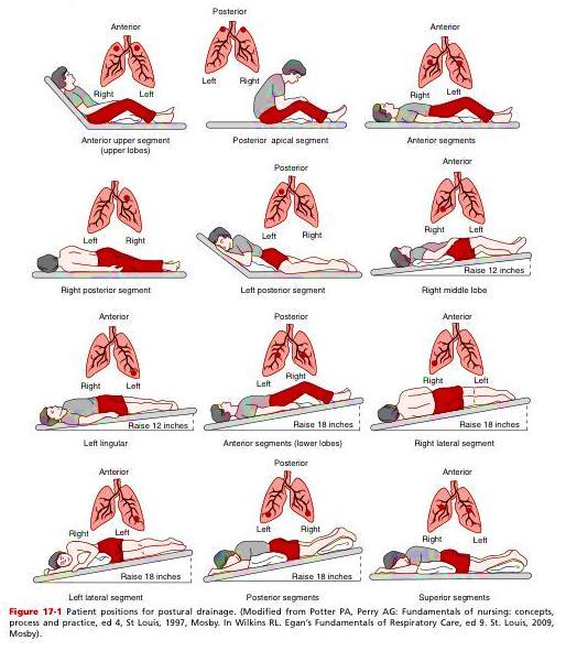
100 Excessive accumulation of bronchial secretions in a lobe or segment of a lung can seriously interfere with the normal flow of air into the alveoli. Furthermore, the stagnation of such secretions is often quickly followed by infection. To aid in the normal drainage of a bronchial segment, a physiotherapist often alters the position of the patient so that gravity assists in the process of drainage. Sound knowledge of the bronchial tree is necessary to determine the optimum position of the patient for good postural drainage. 100
101 101
102 102
slide 23 The lobes in the right and left lungs are divided into segments,which called bronchopulmonary segments
 Done By : Rahmeh Alsukkar Date : 26 /10/2017 slide 23 The lobes in the right and left lungs are divided into segments,which called bronchopulmonary segments Each segmental bronchus passes to a structurally
Lecturer: Ms DS Pillay ROOM 2P24 25 February 2013
 Lecturer: Ms DS Pillay ROOM 2P24 25 February 2013 Thoracic Wall Consists of thoracic cage Muscle Fascia Thoracic Cavity 3 Compartments of the Thorax (Great Vessels) (Heart) Superior thoracic aperture
Lung & Pleura. The Topics :
 Lung & Pleura The Topics : The Trachea. The Bronchi. The Brochopulmonary Segments. The Lungs. The Hilum. The Pleura. The Surface Anatomy Of The Lung & Pleura. The Root & Hilum. - first of all, the lung
Right lung. -fissures:
 -Right lung is shorter and wider because it is compressed by the right copula of the diaphragm by the live.. 2 fissure, 3 lobes.. hilum : 2 bronchi ( ep-arterial, hyp-arterial ), one artery mediastinal
Syllabus: 6 pages (Page 6 lists corresponding figures for Grant's Atlas 11 th & 12 th Eds.)
") PLEURAL CAVITY AND LUNGS Dr. Milton M. Sholley SELF STUDY RESOURCES Essential Clinical Anatomy 3 rd ed. (ECA): pp. 70 81 Syllabus: 6 pages (Page 6 lists corresponding figures for Grant's Atlas 11 th &
PLEURAL CAVITY AND LUNGS Dr. Milton M. Sholley SELF STUDY RESOURCES Essential Clinical Anatomy 3 rd ed. (ECA): pp. 70 81 Syllabus: 6 pages (Page 6 lists corresponding figures for Grant's Atlas 11 th &
Dana Alrafaiah. - Moayyad Al-Shafei. -Mohammad H. Al-Mohtaseb. 1 P a g e
 - 6 - Dana Alrafaiah - Moayyad Al-Shafei -Mohammad H. Al-Mohtaseb 1 P a g e Quick recap: Both lungs have an apex, base, mediastinal and costal surfaces, anterior and posterior borders. The right lung,
Thorax Lecture 2 Thoracic cavity.
 Thorax Lecture 2 Thoracic cavity. Spring 2016 Dr. Maher Hadidi, University of Jordan 1 Enclosed by the thoracic wall. Extends between (thoracic inlet) & (thoracic outlet). Thoracic inlet At root of the
Large veins of the thorax Brachiocephalic veins
 Large veins of the thorax Brachiocephalic veins Right brachiocephalic vein: formed at the root of the neck by the union of the right subclavian & the right internal jugular veins. Left brachiocephalic
Anatomy Lecture 8. In the previous lecture we talked about the lungs, and their surface anatomy:
 Anatomy Lecture 8 In the previous lecture we talked about the lungs, and their surface anatomy: 1-Apex:it lies 1 inch above the medial third of clavicle. 2-Anterior border: it starts from apex to the midpoint
Chapter 5: Other mediastinal structures. The Large Arteries. The Aorta. Ascending aorta
 Chapter 5: Other mediastinal structures The Large Arteries The Aorta The aorta is the main arterial trunk of the systemic circulation and in the healthy state its wall contain a large amount of yellow
Anatomy of the Lungs. Dr. Gondo Gozali Department of anatomy
 Anatomy of the Lungs Dr. Gondo Gozali Department of anatomy 1 Pulmonary Function Ventilation and Respiration Ventilation is the movement of air in and out of the lungs Respiration is the process of gas
Anatomy Sheet #5. In the previous lecture, we finished discussion about the larynx; now we continue with trachea, lungs and pleura.
 Anatomy Sheet #5 In the previous lecture, we finished discussion about the larynx; now we continue with trachea, lungs and pleura. Trachea and lungs The knowledge about the pleura and lungs is very important
Anatomy notes-thorax.
 Anatomy notes-thorax. Thorax: the part extending from the root of the neck to the abdomen. Parts of the thorax: - Thoracic cage (bones). - Thoracic wall. - Thoracic cavity. ** The thoracic cavity is covered
Mediastinum and pericardium
 Mediastinum and pericardium Prof. Abdulameer Al-Nuaimi E-mail: a.al-nuaimi@sheffield.ac.uk E. mail: abdulameerh@yahoo.com The mediastinum: is the central compartment of the thoracic cavity surrounded by
THE GOOFY ANATOMIST QUIZZES
 THE GOOFY ANATOMIST QUIZZES 7. LUNGS Q1. Fill in the blanks: the lung has lobes and fissures. A. Right, three, two. B. Right, two, one. C. Left, three, two. D. Left, two, three. Q2. The base of the lung
PLEURAE and PLEURAL RECESSES
 PLEURAE and PLEURAL RECESSES By Dr Farooq Aman Ullah Khan PMC 26 th April 2018 Introduction When sectioned transversely, it is apparent that the thoracic cavity is kidney shaped: a transversely ovoid space
Lab #3. Mohammad Hisham Al-Mohtaseb. Jumana Jihad. Ammar Ramadan. 0 P a g e
 Lab #3 Mohammad Hisham Al-Mohtaseb Jumana Jihad Ammar Ramadan 0 P a g e Last anatomy lab: Lungs and structure on the mediastinal surfs: 1-the right lung: How do we know it s the right lung??? -the 3 lobes
Dr. Weyrich G07: Superior and Posterior Mediastina. Reading: 1. Gray s Anatomy for Students, chapter 3
 Dr. Weyrich G07: Superior and Posterior Mediastina Reading: 1. Gray s Anatomy for Students, chapter 3 Objectives: 1. Subdivisions of mediastinum 2. Structures in Superior mediastinum 3. Structures in Posterior
10/14/2018 Dr. Shatarat
 2018 Objectives To discuss mediastina and its boundaries To discuss and explain the contents of the superior mediastinum To describe the great veins of the superior mediastinum To describe the Arch of
The External Anatomy of the Lungs. Prof Oluwadiya KS
 The External Anatomy of the Lungs Prof Oluwadiya KS www.oluwadiya.com Introduction The lungs are the vital organs of respiration Their main function is to oxygenate the blood by bringing inspired air into
DESCRIPTION: This is the part of the trunk, which is located between the root of the neck and the superior border of the abdominal region.
 1 THE THORACIC REGION DESCRIPTION: This is the part of the trunk, which is located between the root of the neck and the superior border of the abdominal region. SHAPE : T It has the shape of a truncated
Respiratory System. Functional Anatomy of the Respiratory System
 Respiratory System Overview of the Respiratory System s Job Major Duty Respiration Other important aspects ph control Vocalization Processing incoming air Protection Metabolism (ACE) What structures allow
Organs Histology D. Sahar AL-Sharqi. Respiratory system
 Respiratory system The respiratory system provides for exchange of O2 and CO2 to and from the blood. Respiratory organs include the lungs and a branching system of bronchial tubes that link the sites of
THE DESCENDING THORACIC AORTA
 Intercostal Arteries and Veins Each intercostal space contains a large single posterior intercostal artery and two small anterior intercostal arteries. The anterior intercostal arteries of the lower spaces
The Respiratory System. Dr. Ali Ebneshahidi
 The Respiratory System Dr. Ali Ebneshahidi Functions of The Respiratory System To allow gases from the environment to enter the bronchial tree through inspiration by expanding the thoracic volume. To allow
Chest and cardiovascular
 Module 1 Chest and cardiovascular A. Doss and M. J. Bull 1. Regarding the imaging modalities of the chest: High resolution computed tomography (HRCT) uses a slice thickness of 4 6 mm to identify mass lesions
CHAPTER 22 RESPIRATORY
 pulmonary ventilation move air external respiration exchange gases transportation of gases internal respiration exchange gases CHAPTER 22 RESPIRATORY in / out lungs air - blood blood - cells cell respiration
The Thoracic wall including the diaphragm. Prof Oluwadiya KS
 The Thoracic wall including the diaphragm Prof Oluwadiya KS www.oluwadiya.com Components of the thoracic wall Skin Superficial fascia Chest wall muscles (see upper limb slides) Skeletal framework Intercostal
THE RESPIRATORY SYSTEM
 THE RESPIRATORY SYSTEM Functions of the Respiratory System Provides extensive gas exchange surface area between air and circulating blood Moves air to and from exchange surfaces of lungs Protects respiratory
Mediastinum It is a thick movable partition between the two pleural sacs & lungs. It contains all the structures which lie
 Dr Jamila EL medany OBJECTIVES At the end of the lecture, students should be able to: Define the Mediastinum. Differentiate between the divisions of the mediastinum. List the boundaries and contents of
Bronchioles. Alveoli. Type I alveolar cells are very thin simple squamous epithelial cells and form most of the lining of an alveolus.
 276 Bronchioles Bronchioles continue on to form bronchi. The primary identifying feature is the loss of hyaline cartilage. The epithelium has become simple ciliated columnar, and there is a complete ring
Mohammad Almohtaseb. Lubna Allawi. Ammar Ramadan. 0 P a g e
 5 Mohammad Almohtaseb Lubna Allawi Ammar Ramadan 0 P a g e Trachea and Lungs The trachea The trachea is a flexible tube that extends from lower border of the larynx (lower border of cricoid cartilage at
The Respiratory System:
 The Respiratory System: Respiration Involves both the respiratory and the circulatory systems Four processes that supply the body with O 2 and dispose of CO 2 Respiration Pulmonary ventilation (breathing):
Lecture 2: Clinical anatomy of thoracic cage and cavity II
 Lecture 2: Clinical anatomy of thoracic cage and cavity II Dr. Rehan Asad At the end of this session, the student should be able to: Identify and discuss clinical anatomy of mediastinum such as its deflection,
ANATOMY OF THE PLEURA. Dr Oluwadiya KS
 ANATOMY OF THE PLEURA Dr Oluwadiya KS www.oluwadiya.sitesled.com Introduction The thoracic cavity is divided mainly into: Right pleural cavity Mediastinum Left Pleural cavity Pleural cavity The pleural
STERNUM. Lies in the midline of the anterior chest wall It is a flat bone Divides into three parts:
 STERNUM Lies in the midline of the anterior chest wall It is a flat bone Divides into three parts: 1-Manubrium sterni 2-Body of the sternum 3- Xiphoid process The body of the sternum articulates above
OBJECTIVE: To obtain a fundamental knowledge of the root of the neck with respect to structure and function
 The root of the neck Jeff Dupree, Ph.D. e mail: jldupree@vcu.edu OBJECTIVE: To obtain a fundamental knowledge of the root of the neck with respect to structure and function READING ASSIGNMENT: Moore and
NURSE-UP RESPIRATORY SYSTEM
 NURSE-UP RESPIRATORY SYSTEM FUNCTIONS OF THE RESPIRATORY SYSTEM Pulmonary Ventilation - Breathing Gas exchanger External Respiration between lungs and bloodstream Internal Respiration between bloodstream
Diaphragm and intercostal muscles. Dr. Heba Kalbouneh Associate Professor of Anatomy and Histology
 Diaphragm and intercostal muscles Dr. Heba Kalbouneh Associate Professor of Anatomy and Histology Skeletal System Adult Human contains 206 Bones 2 parts: Axial skeleton (axis): Skull, Vertebral column,
THE THORACIC WALL. Boundaries Posteriorly by the thoracic part of the vertebral column. Anteriorly by the sternum and costal cartilages
 THE THORACIC WALL Boundaries Posteriorly by the thoracic part of the vertebral column Anteriorly by the sternum and costal cartilages Laterally by the ribs and intercostal spaces Superiorly by the suprapleural
Anatomy of the Thorax
 Anatomy of the Thorax A) THE THORACIC WALL Boundaries Posteriorly by the thoracic part of the vertebral column Anteriorly by the sternum and costal cartilages Laterally by the ribs and intercostal spaces
The Respiratory System
 The Respiratory System Cells continually use O2 & release CO2 Respiratory system designed for gas exchange Cardiovascular system transports gases in blood Failure of either system rapid cell death from
Radiological Anatomy of Thorax. Dr. Jamila Elmedany & Prof. Saeed Abuel Makarem
 Radiological Anatomy of Thorax Dr. Jamila Elmedany & Prof. Saeed Abuel Makarem Indications for Chest x - A chest x-ray may be used to diagnose and plan treatment for various conditions, including: Diseases/Fractures
It passes through the diaphragm at the level of the 10th thoracic vertebra to join the stomach
 The esophagus is a tubular structure (muscular, collapsible tube ) about 10 in. (25 cm) long that is continuous above with the laryngeal part of the pharynx opposite the sixth cervical vertebra The esophagus
Unit Nine - The Respiratory System
 Unit Nine - The Respiratory System I. Introduction A. Definition: the respiratory system consists of the nose, nasal cavity, (throat), (voice box), (windpipe), bronchi and lungs (which contain the alveoli).
B. Correct! As air travels through the nasal cavities, it is warmed and humidified.
 Human Anatomy - Problem Drill 20: The Respiratory System Question No. 1 of 10 1. Which of the following statements about the portion of the respiratory system labeled in the image below is correct? Question
The Respiratory System
 PowerPoint Lecture Slide Presentation by Vince Austin Human Anatomy & Physiology FIFTH EDITION Elaine N. Marieb The Respiratory System Dr Nabil Khouri. MD, Ph.D Respiratory System Consists of a conducting
CHAPTER 24. Respiratory System
 CHAPTER 24 Respiratory System RESPIRATION INCLUDES Air moves in and out of lungs Continuous replacement of gases in alveoli (air sacs) Gas exchange between blood and air at alveoli Transport of respiratory
Lecture Overview. Respiratory System. Martini s Visual Anatomy and Physiology First Edition. Chapter 20 - Respiratory System Lecture 11
 Martini s Visual Anatomy and Physiology First Edition Martini Ober Chapter 20 - Respiratory System Lecture 11 1 Lecture Overview Overview of respiration Functions of breathing Organs of the respiratory
Lec #2 histology. Bronchioles:
 Lec #2 histology. Last lecture we talked about the upper respiratory tract histology, this one is about the lower part histology. We will discuss the histology of: -bronchioles -respiratory bronchioles
Cardiovascular system:
 Cardiovascular system: Mediastinum: The mediastinum: lies between the right and left pleura and lungs. It extends from the sternum in front to the vertebral column behind, and from the root of the neck
Bio 322 Human Anatomy Objectives for the laboratory exercise Respiratory System
 Bio 322 Human Anatomy Objectives for the laboratory exercise Respiratory System Required reading before beginning this lab: Saladin, KS: Human Anatomy 5 th ed (2017) Chapter 23 For this lab you will use
Respiratory System. Ling Shucai
 Respiratory System Ling Shucai General Description Ⅰ. Constituents: Respiratory tract Lungs Pleura and plural cavity Ⅱ. Function: exchange O 2 and CO 2 mainly Mediastinum Respiratory tract Upper respiratory
Identify the lines used in anatomical surface descriptions of the thorax. median line mid-axillary line mid-clavicular line
 L 14 A B O R A T O R Y Thorax THORACIC WALL Identify the lines used in anatomical surface descriptions of the thorax. median line mid-axillary line mid-clavicular line Identify the surface landmarks of
Anatomy of thoracic wall
 Anatomy of thoracic wall Topographic Anatomy of the Thorax 1 Bones of Thoracic wall ribs 1-7"true" ribs -those which attach directly to the sternum true ribs actually attach to the sternum by means of
Theme 30. Structure, topography and function of the lungs and pleura. Mediastinum and its contents. X -ray films digestive and respiratory systems.
 Theme 30. Structure, topography and function of the lungs and pleura. Mediastinum and its contents. X -ray films digestive and respiratory systems. STRUCTURE, TOPOGRAPHY AND FUNCTІON OF LUNGS AND PLEURA.
The RESPIRATORY System. Unit 9
 The RESPIRATORY System Unit 9 Respiration The exchange of gases between the atmosphere, blood, and cells Pulmonary Ventilation - the exchange of air between the atmosphere and lungs External (Pulmonary)
The Neck the lower margin of the mandible above the suprasternal notch and the upper border of the clavicle
 The Neck is the region of the body that lies between the lower margin of the mandible above and the suprasternal notch and the upper border of the clavicle below Nerves of the neck Cervical Plexus Is formed
Mohammad Hisham Al-Mohtaseb. Lina Mansour. Enas Ajarma
 6 Mohammad Hisham Al-Mohtaseb Lina Mansour Enas Ajarma Some recommended videos are attached to this sheet ( if u are studying online click on them, if not u can reach them by typing their names on the
Chest X-ray Interpretation
 Chest X-ray Interpretation Introduction Routinely obtained Pulmonary specialist consultation Inherent physical exam limitations Chest x-ray limitations Physical exam and chest x-ray provide compliment
I. Anatomy of the Respiratory System A. Upper Respiratory System Structures 1. Nose a. External Nares (Nostrils) 1) Vestibule Stratified Squamous
 1) Vestibule Stratified Squamous") I. Anatomy of the Respiratory System A. Upper Respiratory System Structures 1. Nose a. External Nares (Nostrils) 1) Vestibule Stratified Squamous Epithelium b. Nasal Cartilages 1) Nasal Cavity Pseudostratified
I. Anatomy of the Respiratory System A. Upper Respiratory System Structures 1. Nose a. External Nares (Nostrils) 1) Vestibule Stratified Squamous Epithelium b. Nasal Cartilages 1) Nasal Cavity Pseudostratified
Ch16: Respiratory System
 Ch16: Respiratory System Function: - O2 in and CO2 out of the blood vessels in the lungs - O2 out and CO2 into the blood vessels around the cells - Gas exchange happens in - Other organs purify, humidify,
Organs of the Respiratory System Laboratory Exercise 52
 Organs of the Respiratory System Laboratory Exercise 52 Background The organs of the respiratory system include the nose, nasal cavity, sinuses, pharynx, larynx, trachea, bronchial tree, and lungs. They
THYROID & PARATHYROID. By Prof. Saeed Abuel Makarem & Dr. Sanaa Al-Sharawy
 THYROID & PARATHYROID By Prof. Saeed Abuel Makarem & Dr. Sanaa Al-Sharawy 1 OBJECTIVES By the end of the lecture, the student should be able to: Describe the shape, position, relations and structure of
Chapter 3: Thorax. Thorax
 Chapter 3: Thorax Thorax Thoracic Cage I. Thoracic Cage Osteology A. Thoracic Vertebrae Basic structure: vertebral body, pedicles, laminae, spinous processes and transverse processes Natural kyphotic shape,
The Respiratory System
 C h a p t e r 24 The Respiratory System PowerPoint Lecture Slides prepared by Jason LaPres North Harris College Houston, Texas Copyright 2009 Pearson Education, Inc., publishing as Pearson Benjamin Cummings
Karachi King s College of Nursing
 Karachi King s College of Nursing Badil Dass Lecturer Respiratory system Respiratory System Respiratory system consist of: Nose Pharynx (Throat) Larynx (Voice Box) Trachea (Wind Pipe) Bronchi Bronchioles
Chapter 16. Respiratory System
 Chapter 16 Respiratory System Introduction Respiration = the entire process of exchanging gases between the atmosphere and body cells 1. Ventilation 2. Gas exchange 3. Gas transport : 4. Cellular respiration
The Respiratory System
 13 PART A The Respiratory System PowerPoint Lecture Slide Presentation by Jerry L. Cook, Sam Houston University ESSENTIALS OF HUMAN ANATOMY & PHYSIOLOGY EIGHTH EDITION ELAINE N. MARIEB Organs of the Respiratory
Chapter 11 The Respiratory System
 Biology 12 Name: Respiratory System Per: Date: Chapter 11 The Respiratory System Complete using BC Biology 12, page 342-371 11.1 The Respiratory System pages 346-350 1. Distinguish between A. ventilation:
5/5/2013. The Respiratory System. Chapter 16 Notes. The Respiratory System. Nasal Cavity. Sinuses
 The Respiratory System Chapter 16 Notes The Respiratory System Objectives List the general functions of the respiratory system. Identify the organs of the respiratory system. Describe the functions of
Pancreas & Biliary System. Dr. Vohra & Dr. Jamila
 Pancreas & Biliary System Dr. Vohra & Dr. Jamila 1 Objectives At the end of the lecture, the student should be able to describe the: Location, surface anatomy, parts, relations & peritoneal reflection
Yara saddam & Dana Qatawneh. Razi kittaneh. Maher hadidi
 1 Yara saddam & Dana Qatawneh Razi kittaneh Maher hadidi LECTURE 10 THORAX The thorax extends from the root of the neck to the abdomen. The thorax has a Thoracic wall Thoracic cavity and it is divided
RESPIRATORY SYSTEM. A. Upper respiratory tract (Fig. 23.1) Use the half-head models.
 Use the half-head models.") RESPIRATORY SYSTEM I. OVERVIEW OF THE RESPIRATORY SYSTEM AND THORAX A. Upper respiratory tract (Fig. 23.1) Use the half-head models. Nasal cavity Pharynx (fare-rinks) B. Lower respiratory tract (Fig. 23.1)
RESPIRATORY SYSTEM I. OVERVIEW OF THE RESPIRATORY SYSTEM AND THORAX A. Upper respiratory tract (Fig. 23.1) Use the half-head models. Nasal cavity Pharynx (fare-rinks) B. Lower respiratory tract (Fig. 23.1)
Unit 9. Respiratory System 16-1
 Unit 9 Respiratory System 16-1 Works together with the circulatory system Exchange of gases between atmosphere, blood, and cells If respiratory system and/or circulatory system fails, death will occur
ORAL CAVITY, ESOPHAGUS AND STOMACH
 ORAL CAVITY, ESOPHAGUS AND STOMACH 1 OBJECTIVES By the end of the lecture you should be able to: Describe the anatomy the oral cavity, (boundaries, parts, nerve supply). Describe the anatomy of the palate,
thoracic cage inlet and outlet landmarks of the anterior chest wall muscles of the thoracic wall sternum joints ribs intercostal spaces diaphragm
 Thoracic Wall Lecture Objectives Describe the shape and outline of the thoracic cage including inlet and outlet. Describe the anatomical landmarks of the anterior chest wall. List various structures making
Endeavour College of Natural Health endeavour.edu.au
 Endeavour College of Natural Health endeavour.edu.au BIOH122 Human Biological Science 2 Session 10 Respiratory System 1 Anatomy & Physiology Bioscience Department Endeavour College of Natural Health endeavour.edu.au
Chapter 13. The Respiratory System.
 Chapter 13 The Respiratory System https://www.youtube.com/watch?v=hc1ytxc_84a https://www.youtube.com/watch?v=9fxm85fy4sq http://ed.ted.com/lessons/what-do-the-lungs-do-emma-bryce Primary Function of Breathing
Lecture 01. The Thyroid & Parathyroid Glands. By: Dr Farooq Khan PMC Date: 12 th March. 2018
 Lecture 01 The Thyroid & Parathyroid Glands By: Dr Farooq Khan PMC Date: 12 th March. 2018 INTRODUCTION LAYERS OF THE NECK The neck has four major compartments or layer which are enclosed by an outer musculofascial
Thyroid and Parathyroid Glands
 Thyroid and Parathyroid Glands Please view our Editing File before studying this lecture to check for any changes. Color Code Important Doctors Notes Notes/ explanation Objectives: By the end of the lecture,
This is not a required assignment but it is recommended.
 SU 12 Name: This is not a required assignment but it is recommended. BIO 116 - Anatomy & Physiology II Practice Assignment 2 - The Respiratory and Cardiovascular Systems 1. The exchange of oxygen and carbon
The Anatomy and Physiology of the Respiratory System
 CHAPTER 1 The Anatomy and Physiology of the Respiratory System Sagittal Section of Upper Airway Fig. 1-1. Sagittal section of upper airway. Structure of the Nose Fig. 1-2. Structure of the nose. Sagittal
CHAPTER 7.1 STRUCTURES OF THE RESPIRATORY SYSTEM
 CHAPTER 7.1 STRUCTURES OF THE RESPIRATORY SYSTEM Pages 244-247 DO NOW What structures, do you think, are active participating in the breathing process? 2 WHAT ARE WE DOING IN TODAY S CLASS Finishing Digestion
11.1 The Aortic Arch General Anatomy of the Ascending Aorta and the Aortic Arch Surgical Anatomy of the Aorta
 456 11 Surgical Anatomy of the Aorta 11.1 The Aortic Arch 11.1.1 General Anatomy of the Ascending Aorta and the Aortic Arch Surgery of the is one of the most challenging areas of cardiac and vascular surgery,
Hyoid Bone. Lower Airway. Aspiration. Larynx. Cartilages of the Larynx. Larynx Tracheobronchial Tree (TB Tree) Trachea Bronchi Bronchioles
 Trachea Bronchi Bronchioles") Lower Airway Larynx Tracheobronchial Tree (TB Tree) Trachea Bronchi Bronchioles Respiratory Terminal Hyoid Bone Not part of the larynx. The Hyoid bone is an anchor for the anterior muscles of the neck
Lower Airway Larynx Tracheobronchial Tree (TB Tree) Trachea Bronchi Bronchioles Respiratory Terminal Hyoid Bone Not part of the larynx. The Hyoid bone is an anchor for the anterior muscles of the neck
Posterior Triangle of the Neck By Prof. Dr. Muhammad Imran Qureshi
 Posterior Triangle of the Neck By Prof. Dr. Muhammad Imran Qureshi For the purpose of anatomical description the neck is sub divided into two major triangles, the Anterior and the Posterior by muscle bellies
Veins of the Face and the Neck
 Veins of the Face and the Neck Facial Vein The facial vein is formed at the medial angle of the eye by the union of the supraorbital and supratrochlear veins. connected through the ophthalmic veins with
LUNGS. Requirements of a Respiratory System
 Respiratory System Requirements of a Respiratory System Gas exchange is the physical method that organisms use to obtain oxygen from their surroundings and remove carbon dioxide. Oxygen is needed for aerobic
The Respiratory System. Supplies body with oxygen Disposes of carbon dioxide Four processes in respiration
 C H A P T E R 22 The Respiratory System The Respiratory System Supplies body with oxygen Disposes of carbon dioxide Four processes in respiration Pulmonary ventilation External respiration Transport of
Circulatory System. and. Respiratory System. Ari Min, Yerim Lee and Min Ji Song THE HEART LUNGS. Monday, May 23, 2011
 Human Anatomy Circulatory System and THE HEART Respiratory System LUNGS Ari Min, Yerim Lee and Min Ji Song Purpose of the Circulatory System Function of circulatory system: exchange gases with cardiovascular
Superior and Posterior Mediastinum. Assoc. Prof. Jenny Hayes
 Superior and Posterior Mediastinum Assoc. Prof. Jenny Hayes WARNING This material has been provided to you pursuant to section 49 of the Copyright Act 1968 (the Act) for the purposes of research or study.
BOGOMOLETS NATIONAL MEDICAL UNIVERSITY DEPARTMENT OF HUMAN ANATOMY. Guidelines. Module 2 Topic of the lesson Aorta. Thoracic aorta.
 BOGOMOLETS NATIONAL MEDICAL UNIVERSITY DEPARTMENT OF HUMAN ANATOMY Guidelines Academic discipline HUMAN ANATOMY Module 2 Topic of the lesson Aorta. Thoracic aorta. Course 1 The number of hours 3 1. The
Respiratory Physiology
 Respiratory Physiology Dr. Aida Korish Associate Prof. Physiology KSU The main goal of respiration is to 1-Provide oxygen to tissues 2- Remove CO2 from the body. Respiratory system consists of: Passages
Chapter 10 Respiration
 1 Chapter 10 Respiration Introduction/Importance of the Respiratory System All eukaryotic organisms need oxygen to perform cellular respiration (production of ATP), either aerobically or anaerobically.
The RESPIRATORY System
 The RESPIRATORY System Respira5on The exchange of gases between the atmosphere, blood, and cells Pulmonary Ven5la5on - the exchange of air between the atmosphere and lungs External (Pulmonary) Respira5on
Sheet. April/14 th /2013. Introduction to Anatomy. Dr. Maher Hadidi. Muna Abu Hijleh. 1 P a g e
 Sheet Introduction to Anatomy Dr. Maher Hadidi Muna Abu Hijleh 1 P a g e 29 April/14 th /2013 Superior & Posterior Mediastinum ***Superior mediastinum * is bounded from: -Anterior by manubrium sterni -posterior
Anatomy of the Thyroid Gland
 Anatomy of the Thyroid Gland Introduction Nomenclature G, thyreos= shield, eidos= like Location Root of the neck ventrally (C5-T1) Function endocrine gland that secretes: Thyroxine (T4) T3 Calcitonin LWW,
The Respiratory System
 Essentials of Human Anatomy & Physiology Elaine N. Marieb Seventh Edition Chapter 13 The Respiratory System Slides 13.1 13.30 Lecture Slides in PowerPoint by Jerry L. Cook Copyright 2003 Pearson Education,
Ventilation 7/28/2013. Clarification of Terminology. Osteology of Ventilation
 Ventilation Clarification of Terminology Ventilation: the mechanical process by which air is inhaled and exhaled through the lungs. It describes only the movement of air. Respiration: a term used to describe