University of Siena. Ph.D in Medical Genetics. Mariangela Amenduni. Supervisor: Prof. Alessandra Renieri. Doctoral school in Oncology and Genetics
|
|
|
- Tobias Parks
- 6 years ago
- Views:
Transcription

1 University of Siena Ph.D in Medical Genetics Establishment and validation of a human cellular model for CDKL5-related disorders Mariangela Amenduni Supervisor: Prof. Alessandra Renieri Doctoral school in Oncology and Genetics Academic year Cicle XXIII
2 INDEX ABSTRACT 2 1. INTRODUCTION CDKL5-associated phenotypes Rett Syndrome ipscs to model human neurologic diseases RATIONALE AND AIMS OF THE STUDY RESULTS ipsc for CDKL5-related disorders 24 Eur J Hum Genet.2011 Jul 13. doi: /ejhg Set up of a protocol for the analysis of morphological alterations in CDKL5 neurons Materials and methods Results Gene expression profiling of ipscs from CDKL5 patients Experimental procedures Results DISCUSSION AND FUTURE PERSPECTIVES REFERENCES 61 ACKNOWLEDGEMENTS 68 1
3 Abstract CDKL5 is an highly conserved kinase expressed in a wide variety of cell-lines and tissues, with the highest levels in brain, testes and thymus. Mutations in this gene are responsible for the early-onset seizures variant of Rett syndrome and a severe encephalopathy with X-linked infantile spasms. However, the molecular mechanisms leading from CDKL5 mutations to disease onset remain largely unknown and the protein is poorly characterized and its function partially elucidated so far. This is mainly due to the unavailability of both a mouse model and a good human cellular model. To overcome these limitations, we employed the approach of genetic reprogramming that allows the generation of induced pluripotent stem (ips) cells directly from patients fibroblasts. We successfully reprogrammed fibroblasts from 2 CDKL5- mutated patients (a male with p.t288i and a female with p.q347x). In order to assess whether ips cells are suitable as an in vitro model to study the pathogenesis of CDKL5-related disorders, we induced these cells toward a neuronal fate. Preliminary characterization of the neuronal differentiation process indicates that ips cells can be differentiated into neurons and that the differentiation follows the stages observed in human embryonic stem cells (hescs). No significant differences respect to neurons from normal controls have been observed. More detailed characterization of the obtained neurons is ongoing. Comparison of expression profiles between CDKL5-mutated and control ipscs pointed out one interesting gene whose expression seems to be reduced in the absence of CDKL5. 2
4 1. INTRODUCTION 3
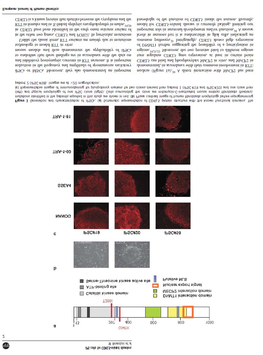
5 1. Introduction Serine Threonine Kinase 9 gene (STK9), also known as CDKL5, was firstly isolated in 1998 by Montini E. and colleagues that were involved in a transcriptional mapping effort in the Xp22 region 1. In 2003 Kalscheuer and colleagues have revisited the complete gene structure of STK9 reporting some discrepancies with the exon number and sizes indicated by Montini in ,2. The main difference is that STK9 contains at least 23 exons, with the first three exons (1, 1a and 1b) being untranslated and determining two splice variants with different 5 UTRs (Fig. 1A). Splice variant I, containing exon 1 is the more abundant while splice variant II, containing exon 1a and 1b is transcribed at very low levels 2. CDKL5 (OMIM #300203), located on Xp22, encodes for a serine-threonine kinase with an N-terminal domain highly homologous to members of the mitogen-activated protein (MAP) and cyclin dependent kinase (CDK) families 3. CDKL5 is a large protein of 1030 aminoacids with an estimated molecular weight of 115 kda containing a conserved serinethreonine kinase domain within its N-terminus and a large C-terminal region (Fig 1B). It is highly conserved and ubiquitously expressed in a wide variety of cell lines and tissues, with the highest level in brain, testes and thymus 3. Very recently an additional exon (16b) within the CDKL5 gene has been identified, showing no homology with other sequences in the human genome and an extremely high level of similarity between species, indicating a functional role that has been conserved during evolution 4. It encodes a further 41 aa, producing a predicted protein of 120 kda in humans. This new transcript has been detected also in mouse brain, with different levels in the various regions, whereas it is not found in other organs, thus suggesting the potential functional importance of the isoform in brain. However the full-length protein containing exon 16b has not yet been identified in tissues, neither by western blotting analysis nor by immunofluorescence studies in human fibroblasts or mouse tissues. Authors suggest that these results do not indicate that the protein is not expressed, but may be due to its low amount that cannot be detected by the methods used. Despite these findings, the presence of the 16b-containing mrna isoform in all brain regions of adult mouse is in favour of the expression of the long CDKL5 isoform in vivo 4. Furthermore another recent publication has reported a novel CDKL5 splice variant containing intron 18 (designated as CDKL5 107 ) and encoding a protein with an alternative C-terminus in both human and mouse. This novel transcript (exon1-intron 18) is widely expressed in all human tissues and cell lines examined, 4
6 and is the predominant isoform in human brain. In view of these results it has been ascertained whether its functional characteristics, such as the catalytic activity and subcellular localization, are similar to those of the CDKL5 115 isoform. The detailed characterization revealed that its subcellular distribution is largely maintained with respect to CDKL5 115 isoform, that its autophosphorylation and heterophosphorylation activity is reduced, but this is compensated by an increase in protein level. Interestingly, this isoform seems to be more stable and resistant to proteasome degradation than the CDKL5 115 isoform 5. However, it has to be taken into consideration that the significance of these in vitro studies for brain function could be better clarified only by studies in vivo and on the primarily affected tissue. Since its molecular characterization started in 2005, when mutations in this gene were identified in patients affected by Rett Syndrome (RTT), CDKL5 protein remains still rather uncharacterized. Its involvement in RTT has been explained by the fact that this kinase seems to work in a molecular pathway common to that of MeCP2, the main gene responsible for RTT 6-8. It has been demonstrated that the two proteins interact both in vivo and in vitro but there are some controversial results regarding the functional role of this interaction; in fact an in vivo phosphorilation of Mecp2 has been reported by Mari and colleagues, but the result has not been confirmed by Li and colleagues 3,6,7 Intriguingly the two genes show an overlapping temporal and spatial expression profile in brain and are simultaneously activated during neuronal maturation 6. Moreover, the two proteins bind to different regions of the N-terminal domain of Dnmt1 strengthening the hypothesis of their belonging to the same molecular pathway 9. More recently Carouge and colleagues addressed the question of the transcriptional control of Cdkl5 by Mecp2 as a potential link between the two genes, taking advantage of MeCP2 induction by cocaine in rat brain structures 8. Their data reveal that over-expression of Mecp2 in transfected cells results in the repression of Cdkl5 expression and that in vivo Mecp2 directly interacts with Cdkl5 gene in a methylation-dependent manner. Taken together these results are consistent with Cdkl5 being a Mecp2-repressed target gene and provide new insights into the mechanism by which mutations in the two genes result in overlapping neurological symptoms 8. At the moment, CDKL5 function during neuronal maturation and activity remains still rather uncharacterized. However since mutations in CDKL5 and MECP2 lead to similar genetic desorders the relationship between these two genes has been extensively investigated. MeCP2 is induced during embryogenesis and its levels remain rather homogeneous during postnatal development until adulthood On the contrary, CDKL5 is highly expressed later in development, and its levels are much more modulated than those of MeCP2, despite an 5
7 overlapping expression profile of these two proteins 13. Moreover, while MeCP2 is an exclusively nuclear protein, CDKL5 shuttles between the nuclear and cytoplasmatic compartments. Interestingly cytoplasmic CDKL5 is localized in the dendritic branches with a distinct punctuate staining. It is noteworthy that CDKL5 expression is mainly cytoplasmatic in late embryonic stages of mouse brains, with a low fraction of the protein in the nucleus. The nuclear fraction increases during early postnatal stages consistently with neuronal maturation and remains as such until adult stages 13. These data suggest that CDKL5 functions might be modulated through mechanisms regulating its shuttling between nucleus and cytoplasm. Intriguingly, it has been demonstrated a fundamental role of the C-terminal tail in regulating CDKL5 function by affecting its stability, kinase activity and its dynamic subcellular distribution 3,7,13. Further studies have showed that CDKL5 co-localizes and is associated with a number of splicing factors that are stored in structures called nuclear speckles, in both cell lines and tissues. In these structures CDKL5 seems to manage the nuclear trafficking of splicing factors and thus indirectly of the splicing machinery 14. It is already known that phosphorylation of the RS domain of Serine-rich (SR) splicing factors is necessary to release these factors from speckles and direct them to sites where pre-mrna processing takes place. Considering that several protein kinases have been described to be able to phosphorylate the RS domain of SR proteins Ricciardi and colleagues have hypothesized that also CDKL5 could have a role in nuclear speckles organization. Interestingly it has been demonstrated that CDKL5 acts on nuclear speckle disassembly determining a redistribution of at least some speckle proteins. 14. In spite of all these findings, our knowledge of the function of CDKL5 inside the neuron and of the underlying molecular mechanisms is still limited by the unavailability of both a mouse model and a good human cellular model for CDKL5-related disease. A recent study in cultured rat cortical neurons has partially overcome this limitation and has demonstrated that CDKL5 down-regulation by RNA interference inhibits neurite outgrowth and dendritic arborization, while its over-expression elicits the opposite effect 15. In several studies CDKL5 has been found to exert its function in the nucleus, while other groups have reported a consistent expression of CDKL5 in the dendrites of cultured neurons, thus suggesting that it might have a cytoplasmatic function independent of its role in the nucleus 3,7,13. Starting from these data Chen and colleagues have inspected its subcellular localization in rat neurons. Interestingly CDKL5 was detected at high levels in the cytoplasmic fraction of cultured cortical neurons. Moreover, immunostaining experiments revealed that it co-localizes with F-actin in the peripheral domain of growth cones, indicating a possible role in actin 6
8 cytosckeleton regulation. Since Rho GTPases are involved in actin dynamics and neuronal morphogenesis, authors hyopthesized a possible interaction with CDKL5. In fact, colocalization and GST pull-down assays revealed that CDKL5 directly or indirectly interacts with Rac1 and that this interaction is enhanced by brain-derived neurotrophic factor (BDNF). The finding that CDKL5 regulates neuronal morphogenesis through a mechanism involving Rac1-BDNF signaling might represent a novel mechanism involved in the pathology of CDKL5 related disorders 15. The recent research recapitulated above has given important glimpses on CDKL5 role in brain. However, due to the absence of a good human cellular model, it has not been possible to date to verify whether all these findings resemble the situation in affected human brain. All the information achieved so far about CDKL5 indicates that it exerts different functions based on the intracellular compartment in which it is localized. However, CDKL5 function if far from being unravelled, precluding an understanding of its role in the pathogenic processes responsible for disease onset. Fig. 1. CDKL5 gene and protein. (A) Genomic structure of CDKL5 gene (previously known as STK9) with exons 1,1a and 1b. The translation start codon ATG is contained in exon2. Image modified from Kalscheuer et al., (B) Schematic representation of the CDKL5 protein with its functional domains. 7
9 1.1 CDKL5-associated phenotypes Several groups have described mutations in CDKL5 in patients with seizures appearing soon after birth and severe mental retardation. The triad of infantile spasm, hypsarrhythmia and severe mental retardation is characteristic of West syndrome or, when X- linked, infantile spasm syndrome (ISSX). The first alterations in this gene have been reported in two unrelated girls with ISSX and X-autosome translocations. In both cases the breakpoints disrupted the CDKL5 gene 2. After that, mutations in CDKL5 have been identified in patients diagnosed with atypical Rett syndrome (RTT), a girl with an autistic disorder and intellectual disability and a boy with severe, early infantile onset neurological disorder Additionally a boy with severe epileptic encephalopathy and bilateral cataract was shown to have an interstitial deletion comprising the CDKL5 gene 19. In summary, the phenotypic spectrum of CDKL5 abnormalities has progressively expanded to include clinical features resembling those of other neurodevelopmental disorders. In particular, most of the cases have been diagnosed with the early-onset seizures variant of RTT suggesting that this could be the predominant phenotype caused by CDKL5 mutations 17,20. The hallmarks of CDKL5-associated diseases are early-onset intractable seizures with infantile spasms, severe intellectual disability and atypical RTT features. The common feature describing patients affected by the early-onset seizures variant of RTT is the appearance of epilepsy during the very first weeks of life and a developmental delay without any period of normal development; in many cases epilepsy becomes refractory to treatment during later stages 21. Moreover, CDKL5 mutated patients clearly exhibit some RTT-like features such as deceleration of head growth, stereotypies and hand apraxia, and sleep disturbances Most patients show a combination of different hand stereotypies without hand gaze and bruxism, although these features become really evident in patients at older age and also able to walk with support. Conversely, dysautonomic features such as breathing disturbances and gastrointestinal dysfunction are rare suggesting a possible better preserved autonomic system in patients with CDKL5 mutations compared with typical RTT ones 25. Although the epileptic disorder is the key feature to identify patients likely to present CDKL5 mutations, there is limited information regarding its precise electro-clinical phenotype. Buoni and colleagues in 2006 described an epileptic pattern consisting of myoclonic epilepsy with refractory seizures and a unique electroencephalogram (EEG) pattern 26. Archer and colleagues have further extended the description of the epilepsy 8
10 phenotype suggesting that the spectrum of severity of the seizure disorder is broader, ranging from no seizures at all to hundreds of seizures a day. However, it is interesting to note that also in the patients described by Archer, seizures are largely resistant to drug treatment 20. Recently another contribution to the definition of the epileptic phenotype in CDKL5 mutations came from Bahi-Buisson and colleagues 21. They investigated the electro-clinical pattern of ISSX patients in order to give a better characterization of the phenotype and thus to facilitate an early diagnosis. Their data suggest an epilepsy phenotype with a three-stage pattern that initiates with early epilepsy, moves on to epileptic encephalopathy and comes to an end with late multifocal and myoclonic epilepsy Rett syndrome Since the early-onset seizures variant of RTT is considered the predominant phenotype caused by CDKL5 mutations, this syndrome will be described in further details 17,20. Rett syndrome (OMIM#312750) is a progressive neurodevelopmental disorder almost exclusively affecting females with an incidence of 1:10000 born females. Due to its incidence it represents the second cause of mental retardation in girls 27,28. The syndrome was first described by Andreas Rett in 1966, but it was internationally recognized as a distinct clinical entity when Hagberg et al. described 35 new cases in In the classic form, Rett girls are characterized by a period of 6-18 months of apparently normal development after which they present a developmental arrest followed by regression in language and motor skills. They usually have normal head circumference at birth followed by postnatal deceleration of head growth leading to microcephaly by the second year of life. As the syndrome progresses, patients lose the purposeful use of hands and develop repetitive stereotypic hand movements. The pattern consists of tortuous hand wringing, hand washing, clapping, flapping or other more bizarre hand automatisms 30. Patients also manifest social withdrawal, communication dysfunction, loss of acquired speech and cognitive impairment, in addition to irritability and self-abusive behaviour. Further characteristics are the appearance of autistic features, including a reduction in interpersonal contact, hypersensitivity to sound, indifference to the surrounding environment and unresponsiveness to social cues 28,31. RTT girls also suffer devastating motor deterioration with the development of ataxia and gait apraxia. Most girls with RTT develop seizures ranging from easily controlled to intractable epilepsy, and suffer respiratory dysfunctions, bruxism and impairment of sleeping patterns. Amelioration of the 9
11 autistic-like behaviour occurs between 5 and 10 years of age, but other somatic and neurological handicaps, such as severe scoliosis, rigidity and reduced somatic growth become evident during this period. As patients get older they develop Parkinsonian features. The condition reaches a plateau and, although sudden death can occur, most people with Rett syndrome survive up to the sixth or seventh decade of life in a profoundly debilitated physical condition 28,31. As experience on this syndrome increased it has become evident that females with RTT may present with a much broader phenotype than originally described. Indeed atypical forms of RTT have been described, with significant differences in disease onset, severity of impairment and profile of clinical course 22,27. The milder forms of RTT include the late regression variant, which is rare and still controversial, and the forme fruste variant presenting with a later age of onset and regression occurring between 1 and 3 years of age. Another form of RTT with a benign course is the Zappella variant previously known as the Preserved Speech Variant (PSV). Affected girls recover the ability to speak in sentences, although not necessarily in context, and display an improvement of purposeful hand movements. The more severe variants include the congenital form, in which girls appear floppy and retarded since the very first months of life, thus lacking the early period of normal development typical of classic RTT, and the early-onset seizures variant with severe epileptic seizures appearing soon after birth. Considering that RTT occurred almost exclusively in females, an X-linked dominant inheritance was suggested, with possible male lethality. However, since more than 99% of RTT cases are sporadic, it was very difficult to map the disease locus by traditional linkage analysis. Using information from rare familial cases, exclusion mapping identified the Xq28 candidate region, and subsequent screening of candidate genes in RTT patients detected mutations in MECP2 gene (Methyl-CpG-binding Protein 2, OMIM #300005). The spectrum of MECP2 mutations includes missense, nonsense and frameshift mutations with over 300 unique pathogenic nucleotide changes described ( 32,33, together with deletions encompassing whole exons 34. Eight recurrent missense and nonsense mutations account for 70% of all changes, while small C- terminal deletions account for another 10%, and complex rearrangements represent 6%. Although RTT was initially believed to occur only in females, this dogma was broken with the identification in males of MECP2 mutations causing a variable phenotype 35. The phenotypes observed in males can be divided in three categories: i) presence of severe 10
12 neonatal encephalopathy. Soon after birth there is severe neurodevelopmental delay and the boys die during early childhood. These male patients bear MECP2 mutations that are also detected in RTT girls; ii) presence of symptoms that are highly similar to those observed in classic RTT females 36,37. This phenotype results from somatic mosaicism for severe mutations or occurs in males presenting a 47XXY karyotype 38,39 ; iii) presence of a phenotype ranging from moderate non specific to severe and syndromic mental retardation or psychiatric disorders These patients usually carry MECP2 mutations that have never been found in RTT girls. Furthermore duplications in Xq28 region including MECP2 have been identified in males as the cause of mental retardation and progressive neurological symptoms 43,44. MECP2 mutations are detected in about 95% of classic RTT patients and in 20-40% of variant cases. Since in some RTT patients no MECP2 mutations could be found, it was proposed that there was at least another gene on the X chromosome responsible for atypical RTT. Indeed in 2005, mutations in CDKL5 were detected in patients with a phenotype overlapping the early-onset seizures variant of RTT 6. So far, CDKL5 disease-causing mutations include chromosome translocations, deletions, insertions, non-sense mutations causing premature termination of transcription, or missense mutations generally occurring within the catalytic domain resulting in decreased catalytic activity 13. Over 50 different pathogenic CDKL5 mutations have been described, 27 of which resulting in the premature truncation of the protein due to nonsense or frame-shift mutations (RettBASEhttp://mecp2.chw.edu.au) 32. Despite intense research efforts no mutations in MECP2 and CDKL5 have been identified for many RTT variant patients, suggesting the involvement of one or more additional genes. In 2008, by array-cgh analysis our group identified a de novo interstitial deletion of chromosome 14 including only five genes in a patient with a complex phenotype classified as RTT-like ( By mutation screening for these genes in RTT patients negative for both MECP2 and CDKL5 mutations our group identified mutations in FOXG1B in patients with a phenotype compatible with the congenital variant of RTT 45. Soon after that, several other groups have identified mutations in FOXG1 in patients fulfilling the criteria of the congenital variant So far 17 molecular alterations in FOXG1, including 10 point mutations, have been reported. Patients with FOXG1 mutations usually present with impaired motor development and absence of voluntary hand use. In contrast with the classic form of RTT, patients exhibit poor eye contact, continuous stereotypic hand movements with hand-washing and hand-mouthing activities and several of them have abnormal tongue 11
13 movements as well as jerky movements of the limbs 49. Scoliosis and autonomic neurovegetative symptoms typical of RTT are frequently present 46. MECP2 MeCP2 protein is encoded by a four-exon gene mapping in Xq28. Alternative splicing of exon 2 generates two different isoforms of the protein that differ only in their N-termini. The first isoform (MeCP2A) uses a translational start site within exon 2, whereas the other isoform (MeCP2B) derives from an mrna in which exon 2 is skipped and a new in-frame ATG in exon 1 is used 50,51. Expression studies in mice demonstrated that MeCp2B is predominantly expressed in brain, while MeCP2A is more abundant in other tissues 51. MeCP2 is a member of the methyl-cpg binding protein family and is composed of three functional domains: the methyl-cpg binding domain (MBD), the transcriptional repression domain (TRD) and a C-terminal domain, together with two nuclear localization signals (NLS) (Fig 2.). The MBD binds to symmetrically methylated CpG islands, while the downstream TRD is able to recruit co-repressor complexes that mediate gene silencing through deacetylation of core histones 52. MeCP2 exerts its function as a transcriptional repressor either through interaction with the corepressor Sin3A and the histone deacetylase complex or through interaction with c-ski and N-CoR, remodelling chromatin that becomes inaccessible to the transcriptional machinery 53,54. In addition, MeCP2 is able to perform histone deacetylase-independent transcriptional repression through the interaction of the TRD domain with the transcription factor TFIIB 55. Furthermore it has been demonstrated that MeCP2 interacts in vivo with Y box-binding protein 1 in an RNA-dependent manner and acts as a splicing regulator 56. Integrated genome-wide promoter analysis of MeCP2 binding, CpG methylation, and gene expression studies unexpectedly revealed that the majority of MeCP2- bound promoters are on active genes and that the promoters with the highest methylation levels are not bound by MeCP2 57. These results contrasted the idea that the primary function of MeCP2 is the silencing of methylated promoters and suggested that MeCP2 is a transcriptional regulator rather than a transcriptional repressor. To uncover the molecular mechanisms leading from MECP2 mutations to RTT onset, different mouse models have been generated, all presenting with clinical manifestations similar to RTT, and extensively characterized 10, These models have contributed to the identification of specific alterations in glutamatergic neurons. Indeed, Chao et al. in

14 demonstrated that glutamatergic neurons lacking Mecp2 display a 46% reduction in synaptic response whereas neurons with doubling of Mecp2 exhibit a two-fold enhancement in synaptic response. Further analysis showed that these changes were primarily due to the number of synapses formed. All together these data indicate that MeCP2 function is critical at the single neuron level and that this protein is crucial in regulating glutamatergic synapse formation in early postnatal development. Despite these results it has not been experimentally demonstrated that RTT is exclusively due to the lack of functional MeCP2 in neurons. To this respect Ballas et al have demonstrated that loss of MeCP2 occurs also in glial cells resulting in a toxic effect on the neighbouring neurons. In fact, using an in vitro co-culture system they have observed that mutant astrocytes from a RTT mouse model are not able to support normal dentritic morphology of either wt or mutant hippocampal neurons. These data suggest that astrocytes with MeCP2 mutations have a non-cell autonomous effect that causes the neuronal damage, probably due to aberrant secreted factors 61. Fig. 2. MeCP2 protein structure with its functional domains. The numbers refers to amino acid positions. FOXG1B FOXG1B encodes for a transcriptional repressor expressed only in fetal and adult brain and testis, namely forkhead box protein G1, FoxG1. This protein interacts with the transcriptional repressor JARID1B and with transcriptional corepressors of the Groucho family and through this interaction it plays a fundamental role in early brain and telencephalon development (Fig.3). Moreover, like MeCP2, FoxG1 associates indirectly with the histone deacetilase 1 protein 62. Despite its early expression in telencephalon, Foxg1 has 13


15 also been detected in the differentiating cortical compartment in postnatal stages, although at lower levels, suggesting that it may have additional functions in differentiating and mature neurons 45. Interestingly FoxG1 expression profile coincides with that of MeCP2 in differentiating and mature neurons and the two proteins share some analogies in their molecular function, suggesting that they can participate into the same molecular pathway. As concerns their subcellular localization FoxG1 localizes in the nuclear compartment, but is excluded from the heterochromatic foci, positive for MeCP2; this finding suggests that, differently from MeCP2, FoxG1 is not a transcriptional repressor stably bound to heterochromatin 45. However the two proteins co-localize in the nuclear compartments outside the heterochromatic foci, thus suggesting that they may belong to a common complex as already demonstrated for MeCP2 and CDKL5. Intriguingly, it has recently been demonstrated that the FOXG1 mutant protein p.r244c localizes in the nuclear speckles suggesting that this specific localization might influence the function of these nuclear domains, involved in the pre-mrna processing in cells. Considering that also the product of CDKL5 gene localizes in nuclear speckles 14, further studies will be required to clarify the functional consequences of the localization of mutant FOXG1 protein in nuclear speckles and to shed light on the possible link with CDKL5. Fig. 3. Schematic representation of the FoXG1 protein with its functional domains. The numbers at the top refers to amino acids positions. 14
16 1.3 ipscs to model human neurologic diseases Even with intensive research efforts, the clarification of the molecular mechanisms of RTT, as well as other neurodevelopmental and neurodegenerative diseases, has been hampered by the lack of satisfactory human in vitro cellular models. Most of our understanding of neurologic disorders has been obtained from the study of mutant mouse models. However this approach is limited to monogenic disorders and mouse models do not always faithfully recapitulate human conditions. Moreover, they do not evaluate the influence of genetic background on disease phenotype. Important breakthroughs on disease-related neuronal phenotypes in humans have been achieved from analysis of post mortem tissues; however, these tissues often represent the end-stage of disease and therefore are not a faithful representation of the disease pathogenesis 63. A completely new perspective for the creation of patient- and disease-specific human cellular models has been opened by the development in 2007 of induced Pluripotent Stem Cells (ipscs) 64. ipscs can be derived from differentiated somatic cells, mainly from skin fibroblasts, through forced expression of a set of transcription factors related to pluripotency, most commonly consisting of OCT-4, SOX-2, c-myc and KLF ipscs are considered functionally and molecularly very similar to human Embryonic Stem Cells (hescs) in terms of morphology, proliferation, surface markers, gene expression, promoter activities, telomerase activities, in vitro differentiation potential and teratoma formation. Like hescs, they can be expanded indefinitely and differentiated in vitro into many different cell types 64. These features make them the ideal tool to study disease mechanisms directly on the primarily affected cells, especially for disorders of the nervous system, where issues of accessibility and the inability of mature neurons to regenerate limit the direct study of human diseased tissue. Furthermore ipscs, thanks to their unlimited growth potential, can offer the opportunity to perform large-scale drug screening for the identification of new therapeutics and they might represent an ideal source of autologous cells for replacement therapies (Fig 4). To pursue all these aims however the fundamental question that has to be addressed is whether or not ipscs, like hescs can be efficiently differentiate into functional cell of various lineages. Great interest has recently arisen on the possibility to model neurodevelopmental and neurodegenerative disorders using ipscs, thus bypassing the main issue of inaccessibility of the nervous system. To this purpose several groups have performed comparison studies between ipsc and hesc, in order to test whether the process of differentiation of ipscs into 15
17 committed neural stem cells and subsequently into functional neurons was similar to hescs 68,69. It was already established that hesc-derived neuroepithelial cells (NE) differentiate into neurons in the first month of the differentiation course, and into astocytes and oligodendrocytes after 2-3 months. Hu and colleagues demonstrated that ipscs follow the same order and timing of neurogenesis and gliogenesis of hescc and normal brain development regardless of if the ipscs were from adult or neonatal cells, if they were generated by transfection with 3 or 4 genes, or if the ipscs were derived with or without transgene integration 68,70,71. Moreover, it has been observed that human ipscs can be patterned in response to morphogens to neurons with specific regional identities of the CNS using methods established for hesc-derived neurons Finally, it has been demonstrated that ipscs are able to differentiate into functional neuronal subtypes such as glutamatergic, GABAergic and dopaminergic neurons. Indeed, ipscs-derived motor-neurons are electrophysiologically active and may form functional synapses with surrounding neurons, with a maturation process similar to that observed in hesc-differentiated neurons 68,71. Similarly, ipscs can be differentiated into functional A9 dopaminergic neurons similar to hesc-derived neurons in terms of time course, neural patterning and efficiency of dopaminergic neurons generation 69,74. Another interesting issue regarding ipscs compared to hescs is represented by the epigenetic status of the somatically silenced X chromosome in female cells. As concerns female hesc lines it seems that there is a highly variable epigenetic status of the X chromosome, even among the same line at different passages, under varying culture conditions or among subclones 75. Regarding ipscs, different groups have recently demonstrated that ipscs retain an inactive X-chromosome in a non-random pattern in contrast with mouse ipscs that reactivate the inactive X-chromosome after reprogramming and exhibit random XCI upon differentiation 66,75,76. Interestingly Marchetto and colleagues showed X-inactivation is erased in reprogrammed ipsc clones and subsequently restored during neuronal differentiation 63. The reasons for this discrepancy remain to be elucidated but a possible effect of differences in reprogramming methods and culturing of ipscs has been hypothesized 76. However, despite these results, the maintenance of XCI in the majority of female ipscs makes these cells an ideal source for the isolation of isogenic control and experimental ipsc lines to study X-linked disorders such as RTT, thus overcoming the main disadvantage of other cellular models in which cultures are mosaic of cells expressing the WT allele and cells bearing the mutated one. 16
18 Up to now, reprogramming of fibroblasts for several neurodegenerative (ALS, SMA, Parkinson, HD, FD) and neurodevelopmental (FRAXA, PW-AS) disorders has been reported, but few studies have actually recapitulated the phenotype of disease in the ipsc-derived neuronal population. Interestingly, ipsc-derived motor neurons from a single SMA patient showed a decrease of survival after 6 weeks of differentiation compared with normal control 77, while ipscs derived from three FD patients revealed a defect in neuronal differentiation and migration 78. These neuronal phenotypes are consistent with alterations identified in patients confirming the potential of this technology to model neurological disorders. ALS and Parkinson s disease have also been studied using ipscs and neuronal differentiation, but no phenotype as yet been observed or reported, suggesting that more subtle analyses will be necessary to reveal phenotypes of diseases with late onset 79,80. As concerns Rett syndrome, ipscs have been recently generated from MECP2 mutated patients fibroblasts and differentiated into glutamatergic neurons 63,76. In particular, Marchetto and colleagues reported that RTT glutamatergic neurons exhibit a reduced number of synapses and dendritic spines when compared with those derived from control ipscs or hescs. Additionally, electrophysiological recordings from RTT neurons revealed an important reduction in the frequency and amplitude of spontaneous excitatory and inhibitory postsynaptic currents. It is worthwhile to note that these findings are in accordance with the data on mouse models, thus confirming the applicability of ipscs technology to model in vitro a neurological and cognitive disorder such as RTT. Cheung A and colleagues, that derived ipscs from a patient with a functionally null mutation in MECP2, obtained similar results 76. Indeed they demonstrated that ipscs retain an inactive X chromosome in a non-random pattern, and this status is maintained upon differentiation into neurons, thus highlighting the possibility to obtain isogenic controls from the same patient eliminating the variance of genetic background between individuals. Furthermore a morphological analysis of ipscs-derived neurons revealed a significant soma size reduction in the mutant compared to the isogenic control in agreement with previous findings
19 Fig. 4. ipsc technology and its role in neurodegenerative and neurodevelopmental diseases modeling. Human ipscs are generated after reprogramming of somatic cells derived from patients and normal controls. ipscs can be subsequently differentiated into specific neuronal subtypes. Detailed phenotypic analysis is performed to evaluate disease-associated defects (neuronal morphology, connectivity and circuitry integration). Once a recognizable phenotype is identified, these cells can be used to develop large scale drug screenings for the identification of new therapeutic compounds, potentially benefiting the patient. (Image taken from Marchetto et al., ). 18
20 2. RATIONALE AND AIMS OF THE STUDY 19
21 2. Rationale and Aims of the study CDKL5 is the gene responsible for the early-onset seizures variant of Rett syndrome and a severe encephalopathy with X-linked infantile spasms. However, the molecular mechanisms leading from CDKL5 mutations to disease onset remain largely unknown and the protein is poorly characterized and its function partially elucidated so far. This is mainly due to the absence of satisfactory in vitro human cellular models. In order to clarify the role of CDKL5 in human affected neurons and establish an innovative cellular model of the disease we decided to employ the approach of genetic reprogramming that allows generating induced pluripotent stem cells (ipscs) directly from patients fibroblasts. To reach this ambitious aim, we have reprogrammed fibroblasts from 2 patients with CDKL5 mutations (a male with a p.t288i and a female with and p.q347x mutation, respectively) into ipscs. In order to asses whether these cells are suitable as an in vitro model to study the pathogenesis of CDKL5- related disorders, we induced the ipscs toward a neuronal fate, following a protocol established for human ESCs (Result 1). ipsc technique has already been applied in the attempt to model several neurodegenerative and neurodevelopmental disorders; however only few studies have actually recapitulated the phenotype of disease in the ipsc-derived neuronal population. In most cases the demonstration of disease-specific pathogenesis and phenotypic rescue in relevant cell types is a current challenge in the field 79. An interesting example of a neurodevelopmental disease with promising results concerning disease modelling by ipsc technology is Rett syndrome. As a matter of fact ipscs have recently been generated from fibroblasts of patients carrying different MECP2 mutations and further differentiated into glutamatergic neurons. Neurons derived from RTT patients exhibit functional and morphological alterations of the synapses and thus have also been used to test the effects of drugs in rescuing synaptic defects 63. Interestingly ipscs-derived neurons carrying different MeCP2 mutations exhibit a decrease in cell soma size when compared to normal controls, in agreement with the recent results obtained by Cheung and colleagues 76. Indeed, in patients with neurodevelopmental disorders such as nonsyndromic mental retardation and RTT an abnormal morphology is a characteristic feature of neurons
22 It is already well established that mutations in MECP2 and CDKL5 are both associated with a RTT phenotype and that the two proteins seem to work in a common molecular pathway. Starting from this consideration it is interesting to ascertain whether CDKL5- mutated neurons present the same or different morphological defects with respect to MECP2- mutated neurons. Indeed, it has recently been demonstrated that in rat brain cdkl5 is essential for neuronal morphogenesis since its absence inhibits neurite growth and dendritic arborization 15. Starting from these data we decided to investigate whether CDKL5-iPSCs derived neurons exhibited any significant morphological alteration, in order to validate the data obtained by Chen and colleagues in a human cellular model for CDKL5 and eventually confirm the phenotype observed in MECP2 ipscs-derived neurons. Considering that neural differentiation results in variable and heterogeneous cultures of neurons, glia and undifferentiated cells, we decided to test two different lentiviruses in order to establish which one was more efficient in the visualisation of neuronal morphology: an ubiquitous one named PLL 3.7 and a neuro-specific lentivirus under the control of synapsin promoter, namely Syn- EGFP. Furthermore we performed cell soma size analysis comparing mutated neurons with normal controls (Results 2). Several studies have underlined so far the possibility that CDKL5 performs MeCP2- independent functions. The fact that the autonomous nervous system, whose malfunction in patients with MECP2 mutations causes severe respiratory problems and constipation, appears to be better preserved in patients with CDKL5 alterations supports separate functions for the two proteins 82. At the same time mutations in CDKL5 are associated with some severe neurological symptoms that are occasionally reported in typical MeCP2 cases, such as infantile spasms, early onset epilepsy and hypsarrhytmia. Interestingly, several clinical studies strongly suggest that inactivation of CDKL5 may be in part responsible for the occurrence of seizures symptoms. Thus it is tempting to speculate that there might be multiple targets of CDKL5 that regulate different neurological activities including seizures 3. Furthermore, while MeCP2 is a nuclear protein, CDKL5 shuttles between the nucleus and the cytoplasm, and has recently been found to be closely associated with nuclear speckles and involved in their structural organization 6,14. Interesting data have been obtained by Chen and colleagues in rat brain, indicating that CDKL5 acts as a critical regulator of neuronal morphogenesis through a signalling pathway involving the Rac1 GTPase. These results suggest a possible cellular mechanism that might explain the disease phenotype observed in CDKL5 patients 15. Starting from these results and considering that the only substrate of CDKL5 identified so far is 21
23 CDKL5 itself, the discovery and characterization of its substrates and interacting partners will contribute to gain new insights into the mechanisms underlying its function in neuronal development. We thus decided to perform gene expression profiling in ipscs cells in order to identify the consequences of CDKL5 deficiency and with the idea to detect differentially expressed genes that might be involved both in neurogenetic processes and in common regulatory circuits with CDKL5, thus playing a possible role in the pathophysiology of CDKL5-related disorders. To this purpose we compared the global gene expression pattern at the transcript level in matched pairs of wild type and mutant ipscs clones from two different patients, a male with a missense mutation affecting the catalytic activity of the protein and a female with an early truncating mutation causing the loss of 2/3 of the protein. 22
24 3. RESULTS 23
25 Result 3.1 ips cells for CDKL5-related disorders Amenduni M, De Filippis R, Cheung AY, Disciglio V, Epistolato MC, Ariani F, Mari F, Mencarelli MA, Hayek Y, Renieri A, Ellis J, Meloni I. Eur J Hum Genet.2011 Jul 13. doi: /ejhg [Epub ahead of print] 24
26 25
27 26
28 27
29 28
30 29
31 30
32 31
33 32
34 33
35 34
36 35
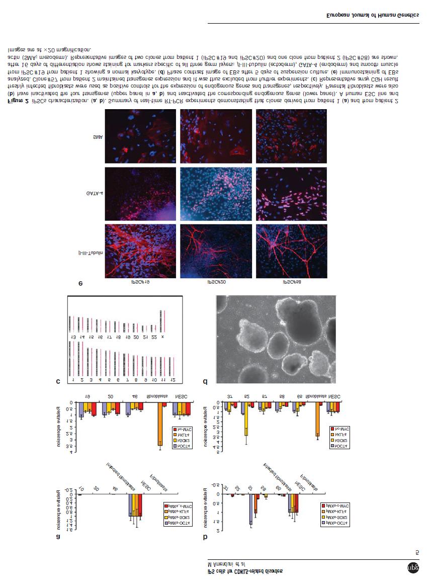
37 Figure S1: Array-CGH analysis. CNVs identified by array-cgh analysis in ips cells are presented. An overview of array results for all chromosomes is shown on the left. On the right, there is an higher magnification of the identified CNVs. Each dot represents a single probe (oligo) spotted on the array. Oligos with equal fluorescence intensity ratio between sample and reference have a value of zero. Copy number gains shift the ratio to the right (value of about +1, red dots). A. A representative image of the duplication in Xq22.2 that was identified in parental fibroblasts and in the three clones from patient 1. B. A trisomy of chromosome 8 is present in clone #52 derived from patient 2. 36
38 Table S1: primers for neuronal markers Gene Forward primer Reverse primer Melting temperature VGLUT1 GTCTGGGCTTCTGCATCAG CTGGAATCTGAGTGACAATG 60 C VGLUT2 ACATCCATGCCAGTCTATGC CTTTCTCACTGTCGTAGTTG 60 C TBR1 TGCATGTGGTGGAAGTGAAC GTCACAGCCGGTGTAGATC 52 C GAD67 TTGACTGCAGAGACACCTTG ACCATCCAACTTCTCTCTC 60 C GFAP GATTGAGTCGCTGGAGGAG TGGAGCGGTACCACTCTTC 60 C 37
39 Result 3.2 Set up of a protocol for the analysis of morphological alterations in CDKL5 neurons 38
40 3.2: Set up of a protocol for the analysis of morphological alterations in CDKL5 neurons Although CDKL5 role in neurons is far from being clarified, recently it has been demonstrated that in rat brain it is essential for neuronal morphogenesis, providing the first evidence of a cellular mechanism that may be responsible for the developmental effects caused by CDKL5 mutations and for the observed disease phenotype 15. However so far it is not clear whether the alterations identified in rat neurons correspond to the situation in affected human neurons. Interestingly, in patients with neurodevelopmental disorders such as nonsyndromic mental retardation and RTT an abnormal morphology is a characteristic feature of neurons 81,83. Neuropathology studies on Mecp2-deficient mice have reported that mutant brains exhibit a substantial reduction in both weight and neuronal cell size 59. The same results are observed throughout the cortical and subcortical region of post-mortem RTT human brains 84. Moreover, recent data on MECP2-iPSCs derived neurons have documented a consistent reduction in cell soma size of neurons carrying different MeCP2 mutations when compared to normal controls 63. To better understand whether CDKL5-iPSCs derived human neurons exhibited any consistent morphological alteration when compared to normal controls we decided to set up a protocol based on the use of lentiviruses expressing EGFP in order to visualize neuronal anatomy. Considering that neural differentiation results in variable and heterogeneous cultures of neurons, glia and undifferentiated cells, we decided to test two different lentiviruses: one driving ubiquitous EGFP expression, and one driving neurospecific GFP expression under the control of synapsin promoter. Furthermore, in order to asses whether the soma size reduction observed for MECP2-iPSCs derived neurons is present also in CDKL5-iPSCs derived neurons, we performed a cell soma size analysis comparing mutated neurons with normal controls. 39
41 3.2.1 Materials and Methods Cell culture. HEK293T and PC12 cells were maintained in DMEM High glucose (Invitrogen) supplemented with 10% (vol/vol) FBS, 10 mm non-essential amino acids, 50 U/ml penicillin, 50 mg/ml streptomycin. Neuronal differentiation of ipscs was performed according to a protocol established for human ESC 73,85 and results 1. (Fig. 1) Lentiviral vectors preparation. PLL3.7 plasmid for ubiquitous GFP expression was obtained from Dr Vania Broccoli (San Raffaele, Milan). Syn-EGFP plasmid, containing EGFP under the control of Synapsin-1 promoter, was obtained from Addgene (ID 19975). Plasmid identity was confirmed by enzymatic digestion and direct sequencing. To produce lentiviral vectors we transfected each plasmid into HEK293T cells together with four packaging plasmids encoding the lentiviral Gag-pol, Tat, Rev and VSV-G genes, also obtained from Addgene (ID 12251, 22502, 12253, 8454). Plasmids were co-transfected into HEK293T cells using Lipofectamine 2000 reagent (Invitrogen) according to the manufacturer s instructions. Two days after plasmid transfection, virus stocks were harvested, filter sterilized and used for virus titration. Virus titer was determined by checking the percentage of EGFP positive cells in PC12 and HEK293T cells. One day before titration cells were seeded at a density of 10 5 cells per well of a 12-well plate containing collagen-coated coverslips. The day after the medium was changed with pre-warmed medium containing polybrene to a final concentration of 8 µg/ml and several virus dilutions were added. Two days after infection cells were fixed with PFA 4% and immunostained for GFP (AbCam). DAPI was used to stain cell nuclei. At least 15 fields for each virus dilution were acquired under the fluorescent microscope and the total number of cells (DAPI nuclear staining) and the number of GFP + cells was determined. The viral titer (IU = infective units) was calculated with the following formula: Viral titer (IU/ml)=[Infected cell number in a well] [EGFP + % / 100]/[Amount of virus used (ml)]. Lentiviral infection of neurons. To test the two lentiviruses we used a normal male control ips clone derived from a male BJ fibroblast line (Cell line n CRL-2522) from ATCC (see Results 1). Neuronal cultures were infected with several amounts of virus and at different days during the differentiation course. Considering that the neuronal differentiation process lasts 70 days and that the percentage of neurons in cultures is variable we decided to not proceed with trypsinization of neuronal cultures and cell count. The amount of virus to use was decided on 40

42 the basis of the viral titer obtained for PC12 and HEK293T cells. Polybrene at 8 µg/ml was added to neuronal medium for the infection. About 16 hours after infection the medium was changed with fresh one. After infection, medium was changed every two days till Day 70. Immunostaining. To confirm the identity of the obtained neurons immunofluorescence was performed with standard protocols (see Results 1) using the generic neuronal marker MAP2 (AbCam). An anti-gfp antibody (AbCam) was used to enhance GFP fluorescence while DAPI was used to stain cell nuclei (Sigma Aldrich). Cells were visualized with an Axioscop 40FL (Zeiss) microscope connected to a computer. Morphometric analysis. To analyse soma size of neurons we acquired pictures at high magnification (100X). Images were analysed using ImageJ software. At least 15 cells for each clone were analysed. In order to avoid bias due to the difficulty to accurately delineate the cell soma, we decided to measure nuclear size. For each neuron, nuclear boundaries were delineated using ImageJ and the program measured the area expressed in arbitrary unit. An ipsc clone bearing the MECP2 mutation p.r306c obtained from Dr James Ellis (Sick Children Hospital, Toronto) was used as internal control to evaluate the accuracy of the method. Fig. 1. Neuronal differentiation Protocol. The scheme summarizes the neuronal differentiation protocol applied to induce ipsc toward neuronal fate. 41
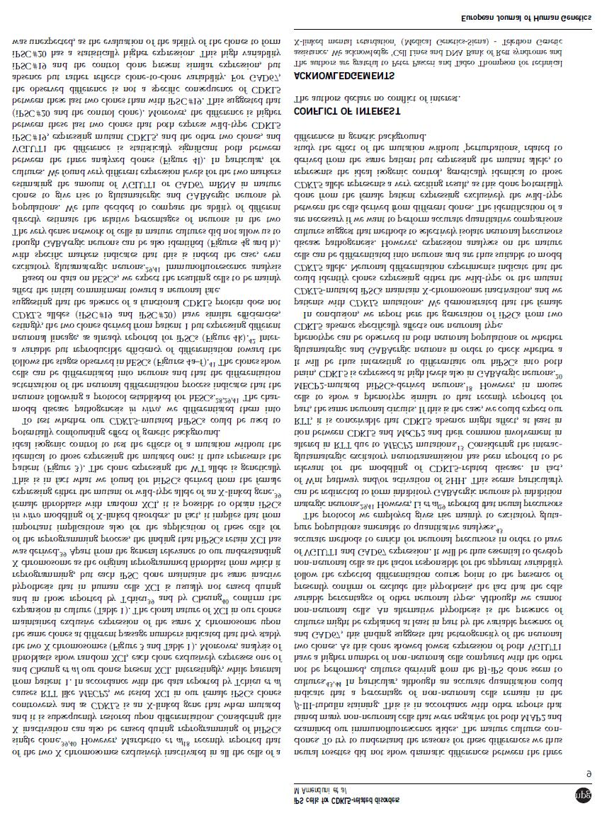
43 3.2.2 RESULTS Lentiviruses testing Neuronal differentiation results in variable and heterogeneous cultures of neurons, glia and undifferentiated cells. In order to obtain isolated neuronal cells suitable for morphological analysis we thus decided to test two different lentiviruses expressing EGFP: one driving ubiquitous GFP expression and one expressing GFP only in neurons, respectively PLL3.7 and Syn-EGFP. The titer of the lentiviral preparations was calculated in PC12 and HEK293T cells and was 3x10 4 IU/ml for Syn-EGFP and 4,9x10 5 IU/ml for PLL3.7. Concerning Syn-EGFP, HEK293T cells did not display GFP expression, confirming its neurospecificity. PLL3.7 Neuronal cultures plated on Coverslips (CVS) were initially infected at Day 66 of neuronal differentiation with 100 µl of PLL3.7 (3x10 3 IU). At Day 70 cells were fixed and immunostained for GFP and MAP2. A fist observation indicated that mature cultures contained mostly non-neuronal GFP+ cells; however, it was possible to distinguish some isolated neurons and some structures resembling dendritic spines could be identified, although GFP intensity was too low (Fig. 2 A-D). Considering these initial results we decided to proceed with another infection, infecting the neuronal cultures 7 days before the end of the differentiation course (day 62), since we hypothesized that more time was necessary for neurons to express GFP at sufficient levels. We used the same amount of virus. Cells were fixed at day 70 and immunostained for GFP and MAP2. With this second attempt we confirmed the result obtained in the first experiment. Mature cultures contained both nonneuronal and neuronal cells positive for GFP, with a prevalence of the first, making it difficult to distinguish isolated neurons although in some isolated cells spines could be detected (FIG 3 A-B). 42

44 Fig. 2. PLL3.7 infection at Day 66. (A) Immunostaining with GFP reveals mostly non-neuronal GFP+ cells. (B) MAP2 staining of the same field shown in (A) to visualize neuronal cells. Comparing figure A and B we can observe that neuronal cells are not GFP+. Images are 20X magnification. (C) Higher magnification showing that GFP+ neurons can be identified but no dendritic spines can be visualized. (D) MAP2 staining confirming neuronal cells identity. Images are 20X magnification in A and B and 100X magnification in C and D. 43
 of SYN-EGFP.")
45 Fig. 3. PLL3.7- Day 62 infection. (A) GFP staining of an isolated neuron showing the presence of dendritic spines. (B) MAP2 staining to confirm neuronal identity. SYN-EGFP Neuronal cultures plated on CVS were initially infected at Day 66 of neuronal differentiation with 500 µl (1.5x10 4 IU) of SYN-EGFP. At Day 70 cells were fixed and immunostained for GFP and MAP2. At a first analysis we observed the presence of too many positive cells and it was difficult to discriminate single GFP+ neurons from the dense neuronal network arising in culture (Fig.4 A-B). In some cases it was however possible to visualize isolated neurons and to recognize some dendritic spines, but GFP intensity was too low to allow an accurate visualization of their morphology (Fig.4 C_D). Considering this initial examination we decided to proceed with another infection, infecting the neuronal cultures 7 days before the end of the differentiation course (day 62). However we decided to use a lower amount of SYN-EGFP virus: 250 µl and 100 µl, respectively. Cells were fixed at day 70 and immunostained for GFP and MAP2. Microscope analysis indicated that the lowest amount of virus was not a good choice to visualize GFP-positive neurons since very few positive cells could be seen in only one out of 3 infected CVS. On the contrary, 250 µl resulted to be a good amount to use in order to visualize isolated cells and identify the presence of spines, since GFP is well distributed along the neurites and spines could be clearly identified (Fig.5 A-B). However, we observed that there was a percentage of GFP+ neurons lacking visible spines, in accordance with recent literature data suggesting that some but not all neurons after the 44

46 differentiation process are completely mature Chamberlain 93. To finally evaluate whether increasing the time lapse between infection and observation could improve dendritic spines visualization without causing stress to neuronal cultures, we decided to proceed with two different infections respectively at 2 weeks and 1 week before the end of the neuronal differentiation course. We decided to use the same amount of virus, 250 µl. Fluorescence microscope analysis of BJ neurons revealed that there was no significant difference between the two experiments and confirmed that 250 µl is a good amount of SYN-EGFP lentivirus to use in further experiments for morphological analysis of neuronal cultures. Fig. 4. SYN-EGFP- Day 66 infection with 500 µl of virus. (A-B) GFP staining revealing the presence of several positive neuronal cells whose identity is confirmed by MAP2 staining. (C-D) Isolated neuronal cell with low GFP intensity hampering accurate visualization of its morphology. Images are 100X magnification. 45

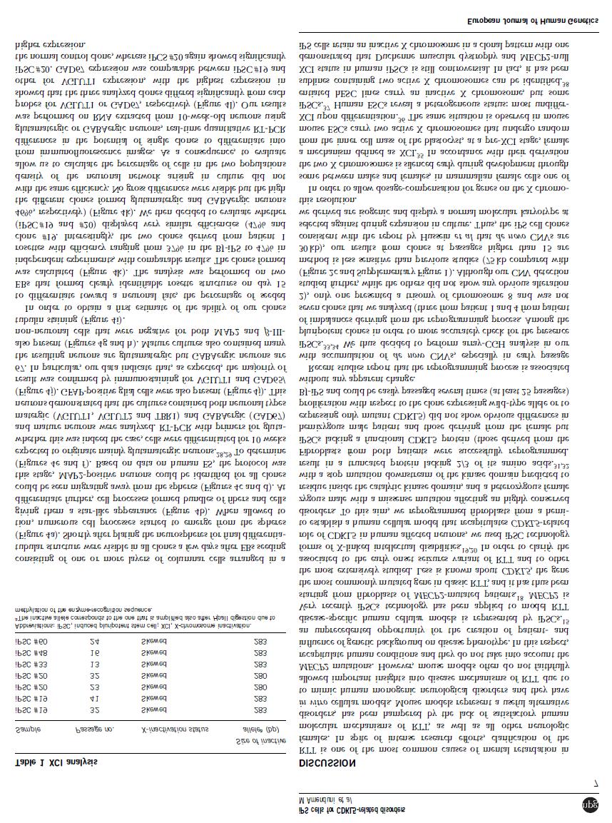
47 Fig. 5. SYN-EGFP Day 62 infection with 250 µl of virus. (A) GFP staining of an isolated neuron showing the presence of dendritic spines. (B) MAP2 staining confirming neuronal identity. Soma size analysis Neuropathology studies on RTT brains and histological results in Mecp2-deficient mice have demonstrated that neurons of RTT patients and mice are abnormal with a significant reduction in soma size 59,84. The same results have been recently obtained by morphological analysis of MECP2-iPSCs derived neurons 63,76. To determine whether similar alterations were present in our CDKL5 mutated neurons compared to normal controls we decided to perform a soma size analysis on our neurons. To this aim, we derived neuronal cultures from 3 different ipsc clones: the ipsc-bj clone (see above and Results 1 for details), used as normal control; an ipsc clone bearing the p-r306c MECP2 mutation, used as internal positive control of the experiment; and the clone ipsc#58, obtained from the male patient reported in Results 1 (Patient2). Mature cultures were fixed at day 70 of neuronal differentiation and immunostained with MAP2 to identify neurons. Soma size measurement is not easy to perform accurately since it is arduous to define soma boundaries; for this reason we decided to measure nuclear size as visualized by DAPI staining. Using ImageJ software we calculated the nuclear area and perimeter of at least 15 cells for each clone (Fig.6 A-C). We observed that the neurons derived from the MECP2-mutated ipsc clone exhibited a significant reduction in nuclear size when compared to the neurons from the BJ control (13% reduction) (Fig.6 C). This result is consistent with previous findings in Mecp2 -/y mice and postmortem brain tissues from RTT patients 59,84 and also with the data already reported for 46

48 MECP2-iPSCs derived neurons 63,76. However, as concerns our CDKL5 patient (Patient 2) no significant difference in nuclear size was observed between BJ neurons and ipsc#58 derived neurons. These results suggest that the use of the nuclear size for soma size calculation is reliable and gives consistent results. Experiments are ongoing to extend the sample size including three additional ipsc clones (#19, #46 and #20) derived from the female CDKL5- mutated patient (see Results 1) in order to confirm the initial results. Fig. 6. Nuclear size evaluation of ipsc-derived neurons. A) Cell nuclei stained with DAPI. The nucleus indicated by the arrow belongs to a neuronal cell and has been used for soma size analysis. B) Neuronal cell stained with MAP2 used for nuclear size calculation. C) Nuclear size comparison between neurons derived from control ipscs (ipsc-bj), MECP2-mutated ipscs (ipsc#r306c) and CDKL5-mutated ipscs (ipsc#58). Bar graph shows the mean nuclear size area of ipsc-derived neurons. ipsc#r306c-derived neurons exhibit a reduction in nuclear size of about 13% compared with ipsc-bj-derived neurons. ipsc#58-derived neurons do not show any difference in nuclear size respect to the normal control. Nuclear size area is expressed in an arbitrary unit as calculated by ImageJ program. MAP2 antibody was used to identify neurons. 47
49 Result 3.3 Gene expression profiling of ipscs from CDKL5 patients 48
50 3.3: Gene expression profiling of ipscs from CDKL5 patients Brain is the primarily affected tissue in RTT patients. However expression studies in this tissue are difficult to carry out for obvious ethical reasons. Some studies have tried to overcome this limitation employing post-mortem tissues. However, this tissue may originate from late stages of the disorder and it might thus not be suitable to investigate the initial pathological processes of the disease 79. Importantly, brain is characterized by cellular heterogeneity that might contribute to mask subtle expression differences. So far several gene expression studies involving MECP2-null samples have been performed on different tissues from RTT individuals and mouse models with Mecp2 loss of function mutations 86,87. However, the lists of mis-regulated genes derived from these studies are not-overlapping, probably due to the different approaches adopted, based on the use of clonal lymphoblastoid cell lines to avoid mosaicism, fibroblast strains from patients or post-mortem RTT brains As concerns mouse models contrasting results may be due to the use of different RTT mouse models and also to the analysis of distinct brain regions such as cerebellum, cortex or midbrain 86,91. As concerns CDKL5, despite intensive research efforts, its function inside the neuron and the underlying molecular mechanisms remain still uncharacterized. CDKL5 is a serinethreonine kinase. Kinase proteins are involved in the regulation of the activity of a huge range of different proteins, including many proteins that play essential roles in signalling cascades resulting in activation/repression of gene transcription. CDKL5 protein seems to work in a molecular pathway common to that of MeCP2, a transcriptional regulator. In fact mutations in CDKL5 are associated with some severe neurological symptoms that are occasionally reported in typical MeCP2 cases such as infantile spasms, early onset epilepsy and hypsarrhytmia. Recent findings regarding a possible role of CDKL5 in nuclear speckles organization, reinforce the hypothesis of an involvement of CDKL5 in the regulation of gene expression 14. However expression profiling studies are hindered by the absence of a CDKL5- null mouse model, together with the absence of a good in vitro human cellular model. Very recently a first attempt of gene expression profiling has been performed on clonal primary cultures of fibroblasts derived from CDKL5-mutated patients leading to the identification of a total of 16 up-regulated and 20 down-regulated genes. Among these genes only MAP3K5, an apoptosis signal regulated kinase, was found to be down-regulated also in neuroblastoma- 49
51 derived SH-SY5Y cells transfected with an shrna targeting CDKL5. MAP3K5 is involved in MAP kinase pathway that mediates signals leading to both differentiation and survival in neuronal cells. According to the authors its significant underexpression in human CDKL5- deficient cells may suggests that neurite outgrowth and synaptic plasticity may be altered in CDKL5 mutated brains 92. These data are in agreement with those obtained by Chen and colleagues, indicating that CDKL5 acts as a critical regulator of neuronal morphogenesis through a signalling pathway involving the Rac1 GTPase 15. CDKL5, as MECP2, is located on the X chromosomes and undergoes X chromosome inactivation, so that female patients result in mosaic expression of mutant or wild type CDKL5 in each of their cells; analysis of gene expression profile in girls will be thus compromised by unpredictable patterns of XCI. However we recently have confirmed previous data on X inactivation in ipscs, indicating that human ipscs can retain an inactive X-chromosome in a clonal pattern, with one of the two X chromosomes exclusively inactivated in all the cells of a single clone 63,75,76. Indeed we have obtained clones derived from the same patient but expressing either the wild type or the mutated CDKL5 allele (Result 1), thus overcoming the functional mosaicism resulting from XCI. In addition these cells are genetically identical and differ only for CDKL5 expression and they thus represent the ideal tool to analyse the consequences of CDKL5 absence without confounding effects due to different genetic background. On the basis of all these considerations we decided to perform gene expression analysis on ipsc clones derived from CDKL5 mutated patients, in order to detect candidate genes that might be involved in neurogenetic processes and thus contribute to gain new insights into the mechanisms underlying CDKL5 function in neuronal development. To this aim, we compared the global gene expression pattern at the transcript level in matched pairs of wild type and mutant ipscs clones from two different patients Experimental procedures ipsc clones. ipscs clones were derived from fibroblasts of two different CDKL5-mutated patients (Result 1): a female patient (Patient 1) with the early-onset seizures variant of RTT and an early truncating mutation (p.q347x) and a male patient (Patient 2) with severe encephalopathy and early-onset intractable epilepsy that carries a missense mutation 50
52 (p.t288i) affecting the catalytic kinase domain. For patient 1 we selected 2 different ips clones, one expressing the wt allele, thus used as normal control (ipsc#20), and the other expressing the mutated CDKL5 allele (ipscs#46) (See Results 1). For patient 1 we selected only one fully characterized clone (ipscs#58) and used a normal male ips clone derived from a male BJ fibroblast line from ATCC as control (ipsc-bj). Clones were maintained in mtesr1 supplied with puromicin using standard procedures. RNA isolation. Total RNA was extracted from ipsc clones with the Qiagen RNeasy kit (Qiagen) according to manufacturer recommended protocol. Prior to array hybridization RNA quality and quantity were assessed with the RNA 6000 Nano Assay using 2100 Bioanalyzer (Agilent Technologies). For clones derived from the female patient, the exclusive expression of only one CDKL5 allele was confirmed by RT-PCR and sequencing. cdna labelling and microarray hybridization. Agilent Whole Human Genome 4X44K microarrays and Two-Color Microarray-Based gene expression Analysis (Quick Amp Labeling) Protocol were used for global gene expression analysis. 500 ng of total RNA from control and mutated samples was used to prepare amplified and labeled crna using Quick- Amp Labeling Kit (Agilent). Control and mutated samples were labelled with Cy3-dCTP and Cy5-dCTP respectively. For each clone, four technical replicates were performed to control technical bias. Following purification with Qiagen RNeasy kit (Qiagen), according to protocol instructions, crna was quantified using NanoDrop ND-1000 UV-VIS Spectrophotometer in order to determine the yield and specific activity of each reaction. For each replicate 825 ng of labeled control crna was combined with 825 ng of labelled mutated crna. The combined reactions were then applied to crna microarray. Hybridization was carried out at 65 C for 17 hr in a hybridization oven. Blocking agent was added according to the protocol. Microarray data analysis. Following hybridization, microarrays were washed and scanned with an Agilent DNA microarray scanner (G2505B) and analysed by Agilent Feature Extraction Software v9.5. The resulting text files were imported into Gene Spring GX software v11.5 for processing and analysis of microarray data. Significantly modulated genes were defined as those with absolute fold change (FC) > 1.5. To translate the data in a more meaningful biological context and to identify candidate genes, functional analysis of the selected genes has been performed using available on-line databases ( in order to 51
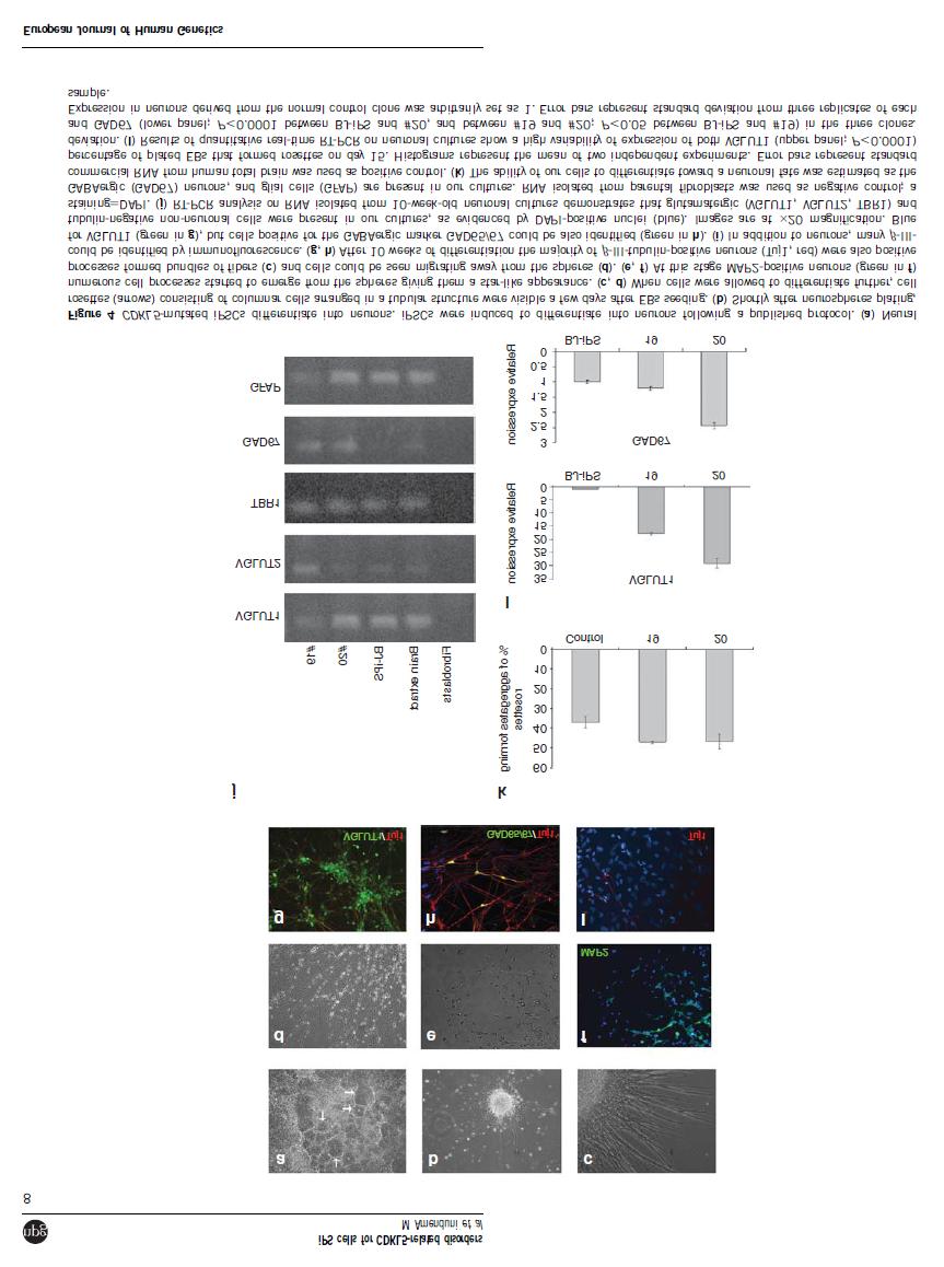
53 identify differentially expressed genes that might be involved in neurogenetic processes, thus playing a possible role in the pathophysiology of CDKL5-related disorders Results Microarray analysis of matched ipsc clones In order to identify genes specifically regulated by CDKL5, we carried out an expression profile experiment on ipsc clones derived from two different CDKL5-mutated patients. For the female patient we selected as normal control the ipsc#20 that expresses only the wt allele due to skewed X chromosome inactivation, while for the male patient we used the BJ clone (see also Results 1). For each patient we performed direct competitive hybridization between WT and mutated clones; hybridization of labelled RNA on Agilent Whole Human Genome 4X44K microarrays was carried out in four technical replicates, providing a total of eight chip hybridizations on two microarray slides. The expression signals were extracted from arrays using Agilent Feature Extraction Software v9.5. The software automatically finds and places microarray grids, accurately determines feature intensities and ratios, flags outlier pixels and calculates statistical confidence ( At the end of this analysis the software generates a QC report and the data files that can be used for further analysis. The QC report (GE 2-color with Agilent spike-ins) allows the evaluation of the correct grid placement and of microarray scan quality and reliability. For further processing and interpretation, we imported the data files into Gene Spring GX software v11.5. Fig. 1A summarizes the analysis procedure applied to our data. For each patient, technical replicates were grouped together. We decided to calculate differences in gene expression level between WT and mutant clones for each patient separately (matched pairs). Before starting the analysis, data were qualitatively assessed using defined flag import settings, in order to remove those entities that are not reliably detected, and pre-processed with the baseline transformation to median of all samples step. As a first approach to gene expression profiling, we selected genes on the basis if their fold change (FC). Significantly altered genes were defined as those with absolute FC > 1.5. The analysis returned a list of 295 dysregulated genes for patient 2 (Entity list 1) and 276 genes for Patient 1 (Entity list 2). Since our aim was to identify genes that might be involved in a common regulatory circuit with CDKL5 and thus be influenced by its deficiency we then compared the gene lists obtained for each patient using the Venn diagram approach in order to verify the presence of 52

54 overlapping genes (Fig 1B). This comparison revealed 11 differentially expressed genes in common between the two patients; two of these genes were altered in opposite directions in the two samples (up-regulated in one patient and down-regulated in the other) and were thus excluded from further analyses (Table 1). We inspected our list on the basis of gene function and expression patterns in order to identify potentially relevant genes. One down-regulated gene GRIP1 (glutamate receptor interacting protein 1) seems particularly interesting since it is present in both glutamatergic and GABAergic synapses and seems to play a role in postsynaptic localization of AMPA receptors that are involved in synaptic plasticity (Song I et al., 2002; Santos SD et al., 2009). Fig. 1. (A) Flow chart summarizing the analysis procedure. (B) Venn Diagram obtained by overlapping the single entity lists for each WT vs mutated couple of samples. A total of 11 genes are in common between the two couples. 53
Objectives. Genetics and Rett syndrome: As easy as apple pie! Chromosome to gene to protein
 Genetics and Rett syndrome: As easy as apple pie! Victoria Mok Siu M.D., FRCPC, FCCMG ORSA conference Ottawa April 24, 2016 Objectives Review chromosomes and genes Understand s Explore the reasons behind
SUPPLEMENTARY INFORMATION
 DOI: 10.1038/ncb2566 Figure S1 CDKL5 protein expression pattern and localization in mouse brain. (a) Multiple-tissue western blot from a postnatal day (P) 21 mouse probed with an antibody against CDKL5.
Proposal form for the evaluation of a genetic test for NHS Service Gene Dossier
 Proposal form for the evaluation of a genetic test for NHS Service Gene Dossier Test Disease Population Triad Disease name Epileptic encephalopathy, early infantile 4. OMIM number for disease 612164 Disease
Fondata nel University of Siena. Ph.D in Medical Genetics
 Fondata nel 1241 University of Siena Ph.D in Medical Genetics FOXG1 and Rett Syndrome: functional characterization and set-up of an in vitro human cellular model Roberta De Filippis Supervisor: Prof. Alessandra
Alpha thalassemia mental retardation X-linked. Acquired alpha-thalassemia myelodysplastic syndrome
 Alpha thalassemia mental retardation X-linked Acquired alpha-thalassemia myelodysplastic syndrome (Alpha thalassemia mental retardation X-linked) Acquired alpha-thalassemia myelodysplastic syndrome Schematic
Muscular Dystrophy. Biol 405 Molecular Medicine
 Muscular Dystrophy Biol 405 Molecular Medicine Duchenne muscular dystrophy Duchenne muscular dystrophy is a neuromuscular disease that occurs in ~ 1/3,500 male births. The disease causes developmental
ips cells to model CDKL5-related disorders
 (211) 19, 1246 1255 & 211 Macmillan Publishers Limited All rights reserved 118-4813/11 www.nature.com/ejhg ARTICLE ips cells to model CDKL5-related disorders Mariangela Amenduni 1,7, Roberta De Filippis
RAS Genes. The ras superfamily of genes encodes small GTP binding proteins that are responsible for the regulation of many cellular processes.
 ۱ RAS Genes The ras superfamily of genes encodes small GTP binding proteins that are responsible for the regulation of many cellular processes. Oncogenic ras genes in human cells include H ras, N ras,
Corporate Medical Policy
 Corporate Medical Policy File Name: Origination: Last CAP Review: Next CAP Review: Last Review: genetic_testing_for_rett_syndrome 7/2012 3/2017 3/2018 5/2017 Description of Procedure or Service Rett syndrome
Overview: Conducting the Genetic Orchestra Prokaryotes and eukaryotes alter gene expression in response to their changing environment
 Overview: Conducting the Genetic Orchestra Prokaryotes and eukaryotes alter gene expression in response to their changing environment In multicellular eukaryotes, gene expression regulates development
Fragile X Syndrome. Genetics, Epigenetics & the Role of Unprogrammed Events in the expression of a Phenotype
 Fragile X Syndrome Genetics, Epigenetics & the Role of Unprogrammed Events in the expression of a Phenotype A loss of function of the FMR-1 gene results in severe learning problems, intellectual disability
Regulation of Gene Expression in Eukaryotes
 Ch. 19 Regulation of Gene Expression in Eukaryotes BIOL 222 Differential Gene Expression in Eukaryotes Signal Cells in a multicellular eukaryotic organism genetically identical differential gene expression
Stem Cells and the Study of Neurodegeneration. Tracy Young-Pearse, PhD September 12, 2014!
 Stem Cells and the Study of Neurodegeneration Tracy Young-Pearse, PhD September 12, 2014! Techniques for studying mechanisms of neurological disease Animal models Human subjects Postmortem analyses, imaging
SUPPLEMENTARY INFORMATION
 SUPPLEMENTARY INFORMATION doi:10.1038/nature19357 Figure 1a Chd8 +/+ Chd8 +/ΔSL Chd8 +/+ Chd8 +/ΔL E10.5_Whole brain E10.5_Whole brain E10.5_Whole brain E14.5_Whole brain E14.5_Whole brain E14.5_Whole
Application of induced pluripotent stem (ips) cells in intractable childhood disorders
 cells in intractable childhood disorders") 10th Annual World Congress on Pediatrics Application of induced pluripotent stem (ips) cells in intractable childhood disorders Lessons from Dravet synd. patient-derived ipscs Shinichi Hirose, MD, PhD
10th Annual World Congress on Pediatrics Application of induced pluripotent stem (ips) cells in intractable childhood disorders Lessons from Dravet synd. patient-derived ipscs Shinichi Hirose, MD, PhD
Molecular Biology (BIOL 4320) Exam #2 May 3, 2004
 Exam #2 May 3, 2004") Molecular Biology (BIOL 4320) Exam #2 May 3, 2004 Name SS# This exam is worth a total of 100 points. The number of points each question is worth is shown in parentheses after the question number. Good
Molecular Biology (BIOL 4320) Exam #2 May 3, 2004 Name SS# This exam is worth a total of 100 points. The number of points each question is worth is shown in parentheses after the question number. Good
Is Intrinsic Hyperexcitability in CA3 the Culprit for Seizures in Rett Syndrome?
 Current Literature In Basic Science Is Intrinsic Hyperexcitability in CA3 the Culprit for Seizures in Rett Syndrome? Network Hyperexcitability in Hippocampal Slices From Mecp2 Mutant Mice Revealed by Voltage-Sensitive
variant led to a premature stop codon p.k316* which resulted in nonsense-mediated mrna decay. Although the exact function of the C19L1 is still
 157 Neurological disorders primarily affect and impair the functioning of the brain and/or neurological system. Structural, electrical or metabolic abnormalities in the brain or neurological system can
Interrogating Rett Syndrome: developing ideas for research that matters. Angus Clarke, Clinical Genetics, Prifysgol Caerdydd, Cymru
 Interrogating Rett Syndrome: developing ideas for research that matters Angus Clarke, Clinical Genetics, Prifysgol Caerdydd, Cymru Research into Rett Syndrome Four Factors: Unique clinical entity Biology
The Biology and Genetics of Cells and Organisms The Biology of Cancer
 The Biology and Genetics of Cells and Organisms The Biology of Cancer Mendel and Genetics How many distinct genes are present in the genomes of mammals? - 21,000 for human. - Genetic information is carried
~Lentivirus production~
 ~Lentivirus production~ May 30, 2008 RNAi core R&D group member Lentivirus Production Session Lentivirus!!! Is it health threatening to lab technician? What s so good about this RNAi library? How to produce
Epigenetic Principles and Mechanisms Underlying Nervous System Function in Health and Disease Mark F. Mehler MD, FAAN
 Epigenetic Principles and Mechanisms Underlying Nervous System Function in Health and Disease Mark F. Mehler MD, FAAN Institute for Brain Disorders and Neural Regeneration F.M. Kirby Program in Neural
Transcriptional and Epigenetic Mechanisms of Addiction
 Transcriptional and Epigenetic Mechanisms of Addiction Eric J. Nestler Mount Sinai School of Medicine New York, NY Dr. Ray Fuller There is every reason to be optimistic that in the future we will find
Problem Set 5 KEY
 2006 7.012 Problem Set 5 KEY ** Due before 5 PM on THURSDAY, November 9, 2006. ** Turn answers in to the box outside of 68-120. PLEASE WRITE YOUR ANSWERS ON THIS PRINTOUT. 1. You are studying the development
22q11.2 DELETION SYNDROME. Anna Mª Cueto González Clinical Geneticist Programa de Medicina Molecular y Genética Hospital Vall d Hebrón (Barcelona)
") 22q11.2 DELETION SYNDROME Anna Mª Cueto González Clinical Geneticist Programa de Medicina Molecular y Genética Hospital Vall d Hebrón (Barcelona) Genomic disorders GENOMICS DISORDERS refers to those diseases
22q11.2 DELETION SYNDROME Anna Mª Cueto González Clinical Geneticist Programa de Medicina Molecular y Genética Hospital Vall d Hebrón (Barcelona) Genomic disorders GENOMICS DISORDERS refers to those diseases
Problem Set 8 Key 1 of 8
 7.06 2003 Problem Set 8 Key 1 of 8 7.06 2003 Problem Set 8 Key 1. As a bright MD/PhD, you are interested in questions about the control of cell number in the body. Recently, you've seen three patients
Potential Treatment and Current Research in Phelan-McDermid Syndrome. 11/16/2016 Frambu Center for Rare Disorders
 Potential Treatment and Current Research in Phelan-McDermid Syndrome 11/16/2016 Frambu Center for Rare Disorders Genetics is Complicated! Deletion 22q13: Therapies Under Investigation Intranasal insulin
Genetics and Genomics: Applications to Developmental Disability
 Tuesday, 12:30 2:00, B1 Objective: Genetics and Genomics: Applications to Developmental Disability Helga Toriello 616-234-2712 toriello@msu.edu Identify advances in clinical assessment and management of
Today. Genomic Imprinting & X-Inactivation
 Today 1. Quiz (~12 min) 2. Genomic imprinting in mammals 3. X-chromosome inactivation in mammals Note that readings on Dosage Compensation and Genomic Imprinting in Mammals are on our web site. Genomic
Tumor suppressor genes D R. S H O S S E I N I - A S L
 Tumor suppressor genes 1 D R. S H O S S E I N I - A S L What is a Tumor Suppressor Gene? 2 A tumor suppressor gene is a type of cancer gene that is created by loss-of function mutations. In contrast to
Clinical Summaries. CLN1 Disease, infantile onset and others
 Clinical Summaries CLN1 Disease, infantile onset and others The gene called CLN1 lies on chromosome 1. CLN1 disease is inherited as an autosomal recessive disorder, which means that both chromosomes carry
Nature Neuroscience: doi: /nn.2275
 Supplementary Figure S1. The presence of MeCP2 in enriched primary glial cultures from rat or mouse brains is not neuronal. Western blot analysis of protein extracts from (a) rat glial and neuronal cultures.
Transcriptional control in Eukaryotes: (chapter 13 pp276) Chromatin structure affects gene expression. Chromatin Array of nuc
 Chromatin structure affects gene expression. Chromatin Array of nuc") Transcriptional control in Eukaryotes: (chapter 13 pp276) Chromatin structure affects gene expression Chromatin Array of nuc 1 Transcriptional control in Eukaryotes: Chromatin undergoes structural changes
Transcriptional control in Eukaryotes: (chapter 13 pp276) Chromatin structure affects gene expression Chromatin Array of nuc 1 Transcriptional control in Eukaryotes: Chromatin undergoes structural changes
JULY 21, Genetics 101: SCN1A. Katie Angione, MS CGC Certified Genetic Counselor CHCO Neurology
 JULY 21, 2018 Genetics 101: SCN1A Katie Angione, MS CGC Certified Genetic Counselor CHCO Neurology Disclosures: I have no financial interests or relationships to disclose. Objectives 1. Review genetic
Part-4. Cell cycle regulatory protein 5 (Cdk5) A novel target of ERK in Carb induced cell death
 A novel target of ERK in Carb induced cell death") Part-4 Cell cycle regulatory protein 5 (Cdk5) A novel target of ERK in Carb induced cell death 95 1. Introduction The process of replicating DNA and dividing cells can be described as a series of coordinated
Part-4 Cell cycle regulatory protein 5 (Cdk5) A novel target of ERK in Carb induced cell death 95 1. Introduction The process of replicating DNA and dividing cells can be described as a series of coordinated
Proposal form for the evaluation of a genetic test for NHS Service Gene Dossier
 Proposal form for the evaluation of a genetic test for NHS Service Gene Dossier Test Disease Population Triad Disease name and description (please provide any alternative names you wish listed) (A)-Testing
Section Chapter 14. Go to Section:
 Section 12-3 Chapter 14 Go to Section: Content Objectives Write these Down! I will be able to identify: The origin of genetic differences among organisms. The possible kinds of different mutations. The
Genetics and Genomics in Medicine Chapter 6 Questions
 Genetics and Genomics in Medicine Chapter 6 Questions Multiple Choice Questions Question 6.1 With respect to the interconversion between open and condensed chromatin shown below: Which of the directions
1 in 68 in US. Autism Update: New research, evidence-based intervention. 1 in 45 in NJ. Selected New References. Autism Prevalence CDC 2014
 Autism Update: New research, evidence-based intervention Martha S. Burns, Ph.D. Joint Appointment Professor Northwestern University. 1 Selected New References Bourgeron, Thomas (2015) From the genetic
Ch. 18 Regulation of Gene Expression
 Ch. 18 Regulation of Gene Expression 1 Human genome has around 23,688 genes (Scientific American 2/2006) Essential Questions: How is transcription regulated? How are genes expressed? 2 Bacteria regulate
Introduction to Cancer Biology
 Introduction to Cancer Biology Robin Hesketh Multiple choice questions (choose the one correct answer from the five choices) Which ONE of the following is a tumour suppressor? a. AKT b. APC c. BCL2 d.
Bio 111 Study Guide Chapter 17 From Gene to Protein
 Bio 111 Study Guide Chapter 17 From Gene to Protein BEFORE CLASS: Reading: Read the introduction on p. 333, skip the beginning of Concept 17.1 from p. 334 to the bottom of the first column on p. 336, and
Development of the Nervous System. Leah Militello, class of 2018
 Development of the Nervous System Leah Militello, class of 2018 Learning Objectives 1. Describe the formation and fate of the neural tube and neural crest including timing and germ layer involved. 2. Describe
Development of the Central Nervous System
 Development of the Central Nervous System an ongoing process, through adolescence and maybe even adult hood? the nervous system is plastic Experience plays a key role Dire consequences when something goes
Fayth K. Yoshimura, Ph.D. September 7, of 7 HIV - BASIC PROPERTIES
 1 of 7 I. Viral Origin. A. Retrovirus - animal lentiviruses. HIV - BASIC PROPERTIES 1. HIV is a member of the Retrovirus family and more specifically it is a member of the Lentivirus genus of this family.
SEX-LINKED INHERITANCE. Dr Rasime Kalkan
 SEX-LINKED INHERITANCE Dr Rasime Kalkan Human Karyotype Picture of Human Chromosomes 22 Autosomes and 2 Sex Chromosomes Autosomal vs. Sex-Linked Traits can be either: Autosomal: traits (genes) are located
Clinical Spectrum and Genetic Mechanism of GLUT1-DS. Yasushi ITO (Tokyo Women s Medical University, Japan)
") Clinical Spectrum and Genetic Mechanism of GLUT1-DS Yasushi ITO (Tokyo Women s Medical University, Japan) Glucose transporter type 1 (GLUT1) deficiency syndrome Mutation in the SLC2A1 / GLUT1 gene Deficiency
Clinical Spectrum and Genetic Mechanism of GLUT1-DS Yasushi ITO (Tokyo Women s Medical University, Japan) Glucose transporter type 1 (GLUT1) deficiency syndrome Mutation in the SLC2A1 / GLUT1 gene Deficiency
A Genetic Program for Embryonic Development
 Concept 18.4: A program of differential gene expression leads to the different cell types in a multicellular organism During embryonic development, a fertilized egg gives rise to many different cell types
Functional Development of Neuronal Networks in Culture -An in vitro Assay System of Developing Brain for Endocrine Disruptors
 Functional Development of Neuronal Networks in Culture -An in vitro Assay System of Developing Brain for Endocrine Disruptors Masahiro Kawahara and Yoichiro Kuroda Tokyo Metropolitan Institute for Neuroscience
7.012 Quiz 3 Answers
 MIT Biology Department 7.012: Introductory Biology - Fall 2004 Instructors: Professor Eric Lander, Professor Robert A. Weinberg, Dr. Claudette Gardel Friday 11/12/04 7.012 Quiz 3 Answers A > 85 B 72-84
Veronika Borbélyová, MSc., PhD.
 Veronika Borbélyová, MSc., PhD. borbelyova.veronika88@gmail.com History Eugen Bleuler autism (from the Greek words autos = self, ismus = orientation, status) the patient reduces the contact with the outside
(a) Schematic diagram of the FS mutation of UVRAG in exon 8 containing the highly instable
 Schematic diagram of the FS mutation of UVRAG in exon 8 containing the highly instable") Supplementary Figure 1. Frameshift (FS) mutation in UVRAG. (a) Schematic diagram of the FS mutation of UVRAG in exon 8 containing the highly instable A 10 DNA repeat, generating a premature stop codon
Supplementary Figure 1. Frameshift (FS) mutation in UVRAG. (a) Schematic diagram of the FS mutation of UVRAG in exon 8 containing the highly instable A 10 DNA repeat, generating a premature stop codon
Gene Expression DNA RNA. Protein. Metabolites, stress, environment
 Gene Expression DNA RNA Protein Metabolites, stress, environment 1 EPIGENETICS The study of alterations in gene function that cannot be explained by changes in DNA sequence. Epigenetic gene regulatory
Are there foreseeable applications of genomic medicine for the management of neuropsychiatric conditions?
 Are there foreseeable applications of genomic medicine for the management of neuropsychiatric conditions? Angus Clarke, Medical Genetics, Cardiff University, Wales Are there foreseeable applications of
Supplemental Information. Induction of Expansion and Folding. in Human Cerebral Organoids
 Cell Stem Cell, Volume 20 Supplemental Information Induction of Expansion and Folding in Human Cerebral Organoids Yun Li, Julien Muffat, Attya Omer, Irene Bosch, Madeline A. Lancaster, Mriganka Sur, Lee
International Graduate Research Programme in Cardiovascular Science
 1 International Graduate Research Programme in Cardiovascular Science This work has been supported by the European Community s Sixth Framework Programme under grant agreement n LSHM-CT-2005-01883 EUGeneHeart.
SUPPLEMENTARY INFORMATION
 SUPPLEMENTARY INFORMATION Human cerebral cortex development from pluripotent stem cells to functional excitatory synapses Yichen Shi 1,2, Peter Kirwan 1,2, James Smith 1,2, Hugh P.C. Robinson 3 and Frederick
mirna Dr. S Hosseini-Asl
 mirna Dr. S Hosseini-Asl 1 2 MicroRNAs (mirnas) are small noncoding RNAs which enhance the cleavage or translational repression of specific mrna with recognition site(s) in the 3 - untranslated region
Name: Answer Key. Question 1.
 2007 7.013 Problem Set 6 Due before 5 PM on FRIDAY, April 27, 2007. Turn answers in to the box outside of 68-120. PLEASE WRITE YOUR ANSWERS ON THIS PRINTOUT. Question 1. 1a. This is a diagram showing changes
CHROMOSOMAL MICROARRAY (CGH+SNP)
") Chromosome imbalances are a significant cause of developmental delay, mental retardation, autism spectrum disorders, dysmorphic features and/or birth defects. The imbalance of genetic material may be due
Chromosome imbalances are a significant cause of developmental delay, mental retardation, autism spectrum disorders, dysmorphic features and/or birth defects. The imbalance of genetic material may be due
Shift 1, 8 July 2018, 09:30-13:00
 Shift 1, 8 July 2018, 09:30-13:00 CNS patterning A001-A014 Stem cells: basic biology and postnatal neurogenesis - part I Development of neural systems: Molecular and genetic characterisationa Epigenetic
CONTRACTING ORGANIZATION: Rutgers, the State University of New Jersey New Brunswick, NJ 08901
 AWARD NUMBER: W81XWH-16-1-0415 TITLE: Cell Type-Specific Contributions to the TSC Neuropathology PRINCIPAL INVESTIGATOR: Gabriella D Arcangelo CONTRACTING ORGANIZATION: Rutgers, the State University of
SUPPLEMENTARY INFORMATION
 Supplementary Discussion The cell cycle machinery and the DNA damage response network are highly interconnected and co-regulated in assuring faithful duplication and partition of genetic materials into
Rare Monogenic Disorders. Function. Pathophysiology
 Rare Monogenic Disorders Function Pathophysiology Protein Gene Episodic Nervous System Diseases Migraine Epilepsy Periodic Paralysis LQTS Episodic Ataxia Paroxysmal Dyskinesias Phenotypes Muscle diseases
General Biology 1004 Chapter 11 Lecture Handout, Summer 2005 Dr. Frisby
 Slide 1 CHAPTER 11 Gene Regulation PowerPoint Lecture Slides for Essential Biology, Second Edition & Essential Biology with Physiology Presentation prepared by Chris C. Romero Neil Campbell, Jane Reece,
Proteins. Length of protein varies from thousands of amino acids to only a few insulin only 51 amino acids
 Proteins Protein carbon, hydrogen, oxygen, nitrogen and often sulphur Length of protein varies from thousands of amino acids to only a few insulin only 51 amino acids During protein synthesis, amino acids
Cells and reagents. Synaptopodin knockdown (1) and dynamin knockdown (2)
 and dynamin knockdown (2)") Supplemental Methods Cells and reagents. Synaptopodin knockdown (1) and dynamin knockdown (2) podocytes were cultured as described previously. Staurosporine, angiotensin II and actinomycin D were all obtained
Supplemental Methods Cells and reagents. Synaptopodin knockdown (1) and dynamin knockdown (2) podocytes were cultured as described previously. Staurosporine, angiotensin II and actinomycin D were all obtained
SUPPLEMENTARY INFORMATION
 sirna pool: Control Tetherin -HA-GFP HA-Tetherin -Tubulin Supplementary Figure S1. Knockdown of HA-tagged tetherin expression by tetherin specific sirnas. HeLa cells were cotransfected with plasmids expressing
MicroRNAs, RNA Modifications, RNA Editing. Bora E. Baysal MD, PhD Oncology for Scientists Lecture Tue, Oct 17, 2017, 3:30 PM - 5:00 PM
 MicroRNAs, RNA Modifications, RNA Editing Bora E. Baysal MD, PhD Oncology for Scientists Lecture Tue, Oct 17, 2017, 3:30 PM - 5:00 PM Expanding world of RNAs mrna, messenger RNA (~20,000) trna, transfer
Ready-to-use Lentiviral Particles for intracelular labeling
 Ready-to-use Lentiviral Particles for intracelular labeling (LocLight TM Living cell imaging lentivirus for sub-cellular localization) LocLight TM cell organelle labeling lentivirus are provided as 200ul/per
Plasticity of Cerebral Cortex in Development
 Plasticity of Cerebral Cortex in Development Jessica R. Newton and Mriganka Sur Department of Brain & Cognitive Sciences Picower Center for Learning & Memory Massachusetts Institute of Technology Cambridge,
Cancer genetics
 Cancer genetics General information about tumorogenesis. Cancer induced by viruses. The role of somatic mutations in cancer production. Oncogenes and Tumor Suppressor Genes (TSG). Hereditary cancer. 1
CURRENT GENETIC TESTING TOOLS IN NEONATAL MEDICINE. Dr. Bahar Naghavi
 2 CURRENT GENETIC TESTING TOOLS IN NEONATAL MEDICINE Dr. Bahar Naghavi Assistant professor of Basic Science Department, Shahid Beheshti University of Medical Sciences, Tehran,Iran 3 Introduction Over 4000
Challenges of CGH array testing in children with developmental delay. Dr Sally Davies 17 th September 2014
 Challenges of CGH array testing in children with developmental delay Dr Sally Davies 17 th September 2014 CGH array What is CGH array? Understanding the test Benefits Results to expect Consent issues Ethical
Discovery of Schizophrenia Drug Targets from DISC1 Mechanisms Atsushi Kamiya M.D., Ph.D.
 Discovery of Schizophrenia Drug Targets 1 Assistant Professor Department of Psychiatry and Behavioral Sciences Johns Hopkins University School of Medicine akamiya1@jhmi.edu How does neurobiology offer
Epigenetics: The Future of Psychology & Neuroscience. Richard E. Brown Psychology Department Dalhousie University Halifax, NS, B3H 4J1
 Epigenetics: The Future of Psychology & Neuroscience Richard E. Brown Psychology Department Dalhousie University Halifax, NS, B3H 4J1 Nature versus Nurture Despite the belief that the Nature vs. Nurture
Induced Pluripotent Stem Cell Modeling of Dravet Syndrome
 Induced Pluripotent Stem Cell Modeling of Dravet Syndrome December 2, 2011 Jack M. Parent, MD, Department of Neurology, University of Michigan Medical Center Disclosures and Acknowledgements Disclosures
Eukaryotic transcription (III)
") Eukaryotic transcription (III) 1. Chromosome and chromatin structure Chromatin, chromatid, and chromosome chromatin Genomes exist as chromatins before or after cell division (interphase) but as chromatids
Eukaryotic transcription (III) 1. Chromosome and chromatin structure Chromatin, chromatid, and chromosome chromatin Genomes exist as chromatins before or after cell division (interphase) but as chromatids
CHAPTER 1 INTRODUCTION
 1 CHAPTER 1 INTRODUCTION 1.1. BACKGROUND Fragile X mental retardation 1 gene (FMR1) is located on the X chromosome and is responsible for producing the fragile X mental retardation protein (FMRP) which
Epigenetics. Lyle Armstrong. UJ Taylor & Francis Group. f'ci Garland Science NEW YORK AND LONDON
 ... Epigenetics Lyle Armstrong f'ci Garland Science UJ Taylor & Francis Group NEW YORK AND LONDON Contents CHAPTER 1 INTRODUCTION TO 3.2 CHROMATIN ARCHITECTURE 21 THE STUDY OF EPIGENETICS 1.1 THE CORE
The functional investigation of the interaction between TATA-associated factor 3 (TAF3) and p53 protein
 and p53 protein") THESIS BOOK The functional investigation of the interaction between TATA-associated factor 3 (TAF3) and p53 protein Orsolya Buzás-Bereczki Supervisors: Dr. Éva Bálint Dr. Imre Miklós Boros University of
THESIS BOOK The functional investigation of the interaction between TATA-associated factor 3 (TAF3) and p53 protein Orsolya Buzás-Bereczki Supervisors: Dr. Éva Bálint Dr. Imre Miklós Boros University of
Early Embryonic Development
 Early Embryonic Development Maternal effect gene products set the stage by controlling the expression of the first embryonic genes. 1. Transcription factors 2. Receptors 3. Regulatory proteins Maternal
Proposal form for the evaluation of a genetic test for NHS Service Gene Dossier
 Proposal form for the evaluation of a genetic test for NHS Service Gene Dossier Test Disease Population Triad Disease name Amyotrophic Lateral Sclerosis 10 (ALS10) and Amyotrophic Lateral Sclerosis 6 (ALS6)
REVIEWS 1. Clinical and biological progress over 50 years in Rett syndrome
 RARE DISEASES Clinical and biological progress over 5 years in Rett syndrome Helen Leonard 1, Stuart Cobb 2 and Jenny Downs 1 Abstract In the 5 years since Andreas Rett first described the syndrome that
Supplementary Information. Supplementary Figure 1
 Supplementary Information Supplementary Figure 1 1 Supplementary Figure 1. Functional assay of the hcas9-2a-mcherry construct (a) Gene correction of a mutant EGFP reporter cell line mediated by hcas9 or
Insulin Resistance. Biol 405 Molecular Medicine
 Insulin Resistance Biol 405 Molecular Medicine Insulin resistance: a subnormal biological response to insulin. Defects of either insulin secretion or insulin action can cause diabetes mellitus. Insulin-dependent
Circular RNAs (circrnas) act a stable mirna sponges
 act a stable mirna sponges") Circular RNAs (circrnas) act a stable mirna sponges cernas compete for mirnas Ancestal mrna (+3 UTR) Pseudogene RNA (+3 UTR homolgy region) The model holds true for all RNAs that share a mirna binding
Circular RNAs (circrnas) act a stable mirna sponges cernas compete for mirnas Ancestal mrna (+3 UTR) Pseudogene RNA (+3 UTR homolgy region) The model holds true for all RNAs that share a mirna binding
Early cell death (FGF) B No RunX transcription factor produced Yes No differentiation
 B No RunX transcription factor produced Yes No differentiation") Solution Key - Practice Questions Question 1 a) A recent publication has shown that the fat stem cells (FSC) can act as bone stem cells to repair cavities in the skull, when transplanted into immuno-compromised
Solution Key - Practice Questions Question 1 a) A recent publication has shown that the fat stem cells (FSC) can act as bone stem cells to repair cavities in the skull, when transplanted into immuno-compromised
Chapter 2. Investigation into mir-346 Regulation of the nachr α5 Subunit
 15 Chapter 2 Investigation into mir-346 Regulation of the nachr α5 Subunit MicroRNA s (mirnas) are small (< 25 base pairs), single stranded, non-coding RNAs that regulate gene expression at the post transcriptional
CHAPTER 5 RESULTS Previous study: cell culture and organotypical slices
 45 CHAPTER 5 RESULTS 5.1. Previous study: cell culture and organotypical slices Initial experiments have been conducted to ensure that the tet-on system works. A neuronal cell culture from mice expressing
Multiple Copy Number Variations in a Patient with Developmental Delay ASCLS- March 31, 2016
 Multiple Copy Number Variations in a Patient with Developmental Delay ASCLS- March 31, 2016 Marwan Tayeh, PhD, FACMG Director, MMGL Molecular Genetics Assistant Professor of Pediatrics Department of Pediatrics
Bio 401 Sp2014. Multiple Choice (Circle the letter corresponding to the correct answer)
") MIDTERM EXAM KEY (60 pts) You should have 5 pages. Please put your name on every page. You will have 70 minutes to complete the exam. You may begin working as soon as you receive the exam. NOTE: the RED
MIDTERM EXAM KEY (60 pts) You should have 5 pages. Please put your name on every page. You will have 70 minutes to complete the exam. You may begin working as soon as you receive the exam. NOTE: the RED
Chapter 11 How Genes Are Controlled
 Chapter 11 How Genes Are Controlled PowerPoint Lectures for Biology: Concepts & Connections, Sixth Edition Campbell, Reece, Taylor, Simon, and Dickey Copyright 2009 Pearson Education, Inc. Lecture by Mary
BIOL212 Biochemistry of Disease. Metabolic Disorders - Obesity
 BIOL212 Biochemistry of Disease Metabolic Disorders - Obesity Obesity Approx. 23% of adults are obese in the U.K. The number of obese children has tripled in 20 years. 10% of six year olds are obese, rising
Introduction. Cancer Biology. Tumor-suppressor genes. Proto-oncogenes. DNA stability genes. Mechanisms of carcinogenesis.
 Cancer Biology Chapter 18 Eric J. Hall., Amato Giaccia, Radiobiology for the Radiologist Introduction Tissue homeostasis depends on the regulated cell division and self-elimination (programmed cell death)
Nature Neuroscience: doi: /nn Supplementary Figure 1. MADM labeling of thalamic clones.
 Supplementary Figure 1 MADM labeling of thalamic clones. (a) Confocal images of an E12 Nestin-CreERT2;Ai9-tdTomato brain treated with TM at E10 and stained for BLBP (green), a radial glial progenitor-specific
Transcriptional repression of Xi
 Transcriptional repression of Xi Xist Transcription of Xist Xist RNA Spreading of Xist Recruitment of repression factors. Stable repression Translocated Xic cannot efficiently silence autosome regions.
Epigenetics and Human Disease
 Epigenetics and Human Disease May 28, 2014 1 Angelman Syndrome & Prader-Willi Syndrome Sister Syndromes Angelman Syndrome ~1/20,000 births happy disposition smile often bouts of laughter minimal verbal
Neurotrophic factor GDNF and camp suppress glucocorticoid-inducible PNMT expression in a mouse pheochromocytoma model.
 161 Neurotrophic factor GDNF and camp suppress glucocorticoid-inducible PNMT expression in a mouse pheochromocytoma model. Marian J. Evinger a, James F. Powers b and Arthur S. Tischler b a. Department
MicroRNA and Male Infertility: A Potential for Diagnosis
 Review Article MicroRNA and Male Infertility: A Potential for Diagnosis * Abstract MicroRNAs (mirnas) are small non-coding single stranded RNA molecules that are physiologically produced in eukaryotic