MRI-Based Classification Techniques of Autistic vs. Typically Developing Brain
|
|
|
- Colleen Webster
- 6 years ago
- Views:
Transcription

/ ECE Dept.")
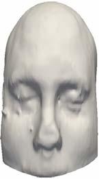
1 MRI-Based Classification Techniques of Autistic vs. Typically Developing Brain Presented by: Rachid Fahmi 1 2 Collaborators: Ayman Elbaz, Aly A. Farag 1, Hossam Hassan 1, and Manuel F. Casanova3 1Computer Vision & Image Processing Lab (CVIP Lab.)/ ECE Dept. 2Bioengineering Department 3 Dept. of Psychiatry and Behavioral Sciences
2 Research Motivation Autism is neuro-developmental disorder Impairments in social interaction, communication Unusual behaviors and interests According to CDC, 1/150 American kids are autistic (4:1 ratio of boys to girls) Challenges: No definitive medical test for diagnosis No reliable cause is identified No cure. BUT: Therapies for specific symptoms


3 Autistic Brain Brain Size Enlarged Brain weight & Head Circumference (first noticed by Kanner in 1943). Normal brain size at birth and rapid expansion by age 2 By age 4 autistic children have brain the size of a typical 13-yrs old The limbic system (Center of emotions) Increased cell packing density and reduced cell size in the following brain regions: hippocampus Subiculum Amygdala Diminished width for pyramidal cell arrays in the neocortex (Casanova et al. 02, 04, 06).


4 Autistic Brain Brainstem Smaller areas of brainstem sub-regions in mentally and non-mentally retarded juvenile young adults individuals with autism relative to controls. Cerebellum (Sensory perceptions integration) Decreased number of Purkinje cell in cerebella hemisphere, vermis, and cerebellum. Enlarged total GM and WM volumes in cerebellum have been reported by well designed controlled MRI studies in autistic children and young adults mostly moderate-tohigh functioning.

 reported abnormal anatomy")

5 Corpus Callosum Autistic Brain Increasing agreement from structural imaging studies on the deficits in the size of the Corpus Callosum and its sub-regions in patients with autism. White Matter Several imaging studies (MRI, DTI) reported abnormal anatomy of the WM. Excessive WM in many brain areas. Asymmetry/Increase in the outer radiate compartment of WM (Herbert et al. 04). Reduced Fractional Anisotropy (Keller et al. 06). White Matter Interface
6 Proposed Classification Approaches Taking advantage of the abnormalities of some brain regions in order to devise new techniques that allow the classification of autistic subjects vs. typically developing ones by analyzing their respective brain MR images. Focus on analyzing the white matter (WM) and the corpus callosum (CC). Increasing agreement from structural imaging studies on the abnormal anatomy of the WM in autistic brains (e.g., asymmetry, volume, fractional anisotropy, ). The deficits in the size of the corpus callosum and its sub-regions in patients with autism relative to controls is well established. Two types of MRI data are used: Post-mortem Proton density MRI Pre-mortem T1- weighted MRI Data set size: 256x256x124 Data Resolution: x0.9375x1.5


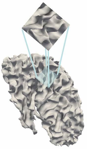
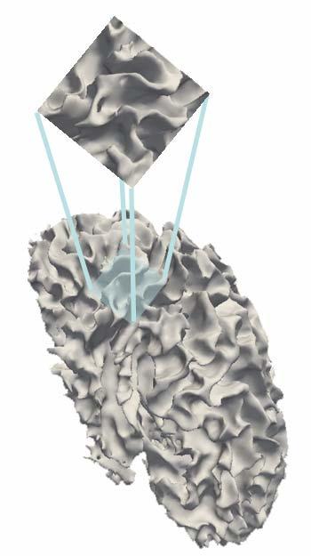
7 White Matter Analysis Fixed data Moving data Data Alignment Moving data


8 Preprocessing of postmortem data sets Due to distortions, post-mortem data sets are segmented slice by slice using our level set based approach. Stack of Images Whole 3D Head Model WM Visualization 2D Slice Extracted WM (Red)/GM (Blue) Binary Representation of WM


9 Preprocessing of Pre-mortem Data Sets Each data set is smoothed, skull stripped and then segmented into WM, GM, and CSF. Original slice Smoothed Skull Stripped Segmentation into WM, GM, and CSF


10 Segmented WM for Control and Autistic brains Autistic Control

11 Distance Map inside the WM We used the Fast Marching Level Sets to compute the 3D distance map inside the WM


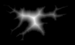
12 Example Of Generated Distance Maps
13 Training Samples Cumulative distributions of the distance map inside the WM of a training set from each group are computed, normalized, and averaged.
14 Classification Procedure Given a test subject (green curves): Compute and normalize the DM inside its WM. Compute corresponding CDF and compare it to the two averaged CDF s using the Levy distance.
15 Total Classification Results Accuracy of classification based on WM shape analysis Confidence rate Autistic Control Post-mortem Pre-mortem Post-mortem Pre-mortem 85% 14 /14 13/15 12/12 30/30 90% 13/14 12/15 12/12 30/30 95% 13/14 11/15 11/12 28/30
16 New Non-rigid Registration method (EMBC 06) New feature descriptors are built as voxel signatures using scale space theory. The global motion of the imaged object is modeled by matching these descriptors. Iso-surface evolved in target image to match those of source image in four steps Generate the distance map inside of the imaged organ Use this distance map to generate iso-surfaces Number to be set by user Not necessarily the same for both volumes Find Correspondences between iso-surfaces Evolve iso-surfaces in volume A to match those in volume B


17 Example: 3D Registration of brain MRI s Before Registration Rigid Alignment Non-rigid Alignment

18 Corpus Callosum Analysis Only the pre-mortem data is considered. Corpus callosum is manually segmented from the segmented brain date sets. Illustration of the proposed non-rigid registration approach.
19 Training Sample For each group: Choose a reference within a training set. Register the rest to the reference. Compute CDF of the corresponding deformation fields. Average the deformation fields.
20 The CC-registration based classification approach
21 Classification Performance Accuracy of classification based on CC analysis Confidence level Autistic Control 85 % 10/15 29/30 90 % 9/15 29/30 95 % 7/15 25/30 Combination of the two classification approaches Confidence level Autistic Control 85 % 14/15 30/30 90 % 14/15 30/30 95 % 13/15 30/30
22 Conclusions & Future Directions Two new neuroimaging-based classification techniques have been proposed to discriminate between autistic and typically developing brains. Our methods are based on shape & geometrical models and then overcome the shortcomings of the traditional volumetric approaches. Methods tested on post and pre-mortem MRI s and show great performances. More brain structures and other image modalities will be considered in future work. Part of this work is supported by the NSF This work is part of a joint R01 submitted to the NIH with Dr. M. F. Casanova and other collaborators.
23 Some References L. Kanner, Autistic disturbances of affective contact. Nervous Child, vol. 2, pp , R. Courchesne, R. Carper, and N. Akshoomoff, Evidence of brain overgrowth in the first year of life in autism. JAMA, vol. 290, pp , E. Courchesne et al. Unusual brain growth patterns in early life in patients with autistic disorder: an mri study. Neurology, vol. 57, no. 2, pp , C. Vidal et al. Mapping corpus callosum deficits in autism: An index of aberrant cortical connectivity. Biol. Psychiatry., vol. 60, no. 3, pp , A. A. Farag and H. S. Hassan, Adaptive segmentation of multi-modal 3d data using robust level set techniques. in (MICCAI 04), Saint-Malo, France, pp R. Fahmi et al. New deformable registration technique using scale space and curve evolution theory and a finite element based validation framework. IEEE/EMBC 06. M. F. Casanova et al. Minicolumnar abnormalities in autism. Acta Neuropathological, 2006 in press (Available online). J.E. Lainhart et al. The brain during life in autism: advances in neuroimaging research. In Casanova, MF editor. Recent Advances in Autism Research, New York: NOVA Biomedical, p , M. Herbert et al. Localization of white matter volume increase in autism and developmental language disorder. Ann. Neurol., vol. 55, pp , 2004.
24
Shape Modeling of the Corpus Callosum for Neuroimaging Studies of the Brain (Part I) Dongqing Chen, Ph.D.
 Dongqing Chen, Ph.D.") The University of Louisville CVIP Lab Shape Modeling of the Corpus Callosum for Neuroimaging Studies of the Brain (Part I) Dongqing Chen, Ph.D. Computer Vision & Image Processing (CVIP) Laboratory Department
The University of Louisville CVIP Lab Shape Modeling of the Corpus Callosum for Neuroimaging Studies of the Brain (Part I) Dongqing Chen, Ph.D. Computer Vision & Image Processing (CVIP) Laboratory Department
Visualization strategies for major white matter tracts identified by diffusion tensor imaging for intraoperative use
 International Congress Series 1281 (2005) 793 797 www.ics-elsevier.com Visualization strategies for major white matter tracts identified by diffusion tensor imaging for intraoperative use Ch. Nimsky a,b,
Automated detection of abnormal changes in cortical thickness: A tool to help diagnosis in neocortical focal epilepsy
 Automated detection of abnormal changes in cortical thickness: A tool to help diagnosis in neocortical focal epilepsy 1. Introduction Epilepsy is a common neurological disorder, which affects about 1 %
Discriminative Analysis for Image-Based Studies
 Discriminative Analysis for Image-Based Studies Polina Golland 1, Bruce Fischl 2, Mona Spiridon 3, Nancy Kanwisher 3, Randy L. Buckner 4, Martha E. Shenton 5, Ron Kikinis 6, Anders Dale 2, and W. Eric
International Journal of Research (IJR) Vol-1, Issue-6, July 2014 ISSN
 Vol-1, Issue-6, July 2014 ISSN") Developing an Approach to Brain MRI Image Preprocessing for Tumor Detection Mr. B.Venkateswara Reddy 1, Dr. P. Bhaskara Reddy 2, Dr P. Satish Kumar 3, Dr. S. Siva Reddy 4 1. Associate Professor, ECE Dept,
Developing an Approach to Brain MRI Image Preprocessing for Tumor Detection Mr. B.Venkateswara Reddy 1, Dr. P. Bhaskara Reddy 2, Dr P. Satish Kumar 3, Dr. S. Siva Reddy 4 1. Associate Professor, ECE Dept,
APPLICATION OF PHOTOGRAMMETRY TO BRAIN ANATOMY
 http://medifitbiologicals.com/central-nervous-system-cns/ 25/06/2017 PSBB17 ISPRS International Workshop APPLICATION OF PHOTOGRAMMETRY TO BRAIN ANATOMY E. Nocerino, F. Menna, F. Remondino, S. Sarubbo,
Discriminative Analysis for Image-Based Population Comparisons
 Discriminative Analysis for Image-Based Population Comparisons Polina Golland 1,BruceFischl 2, Mona Spiridon 3, Nancy Kanwisher 3, Randy L. Buckner 4, Martha E. Shenton 5, Ron Kikinis 6, and W. Eric L.
Shape analysis of the corpus callosum of autistic and normal subjects in neuroimaging.
 University of Louisville ThinkIR: The University of Louisville's Institutional Repository Electronic Theses and Dissertations 8-2009 Shape analysis of the corpus callosum of autistic and normal subjects
Computer based delineation and follow-up multisite abdominal tumors in longitudinal CT studies
 Research plan submitted for approval as a PhD thesis Submitted by: Refael Vivanti Supervisor: Professor Leo Joskowicz School of Engineering and Computer Science, The Hebrew University of Jerusalem Computer
CISC 3250 Systems Neuroscience
 CISC 3250 Systems Neuroscience Levels of organization Central Nervous System 1m 10 11 neurons Neural systems and neuroanatomy Systems 10cm Networks 1mm Neurons 100μm 10 8 neurons Professor Daniel Leeds
Online appendices are unedited and posted as supplied by the authors. SUPPLEMENTARY MATERIAL
 Appendix 1 to Sehmbi M, Rowley CD, Minuzzi L, et al. Age-related deficits in intracortical myelination in young adults with bipolar SUPPLEMENTARY MATERIAL Supplementary Methods Intracortical Myelin (ICM)
A new Method on Brain MRI Image Preprocessing for Tumor Detection
 2015 IJSRSET Volume 1 Issue 1 Print ISSN : 2395-1990 Online ISSN : 2394-4099 Themed Section: Engineering and Technology A new Method on Brain MRI Preprocessing for Tumor Detection ABSTRACT D. Arun Kumar
Leah Militello, class of 2018
 Leah Militello, class of 2018 Objectives 1. Describe the general organization of cerebral hemispheres. 2. Describe the locations and features of the different functional areas of cortex. 3. Understand
Quantitative Neuroimaging- Gray and white matter Alteration in Multiple Sclerosis. Lior Or-Bach Instructors: Prof. Anat Achiron Dr.
 Quantitative Neuroimaging- Gray and white matter Alteration in Multiple Sclerosis Lior Or-Bach Instructors: Prof. Anat Achiron Dr. Shmulik Miron INTRODUCTION Multiple Sclerosis general background Gray
fmri (functional MRI)
") Lesion fmri (functional MRI) Electroencephalogram (EEG) Brainstem CT (computed tomography) Scan Medulla PET (positron emission tomography) Scan Reticular Formation MRI (magnetic resonance imaging) Thalamus
Lesion fmri (functional MRI) Electroencephalogram (EEG) Brainstem CT (computed tomography) Scan Medulla PET (positron emission tomography) Scan Reticular Formation MRI (magnetic resonance imaging) Thalamus
The Cause of Autism: Its Footprint Tells
 The Cause of Autism: Its Footprint Tells Inaugural Autism Symposium March 11, 2009 Nancy Minshew, MD Professor Psychiatry & Neurology University of Pittsburgh USA Convergence The Top of 10 Clinical of
CNS Tour (Lecture 12)
") A. Introduction CNS Tour (Lecture 12) There are to a chemical pathways in the nervous system. These pathways also form different neurological structures B. Spinal Cord Receives sensory neurons from skin
A. Introduction CNS Tour (Lecture 12) There are to a chemical pathways in the nervous system. These pathways also form different neurological structures B. Spinal Cord Receives sensory neurons from skin
Neuroanatomy of autism
 Review Neuroanatomy of autism David G. Amaral 1, Cynthia Mills Schumann 2 and Christine Wu Nordahl 1 1 The M.I.N.D. Institute, Department of Psychiatry and Behavioral Sciences, University of California,
Neuroanatomic observations of the brain in autism: a review and future directions
 Int. J. Devl Neuroscience 23 (2005) 183 187 Review Neuroanatomic observations of the brain in autism: a review and future directions Margaret L. Bauman *, Thomas L. Kemper Children s Neurology Service,
III. Studying The Brain and Other Structures
 III. Studying The Brain and Other Structures 1. Accidents (case study) In 1848, a railroad worker named Phineas Gage was involved in an accident that damaged the front part of his brain. Gage s doctor
mr brain volume analysis using brain assist
 mr brain volume analysis using brain assist This Paper describes the tool named BrainAssist, which can be used for the study and analysis of brain abnormalities like Focal Cortical Dysplasia (FCD), Heterotopia
Diffusion Tensor Imaging in Psychiatry
 2003 KHBM DTI in Psychiatry Diffusion Tensor Imaging in Psychiatry KHBM 2003. 11. 21. 서울대학교 의과대학 정신과학교실 권준수 Neuropsychiatric conditions DTI has been studied in Alzheimer s disease Schizophrenia Alcoholism
IV. The Divisions of the Brain. Slide # 1
 IV. The Divisions of the Brain Slide # 1 The Hindbrain Hindbrain, located at the rear base of the skull, controlling automatic functions Contains: Cerebellum (balance & coordination) Medulla (heartbeat,
Review of Longitudinal MRI Analysis for Brain Tumors. Elsa Angelini 17 Nov. 2006
 Review of Longitudinal MRI Analysis for Brain Tumors Elsa Angelini 17 Nov. 2006 MRI Difference maps «Longitudinal study of brain morphometrics using quantitative MRI and difference analysis», Liu,Lemieux,
Myers Psychology for AP*
 Myers Psychology for AP* David G. Myers PowerPoint Presentation Slides by Kent Korek Germantown High School Worth Publishers, 2010 *AP is a trademark registered and/or owned by the College Board, which
Brain Structure and Function in Nephropathic Cystinosis
 Brain Structure and Function in Nephropathic Cystinosis Doris A. Trauner M.D. Professor, Depts. of Neurosciences and Pediatrics University of California San Diego School of Medicine La Jolla, CA USA Cystinosis
P. Hitchcock, Ph.D. Department of Cell and Developmental Biology Kellogg Eye Center. Wednesday, 16 March 2009, 1:00p.m. 2:00p.m.
 Normal CNS, Special Senses, Head and Neck TOPIC: CEREBRAL HEMISPHERES FACULTY: LECTURE: READING: P. Hitchcock, Ph.D. Department of Cell and Developmental Biology Kellogg Eye Center Wednesday, 16 March
Classification and Statistical Analysis of Auditory FMRI Data Using Linear Discriminative Analysis and Quadratic Discriminative Analysis
 International Journal of Innovative Research in Computer Science & Technology (IJIRCST) ISSN: 2347-5552, Volume-2, Issue-6, November-2014 Classification and Statistical Analysis of Auditory FMRI Data Using
In Press: Carol Armstrong, Ed., Handbook of Medical Neuropsychology. New York: Springer Science.
 In Press: Carol Armstrong, Ed., Handbook of Medical Neuropsychology. New York: Springer Science. Autism and Asperger s Syndrome: A Cognitive Neuroscience Perspective Jeanne Townsend, Ph.D., Marissa Westerfield,
Automated Volumetric Cardiac Ultrasound Analysis
 Whitepaper Automated Volumetric Cardiac Ultrasound Analysis ACUSON SC2000 Volume Imaging Ultrasound System Bogdan Georgescu, Ph.D. Siemens Corporate Research Princeton, New Jersey USA Answers for life.
QIBA/NIBIB Final Progress Report
 QIBA/NIBIB Final Progress Report Amyloid Profile Continued Support with Brain Phantom Development Larry Pierce, David Haynor, John Sunderland, Paul Kinahan August 29, 2015 Title: Amyloid Profile Continued
Imaging and Hemodynamics in Aneurysm Evolution
 Disclosures Imaging and Hemodynamics in Aneurysm Evolution David Saloner, PhD There are no financial conflicts of interest to report Department of Radiology and Biomedical Imaging VA Medical Center San
10/3/2016. T1 Anatomical structures are clearly identified, white matter (which has a high fat content) appears bright.
 appears bright.") H2O -2 atoms of Hydrogen, 1 of Oxygen Hydrogen just has one single proton and orbited by one single electron Proton has a magnetic moment similar to the earths magnetic pole Also similar to earth in that
H2O -2 atoms of Hydrogen, 1 of Oxygen Hydrogen just has one single proton and orbited by one single electron Proton has a magnetic moment similar to the earths magnetic pole Also similar to earth in that
Today s goals. Today s reading. Autistic Spectrum Disorder. INF1-CG 2014 Lecture 27
 INF1-CG 2014 Lecture 27 Autistic Spectrum Disorder Richard Shillcock 1 /26 Today s goals Look at Autistic Spectrum Disorder (ASD) and cognition, with particular attention to language and the linguistic
Automated Whole Brain Segmentation Using FreeSurfer
 Automated Whole Brain Segmentation Using FreeSurfer https://surfer.nmr.mgh.harvard.edu/ FreeSurfer (FS) is a free software package developed at the Martinos Center for Biomedical Imaging used for three
The Cause of Autism: Its Footprint Tells. Autism Symposium-Part II
 The Cause of Autism: Its Footprint Tells Autism Symposium-Part II May 22, 2009 Nancy Minshew, MD Professor Psychiatry & Neurology University of Pittsburgh USA Convergence The Top of 10 Clinical of 2007
Cognitive Neuroscience of Autism
 Townsend, J., Westerfield, M. (in press). Autism and Asperger s Syndrome: A Cognitive Neuroscience Perspective. In, Carol Armstrong, Ed., Handbook of Medical Neuropsychology. New York: Springer Science.
Group-Wise FMRI Activation Detection on Corresponding Cortical Landmarks
 Group-Wise FMRI Activation Detection on Corresponding Cortical Landmarks Jinglei Lv 1,2, Dajiang Zhu 2, Xintao Hu 1, Xin Zhang 1,2, Tuo Zhang 1,2, Junwei Han 1, Lei Guo 1,2, and Tianming Liu 2 1 School
Big brains may hold clues to origins of autism
 VIEWPOINT Big brains may hold clues to origins of autism BY KONSTANTINOS ZARBALIS 23 FEBRUARY 2016 A persistent challenge to improving our understanding of autism is the fact that no single neurological
Detection of Mild Cognitive Impairment using Image Differences and Clinical Features
 Detection of Mild Cognitive Impairment using Image Differences and Clinical Features L I N L I S C H O O L O F C O M P U T I N G C L E M S O N U N I V E R S I T Y Copyright notice Many of the images in
Regional and Lobe Parcellation Rhesus Monkey Brain Atlas. Manual Tracing for Parcellation Template
 Regional and Lobe Parcellation Rhesus Monkey Brain Atlas Manual Tracing for Parcellation Template Overview of Tracing Guidelines A) Traces are performed in a systematic order they, allowing the more easily
Cerebral Cortex 1. Sarah Heilbronner
 Cerebral Cortex 1 Sarah Heilbronner heilb028@umn.edu Want to meet? Coffee hour 10-11am Tuesday 11/27 Surdyk s Overview and organization of the cerebral cortex What is the cerebral cortex? Where is each
Activated Fibers: Fiber-centered Activation Detection in Task-based FMRI
 Activated Fibers: Fiber-centered Activation Detection in Task-based FMRI Jinglei Lv 1, Lei Guo 1, Kaiming Li 1,2, Xintao Hu 1, Dajiang Zhu 2, Junwei Han 1, Tianming Liu 2 1 School of Automation, Northwestern
Basic Brain Structure
 The Human Brain Basic Brain Structure Composed of 100 billion cells Makes up 2% of bodies weight Contains 15% of bodies blood supply Uses 20% of bodies oxygen and glucose Brain Protection Surrounded by
Biocomputer Wired for Action MWABBYH CTBIR LOBES
 Biocomputer Wired for Action MWABBYH CTBIR LOBES 100 100 100 100 100 200 200 200 200 200 300 300 300 300 300 400 400 400 400 400 500 500 500 500 500 Biocomputer Wired for Action MWABBYH CTBIR LOBES 100
Structural And Functional Integration: Why all imaging requires you to be a structural imager. David H. Salat
 Structural And Functional Integration: Why all imaging requires you to be a structural imager David H. Salat salat@nmr.mgh.harvard.edu Salat:StructFunct:HST.583:2015 Structural Information is Critical
Department of Human Anatomy GUIDELINES. nuclei. The lateral ventricles. White substance of cerebral hemispheres. course 1
 Department of Human Anatomy GUIDELINES Academic discipline Human Anatomy Module 2 Content module 11 Study subject The olfactory brain. Basal nuclei. The lateral ventricles. White substance of cerebral
The Nervous System. Divisions of the Nervous System. Branches of the Autonomic Nervous System. Central versus Peripheral
 The Nervous System Divisions of the Nervous System Central versus Peripheral Central Brain and spinal cord Peripheral Everything else Somatic versus Autonomic Somatic Nerves serving conscious sensations
Early Diagnosis of Autism Disease by Multi-channel CNNs
 Early Diagnosis of Autism Disease by Multi-channel CNNs Guannan Li 1,2, Mingxia Liu 2, Quansen Sun 1(&), Dinggang Shen 2(&), and Li Wang 2(&) 1 School of Computer Science and Engineering, Nanjing University
Cover Page. The handle holds various files of this Leiden University dissertation
 Cover Page The handle http://hdl.handle.net/1887/26921 holds various files of this Leiden University dissertation Author: Doan, Nhat Trung Title: Quantitative analysis of human brain MR images at ultrahigh
By Lauren Stowe, PhD, CCC-SLP & Gina Rotondo, MS, CCC-SLP The Speech Therapy Group
 By Lauren Stowe, PhD, CCC-SLP & Gina Rotondo, MS, CCC-SLP The Speech Therapy Group http://www.acquiredbraininjury.com/interactive brain/interactivebrain.swf 1. Hormones make the science messy 2. Difference
Image Fusion, Contouring, and Margins in SRS
 Image Fusion, Contouring, and Margins in SRS Sarah Geneser, Ph.D. Department of Radiation Oncology University of California, San Francisco Overview Review SRS uncertainties due to: image registration contouring
Assessing Brain Volumes Using MorphoBox Prototype
 MAGNETOM Flash (68) 2/207 33 Assessing Brain Volumes Using MorphoBox Prototype Alexis Roche,2,3 ; Bénédicte Maréchal,2,3 ; Tobias Kober,2,3 ; Gunnar Krueger 4 ; Patric Hagmann ; Philippe Maeder ; Reto
Spatial Normalisation, Atlases, & Functional Variability
 Spatial Normalisation, Atlases, & Functional Variability Jörn Diedrichsen Institute of Cognitive Neuroscience, University College London Overview Cerebellar normalisation Anatomical reference High-resolution
Cetacean Brains Cogs 143 * UCSD
 Cetacean Brains Cogs 143 * UCSD EQ -- Encephalization Quotient EQ = Actual brain mass / Expected brain mass Where expected = 0.12 x (Body mass) 2/3 EQ -- Encephalization Quotient Chimp Human Dolphin Globular
What we can infer by comparing the shape of anatomical structures? Comparative anatomy of the parietal cortex in human and chimpanzee
 Interdisciplinary Workshop on 3D Paleo-Anthropology, Anatomy, Computer Science & Engineering - Synergies for the Future - June, 19-20 Musée de Toulouse, France What we can infer by comparing the shape
Running head: NEURODEVELOPMENT AND NEUROANATOMY OF AUTISM 1. A Review: Neurodevelopment and Neuroanatomy of Individuals with Autism Spectrum
 Running head: NEURODEVELOPMENT AND NEUROANATOMY OF AUTISM 1 A Review: Neurodevelopment and Neuroanatomy of Individuals with Autism Spectrum Disorder; Implications for Therapist A Research Paper Presented
Automated morphometry in adolescents with OCD and controls, using MR images with incomplete brain coverage
 Automated morphometry in adolescents with OCD and controls, using MR images with incomplete brain coverage M.Sc. Thesis Oscar Gustafsson gusgustaos@student.gu.se Supervisors: Göran Starck Maria Ljungberg
NIH Public Access Author Manuscript Proc SPIE. Author manuscript; available in PMC 2014 February 07.
 NIH Public Access Author Manuscript Published in final edited form as: Proc SPIE. 2007 March 5; 6512: 651236. doi:10.1117/12.708950. Semi-Automatic Parcellation of the Corpus Striatum Ramsey Al-Hakim a,
Autism shares features with cerebellar syndromes
 NEWS Autism shares features with cerebellar syndromes BY KELLY RAE CHI 3 DECEMBER 2009 1 / 7 2 / 7 Misshapen structures: MRI scans show that, compared with controls (A), the cerebellum of a child with
Fibre orientation dispersion in the corpus callosum relates to interhemispheric functional connectivity
 Fibre orientation dispersion in the corpus callosum relates to interhemispheric functional connectivity ISMRM 2017: http://submissions.mirasmart.com/ismrm2017/viewsubmissionpublic.aspx?sei=8t1bikppq Jeroen
Diffusion-Weighted and Conventional MR Imaging Findings of Neuroaxonal Dystrophy
 AJNR Am J Neuroradiol 25:1269 1273, August 2004 Diffusion-Weighted and Conventional MR Imaging Findings of Neuroaxonal Dystrophy R. Nuri Sener BACKGROUND AND PURPOSE: Neuroaxonal dystrophy is a rare progressive
Optic Nerve Hypoplasia Part 2: Clinical Problems
 Optic Nerve Hypoplasia Part 2: Clinical Problems Hypopituitarism Deficiencies in: Growth hormone Thyroid hormone ACTH (cortisol) Anti-diuretic hormone (diabetes insipidus) Sex hormones Hypothalamic Dysfunction:
Introducing a NEW update for the in-office design software that facilitates better outcomes
 DWOS Chairside 2 Release Info May 2018 DWOS Chairside 2 Introducing a NEW update for the in-office design software that facilitates better outcomes for patients DWOS Chairside 2 builds on the innovative
Diffusion tensor imaging of the infant brain: From technical problems to neuroscientific breakthroughs Jessica Dubois
 Diffusion tensor imaging of the infant brain: From technical problems to neuroscientific breakthroughs Jessica Dubois L. Hertz-Pannier, G. Dehaene-Lambertz, J.F. Mangin, D. Le Bihan Inserm U56, U663; NeuroSpin
meninges Outermost layer of the meninge dura mater arachnoid mater pia mater membranes located between bone and soft tissue of the nervous system
 membranes located between bone and soft tissue of the nervous system meninges Outermost layer of the meninge dura mater middle layer of the meninges, contains no blood vessels arachnoid mater Innermost
Summary of findings from the previous meta-analyses of DTI studies in MDD patients. SDM (39) 221 Left superior longitudinal
 221 Left superior longitudinal") Supplemental Data Table S1 Summary of findings from the previous meta-analyses of DTI studies in MDD patients Study Analysis Method Included studies, n MDD (medicated) HC Results (MDDHC)
Supplemental Data Table S1 Summary of findings from the previous meta-analyses of DTI studies in MDD patients Study Analysis Method Included studies, n MDD (medicated) HC Results (MDDHC)
XIXth Century: Localization of Functions to Different Parts of the Brain
 XIXth Century: Localization of Functions to Different Parts of the Brain Studies by Bell and Magendie initiated an extremely important scientific procedure,, where a specific part of the nervous system
Taken From The Brain Top to Bottom //
 Taken From The Brain Top to Bottom // http://thebrain.mcgill.ca/flash/d/d_03/d_03_cl/d_03_cl_que/d_03_cl_que.html THE EVOLUTIONARY LAYERS OF THE HUMAN BRAIN The first time you observe the anatomy of the
The Brain Studying & Structures. Unit 3
 The Brain Studying & Structures Unit 3 Modified PowerPoint from: Aneeq Ahmad -- Henderson State University. Worth Publishers 2007 Learning Objectives Describe the nervous system and its subdivisions and
Children Born to Women with Hypothyroidism during Pregnancy Show Abnormal Corpus Callosum Development
 Children Born to Women with Hypothyroidism during Pregnancy Show Abnormal Corpus Callosum Development Arash Samadi, Jovanka Skocic, Joanne Rovet AMERICAN THYROID ASSOCIATION ANNUAL MEETING SAN JUAN PUERTO
8/10/2016. PET/CT Radiomics for Tumor. Anatomic Tumor Response Assessment in CT or MRI. Metabolic Tumor Response Assessment in FDG-PET
 PET/CT Radiomics for Tumor Response Evaluation August 1, 2016 Wei Lu, PhD Department of Medical Physics www.mskcc.org Department of Radiation Oncology www.umaryland.edu Anatomic Tumor Response Assessment
Characterizing Anatomical Variability And Alzheimer s Disease Related Cortical Thinning in the Medial Temporal Lobe
 Characterizing Anatomical Variability And Alzheimer s Disease Related Cortical Thinning in the Medial Temporal Lobe Long Xie, Laura Wisse, Sandhitsu Das, Ranjit Ittyerah, Jiancong Wang, David Wolk, Paul
1. Processes nutrients and provides energy for the neuron to function; contains the cell's nucleus; also called the soma.
 1. Base of brainstem; controls heartbeat and breathing 2. tissue destruction; a brain lesion is a naturally or experimentally caused destruction of brain tissue 3. A thick band of axons that connects the
Lecture 35 Association Cortices and Hemispheric Asymmetries -- M. Goldberg
 Lecture 35 Association Cortices and Hemispheric Asymmetries -- M. Goldberg The concept that different parts of the brain did different things started with Spurzheim and Gall, whose phrenology became quite
What is Autism? -Those with the most severe disability need a lot of help with their daily lives whereas those that are least affected may not.
 Autism Summary Autism What is Autism? The Autism Spectrum Disorder (ASD) is a developmental disability that can have significant implications on a child's ability to function and interface with the world
Autism Updates Mike Miklos PATTAN Autism Initiative ABA Supports. Pennsylvania Training and Technical Assistance Network
 Autism Updates 2018 Mike Miklos PATTAN Autism Initiative ABA Supports Pennsylvania Training and Technical Assistance Network Change in Prevalence Data CDC has announced that the prevalence rate for Autism
XIXth Century: Localization of Functions to Different Parts of the Brain
 XIXth Century: Localization of Functions to Different Parts of the Brain Studies by Bell and Magendie initiated an extremely important scientific procedure,, where a specific part of the nervous system
Cephalization. Nervous Systems Chapter 49 11/10/2013. Nervous systems consist of circuits of neurons and supporting cells
 Nervous Systems Chapter 49 Cephalization Nervous systems consist of circuits of neurons and supporting cells Nervous system organization usually correlates with lifestyle Organization of the vertebrate
The autistic brain: birth through adulthood Eric Courchesne a,c, Elizabeth Redcay b and Daniel P. Kennedy a
 The autistic brain: birth through adulthood Eric Courchesne a,c, Elizabeth Redcay b and Daniel P Kennedy a Purpose of review We discuss evidence of brain maldevelopment in the first years of life in autism
PsychoBrain. 31 st January Dr Christos Pliatsikas. Lecturer in Psycholinguistics in Bi-/Multilinguals University of Reading
 PsychoBrain 31 st January 2018 Dr Christos Pliatsikas Lecturer in Psycholinguistics in Bi-/Multilinguals University of Reading By the end of today s lecture you will understand Structure and function of
The Nervous System and the Endocrine System
 The Nervous System and the Endocrine System Neurons: The Building Blocks of the Nervous System Nervous System The electrochemical communication system of the body Sends messages from the brain to the
Prof. Greg Francis 5/23/08
 Brain parts The brain IIE 269: Cognitive Psychology Greg Francis Lecture 02 The source of cognition (consider transplant!) Weighs about 3 pounds Damage to some parts result in immediate death or disability
Pediatric MS MRI Study Methodology
 General Pediatric MS MRI Study Methodology SCAN PREPARATION axial T2-weighted scans and/or axial FLAIR scans were obtained for all subjects when available, both T2 and FLAIR scans were scored. In order
Hanan Azouz, Hesham Kouzou,Mona Khalil, Rania Abdou, Mohamed sakr
 Hanan Azouz, Hesham Kouzou,Mona Khalil, Rania Abdou, Mohamed sakr Autism spectrum disorder (ASD), a major neuropsychiatric condition in children, is generally recognized as a developmental condition in
To understand AD, it is important to
 To understand AD, it is important to know a bit about the brain. This part of Unraveling the Mystery gives an inside view of the normal brain, how it works, and what happens during aging. The brain is
Contributions to Brain MRI Processing and Analysis
 Contributions to Brain MRI Processing and Analysis Dissertation presented to the Department of Computer Science and Artificial Intelligence By María Teresa García Sebastián PhD Advisor: Prof. Manuel Graña
Head Injury: Classification Most Severe to Least Severe
 Head Injury: Classification Most Severe to Least Severe Douglas I. Katz, MD Professor, Dept. Neurology, Boston University School of Medicine, Boston MA Medical Director Brain Injury Program, HealthSouth
Functional MRI and Diffusion Tensor Imaging
 Functional MRI and Diffusion Tensor Imaging Andrew Steven March 23, 2018 Ochsner Neuroscience Symposium None Disclosure 1 Objectives Review basic principles of BOLD fmri and DTI. Discuss indications and
1 in 68 in US. Autism Update: New research, evidence-based intervention. 1 in 45 in NJ. Selected New References. Autism Prevalence CDC 2014
 Autism Update: New research, evidence-based intervention Martha S. Burns, Ph.D. Joint Appointment Professor Northwestern University. 1 Selected New References Bourgeron, Thomas (2015) From the genetic
Brain-Behavior Network. Central Nervous System. Cerebral Cortex Gyrus and Sulcus. Nervous System
 Brain-Behavior Network Nervous System Sensory information comes into and decisions come out of the central nervous system (CNS) Central Nervous System The nerves outside the CNS are called the peripheral
biological psychology, p. 40 The study of the nervous system, especially the brain. neuroscience, p. 40
 biological psychology, p. 40 The specialized branch of psychology that studies the relationship between behavior and bodily processes and system; also called biopsychology or psychobiology. neuroscience,
Jack Noble, PhD, René Gifford, PhD, Benoit Dawant, PhD, and Robert Labadie, MD, PhD
 Jack Noble, PhD, René Gifford, PhD, Benoit Dawant, PhD, and Robert Labadie, MD, PhD Overview The position of implanted electrodes relative to stimulation targets can be used to aid programming Individualized
Advanced multimodal imaging in malformations of cortical development
 Advanced multimodal imaging in malformations of cortical development Seok Jun Hong (sjhong@bic.mni.mcgill.ca) NOEL Neuroimaging of Epilepsy Lab MICA Multimodal Imaging and Connectome Analysis Lab w4 w5
Modeling of early-infant brain growth using longitudinal data from diffusion tensor imaging.
 Modeling of early-infant brain growth using longitudinal data from diffusion tensor imaging. Guido Gerig, Neda Sadeghi, PhD, Marcel Prastawa, Tom Fletcher, Clement Vachet Scientific Computing and Imaging
Patterns of Brain Tumor Recurrence Predicted From DTI Tractography
 Patterns of Brain Tumor Recurrence Predicted From DTI Tractography Anitha Priya Krishnan 1, Isaac Asher 2, Dave Fuller 2, Delphine Davis 3, Paul Okunieff 2, Walter O Dell 1,2 Department of Biomedical Engineering
Chapter 3. Biological Processes
 Biological Processes Psychology, Fifth Edition, James S. Nairne What s It For? Biological Solutions Communicating internally Initiating and coordinating behavior Regulating growth and other internal functions
Mounting evidence implicates cerebellum in autism
 NEWS Mounting evidence implicates cerebellum in autism BY SARAH DEWEERDT 6 JANUARY 2014 Young children who don t point at interesting objects or make eye contact may be showing early warning signs of autism.
FAILURES OF OBJECT RECOGNITION. Dr. Walter S. Marcantoni
 FAILURES OF OBJECT RECOGNITION Dr. Walter S. Marcantoni VISUAL AGNOSIA -damage to the extrastriate visual regions (occipital, parietal and temporal lobes) disrupts recognition of complex visual stimuli
The origins of localization
 Association Cortex, Asymmetries, and Cortical Localization of Affective and Cognitive Functions Michael E. Goldberg, M.D. The origins of localization The concept that different parts of the brain did different