5 th International Conference on Non-Invasive Brain Stimulation
|
|
|
- Calvin O’Neal’
- 6 years ago
- Views:
Transcription


1 5 th International Conference on Non-Invasive Brain Stimulation March 2013 Leipzig Abstracts
2 Index Abstracts of Invited Speakers... 3 Abstracts of Selected Oral Presentations Abstracts of Poster Session I Basic Physiology Cognitive Neuroscience I Motor Learning and Plasticity I Therapeutic Applications I Abstracts of Poster Session II Cognitive Neuroscience II Motor Learning and Plasticity II Multimodal Approaches Novel Techniques Therapeutic Applications II
3 Invited Speaker IS 1 The electric field in the brain during tdcs *P. C. Miranda 1 1 IBEB, Faculty of Science of the University of Lisbon, Lisbon, Portugal Introduction: Transcranial brain stimulation involves the interaction between an applied electric field and cells in the brain. In transcranial direct current stimulation (tdcs), this field is created by passing a weak current between two or more electrodes, at least one of which is placed on the scalp. The spatial distribution of the electric field and its orientation relative to the brain cells are two important factors that influence the outcome of stimulation. Objectives: To describe how the shape and the electrical properties of tissues affect the electric field distribution and to show to what extent this distribution can be altered by using multiple electrodes. Materials & methods: We developed a realistic head model based on anatomical MR images to compute the electric field using the finite element method. The values for the conductivity of the tissues were obtained from the literature. The electrode montage typically used to stimulate M1 was chosen. Results: The calculations show that the low conductivity of the skull and the high conductivity of the cerebrospinal fluid (CSF) have a strong impact on the electric field. As expected, the effect of the skull is to decrease the electric field intensity and to broaden its spatial variation in the brain. On the other hand, the presence of the CSF leads to very distinct distributions of the normal and tangential components of the electric field on the cortical surface. The normal component is strongest near the bottom of the sulci under the electrodes whereas the tangential component is strongest in the crowns of the gyri between electrodes. Whereas the high values of the normal component are confined to the bottom or walls of the sulci, the high values of the tangential component spread out over the crowns. One of the shortcomings of the traditional montages that employ two identical electrodes is that the electric field has practically the same magnitude (and opposite polarity) under the two electrodes. Using our model we show that one way to overcome this limitation efficiently is to use one active electrode and multiple return electrodes to reduce the magnitude of the electric field under the return electrodes relative to that under the active electrode. Conclusion: This work provides novel insights into the electric field distribution in the brain during tdcs. The most pressing issue for the future is the physiological validation of its predictions. Also, the accuracy of the calculations can be improved by incorporating the anisotropy of skull and white matter in the model. Likely future developments include the rapid generation of subject specific head models or the optimization of electric field delivery using multiple electrodes. Acknowledgments: This work was supported in part by the Foundation for Science and Technology (FCT), Portugal and by project HIVE. The project HIVE acknowledges the financial support of the Future and Emerging Technologies (FET) programme within the Seventh Framework Programme for Research of the European Commission, FET-Open grant
4 Invited Speaker IS 2 Using Computational Models in tdcs research and clinical trials *M. Bikson 1 1 The City College of New York of CUNY, Department of Biomedical Engineering, New York, United States Computational models of tdcs link tdcs dose (electrode montage) with the resulting brain current flow. This presentation summarizes computational tools and approaches currently available to clinicians and researchers that can be leveraged in the optimization and customization of tdcs. Even as increasingly complex and advanced modeling techniques are developed by engineering groups, clinicians and cognitive neuroscience researchers require software that is readily accessible, and does not require specialized training or significant computational resources. Approaches developed by the CCNY group to provide clinicians and researchers with such a toolbox including reducing the burden automatic individual model creation, instant current calculation, and true montage optimization (eliminating the need for iterative search) are explained. Currently available software is summarized. 4
5 Invited Speaker IS 3 TMS field modelling - Status and next steps *A. Thielscher 1,2,3 1 Copenhagen University Hospital Hvidovre, Danish Research Center for Magnetic Resonance, Copenhagen, Denmark 2 Technical University of Denmark, Kgs. Lyngby, Denmark 3 Max-Planck-Institute for Biological Cybernetics, Tübingen, Germany In the recent years, an increasing number of studies used geometrically accurate head models and Finite Element (FEM) or Finite Difference Methods (FDM) to estimate the electric field induced by non-invasive neurostimulation techniques such as transcranial magnetic stimulation (TMS) or transcranial weak current stimulation (tcs; e.g., [1, 2]). A general outcome was that the field estimates based on these more realistic models differ substantially from the results obtained with simpler head models. This suggests that the former models are indeed needed to realistically capture the field distribution in the brain. However, it is unclear how accurate even these more advanced models are and, in particular, to which extent they allow predicting the physiological outcome of stimulation. An experimental validation of the novel methods for field calculation is thus necessary. Focusing on motor cortex stimulation by TMS, our goal is to explore to which extent the field estimates based on advanced models correlate with the physiological stimulation effects. For example, we aim at testing whether interindividual differences in the field estimates are also reflected in differences in the MEP responses. This would indicate that the field calculations accurately capture the impact of individual macroanatomical features of the head and brain on the induced field distribution, in turn strongly supporting their plausibility. Our approach is based on the SimNIBS software pipeline ( that allows for the automatic creation of accurate head models from structural and diffusion-weighted magnetic resonance images (MRI) [3]. This enables us to perform field calculations for multiple subjects, as required in neuroscientific studies. We substantially improved the software in order to improve its usability in a group analysis. At the moment, we are performing field calculations and are acquiring motor mapping data in a group of subjects for a systematic comparison of both data sets. I will give an overview on the status of the SimNIBS project. I will start by summarizing the key findings on how the individual brain anatomy shapes the electric field induced by TMS [2, 4]. The putative link between the modeling results and basic physiological TMS effects is highlighted. I will then introduce the novel features of SimNIBS that include the import of coil positions from neuronavigation systems, improved support for diffusion-weighted MRI and transformation of the estimated fields into MNI space for group analysis. Preliminary results on the comparison between field estimates and motor mapping data will be presented. To summarize, field estimates based on accurate head models have already proven highly useful for a better understanding of the biophysics of non-invasive brain stimulation. The improved software tools now allow for systematic tests of the links between the estimated fields and the physiological effects in multisubject studies. This will give the knowledge needed, e.g. for a more accurate spatial targeting of specific brain areas by TMS. References 1. Datta, A., M. Bikson, and F. Fregni, Transcranial direct current stimulation in patients with skull defects and skull plates: high-resolution computational FEM study of factors altering cortical current flow. Neuroimage, (4): p Thielscher, A., A. Opitz, and M. Windhoff, Impact of the gyral geometry on the electric field induced by transcranial magnetic stimulation. Neuroimage, (1): p Windhoff, M., A. Opitz, and A. Thielscher, Field calculations in brain stimulation based on finite elements: An optimized processing pipeline for the generation and usage of accurate individual head models. Human Brain Mapping, epub. 4. Opitz, A., et al., How the brain tissue shapes the electric field induced by transcranial magnetic stimulation. Neuroimage, (3): p
6 Invited Speaker IS 4 Modelling TBS *Y.- Z. Huang 1 1 Chang Gung University, Taipei, Taiwan Theta burst stimulation, a form of repetitive transcranial magnetic stimulation, can induce lasting changes in corticospinal excitability through plasticity-like mechanisms on cortical synapses. Interestingly, the direction of the effect on synaptic efficiency depends on whether the bursts are delivered continuously (ctbs, producing long-term depression (LTD)-like effects) or intermittently (itbs, producing long-term potentiation (LTP)-like effects). We firstly built a simple phenomenological model based on knowledge of calciumdependent mechanisms of post-synaptic plasticity to successfully explain this by postulating (1) that burst stimulation induces a mixture of excitatory and inhibitory effects, (2) those effects may cascade to produce long-lasting effects and (3) the final effect of TBS (potentiation or depression) depends on the summation of these two effects. Furthermore, we went on to extend the model by including spike timing dependent plasticity with detailed calcium dynamics based on kinetic equations that mimic protein kinase interactions at the cellular and molecular levels. However, the post-synaptic calcium dependent plasticity model alone was not sufficient for describing diverse plasticity effects aroused by different rtms protocols. We then further recruited the pre-synaptic mechanism for the extended model, because we noticed that shot-term pre-synaptic depression due to vesicle depletion could play a critical key in the regulation of long-term plasticity in postsynaptic neurons. In results, the new improved synaptic model has successfully simulated not only the results of TBS but also those of conventional rtms protocols. 6
7 Invited Speaker IS 5 Clinical efficacy of non-invasive transorbital alternating current stimulation in optic neuropathy: a double-blind, randomized, sham-controlled multi-center study *C. Gall 1, A. Federov 1,2, A. Antal 3, M. Schittkowski 4, S. Kropf 5, A. Mante 6, S. Schmidt 6, B. Sabel 1 1 University of Magdeburg, Medical Faculty, Institute of Medical Psychology, Magdeburg, Germany 2 EBS Technologies GmbH, Kleinmachnow, Germany 3 Georg-August University Göttingen, Department of Clinical Neurophysiology, Göttingen, Germany 4 Georg-August University Göttingen, Department of Ophthalmology, Göttingen, Germany 5 University of Magdeburg, Medical Faculty, Institute of Biometry and Medical Informatics, Magdeburg, Germany 6 Universitätsmedizin Charité, Department of Neurology, Berlin, Germany Question: Non-invasive brain current stimulation enhances neuronal plasticity in the visual system both in normal subjects and in patients with visual field loss (Antal et al., 2012; Sabel et al., 2011). In order to improve visual functions in patients with optic nerve damage we have now validated the efficacy of repetitive transorbital alternating current stimulation (rtacs) for the treatment of optic nerve damage in a randomized, multi-center clinical trial. Methods: A total of 98 patients were randomized in rtacs and sham group using stratified block randomisation considering the study center (3 levels) and the defect depth of visual fields at baseline (2 levels) as a potential prognostic factor. Patients were stimulated with 4 stimulation electrodes positioned near the closed eyes on 2x5 consecutive weekdays for min daily with square pulse rtacs at 125% of individual phosphene threshold based on a preceding parameter optimization study (reported in an accompanying abstract). The sham group received a minimal dose of short-lasting single pulse phosphenes once per minute. Diagnostic sessions assessing visual field parameters, reaction times, visual acuity, and EEG were conducted 2 days before and after stimulation and at a 2-months follow-up. Eightytwo patients were finally included in the analyses. Results: The primary analysis was directed to the comparison of percentage changes in detection rates in visual field diagnostics using the high resolution perimetry method (HRP) between the two treatment arms (rtacs vs. sham) with a significant difference in favor of rtacs (p = 0.011; one-sided Mann-Whitney U). RtACS-treated patients showed significant improvements after treatment (Hodges-Lehmann estimator for median increase 6.4%, 95%-CI (2.9%, 11.6%); p<0.001 (Wilcoxon signed rank test one-sided). No significant change was seen in the sham-group (median change 1.1%, 95%-CI (-2.0%; 4.3%); p= Due to a higher number of responders in the rtacs-group the mean improvement of visual fields was larger for visual field parameters after rtacs (23.96%) compared to sham (2.53%). RtACS-induced visual field changes were stable at a 2-months follow-up. Secondary perimetry measures confirmed this result, the most pronounced visual field changes being located in the central 5. Covariance analysis for the logarithms of visual field change parameters did not reveal any significant differences between the study centers, neither as main factor nor as interaction with the treatment arm. EEG findings are reported in an accompanying abstract. Conclusions: RtACS treatment led to visual field improvement in patients with optic neuropathy suggesting that it is a viable clinical tool to improve visual fields in patients with long-lasting visual field loss. Together with the changes in EEG power spectra this finding supports the notion that visual system plasticity can be altered by non-invasive alternating current stimulation. Funding The study was funded by EBS Technologies GmbH (Kleinmachnow, Germany) and the University of Magdeburg. References Antal A, Paulus W, Nitsche MA (2012) Electrical stimulation and visual perception. Restor Neurol Neurosci 29: Sabel BA, Fedorov AB, Naue N, Borrmann A, Herrmann C, Gall C. (2011) Non-invasive alternating current stimulation improves vision in optic neuropathy. Restor Neurol Neurosci 29:
8 Invited Speaker IS 6 Trials in psychiatry *A. J. Fallgatter 1, T. Dresler 1, A.- C. Ehlis 1, P. Zwanzger 2, C. Plewnia 1 1 University of Tuebingen, Dept. of Psychiatry, Tuebingen, Germany 2 University of Muenster, Dept. of Psychiatry, Muenster, Germany Objectives: Psychiatric disorders like depression, anxiety and schizophrenias are currently mainly treated with pharmacotherapeutic and psychotherapeutic methods. The success measured as improvement of symptoms under pharmacotherapeutic and psychotherapeutic treatment strategies is surprisingly good with high effect sizes (>0.8) in randomized controlled trials. However, there is still room and need for improvement. Methods: Non-invasive brain stimulation methods like repetitive transcranial magnetic stimulation (rtms) and transcranial direct current stimulation (tdcs) are increasingly applied (1) in combination with neuroimaging methods like fmri, NIRS or EEG in order to better understand the pathophysiological background of the above-mentioned psychiatric disorders or disorder-related symptoms and (2) as an alternative or add-on therapeutic approach. Results: Evidence from multiple studies with rtms and in the last years also with tdcs points to a major role of a dysfunction of the lateral prefrontal cortex in depression and anxiety and of the temporal cortex in auditory hallucinations in schizophrenic patients. Treatment protocols with rtms and tdcs focussing on a functional improvement of these brain regions in the respective disorders provide first evidence for a therapeutic application. Conclusion: Both rtms and tdcs are of increasing importance as methods for an improved understanding of the brain pathophysiology underlying depression, anxiety and schizophrenias as well as methods suitable for a direct therapeutic application in these disorders. 8
9 Invited Speaker IS 7 News about short interval intracortical inhibition (SICI) *R. Hanajima 1 1 University of Tokyo Hospital, Neurology, Tokyo, Japan Conditioned transcranial magnetic stimulation (TMS) techniques have been proposed to reflex many intracortical inhibition or facilitation mechanisms of the primary motor cotex (M1). Conditioning stimuli (CS) at intensity below threshold over M1 suppressed MEP to test stimuli given through the same coil at interstimulus intervals (ISIs) of 1-5ms (Kujirai methods). The inhibition is called as short interval intracortical inhibition (SICI) and has been considered as one of basic tool to evaluate cortical excitability changes. 1. Physiological features of SICI SICI was reported to be a cortical inhibition, because neither H-waves nor MEPs to transcranial electrical stimulation were affected. Only later I-waves, probably induced though several synapses in M1, are suppressed by CS, but I1-waves are not. Pharmacological studies showed that GABAA mediated mechanisms could contribute to SICI. The GABAergic inhibition, however, lasts longer than 5ms even though SICI lasts 5ms. The GABAergic inhibition may not explain SICI at all 1-5ms intervals. Inhibition at 1ms interval mainly reflects the refractory period. Those at 2-5ms intervals may be produced mainly by GABAergic inhibition. Another point to mind is the mask of inhibition by some facilitation mechanisms. The intracortical facilitation overlaps GABAergic inhibition at later intervals, which must mask the longer part of inhibition. Even at short intervals, short intervals intracortical facilitation (SICF) overlaps it when the CS intensity is too high. When the amount of SICI is reduced in the experiment, we should not simply conclude that the GABAergic inhibition is reduced and consider contamination of these cofounding mechanisms. We should consider the possibility that the MEP could are composed mainly by D-waves or I1-waves and the possibility that the some enhanced intracortical facilitation mask normal inhibition. I will present some example. SICI test showed inhibition reduction in Parkinson s disease (PD) with usual Kujirai methods. However, when CS was set at 80%RMT, the amount of SICI at an ISI of 3ms in PD was similar to that in healthy volunteers. Moreover, when using TMS pulse with AP directed currents, normal inhibition was induced and it continued longer than 5ms probably because AP directed currents produces mainly I3-waves. These findings suggested that M1 GABAergic inhibition is not affected in PD, but some confounding factors may affect the results of SICI experiment. 2. SICI and cortical plasticity Repetitive TMS (rtms) noninvasively induces long lasting effects on M1. The lasting effects are considered to be a kind of synaptic plasticity induction in cortical neurons. SICI is used to evaluate the M1 excitability state after these inductions. Conventional rtms at 5Hz enhances MEP size and reduces the amount of SICI. However, prolonged 5Hz rtms with 1800 pulses does not affect SICI, even though MEP is still enlarged. On the other hand, continuous theta burst stimulation (TBS) decreases both MEP and SICI. Another intervention method of paired associative stimulation (PAS) at interval of 25ms induces MEP size enlargement without any changes in SICI. Quadri-pulse stimulation (QPS) does not affect SICI in either LTP or LTD like effect inductions. These results indicate that MEP changes by rtms occur independently of changes in GABAergic inhibition. Those interventions could also induce synaptic plasticity in the cortical GABAergic inter-neuronal networks. However, neuro-plastic characters of M1 GABAergic inter-neuronal networks may be different from that of M1 excitatory neuronal networks. SICI could give us some more information about mechanisms for synaptic plasticity in M1. 9
10 Invited Speaker IS 8 Possible role of glia in dc stimulation J. Ruohonen 1, *J. Karhu 1 1 Nexstim Ltd., Helsinki, Finland Introduction: We evaluated the theoretical factors and possibilities of glial contribution in dc stimulation. Methods: Cable theory is used to estimate roughly transmembrane potential in neurons and glial cells. tdcs is additionally compared to neuronal stimulation techniques for which the mechanisms are better known. Results: Theoretical calculations indicated that tdcs can affect the glial transmembrane potential. The change is similar to what is physiologically observed in astrocytes during neuronal activation. In neurons, corresponding transmembrane potential changes are much weaker than the threshold for eliciting action potentials. Conclusions: tdcs may affect glial cells' transmembrane potential and thereby the overall balance, or state, of neural networks. No physiological evidence or proof is yet available, however. Significance: Glial cells have multiple essential roles in the human brain. The possibility that tdcs can manipulate glia opens insights to the therapeutic potential of tdcs, and provides a new framework for future empirical studies. 10
11 Invited Speaker IS 9 Modulation of Cortical Inhibition by rtms - a Rat Model *K. Funke 1, K. Hoppenrath 1 1 Ruhr-University Bochum, Department of Neurophysiology, Medical School, Bochum, Germany Introduction: Human cortical excitability can be modified by repetitive transcranial magnetic stimulation (rtms) but the factors that determine the direction of change remain still elusive. Classical forms of synaptic plasticity and metaplastic processes like homeostatic synaptic scaling and priming-dependent synaptic plasticity are possible mechanistic scenarios. Although the activity of excitatory neurons finally represents the degree of cortical activation, a multitude of inhibitory cortical systems governs about magnitude and pattern of cortical activity and the degree of plasticity allowed without loosing essential excitation-inhibition balance. It is thus likely that rtms-induced changes in cortical excitability are associated with changes in cortical inhibition and that classes of interneurons differently contribute. Objectives: We established a rat model of rtms enabling to study cellular effects of rtms in more detail and with possible relation to behavioural changes. To focus on changes in inhibitory cortical activity, we studied the expression of certain activity markers which are specific to inhibitory neurons in general, like the two isoforms of the glutamic acid decarboxylase GAD65 and GAD67, or relate to a certain class of interneurons, like the expression of the calcium-binding proteins parvalbumin (PV), calbindin (CB) and calretinin (CR). Our recent studies focus on the temporal dynamics of these activity markers which is of particular interest in case of combining rtms with other artificial or natural interventions or repeated rtms. Materials & methods: Adult male Sprague-Dawley rats (n=42,15 weeks old) were trained to tolerate the procedure of manual restrain and rtms without signs of stress before receiving a single block (600 pulses) of either verum or sham intermittent theta-burst stimulation protocol (itbs) at an intensity just subthreshold for evoking motor responses (MagStim rapid, 70mm figure-of-eight coil). Then, rat were deeply anesthetized and perfused at different intervals post-rtms (10, 20, 40, 80, 160 min) for immunohistochemical analysis of neuronal activity marker expression (GAD65/67, PV, CB for inhibitory neurons; c-fos and zif268 mainly for excitatory neurons). Results: Three phases of changes in neuronal activity marker expression could be distinguished: an early phase (10-20 min) of strongly increased c-fos and GAD65 expression, a following phase (40-80 min) of reduced GAD67, PV and CB expression and a late phase (>160 min) of reduced c-fos and GAD65 expression. Conclusion: Early increased levels of c-fos and GAD65 indicate a strong co-activation of excitatory and inhibitory neurons due to itbs. The delayed decrease in GAD67, PV and CB expression indicates a secondary depression of activity of inhibitory neurons without signs of increased excitatory activity (c-fos, zif268). The late decrease in c-fos and GAD65 finally indicates a matched reduction in the activity of both excitatory and inhibitory activities. Obviously, excitation-inhibition balance is never violated but the phases of changed synaptic (GAD65) and somatic (GAD67, PV, CB) inhibitory neuron activity to not co-vary in time. This study has been supported by the Deutsche Forschungsgemeinschaft, DFG (FU 256/3-2, SFB 874, TP A4). 11
12 Invited Speaker IS 10 What have we learned from motor learning? *M. Hallett 1 1 Human Motor Control Section, NINDS, NIH, Bethesda, United States There are several types of motor learning and here the issue of skill learning will be considered. Skill learning is the development of a new motor ability with practice. The process sequentially consists of initial learning, consolidation, reconsolidation, off line improvement and development of automaticity. Studies with EEG and neuroimaging as well as TMS have illuminated the physiology. Initial learning involves the primary motor cortex, and the capability for learning appears to depend on the state of synaptic excitability within a homeostatic range. For example, TMS methods that increase motor cortex excitability can reduce the ability to learn. Consolidation and reconsolidation depend on continuing activity within motor cortex in a short period following practice. This can be demonstrated by showing a loss of learning with inhibitory TMS immediately following learning. In the long term, there is evidence that the motor program is stored in a parietal-premotor network. Changes in parietal-motor excitability assessed by TMS can also be seen in the short period after practice. In early learning the excitability of the motor cortex rises and then falls; with later learning the activity of the parietal-premotor regions increases, but then decreases with automaticity, together with stronger connectivity. The parietal-premotor network can be studied with a cortico-cortical paired-associative stimulation (PAS) method and that method should be useful in future to probe the changes in this network in the later phases of skilled learning. 12
13 Invited Speaker IS 11 Timing-dependent effects of brain stimulation on motor learning *M. A. Nitsche 1 1 Georg-August-University, Dept. Clinical Neurophysiology, University Medical Center, Goettingen, Germany Non-invasive brain stimulation enables the induction of plasticity in the humans brain. Since neuroplastic alterations of synaptic connections are an important pre-condition for learning and memory formation, the option to improve these processes by stimulation approaches was evaluated in the last years. In accordance, the results of numerous studies show beneficial effects of brain stimulation on motor learning in healthy subjects, and patients with brain lesions. So far not much is known however, which stimulation protocols induce optimal performance improvements. Due to metaplastic mechanisms of plasticity, timing of stimulation in relation to task performance might be an important aspect. Here we show that indeed dependent on the time point of stimulation (before, during, and after task performance), different effects on motor learning are obtained. Together with new data about optimized stimulation protocols, this might offer new venues for the development of maximum effective stimulation for motor rehabilitation. 13
14 Invited Speaker IS 12 Plasticity in stroke patients: Why brain stimulation may (not) work *N. Ward 1 1 UCL Institute of Neurology, Sobell Dept. Of Motor Neuroscience, London, United Kingdom Advances in brain imaging techniques allow us to study not just what the brain looks like but how it works. When applied to people who have suffered a stroke this technology has demonstrated reorganization of the way surviving brain regions function. These findings give hope to the idea that new treatments can be designed and more effectively targeted towards individual patients. So how can we measure these changes in organization in the human brain? Brain imaging techniques such as functional magnetic resonance imaging (fmri) have developed to the point where a detailed appreciation of the damage to brain structures and their connections is possible. Not only this, but we can determine whether an apparently healthy parts of the brain are functioning normally. Changes in the pattern of brain activation during movement of an affected limb can be measured over the first few weeks and months after stroke. Studies have already suggested that after stroke-related damage reorganization within surviving brain regions and networks can help maximize recovery. For example, parts of the brain normally only involved in more complex movements help recovery of simple movements after stroke. Over time, it seems that these more extensive patterns of brain activity can be modified towards a more normal pattern during recovery. This focusing is very similar to that seen during learning of a new complex motor task in healthy adults. However, this reorganisation can only take place within brain regions left undamaged and the quality of recovery will depend on how effective these regions are at generating signals to the hand muscles. Brain stimulation techniques probably work by enhancing the effects of learning during task specific practice. Much work has been done in healthy subjects, but questions must remain over the overall efficacy in patients with focal brain damage. There are two issues to consider. The first question is - when? There is evidence from animal models that plasticity is enhanced for weeks after focal brain damage. Can cortical stimulation add to this or should we be attempting to prolong the window of enhanced plasticity by concentrating on the chronic phase (or both)? Secondly, there is the question of - where? In healthy subjects, the general idea is to enhance plasticity in primary motor cortex. The evidence from fmri and MEG studies of stroke patients is that, for some, primary motor cortex may not be the key node in the motor network anymore. In this case, how do we go about targeting cortical stimulation, or actually does it matter? Both of these questions need to be addressed by considering stratification of patients much more closely, and as always, this can only be approached through consideration of mechanisms (both of cortical stimulation and of recovery). Ward NS. Assessment of cortical reorganisation for hand function after stroke. J Physiol. 2011;589:
15 Invited Speaker IS 13 Mean corticospinal excitability and its variability during decisions for actions *S. Bestmann 1 1 Institutional Research Information Service, Institute of Neurology, London, United Kingdom The neural underpinnings that link decision making and action selection remain largely undetermined. It is thought that action selection in motor regions originates from a competitive process that is gradually biased by evidence signals originating in other regions, such as those specialized in value computations. Biases reflecting the evaluation of choice options should thus emerge in the motor system before the decision process is complete. We here review recent evidence that this is indeed the case. Mean changes in cortico-spinal excitability (CSE) for chosen versus unchosen actions distinguish forthcoming choices before completion of the decision process. Additionally, the variability of CSE prior to movement declines contralateral to the responding hand, and more strongly so on fast compared to slow response trials; this is consistent with data in non-human primates. For the non-responding hand, CSE variability also decreased, but only on choice trials and earlier compared to the responding hand, possibly reflecting choice-specific suppression of unselected actions. Internally generated (value-based) decisions thus influence the competition between action representations in motor cortex before the decision process is complete. In addition, the variability of human CSE elicited by TMS over M1 tracks the state of motor preparation, and may reflect the optimization of preparatory population activity. Collectively these results show how the mean changes in CSE and their variability reveal distinct influences of decision processes on regions specified in action preparation and selection. 15


16 Invited Speaker IS 14 Quadripulse stimulation (QPS) and octapulse stimulation (OPS) *Y. Ugawa 1 1 Fukushima Medical University, Fukushima, Japan We have proposed that QPS or OPS are have some superior points as compared with several stimulation methods for inducing long lasting effects (LTP?LTD like effects) in humans, especially in the motor cortex. In the present communication, I will summarize several issues for QPS or OPS. QPS Leopard Several published papers are listed below, and see their details in those papers. I mention here some issues briefly. QPS induces LTP/LTD like effects on the primary motor cortex (M1) and also the primary sensory cortex (S1). It also has a metaplastic characteristic. Monophasic pulses; Monophasic pulses used in QPS may explain that QPS is not influenced by BDNF polymorphism or not much influenced by voluntary contraction after QPS. We compared lasting effects between monophasic QPS and biphasic QPS. Monophasic QPS had more powerful and longer bidirectional effects than biphasic one. Duration of QPS or OPS: We compared lasting effects among several durations of QPS or OPS. The duration of period of QPS had more influenced the after-effects than the total number of pulses. This may be explained by the processes involved in the LTP/LTD. Voluntary contraction effects on LTP/LTD induction: Voluntary contraction abolished some phase of lasting effects transiently, but later phase effects reappear again after some lag-time. This may explain that the LTP/LTD induction has several processes, and one process may trigger the next process non-linearly to the degree of its induction. NIRS studies: The on-line changes in NIRS are not compatible with bi-directionality of QPS. This is compatible with the idea that the degree of LTP/LTD induction has no linear correlation with the amount of stimulation, whereas NIRS results have a linear relation with it. LOPA and QPS: L-dopa intake enhanced bidirectional effects of QPS even though agonist had different effects. OPS octopus The duration of aftereffects by QPS may be still too short to be applied as a clinical therapeutic tool. We hypothesized that increasing the number of pulses to eight (Octo-Pulse Stimulation: OPS) within one burst prolongs the duration of aftereffects. In ten healthy subjects the effects of higher and lower frequency OPS M1 on M1 excitability were compared with those of QPS. Motor evoked potentials were used as measure for cortical excitability. Both OPS and QPS induced sustained bidirectional excitability changes of M1 depending on the stimulation frequency as compared with sham stimulation. Moreover, the duration of aftereffects was longer after OPS compared to QPS. OPS and QPS induce powerful bidirectional plasticity of M1. No more pulses from Magstim company cuttlefish scolopendra References Bidirectional long-term motor cortical plasticity and metaplasticity induced by quadripulse transcranial magnetic stimulation. J Physiol : Primary motor cortical metaplasticity induced by priming over the supplementary motor area. J Physiol 587: , 2009 Quadri-pulse stimulation (QPS) induced LTP/LTD was not affected by Val66Met polymorphism in the brain-derived neurotrophic factor (BDNF) gene. Neurosci Letts 487: , Quadri-pulse stimulation induces stimulation frequency dependent cortical hemoglobin concentration changes within the ipsilateral motor cortical network. Brain Stimulation (in press) Bidirectional modulation of sensory cortical excitability by quadripulse magnetic stimulation (QPS) in humans. Clin Neurophysiol 123: ,
17 Invited Speaker IS 15 Flashes of insight: Non-conventional NIBS reveals novel ways to stimulate the brain *A. Antal 1, L. Chaieb 1, G. G. Ambrus 1, W. Paulus 1 1 Georg-August University, Department of Clinical Neurophysiology, Göttingen, Germany Introduction: Modulating, disrupting or otherwise interfering with the activity of the cerebral cortex by noninvasive, external stimulation methods not only offers the possibility of clinical intervention in neurological and psychiatric diseases, but provides us with a powerful research tool for understanding the workings of the intact human brain. In the past years new non-conventional, non-invasive brain stimulation (NIBS) techniques have been developed. Objective: Application of near-infrared light to the scalp has recently been introduced in order to treat neurological conditions such as stroke and the treatment of traumatic brain disorders (Hashmi et al., 2010). Another very simple method, the application of static magnetic fields (tsms) through the scalp by using small cylindrical magnets, has been shown to induce robust effects on levels of cortical excitability in healthy subjects (Oliviero et al., 2011). Nevertheless, a deeper knowledge of the mechanisms of how these methods work on a neuronal level, is required. Results: In our laboratory we have demonstrated that applying near-infrared light at a wavelength of 810nm for 10minutes to the primary motor cortex (M1), results in cortical excitability changes, assessed by means of motor-evoked potentials (MEP). MEP amplitudes showed a 20-40% decrease compared to sham stimulation, for up to 1hour post-stimulation. Furthermore, it was observed that the stimulation modified the long intracortical inhibition. Similarly, with the application of tsms to the M1, an inhibition could be observed. On the functional level it has been demonstrated that both of these methods can modify implicit motor learning. Conclusion: These non-conventional NIBS techniques may thus be opening new research domains for influencing brain activity and to treat neurological and psychiatric disorders in a non-invasive way. Hashmi JT, Huang YY, Osmani BZ, Sharma SK, Naeser MA, Hamblin MR (2010) Role of low-level laser therapy in neurorehabilitation. PM R 2:S Oliviero A, Mordillo-Mateos L, Arias P, Panyavin I, Foffani G, Aguilar J (2011) Transcranial static magnetic field stimulation of the human motor cortex. The Journal of physiology 589:
18 Invited Speaker IS 16 Variability of the response to brain stimulation plasticity protocols: a concealed problem *J. Rothwell 1 1 UCL Institute of Neurology, London, United Kingdom The after effects that are produced by a variety of brain stimulation plasticity protocols such as repetitive TMS, TDCS and paired associative stimulation (PAS) are variable from individual to individual and from session to session in any one individual. The problem, at least in the motor cortex, where measurement is relatively easy, is the extent of the variation. In a series of studies on 50+ individuals we find that conventional theta burst stimulation (TBS) produces the expected after-effect in only 50% young volunteers; 2mA anodal TDCS produces the expected facilitation in approx. 60% whereas with 2mA cathodal TDCS, inhibition is seen in only 45%; conventional paired associative stimulation with an interstimulus interval of 25ms gives facilitation in about two thirds of volunteers. A large number of factors can influence the response including prior activity, time of day, genetics etc. Some of these are controllable while the effects of others (e.g. genetics) is small. We have therefore tested whether other factors can predict response and give some insight into possible mechanisms of the effects. In a study on 22 people, we found that the 50% of the variation in the excitatory effect of PAS could be predicted from a person s response to short interval intracortical inhibition (SICI). The more SICI, the better the PAS response. For anodal TDCS and TBS, good predictability was obtained by measuring the onset latency of MEPs evoked by anterior-posterior stimulation relative to D-wave activation. Many of the same protocols are currently being used in clinical trials. If the clinical effectiveness is related to the effect on motor cortex excitability then this poses a problem. If we include all eligible participants in a trial then the fact that up to 50% may not respond (or may even have the opposite response) will make it very difficult to determine whether the protocol is effective in the remainder. A solution is to test whether clinical effectiveness relates to after-effects on motor cortex excitability and then test for responsiveness prior to entering a trial, or to use one of the predictive factors to exclude individuals least likely to show a effect. 18
19 Invited Speaker IS 17 Effects of focal static magnetic fields on the human cortex *A. Oliviero 1 1 Hospital Nacional de Paraplejicos, Toledo, Spain The non-invasive modulation of motor cortex excitability by the application of static magnetic fields through the scalp was investgated in healthy humans. Static magnetic fields were obtained by using cylindrical NdFeB magnets. (tsms) in conscious subjects. We observed an average reduction of motor cortex excitability of up to 25%, as revealed by TMS, which lasted for several minutes after the end of 10 minutes of static magnetic field stimulation (tsms). The effect of tsms was dose-dependent (intensity of the magnetic field) and duration dependent, but not polarity-dependent. We used transcranial electric stimulation (TES) to establish that the tsms-induced reduction of motor cortex excitability was not due to corticospinal axon and/or spinal excitability, but specifically involved intracortical networks.. We further explored the tsms effects on EEG oscillations in the visual cortex and during visual attentional performance in healthy humans. We specifically examined the hypothesis that these effects could be related to an increase of alpha band activity, and therefore, associated to an inhibitory effect. During real but not sham tsms over the visual cortex, there was a significant increase of the alpha band power. Moreover, we observed a similar reaction time (RTs) pattern during real and sham tsms for most of trials. However, a significant slowing of RTs emerged across those trials with a higher difficulty levels during real in comparison to sham tsms. Further studies using tsms are required to extend the knowledge of the functional significance of cortical excitability changes and brain oscillations changes induced by the application of small magnets over the scalp. These results suggest that tsms using small static magnets may be a promising tool to modulate cerebral excitability in a non-invasive, painless, and reversible way 19
20 Invited Speaker IS 18 Interneuron networks *M. Hamada 1 1 UCL Institute of Neurology, Sobell Department of Motor Neuroscience and Movement Disorders, London, United Kingdom Transcranial magnetic stimulation (TMS) is known to activate several populations of interneuron networks in cerebral cortex as demonstrated by the existence of the well-known early and later indirect wave (I-wave) pathways which can be differentially stimulated by changing the direction of induced current in the brain. The question we ask here is whether these circuits are epiphenomena that only function when activated by a non-physiological TMS pulse, or whether they represent activity in intrinsic collections of interneurons that contribute differentially to specific types of motor behaviour. That is, can the electroanatomy of TMS tell us something about the functional anatomy of the cerebral cortex? A series of experiments suggests that this may be the case. First, we have devised a new intracortical paired associative stimulation (icpas), to show that the difference in synaptic inputs recruited by anterior-to-posterior (AP) and posterior-to-anterior (PA) stimulation determines the direction of change in presumed synaptic plasticity. Second, we have recently found that variation in response to plasticity probing protocols, such as theta burst stimulation (TBS), is strongly influenced by which neuronal networks are likely to be recruited by each TMS pulse. The responses to TBS protocols were highly variable between individuals: about 50% of this variation was predicted by our postulated marker for the efficiency of late I-wave recruitment. Finally, the differential modulation of these circuits can interact with motor practice, measured by enhancement of acceleration of ballistic finger movements. These findings highlights the importance of differential role of interneuron networks within primary motor cortex in stimulation induced plasticity as well as voluntary motor learning. 20
21 Invited Speaker IS 19 Using Non-Invasive Brain Stimulation to Study Cerebellar Contributions to Motor Learning *P. Celnik 1 1 Johns Hopkins Hospital, Department of Physical Medicine and Rehabilitation, Baltimore, United States The cerebellum is a crucial structure involved in motor control and learning processes. Although imaging and studies in patients with cerebellar disease have helped understand the cerebellar role in different motor behaviors, little is known about the neurophysiological changes occurring in the cerebellum during human motor learning. In this talk, I will present a series of recent studies that assessed cerebellar neurophysiological contributions to learning and how manipulation of cerebellar excitability affects motor learning processes. First, I will describe how cerebellar excitability is specifically modulated in association to motor learning. Then, I will show that it is possible to modulate cerebellar excitability in humans using transcranial direct current stimulation (tdcs). Finally, I will present 2 studies that found that enhancing cerebellar excitability with anodal tdcs improves learning of a hand and a locomotor behavior. These investigations indicate that it is possible to determine neurophysiological processes underlying behaviors that involve the cerebellum, that we can up- and down-regulate cerebellar excitability using noninvasive brain stimulation techniques, and that this modulation has an impact in behavior. These results are promising not only to advance our understanding of the role of the cerebellum in motor control, but also to develop strategies to enhance performance, learning and possibly recovery in patients with brain lesions. 21
22 Invited Speaker IS 20 Spinal DC stimulation *A. Priori 1,2 1 Fondazione IRCCS Ca' Granda, Centro Clinico per la Neurostimolazione, le Neurotecnologie ed i Disordini del Movimento, Milano, Italy 2 Università degli Studi di Milano, Dipartimento di Fisiopatologia medico-chirurgica e dei Trapianti, Milano, Italy In the past 10 years renewed interest has centered on non-invasive transcutaneous weak direct currents applied over the scalp to modulate cortical excitability ( brain polarization or transcranial direct current stimulation, tdcs). Extensive literature shows that tdcs induces marked changes in cortical excitability that outlast stimulation. Aiming at developing a new, non-invasive, approach to spinal cord neuromodulation we assessed the after-effects of thoracic transcutaneous spinal DC stimulation (tsdcs) on somatosensory potentials (SEPs) evoked in healthy subjects by posterior tibial nerve (PTN) stimulation. Our findings showed that thoracic anodal tsdcs depresses the cervico-medullary PTN-SEP component (P30) without eliciting adverse effects. tsdcs also modulates post-activation H-reflex dynamics. Later works further confirmed that transcutaneous electric fields modulate spinal cord function. Subsequent studies in our laboratory showed that tsdcs modulates the flexion reflex in the human lower limb. Besides influencing the laser evoked potentials (LEPs), tsdcs increases pain tolerance in healthy subjects. Hence, though the underlying mechanisms remain speculative, tsdcs modulates activity in lemniscal, spinothalamic, and segmental motor systems. Here we review currently available experimental evidence that non-invasive spinal cord stimulation (SCS) influences spinal function in humans and argue that, by focally modulating spinal excitability, tsdcs could provide a novel therapeutic tool complementary to drugs and invasive SCS in managing various pathologic conditions, including pain. 22
23 Invited Speaker IS 21 tdcs effects on ipsilateral motor control: Robbing Peter to pay Paul? *W. D. Byblow 1,2, L. V. Bradnam 3, P. A. Barber 4,2, C. M. Stinear 4,2 1 The University of Auckland, Movement Neuroscience Laboratory, Department of Sport & exercise Science, Auckland, New Zealand 2 The University of Auckland, Centre for Brain Research, Auckland, New Zealand 3 Flinders University, Brain Research Laboratory, Centre for Neuroscience, School of Medicine, Adelaide, New Zealand 4 The University of Auckland, Neurology Research Group, Department of Medicine, Auckland, New Zealand Introduction: In humans both cerebral hemispheres play an essential role in controlling the upper limb. However, the neurophysiological effects of noninvasive brain stimulation (NBS) of primary motor cortex (M1) have been investigated primarily from motor evoked potentials (MEPs) recorded in contralateral distal hand muscles alone. This may present an incomplete picture. For example, the idea of using NBS after stroke to suppress excitability of the contralesional hemisphere (cm1), or re-balance excitability, may be too simplistic, and may lead to undesirable outcomes for some patients. Objectives: The purpose of this presentation is to show how NBS alters excitability of projections to contralateral and ipsilateral motoneurons (MNs) controlling the proximal upper limb, and in particular, the cortico-reticulo-propriospinal pathway (CRPP). This has implications for the use of NBS as adjuvants to upper limb rehabilitation after stroke. Materials & Methods: Experiments involved TMS to elicit MEPs in distal and proximal upper limb muscles, M1 cathodal t-dcs (c-tdcs), and sham control. Effects of M1 c-tdcs and sham were compared in healthy participants and patients with upper limb weakness after sub-cortical stroke. Results: M1 c-tdcs modulated ipsilateral and contralateral MEPs in proximal upper limb muscles in a task-dependent manner, at least in part via the CRPP. In stroke patients, cm1 c-tdcs modified selectivity of paretic biceps brachii (BB). Mildly impaired patients showed improved selectivity, whereas moderateseverely impaired patients showed worsened selectivity, and modulation of ipsilateral BB MEPs after cm1 c-tdcs correlated with clinical, neurophysiological and neuroimaging measures. Conclusions: In patients with upper limb weakness after stroke, the role of the cm1 for upper limb control can vary between individuals, depending on extent of damage to the corticospinal pathway. We present a neurophysiological model that explains how the cm1 may gain control of the paretic arm via the CRPP and why suppression of cm1 with NBS can be beneficial for some patients, but detrimental for others. Finally we describe ways to predict whether cm1 suppression may be indicated or contraindicated for an individual patient after stroke. 23
24 Invited Speaker IS 22 Coupling of motor imagination and nervous system stimulation to induce cortical plasticity *N. Mrachacz-Kersting 1, N. Jiang 2,3, K. Dremstrup 1, D. Farina 3 1 Aalborg University, Center for Sensory-Motor Interaction, Department of Health Science and Technology, Aalborg, Denmark 2 Otto Bock HealthCare GmbH, Strategic Technology Management, Duderstadt, Germany 3 University Medical Center, Neurorehabilitation Engineering Bernstein Center for Computational Neuroscience, Göttingen, Germany We recently developed a novel technique for inducing plasticity in the human motor cortex by combining the physiologically generated signal when a person imagines a simple dorsiflexion task with the peripheral stimulation of the nerve that innervates the muscle involved in the task 1. The subject activates the relevant brain areas via imagination and is provided with the expected afferent feedback via the single peripheral electrical stimulation to the target nerve. This protocol induced significant plasticity only when the afferent volley was timed to arrive during the peak negativity (PN) of the movement-related cortical potential (MRCP) generated in the corresponding brain area during the imagined task. The changes were specific to the target muscle, long lasting and rapidly evolving. These changes could be induced independent of if the subject was cued to start the movement or self-selected when to perform 1, 2. Only 50 repetitions of this combined stimulation paradigm are required to have an effect that outlasts the intervention. In a recent study with our collaborator Professor Kostic of the Department of Neurology, University of Belgrade, Serbia, we applied this intervention in a group of 13 chronic stroke subjects and evaluated neural plasticity and functional changes as quantified by the 10m walk test and a foot tapping task. Patients attended five separate sessions. In the first and last session clinical measures and the MRCP during 50 attempted dorsiflexion movements were collected. In sessions two to four they were exposed to the intervention as described above. Across all subjects the motor evoked potentials (MEPs), quantified prior to and following the interventions increased significantly (80% on average). Patients were able to walk faster (on average 8% in 10m walk test) and improved foot tapping frequency by 18%. Interestingly a finger tapping task showed no changes, thus further supporting the specificity of this intervention. Our initial results using the novel BCI interface leave a number of open issues. Firstly, the MRCP generated during task execution or imagination has several generators that differ depending on whether the task is cue based or self-paced 3, yet our results show that our intervention is successful in both paradigms 1, 2. In an ongoing study we are randomly exposing healthy subjects to either paradigm following a selective blocking of either PMd or SMA to further enhance our understanding of the generators of the MRCP. Secondly, we have shown in the past that we can differentiate between ballistic versus slow movements as well as between low force and high force movements 4, 5, also from single trial MRCPs. The implications for the development of a brain-driven electrical stimulator that will provide the exact amount of afferent feedback necessary during an imagined movement are substantial. It will allow a more engaging approach to the rehabilitation of affected function that is tailored to the exact need of the patient. Current work aims at refining our BCI to allow this. The goal is to ask subjects to imagine a dorsiflexion consisting of different movement parameters (force and speed) and to provide the afferent feedback that would have been generated had the movement been performed rather than imagined. We alter both the rate and intensity of the peripheral nerve stimulation to meet this demand. Preliminary results show that cortical plasticity as assessed by changes in MEP size is further enhanced when the afferent feedback is matched to the characteristics of the imagined movement. 1. Mrachacz-Kersting,N., Kristensen,S.R., Niazi,I.K., & Farina,D. Precise temporal association between cortical potentials evoked by motor imagination and afference induces cortical plasticity. J Physiol 590, (2012). 2. Niazi,I.K., Mrachacz-Kersting,N., Jiang,N., Dremstrup,K., & Farina,D. Peripheral electrical stimulation triggered by self-paced detection of motor intention enhances motor evoked potentials. IEEE Trans. Neural Syst. Rehabil Eng 20, (2012). 3. Lu,M.K., Arai,N., Tsai,C.H., & Ziemann,U. Movement related cortical potentials of cued versus self-initiated movements: Double dissociated modulation by dorsal premotor cortex versus supplementary motor area rtms. Hum. Brain Mapp. 33, (2012). 4. do Nascimento,O.F. & Farina,D. Movement-related cortical potentials allow discrimination of rate of torque development in imaginary isometric plantar flexion. IEEE Trans. Biomed. Eng 55, (2008). 5. Gu,Y., do Nascimento,O., Lucas,M.F., & Farina,D. Identification of task parameters from movement-related cortical potentials. Medical and Biological Engineering and Computing 47, (2009). 24
25 Invited Speaker IS 23 Testing cortical connectivity with multifocal TMS *G. Koch 1,2 1 Santa Lucia Foundation IRCCS, Non-invasive Brain Stimulation Unit, Rome, Italy 2 Policlinico Tor Vergata, Stroke Unit, Rome, Italy Multifocal transcranial magnetic stimulation (TMS) has been recently investigated as a powerful method able to detect, within a millisecond time scale, direct information on the causal connectivity between distant cortical areas. A conditioning stimulus (CS) is first used to activate putative pathways to the motor cortex from, for example, the posterior parietal cortex or the premotor cortex, while a second, test stimulus (TS), delivered over the M1 a few milliseconds later probes any changes in excitability that are produced by the CS. When tested at rest, the activation of these cortico-cortical projections may induce either a transient facilitation or inhibition in the M1 ipsilateral or contralateral to the site of conditioning. These interactions are not fixed, but may change critically during a certain motor task, giving important information on how the strength of the connection changes over time and during a specific task, and providing crucial information on the causal effects that a specific cortical region exerts over the M1. For instance the changes in the excitability of the connections between the parietal or the prefrontal cortex with the primary motor cortex (M1) explain how their activity may modulate the pattern of output from primary motor areas preceding execution of a movement. Moreover, it is possible to combine these measurements with magnetic resonance imaging methods such as resting-state fmri or diffusion tensor imaging (DTI) to obtain further insight into the anatomical pathways that mediate these interactions. Finally, I will briefly discuss the implications of these methods for clinical research, especially in the field of stroke recovery. 25
26 Invited Speaker IS 24 TMS-EEG to measure excitability and connectivity of human cortical circuits *M. Rosanova 1 1 University of Milan, Department of Biomedical and Clinical Sciences L. Sacco, Milan, Italy Recording TMS-evoked EEG potentials (TEPs) that are unconfounded by electromagnetic and biological artifacts is challenging. Neuronavigation guided TMS-EEG recordings performed in brain-injured patients showed that, when confounding factors are appropriately controlled, TMS elicits a significant EEG response only when is targeted on a scalp region overlying healthy cortical tissue and no response when is targeted over scalp areas overlying dead portions of the cortex while, in healthy subjects TEPs are highly reproducible over time. Thus, TEPs seem to purely reflect the response of the cortical tissue to TMS and can be used reliably to detect changes of excitability and connectivity in physiological, psychiatric and neurological conditions. For instance, in awake healthy subjects each cortical region tends to generate TEPs characterized by a dominant frequency (natural frequency) either when directly or indirectly activated by a TMS pulse. On the contrary, the tendency of different cortical circuits to oscillate at a specific frequency when directly perturbed is lost in schizophrenic patients. Most importantly, TMS-EEG measurements showed that effective connectivity is reduced during loss of consciousness in deep sleep, anesthesia and vegetative state. Interestingly, effective cortical connectivity recovered during REM sleep, and in acute brain injured patients before they could reestablish a functional communication with the environment. Overall, these observations suggest that TMS-EEG may represent a valuable tool to probe directly and non-invasively cortical excitability and connectivity in humans. 26
27 Invited Speaker IS 25 tdcs & resting state fmri *R. Polania 1 1 University Medical Center, Deptartment of Clinica Neurophysiology, Göttingen, Germany In the last decade functional connectivity of the spontaneous cerebral activity measured by BOLD-fMRI has enhanced our understanding of the human brain functional architecture. Analysis with resting state fmri shows consistent large-scale patterns of coherent signals which correlate with the underlying structural and functional anatomy of brain regions related to task performance. We applied this methodology to investigate the impact of tdcs on human large-scale brain connectivity. We hypothesized that the relatively long-lasting synaptic modification induced by tdcs over target cortical regions results in the alteration of associations among populations of neurons which may be reflected in a change of its functional architecture. Our results suggest that tdcs induces widespread alterations and reorganization of functional connectivity in the human cerebral cortex. 27
28 Invited Speaker IS 26 Transcranial magnetic stimulation (TMS) and functional magnetic resonance imaging (fmri) *H. R. Siebner 1 1 Copenhagen University Hospital-Hvidovre, Danish Research Center for Magnetic Resonance, Copenhagen, Denmark The combination of transcranial magnetic stimulation (TMS) and functional magnetic resonance imaging (fmri) is a powerful multimodal approach to study human brain function. fmri uses the blood oxygen level dependent (BOLD) signal to map changes in regional neural activity at high spatial resolution. When TMS is interleaved with fmri (online approach), it is possible to assess how focal cortex stimulation acutely modifies the activity and connectivity in the stimulated neuronal circuits. By manipulating the neural state of the cortical target area, one can examine how a change in context alters the regional and distributed BOLD response to TMS. TMS and fmri can also be separated in time (offline approach). A conditioning session of repetitive TMS (rtms) may be used to induce rapid reorganization in functional brain networks. The temporospatial patterns of TMS-induced reorganization can be subsequently mapped using fmri. Alternatively, fmri can be performed before TMS to localize brain areas that are involved in a given task. The temporospatial activation pattern obtained by neuroimaging can then be used to define the optimal site of stimulation for subsequent TMS. In this lecture, I will address some general methodologic issues that need to be taken into account when combining TMS and fmri. I will then discuss how the combination of fmri and TMS can be used to study the selection of actions in the human brain. 28
29 Invited Speaker IS 27 Brain Stimulation in Stroke Therapy *F. C. Hummel 1 1 Universitätsklinikum Hamburg-Eppendorf, Abteilung für Neurologie, Hamburg, Germany Non-invasive brain stimulation has shown its potential to modulate brain plasticity and enhance the effects of training in humans (Zimerman et al., Ann Neurol 2012). Endeavour has been made to utilize brain stimulation in neurological diseases to enhance adaptive processes and prevent potential maladaptive ones. First studies presented evidence that non-invasive brain stimulation might not only transiently improve functions of the paretic hand, but can also modulate processes of learning (Zimerman et al., Stroke 2012), a basis to achieve longer lasting effects. Based on this enhancement of functional recovery of both, sensorimotor and higher cognitive impairment (such as aphasia and neglect), by brain stimulation has been addressed in stroke. In the present talk, an update of the field of non-invasive brain stimulation to improve motor and higher cognitive functions in patients suffering from stroke will be presented. The recent pathophysiological grounds for therapeutic approaches based on brain stimulation will be provided in the framework of the actual controversial discussion of the field. At the end briefly the potential developments and future directions of this research topic will be discussed. 29
30 Invited Speaker IS 28 Non-invasive Brain Stimulation in Parkinson s disease *R. Chen 1 1 University of Toronto, Catherine Manson Chair in Movement Disorders, Professor of Medicine (Neurology), Toronto, Canada Paired transcranial magnetic stimulation (TMS) has been used to investigate the pathophysiology of Parkinson s disease (PD). While previous studies reported conflicting results on short-interval intracortical inhibition (SICI, related to GABA A receptor inhibition) in PD, a detailed study found decreased SICI and increased short-interval intracortical facilitation (SICF) in the off medication state. SICI increased with dopaminergic medications. Normalization of SICF with dopaminergic medications correlated with the degree of motor improvement. The interaction between long-interval intracortical inhibition (LICI, related to GABA B receptor inhibition) and SICI, likely mediated by presynaptic inhibition, is impaired in PD and did not improve with medications. The excitability of the cerebellothalamocortical pathway is also reduced in PD. Sensorimotor integration measured with short (SAI) and long (LAI) latency afferent inhibition are decreased in PD and normalized with subthalamic nucleus deep brain stimulation (STN DBS). Cortical plasticity measured with paired-associative stimulation is decreased in PD, increased with medications in nondyskinetic patients and is improved by STN DBS. The results of theta burst stimulation and other plasticity studies in PD may depend on the disease stage and medication status. Many studies tested non-invasive brain stimulation as a treatment for PD. A double-blinded, shamcontrolled trial of anodal transcranial direct current stimulation in PD showed no effect on gait and Unified Parkinson s Disease Rating Scale, although bradykinesia measured by a timed test was slightly improved. A meta-analysis found that high frequency (>1 Hz) repetitive TMS (rtms) had beneficial effect while low frequency (1 Hz or lower) rtms did not change PD motor signs. Cortical low frequency (~1 Hz) rtms or cerebellar rtms are potential treatments for levodopa-induced dyskinesias. However, the published rtms studies in PD are small and the protocols used are highly variable. The efficacy of rtms in PD needs to be examined in a large, sham-controlled study. In summary, non-invasive brain stimulation techniques provide is useful methods to investigate the pathophysiology and effects of treatment in PD. Further studies are needed to determine whether it is a useful treatment for PD. 30
31 Invited Speaker IS 29 Effect of rtms on Freezing of Gait *Y.- H. Kim 1, W. H. Chang 1, S. Y. Lee 1, J.- Y. Youn 2, J.- W. Cho 2, S. Y. Jang 1 1 Sungkyunkwan University School of Medicine, Department of Physical and Rehabilitation Medicine, Stroke and Cerebrovascular Center, Samsung Medical Center, Seoul, Korea, Republic of 2 Sungkyunkwan University School of Medicine, Department of Neurology, Samsung Medical Center, Seoul, Korea, Republic of Freezing of Gait (FOG) is a disabling symptom that commonly affects patients with Parkinson s disease (PD) and current treatments are very limited. Previous studies have reported that repetitive transcranial magnetic stimulation (rtms) can improve the motor symptoms of various neurologic diseases. In this experiment, we investigated the site-specific modulation effects of rtms on FOG in PD. Twenty patients with PD were recruited. All participants received randomly arranged 4 sessions of rtms with more than 24 hrs. of washout period between the session. In each session, 10 Hz rtms (90% of RMT, total 1,000 pulses) was applied with double cone coil over the primary motor cortex (M1) for lower limb, the supplementary motor cortex, or the dorsolateral prefrontal cortex (DLPFC)) of dominant hemisphere. Sham rtms was performed with the coil at an angle of 90 degrees from the tangential plane to the vertex using the same stimulation parameters. Unified Parkinson s Disease Rating Scale (UPDRS), timed 6m walk test, and SS- 180 turning test were evaluated before and immediately after each rtms session. Motor cortical excitability was also assessed. When rtms was given over M1, significant beneficial effects on UPDRS and gait speed were observed (p<0.05). After rtms over M1 and DLPFC, there was significant improvements in SS-180 turning test (p<0.05). Cortical excitability showed significantly higher MEP amplitude and enhanced intracortical facilitation after M1 and DLPFC stimulation (p<0.05). These results support the perspective of the M1 as a target for improving motor function and FOG in PD, and DLPFC as a possible target for FOG (Supported by Samsung Medical Center Grant (#CRO112051) and by the NRF of Korea grant (No ). 31
32 Invited Speaker IS 30 Predicting recovery of motor function after stroke - an essential role for TMS *C. Stinear 1,2, S. J. Ackerley 1,2, M. A. Petoe 1,2, P. A. Barber 1,2, W. D. Byblow 2,3 1 The University of Auckland, Neurology Research Group, Department of Medicine, Auckland, New Zealand 2 The University of Auckland, Centre for Brain Research, Auckland, New Zealand 3 The University of Auckland, Movement Neuroscience Laboratory, Department of Sport & Exercise Science, Auckland, New Zealand Introduction: Accurate prognosis of motor recovery assists rehabilitation planning and efficient resource allocation by clinicians and patients. However, the relationship between initial impairment and subsequent recovery is highly variable, making accurate prognosis for individual patients difficult. Neurophysiological and neuroimaging techniques offer more predictive power than clinical assessment, and can be used to identify patients with previously unrecognised potential for recovery. Objectives: We have recently developed the PREP algorithm for Predicting Recovery Potential after stroke, which uses TMS to test the functional integrity of descending motor pathways to the paretic upper limb. The algorithm had positive predictive power of 88%, negative predictive power of 83%, specificity of 88% and sensitivity of 73%. PREP is currently being evaluated in a 3-year pragmatic clinical trial of patients admitted to Auckland City hospital with stroke and upper limb weakness. Data acquired in the first 12 months confirm an essential role for TMS in the prognosis of recovery of upper limb function. Materials & Methods: We use clinical, neurophysiological and neuroimaging measures to predict the recovery of upper limb function. Upper limb impairment is evaluated within 72 hours of stroke by grading shoulder abduction and finger extension strength. Functional integrity of descending motor pathways is evaluated within 10 days after stroke by determining whether motor evoked potentials could be elicited from the paretic wrist extensor muscles. Structural integrity of the posterior limbs of the internal capsules is evaluated with diffusion-weighted magnetic resonance imaging, conducted within 14 days after stroke. These measures are combined in the PREP algorithm to predict paretic upper limb function at 12 weeks after stroke for each patient. Results: To date, we are finding further evidence that a combination of clinical and TMS assessment can provide an accurate prognosis for 80% of the patients studied, with MRI required to resolve uncertainty for the remaining 20%. This highlights the valuable information provided by a relatively simple neurophysiological assessment. Conclusions: TMS is a useful tool for predicting recovery of upper limb function after stroke, particularly in combination with clinical and imaging measures, as part of the PREP algorithm. The clinical benefits of tailoring rehabilitation based on the individual s capacity for motor recovery will be discussed. 32
33 Invited Speaker IS 31 TMS for Treating Depression *M. S. George 1 1 Medical University of South Carolina, Psychiatry, Radiology and Neuroscience, Charleston, United States TMS as a treatment for acute depression has been one of the most successful clinical applications of the non-invasive brain stimulation (NBS) methods. There are now 2 different device manufacturers who have US FDA approval to market their devices for treating depression, and many governments and insurance companies are paying for this treatment, which is included in most professional society depression treatment guidelines. Early on, things did not look as promising. For several reasons, clinical depression was perhaps an unlikely initial choice. The neuroanatomical circuits involved were only minimally described, and the actual source and nature of the dysfunction in depression is still unknown, especially when compared with other CNS diseases like movement disorders, or tinnitus. However, ECT was and is a mainstay of depression treatment and stood as a lighthouse beacon example that other focal NBS methods might be devised that would have clinical utility. In 2013, approximately 20 years after the first antidepressant studies, there appears to be at least one general form of TMS treatment that has substantial class I evidence of acute efficacy. This involves intermittent stimulation of the left prefrontal cortex at intensities equal to or greater than motor threshold, at 10 Hz or greater, for at least 2000 stimuli per day, weekly for 4-6 weeks. Remission and response rates with these parameters are between 30-60% in patients who have tried and failed other medications. This effect is likely better than would be seen if these patients had taken yet another additional medication. It is unclear how TMS compares with ECT results, but it is likely slightly less effective, but clearly with a side effect and ease of use advantage. The treatment effect appears clinically durable if patients return to their pretreatment medications, or in some studies even medication free, with relapse rates at 3-6 months in the 20% range. Patients who respond once to TMS tend to re-respond at high percentages, and loss of efficacy appears to be rare. Maintenance studies are underway. However because of the way that TMS was researched and approved with a rush to test and get to market something that worked, the parameters and dosing regimens described above are only first approximations of how to best use TMS to treat depression. Current active areas of research involve delivering higher doses (number of stimuli) in a more concentrated fashion, determining better targeting strategies, using larger less focal coils, stimulation at other brain regions, different frequencies of stimulation, or coupling TMS with forms of behavioral therapy. It is likely that further understanding of the pathophysiology of the depressions, coupled with enhanced understanding of how to use the NBS methods to change and modify the brain, will continue to improve on the successful early launch of TMS to treat depression. 33
34 motor learning and plasticity IS 32 Impaired cortical plasticity in schizophrenia *A. Hasan 1, T. Wobrock 2, P. Falkai 1 1 LMU, Psychiatry, Munich, Germany 2 University of Goettingen, Goettingen, Germany Questions: Neural plasticity involves reorganization of synaptic connections and represents the brain s capacity to reorganize its function in response to a challenge or to a stimulus. Plasticity implies changing synaptic activity and connectivity and the underlying mechanisms are long-term potentiation (LTP) and long-term depression (LTD). Methods: In four studies, using excitability-enhancing anodal and excitability-diminishing cathodal transcranial direct current stimulation (tdcs) and transcranial theta-burst stimulation (TBS), we performed an in vivo assessment of glutamatedependent LTP-like and LTD-like cortical plasticity in schizophrenia patients, their relatives and healthy controls with no family history of schizophrenia. To determine the physiological basis of possible plasticity changes, different inhibitory and facilitatory networks were investigated. Furthermore, the impact of the brain-derived-neurotrophic factor on cortical plasticity was analysed. Results: Multi-episode schizophrenia patients showed significantly reduced LTP-like plasticity compared to recent-onset schizophrenia patients and healthy controls. All schizophrenia patients presented abolished LTD-like plasticity and schizophrenia patients showed a dysfunctional plasticity spread (LTD) across hemispheres. First-degree relatives showed an inversed plasticity reaction compared to healthy controls and schizophrenia patients on the non-stimulated hemisphere. Conclusions: The pattern of our results provides evidence for a specific plasticity deficit in schizophrenia patients which might be associated with a hyperglutamatergicstate, dysfunctional N-methyl-D-aspartatereceptors and dysfunctional intracortical inhibitory networks across hemispheres. These results indicate that the available transcranial stimulation techniques might be not optimal for the treatment of schizophrenia. New treatment tools need to be developed and the effects of the disease state, medication and the genetic background need to be taken into consideration 34
35 Invited Speaker IS 33 NBS in schizophrenia *Z. Daskalakis 1 1 University of Toronto, Temerty centre for therapeutic brain intervention, Toronto, Canada Background: Working memory represents a core cognitive domain that is impaired in schizophrenia for which there are currently no satisfactory treatments. Repetitive transcranial magnetic stimulation (TMS) targeted over the dorsolateral prefrontal cortex (DLPFC) has been shown to modulate neurophysiological mechanisms linked to working memory in schizophrenia and improves working memory performance in healthy subjects, and may therefore represent a treatment modality for schizophrenia patients. Objectives: To evaluate the effects of rtms on working memory performance in schizophrenia patients; and evaluate if rtms normalizes performance to healthy subject levels. Design: 4-week randomized double-blind sham-controlled pilot study design. Setting: Centre for Addiction and Mental Health (a university teaching hospital that provides psychiatric care to a large urban catchment area and serves as a tertiary referral centre for the province of Ontario). Participants: 27 medicated schizophrenia patients performed the verbal working memory N-back task before and after rtms. Intervention: rtms was targeted bilaterally sequentially to left and right DLPFC 750 pulses per side at 20 Hz for 20 treatments using MRI-targeted methods. Main Outcome Measures: Mean magnitude of change in the N-back accuracy for target responses with active (n=13) or sham (n=12) rtms treatment course. Results: rtms significantly improved 3-back accuracy for targets compared to placebo sham. The improvement in 3-back accuracy was also found to be at a level comparable to healthy subjects. Conclusions: These pilot data suggest that bilateral rtms may be a novel, efficacious, and safe treatment for working memory deficits in patients with schizophrenia. rtms for the treatment on neurophysiology and non-cognitive symptoms for schizophrenia will also be reviewed in the context of this trial. 35
36 Invited Speaker IS 34 Neuromodulatory influences of cortisol *M. Ridding 1 1 University of Adelaide, School of Paediatrics and Reproductive Health, Adelaide, Australia In recent years there has been great interest in using non-invasive brain stimulation (NBS) techniques to induce functionally relevant neuroplastic change in the human cortex. Indeed, a number of approaches have been developed that can induce short lasting neuroplastic changes that interact with behavior in both healthy subjects and patients with neurological impairment (Ridding and Rothwell, 2007) and likely involve the mechanisms of long-term potentiation (LTP) and long-term depression (LTD). However, the effects are, by and large, small and highly variable. There are many causes of this variability including age, gender and genetics (Ridding and Ziemann, 2010). One area that has so far been little studied is the influence of hormones. One hormone that might be expected to exert important influences on the response to NBS techniques is the stress hormone cortisol. Cortisol has well described effects on learning and memory function that is underpinned by LTP and LTD. We, and others, have provided preliminary data that cortisol might be an important modulator of the neuroplastic response to a number of NBS including paired associative stimulation (PAS) and continuous theta burst stimulation (ctbs). In my talk I will describe these results and suggest future studies that might help further define these important influences on the response to NBS. Such information will be critical for optimal development of therapeutic NBS approaches. References Ridding MC, Rothwell JC (2007) Is there a future for therapeutic use of transcranial magnetic stimulation? NatRevNeurosci 8: Ridding MC, Ziemann U (2010) Determinants of the induction of cortical plasticity by non-invasive brain stimulation in healthy subjects. J Physiol 588:
37 Invited Speaker IS 35 Multimodal Approaches: Structural/ Molecular / Genetics Synaptic and molecular mechanisms of direct current stimulation - implications for motor learning and neurorehabilitation *J. Reis 1 1 University of Freiburg, Dept. of Neurology, Freiburg, Germany This talk will focus on the synaptic and molecular mechanisms underlying direct current stimulation (DCS) and its impact on neuroplastic processes in humans, e.g. motor learning. We use a multimodal approach - in vitro slice recordings in regular mouse strains and transgenic mice, molecular biological analysis, immunohistochemistry and behavioral assessment - to tackle the mechanisms and consequences of DCS induced long-lasting synaptic potentiation (DCS-LTP), which is polarity specific, NMDA receptor dependent, and requires coupling of DCS with low-frequency synaptic activation. Similar neuroplastic effects are observable during motor learning. I will demonstrate how genetic factors across species (rodent and human) affect neuroplastic processes in general and motor learning in particular. Since DCS-LTP and learning-induced LTP may share common mechanisms, it is of great relevance to understand their interaction. This will allow a better determination of efficacy of DCS to augment motor learning and potentially neurorehabilitative processes in the future. 37
38 Invited Speaker IS 36 Molecular and cellular effects of repetitive magnetic stimulation *U. Ziemann 1 1 Universitätsklinikum Tübingen, Hertie-Institut für klinische Hirnforschung, Tübingen, Germany Repetitive transcranial magnetic stimulation (rtms) is a non-invasive brain stimulation technique, which can alter cortical excitability in human subjects for hours beyond the period of stimulation. Therefore, it has therapeutic potential in neurological and psychiatric disorders associated with alterations in cortical excitability. However, rtms-induced effects remain poorly understood at the molecular and cellular level. This presentation will provide a review on recent findings from other groups on repetitive magnetic stimulation effects on immediate early gene expression (such as c-fos and zif268), protein expression (such as parvalbumin and calbindin in inhibitory interneurons), or markers of neuroplasticity (such as BDNF and GluR1 subunit of AMPA receptor). Furthermore, recent evidence from our group will be provided that repetitive magnetic stimulation can induce a long-lasting increase in glutamatergic synaptic strength, which is accompanied by structural remodeling of dendritic spines and postsynaptic NMDA-receptor-mediated accumulation of GluA1- containing AMPA-receptors. Together, there is accumulating evidence that the repetitive magnetic stimulation induces specific changes of gene and protein regulation and ultimately functional and structural synaptic plasticity at the cellular level. Repetitive magnetic stimulation in small animals, slices and cell cultures may provide useful experimental models for development and prediction of therapeutically effective rtms protocols in humans. 38
39 Invited Speaker IS 37 Repetitive transcranial magnetic stimulation enhances BDNF-TrkB signaling in both brain and lymphocyte *A. Quartarone 1 1 University of Messina, Department of Neurosciences, Messina, Italy Repetitive transcranial magnetic stimulation (rtms) is a noninvasive brain-stimulation procedure noted for its effects on emotional, cognitive, sensory, and motor functions in patients with neuropsychiatric diseases. Despite the large use of rtms in different neuroscience fields the precise mechanism of this technique remain poorly understood. It is likely that rtms induces long-term potentiation (LTP) or depression, which, in turn, produce lasting changes on neocortical excitability and synaptic connections. In recent years, brainderived neurotrophic factor (BDNF) and its cognate receptor tyrosine receptor kinase B (TrkB), a member of the neurotrophin receptor tyrosine kinase family, have emerged as important upstream regulators of LTP in brain regions, including hippocampus and neocortex (Minichiello, 2009; Fritsch et al., 2010). In keeping with these findings we have recently reported that daily 5 Hz rtms for 5 d improves BDNF-TrkB signaling in rats by increasing the affinity of BDNF for TrkB, which results in higher tyrosine-phosphorylated TrkB, increased recruitment of PLC-γ1 and shc/n-shc to TrkB, and heightened downstream ERK2 and PI-3K activities in prefrontal cortex and in lymphocytes. The elevated BDNF-TrkB signaling is accompanied by an increased association between the activated TrkB and NMDA receptor (NMDAR). In normal human subjects, 5 d rtms to motor cortex decreased resting motor threshold, which correlates with heightened BDNF-TrkB signaling and intensified TrkB-NMDAR association in lymphocytes. These findings suggest that rtms to cortex facilitates BDNF-TrkB-NMDAR functioning in both cortex and lymphocytes. We propose TMS as a non invasive tool to test the NMDA receptor machinery at a molecular level. 39
40 Invited Speaker IS 38 Non-Invasive Brain Stimulation In Cognitive Neuroscience *C. Miniussi 1,2, J. A. Harris 3, M. Ruzzoli 4 1 University of Brescia, Department of Clinical and Experimental Sciences, Brescia, Italy 2 IRCCS Centro San Giovanni di Dio Fatebenefratelli, Cognitive Neuroscience Section, Brescia, Italy 3 The University of Sydney, School of Psychology, Sydney, Australia 4 Universitat Pompeu Fabra, Department de Tecnologies de la Informació i les Comunicacions, Barcelona, Spain Non-invasive brain stimulation (NIBS) is a unique method to study cognitive functions. NIBS offers the opportunity to study brain mechanisms beyond process localisation, providing information about when activity in a given brain region is involved in a cognitive process, and even how it is involved. We know that NIBS techniques have the potential to transiently influence behaviour by altering neuronal activity, which may have facilitatory or inhibitory behavioural effects, and these alterations can be used to understand how the brain works. NIBS techniques include transcranial magnetic and electric stimulation (TMS and tes). The mechanisms underlying TMS and tes seem to be different, nevertheless, the final behavioural effects induced by TMS and tes are often very similar. In this presentation I will describe the mutual interactions between NIBS and brain activity and provide an updated perspective on the theoretical frameworks of NIBS and their impact on cognitive neuroscience. Given that NIBS necessarily involves the relatively indiscriminate activation of large numbers of neurons, its impact on a neural system can be easily understood as modulation of neural activity that changes the relation between noise and signal. The framework that I wish to proposed here offers the opportunity to understand how NIBS, by altering levels of noise, could usually impair, but sometimes improve performance on a task. Depending on the amount of noise introduced by NIBS, the existing level of noise in the system or in the task due to the state of the subject, it is possible to evaluate the final result. By transitioning the discussion from one aspect (NIBS) to the other (cognition), the aim is to provide insights to guide future research. 40
41 Invited Speaker IS 39 Enhanced visuo-spatial and language learning with tdcs *A. Flöel 1, W. Suttorp 2, M. Meinzer 1, C. Breitenstein 2 1 Charite Universitätsmedizin, Department of Neurology, Center for Stroke Research Berlin & Cluster of Excellence NeuroCure, Berlin, Germany 2 University of Münster, Department of Neurology, Münster, Germany Efficacy and effectiveness of training in conditions like chronic post-stroke aphasia or neglect, or neurodegenerative disorders like mild cognitive impairment (MCI) and Alzheimer s disease, remain moderate only. Thus, novel strategies to enhance training success and overall behavioural outcome are urgently needed. Transcranial direct current stimulation (tdcs) is a noninvasive brain stimulation tool that is now being widely used in neuroscientific and clinical research in humans, modulating cortical excitability by application of weak electrical currents in the form of direct current brain polarization. In a series of studies, we assessed the use of tdcs to enhance learning in the language and the visuospatial domain in healthy individuals 1-4, patients in the chronic phase after stroke 5, and patients with neurodegenerative disorders (in prep). Moreover, we evaluated the mechanisms underlying the effects using both resting state and task-related functional magnetic resonance imaging 4. The results of these studies indicate that tdcs may effectively enhance linguistic and visuo-spatial functions in healthy individuals and patients. Mechanisms underlying this effect may include increased connectivity of stimulated brain areas with additional major hubs of the respective functional network and more efficient processing in critical task-relevant areas. 1. de Vries MH, Barth AC, Maiworm S, Knecht S, Zwitserlood P, Floel A. Electrical stimulation of broca's area enhances implicit learning of an artificial grammar. J Cogn Neurosci. 2010;22: Floel A, Rosser N, Michka O, Knecht S, Breitenstein C. Noninvasive brain stimulation improves language learning. J Cogn Neurosci. 2008;20: Floel A, Suttorp W, Kohl O, Kurten J, Lohmann H, Breitenstein C, Knecht S. Non-invasive brain stimulation improves objectlocation learning in the elderly. Neurobiol Aging Meinzer M, Antonenko D, Lindenberg RU, L., Avirame K, Flaisch T, Floel A. Electrical brain stimulation improves cognitive performance by modulating functional connectivity and task-specific activation. J Neurosci. 2012;32: Floel A, Meinzer M, Kirstein R, Nijhof S, Deppe M, Knecht S, Breitenstein C. Short-term anomia training and electrical brain stimulation. Stroke
42 Invited Speaker IS 40 Visual exploration *R. M. Müri 1 1 Neurologische Universitätsklinik, Abteilung für Neuropsychologische, Bern, Switzerland Traditionally, attention is seen as the preferential processing of objects in detriment of others. Attentional selection of an object of the visual space can be made in two different ways: Overt attention is the term used to describe attending by means of looking, and covert attention is used to describe attending without looking. Thus, visual exploration reflects the complex interaction between eye movements, visual attention and visual processing. The network involved in visual attention is well known, and parietal and frontal regions involved in attentional control are also involved in eye movement control. Visual exploration may by disturbed by attentional deficits such as hemineglect, or by visual field defects. The analysis of the visual exploration is useful to study mechanisms of attention and its disorders. Non-invasive brain stimulation (NIBS) may be used to interfere specifically with the attentional network. In healthy subjects, we are able to induce neglect-like visual exploration by the stimulation of the posterior parietal cortex. In patients with neglect, NIBS of the contralesional hemisphere is able to influence visual exploration by interfering with interhemispheric balance. The aim of the presentation is to overview the effect of NIBS on visual exploration in both healthy subjects and patients with visual hemineglect. 42
43 Invited Speaker IS 41 Attention, brain oscillations and frequency-tuned TMS *G. Thut 1 1 University of Glasgow, Deptartment of Psychology, Glasgow, United Kingdom Brain oscillations reflect interactions between neuronal elements which functionally assemble through synchronization in specific frequency bands, depending on the state of the brain and on the task that is currently being executed. This gives rise to brain rhythms that can be measured on the scalp by electroencephalography (EEG). Transcranial magnetic stimulation (TMS) can be used to stimulate cortical areas in rhythmic pulse-trains, at frequencies that characterize EEG-signals. This raises two intriguing questions: Could frequency-tuned TMS be used to transiently entrain brain oscillations, and would this result in behavioural consequences? My talk covers (1) EEG-signatures that carry information on the excitability of visual cortex (amenable to attention control) and predict perception of an upcoming visual event. It then addresses the questions whether (2) these signatures can be transiently entrained by frequency-tuned rhythmic TMS, and whether (3) this alters perception in expected directions, i.e. in line with the proposed functional roles of these oscillations in perception and attention. 43
44 Invited Speaker IS 42 Brain stimulation in pain treatment *J.- P. Lefaucheur 1, S. S. Ayache 1, W. H. Farhat 1, C. Goujon 1 1 Hôpital Henri Mondor, Université Paris-Est-Créteil, Service de Physiologie Explorations Fonctionnelles, EA 4391, Créteil, France Chronic epidural motor cortex stimulation (EMCS) induces significant pain relief in patients suffering from chronic neuropathic pain. Repetitive transcranial magnetic stimulation (rtms) allowed several questions to be addressed: (i) could the analgesic effects provided by EMCS be also produced by non-invasive rtms? (ii) Could a therapeutic application of rtms be conceivable in patients with chronic pain? (iii) Were these effects predictive for the outcome of EMCS? (iv) Could rtms help in understanding the mechanisms of action of EMCS? We performed various experiments based on the assessment of rtms-induced pain relief with respect to: stimulus frequency; stimulation site; time course of the effects; influence of sensory deficit within the painful zone; combination with acute provoked pain. The assessment included clinical scoring of pain level; quantified sensory testing; cortical excitability studies. We found that pain can be transiently relieved in patients with chronic neuropathic pain by applying rtms at Hz over the motor cortex corresponding to the painful zone. The targeting within the precentral gyrus influenced the results, justifying the use of an image-guided navigated approach. Pain relief was associated with changes in sensory discrimination within the painful zone and with the restoration of intracortical inhibitory processes. Cortical stimulation over motor areas is able to produce analgesic effects. Regarding rtms, the parameters of stimulation and the way of managing rtms therapy remained to be optimized before considering this technique as a therapeutic tool. Conversely, motor cortex rtms could be used to select good candidates for the surgical implantation of a cortical stimulator. 44
45 Invited Speaker IS43 tdcs - from resting state network modulation to enhancing psychotherapy *F. Padberg 1,2, T. Rüther 2, U. Palm 2, A. Hasan 2, A. Linhardt 2, B. Ertl-Wagner 3, D. Keeser 2,3 1 Ludwig-Maximilians-Universität München, Klinik für Psychiatrie und Psychotherapie, München, Germany 2 Ludwig-Maximilian University Munich, Department of Psychiatry and Psychotherapy, Munich, Germany 3 Ludwig-Maximilian University Munich, Department for Clinical Radiology, Munich, Germany Introduction: Anodal tdcs of the dorsolateral prefrontal cortex (DLPFC) has been associated with improvement of cognitive performance and suggested as therapeutic intervention for several neurological and psychiatric disorders. Most recently, anodal tdcs has been shown to improve deficient cognitive control in major depression. Based on previous behavioral findings and preclinical data of tdcs inducing long-term NMDA receptor and/or BDNF-dependent plasticity, anodal tdcs of the DLPFC may be theoretically suitable for enhancing the effects of psychotherapeutic interventions similar to pharmacological strategies like d-cycloserine. Objectives: To investigate whether anodal tdcs of the DLPFC applied as group intervention enhances the effect of a behavioral group therapy program for nicotine dependency. Material and Methods: Following the investigation of tdcs mediated effects in resting state EEG activity and event related potentials, these effects were further characterized using resting state fmri in healthy subjects who underwent real and sham tdcs in a randomized cross-over design. tdcs was applied for 20 min at 2 ma with the anode positioned over the left DLPFC and the cathode over the right supra-orbital region. Resting state brain activity was measured before and after tdcs with 3T fmri. Subsequently, the same tdcs protocol was used in subjects who underwent a behavioral psychotherapy group program for nicotine dependency. tdcs is applied in a placebo-controlled, double-blind pilot study in 36 subjects who want to quit smoking. This study is still ongoing (ClinicalTrials.gov: NCT ). Results: In healthy subjects, significant changes of regional brain activity were found for the default mode network (DMN) and both frontal-parietal networks (FPN) close to the primary stimulation site and in connected brain regions after real and compared to sham tdcs. The tdcs group intervention in nicotine dependency was feasible and well tolerated so far. The study is still ongoing and preliminary findings will be presented. Conclusion: These findings show that prefrontal tdcs modulates connectivity in resting state networks. Such modulation may theoretically allow facilitating the effects of learning in psychotherapy. Moreover, a tdcs group intervention appears to be feasible in adjunction to a behavioral therapy program and final results of this pilot study in nicotine dependency will allow a first evaluation of its therapeutic efficacy. Reference: Keeser D, Meindl T, Bor J, Palm U, Pogarell O, Mulert C, Brunelin J, Möller HJ, Reiser R, Padberg F (2011) Prefrontal transcranial direct current stimulation (tdcs) changes activity of resting-state networks during functional magnetic resonance imaging (fmri). J. Neurosci., 31,
46 Invited Speaker IS44 Development and applications of deep rtms in depression and addiction studies *A. Zangen 1 1 Ben-Gurion University, Beer-Sheva, Israel Several psychiatric disorders involve impaired excitability and function of reward-related circuitries. Repeated stimulation of these circuitries can enhance neuroplasticity and induce long-lasting alterations in excitability and function, thereby becoming a novel therapeutic approach. Our animal studies revealed that multiple sessions of localized stimulation of the prefrontal cortex unilaterally or bilaterally can alter molecular and behavioral features of depression or addiction, respectively. In order to affect the relevant circuitries without a surgery, we have designed special transcranial magnetic stimulation (TMS) coils that enable stimulation of deeper and larger regions relative to those directly affected by standard TMS. These coils, termed H-coils, were tested for their safety and ability to reach deeper brain regions, and evaluation of their antidepressant potential when applied over the prefrontal cortex of medication-resistant depressive patients showed high rates of remission in a large multi-center study. The therapeutic potential of other versions of the H-coils are evaluated in several psychiatric disorders including addiction. We have recently completed a study in heavy smokers in which the effectiveness of several stimulation parameters were evaluated in combination with or without cue-induced craving. It was found that high-frequency stimulation of the prefrontal cortex and the insular cortex combined with craving induction causes significant lasting reductions in cigarette consumption, urine cotinine levels and cigarette craving. Such effect was not observed when stimulation did not follow psychological activation of craving or when stimulation was applied at low frequency. The choice of coil and target in TMS studies may be critical in both psychiatric and basic brain research. The ability to induce direct stimulation of deeper brain regions opens a wide range of therapeutic and research options. Optimization of stimulation parameters requires further investigation into mechanisms utilizing imaging and electrophysiological techniques. 46
47 therapeutic applications IS 45 Brain stimulation-enhanced therapy for visual neglect *J. O'Shea 1,2, P. Revol 2, H. Cousijn 3, J. Near 3, C. Stagg 3, G. Rode 2,4, Y. Rossetti 2,4 1 University of Oxford, Clinical Neurosciences, Oxford, United Kingdom 2 Centre de Recherche en Neuroscience de Lyon, "ImpAct", INSERM U1028, Bron, France 3 University of Oxford, FMRIB, Oxford, United Kingdom 4 Hopital Henry Gabrielle, Neurological rehabilitation, Lyon, France Background: Neglect is a common disabling neurological syndrome after right hemisphere stroke. It is a multifaceted disorder in which patients lose the capacity to voluntarily attend to the left half of space, objects or the body. While spontaneous remission can occur over time, one third of patients suffer chronic neglect and life-long disability. Prism adaptation (PA) is a behavioural intervention in which patients learn to adapt to a rightward optical (prismatic) shift, by re-calibrating their pointing behaviour from the right (intact) into the left (neglected) hemi-space. Following PA, patients exhibit a leftward shift in pointing behaviour, known as a prism aftereffect. While this after-effect is present, neglect symptoms are improved (Rossetti et al Nature 395). A few minutes of PA can induce transient gains across a remarkable range of deficits (eg: visual search, drawing, postural control, wheelchair driving and reading; Pisella et al Restor Neurol Neurosci. 24). However, not all patients respond, and in those who do, repeated interventions are required to produce lasting benefit. Aims: Our goal was to devise a brain stimulation protocol that would enhance the efficacy of prism therapy for neglect. Specifically, we aimed to test the hypothesis that a plasticity-promoting protocol would enhance consolidation of prism after-effects, and thus cause lasting improvements in visual neglect. Methods: We used a bench-to-bedside translational strategy, devising protocols and testing hypotheses first in healthy controls (N=52), and then transferring the most promising intervention to patients. In a series of six experiments, we tested the effect of transcranial direct current stimulation (TDCS) to a variety of cortical regions on learning and retention rates for PA. Next, in a series of longitudinal studies in three patients, we tested the impact of the most promising stimulation protocol on performance of a battery of neglect tests. Results: The experiments in healthy volunteers enabled us to identify a protocol that enhanced consolidation of prism after-effects over a time-scale of several days, in a manner that was functionally, anatomically and neurochemically specific, as well as cognitive state dependent. When we translated this protocol to patients, we found that a single 20-minute intervention significantly improved neglect [relative gain: 66% (patient 1 session 1), 45% (session 2), 53% (patient 2), 25% (patient 3)]. These effects lasted weeks to months and did not return to baseline [duration of benefit: 80 days (patient 1 session 1), 46 days (session 2), 46 days (patient 2), 21 days (patient 3)]. Notably, this occurred in patients with chronic, severe, neglect who showed no response to prism therapy alone. In a single-case double crossover design (patient 1) comparing real versus sham TDCS, the patient exhibited a large positive response to each real TDCS, and the effects accumulated, suggesting that repeated treatments might induce gradual recovery. Conclusions: This is the first proof-of-principle demonstration that a single-shot, simple behavioural procedure combined with TDCS can remediate treatment-unresponsive chronic visual neglect. TDCS provoked a positive therapeutic response in patients who did not otherwise respond to the behavioural therapy. By enhancing the consolidation of prism therapy, TDCS increased both the gain and longevity of therapeutic response, yielding large, long-lasting improvements in visual neglect. 47
48 Invited Speaker IS 46 State-dependent TMS in cognitive neuroscience *J. Silvanto 1 1 Aalto University School of Science, Brain Research Unit, OV Lounasmaa Laboratory, Aalto, Finland I will describe a methodology which enhances the functional resolution of TMS to a level that allows for differential stimulation of functionally distinct neuronal representations within a cortical area, and allows investigations of activation states associated with perceptual and cognitive functions. The methodological refinement comes from exploiting factors which modulate neural susceptibility to TMS. It is based on the well-known principle of state-dependency: a phenomenon whereby the response of a system to an external stimulus is affected not only by the properties of that stimulus, but also by the internal state of the system. In TMS, the neural impact of the pulse is determined not only by the stimulation parameters but also by the initial activation state of the affected neurons. Therefore, neurons within a cortical area will be differentially affected by TMS if their initial activation states at the time of stimulation are dissimilar. The basic idea in state-dependent TMS is to exploit this differential susceptibility to gain an insight into neuronal representations underlying perceptual and cognitive functions. 48
49 cognitive neuroscience IS 47 Frequency-dependent boosting of fluid intelligence during weak prefrontal alternate current stimulation. *S. Rossi 1, E. Santarnecchi 1, M. Godone 1, N. R. Polizzotto 1, F. Giovannelli 1, M. Feurra 1, A. Rossi 1 1 Azienda Ospedaliera Universitaria Senese, Neuroscience, Siena, Italy Background: modulation of ongoing brain activity through transcranial direct and alternate current stimulation have been already demonstrated, using both steady state cortical excitability and task-related motor performances as final outcome indexes. The opportunity to modulate, i.e. enhance or inhibit, higher level cognitive abilities in the same way is an intriguing scenario, especially for a cognitive rehabilitation perspective, although no evidences have been provided so far. Objective: to originally investigate the possibility to modulate abstract reasoning and logical abilities through transcranial alternate current stimulation (tacs), 15 healthy subjects, naïve to tacs, underwent a Raven progressive matrices-alike cognitive task, composed by 60 randomized stimuli divided into 4 increasing difficulty levels (namely 1, 2, 3 Relations and Logic matrices). Five parallel version of the task have been created using SANDIA software and used during 5 randomized separate tacs sessions (5/10/20/40Hz/sham at 1 ma stimulation intensity), carried out using a bipolar montage (active electrode on left middle frontal gyrus, vertex reference). As a control task, additional reaction times (RTs) were measured throughout the experimental sessions, by using a low-cognitive load odd/even pc-based task. Methods: The relationship between performance and the predictors of interest, i.e. tacs (sham/5/10/20/40hz) and kind of test (LOGIC and 1/2/3 RELATIONS) was investigated through gamma (for reaction time) and logistic (for accuracy) regressions. Results: A main effect of Task on both accuracy (X 2 =3668.5, p<0.001) and RTs (X 2 =424.6, p<0.001) underscored how the task reliably captured the greater difficulty of the presentations assessing Logic and 3 Relations. The overall performance was affected by tacs in a frequency specific manner: 40Hz stimulation (gamma band) speeded up correct responses, sustaining a main effect of stimulation type on RTs (X 2 =13.6,p<0.001). This effect could not be explained by speed-accuracy tradeoff changes, as there was no effect of stimulation type on accuracy (X 2 =0.3, n.s). Consistently, the presence of interaction between tacs and task for RTs (40Hz; X 2 =97.7, p<0.001) and accuracy (10Hz; X 2 =125.6, p<0.001) was related to performance in the aforementioned most difficult cognitive tasks. Conclusions: results originally show a possible online boosting of higher order cognitive abilities using tacs. The frequency-dependent gamma-band tacs effect on RTs offers an interesting hypothesis about a cognitive flexibility improvement during high loading tasks (3 relations and logic), possibly induced by an entrainment of gamma stimulation. Such an entrainment might cause an increase of gamma bursts occurrence typically observed during problem solving. 49
50 Invited Speaker IS 48 NBS and language *G. Hartwigsen 1 1 University of Leipzig, Department of Neurology, Leipzig, Germany Using non-invasive stimulation techniques in the healthy human brain allows for the investigation of acute focal lesion effects that are not confounded by chronic processes mediating functional recovery. In this talk, I will outline how TMS can be used to characterize the involvement of homologous right hemisphere brain regions in language. The first part of my talk focuses on the use of multifocal dual-site online TMS approaches in which TMS is applied either unilaterally over homologous areas in the left or right hemisphere or simultaneously to both hemispheres during different language comprehension tasks. Here, online TMS is used to characterize the contribution of the stimulated area to a specific language function. The dual-site TMS approach further offers the possibility to study interhemispheric compensation by comparing the effects of unilateral TMS over either hemisphere with the effects of bilateral stimulation. I will also introduce the condition-and-perturb TMS approach which combines the application of offline and online TMS over different nodes within a functional network. This approach allows for the investigation of intact interactions and adaptive short-term reorganisation processes within intrahemispheric language networks. The second part of my talk focuses on the combination of offline TMS (i.e., TMS before a task) and functional MRI to investigate compensatory short-term reorganisation and changes in effective connectivity in the language network. 50
51 Selected Oral Presentations OP 1 Boosting sleep slow oscillations by oscillatory transcranial direct current stimulation enhances memory consolidation in rats *S. Binder 1, K. Berg 1, F. Gasca 1, J. Born 2, L. Marshall 1 1 University of Luebeck, Luebeck, Germany 2 University of Tübingen, Tübingen, Germany The importance of slow-wave sleep (SWS), hallmarked by the occurrence of sleep slow oscillations (SO), for the consolidation of hippocampus-dependent memories has been shown. Previously, the application of transcranial direct current stimulation, oscillating at the frequency of endogenous slow oscillations, during SWS enhanced memory consolidation for a hippocampus dependent task in human subjects and suggested a causal role of SOs for sleep dependent memory consolidation (1). Objectives: We aimed to replicate and extend these findings to a rodent model. Methods: Slow oscillatory direct transcranial current stimulation (so-tdcs) was applied over the frontal cortex of rats during slow-wave sleep (SWS) and its effects on memory consolidation in the one-trial objectplace recognition task (OPR) were examined. The OPR is a spatial memory task, and has been shown to depend critically on intact hippocampal function (2,3). It does not involve stressful procedures or food deprivation; it is based on the rodents natural behavior, i.e., novelty preference (4) and allows for intra-subject testing for stimulation and sham conditions. Previous findings showed the dependency of this task on sleep containing a large amount of slow wave activity within the retention interval (5,6). In a within subject design, 12 rats received one so-tdcs over the frontal cortex and one sham-tdcs session immediately after learning during early SWS. 24 h after learning, the test session in the OPR task took place to investigate the effects of so-tdcs on long-term memory. Results: As depicted in figure 1, animals were only able to solve the task following so-tdcs, but failed to do so in the sham condition lacking so-tdcs. EEG spectral power indicated a transitory enhancement of endogenous SO activity after cessation of so-tdcs. Conclusion: We conclude that SO play a causal role for sleep dependent memory consolidation, and state that oscillatory tdcs is a highly valuable tool to further investigate the function of endogenous cortical network activity. References [1] Marshall L, Helgadottir H, Mölle M, Born J (2006) Boosting slow oscillations during sleep potentiates memory. Nature 444: [2] Bussey TJ, Duck J, Muir JL, Aggleton JP (2000) Distinct patterns of behavioural impairments resulting from fornix transection or neurotoxic lesions of the perirhinal and postrhinal cortices in the rat. Behav Brain Res 111: [3] Mumby DG, Gaskin S, Glenn MJ, Schramek TE, Lehmann H (2002) Hippocampal damage and exploratory preferences in rats: memory for objects, places, and contexts. Learn Mem 9: [4] Ennaceur A, Neave N, Aggleton JP (1997) Spontaneous object recognition and object location memory in rats: the effects of lesions in the cingulate cortices, the medial prefrontal cortex, the cingulum bundle and the fornix. Exp Brain Res 113: [5] Binder S, Baier PC, Mölle M, Inostroza M, Born J, Marshall L (2012) Sleep enhances memory consolidation in the hippocampusdependent object-place recognition task in rats. Neurobiol Learn Mem 97: [6] Inostroza, M., Binder, S., Born, J. (2012) Sleep-dependency of episodic-like memory consolidation in rats. Behav Brain Res. doi: /j.bbr
52 Figure 1. Object-Place Recognition task. A) Two objects are presented in an open field during a Sample trial, and after a 24-h retention interval the same objects are presented in a Test trial with one of the objects displaced. B) Preference-Index (mean +/- SEM) for the displaced object during the Test trials for both conditions, separated for the 1st and the total 2 min of the trial. An exploration pattern above chance level is only observed in the STIM condition (black bars) but not in the SHAM condition (white bars). * p < 0.05, ** p
53 Selected Oral Presentations OP 2 Transfer of cognitive training across magnitude dimensions achieved with concurrent brain stimulation of the parietal lobe *M. Cappelletti 1, E. Gessaroli 1, R. Hithersay 1, M. Mitolo 1, R. Kanai 1, R. Cohn Kadosh 1, V. Walsh 1 1 University College London, Institute of Cognitive Neuroscience, London, United Kingdom Question: can training-induced changes be maintained long term and can they be extended to other related but untrained skills? Method: We (i) measured the independent and combined contribution of intensive, 5-day cognitive training and brain stimulation applied to critical and control brain region using transcranial random noise stimulation, trns; (ii) tested whether any improvement in a trained cognitive skill transferred on to non-trained skills; and (iii) tested the possible long-term effects of any cognitive improvement. Participants were trained on a well-known, parametrically-designed numerosity discrimination task, requiring them to judge the most numerous of two sets i.e. the set containing more dots (see Figure 1). Results: There was a significant improvement (on average about 18%) in performing a numerosity discrimination task following intensive and continuous repetition of the task. However, there was an even larger improvement (about 37%) when this repetition was accompanied by trns to brain areas that are critical for numerosity discrimination, i.e. the left and right parietal lobes. The improvement was much smaller when stimulation was not associated to cognitive training or when trns was applied to brain regions irrelevant for the trained task, i.e. the motor areas. Improvements in numerosity discrimination following cognitive training combined to parietal trns were maintained up to 16 weeks post-training. In contrast, the other experimental conditions showed no such steadiness of learning. When intense cognitive training was associated to parietal trns, improvement in number acuity transferred to the ability to discriminate other types of quantity, specifically time and space that are thought to share common neural and cognitive substrates with numerosity discrimination. There was no transfer to other cognitive skills such as face perception and arithmetic. Conclusions: These results indicate that with a suitably chosen task and stimulation, cognitive training can cause large long-lasting enhancement of cognitive functions that are shared across associated tasks. 53
 while receiving real (P & PT groups) or fake (ST group)")
54 Figure 1. Design of the training paradigm and of the tasks used. Participants in the P, ST and PT groups were trained intensively on a numerosity discrimination task for 5 consecutive days (day 2 to 6) while receiving real (P & PT groups) or fake (ST group) stimulation to the parietal areas or to a control region (motor areas, MT). Prior to the training (day 1, pre-training), participants were tested with the numerosity discrimination task and with other quantity tasks (time & space discrimination), mathematical and control tasks. The same tasks were repeated at the end of the training (day 7, post-training) to test for any training-induced change, and again at week 4, 8, 12, and 16 post-training to test for any long-term training-effect. Figure 2. Results. (A) Performance in the numerosity discrimination task in the four groups for each of the training days, the post-training and at week 4, 8, 12 and 16 after training. Performance corresponds to the percent change of the weber fraction from pre-training. Performance in the (B) time and (C) space discrimination tasks in the four groups at post-training and at week 4, 8, 12 and 16 after training. Performance is calculated as percent change of the just noticeable difference from pre-training. 54
55 Selected Oral Presentations OP3 Influence of anodal stimulation of the left dorsolateral prefrontal cortex on evaluation of moral dilemmas *M. Kühne 1, K. Heimrath 1, H.- J. Heinze 1, T. Zähle 1 1 Medical School, Otto-von-Guericke University Magdeburg, Neurology, Magdeburg, Germany Introduction: Social cognition and social behavior develop in humans to a uniquely form that is specified by an attitude to morality. Cultural norms and worths define morally right or wrong behavior. Social cognition plays key role when judging about the moral righteousness of motives and actions (Prehn, Wartenburger et al. 2008). Moral behavior is controlled by a widespread neural network (Fumagalli and Priori 2012), including the dorsolateral prefrontal cortex (DLPFC), which plays an important role in the case of problem solving when a decision must be made (Greene, Nystrom et al. 2004). Objectives: In the present study we tested whether neuromodulation of the DLPFC by means of transcranial direct current stimulation (tdcs) has an influence on the process of moral reasoning. Materials and Methods: 15 subjects took part in the study. In 2 sessions they were once stimulated anodal with 2mA, the other time they have got sham stimulation. Both times the anode was placed over the left DLPFC. During the stimulation the subjects were asked to value 11 hard personal moral dilemmas (Greene et al. 2004) and to rate the appropriateness of the action descripted (c.f. Figure 1 for an example of a dilemma). The valuation was marked on a visual analog scale. Results: A paired sample t-test revealed a significant stimulation effect (p < 0.05) for the calculated average distances on visual analog scale. During anodal stimulation subjects valued the dilemmas as more inappropriate as in the sham stimulation condition. Thus, in conflict situations during anodal tdcs of the left DLPFC participants rated a passive behavior, which avoided harming another person, but in consequence accepted the harm of several people, as more appropriate than the same participants rated in the shamstimulation condition. Conclusion: Due to anodal tdcs of the left DLPFC, participants moral judgments tended towards passive behavior. This shift in moral reasoning might imply that those emotion induced when one is personally harming another person seems to become more important than the emotion that develops when the life of several people depends on one s own non-action. Our results indicate that the left DLPFC is an important component within the process of moral judging and that the process of moral judging can be changed online by tdcs. References: Fumagalli, M. and A. Priori (2012). Functional and clinical neuroanatomy of morality. 135: Greene, J. D., L. E. Nystrom, et al. (2004). "The Neural Bases of Cognitive Conflict and Control in Moral Judgment." Neuron 44(2): Prehn, K., I. Wartenburger, et al. (2008). Individual differences in moral judgment competence influence neural correlates of socionormative judgments. 3:
56 Figure 1: Example of dilemma task including visual analog scale (translated into English). 56
57 Selected Oral Presentations OP4 Psychopathic traits and the psychophysiology of deception: A transcranial direct current stimulation (tdcs) study *A. Karim 1,2,3, M. Schneider 2, J. Kipping 2, B. Kotchoubey 2, E. Khedr 4, A. Fallgatter 1, N. Birbaumer 2,5 1 University Clinic for Psychiatry and Psychotherapy, Tübingen, Germany 2 University of Tübingen, Institute of Medical Psychology and Behavioral Neurobiology, Tübingen, Germany 3 University of Applied Sciences Arnstadt-Balingen, Balingen, Germany 4 Assiut University Hospital, Department of Neurology, Assiut, Egypt 5 Ospedale San Camillo, Instituto di Ricovero e Cura a Carattere Scientifico, Venezia, Italy Recent neuroimaging studies have suggested that prefrontal cortical regions are involved in the neural control of deception. However, to overcome the correlative restrictions of these studies Karim et al. (2006; 2009) have investigated the effects of modulating cortical excitability by transcranial direct current stimulation (tdcs) and have shown that inhibition of the frontopolar cortex (BA9/10) can facilitate skillful lying and decrease skin conductance response (SCR) and feelings of guilt while deceiving an interrogator of a mock crime. Whereas these findings support the hypotheses that a dysfunction of BA9/10 may underlie certain psychopathic symptoms characterized by the lack of feelings of guilt and the absence of sympathetic arousal during deception, the relationship between the frontopolar cortex and psychopathic personality traits remain elusive. We report here for the first time that psychopathic personality traits modulate the effects of frontopolar tdcs on deceptive behavior and SCR. Twenty-one volunteers participated in a thief role play in which they were supposed to steal money and then to attend an interrogation with the Guilty Knowledge Test. Psychopathic traits were assessed using the Psychopathic Personality Inventory (PPI). Remarkably, the only PPI-factor showing a significant correlation with SCR during deception was machiavellian egocentricity (ME) which assesses narcissistic and ruthless attitudes in interpersonal functioning. Moreover, only in subjects with low ME, inhibition of the frontopolar cortex led to a significant reduction of SCR and feelings of guilt. However, in subjects with high ME, inhibition of the frontopolar cortex showed no effects. The combination of transcranial cortex stimulation with psychophysiological data and personality factors is a novel approach that might provide important insights into the neurobiology of deception and the pathophysiology of psychopathic traits. References Karim AA, Lotze M, Schneider M, Weber C, Braun C, Birbaumer N Inhibition of the anterior prefrontal cortex improves deceptive behavior. Psychophysiology 43:S50. Karim AA, Schneider M, Lotze M, Veit R, Sauseng P, Braun C, Birbaumer N. (2009). The truth about lying: Inhibition of the anterior prefrontal cortex improves deceptive behavior. Cerebral Cortex 20(1): Karim AA, Schneider M, Birbaumer N. (2009). Zur Neurobiologie des Lügens. In: Mülller JL, editor. Neurobiologie forensischrelevanter Störungen. Stuttgart: Kohlhammer. Karim AA, Birbaumer N., Fallgatter AJ (2012). Die Wahrheit über das Lügen: Neurophysiologische und Psychopathische Persönlichkeitszüge. In: J. Müller, M. Rösler, P. Briken, editor. Empirische Forschung in der forensischen Psychiatrie, Psychologie und Psychotherapie. Berlin: Medizinisch Wissenschaftliche Verlagsgesellschaft. Karim AA, Schneider M, Kipping J, Kotchoubey B, Khedr E, Fallgatter AJ, Birbaumer N. (submitted). Psychopathic traits and the psychophysiology of deception: A transcranial direct current stimulation (tdcs) study. Frontiers in Neuroscience. Krippl M & Karim AA, (2011). "Theory of mind" and its neuronal correlates in forensically relevant disorders. Nervenarzt. 82:
58 Selected Oral Presentations OP5 Combined left- and right-prefrontal rtms in depression: theta-burst vs. tonic rtms vs. sham stimulation *M. Landgrebe 1,2, M. Schecklmann 2, P. Kreuzer 2, E. Frank 2, T. Pöppl 2, J. Prasser 2, B. Langguth 2 1 Sozialstiftung Bamberg, Department of Psychiatry, Bamberg, Germany 2 University of Regensburg, Department of Psychiatry, Regensburg, Germany Repetitive transcranial magnetic stimulation (rtms) has been proposed to exert antidepressant effects superior to placebo treatment, though effect sizes are moderate. However, data from a randomised multicentre trial cast doubt on an augmenting or accelerating antidepressant effect of continuous rtms and substantiate the need for exploration of new stimulation protocols. Theta burst stimulation (TBS) provides controllable, consistent, long-lasting, and powerful effects on the cortex physiology and might serve as a rather effective tool than continuous rtms. Its efficacy has not yet been evaluated though. 61 (minus 7 drop outs) patients suffering from a moderate to severe depressive episode (unipolar, bipolar, or major depression) without psychotic symptoms and scoring at least 18 points on the Hamilton rating scale for depression (HAMD) were included. Patients received 15 sessions (one per day) of either tonic or theta burst or sham rtms over the dorsolateral prefrontal cortex (DLPFC) as an add-on to regular pharmacotherapy in a randomized, double-blind controlled trial. In detail, we investigated the combination of high frequency (10 Hz) left frontal and low frequency (1 Hz) right frontal versus the combination of intermittent theta burst (itbs) left frontal and continuous theta burst (ctbs) right frontal and versus placebo treatment. 20 patients were assigned to the theta burst, 17 to the tonic and 17 to the sham rtms group. Groups were comparable with respect to age and sex. The primary outcome analysis indicates a significant effect of treatment, i.e., all patients exhibited a significant amelioration of symptoms as assessed by HAMD scores. There was no significant difference between the three interventions. However, power analyses indicate an advantage of TBS over tonic and sham rtms with small effect sizes with negligible effect size between tonic and sham rtms. Secondary analysis showed that eight weeks after treatment differences between groups get bigger with a linear increase of amelioration of symptoms from sham over tonic to theta burst rtms. Further analysis with the global assessment of functioning scale and the clinical global impression scale affirmed these findings. For the Beck depression inventory no effects could be found. In conclusion, we found firstly superiority of combined left and right prefrontal rtms over sham rtms in depressive patients as an add-on to regular pharmacotherapy. Secondly, we also could demonstrate a superiority of TBS over tonic rtms for the treatment of depression. Thirdly, effects were small and were highest at the last follow up visit. 58
59 Selected Oral Presentations OP6 Stimulating the brain while playing a computer-based maths game to enhance domain-specific and domain-general cognitive abilities *C. Y. Looi 1, M. Duta 1, S. Huber 2, H.- C. Nuerk 2, R. Cohen Kadosh 1 1 University of Oxford, Oxford, United Kingdom 2 Eberhard Karls University of Tübingen, Tübingen, Germany Introduction: Effective processing of the spatial representation of number magnitude is crucial for the development of mathematical skills. Recent research has shown that: 1) bodily spatial experiences of number magnitude resulted in pronounced improvement in numerical development [1], and 2) competence with fractions predicted gains in mathematical achievement [2]. Objectives: We examined here whether cognitive training that included these components coupled with transcranial direct current stimulation (tdcs) could: 1) affect mathematical performance during training; and 2) impact other cognitive functions that are involved in mathematics such as working memory. Materials & methods: We designed an adaptive computer-based mathematics game which combined a motion-sensing input device (KINECT, Microsoft) with wireless tdcs (StarStim, NeuroElectrics) (Figure 1). We delivered anodal tdcs to the right dorsolateral prefrontal cortex (DLPFC), and cathodal tdcs to the left DLPFC to modulate neuronal excitability and neuroplasticity during the mathematical game. Twenty participants completed two 30-minute training sessions on two separate days. They indicated the location of fractions on a visually presented number line by physically moving side-to-side. Trial difficulty increased as a function of performance. Results: Compared to sham stimulation, TDCS led to more accurate performance and faster reaction times at higher levels of difficulty. One of the important effects was a dissociation between the success in the game (the highest level reached at the end of training) and mathematical abilities before starting the game (Figure 2). While the predicted pattern of positive correlation between success in the game and mathematical ability were observed for sham stimulation, in the case of tdcs a negative correlation was observed. Namely, in the tdcs group, those who had lower mathematical abilities were able improve more (i.e., as shown by the levels raised) than those how had higher mathematical abilities prior to training. In addition, tdcs led to a transfer effect in which participants who received tdcs showed after the training a significant increase in verbal working memory performance compared to the sham group, but not on visuospatial working memory. Conclusion: Our unique combination of a computer-based mathematics game and brain stimulation has shown to lead to enhanced performance during the training, with an effect on verbal working memory after the completion of the training. In addition, the results indicate the efficacy of tdcs in improving the performance especially of those with less competent mathematical abilities, therefore having important neuroscientific, societal, educational and ethical implications. References 1. Moeller, K., et al. (2012). Learning and development of embodied numerosity. Cogn Process, 13, Bailey, D. H., et al. (2012). Competence with fractions predicts gains in mathematics achievement. J Exp Child Psychol, 113,

60 An example of a participant trying to place herself in the right spatial location during the computer-game and tdcs. A dissociation between the success in the computer-based game, indicated by the highest level reached at the end of training, and mathematical abilities before starting the game. While the predicted pattern of positive correlation between success in the game and mathematical abilities was observed for sham stimulation, in the case of tdcs a negative correlation was observed. Namely, in the tdcs group those who had scored low on a standardized mathematical test (WIAT II-UK) were able to reach higher level than those who had higher mathematical abilities before the training started. 60
61 Selected Oral Presentations OP7 Anodal transcranial direct current stimulation of the motor cortex induces opposite modulations of reciprocal inhibition in wrist extensors and flexors. *A. Lackmy-Vallee 1, W. Klomjai 1, B. Bussel 1, R. Katz 1, N. Roche 1 1 Er6 UPMC, Université Pierre et Marie Curie, service MPR Hopital Pitie Salpetriere, Paris, France Development of non-invasive brain stimulation techniques in humans allowed to change cortex excitability and thus to study physiological and physio-pathological mechanisms of cortical plasticity. Recently, it was shown that transcranial direct current stimulation (tdcs) applied over the motor cortex can also affect neural networks in the spinal cord. This opens the way to use tdcs to explore cortical descending control onto spinal neural networks. In the present study, tdcs (in anodal polarity) was applied over the hand motor area so as to explore effects of increased motor cortex excitability onto reciprocal inhibition pathways innervating wrist flexors and wrist extensors. For the first time, tdcs effects were tested in parallel on reciprocal inhibition directed from wrist flexors to wrist extensors and in reverse situation i.e reciprocal inhibition directed from wrist extensors to wrist flexors. We showed that modulations of reciprocal inhibition between flexors and extensors during anodal tdcs application are opposite: tdcs depresses reciprocal inhibition directed from flexors to extensors but increases reciprocal inhibition from extensors to flexors. This asymmetry but to a lesser extent was also observed when ipsilateral motor cortex was stimulated by tdcs. Our results suggest that reciprocal inhibition between flexors and extensors at wrist level is not symmetrical. The functional significance is that reciprocal inhibition is arranged to favor the extensor contraction which is required in a large sample of wrist and finger movements in humans. We will study the effect of tdcs in patients with central nervous system lesion to explore if anodal tdcs applied over the hand motor cortex area may be used as a tool to favor extensor contraction. 61
62 Selected Oral Presentations OP8 Modulating tactile perception and learning processes by tcs in animal models: hyperinteraction viability experiments (HIVE) *J. Márquez-Ruiz 1, C. Ammann 1, R. Leal-Campanario 1, F. Wendling 2,3, G. Ruffini 4, A. Gruart 1, J. Delgado-García 1 1 University Pablo de Olavide, Seville, Spain 2 INSERM, U642, Rennes, France 3 Université de Rennes, 1, LTSI, Rennes, France 4 Starlab Barcelona SL, Teodor Roviralta, 45, Barcelona, Spain Introduction: The mission of the European HIVE (Hyperinteraction viability experiments) project is to develop new stimulation paradigms and to create a new generation of non-invasive brain stimulation technologies. The transcranial current stimulation (tcs) is a non-invasive brain stimulation technique that has been successfully applied in both basic and clinical researches. Whereas transcranial direct-current stimulation (tdcs) is capable of inducing changes in neuronal membrane potentials in a polarity-dependent way, transcranial alternating-current stimulation (tacs) has been proposed to interact with ongoing cortical oscillations enhancing or diminishing specific frequencies of cortical neural activities. Nevertheless, little is known about tcs effects on cortical circuits and its implications in sensory perception processes. Objectives: The aim of the HIVE project is to investigate the biophysics of non-invasive brain stimulation at the theoretical, computational and experimental level --both in humans and animals. Specifically, the objective of this study was to investigate tdcs effects on the plastic properties of the somatosensory cortex, to provide a new animal model to study the effects of tcs in learning, and to test for the viability of hyperinteraction experiments, implying the direct generation of perceptions and, hence, transmission of information, by transcranial stimulation of the cerebral cortex. Materials and Methods: Rabbits were prepared for the chronic recording of local field potentials (LFP) in the somatosensory cortex (SS) in response to whisker and/or ventroposterior medial (VPM) thalamic nucleus stimulations in the presence of tdcs and tacs. A second group of animals was prepared for classical eyeblink conditioning and simultaneous tcs. Results: tdcs and tacs applied over the SS modulated cerebral cortical processes subsequent to the localized stimulation of the whisker pad or of the corresponding area of the VPM nucleus. Longer stimulation periods indicate that post-stimulation effects were only observed in the SS after cathodal tdcs. Consistently with this polarity-specific effects, the acquisition of a classical eyeblink conditioning was potentiated or depressed by the application of anodal or cathodal tdcs respectively, when stimulation of whisker pad was used as a conditioned stimulus (CS). In addition, we noticed that tacs (100 ms, 30 Hz) can successfully substitute for whisker CS during an associative learning task. tacs-cs induced conditioned responses similar to those observed when a direct stimulation of the whisker pad was carried out. We also studied the putative mechanisms underlying immediate and after-effect of tdcs observed in the SS. Pairs of pulses applied to the thalamic VPM nucleus suggested that tdcs modifies thalamocortical synapses at presynaptic sites. In addition, blocking activation of adenosine A1 receptors prevented the long-term depression evoked in the SS following cathodal tdcs. Conclusion: Results reported here confirm earlier studies in humans regarding the effects of tdcs and tacs on the cerebral cortex, highlighting the potential of this technique for modulating associative learning. In addition, it is shown the participation of adenosine A1 receptors in tdcs actions on cortical circuits. We also show that peripheral whisker stimulation can be substituted by tacs as CS when the proper stimulation frequencies are applied. Acknowledgements Supported by grants BFU , P07-CVI to J.M.D.-G., and BFU , P07-CVI to A.G., from Spain. In addition, this work was funded by the EU FP7 FET-Open HIVE (222079) project. 62
63 Selected Oral Presentations OP9 Tailoring transcranial current stimulation to modulate cortical oscillations in computer simulations, ferrets, and humans *F. Frohlich 1, K. Sellers 1, M. Boyle 1, M. Ali 1, A. Cordle 1, B. Vaughn 1, J. Gilmore 1 1 UNC - Chapel Hill, Psychiatry, Chapel Hill, United States Introduction: Non-invasive transcranial current stimulation (TCS) has emerged as a highly promising therapeutic neuromodulation approach for a broad range of neurological and psychiatric illnesses. The majority of TCS studies are performed with direct current (tdcs). TCS with sine-wave stimulation waveforms (tacs) represents an attractive alternative to enhance cortical oscillations [1]. Yet, little is known about how weak sine-wave electric fields interact with endogenous network dynamics [2]. We hypothesize that more closely matching stimulation waveforms to ongoing cortical activity may increase and focus the effect of electrical stimulation. Objectives: We utilized a multi-technique approach to determine the interaction dynamics between endogenous cortical activity and applied sine-wave stimulation (large-scale computer simulations, in vivo ferret animal model, awake healthy human subjects). Materials and Methods: Large-scale computer simulations of cortical networks (Izhikevich neuron models, pyramidal cells and fast-spiking inhibitory interneurons) were performed with CUDA-enhanced, customwritten C. TCS was modeled by injection of electric current equal to that caused by application of the electric field. tacs of varying frequencies and tdcs were used to elucidate the underlying mechanisms of how global weak pertubations of the membrane voltage interact with macroscopic network dynamics. An animal model with a gyrencephalic cortex was used to test tacs in vivo. Isoflurane/xylazine anesthesia induced slow endogenous cortical oscillations. tacs matching intrinsic activity from 0.5Hz - 2Hz (in steps of 0.5Hz) was applied transcranially. Extracellular electrophysiology was conducted to measure modulation of oscillation structure. A pre-clinical study in awake, healthy human subjects assessed differential effects of 0.75Hz and 40Hz tacs stimulation. Subjects (N = 16) were fitted with EEG electrodes and two 5x7 cm TCS electrodes. Each subject underwent three sessions of tacs (active sham, 0.75Hz: frequency did not match intrinsic activity of awake subjects, 40Hz: frequency matched ongoing activity). Five blocks of stimulation (five minutes each) were interleaved with one-minute blocks of EEG recordings. Results: The computational simulations revealed tacs frequencies matched to intrinsic oscillations require the lowest amplitude to achieve entrainment. tacs was more effective than tdcs at entrainment. Entrainment of endogenous oscillations by weak global perturbations is mediated by a non-linear threshold effect where stimulation increases the number of hotspots that give rise to network-wide activation patterns. tacs in ferrets increased oscillation power at the frequency matched to stimulation. In our human study, we found that 40Hz tacs targets ongoing oscillations in the awake cortex more specifically than 0.75Hz tacs. 40Hz tacs amplifies the balance between resting and activated states mediated by alpha and gamma oscillations (ratio of normalized stimulation effect to sham: alpha =.944, p =.027; gamma = 1.08, p =.022). Discussion: Our computer simulations, in vivo animal experiments, and pre-clinical human trial demonstrate that tacs has the potential to become an effective modulator of cortical oscillations, particularly when stimulation frequency is matched to ongoing endogenous cortical oscillations. References 1. Marshall, L., et al., Boosting slow oscillations during sleep potentiates memory. Nature, (7119): p Frohlich, F. and D.A. McCormick, Endogenous electric fields may guide neocortical network activity. Neuron, (1): p
64 Selected Oral Presentations OP10 Phasic modulation of somatosensory perception by means of transcranial alternating current stimulation *C. Gundlach 1, M. M. Müller 2, T. Nierhaus 1, A. Villringer 1, B. Sehm 1 1 Max Planck Institute for Human Cognitive and Brain Sciences, Leipzig, Germany 2 University of Leipzig, Institute of Psychology, Leipzig, Germany Introduction: Ongoing oscillations are associated with brain functions such as somatosensory perception. For example, the amplitude of the sensorimotor mu rhythm can be linked to the perception of neardetection-threshold somatosensory stimuli (Linkenkaer-Hansen et al., 2004). Furthermore the phase of neuronal oscillations affects the perception of near-threshold stimuli (Busch et al., 2009). Transcranial alternating current stimulation (tacs) may offer the possibility to modulate oscillatory activity. Recently it was shown that tacs increased the amplitude of visual alpha oscillations (Zaehle et al., 2010) and had a phase dependent influence on auditory perception (Neuling et al., 2012). Objectives: We examined the effect of tacs applied at participants individual mu frequency on threshold levels of somatosensory perception. We hypothesized that (a) tacs modulates somatosensory perception thresholds as compared to sham and (b) perception thresholds vary as a function of the phase of tacs. Methods: In a randomized, single-blinded, crossover design, 17 participants (mean age: 27; female: 10) underwent a combined EEG/tACS experiment in two separate sessions (real or sham tacs). In the beginning, subject s individual mu frequency was derived from the event-related desynchronization over the left somatosensory cortex (S1) induced by electric pulses to the right index finger (Fig. 1b). Subsequently, somatosensory detection thresholds were determined in a block of 16 minutes using an adaptive staircase procedure of weak electric stimuli that were presented with electrodes at the right index finger. During the second third of the task 5 minutes of tacs was applied at the individual mu frequency in a bilateral montage over both primary somatosensory cortices (S1). For sham, 30 s of 1 ma random noise stimulation was applied (Fig. 1a). Behavioral performance was assessed with respect to (i) an average effect of tacs as compared to sham and (ii) a modulation dependent on the tacs phase. Results: No differences in the average somatosensory perception thresholds were observed between real and sham stimulation. However, during tacs, somatosensory detection thresholds changed as a function of the phase of tacs. Thresholds were differing maximally for stimuli presented at opposite phases in both maxima of the tacs signal curve (Fig. 2). Conclusion: We conclude that tacs applied at the individual mu frequency over S1 is capable of modulating perception of near-threshold somatosensory stimuli in a phase-dependent manner. Our findings suggest that functionally relevant intrinsic oscillations may be modulated using non-invasive brain stimulation. References Busch, N. A., Dubois, J., & VanRullen, R. (2009). The Phase of Ongoing EEG Oscillations Predicts Visual Perception. J. Neurosci., 29(24), Linkenkaer-Hansen, K., Nikulin, V. V., Palva, S., Ilmoniemi, R. J., & Palva, J. M. (2004). Prestimulus Oscillations Enhance Psychophysical Performance in Humans. J. Neurosci., 24(45), Neuling, T., Rach, S., Wagner, S., Wolters, C. H., & Herrmann, C. S. (2012). Good vibrations: Oscillatory phase shapes perception. NeuroImage, 63(2), Zaehle, T., Rach, S., & Herrmann, C. S. (2010). Transcranial alternating current stimulation enhances individual alpha activity in human EEG. PLoS ONE, 5(11), e
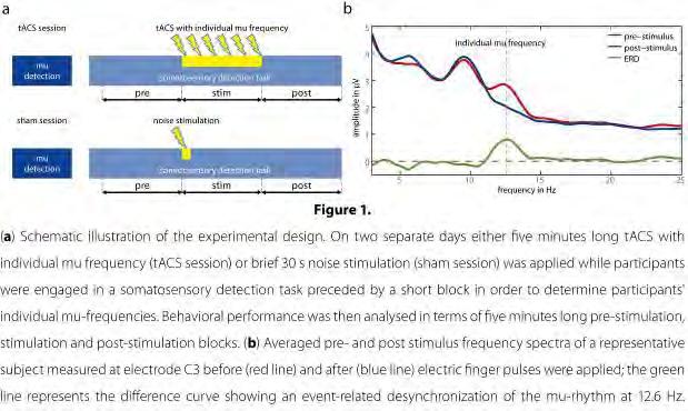

65 65
66 Selected Oral Presentations OP11 Optimized tdcs electrode configurations for five targets determined via an inverse FE modeling approach *S. Rampersad 1,2, D. Stegeman 1,2,3, T. Oostendorp 4,2 1 Radboud University Nijmegen Medical Centre, Neurology, Nijmegen, Netherlands 2 Donders Institute for Brain, Cognition and Behaviour, Nijmegen, Netherlands 3 VU University, Research Institute MOVE, Amsterdam, Netherlands 4 Radboud University Nijmegen Medical Centre, Cognitive Neuroscience, Nijmegen, Netherlands Introduction: Transcranial direct current stimulation (tdcs) has shown potential in improving brain function in both healthy subjects and patients suffering from a wide range of neuropathologies. Unfortunately, the effects are too small and short-lived for tdcs to be used as a clinical therapy. Increasing the effect size of tdcs could possibly be achieved by better targeting the current, both in direction and amplitude. Volume conduction modeling has shown that the areas with the highest electric fields strengths do not, as is often assumed, lie beneath the electrodes [1]. Attempts at optimization have been published for point and ring electrodes [2,3], but not for the square patches used in most labs. In order to find for these electrodes, configurations that do result in maximum stimulation at the target area, we propose an inverse modeling approach. Objectives: We simulate tdcs for ~7000 configurations and determine which of these lead to optimal electric fields, both in strength and direction, at the five most target locations in tdcs research: motor cortex (M1), dorsolateral prefrontal cortex, inferior frontal gyrus, occipital cortex and cerebellum. Methods: A detailed finite element (FE) head model was made by automatic segmentation of MR images aided by manual corrections. The model contains over 4 million elements and 11 tissue types. Special attention was given to the skull, the main barrier for the tdcs current, by including the spongiosa layer and skull holes. Brain anisotropy was derived from DTI measurements. On the skin surface, a grid of 89 points was placed consisting of the standard EEG system and extra points on the cheeks and neck. For each combination of 2 points, we placed 5 x 5 cm electrode patches onto the model, centred on the two points, and simulated 1 ma tdcs. At each target, we placed a cylindrical volume in the brain and selected the configurations leading to highest mean field strength or optimal direction in the target volume. Results: For all targets, the optimized configurations did not include the commonly used configurations. Often, highest field strengths were found in configurations that are near-perpendicular to the standard configurations. Optimization based on either strength or direction of the field lead to completely different configurations. Conclusion: The optimized configurations found in this study suggest that improved results of tdcs can be expected. We found different optimized configurations by looking at either strength or direction of the field. Comparing these optimized configurations experimentally will not only verify our modeling approach, but also provide valuable information on the mechanisms behind tdcs. This approach can be used to optimize stimulation for any target location. References 1. Datta et al. Brain Stimul. 2009; 2(4): Im et al. Phys Med Biol. 2008; 53(11):N Dmochowski et al. J Neural Eng. 2011; 8(4):

67 Figure 1: Setup of this study. Figure 2: Example of results for M1 stimulation. All electrode locations are condensed into one direction (as if viewed from the top of the head). The red line represents the standard configuration for M1 stimulation. The green lines represent configurations that result in a mean electric field strength in the target volume higher than 90% of the maximum found in all configurations. 67
68 Selected Oral Presentations OP12 Effects of transcranial static magnetic field stimulation (tsms) over the human visual cortex: Behavioural and electrophysiological effects *J. J. Gonzalez-Rosa 1, V. Soto-Leon 2, G. Foffani 2, B. Strange 1, A. Oliviero 2 1 Centre of Biomedical Technology (CTB) - Technical University of Madrid (UPM), Laboratory for Clinical Neuroscience, Madrid, Spain 2 Hospital Nacional de Parapléjicos, FENNSI Group, Toledo, Spain Question: We previously reported that continuous application of Transcranial Static Magnetic Field Stimulation (tsms) over the motor cortex induces a reduction of cortical excitability lasting several minutes after the end of tsms. In the present study, we further explore static magnetic field effects on EEG oscillations in the visual cortex and during visual attentional performance in healthy humans. We specifically test a hypothesis that our previous tsms effects in motor cortex could be related to an increase of alpha band activity, and therefore, associated with an inhibitory effect. Methods: Thus, we investigated the effects of tsms (real or sham) placed over visual cortex in two studies: a first experiment, in which tsms was simultaneous applied during ten minutes eyes-open resting EEG recording; a second experiment, in which visual attention was measured in the context of tsms during performance of a visual search paradigm. In both experiments, EEG and behavioural measurements were made during, and 10 minutes following, tsms application. Results: As predicted, the application of real, but not sham, tsms over the visual cortex resulted in a significant increase in alpha band power. In the visual search task, real and sham tsms groups displayed a similar behavioural profile, with reaction times (RTs) increasing with increasing task demands. Critically, however, a significant slowing of RTs emerged on trials with the highest difficulty levels during real in comparison to sham tsms. Conclusions: Our results therefore suggest that tsms has a profound influence on oscillatory alpha activity, which we suggest reflects a modulation of visual cortical excitability. Secondly, our behavioural results indicate that tsms over the occiput alters visual search performance, slowing responses to stimulus detection. Importantly, this was not a general task impairment, but a tsms-induced slowing of visual search specific to high attentional load levels. We speculate that this slowing is secondary to a decrease in underlying alpha oscillations. Further studies using tsms are required to extend the knowledge of the functional significance of brain oscillations changes induced by the application of small magnets over the scalp. 68
69 Poster Session I Basic Physiology P 1 Repetition suppression in sensorimotor system to adapt cortex to stimuli O. Löfberg 1, *P. Julkunen 1, P. Tiihonen 1, A. Pääkkönen 1, J. Karhu 2 1 Kuopio University Hospital, Kuopio, Finland 2 Nexstim Oy, Helsinki, Finland Introduction: Repetition suppression (RS) [1] of auditory evoked potentials (AEPs) is well known. As the sound stimulus is repeated, AEPs (N100 responses) decrease in amplitude and habituate [2]. Recently, RS was demonstrated during mental imaging of movement [3] and repetition of hand gestures [4]. These studies suggest that RS could have a role in motor control, while lacking evidence of causal relation between neuroimaging data and cortical motor output. This can be studied using transcranial magnetic stimulation (TMS). We studied whether the output of central motor network is controlled by RS with the hypothesis that RS could be a part of motor control mechanisms. Methods: Six right-handed healthy subjects (5 male) aged 22 to 58 years participated in the study. We measured RS of the cortical N100 responses with electroencephalography (EEG) [5]. We utilized a standard protocol in studying auditory habituation. Tones were delivered to the subject s right ear at 60 db above hearing threshold. The paradigm comprised of 160 tones in 40 trains, 4 tones within a train. The inter-train interval (ITI) was 20s while the ISI between the tones within a train was 1s [6]. Subsequently, the primary motor cortex area was comprehensively mapped using navigated TMS to locate the optimal cortical representation area of right first dorsal interosseous muscle. The resting motor threshold (rmt) was determined for each subject. Subsequently, we conducted a stimulation protocol with TMS at 120% of the rmt similar to that used in the auditory stimulation. During this, EEG and EMG responses were recorded. Results: RS was evident in the cortical N100 response to auditory stimuli (31-37% decrease in amplitude) as well as to TMS (43-58% decrease in amplitude). More importantly, we also observed RS in the MEPs. The MEPs to the 2nd, 3rd and 4th stimulus in the train were 42-46% lower in amplitude than that to the 1st stimulus (Figure). The effect was similar in all subjects. In a control measurement with peripheral electric stimulation of ulnar nerve, we found no such effect. Conclusions: Our finding of RS in the central motor network is crucial for understanding the general control mechanisms of the motor cortex. However, the mechanism cannot be yet explained unequivocally. It appears that like more complex sensory networks, basic motor network in M1 is able to habituate to repeated stimuli. The recorded suppression of MEPs and TMS-EEG activity resembles the auditory habituation, which suggests that they all share same neuronal mechanisms of suppression. Hypothetically, the suprathreshold TMS may modulate the release of synaptic transmitters of the motor cortex and hence affect cortical excitability. One possible explanation for RS in a motor network is a servo-like mechanism by negative feedback from S1 after sensory feedback from movement [7]. We suggest that RS may be a general servomechanism of the cortex not limited to perception and control of the cortical input. References: [1] Grill-Spektor et al, 2006; [2] Fruhstorfer, 1971; [3] Hohlefeld et al, 2011; [4] Hamilton and Grafton, 2009; [5] Näätänen and Picton, 1987; [6] Furubayashi et al, 2000; [7] Catani et al,
70 Figure: Mean response amplitudes during the RS with respect to the first response of each stimulus train: during auditory stimulation observed in cortical N100 amplitude, during TMS observed in cortical N100 amplitude and during TMS observed in MEPs of the contralateral hand. The effect size and p-value has been indicated with post-hoc p-values. 70
71 Poster Session I Basic Physiology P 2 Modeling the active neurodynamic behavior of rtms in the prefrontal cortex of a realistic human head *N. De Geeter 1, G. Crevecoeur 1, L. Dupré 1 1 Ghent University, Department Electrical Energy, Systems and Automation, Ghent, Belgium Question: Transcranial magnetic stimulation (TMS) is an established tool for non-invasive brain stimulation. It acts via a time dependent magnetic field, generated by an external coil, inducing an electric field in the brain, which can interact with the neural system. Repetitive TMS (rtms) to the left dorsolateral prefrontal cortex (DLPFC) is clinically used for the treatment of medication-resistant depression. Although this technique is widely used, the underlying neurophysiological mechanism remains unclear. Moreover, optimal parameters such as frequency of stimulation, intensity and duration are still unknown. Methods: Therefore, we modulate the rtms response in the prefrontal cortex of a realistic human brain. This head model is constructed from T1-weighted magnetic resonance images (MRI) and segmented into scalp, skull, cerebrospinal fluid, grey and white matter. The anisotropic material properties are obtained from the 4-Cole-Cole model [1]. To track the realistic oriented pathways of nerve fibers located in the stimulated target, namely the left DLPFC, we applied MR tractography [2] based on diffusion tensor images (DTI). These fibers are considered as one myelinated nerve bundle (Fig1). The 20mm figure-of-eight TMS coil (MagStim, UK) is positioned above the left DLPFC, perpendicular to the skull. We modeled the neurophysiological response of a sinusoidal stimulation (pulse width 450 μs) at different intensities on the Rapid² stimulator. The induced electric field is calculated using the recently developed independent impedance method [3]. Studies have shown that nerves are primarily activated by the gradient of the component of the electric field along the nerves, the so-called activating function [4]. This affects the membrane potential of the nerve bundle and can cause a generation and propagation of action potentials (AP). To describe this neurodynamic behavior along the nerve bundle we use the active cable equation [5], discretized by the Crank-Nicholson method. Results: When applying one biphasic stimulation pulse at an intensity just below activation threshold, no AP is generated (Fig2 a). Repetitive stimulation at the same intensity generated a single spike (Fig2 b). As the intensity of rtms increases, a second AP is initiated (Fig2 c). For an even higher intensity of the sustained stimulation a train of spikes at a fixed frequency is rendered. Remark how this spiking frequency first increases with increasing stimulation intensity, but then decreases again (Fig2 d,e,f). Conclusions: These results point out the advantage of repetitive stimulation in comparison to a single pulse stimulation. More important, they indicate the existence of an optimal stimulation intensity for a specific TMS set-up and target, whereby a train of spikes at a maximum spiking frequency is generated. This study highlights the importance of numerical modeling in TMS, either in the determination of the underlying neurophysiological mechanism or the effect of different stimulation parameters. In future research, we aim to simulate the propagation of TMS effects in depression through anatomical connections to deeper limbic regions and to optimize this non-invasive brain stimulation technique. References [1] Cole KS and Cole RH (1941) Chem Phys, 9: [2] Leemans A et al (2009) Reson Med, [3] De Geeter N et al (2012) Phys Med Biol, 57: [4] Roth BJ and Basser PJ (1990) IEEE Trans Biomed Eng, 37: [5] Wesselink WA et al (1999) Med Biol Eng Comp, 37:
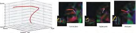
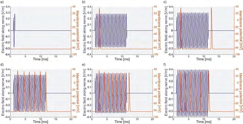
72 72
73 Poster Session I Basic Physiology P 3 Dose-dependence of Changes in Cortical Protein Expression Induced by Theta Burst Stimulation in the Rat *L. J. Volz 1,2,3, A. Mix 3, A. Benali 3, K. Funke 3 1 Max-Planck Institute for neurological Research, Neuromodulation & Neurorehabilitation, Köln, Germany 2 University Hospital, Neurology, Cologne, Germany 3 Medical Faculty, Ruhr-University Bochum, Neurophysiology, Bochum, Germany Introduction: Theta Burst stimulation (TBS) applied via transcranial magnetic stimulation (TMS) is an effective tool to modulate human neocortical excitability [1]. Repeated application of the same TBS protocol or variation of the number of stimuli has been shown to alter the strength and direction of changes in cortical excitability compared to the standard TBS protocols [2,3]. TBS applied to rat cortex affected the expression of activity-dependent proteins related to the cortical inhibitory systems, suggesting altered cortical inhibition contributing to the TBS after-effects [4,5]. Objectives: Our aim was to investigate the impact of varying numbers of TBS-stimuli applied as multiple blocks of intermittend TBS (itbs) or continuous TBS (ctbs) on cortical protein expression in the rat, to further our insights in physiological mechanisms underlying TBS-induced changes in cortical excitability. Materials & methods: Nine groups of anesthetized rats (male Sprague Dawley, g) received TMS. Eight groups received a different number of itbs/ctbs-blocks summing up to either 600, 1200, 1800, or 2400 stimuli, applied with breaks of 15 min between blocks of 600 stimuli. Sham stimulation (coil more distant to the head) was applied to a control group. Rats were sacrificed for immunohistochemical analysis or western blotting focussing on frontal, motor, sensory and visual cortex. Results: In general, quite similar effects for itbs and ctbs were observed. The expression of the 65-kD isoform of glutamic acid decarboxylase (GAD65) increased, while that of the 67-kD isoform (GAD67) and that of the calcium-binding proteins Parvalbumin (PV) and Calbindin (CB) progressively decreased. Also the expression of the immediate early gene c-fos decreased with an increasing number of blocks. A more detailed analysis, however, revealed that the sensitivity of distinct proteins to stimulation varied with the number of stimuli and type of stimulation. Conclusion: Our findings show that both itbs and ctbs affect the activity of inhibitory interneurons and indicate that repeated TBS elicits no simple accumulative dose-dependent effect for all activity-markers but distinct profiles with threshold characteristics and a waxing-and-waning effect especially for two markers of inhibitory activity, CB and GAD67. Thus, our data do not suggest fundamentally different modulation of the cortical inhibitory systems by itbs and ctbs. Subtle differences in stimulation after-effects on different neuronal subsystems might contribute to the opposite impact on cortical excitability following itbs and ctbs and the switching sign of effect with repeated stimulation. References [1] Huang YZ et al. (2005) Theta burst stimulation of the human motor cortex. Neuron 45: [2] Gentner R et al. (2008) Depression of human corticospinal excitability induced by magnetic theta-burst stimulation: evidence of rapid polarity-reversing metaplasticity. Cereb Cortex 18: [3] Gamboa OL et al. (2010) Simply longer is not better: reversal of theta burst after-effect with prolonged stimulation. Exp Brain Res 204: [4] Benali A et al. (2011) Theta-Burst Transcranial Magnetic Stimulation alters cortical inhibition. J Neurosci 31(4): [5] Funke K, Benali A (2011) Modulation of cortical inhibition by rtms - findings obtained from animal models. J Physiol :
74 Poster Session I Basic Physiology P 4 Cerebral Blood Flow Modulation by Transcutaneous Cranial Electrical Stimulation with Limoge s Current. *D. Gense de Beaufort 1, M. Sesay 1, L. Stinus 2, R. Thiebaut 3,4, M. Auriacombe 4, V. Dousset 1,5 1 Hopital Pellegrin, Neuroradiology, Bordeaux, France 2 CNRS UMR-5541, Bordeaux, France 3 INSERM U897, Bordeaux, France 4 EA4139 INSERM IFR99, University Bordeaux 2, Bordeaux, France 5 University Bordeaux 2, Bordeaux, France Objectives: TCES delivers a high-frequency (166 khz) pulsed biphasic balanced current with a pulse repetition frequency of 100 Hz with 40% duty cycle through a negative electrode and two positive electrodes over the skull. TCES has a proven ability to potentiate anesthesia and analgesia, although the physiological mechanisms of this effect remain unclear. We hypothesized that the mechanism is a modulation of CBF in the central endogenous opioid system. This study aimed at determining the effects of TCES on CBF to elucidate its physiological mechanism. Methods: Thirty-six healthy volunteers were randomly assigned to active or placebo TCES, and all assessments were double blind. TCES was performed using the Anesthelec device. In the stimulated group, an active cable was used, and in the control group (sham), the cable was inactive. CBF was measured by XeCT before and after two hours of TCES. Results: Globally, CBF was unchanged by TCES. However, locally, TCES induced a significant CBF decrease in the brainstem and thalamus, which are structures involved in pain and anxiety (TCES and control CBF decrease were 18.5 and 11.9 ml/100 g brain tissue/min, respectively). Conclusion: TCES can modulate local CBF but it has no effect on overall CBF. [Clinical Trials.gov number: NCT ] 74
75 Poster Session I Basic Physiology P 5 Motor cortical excitability in psychiatric disorders probed by transcranial magnetic stimulation: a systematic review *T. Bunse 1, W. Strube 1, T. Wobrock 2, F. Padberg 1, P. Falkai 1, A. Hasan 1 1 Ludwig Maximilian Universität Munich, Department of Psychiatry and Psychotherapy, Munich, Germany 2 Darmstadt-Dieburg Clinics, Centre of Mental Health, Darmstadt, Germany Introduction: Since many years, transcranial magnetic stimulation (TMS) has been used to probe cortical physiology in awake humans. In particular, single- and paired-pulse TMS-protocols applied to the human motor cortex allowed the exploration of various intracortical inhibitory and facilitatory networks. In psychiatry, TMS offers the possibility to access motor-cortical excitability and to link the findings to possible neurotransmitter alterations with regard to the investigated disorder. However, little is known about general patterns of cortical excitability across psychiatric disorders. Objectives: We aimed to review the evidence for impaired cortical excitability in psychiatric disorders using TMS. Methods: A systematically literature research for this review was conducted via the internet database PubMed ( ) using predefined search-algorithms. This search offered 684 publications in total. After screening of the abstracts and careful review of the content by two independent researchers with regard to the objectives of this review, a number of 82 publications was considered to be suitable. Results: Most studies (n=20) investigated the effects of TMS in patients with schizophrenia. Remarkably, all major psychiatric disorders were investigated by TMS: dementia (n=19), attention deficit hyperactivity disorder (n=13), Tourette syndrome (n=6), cocaine dependence (n=5), depression (n=4), alcohol dependence (n=3), cannabis-dependence (n=3), nicotine dependence (n=2), obsessive compulsive disorder (n=2), borderline personality disorder (n=1), bipolar disorder (n=1), posttraumatic stress disorder (n=1), altered personality traits (n=1) and subjects at risk to develop psychosis (n=1). In general, psychiatric illness can be linked to reduced cortical inhibition and in parts to enhanced cortical facilitation, resulting in a motor-cortex hyperexcitability. Detailed descriptions and exceptions will be presented and discussed at the conference. Conclusion: Our results show that motor-cortex TMS is currently not suitable as a general diagnostic tool for psychiatric disorders. The disease state, different phenotypes of the same diseases and the influence of neuroactive medication can be discussed as major confounding factors. While it seems to be possible to hypothetically transmit results from motor-cortex TMS to other cortical and subcortical structures, further investigations are needed to understand the meaning of alterations in motor-cortex excitability in psychiatric disorders. 75
76 Poster Session I Basic Physiology P 6 tdcs-induced effects on paired-pulse inhibition in the primary somatosensory cortex *M. Hoff 1, E. Kaminski 1, C. Gundlach 1, B. Sehm 1, A. Villringer 1, P. Ragert 1 1 Max Planck Institute for human cognitive and brain sciences Leipzig, Neurology, Leipzig, Germany Introduction: Transcranial direct current stimulation (tdcs) is a non-invasive brain stimulation technique used to modulate cortical excitability. In the motor domain, there is consensus that anodal tdcs increases cortical excitability, whereas cathodal tdcs decreases it. On the other hand, inconsistent results of tdcsinduced effects in the sensory domain have been reported. Objective: The aim of the present study was to investigate (A) changes in cortical excitability within primary somatosensory cortex (S1) by means of single-pulse somatosensory evoked potentials (SEPs) and (B) intracortical inhibition by means of paired-pulse SEPs when tdcs was applied over left SM1. Materials & Methods: 10 minutes of anodal, cathodal or sham tdcs was applied over the left SM1 using saline-soaked sponge electrodes (35 cm^2 electrodes, 1mA, current density ma/cm^2). Before, immediately after as well as 10 minutes after termination of tdcs, single- and paired-pulse SEP recordings were performed. We hypothesized that tdcs will induce polarity specific changes in cortical excitability within left S1. Furthermore, we reasoned that anodal tdcs will reduce paired-pulse inhibition within left S1 while cathodal tdcs will result in an augmentation of inhibition relative to sham stimulation. Results: 10 minutes of anodal and cathodal tdcs over SM1 did not result in any significant excitability changes within left S1. However, anodal tdcs resulted in a reduction of paired-pulse inhibition within left S1 10 minutes after termination of stimulation. No change in paired-pulse inhibition could be observed after cathodal tdcs. Conclusion: Here we provide novel evidence that anodal tdcs affects inhibitory processing within S1, a finding that might improve our understanding about the underlying neural mechanisms of tdcs on somatosensory processing. 76
77 Poster Session I Basic Physiology P 7 Effects of transcutaneous spinal direct current stimulation (tsdcs) on long-term potentiation (LTP)- like central sensitization of pain sensation *C. Meyer-Frießem 1, *L. Haag 1,2, T. Schmidt-Wilcke 2, W. Magerl 3, P. Zahn 1, M. Tegenthoff 2 1 BG-Universitätskliniken Bergmannsheil Bochum, Klinik für Anästhesie, Intensiv-, Palliativ- und Schmerzmedizin, Bochum, Germany 2 BG-Universitätskliniken Bergmannsheil Bochum, Neurologische Klinik und Poliklinik, Bochum, Germany 3 Medizinische Fakultät Mannheim, Universität Heidelberg, Lehrstuhl für Neurophysiologie, Mannheim, Germany Background: Strong stimulation of nociceptive afferents results in hyperalgesia based on facilitation of spinal nociceptive pathways, which plays a role in pathological pain conditions. This central sensitization can be experimentally induced via nociceptive high frequency stimulation (HFS) at high intensities in animals (nociceptive LTP) and humans (pain-ltp), and has many mechanistic and molecular similarities with LTP described in the hippocampus. Since LTP in the hippocampus can be inhibited by post-synaptic hyperpolarization, altering the resting membrane potential (RMP) of neurons in the spinal cord could also modulate pain sensitivity and experimentally-induced hyperalgesia. Transcranial direct current stimulation over M1 is thought to enhance or reduce motor neuron excitability via an NMDA-dependent mechanism in a polarity-specific manner. When applied over the spine (tsdcs) this technique may have a similar effect on the spinal circuitry and has in fact already been shown to affect motor and nociceptive reflex responses. We therefore postulated that tsdcs could alter the hyperalgesia induced by nociceptive HFS in a polarity-specific manner. Methods: In a double-blind, randomized study 14 healthy right-handed male volunteers each took part in 3 sessions separated by at least 2 weeks. Painful electrical trains of HFS (5 trains of 100 Hz for 1 sec, 9 sec onset interval) were given to the right distal thigh. TsDCS was applied directly following HFS via two 7x5cm saline-soaked sponges over T12 vertebral process and the left shoulder. Anodal, cathodal (2.5 ma, 15 min) and sham (1.5 ma, 45 sec) stimulation polarities referred to the electrode over T12. Sensory perception was tested on both sides using electrical and mechanical stimuli before (baseline) and at 3 time points (0, 30, 60 min) post-tsdcs. Subjects gave pain ratings from 0 (non pain) to 100 (worst pain imaginable), resulting in electrical detection threshold (EDT), mechanical pain threshold (MPT), and electrical and mechanical pain sensitivity (EPS and MPS) scores. Baseline and post-stimulation scores were compared using repeated measures ANOVA and Tukey HSD post-hoc tests to identify time and stimulation effects. Results: HFS induced significant hyperalgesia after not only sham tsdcs but also following anodal and cathodal polarities. MPS and EPS pain ratings were significantly elevated for all three post-stimulationtime points in all sessions (p< ). EDT was significantly reduced at T0 in all sessions (p<0.03) whereas MPT decreased marginally, which never reached significance at any time point in any session. None of the responses differed significantly between tsdcs and sham condition. Discussion: This study confirmed that LTP-like central sensitization induced by HFS is a robust effect and for the first time showed that tsdcs applied immediately following HFS does not influence the resulting hyperalgesia seen within the first hour. Whether sensitized neurons are more resistant to the applied current or whether RMP changes from tsdcs occur in neurons insignificant to the development of hyperalgesia is unknown. To test this first hypothesis we plan to repeat this study, this time applying tsdcs before HFS. 77
78 Poster Session I Basic Physiology P 8 Bringing TMS-based corticomotor mapping into shape: Shape-based TMS mapping reveals motor finger somatotopy in M1-HAND *E. Raffin 1, G. Pellegrino 1, A. Tielscher 1, H. R. Siebner 1 1 DRCMR, Hvidovre, Denmark Background: Movements of the fingers involve overlapping neural representations in the hand region of the primary motor cortex (M1 HAND ). Despite of these overlapping representations, somatotopic gradients of cortical motor representations exist in M1 HAND, but cortical motor somatotopy is difficult to capture with conventional transcranial magnetic stimulation (TMS) mapping techniques. Aim: To develop a novel TMS mapping approach, which takes into account the sulcal shape to map motor finger somatotopy in human M1 HAND. Method: In 9 healthy young subjects, neuronavigated single-pulse TMS was given to the left M1 HAND via a small figure-of-eight coil to map muscle-specific corticomotor representations. We compared three different mapping approaches which differed in terms of target grids and coil orientations: (1) Line-45 O condition: Six targets located on a straight medio-to-lateral line which corresponded to the overall orientation of the central sulcus using a fixed coil orientation that induced a posterior-to-anterior current having a direction of 45 relative to the mid-sagittal line, (2) Shape-45 O condition: Seven targets which followed the bending of the central sulcus with a fixed coil orientation as in the first condition, and (3) Shape-90 O condition: Seven targets which were individually positioned following the bending of the central sulcus as in condition 2 but with the coil orientation adjusted to produce a current direction perpendicular to the central sulcus. We compared cortico-motor representations of the abductor digiti minimi (ADM) and the first dorsal interosseus (FDI) muscle at rest, during isometric contraction of either the FDI or ADM muscles or during co-contraction of both muscles. Based on the amplitudes of the motor evoked potential (MEP) at each target site, we generated muscle excitability profiles, the position of the weighted mean MEP amplitude. Results: In contrast to the other mapping conditions, the shape-90 O condition demonstrated clear somatotopy of the FDI and ADM muscle representations in M1 HAND and yielded state-dependant shifts in the weighted mean positions of the curves when switching from rest to active conditions. Conclusion: Within-hand motor somatotopy in M1 HAND can be readily studied with neuronavigated TMS that follows the sulcal shape and creates a tissue current perpendicular to the central sulcus at all mapping sites 78
79 79
80 Poster Session I Basic Physiology P 9 Appearance of late cortical silent period is dependent on stimulation intensity *E. Kallioniemi 1, L. Säisänen 1, M. Könönen 1, J. Karhu 2, P. Julkunen 1 1 Kuopio University Hospital, Kuopio, Finland 2 Nexstim Oy, Helsinki, Finland Question: Cortical silent period (csp) is a temporary suppression in electromyography (EMG) induced by transcranial magnetic stimulation (TMS) of cortex [1]. The duration of csp is used as a measure of inhibition in the corresponding cortical circuits [2]. Commonly, the csps are induced with a stimulation intensity (SI) of >120% of the resting motor threshold (rmt) while the subject is performing a voluntary contraction of the target muscle [3]. The duration of csp is highly dependent on SI as with higher intensity, the duration of the csp increases [4,5]. The level of muscle contraction has no effect on the duration of the csp [6,7] however, for the detection of the csp a reasonable contraction level is needed. Generally, the csps are observed as an early effect, right after the motor evoked potential. However, in some cases after the csp and recovery of muscle contraction another later csp may occur (Figure 1). Our objective was to characterize and evaluate the late csp as a function of SI.So far, the characterization or the investigation on the origin of late csps have not been performed. Methods: For the study, eight subjects were recruited (3 male, 5 female, age range : years). The stimulation was focused on the left M1 using neuronavigation and EMG was measured from first dorsal interosseous muscle (FDI) and abductor pollicis brevis (APB). The analyzed csps were induced at eight different SIs normalized to the rmt of each subject (60, 70, 80, 90, 100, 110, 120, 130 % of rmt). 10 trials were performed at each SI in a randomized sequence at an inter-trial interval of 12s. During the TMS, subjects were asked to keep a constant contraction of the target muscles against resistance of approximately 1 mv of peak-to-peak EMG. Late csps were identified with a minimum duration of 10 ms and maximum difference of ~50 ms between the offset of csp and onset of late csp. The late csps were analyzed by taking the mean duration at each SI for all the individual subjects. To confirm the presence of late csps at different SIs, we used ANOVA with normalized SI as fixed factor and subject as random factor. Post-hoc tests for the SI effect were analyzed and p-values were adjusted with least significant difference (LSD). Results: We found that the time between the offset of the csp and the onset of the late csp was ~40 ms. ANOVA confirmed that late csps were present in most subjects. The SI had a significant effect on the appearance of the late csp in EMG recorded both from FDI (F=4.504, p=001, Figure 2) and from APB (F=3.882, p=002). The post-hoc test revealed that SI had a significant effect on the appearance of late csps at certain SIs (Figure 2). Especially, TMS with SIs close to rmt was able to induce a late csp, while at higher SIs the late csps disappeared. Conclusion: We have confirmed the appearance of late csp at SIs around rmt. The origin of the late csp is not yet known and requires further investigation. References : [1] Fuhr et al [2] Orth et al [3] Kessler et al [4] Hess et al [5] Roick et al [6] Rossini, 1990 [7] Uozumi et al

81 Figure 1: Demonstration of csp and late csp at subthreshold SI. Figure 2: Group mean of the late csp duration as a function of normalized SI. The grey area surrounding the group mean represents standard error. SIs at which the late csps were confirmed statistically to appear are indicated by red dots. The level of significance based on the post-hoc test is also indicated above the red dots. 81
82 Poster Session I Basic Physiology P 10 Acute state-dependent changes in corticomotor threshold in the absence of overt motor activity *A. Karabanov 1, E. Raffin 1, H. Siebner 1 1 DRCMR, Hvidovre, Denmark Background: The cortical motor threshold (CMT) is often used to individually adjust the intensity of transcranial magnetic stimulation (TMS). It reflects the integrated excitability of the corticomotor projection, including the excitability at the spinal level. The CMT reflects axonal and synaptic excitability as CMT can be modified by drugs that voltage-gated sodium channel blockers or non-nmda glutamatergic drugs. Aims and hypothesis: The CMT is not a static measure but is subject to state-dependent fluctuations. To test this hypothesis we used an adaptive stair-case procedure to track acute changes in CMT associated with motor imagery (MI) or shifts in visuospatial attention (VA). Method: Eight healthy subjects were asked to imagine a continuous thumb-index movement with the left or right hand (MI-left and MI-right), to shift their visuospatial attention to the left or right FDI (VA-left and VAright) or to fixate a cross in the central visual field. Using a fast (<15 pulses) threshold-hunting procedure (Borckardt, 2006), we assessed task-dependent CMT changes in the left primary motor hand area (M1) during each task. CMT was defined as the stimulus intensity, at which a single TMS pulse elicited a MEP of 50 µv in the relaxed right first dorsal interosseus muscle (FDI) with a probability of 0.5. The CMT measurements during the VA and MI task were normalized to the control condition. All participants underwent two experimental sessions on separate days, in which the TMS pulse either induced a posterior-to-anterior (PA) or anterior-to-posterior (AP) current in M1. Results: The number of pulses needed to determine rmt did not significantly differ across conditions (average: 13.5). Using the normalized CMT values as dependent variable, we computed a repeated measures ANOVA with the factors Current (AP/PA), Task (MI/VA) and Site (Left/Right). We found a significant Task-by-Site interaction (p = 0.004). Post hoc testing confirmed that CMT during MI-right was significantly lower than during MI-left or during VA-right. This was the case for AP and PA current direction. Discussion: The resting CMT is subject to rapid state-dependent changes in the absence of overt motor activity. We show that these acute CMT changes can be captured with a threshold-hunting procedure. The CMT tracking approach might be particularly useful to assess the temporal dynamics of corticomotor excitability changes induced by interventional brain stimulation protocols. 82
83 Poster Session I Basic Physiology P 11 Cortical Inhibition within Motor and Frontal Regions in Alcohol Dependence: A TMS-EEG study. *J. Naim-Feil 1,2, J. L. Bradshaw 3, N. Rogasch 1, Z. J. Daskalakis 1,4, D. Sheppard 5, D. I. Lubman 6, P. B. Fitzgerald 1 1 Monash University, Monash Alfred Psychiatry Research Centre, Melbourne, Australia 2 Weizmann Institute of Science, Physics of Complex Systems, Rehovot, Australia 3 Monash University, Experimental Neuropsychology Research Unit, Melbourne, Australia 4 Centre for Addiction and Mental Health, University of Toronto, Toronto, Canada 5 Monash Injury Research Institute, Melbourne, Australia 6 Turning Point Alcohol and Drug Centre, Melbourne, Australia Background: Preclinical studies suggest that alterations within the frontal cortex play a critical role in the neurophysiology of alcohol dependence. The combination of transcranial magnetic stimulation and electroencephalography (TMS-EEG) allows a direct assessment of cortical excitability and inhibition within the frontal cortex in human subjects. We report the first application of TMS-EEG to measure these indices within the frontal cortices of patients with alcohol dependence. Methods: Cortical inhibition was assessed in 12 patients with alcohol dependence and 14 healthy controls through single and paired-pulse transcranial magnetic stimulations (TMS) paradigms delivered to both the frontal and motor cortices. Long interval cortical inhibition (LICI) was used to index inhibition in the frontal cortex. Short intracortical inhibition (SICI) and cortical silent period (CSP) was used to index inhibition, while intracortical facilitation (ICF) measured facilitation, in the motor cortex. Cortical excitability was indexed by the resting motor threshold (RMT) and active motor threshold (AMT). Results: The alcohol dependent group demonstrated deficits in LICI across both the left and right dorsolateral prefrontal cortex relative to healthy controls. The alcohol dependent group also exhibited reduced RMT and AMT. In terms of motor cortex inhibition, there were no significant differences in SICI, ICF or CSP, although increased intra-trial-variability in SICI was observed in the alcohol dependent group. Conclusions: The current study provides the first direct evidence of reduced cortical inhibition that is specific to the frontal cortex of patients with alcohol dependence. Our study also revealed evidence of altered cortical excitability in the motor cortex of patients with alcohol dependence; however, the utility of using the motor cortex to index cortical alterations related to alcohol dependence remains unclear. Although these findings are preliminary, they provide critical neurophysiological evidence of disrupted cortical excitability within the frontal cortex of alcohol dependent patients. 83
84 Poster Session I Basic Physiology P 12 Transcranial alternating current stimulation enhances endogenous alpha for 30 minutes only for moderate alpha levels *T. Neuling 1, S. Rach 1, C. Herrmann 1 1 Carl von Ossietzky Universität Oldenburg, Department for Experimental Psychology, Oldenburg, Germany Introduction: Transcranial alternating current stimulation (tacs) has been proven to successfully influence endogenous brain oscillations in a frequency specific manner (Zaehle, 2010). Besides effects on the electrophysiology, tacs is also able to influence perception and behavior (Neuling, 2012a). After tacs, the endogenous power of the stimulated frequency is enhanced when compared a pre-stimulation period, however, little is known about the duration of this effect and which parameters contribute to this effect. Objectives: Our goal was to discern how long endogenous brain oscillations are enhanced post tacs and how pre-stimulation power of the oscillation modulates this effect. Materials & methods: Two experiments were conducted whereby subjects had their eyes either open or closed. Experimental procedure is demonstrated in Fig. 1. Participants were stimulated with their individual alpha frequency (IAF) for 20 minutes or received sham stimulation. Stimulation electrode positions were chosen in order to effect occipital alpha (Neuling, 2012b). Five minutes before (pre) and 30 minutes after stimulation (post), the electroencephalogram (EEG) was recorded. During the experiment, subjects performed a simple auditory detection task to ensure vigilance (Fig. 1C). Results: In the eyes closed experiment, no effects on the oscillatory power could be demonstrated (Fig. 2AI). After stimulation, both in the stimulation and in the sham group, IAF power was not significantly different than pre-stimulation levels (Fig. 2BI). Likewise, no significant effects in the entire post period could be revealed (Fig. 2CI). Contrary to the results in the eyed closed experiment, an effect on the oscillatory power, limited to the alpha range, was found in the eyes open experiment (Fig. 2AII). The power increase from the pre- to the post stimulation period was significant in the stimulated group, but not in the sham group (Fig. 2BII). This enhancement effect was significant over the entire 30 minute post-stimulation recording (Fig 2CII). Conclusion: If the endogenous alpha oscillations are high, stimulation with tacs fails to increase alpha power due to ceiling effects. The neuronal network activity is unaffected by external oscillations, at least in terms of oscillatory power. Endogenous oscillations exhibiting lower power are more prone to effects of tacs. Here, tacs is able to enhance endogenous oscillations over a long duration. It might be speculated that the enhancement effect would last even longer than 30 minutes. The results of our study illustrate the feasibility of tacs as a tool for non-invasive brain stimulation and demonstrate the potential for therapeutic application to balance altered brain oscillations. References: Zaehle, T., Rach, S. & Herrmann, C.S. (2010), Transcranial Alternating Current Stimulation Enhances Individual Alpha Activity in Human EEG, PLoS ONE 5(11): e doi: /journal.pone Neuling, T., Rach, S., Wagner, S., Wolters, C.H. & Herrmann, C.S. (2012a), Good Vibrations: Oscillatory Phase Shapes Perception, Neuroimage 63(2), Neuling, T., Wagner, S., Wolters, C.H., Zaehle, T. & Herrmann CS (2012b), Finite-element model predicts current density distribution for clinical applications of tdcs and tacs, Front. Psychiatry 3(83). doi: /fpsyt
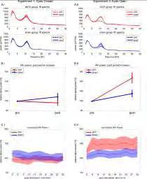
85 85
86 Poster Session I Basic Physiology P 13 Intersession reliability of cortical motor maps with navigated transcranial magnetic stimulation *M. Roennefarth 1, S. Schmidt 1, R. Bathe-Peters 1, R. Fleischmann 1, A. Mante 1, A. Althoff 1, S. A. Brandt 1 1 Charité-Universitätsmedizin Berlin, Department of Neurology, Berlin, Germany Introduction: Navigated transcranial magnetic stimulation (ntms) is a non-invasive highly effective tool for mapping individual muscle representations in the motor cortex (Julkunen et al. 2009, Picht et al. 2011). Optical navigation and model based estimates of intracortical target sites and stimulation strength suggest that ntms should provide spatially more precise cartography than using external landmarks for defining a motor hot-spot. The remaining outstanding challenges are a.) a high inter- and intra-subject variability of motor evoked potentials (MEP) (Schmidt et al.2009, Brasil-Neto et al. 1992), b.) the extent to which anatomical mapping relative to the underlying gyrification is preferable and c.) hemispheric functional asymmetries possibly reflecting functional lateralization. These factors are particularly relevant in a clinical context, where typically cartography will be used with single pulses per stimulation location. Further, the definition of a representation area is strongly confounded by the variability of fringe neurons or overlapping cortical representations (Schieber and Hibbard 1993). Objectives: The aim was to study the extent to which single pulse cartography can assure reproducible results. In the present analysis we focus on the concept of the hot-spot, i.e. point of maximum MEP and Center of Gravity (CoG). Materials and Methods: 15 subjects were examined in two sessions by two different researchers. Stimulation was performed using a computer based navigated TMS system with a figure-of-eight-coil (eximia, Nextim Ltd., Helsinki, Finland). Mapping of the FDI over both hemispheres was performed with perpendicular (fine) and changing (random) coil orientation with an average of 79 (+/-35.91) stimuli at 110% resting motor threshold. Margins were defined as scalp locations where a stimulus resulted in an MEP < 50µV. To assess intersession variability we calculated the intraclass correlation coefficient (ICC), which is considered to reflect high reliability when higher than 75% (Mortifee et al. 1994). Results: Stimulus location for the FDI- hot-spot in the dominant hemisphere showed a high intersession correlation (ICC: 81-87%). In the non-dominant hemisphere the ICC was lower (45-73%). There was no significant difference in ICC using intracortical instead of scalp stimulus location. Weighted hot-spots as expressed by the CoG showed a very high intersession reliability (ICC: 84-88%), the side of maximum MEP a rather poor intersession reliability (ICC: 9-72%). Perpendicular mapping was associated with significant higher test-retest correlation than random mapping (ICC for random mapping 22-38%). Conclusion: Stimulus locations ( hot-spots ) in the dominant hemisphere are more precise than in the nondominant hemisphere. Intracortical locations are not superior to scalp location for hot-spot -measurements. Weighted hot-spots are more precise than maximal MEPs for hot-spot - definition and inter-rater reproducible. Perpendicular mapping is clearly spatially more accurate than random mapping. References: Julkunen P et al., NeuroImage 2009 Picht et al., Neurosurgery 2011 Schmidt et al., Clin Neurophysiol 2009 Brasil-Neto et al., EEG Clin Neurophysiol 1992 Schieber and Hibbard, Science 1993Mortifee et al, EEG Clin Neurophysiol
87 Poster Session I Basic Physiology P 14 A detailed evaluation of surround inhibition in human motor cortex using transcranial magnetic stimulation *N. Thirugnanasambandam 1, R. Khera 1, H. Wang 1, S. Kukke 1, M. Hallett 1 1 NIH, Bethesda, United States Surround inhibition (SI) is a feature of motor control for which the physiology is still unknown (Beck and Hallett, 2011). This phenomenon is impaired in patients with focal hand dystonia (Sohn and Hallett, 2004a, Beck et al., 2008) and hence it becomes relevant to study it more in detail. Several studies have demonstrated SI in the human motor system using transcranial magnetic stimulation (TMS) (Beck et al., 2008, Sohn and Hallett, 2004b, Kassavetis et al., 2012). In all these studies SI was measured by applying a single suprathreshold TMS pulse during the movement initiation phase. The figure of eight coil was positioned such that it induced current along the postero-anterior direction in the brain. We were interested to study SI not just at a single intensity, but at intensities ranging from sub-threshold to supra-threshold levels. Also, we wanted to explore if SI could be elicited when the motor cortex was stimulated along other directions (antero-posterior, latero-medial). 10 healthy volunteers participated in the study. Their task was to perform a brief isometric index finger flexion on hearing a tone. EMG was recorded from the first dorsal interosseous (active muscle) and the abductor pollicis brevis (surround muscle). Single pulse TMS was applied at intensities ranging from 20% to 100% of maximum stimulator output at 5% intervals either before the tone (rest) or after the tone ~75 ms before expected onset of muscle activity (SI). The MEP amplitudes were then plotted against stimulation intensities to obtain the MEP recruitment curves for the rest and SI conditions separately. The recruitment curves were plotted with the coil held in three directions (P-A, A-P, L-M) for every subject. We found that SI can be elicited when the coil was positioned in the P-A direction and only within a certain range of supra-threshold intensities. Maximum SI was seen around 150%MT. Stimulating the motor cortex along the A-P direction did not elicit SI, rather showed facilitation that peaked at around 120%MT. We can therefore conclude that SI can be elicited in those neuronal groups that can be stimulated best along the PA direction. References: Beck S, Hallett M. Surround inhibition in the motor system. Experimental brain research Experimentelle Hirnforschung Experimentation cerebrale. 2011; 210(2): Sohn YH, Hallett M. Disturbed surround inhibition in focal hand dystonia. Annals of neurology. 2004a; 56(4): Beck S, Richardson SP, Shamim EA, Dang N, Schubert M, Hallett M. Short intracortical and surround inhibition are selectively reduced during movement initiation in focal hand dystonia. The Journal of neuroscience : the official journal of the Society for Neuroscience. 2008; 28(41): Sohn YH, Hallett M. Surround inhibition in human motor system. Experimental brain research Experimentelle Hirnforschung Experimentation cerebrale. 2004b; 158(4): Kassavetis P, Saifee T, Sadnicka A, Pareés I, Kojovic M, Rothwell J, et al. Adaptation of surround inhibition in the human motor system. Experimental Brain Research. 2012; 222(3):
88 Poster Session I Basic Physiology P 15 Delta waves increase after cortical plasticity induction during wakefulness. *G. Assenza 1, G. Pellegrino 1, M. Tombini 1, F. Tecchio 2, L. Tomasevic 2, G. Di Pino 1, V. Di Lazzaro 1 1 Campus Biomedico University, Clinical Neurology, Rome, Italy 2 CNR, ISTC LET'S, Rome, Italy Objective: Delta waves are the most prominent electroencephalographic (EEG) feature of non-rapid eye movement sleep with cortical origin. Several human studies suggested they are sensor of synaptic weight and possible effectors of sleep-dependent synaptic plasticity. During wakefulness delta waves are almost absent in physiological conditions, but they come out when a subcortical brain lesion occurs. For these reasons delta waves during wakefulness are considered as a lesional sign but no definitive data about its functional significance were provided. Our aim was to verify whether delta waves can be a sign of plastic cortical modifications. Materials and methods: 11 young healthy subjects (28±3) were enrolled in the study. Intermittent theta burst stimulation (itbs) was administered on left primary motor cortex to induce enduring cortical motor plasticity. High density scalp EEG (32 channels) was recorded all along the experiment. PSD for standard frequency bands (delta Hz, theta Hz, alpha 8-12 Hz, beta 13-22Hz) was calculated in each longitudinal bipolar derivation. 5-minutes resting opened-eyes EEG was analyzed pre-itbs (T0) and immediately after itbs (T1), after 15 (T2) and 30 (T3) minutes. MEP amplitude and visual analogic scales about sleepiness, alertness, and the Stanford Sleepiness Scale were analyzed at the same time-points to explore motor plasticity induction and alertness status. Results: itbs induced a 9%-increase of MEP amplitude which remained stable until 30 minutes after stimulation (T1vsT0 p=0.005; T2vsT1 p=0.560; T3vsT2 p=0.158). A selective increase (+87.5%) in delta band PSD was evident at T1 vs T0, remaining stable all along the experiment (T1vsT0 p=0.019, T2vsT1 p=0.724, T3vsT2 p=1). Significant changes were bilaterally present only in fronto-central derivations (p<0.05). No itbs changes on behavioural scales were found. Discussion: Our experiment firstly demonstrated a large and enduring increase in EEG delta activity in cortical areas undergoing plasticity induction during wakefulness. Present results may open new scenarios in interpreting scalp EEG slow waves after brain lesions not only as sign of lesion but also as sign of neuronal plastic rearrangement not with a negative value. 88
89 Poster Session I Basic Physiology P 16 Modulating emerging cortical activity with electric fields in brain slices *J. Weinert 1, L. Perez-Mendez 1, M. Perez-Zabalza 1, M. Sanchez-Vives 1 1 Institut d'investigacions Biomèdiques August Pi i Sunyer, Barcelona, Spain Introduction: During slow-wave-sleep (SWS) or deep anesthesia the EEG activity displays low frequency (< 1 Hz), high amplitude low oscillatory rhythm mostly of cortical origin that was found to dominate slow wave sleep periods (1). This rhythm consists in an alternation of Up states (periods of cortical activation) and Down states (periods of silence). It is during Up states or active periods that activity synchronizes at least during some periods in beta and even gamma frequencies (2, 3). The advantage provided by cortical slices is that they contain an isolated cortical network, close to a two-dimensional one (400 microns thick) and with no inputs or outputs to other cortical areas or brain nuclei. Still, they contain enough network to generate slow oscillations (4) and even fast oscillations (5). Transcranial brain stimulation through electric fields is nowadays being used for treatment in different neurological or psychiatric conditions. Benefits of brain stimulation are reported in numerous studies, however, the network and cellular mechanisms underlying these effects are not yet understood in detail. A preparation of active brain slices in vitro provides us the opportunity to analyze the modulation of the network dynamics by electric fields in an accessible and controlled way. Objectives: Our objective has been to quantify the modulation of the emergent activity and network entrainment that we could achieve by exposing the cortical slices to a varying electric field. Material and Methods: Slow oscillations were obtained from visual and prefrontal cortex slices as described in (6). A uniform electric field was generated by passing current between two parallel AgClcoated silver wires placed in parallel to the cortical layers. The intensity of the electric field was varied on time. Extracellular recordings of field potentials were obtained using tungsten electrodes or multielectrode arrays. Results: The application of a constant current through the stimulating electrodes generates an electric field parallel to the apical-dendritic axis of pyramidal cortex neurons which is capable of depolarize or hyperpolarize the neuron's resting membrane potentials. We find that electric fields of the appropriate orientation are indeed powerful modulators of the emergent activity patterns through excitability changes, in agreement with other authors (7). Furthermore, we obtain as well a systematic modulation of higher frequencies of oscillations within the beta and gamma ranges. Conclusions: These encouraging results suggest that proper manipulation of the electric fields can achieve highly specific spatio-temporal control of the activity. This provides a valuable testbed for the study of cortical stimulation of the intervention in different models of neurological alterations. Funded by BFU (Spain) References: 1. Steriade, M., Nunez, A. & Amzica, F. (1993) J Neurosci 13, Steriade, M., Amzica, F. & Contreras, D. (1996) J Neurosci 16, Hasenstaub, A., Shu, Y., Haider, B., Kraushaar, U., Duque, A. & McCormick, D. A. (2005) Neuron 47, Sanchez-Vives, M. V. & McCormick, D. A. (2000) Nat Neurosci 3, Compte, A., Reig, R., Descalzo, V. F., Harvey, M. A., Puccini, G. D. & Sanchez-Vives, M. V. (2008) J Neurosci 28, Sanchez-Vives, M. V. (2012) Isolated Central Nervous System Circuits (Ed K Ballanyi), Neuromethods Series 73, Frohlich, F. & McCormick, D. A. (2010) Neuron 67,
90 Poster Session I Basic Physiology P 17 Cortical inhibition in schizophrenia: a retrospective pooled cross-sectional analysis *W. Strube 1, T. Bunse 1, T. Wobrock 2, P. Falkai 1, A. Hasan 1 1 University of Munich, Clinic of Psychiatry and Psychotherapy, Munich, Germany 2 Centre of Mental Health, Darmstadt-Dieburg Clinics, Darmstadt, Germany Introduction: Transcranial magnetic stimulation (TMS) has been established as a method to probe inhibitory and facilitatory networks in the motor cortex. Reduced motor-cortical inhibition is a common, yet not fully understood finding in patients with schizophrenia. Based on neuropathological findings, the reduced cortical inhibition in schizophrenia has been linked to alterations in GABAergic networks. Antipsychotic treatment and the course of the disease have been discussed to influence inhibitory cortical networks. Objectives: The aim of the present study was to investigate the impact of disease state and antipsychotic medications on cortical inhibition. Materials & Methods: Cortical inhibition was investigated in a pooled cross-sectional sample of firstepisode-schizophrenia patients (FE-SZ), chronically ill schizophrenia patients (CH-SZ) and healthy controls (HC) using single and paired pulse TMS to the left primary motor cortex. TMS in all participants was conducted using a standard 70 mm-tms figure-of-eight magnetic coil connected to a MagPro X 100 magnetic stimulator. Analyses were focused on resting-motor threshold (RMT), short-interval intracortical inhibition (SICI at 3 ms), intracortical facilitation (ICF at 12 and 15 ms) and cortical silent period (CSP at 120% RMT). Results: Our pooled analyses included n=36 FE-SZ, n=67 CH-SZ and n=61 HC participants (all numbers presented under reserve of further examination). Cortical inhibition was in general reduced in all schizophrenia patients compared to healthy control. Further results (FE-SZ vs. CH-SZ vs. HC; medication effects) will be presented at the conference. Conclusion: This is the largest homogenous sample presenting various parameters of cortical excitability in schizophrenia patients. The findings allow to reduce the variance of different smaller samples and give new insights into the influence of antipsychotics and disease state on cortical excitability. 90
91 Poster Session I Basic Physiology P 19 Threshold for neurodegeneration and microstructural changes in different cell types induced by anodal transcranial direct current stimulation *A.- K. Gellner 1, J. Reis 1, V. E. Ludwig 1, R. Galbusera 1, C. Weiller 1, B. Fritsch 1 1 Universitätsklinik Freiburg, Neurologie, Freiburg, Germany Introduction: Transcranial direct current stimulation (tdcs) has been shown to modulate cortical excitability and to improve motor learning (Nitsche and Paulus 2000, Reis et al. 2009, Fritsch et al. 2010). To date there is no data available on the safety aspects of tdcs covering a broad range of stimulation intensities, thus limitations for dosages applied to humans are still based empirically on other types of electrical brain stimulation (Agnew and McCreery 1987). Objectives: The aim of this study was to investigate lesion threshold, accompanying neurodegeneration and possible microstructural changes in glial cells in an animal model of tdcs. Methods: The tdcs electrode was placed in a screw tube surgically fixed on the skull above the left primary motor cortex of 47 Male Sprague Dawley rats. Anodal tdcs was applied in different intensities (254.8, 127.4, 63.7, 31.8, 15.9 and 8 A/sqm) or as sham stimulation for 20 minutes under slight isoflurane anesthesia. For each intensity animals were sacrificed either 24 hrs (acute effects) or 7 days (chronic effects) after intervention. Histological analyses of neurodegeneration (HE-staining, Fluoro-Jade C), astrocyte (anti-gfap) and microglial (anti-cd11b [OX-42]) cell counts/density and/or morphological changes were evaluated. Results: Macroscopical lesions could be observed at tdcs intensity of 127 A/sqm and above whereas the lesion dose 50 (LD 50) for microscopical neurodegeneration was determined at 60 A/sqm. Threshold for damage in neuronal and glial cells and change in morphology (activation) was identical. These changes could be detected both in the acute and chronic group. Notably, signs of still ongoing neurodegeneration, though to a slighter extent, were seen 7 days after stimulation. Brain swelling, which was seen underneath the stimulation electrode 24 hours following the highest intensity used, was not present one week after stimulation. Conclusions: Our results demonstrate that the LD 50 intensity of anodal tdcs for unwanted structural effects is 75 times higher than the intensities applied to humans to date. Additional safety derives from the fact that structures between brain and stimulation electrode, which lead to energy absorbtion, are thicker in humans compared to rats. Cell death, as a trigger for glial reaction, continues even 7 days after the intervention. Based on our results we conclude that higher tdcs intensities in human studies can be applied without risking structural damage. References: Nitsche MA, Paulus W,J Physiol Sep 15;527 Pt 3:633-9 Reis J et al.,proc Natl Acad Sci U S A Feb 3;106(5): Fritsch B et al.,neuron Apr 29;66(2): Agnew WF, McCreery DB,Neurosurgery Jan;20(1):
92 Poster Session I Basic Physiology P 20 Corticobulbar projection and intracortical circuits in the facial motor cortex innervating lower facial muscles in healthy humans *G. Pilurzi 1,2, H. Alkomiet 3, T. A. Saifee 4, A. Manca 1, E. Tolu 1, J. C. Rothwell 4, F. Deriu 1 1 University of Sassari, Department of Biomedical Sciences, Sassari, Italy 2 University of Sassari, Department of Clinical and Experimental Medicine, Sassari, Italy 3 Ludwig-Maximilans-University, Department of Psychiatry and Psychotherapy, Munich - Germany, Germany 4 UCL Institute of Neurology, Sobell Department of Motor Neuroscience and Movement Disorders, London, United Kingdom Introduction: Despite the importance of facial muscles in social life and their frequent and disabling involvement in stroke or movement disorders, conclusive data concerning bilaterality, symmetry and hemisphere dominance in the control of lower facial muscles are not available yet. In addition, the role of intracortical circuits in the modulation of resting and active facial muscles is still debated. Objectives: 1) To reassess controversy about the symmetry of cortical output to lower facial muscles, arising from the left and right hemisphere. 2) To clarify whether intracortical excitability modulates the ipsi- and contralateral corticobulbar projection, depending on the muscle state and on the stimulated hemisphere. Methods: In nine healthy subjects, left and right hemisphere were tested using single and paired pulse TMS methods to probe hot spot, motor evoked potentials (MEPs) latencies and amplitudes as well as short-interval intracortical inhibition (SICI) and intracortical facilitation (ICF) in the ipsi- and contralateral depressor anguli oris (DAO) muscles both at rest and during activation at 10% of maximal voluntary contraction. Resting and active motor thresholds (RMT and AMT, respectively) were determined following TMS of both hemispheres. Test and conditioning stimuli were 120% and 70% of motor threshold, respectively, and interstimulus intervals (ISIs) were 2ms for SICI and 10ms for ICF. Student s paired t-test, ANOVA and post hoc t-test have been performed for single paired pulse TMS on the left and right hemispheres. Results: Resting and active MEPs were evoked bilaterally in the relaxed and active DAO following left and right hemisphere stimulations. Student s paired t test showed no differences between left and right hemisphere as for hot spot, RMT, AMT, MEP amplitudes and onset latencies. In both muscle states, the contralateral MEP had a significantly shorter latency (p=0.005) and larger amplitude (p=0.02) than the ipsilateral MEP, with no significant differences according to the side of the stimulated hemisphere. Repeated measures ANOVA showed a significant effect of the paired pulse protocol (p<0.001) at rest and during voluntary contraction, regardless the side of the stimulated hemisphere. Post hoc analysis showed, in both muscle states and for both hemispheres, significant SICI (p<0.01) at 2ms and ICF (p<0.05) at 10ms. SICI effects were smaller in the ipsilateral respect to the contralateral DAO at rest (p=0.003) and during voluntary contraction (p=0.032); ICF was reduced ipsilaterally only in the resting state (p=0.037). The amount of SICI and ICF was significantly smaller during voluntary contraction in the ipsi- and contralateral DAO (p=0.026 and p=0.005, respectively). Conclusions: TMS applied to the left and right facial motor cortex evokes, in the DAO muscle, bilateral and asymmetric MEPs, with contralateral predominance regardless of the stimulated hemisphere. SICI and ICF are present bilaterally at rest and during voluntary activation and show similar extent following stimulation of both hemispheres. In conclusion, corticobulbar responses as well as intracortical excitability of the left and right facial motor cortex innervating lower facial muscles show similar physiological properties. These data may provide further insight into studies of pathologies affecting the facial motor system. This research was founded by Fondazione Italiana Sclerosi Multipla (FISM 2011/R/17) 92

93 Poster Session I Basic Physiology P 21 Temporal cortex transcranial direct current stimulation modulates the performance in an audiovisual integration task *K. Heimrath 1, S. Tyll 2, M. Kuehne 1, H.- J. Heinze 1, T. Noesselt 1, T. Zaehle 1 1 Universitätsklinikum, Neurology, Magdeburg, Germany 2 Institute of Cognitive Neurology and Dementia Research, Magdeburg, Germany Introduction: Organisms are able to combine information from different sensory channels. This enables them to obtain a coherent representation of the environment by a process termed multisensory integration. The interaction of different sensory inputs minimizes perceptual uncertainty in localizing and detecting objects in the observers surrounding (MacLeod and Summerfield, 1990). Thus, the integration of information arising from the auditory and visual domain is a crucial cognitive function for the perception of objects in a certain distance (Welch and Warren, 1986). Objectives: While audio-visual integration has been studied in the past showing multimodal and unimodal areas involved in this dual process, the influence of transcranial direct current stimulation (tdcs) on these cortical regions has been poorly explored. The aim of the present study was to investigate the behavioral and electrophysiological effects of tdcs applied over the left temporal cortex during an audio-visual integration task and in parallel to determine electrophysiological correlates of multisensory integration by means of electroencephalography (EEG). Materials & Methods: 10 healthy subjects received in a randomized order at 3 consecutive days anodal, cathodal or sham tdcs (referenced over the right supraorbital area) over the left temporal cortex for 30min with a current strength of 2mA. Subsequently, an audio-visual task (Noesselt et al., 2010) and simultaneous EEG recording started. Participants had to detect Gabor Patches which were presented either solely or with a tone (cf. Fig.1). These Gabor Patches differ in contrast sensitivity (high or low salience). Figure 1: The audio-visual integration task with one exemplary trial showing a high salient Gabor Patch, which was presented constantly right above. Results: Results show that tdcs over the left temporal cortex modulates behavioral performance in a multisensory integration task. In parallel, we found tdcs-induced modulations of event-related potential components demonstrating that tdcs can interact with the process of multisensory audio-visual integration. Conclusion: This study demonstrates tdcs-induced changes in multisensory processing. Modulating the reactivity of the auditory cortex by means of tdcs resulted in a modified performance and related electrophysiological responses in an audio-visual integration task. References: MacLeod A, Summerfield Q (1990) A procedure for measuring auditory and audio visual speech reception thresholds for sentences in noise: rationale, evaluation, and recommendations for use. Br J Audiol 24: Noesselt T, Tyll S, Boehler CN, Budinger E, Heinze H-J, Driver J (2010) Sound-Induced Enhancement of Low-Intensity Vision: Multisensory Influences on Human Sensory-Specific Cortices and Thalamic Bodies Relate to Perceptual Enhancement of Visual Detection Sensitivity. The Journal of Neuroscience 30(41): Welch R, Warren D (1986) Handbook of Perception and Human Performance. New York: Wiley. 93
94 Poster Session I Basic Physiology P 22 Transcranial direct current stimulation (tdcs) and peripheral lymphocytes. *G. ARDOLINO 1, E. SCELZO 2, F. COGIAMANIAN 1, P. BONARA 3, S. BARBIERI 1, M. ROSA 2, A. PRIORI 2 1 Fondazione IRCCS Ca' Granda Ospedale Maggiore Policlinico, Neurophysiology, Milan, Italy 2 Clinical Center for Neurotechnology, Neurostimulation & Movement Disorders Fondazione IRCCS Cà Granda - Ospedale Maggiore di Milano Department of Medical-Surgical Pathophysiology and Transplants Section of Neurosciences, Milan, Italy 3 Fondazione IRCCS Ca' Granda Ospedale Maggiore Policlinico, Department of Internal Medicine, Milan, Italy Introduction: Non-invasive brain stimulation techniques modulate cortical excitability in healthy subjects and in patients. Electric fields can induce directional cell migration (i.e. electrotaxis) of epithelial cells, endothelial cells, fibroblasts, and neutrophils. Human lymphocytes respond in vitro to electric fields migrating toward the cathode. Whereas repetitive transcranial magnetic stimulation (rtms) increases peripheral lymphocytes number, no data are available about transcranial direct current stimulation (tdcs).the possible effect of tdcs on lymphocytes has potential implication on its safety. Objective: The aim of the present study is to investigate the effect of tdcs on peripheral lymphocytes in humans. Materials and Methods: Eight healthy right-handed subjects (six women and two men aged 23-39; mean+sd: 29,6±5,64) participated in the study. Subjects underwent anodal and sham tdcs in a cross over design. Anodal tdcs was performed with the active electrode (anode) placed over the left temporal lobe (T3) and the reference (cathode) over the right arm (current intensity at 2mA, 20 minutes, electrode size of 35 cm 2 ). Peripheral blood samples for immunological testing were collected at baseline (T0) and one (T1) and four hours (T2) after tdcs offset and monoclonal antibodies identified the following lymphocyte subsets: T-lymphocytes (CD3+, CD4+3+, CD8+3+ and CD3+DR+), B-lymphocytes (CD20+), IL-2 receptor (CD25+) and natural killer cells (CD ). Monocytes were also tested (CD14+, CD14+DR+). Results: Baseline values were comparable between sham and active tdcs. While T-lymphocytes and B- lymphocytes significantly increased after stimulation offset (factor time point : total lymphocytes, p: 0,035; CD3+, p: 0,010, CD4+3+, p: 0,012; CD20, p: <0,001 ) NK cells and monocytes decreased (factor time point : NK, p:0,042, total monocytes, p: 0,004; CD14+, p: 0,005; CD14+DR+, p: 0,003) in both stimulation conditions without significant differences. Discussion: In the present study the extra-cephalic reference anodal tdcs on dominant hemisphere did not affect the number of circulating lymphocytes and monocytes. Changes in lymphocytes number both in anodal and sham stimulation could depend onlymphocytes circadian rhythm.these results could have important implications for anodal tdcs clinical use and safety especially in immunodepressed and stroke patients. Infact, cerebrovascular lesions in the dominant hemisphere are associated to decreased total blood lymphocytes countand greater morbidity.in conclusion, unlike TMS, anodal tdcs does not influence peripheral lymphocytes thus further supporting the systemic safety of this technique. 94
95 Poster Session I Basic Physiology P 23 Safety aspects of anodal transcranial direct current stimulation: Dose-response effects on EEG and sensory evoked potentials *H. Hulshof 1, J. Reis 1, A.- K. Gellner 1, C. Weiller 1, B. Fritsch 1 1 University of Freiburg, Dept. of Neurology, Freiburg, Germany Introduction: Anodal transcranial direct-current stimulation (tdcs) has been shown to modulate cortical excitability and to improve motor learning (Nitsche & Paulus, 2001; Reis et al., 2009). The human studies on tdcs generally use low stimulation intensities - approximately A/m² - to ensure safe levels. However, the dose-response relationship of tdcs on functional measures of cortical excitability and possible adverse effects of tdcs have not yet been investigated. Objective: In this in vivo animal model of tdcs dose-dependent effects of anodal tdcs on cortical excitability were investigated and safety limits for safe application were determined. Methods: Adult male Sprague-Dawley rats were equipped with electrodes for tdcs stimulation at the left primary motor cortex and the chest region. Furthermore, EEG electrodes were surgically placed above the somatosensory cortices and the cerebellum (grounding electrode). In separate sessions, the rats were then exposed to anodal tdcs of different intensities (between 2 A/m² and 31.8 A/m²) for 20 min. EEG and sensory evoked potentials were continuously recorded prior to, during and after tdcs, in freely moving (EEG) or anaesthetised (SEP) animals. Results: EEG data clearly showed that low stimulation intensities did not affect EEG patterns. Mild EEG alterations - showing more spiky waves and increased amplitudes - were present at the lowest stimulation intensity of 2 A/m², while at an intensity of 31.8 A/m² EEG seizure patterns were observed, also associated with clinical seizures (facial twitch, head nodding, i.e. stage 1-2 on the Racine seizure scale). The calculated threshold (ED1, effective dose 1%) for first occurrence of EEG alterations was 1.35 A/m², the ED50 was estimated at 9.2 A/m². SEP data are currently under investigation. Conclusions: Since only a high stimulation intensity, approximately 40 times higher than that applied to humans, was found to cause seizures, the stimulation intensities applied in human studies are unlikely to be harmful. This suggests that higher stimulation intensities could be applied in human studies, thereby possibly further improving plasticity, i.e. learning, without putting the brain at risk. References: 1: Nitsche MA, Paulus W. Excitability changes induced in the human motor cortex by weak transcranial direct current stimulation. J Physiol Sep 15;527 Pt 3: : Reis J, Schambra HM, Cohen LG, Buch ER, Fritsch B, Zarahn E, Celnik PA, Krakauer JW. Noninvasive cortical stimulation enhances motor skill acquisition over multiple days through an effect on consolidation. Proc Natl Acad Sci U S A Feb 3;106(5):
96 Poster Session I Basic Physiology P 24 Navigated transcranial magnetic brain stimulation: does the premotor cortex contain direct hand motor connections? *P. Spangenberg 1, S. Luecke 1, M. Meschede 1, K. Schmieder 1 1 University Hospital Bochum, Neurosurgery, Bochum, Germany Question: Navigated magnetic brain stimulation (NBS) has become a valuable tool for mapping the motor cortex in the workup of brain tumor operations. The premotor cortex is known to modulate motor executions and has not been regarded as a target for NBS mapping. We wanted to clarify the usefulness of premotor NBS for mapping of the hand motor cortex. Methods: 10 human right handed adults were studied, 9 of which harbored a unilateral brain tumor. We used NBS for mapping of the precentral gyrus and premotor frontal cortex. 3D surface segmentation of high resolution T1 Dicom MR images at 25 mm from the scalp at the region of the hand motor knob were used as mapping surface. During NBS, muscle activity was monitored online by electromyography. The motor threshold, the amplitudes and latencies for the abductor digiti minimi (ADM) and abductor pollicis brevis (APB) muscles were determined. To visualize the cortical areas representing the hand muscles, stimulation sites that elicited motor evoked potentials were marked to the cortex surface at a depth of 25 mm. Diffusion tensor imaging fiber tracking was performed bilaterally in one patient with a precentral tumor. Results: Mapping of the precentral gyrus showed the typical representation of the APB and ADM muscles in the hand knob of the precentral gyrus as described in Penfield s homunculus. We found in 14 out of 20 hemispheres an additional hand motor area in the premotor cortex rostral to the hand knob of the precentral gyrus. This cortex area was mapped positive for both APB and ADM stimulation and had a smaller size of the hand knob itself and a maximum length of 2 cm. This premotor hand area reacted to NBS by hand muscle activation as fast as the hand knob of the precentral gyrus. The motor threshold and latencies were similar in both cortical areas. This premotor hand motor cortex could also be detected in those hemispheres with a tumor residing in the central cortex. DTI fiber tracking in one patient revealed direct corticospinal fiber connection of the NBS positive premotor area. Conclusion: The present study indicates that there are distinct cortical representations for hand motor executions rostral to the well known hand knob of the precentral gyrus. This premotor hand area can be detected by NBS in healthy hemispheres and also in cortices altered by a space-occupying lesions and seems to have direct corticospinal connections. 96
97 Poster Session I Basic Physiology P 25 Comparing brain reactivity and plasticity in medicated Alzheimer s patients and healthy elderly *A.- K. Brem 1, L. Schilberg 1,2, N. Atkinson 1, E. Seligson 1, A. Pascual-Leone 1 1 Berenson-Allen Center for Noninvasive Brain Stimulation, Harvard Medical School, Neurology, Boston, United States 2 University of Maastricht, Cognitive Neuroscience, Maastricht, United States Introduction: Alterations in neuroplasticity and synaptic functions in Alzheimer s Disease (AD) have been previously demonstrated. Transcranial magnetic stimulation (TMS) measures can be used to explore these alterations. A recent study using TMS measures found significant differences in active motor threshold and plasticity measures in non-medicated AD patients as compared to healthy controls (HC) (Koch et al., 2012). However, little is known about the possible impact of anticholinesterase medications on such measures. Objective: Evaluate the effect of pharmacological treatment on TMS measures of cortical reactivity and plasticity in patients with AD. Materials & methods: We compared brain reactivity and plasticity measures in medicated patients with AD and age-matched elderly subjects. All AD patients were treated with acetylcholinesterase inhibitors (AChEinh). Intermittent theta-burst stimulation (itbs) was applied over the left motor cortex at 80% of active motor threshold. Motor evoked potentials (MEPs) were recorded from the first dorsal interosseous muscle (FDI) with single-pulse TMS at 120% of resting motor threshold before and at different time-points (T5, 10, 20, 30) after itbs. Brain reactivity was defined as the amplitude of MEPs at baseline. Plasticity was defined as the ratio of post-itbs MEPs to baseline MEPs. Intracortical facilitation (ICF) as well as short-interval and long-interval intracortical inhibition (SICI, LICI) were explored with paired-pulse TMS. Results: We found that patients with AD on AChE-inh showed significantly smaller MEP amplitudes at baseline and up to 30 minutes post-itbs as compared to HC (Figure 1A). Plasticity measures were marginally reduced at T5/T10 (Figure 1B). LICI was significantly reduced, while SICI and ICF were comparable in both groups (Figure 2). We found no significant differences in resting and active motor threshold. Conclusions: Here we show that alterations in cortical reactivity and plasticity persist in patients with AD despite treatment with AChE-inh. Thus, pharmacological treatment does not lead to normalization of aberrant plasticity in AD. TMS can be used to identify changes in brain reactivity and plasticity and assess the impact of medication on these measures. Acknowledgments: We thank Catarina Freitas, Léonie Asboth, Edward Gold, Christina Carbone, and Ilya Vidrin for their assistance with data collection. Funding: Nexstim, Neuronix, Harvard Catalyst, National Institute of Health (NIH) References: Koch G, Di Lorenzo F, Bonnì S, Ponzo V, Caltagirone C, Martorana A. Impaired LTP- but not LTD-like cortical plasticity in Alzheimer s disease patients. J. Alzheimers Dis. 2012;31(3):
98 Figure 1. Brain reactivity and plasticity in AD and HC. Figure 2. Paired-pulse measures in AD and HC. 98
99 Poster Session I Basic Physiology P 26 Long lasting intracortical inhibition and facilitation in the human primary motor cortex. *A.- M. Vallence 1, L. Schneider 1, J. Pitcher 1, M. Ridding 1 1 The University of Adelaide, The Robinson Institute, School of Paediatrics & Reproductive Health, Adelaide, Australia Introduction: Intracortical inhibitory processes in the primary motor cortex (M1) play an important role in both the preparation and execution of motor tasks. Long-interval intracortical inhibition (LICI), measured with paired-pulse transcranial magnetic stimulation (TMS), is generally thought to reflect a single, longlasting inhibitory process mediated by GABA B receptor activity. Recently, however, preliminary evidence has emerged to suggest that independent LICI processes with different time courses exist (1). Furthermore, there is one report of long-lasting facilitation in response to paired-pulse TMS; the original work of Valls- Sole and colleagues (2) showed that the conditioned MEP was facilitated when paired stimuli of sub- or near-threshold intensities were delivered. Objectives: Here, we were interested in (1) investigating whether LICI measured with an inter-stimulus interval (ISI) of 100 ms and LICI measured with an ISI of 150 ms are independent processes, and (2) characterising long-lasting facilitation evident with subthreshold CS intensities and a suprathreshold TS intensity. Materials and Methods: Recruitment curves were obtained by delivering paired-pulse TMS with varying conditioning stimulus (CS) intensities. Single pulse test stimuli (TS) and paired-pulse stimuli were delivered to the left M1. CS intensity ranged from 50% to 100% resting motor threshold (rmt), increasing in increments of 5% rmt, TS intensity was set at an intensity that evoked a MEP of approximately 1 mv, and ISIs were set to 100 ms and 150 ms. Results: Significant inhibition of the conditioned MEPs was evident at a lower CS intensity when the ISI was set to 100 ms (LICI 100 ) than 150 ms (LICI 150 ). Significant facilitation of the conditioned MEP was evident at a range of subthreshold CS intensities, from 75-90% rmt, when ISI was set to 100 ms. Conclusions: The emergence of LICI 100 at a lower CS intensity than LICI 150 provides support for the suggestion that independent LICI processes with different time courses exist. It is plausible that LICI 100 and LICI 150 are mediated by pre- and postsynaptic GABA B receptor activity respectively (1); this suggestion is supported by evidence from animal studies showing peak activation of presynaptic GABA B receptors at 100 ms and postsynaptic GABA B receptors at 150 ms (3). The identification of independent LICI processes with different time courses might have important implications for studies examining the role of GABA B inhibition in motor function and plasticity. The current results also provide evidence for a long-lasting corticospinal facilitation or disinhibition, however, at present, the mechanism(s) underlying this process remains unknown. 99
100 Figure 1. Mean recruitment curves showing long-lasting inhibition and facilitation as a function of CS intensity at ISIs of 100 ms (open symbols) and 150 ms (closed symbols). Inhibition/facilitation is expressed as a ratio of paired-pulse to single-pulse MEP amplitude (ration greater than 1: facilitation; ratio less than 1: inhibition). Error bars show +/- SEM. 1. Chu J, Gunraj C, Chen R. Possible differences between the time courses of presynaptic and postsynaptic GABA(B) mediated inhibition in the human motor cortex. Exp Brain Res. 2008;184(4): Valls-Sole J, Pascual-Leone A, Wassermann EM, Hallett M. Human motor evoked-responses to paired transcranial magnetic stimuli. Electroencephalography and Clinical Neurophysiology. 1992;85(6): Davies CH, Davies SN, Collingridge GL. Paired-pulse depression of monosynaptic GABA-mediated inhibitory postsynaptic responses in rat hippocampus. The Journal of Physiology. 1990;424(1):
101 Poster Session I Basic Physiology P 27 rtms and eeg - investigations in healthy subjects *C. Berger 1, T. Domning 1, J. Thome 1, J. Höppner 1 1 University of Rostock, department of psychiatry, Rostock, Germany Question: Depending on the frequency of rtms protocols, previous combined EEG-rTMS-studies have shown different effects on the cortical excitiability. Regarding conventional repetitive protocols, facilitation was observed after high frequency stimulation, whereas inhibition on cortical activity was shown after the application of low frequency protocols. Despite conclusions about the direction of rtms effects, the after rtms-effects where mainly observed in Event Related Potential (ERP) - studies. The results of oscillatory EEG studies are much more non-uniform, and still remain uncertainty about the nature of after rtms effects on quantitative EEG measures, because of different rtms protocols including high variablity of experimental conditions such as investigated subjects (healthy probands versus patients), stimulation location, rtms parameters and EEG-measure procedures. In this study we analyzed the frequency dependend effects of rtms over the right dorsolateral prefrontal cortex (DLPFC) on the quantitative EEG in healthy female subjects. We prefered the right frontal stimulation regarding the prior findings in reduction of anxiety and depressive symptoms. Additionally we analyzed connectivity pattern in the default mode network (DMN). Our hypothesis was, that the right frontal low frequency rtms inhibites cortical activity. Furthermore, we included behavioral measures to measure the interaction of rtms with affective state. Methods: Twenty out of 40 healthy female subjects received one session of 1 Hz rtms on the right DLPFC, 20 subjects one session of 10 Hz rtms. In a cross over design a placebo stimulation was applied two weeks later. Pre and post rtms a 10 minutes resting state EEG was analyzed. After artefact control we used sloreta (standardized low-resolution brain electromagnetic tomography), in order to analyse the three dimensional cortical distribution of electrical current density for the main EEG frequency bands. For behavioral measures we used the Beck Depression Inventar (BDI) questionaire as instrument for affective trait markers and the Multidimensional Mental State Measure Questionaire (german MDBF). Results: After 1 Hz rtms a reduction of temporal theta EEG activity ipsilaterally to the stimulation side was observed. In the DMN, after high frequency rtms an increase of connectivity in alpha-band and after low frequency rtms a decrease of beta activity, both between medial prefrontal cotex and left inferior parietal lobule, was found. Furthermore, an increase of frontal alpha-assymmetry towards left hemisphere after 1 Hz rtms could be shown in participants with higher baseline BDI-scores. Clinically relevant changes of mood, however, were not present. Conclusions: Both rtms protocols have shown a modulating effect on the activity of the DMN in healthy females. In order to get more conclusions about translative rtms effects on behavior, further analysis includingmore precise behavioral measures are necessary. 101
102 Poster Session I Basic Physiology P 28 Comparison of a focal High intensity-coil and common round-coil for standard MEP diagnostics *A. Szelényi 1, J. Wölfle 1, M. Sabel 1 1 Department for Neurosurgery, University Hospital Düsseldorf, Düsseldorf, Germany Introduction: Diagnostic Motor Evoked Potentials (MEPs) are commonly elicited with a round coil for cortical and spinal stimulation in combination with peripheral electric stimulation. Especially for lower extremity MEPs, the stimulator output often limits the efficacy of the examination. This raises the issue, whether the lack of MEPs is the result of true pathophysiological changes or the result of non-optimal stimulation. Objectives: High-intensity coils (HIC) induce a higher electric field thought for deeper tissue penetration. Thus, those might be useful for standard diagnostic setting especially for lower extremity muscles, as well for spinal and peripheral nerve stimulation. Another advantage is that the exam could be performed with one coil only. Thus motor thresholds and latencies obtained with an H-coil and a round coil were compared. Methods: In 25 healthy volunteers (29 ± 11.6; 11 m), bilateral Abductor pollicis brevis muscle (APB) and Abd. hallucis muscle (AH) MEPs were elicited with the following protocol: (a) high-intensity-coil (Max e- field at 25 mm depth 204 V/m; Nexstim Co., Finland) for cortical, spinal (cervical and lumbar) and peripheral (median nerve at wrist, tibial nerve at medial malleolus) and (b) with a commercial round-coil (9 cm diameter, Magstim Co., UK) for cortical, spinal (cervical and lumbar) and electric anodal stimulation at the median nerve at wrist and tibial nerve at the medial malleolus. Cortical stimulation was performed with slight preinnervation, all other stimulations were performed in relaxed muscle. Motor thresholds and latencies were compared with the paired t-test, the level of significance was set at Results: MEPs and peripheral responses could be elicited in all healthy volunteers. There were any significant differences between right and left muscles allowing for common comparison into one group. 102
103 High intensity-coil Round coil Electrical stim. muscle Cortical Spinal Peripheral Cortical Spinal Peripheral APB MT [ma] 34.0 ± 36 ± ± ± 33.9 ± 10.9 ± Paired ttest HIC vs < n/a RC Latency [ms] 22.8 ± 15.3 ± 3.8 ± ± ± 3.7 ± Latency (median; ms) 22. Jul 15. Mrz Mrz Mrz 15. Jun 03. Jun Paired ttest HIC vs RC AH MT [ma] 53.4 ± 31.0 ± 32.1 ± ± 33.0 ± 14.8 ± Paired ttest HIC vs < n/a RC Latency [ms] 44.7 ± 27.4 ± 5.1 ± ± 28.1 ± 5.7 ± Latency (median; ms) Apr 05. Feb Apr 05. Mai Paired ttest HIC vs RC Conclusions: MT for HIC was significantly shorter compared to the round coil, demonstrating the efficacy this coil type. As both coils were used at motor threshold, average latencies elicited with the H-coil were only 0.1 ms shorter, which reached a weak significance for cortical stimulation with the APB and for spinal stimulation for the AH, as well as for AH elicited by peripheral stimulation. This is explained by the deeper penetration of the electromagnetic field by the H-coil and in case of the peripheral stimulation by smaller distance to the recording electrode. The HIC may be used for standard diagnostics, but requires new normative values. 103
104 Poster Session I Basic Physiology P 29 BDNF val66met polymorphism influences the time course of changes in corticospinal excitability induced by paired associative and transcranial direct current stimulation. B. D. Nelson 1, *R. G. Carson 2,1 1 Queen's University Belfast, School of Psychology, Belfast, Ireland 2 Trinity College DUblin, Trinity College Institute of Neuroscience, Dublin, Ireland It is widely appreciated that brain-derived neurotrophic factor (BDNF) is released in an activity dependent manner within the brain, and that it plays a key role in regulating synaptic plasticity and long-term potentiation (LTP). A single nucleotide polymorphism (SNP) arises at nucleotide 196 of the eponymous gene in humans. This gives rise to a valine to methionine substitution at codon 66 of the precursor protein. When assessed in animal models, the presence of this substitution is associated with impaired intracellular transport and reduced capacity for LTP. Differences between met carriers and val homozygotes have been reported in studies investigating the plasticity of human motor cortex in response to various forms of brain stimulation. Those who are homozygous for the val allele show elevations in corticospinal excitability following paired associative stimulation (PAS) (Cirillo et al., 2012) and theta burst repetitive transcranial magnetic stimulation (rtms) (Cheeran et al., 2008), that are larger than those exhibited by individuals with a met allele. In contrast, Antal et al. (2010) reported that met carriers show an accentuated response to transcranial direct current stimulation (tdcs). Given the clinical potential of brain stimulation techniques, there is a pressing need to establish the basis of these contrarieties, and the generality of effects attributable to BDNF genotype. In the present study, we focused on corticospinal projections to muscles of the forearm that are of particular significance in rehabilitation following stroke. Consideration was given to two forms of brain stimulation - PAS and tdcs that putatively have utility in this context. A total of 56 participants (29 Female) aged (Mean ± SD: ± 3.42 years) underwent PAS. There were 53 participants (30 Female) aged between 18 and 32 (Mean ± SD: ± 3.44) who took part in the tdcs condition. PAS consisted of a train of electrical stimulation (1ms pulse width, 10Hz, 500 ms duration) delivered to the right Flexor Carpi Radialis (FCR) motor point, followed 25 ms later by a TMS pulse delivered to M1. This was repeated every 10 seconds for thirty minutes (total 180 pairs). Anodal 1mA tdcs was delivered to M1 for 30 minutes. Motor evoked potentials (MEPs) in response to TMS of the target area were obtained on prior to the intervention, and at 0, 10, 20 and 30 minutes following the cessation of stimulation. With respect to FCR, val homozygotes exhibited increased corticospinal excitability following tdcs that emerged after 10 mins and remained elevated thereafter. In met carriers there was a transient increase in MEP amplitude that peaked 10 mins post-intervention. The val homozygotes exhibited a qualitatively similar pattern of response to PAS, whereby increases in corticospinal excitability were delayed. Although weaker overall, the elevations of MEP amplitude exhibited by met carriers were larger immediately following the cessation of stimulation than subsequently. In relation to ECR, the profile of response to tdcs was similar to that manifested in FCR. The val homozygotes exhibited an immediate and sustained elevation in ECR MEP amplitude following PAS. No such changes were present for met carriers. In summary, these findings suggest that met carriers respond transiently, albeit weakly, to brain stimulation. Responses to tdcs are more prominent than those to PAS. Val homozygotes exhibit responses to both forms of stimulation that tend to increase in magnitude over 30 mins post-intervention. 104
105 Poster Session I Basic Physiology P 30 The effects of transcranial direct current stimulation on conscious perception of sensory inputs from hand palm and dorsum *A. Westgeest 1, J. Valls-Sole 1, M. Morales 1, S. Rudilosso 1, A. Renu 1 1 Hospital Clinic, Neurology, Barcelona, Spain Introduction: Conscious perception of sensory inputs is dependent on the salience and relevance given to them. As a consequence, there can be differences in subjective awareness (AW) of the timing of sensory perception according to site and type of the stimulus. Since this is a cortical function, it might be susceptible to modification through brain stimulation. Objectives: To characterize differences in AW and investigate the effects of transcranial direct current stimulation (tdcs) on the conscious perception of sensory inputs from glabrous and hairy skin. Materials and Methods: In 14 healthy volunteers, we applied heat pain and weak electrical stimuli to the hand palm and dorsum while subjects were assessing the time of sensory perception through the Libet s clock (Libet et al., Brain 1983;106: ). In brief, subjects were requested to state the position of the clock s hand at the time they felt the stimulus. Latency of AW was calculated as the difference between the actual time at which the stimulus was issued and its subjective assessment. In order to assess whether the cortex s role in processing sensory inputs differs for stimulus site and type, we examined the effects on AW of cathodal (inhibitory) tdcs over the parietal cortex contralateral to the hand receiving thermal and electrical stimuli. Results: Latency of AW was significantly longer for thermal than for electrical stimuli, in corrrespondence with the different conduction velocity of the axons involved. It was also longer for palm than for dorsum for thermal stimulation (p<0.01) but no differences between stimulation site were observed for the electrical stimuli. Furthermore, tdcs induced a significant shortening of AW to thermal stimuli in the palm but not in the dorsum and no effects were seen with electrical stimuli. Conclusion: Longer latency of AW in the palm than in the dorsum is attributable to the thickness of the skin in the hand palm, which leads to a deeper location of the receptors. These differences are not seen with electrical stimuli because they activate directly the subcutaneous myelinated axons. The finding that tdcs caused a change in latency of AW exclusively to thermal stimulation of the glabrous skin indicates a cortical control of nociceptive inputs that is specific for hand palm and could be related to the need to hold potentially harmful objects. 105
106 Poster Session I Basic Physiology P 31 Effects of transcutaneous spinal Direct Current Stimulation (tsdcs) on the Conduction Along the Human Corticospinal System *T. Bocci 1,2,3, M. Vergari 1, F. Cogiamanian 1, G. Ardolino 1, S. Barbieri 1, A. Priori 1 1 Fondazione IRCCS Ca Granda Ospedale Maggiore di Milano, Department of Medical-Surgical Pathophysiology and Transplants, Milano, Italy 2 Neurology and Clinical Neurophysiology Section, Azienda Ospedaliera Universitaria Senese, Department of Neurological and Neurosensorial Sciences, Siena, Italy 3 Unit of Neurology, Pisa University Medical School, Pisa, Italy Objective: there are to date few studies investigating the effects of spinal direct current stimulation (tsdcs) on spinal ascending tract and reflexes. Whether tsdcs interferes with conduction along pyramidal tract at a spinal level is unknown. Methods: eleven healthy subjects underwent anodal and cathodal tsdcs of the thoracic spinal cord, at the T10-T12 level (2.5 ma, 20 min). The two conditions were tested in random order and at least 1 week elapsed between sessions. The subjects were blinded about the polarity of conditioning stimulation. MEPs elicited by transcranial magnetic stimulation (TMS) were recorded from abductor hallucis (AH) and abductor digiti minimi (ADM) before tsdcs and after the current offset. We also evaluated AH and ADM F-waves and soleus H-reflex. Results: Baseline neurophysiological variables (MEPs threshold, area and latency; H-reflex threshold, latency, amplitude and Hmax/Mmax ratio; F-wave mean latency, minimal latency, amplitude and chronodispersion) from upper and lower limb were comparable in anodal and cathodal sessions. After tsdcs, whereas cathodal tsdcs left MEP threshold unchanged (P >0.1), anodal tsdcs increased significantly MEP threshold (two-way ANOVA as time and stimulation as factors: F (2,20) = 5.01, p = 0.017). Similarly, lower limb MEPs area significantly decreased after anodal tsdcs, increasing however after cathodal stimulation (two-way ANOVA as time and stimulation as factors: F (2,20) = 10.2, p = ). Both anodal and cathodal tsdcs failed to affect lower limb MEP latency, upper limb MEPs threshold, area and latency, H-reflex and F-wave. Discussion:. tsdcs modulates the function of human corticospinal pathways. The effects occur selectively in muscles controlled by corticospinal axons running below the tsdcs electrode over the lower thoracic spinal cord without concomitant effect on both upper limb muscles and motorneuronal excitability. Overall these findings suggest a direct effect of tsdcs on the corticospinal system below the stimulating electrode: among the possible explanations, anodal tsdcs could block impulse conduction along corticospinal axons. A further possibility is that changes of corticospinal function in the brain after tsdcs could be indirectly elicited by its effect on tonic resting activity of ascending spinal pathways -an important input to motor areas-- ultimately influencing the excitability of corticospinal neurons at cortical level. Conclusions: tsdcs over the dorsal spinal cord can modulate the corticospinal input to sacral motoneurons. The possibility of modulating corticospinal input to motorneurons has potential clinical implications. 106
107 Poster Session I Basic Physiology P 32 A comparison of TMS induced electric fields over multiple cortical areas using the finite element method *A. Janssen 1, D. Stegeman 1, T. Oostendorp 2 1 Radboud University Medical Centre, Neurology, Nijmegen, Netherlands 2 Radboud University Medical Centre, Cognitive Neuroscience, Nijmegen, Netherlands Introduction: Transcranial magnetic stimulation (TMS) has proven to be a powerful non-invasive technique in the field of neuroscience and in clinical studies. The first applications were developed for stimulation of the motor cortex (M1), but nowadays many cortical areas have been studied with TMS. Although this widespread usage over all possible cortical areas, most of the protocols are still based on the standards developed for M1. In the specific case of M1 stimulation a motor evoked potential (MEP) can be measured with the use of electromyography (EMG). The minimal intensity needed to evoke such an MEP is called the motor threshold (MT) and it varies between individuals. This threshold is used to adapt the stimulation intensity in single pulse, paired pulse or repetitive stimulation protocols per individual. Because most of the cortical areas outside M1 do not have a similar outcome measure like the MEP, the MT found over M1 is commonly used. There are, however, next to intra-individual differences also interindividual differences in anatomy and physiology between cortical areas. These differences are for example the distance to the cortex, the thickness of the several tissue layers or the local cortical folding. Therefore, the intensity found over M1 can be sub-optimal for the cortical areas outside M1. By including the interindividual differences between M1 and other cortical areas for the determination of the stimulation intensity, the induced TMS effects could possibly be optimized. However, before TMS protocols can be adapted, a verification of these inter-individual differences is advisable. A way to perform this verification is by TMS simulations. Objectives: Compare the TMS induced electric field for multiple cortical areas with the induced electric field found for M1 stimulation. Materials & methods: To simulate the TMS induced electric field a bioelectric problem has to be solved for a volume that represents a human head. We constructed a highly realistic head model based on Magnetic Resonance Imaging (MRI) and Diffusion Tensor Imaging (DTI) data. This model includes 8 tissue types and brain anisotropy. To solve the bioelectric problem the finite element method (FEM) was used. The electric field was calculated for multiple cortical locations, including cerebellum and frontal areas. The locations were based on experimental studies. The strength of these induced fields were then compared with the strength of the field over M1. Results: The results show that the magnitude of the induced electric field differs largely between cortical locations, as expected (Figure 1). The distance between the cortical location and the coil has the most prominent effect on the electric field magnitude, but also the local anatomy and conductivity has an influence that cannot be ignored. Especially, the cerebellar locations (Figure 1B) and the locations along the sagittal midline (Figure 1C) display a field strength that is influenced by the surrounding tissue distribution. Conclusion: The results indicate that an increase in intensity for cortical areas more distant from the coil is needed to induce a similar electric field magnitude as for M1. However, a correction in stimulation intensity solely based on the distance between the TMS coil and the cortex will probably not suffice. However, at this moment in time the optimal stimulation intensity per individual for locations outside M1 can only be determined with TMS simulations. 107
 supplementary motor area 30 mm anterior to Cz.")
108 (A) The electric field distribution on the cortical surface for stimulation over left hemispheric M1, (B) right lateral cerebellum and (C) supplementary motor area 30 mm anterior to Cz. The field strength is displayed on a scale from 0 to 150 V/m. 108
109 Poster Session I Basic Physiology P 33 Sub-threshold TMS consistently excites corticospinal motor neurons *M. Bedulli 1, M. A. Kaoumi-Stephan 1, D. Benninger 1 1 Centre Hospitalier Universitaire Vaudois (CHUV), Départment des Neurosciences Cliniques, Lausanne, Switzerland Background/ Objective: The Triple Stimulation Technique (TST) has proven the potential of TMS to achieve depolarization of all cortical and spinal motor neurons of a target muscle and its utility in a more precise quantification of the corticospinal conduction. Here we investigated with the TST whether conventional sub-threshold TMS excites the corticospinal motor neurons. Methods: In 10 right-handed young healthy participants resting and active motor thresholds (rmt and amt) were assessed by the adaptive method (Groppa S 2012) with TMS alone and by using the TST. We investigated motor potentials evoked by TMS alone and with the TST of the right first dorsal interosseus (FDI) muscle at sub-threshold intensity (80% rmt as determined with TMS). The order of assessments was randomized. Results: Both active and resting MTs were significantly lower when assessed with the TST than with TMS alone, and active MT was significantly lower than the rmt, in both methods. TST consistently demonstrated MEPs at sub-threshold stimulation intensity, which could not be shown with TMS alone. Conclusion: The TST provides evidence for lower motor cortex threshold levels than determined by conventional methods, also as motor potentials proving depolarization of corticospinal motor neurons are evoked at conventionally considered sub-threshold TMS intensities. This needs to be taken into account in conditioning paradigms. 109
110 Poster Session I Basic Physiology P 34 Inter-Individual Variation during Transcranial Direct Current Stimulation and Normalization of Dose Using MRI-Derived Computational Models D. Truong 1, *B. Guleyupoglu 1, A. Datta 1, P. Minhas 1, L. Parra 1, M. Bikson 1 1 The City College of New York, Biomedical Engineering, New York, United States Transcranial Direct Current Stimulation (tdcs) is a non-invasive, versatile, and safe neuromodulation technology under investigation for the treatment of neuropsychiatric disorders, adjunct to rehabilitation, and cognitive enhancement in healthy adults. Despite promising results, there is variability in responsiveness. One potential source of variability is the intensity of current delivered to the brain which is a function of both the operator controlled tdcs dose (electrode montage and total applied current) and subject specific anatomy. We are interested in both the scale of this variability across anatomical typical adults and methods to normalize inter-individual variation by customizing tdcs dose. Computational FEM simulations are a standard technique to predict brain current flow during tdcs and can be based on subject specific anatomical MRI. To investigate this variability, we modeled multiple tdcs and HD-tDCS montages across eight adults. The current flow profile across all subjects and montages was influenced by gross anatomy as well as details in cortical gyri/sulci. This data suggests that subject specific modeling can facilitate consistent and more efficacious tdcs. 110
111 Poster Session I Cognitive Neuroscience I P 35 Investigating a causal role of the supramarginal gyrus for pitch memory using transcranial direct current stimulation *N. K. Schaal 1, V. J. Williamson 2, M. J. Banissy 2 1 Heinrich-Heine-Universität, Experimentelle Psychologie, Abteilung Allgemeine Psychologie, Düsseldorf, Germany 2 Goldsmiths, University of London, London, United Kingdom Functional neuroimaging studies have shown an activation of the supramarginal gyrus during recognition pitch memory tasks and also showing a positive correlation of stronger activation of the left supramarginal gyrus and better task performance on the pitch memory task (Gaab et al., 2003; Gaab, Gaser & Schlaug, 2006). A previous transcranial direct current stimulation study using cathodal stimulation over the left supramarginal gyrus reported a detrimental effect on short-term pitch memory performance; indicating an important role of the left supramarginal gyrus for pitch memory (Vines, Schnider & Schlaug, 2006). The current study investigated a causal involvement of the left supramarginal gyrus for the pitch memory process in non-musicians by using anodal and sham transcranial direct current stimulation to see whether this has a significant effect on the performance across different pitch memory paradigms (a recognition and a recall pitch memory task were used). A face memory task, used as a visual control task, was included to determine whether effects are specific to pitch memory. A between subject design was used. The two groups, which were matched by age, gender and pitch memory performance (evaluated in a preliminary test), either received anodal or sham stimulation over the left supramarginal gyrus and completed the three tasks in randomised order (between-subject design). The results show that the anodal group performed significantly better on both pitch memory tasks (see figures 1 and 2) but performance did not differ on the face memory task. These findings provide strong support for the causal involvement of the supramarginal gyrus specifically for the pitch memory process. Anodal stimulation facilitates pitch memory. Additionally, a post-hoc analysis of the serial position curve of pitch items in the recall task showed that anodal transcranial direct current stimulation does not effect a particular point of the working-memory process (recency or primacy effects) but boosts pitch memory performance in general. Anodal stimulation over the supramarginal gyrus increased pitch memory performance significantly suggesting that the supramarginal gyrus could be responsible for the storage of pitch information in the memory process. References: - Gaab, N., Gaser, C., Zaehle, T., Jäncke, L. & Schlaug, G. (2003a). Functional anatomy of pitch memory- an fmri study with sparse temporal sampling. NeuroImage, 19, Gaab, N, Gaser, C. & Schlaug, G. (2006). Improvement-related functional plasticity following pitch memory training. NeuroImage, 31, Vines, B. W., Schnider, N. M., Schlaug, G. (2006). Testing for causality with transcranial direct current stimulation: pitch memory and the left supramarginal gyrus. Neuroreport, 17(10),
, therefore anodal tdcs over the lsmg improved pitch memory performance.")
.")
112 Figure 1: On the pitch memory recognition task the anodal group outperformed the sham group significantly (p <.05), therefore anodal tdcs over the lsmg improved pitch memory performance. Figure 2: On the pitch memory recall task the anodal group outperformed the sham group significantly at every sequence length (p <.05). A main effect of sequence length was found for both groups (p <.05). 112
113 Poster Session I Cognitive Neuroscience I P 36 Preserved automatic inhibition effect after 1 Hz repetitive transcranial magnetic stimulation over the supplementary motor area *K. D'Ostilio 1, J. Cremers 1, V. Delvaux 1, G. Garraux 1 1 University of Liège, Cyclotron Research Center, Liège, Belgium Background: It is widely accepted that medial frontal regions are involved in voluntary action control. Indeed, Sumner et al. (2007) have recently suggested that one of the mechanisms through which the supplementary motor area (SMA) contributes to voluntary control is automatic and unconscious motor inhibition. In this study, they administered a visuo-motor subliminal masked prime task (Eimer & Schlaghecken, 2003) to two patients with micro-lesions of the SMA and demonstrated an absence of automatic and unconscious inhibition as evoked by masked prime stimuli. This finding has been supported by neuroimaging data (D'Ostilio et al., 2012). Here, the aim of our research was to corroborate this result by means of a virtual lesion approach. Methods: For this purpose, we examined the effects of 1 Hz rtms (train of 20 min; stimulus intensity 120 % of resting motor threshold) over the SMA of ten healthy volunteers, previously localized by functional magnetic resonance imaging (fmri), on reaction time (RT) performance in the subliminal masked prime task. The functional localizer experiment consisted of four blocks of sequential finger tapping and 15 s of rest after each block. Imaging data were analyzed with SPM 8 and then were imported into the Brainsight software version With such system, we were able to navigate across the subjects brain. The peak voxel in the SMA for each subject (at a statistical threshold of p < 0.05 uncorrected) was used as a target point for the rtms session. Results: The mean motor threshold was 50.9 % of maximal stimulator output (SD: ± 4.86 %). Wilcoxon tests showed a significant effect of compatibility on RTs (sham: Z = 2.7, p = 0.007; rtms: Z = 2.8, p = 0.005) and accuracy rate (sham: Z = 2.5, p = 0.01; rtms: Z = 2.1, p = 0.03), subjects being slower and making more errors in compatible trials (sham: ± 52 ms, 87.3 % of accuracy; rtms: ± 37 ms, 86.3 % of accuracy) in comparison to incompatible trials (sham: ± 36 ms, 92.5 % of accuracy; rtms: ± 28 ms, 92.7 % of accuracy), suggesting motor inhibition. However, this NCE was preserved after rtms over the SMA (RTs: Z = 0.87, p = 0.39; accuracy rate: Z = 0.71, p = 0.47). Conclusions: We conclude that long trains of low intensity 1 Hz rtms did not affect the modulation of RT by subliminal stimuli, suggesting that the SMA might not be mandatory for the implementation of this automatic process. The limitation of this study is relative to the neural efficacy argument because we are not sure that TMS was strong enough to disturb the redundant organizational processing in the SMA or that other regions were not able to compensate for the virtually lesioned area. D'Ostilio, K., Collette, F., Phillips, C. & Garraux, G. (2012) Evidence for a role of a cortico-subcortical network for automatic and unconscious motor inhibition of manual responses. Plos One, 7, e Eimer, M., & Schlaghecken, F. (2003). Response facilitation and inhibition in subliminal priming. Biological Psychology, 64(1-2), Sumner, P., Nachev, P., Morris, P., Peters, A. M., Jackson, S. R., Kennard, C., et al. (2007). Human medial frontal cortex mediates unconscious inhibition of voluntary action. Neuron, 54(5),
114 Poster Session I Cognitive Neuroscience I P 37 Hemispheric asymmetries in language processing: Evidence from false memories for lists, texts, and ambiguous words *E. Ben-Artzi 1, M. Faust 2 1 Center for Academic Studies, Psychology, Or Yehuda, Israel 2 Bar Ilan University, Psychology, Bar Ilan, Israel Previous research suggests that, whereas the left cerebral hemisphere (LH) is dominant for language processing, the right hemisphere (RH) may play a unique role in the comprehension of natural language, i.e. in processing language in contexts that extend beyond the meanings of individual words or sentences such as stories, conversations, or texts, and thus may contribute uniquely to the processing of lexical ambiguity by activating and maintaining a wide range of meanings, including subordinate. A series of studies used the word-lists false memory paradigm (Roediger & McDermott, 1995) to examine the difference between the two cerebral hemispheres in language processing. Specifically, we tested the susceptibility of the LH and RH to unpresented target words following the presentation of semantically related words appearing in either word lists or short texts (Study 1) and whether these differences between the two cerebral hemispheres in semantic processing also affect memory representations for different meanings of ambiguous words (Study 2). Specifically, we tested the differences between the LH and RH in recollecting unpresented, semantically related, ambiguous words following the presentation of lists of words all related to either the dominant or the subordinate meanings of these ambiguous words. Findings of Study 1 showed that the RH produced more false alarms than the LH for unpresented target words following either word lists or texts. These findings reveal hemispheric differences in memory for semantically related information and suggest that RH advantage in long-term maintenance of a wide range of text-related word meanings may be one aspect of its unique contribution to the construction of a discourse model. Findings of Study 2 indicated that for the unpresented ambiguous words, the LH made more false alarms than the RH for the dominant lists, whereas the opposite pattern emerged for subordinate lists. These results support the RH coarse semantic coding theory (Beeman, 1998) indicating that during word recognition, the RH activates and maintains a broader and less differentiated range of related meanings than the LH, including both dominant and subordinate meanings of ambiguous words. Furthermore, the findings suggest that hemispheric differences in ambiguity resolution during language processing extend also to verbal memory. 114
115 Poster Session I Cognitive Neuroscience I P 38 Motor Simulation Coordinates Joint Actions in Real Time: Music Performance meets on-line doublepulse TMS. *G. Novembre 1, L. Ticini 1, S. Schütz-Bosbach 1, P. Keller 2,1 1 Max Planck Institute for Human Cognitive and Brain Sciences, Leipzig, Germany 2 the MARCS Institute, Sydney, Germany Question: Joint action is grounded in the ability to integrate simultaneous self- and other-related behaviour in real time. We investigated whether this function is underpinned by motor simulation, i.e. the capacity to represent a perceived action in terms of the neural resources required to execute it (Jeannerod 2000). This was tested in a music performance experiment wherein on-line brain stimulation (double-pulse Transcranial Magnetic Stimulation, dtms) was employed to interfere with motor simulation. Methods: Ten pianists (13.81 ± 3.86 years of musical training) played the right-hand part of piano pieces in synchrony with a recording of the left-hand part, which had (Trained) or had not (Untrained) been practiced beforehand. Training was assumed to enhance motor simulation of the audible left-hand part (cf Haueisen & Knösche, 2001, D Ausilio et al., 2006, Novembre et al., 2012). The task required adaptation to occasional tempo changes (i.e. accelerations or decelerations of large or small magnitude). In order to interfere with motor simulation processes, dtms (two pulses, with an interonset-interval of 100 ms and intensity at 110% of the individual s resting motor threshold) or sham was delivered over the right primary motor cortex prior to each tempo change. Tempo adaptation accuracy was quantified on the basis of the ratio between produced keystroke interonset intervals and target inter-onset intervals in recorded bassline parts (cf. Repp et al., 2005). Participant s perspective taking tendencies (i.e., how spontaneously they adopt others perspectives) were assessed at the end of the experiment via an empathy questionnaire based on the Interpersonal Reactivity Index (Davis 1980). Results: Results indicated that that dtms impaired tempo adaptation accuracy only when pianists had practiced the left-hand bassline part beforehand, i.e. when motor simulation was presumably enhanced. This impairment, which was associated with a deceleration in the performance with respect to the audible bassline, occurred irrespective of tempo change direction (acceleration or deceleration) and magnitude (small or large). Finally, the decelerating effect of dtms on performance tempo was greater in empathic individuals who possessed a stronger tendency to adopt others perspectives (cf. Novembre et al., 2012). Conclusions: These findings indicate that motor simulation provides a functional resource for the temporal coordination of one s own behaviour with others in dynamic social contexts. References: Davis (1980) A multidimensional approach to individual differences in empathy. JSAS Catalog of selected documents in psychology. D Ausilio et al., (2006) Cross-modal plasticity of the motor cortex while listening to a rehearsed musical piece. The Eur J Neurosci. Haueisen J, Knösche TR (2001) Involuntary motor activity in pianists evoked by music perception. J Cognitive Neurosci. Jeannerod M (2001) Neural simulation of action: a unifying mechanism for motor cognition. NeuroImage. Novembre et al., (2012) Distinguishing Self and Other in Joint Action. Evidence from a Musical Paradigm. Cereb. Cortex. Repp (2005) Sensorimotor synchronization: A review of the tapping literature. Psychon B Rev. 115
116 Poster Session I Cognitive Neuroscience I P 39 Functional causality of the dorsal stream in sensorimotor integration of speech repetition *T. Murakami 1, C. Kell 2, J. Restle 2, Y. Ugawa 1, U. Ziemann 3 1 Fukushima Medical University, Neurology, Fukushima, Japan 2 Goethe-University, Neurology, Frankfurt am Main, Germany 3 Eberhard-Karls-University, Neurology, Tübingen, Germany Introduction: Acquisition of language skills is necessary for social interactions in humans, and speech repetition plays a fundamental role in its acquisition by mapping auditory speech input onto matching speech output. At the level of neuronal networks in the brain, speech repetition occurs within the auditory dorsal stream, which is constituted by a temporo-parieto-frontal network formed by the posterior part of superior temporal sulcus (psts), the posterior inferior frontal gyrus (pifg), and the temporo-parietal junction (Tpj). However, it is less known which component of the dorsal stream plays an essential role in the modulation of speech repetition. Objectives: To investigate the issue of causality of the dorsal stream in sensorimotor integration of speech repetition, we employed continuous theta-burst transcranial magnetic stimulation (ctbs) in order to disrupt neuronal activity in the stimulated areas. Methods: Nineteen right-handed German volunteers performed four behavioral speech tasks. (1) Wordpicture matching test: subjects listened to a German noun and were asked to choose a picture which fit to the word heard from four different pictures; the target picture, pictures implying a phonemic or semantic error, and an unrelated picture. (2) Syllable repetition test: subjects listened to one of six different syllables and repeated the perceived syllable immediately. (3) Pseudo-word repetition test: subjects listened to meaningless pseudo-words and immediately repeated it. (4) Sentence repetition test: subjects listened to German sentences and immediately repeated it. In all the tests, error rates (ER) and reaction time (RT) were calculated. Inhibitory ctbs was applied over the individual activated regions of the left pifg, Tpj, psts by using an fmri-guided TMS neuronavigation system. Left middle occipital gyrus (MOG) was also stimulated as a control region to clarify the topographical specificity of ctbs effects because it is assumed that the MOG is concerned with visual processing. The behavioral speech tests were performed after ctbs over four sites mentioned above, and they were compared by repeated measures ANOVAs. Results: (1) In word-picture matching test, ctbs of Tpj, psts and pifg increased phonemic errors compared to ctbs of MOG. No other types of errors were affected by any sites of ctbs. (2) The syllable repetition ERs were significantly larger after ctbs of Tpj, psts and pifg than after ctbs over MOG. (3) Pseudo-word repetition test demonstrated that ERs significantly increased after ctbs of Tpj and psts than after ctbs of MOG and pifg. (4) In the sentence repetition test, no differences were observed in ERs between sites of ctbs. Throughout all the behavioral tests, RTs were not significantly different between sites of ctbs. Conclusion: Disruption of the dorsal stream led to increases of phonemic errors in word-picture matching test and to increases of errors of syllable and pseudo-word repetitions, demonstrating that the dorsal stream plays a crucial role in sensorimotor integration of phonological processing. The lack of ctbs effects on native sentences may be explained by the idea that the lexical/semantic material processing is done by the ventral auditory stream. The sensory phonological system performs critical modulation of subsequent articulation in the motor system via a sensorimotor interface, supporting evidence that the sensory system is situated hierarchically at higher level to modulate the motor articulation. 116

 activation (Balconi & Caldiroli, 2011; Balconi & Vitaloni, 2012).")
117 Poster Session I Cognitive Neuroscience I P 40 Representation of the incorrect use of an object is supported by the DLPFC. The tdcs effect on ERP N400 for visual and linguistic stimuli *M. Balconi 1, S. Vitaloni 1 1 Catholic university of milan, Milan, Italy In the present study, we explored the representation of an incongruent action (instrumentally incorrect use of an object) in comparison with sentences ending with an incongruent action word, taking into account the role of left dorsolateral prefrontal cortex (DLPFC) activation (Balconi & Caldiroli, 2011; Balconi & Vitaloni, 2012). This activity was appositely modulated by tdcs (transcranial direct current stimulation). First, we sought to analyse the direct effect of tdcs on the ERP profile in response to the semantic task. Two different tasks were examined: a first object-related action representation induced by an action sequence (Experiment 1) and a second object-related action representation induced by a sentence (Experiment 2). The effect of tdcs when subjects processed congruent/incongruent object-related actions or sentences was verified by measuring changes in the ERP (event-related potential) N400, ERs (Error Rates) and RTs (Response Times). In Experiment 1, thirty subjects performed the detection task within a dynamic context (video tapes representing a sequence of four action frames). In Experiment 2, twenty-eight subjects read sentences that represented object-related actions. The stimulation effect (a cathode applied to the DLPFC and an anode to the right supraorbital region) was analysed by comparing the ER, RT and ERP profiles before and after stimulation (or sham treatment). A significant reduction of the N400 was observed for incongruent stimuli in the case of cathodal (inhibitory) stimulation of the DLPFC compared with prestimulation conditions for Experiment 1 and Experiment 2. Moreover, ERs were increased, and RTs were reduced in response to incongruent conditions after tdcs, but not after sham stimulation in Experiment 1 and 2. It was suggested that perturbation of the DLPFC may limit the ability to analyse a semantically anomalous action sequence, with a reduced N400 ERP effect and increased random responses being observed. Finally, the contribution of the frontal area to the semantic processing of action was discussed (Balconi, Caldiroli & Vitaloni, 2012). Balconi, M., Caldiroli, C. (2011). Semantic violation effect on object-related action comprehension. N400-like event-related potentials for unusual and incorrect use, «Neuroscience», 197, Balconi, M., Vitaloni, S. (2012) The tdcs effect on alpha brain oscillation for correct vs. incorrect object use. The contribution of the left DLPFC. «Neuroscience Letters», 517, Balconi, M., Caldiroli, C., Vitaloni, S. (2012). tdcs effect on EEG profile in response to semantic motor anomaly detection «Neurorehabilitation and Neural Repair», 26,
118 Poster Session I Cognitive Neuroscience I P 41 Motor evoked potentials as biomarker for sexual urge *M. Schecklmann 1, J. Konzog 2, T. B. Poeppl 1, M. Greenlee 2, B. Langguth 1 1 University of Regensburg, Department of Psychiatry, Regensburg, Germany 2 University of Regensburg, Department of Experimental Psychology, Regensburg, Germany Introduction: Previous work documents that motor cortex excitability is modulated by emotional states - induced by music for example. Recently, motivation and reward anticipation - operationalized by the viewing of food or money stimuli and by the use of a slot machine respectively - increased motor evoked potentials (MEPs). Motivational besides emotional, cognitive, and physiological processing is one component of a four component model of sexual arousal. Activity in limbic (emotional) areas is considered to spill over to motor cortex via the basal ganglia (motivation) with top-down control of frontal (cognitive) areas. Objectives: Here, we aim to investigate the influence of sexual urge on motor evoked potentials. Materials & Methods: 24 heterosexual healthy men viewed single stimuli of naked women or men genitals covered with black bars. Control stimuli were generated by Fourier transformation of these stimuli. Each stimulus category was shown 60 times without repetition of one stimulus. Subjects had to judge if they want to see the person naked. Subjects received a transcranial magnetic resonance pulse during the motor preparation phase. Ratings with respect to emotional palatableness, sexual arousal, and sexual motivation were done after the MEP paradigm on a five-point scale. Results: Heterosexual men showed increased motor evoked potentials for viewing female in contrast to male sexual stimuli. There were no significant correlations of the ratings with motor evoked potentials. Conclusion: Increased motor cortex excitability might be not only related to the attractiveness of the female pictures but also to the aversiveness of the male pictures. Data with respect to homosexual subjects might add further hints for motor cortex excitability as biomarker for sexual urge. 118
119 Poster Session I Cognitive Neuroscience I P 42 Modulation of healthy and disturbed cognitive control with transcranial direct current stimulation. *C. Plewnia 1, M. Zeiller 1, L. Wolkenstein 2 1 University of Tübingen, Psychiatry and Psychotherapy, Tübingen, Germany 2 Universitiy of Tübingen, Clinical Psychology, Tübingen, Germany Introduction: Cognitive control (CC) is of decisive relevance for adaptive human behavior. Deficient CC is a central characteristic of major depressive disorder (MDD). Insufficient activation of the dorsolateral prefrontal cortex (dlpfc) has been linked with this deficit. The activity of distinctive brain areas can be transiently modulated by transcranial direct current stimulation (tdcs). Objectives: First, to explore the role of the dlpfc in CC and its disturbance in MDD by means of tdcs. Second, to evaluate the feasibility of polarity-specific, selective modulation of CC with tdcs. Third, to investigate the potential of anodal tdcs to ameliorate disturbed CC in MDD. Materials & Methods: CC was assessed by emotional distractibility in a delayed working memory task (DWM) and an attentional bias task (ABT). In a randomized cross-over design, activity enhancing anodal tdcs or sham stimulaiton was applied to the left dlpfc (1mA, 20 min, cathode at right upper arm) during DWM performance enhanced working memory in healthy subjects (n=22) and MDD patients (n=22). In turn, inhibiting, cathodal tdcs was to the left dlpfc (1mA, 20 min, anode at right upper arm) was compared to sham stimulation in healthy subjects (n=28) Results: We found that activity enhancing anodal tdcs to the left dlpfc during DWM performance enhanced working memory in healthy subjects and MDD patients. Most importantly, it ameliorated emotional distraction prevalent in patients with MDD. Reciprocally, inhibiting, cathodal tdcs impairs CC on the influence of negative stimuli in healthy subjects reflected by a decrease of correct responses in the DWM and an increase in reaction time in the following ABT. Conclusion: These findings demonstrate a tdcs-polarity specific plasticity of CC and thus verify the important role of left dlpfc activity for this key feature of human cognition and its disturbance in MDD. Therefore our data point towards new opportunities for the treatment of MDD by a specific combination of brain stimulation techniques and cognitive-behavioral treatment strategies. 119
120 Poster Session I Cognitive Neuroscience I P 43 Functional connectivity between the dorsolateral prefrontal cortex and the ipsilateral primary motor cortex *A. Hasan 1, J. Galea 2, E. Casula 3, P. Falkai 1, S. Bestmann 3, J. Rothwell 3 1 LMU, Psychiatry, Munich, Germany 2 University of Birmingham, Birmingham, United Kingdom 3 Sobell Department, London, United Kingdom Introduction: The prefrontal cortex has a crucial role in higher cognitive functions and various line of evidence point to a tight connectivity network between the dorsolateral prefrontal cortex (DFLPC, BA46) and the ipsilateral primary motor cortex (M1). Objectives: The main objective of this study was to determine the precise timing and the spatial specifity of this functional connectivity during the performance of a choice-reaction task. Materials & Methods: Twin coil, neuronavigated transcranial magnetic stimulation (TMS) was used during the performance of a choice-reaction task. By varying the time of stimulation (ISI 6, 8, 12 ms) after a cue (stimulus-onset asynchronies 75, 100, 125 ms) which signalled either a free selection or specified finger movement, the interactions between BA46 and M1 could be investigated. Furthermore, we tested whether the influence of a BA46 stimulation is specific to muscles involved in the task or not by investigating task involved and not-involved muscles. Results: Our results indicate that in unselected muscles during trials with externally specified responses, stimulation of BA46 increases excitability of M1 at a SOA of 75 ms. In freely selected trials, stimulation of BA46 at a SOA of 100 ms facilitates M1 excitability. Our data suggested that the main effects occured at a ISI of 12 ms pointing to an indirect connectivity. In selected muscles this differences disappeared and the M1 output to these muscles was influenced by whether or not the muscle was selected or not. No effects could be observed when BA9 was stimulated. Conclusion: The present results suggest that there is anatomically specific functional connectivity between left BA46 and left M1 during free and specified selection of a movement. This is the first study allowing to draw conclusions about the precise timing of this important functional connectivity. *The results of this study has been accepted for publication in the Journal of Cognitive Neuroscience in November 2012, but not yet been published. 120
121 Poster Session I Cognitive Neuroscience I P 44 Behavioral and neurophysiological correlates of a motor placebo: a TMS study *M. Fiorio 1, M. Emadi Andani 1, A. Marotta 1, M. Tinazzi 1 1 University of Verona, Department of Neurological, Neuropsychological, Morphological and Movement Sciences, Verona, Italy Introduction: Our perception of the external world is strongly modulated by our own theories, experiences, beliefs and expectations. An emblematic case of this phenomenon is represented by the so-called placebo effect. Until now, most of the studies investigated the effects of placebo on the sensory systems, in particular on nociception. Objective: In the current study we aimed at addressing a still partially uncovered issue, which is the neurophysiologic bases of the effect of expectation, induced through a placebo procedure, on motor performance. Materials and methods: Subjects were assigned to three groups: experimental (N=13), control-1 (N=13) and control-2 (N=13). Participants had to press a piston connected to a force transducer, with the index finger. Finger pressures against the piston were converted into vertical cursor s displacements on a PC monitor. Subjects were asked to press as strongest and fastest as possible. The protocol included three sessions: baseline, experimental manipulation and final. In control-2 group all the sessions were equal. In the experimental and control-1 groups, the baseline and final sessions were identical, whereas the experimental manipulation consisted in the application of a fake treatment (low frequency transcutaneous electrical nerve stimulation, TENS), accompanied by different verbal instructions to the two groups. The experimental group was told that TENS was an effective way to enhance force, whereas the control-1 group was told that TENS was inefficient in influencing force. In addition, soon after TENS, the experimental group underwent a conditioning procedure consisting in the surreptitious magnification of the cursor's excursion range, so as to make subjects believe that the treatment was really effective. All the participants underwent transcranial magnetic stimulation (TMS) over the primary motor cortex before and after treatment (for control-2 we applied TMS pulse at the same time as in the other groups, but without treatment), and motor evoked potentials (MEP) were measured from the FDI and ADM muscles. TMS was delivered for all participants at the 30% of the maximal voluntary contraction. Neurophysiological (MEP amplitude and cortical silent period, CSP, duration) and behavioral measures (force level, subjective feeling of force, sense of extent) were compared in the baseline and final sessions and across groups. Results: With respect to the baseline, in the final session the experimental group showed higher force levels (p = 0.050), higher MEP amplitude (p = 0.022) and lower CSP duration (p = 0.007). On the contrary, in the two control groups the level of force, MEP and CSP did not change between the two sessions (p > 0.050). Conclusion: These findings suggest that the proposed protocol is successful in inducing improvement of motor performance and hint at an enhancement of cortico-spinal activation due to a motor-placebo-like procedure. 121
122 Poster Session I Cognitive Neuroscience I P 45 The right planum temporale is involved in auditory attention - evidence from TMS *M. Hirnstein 1, R. Westerhausen 1, K. Hugdahl 1 1 University of Bergen, Department of biological & medical Psychology, Bergen, Norway Introduction: It is well known that the planum temporale (PT) carries out spectro-temporal analysis of auditory stimuli, which is crucial for speech, for example. There are suggestions, however, that the PT is also involved in auditory attention, specifically in the discrimination and selection of left and right ear stimuli. Objectives: We aimed to provide a direct test for the role of the PT in auditory attention. Materials & Methods: Fourteen participants (7 men, 7 women) completed the Bergen Dichotic Listening Test. In this test two different consonant-vowel syllables (e.g., ba and da ) are presented simultaneously, one to each ear, and participants are asked to report the sound they heard best or most clearly. Each participant completed the test three times, that is, after repetitive transcranial magnetic stimulation (rtms) for 10 minutes at a frequency of 1Hz had been applied to the left and the right PT (located with anatomical brain scans), and after sham stimulation with a placebo coil (control). The order of left, right and sham PT stimulation was counterbalanced. Results: After sham stimulation the typical right ear advantage emerged, that is, participants reported relatively more right than left ear stimuli reflecting a left-hemispheric dominance for language. However, rtms over the right but not left PT significantly reduced the right ear advantage. This was the result of participants reporting more left and fewer right ear stimuli after right PT stimulation, suggesting there was a leftward shift in stimulus selection. Conclusion: Our findings support the notion that the PT, and the right PT in particular, is involved in stimulus selection and auditory attention, although its primary function is auditory perception. 122
123 Poster Session I Cognitive Neuroscience I P 46 Featural and Configural processing of faces are dissociated in the dorsolateral prefrontal cortex: a TMS study *C. Renzi 1,2, S. Schiavi 1, C.- C. Carbon 1,3, T. Vecchi 1,2, J. Silvanto 4, Z. Cattaneo 2,5 1 University of Pavia, Brain and Behavioral Sciences, Pavia, Italy 2 IRCCS Mondino, Brain Connectivity Center, Pavia, Italy 3 University of Bamberg, Department of General Psychology and Methodology, Bamberg, Germany 4 Aalto University, 4. Brain Research Unit, O.V. Lounasmaa Laboratory, Espoo, Finland 5 University of Milano-Bicocca, Department of Psychology, Milano, Italy Facial recognition relies on distinct and parallel types of processing: featural processing focuses on the individual components of a face (e.g., the shape or the size of the eyes), whereas configural (or relational ) processing considers the spatial interrelationships among the single facial components (e.g., distance of the mouth from the nose). Previous neuroimaging evidence has suggested that featural and configural processes may rely on different brain circuits. By using rtms, here we show for the first time a double dissociation in dorsolateral prefrontal cortex for different aspects of face processing. Twelve participants were asked to perform a same-different judgment task ("Jane faces" task, Mondloch et al., 2002, Perception 31: ) on two shortly consecutive presented faces. Two sets of stimuli (faces differing either for featural or configural aspects) were run separately to allow time for each style of processing to emerge. Triple-pulse TMS was delivered between 100 and 150 ms after the onset of the second face stimulus over the left middle frontal gyrus, the right middle frontal gyrus, or the vertex, in addition to a condition where no TMS was administered. TMS over the left middle frontal gyrus selectively disrupted featural processing, whereas TMS over the right frontal gyrus selectively interfered with configural processing of faces. By establishing a causal link between activation in left and right prefrontal areas and different modes of face processing, our data extend previous neuroimaging evidence and may have important implications in the study of face-processing deficits, such as those manifested in prosopagnosia and autistic spectrum disorders. 123
124 Poster Session I Cognitive Neuroscience I P 47 Does intermittent theta burst stimulation on visual cortex modulate visual acuity? *S. Brückner 1, T. Kammer 1 1 University of Ulm, Dept. of Psychiatry, Ulm, Germany Introduction: Repetitive transcranial magnetic stimulation (rtms) in form of theta burst stimulation (TBS) is a potent tool to modulate motor cortex function. Several studies demonstrate effects of TBS on non-motor cortical areas. However, it is not yet clear whether TBS can change network properties in any cortical area. Objectives: Our aim was to determine the effect of intermittent theta burst stimulation (itbs) applied to the visual cortex on visual acuity. Expecting a facilitation, i.e. an increase in acuity, we tested several stimulation intensities. Methods: Eleven healthy subjects participated in the study. First, a coil position at left occipital pole was determined to evoke a bright phosphene in the right lower quadrant of the visual field. The position was registered by neuronavigation. In four separate sessions itbs ( 600 pulses) was applied with the following stimulation intensities: 60%, 80%, 100% and 120% of individual phosphene threshold, respectively. In order to control for putative carry-over effects, we started the four sessions with either 60% or 120% intensity. The visual task consisted of a simultaneous interleaved acuity measurement in the four quadrants of the visual field. Four rings were presented simultaneously in the four quadrants (eccentricity 10.6 ). Three rings were closed, and one ring had a gap (Landolt C optotype) oriented either up, right, down, or left. The rings were displayed for 100 ms followed by a mask consisting of random noise pixels. Subjects had only to report on the direction of the gap neglecting the quadrant of presentation (four alternatives forced choice). Acuity was determined for each quadrant separately by varying the diameter of the Landolt C using an adaptive method. In each presentation the diameter of the three closed rings was identical with the optotype. The variation occurred in steps of one pixel ( ). In the sessions, prior to itbs, subjects had to complete 4 acuity trials of the visual acuity task. Immediately after stimulation they passed another acuity trial. Results: Only 8 of 11 subjects tolerated itbs at 120% phosphene threshold. Intensities up to 100% were tolerated by each of the subjects. Visual acuity in the right lower quadrant decreased with 60% itbs. After 100% itbs we observed an increase of visual acuity in this quadrant. No significant changes were found either with 80% or with 120% intensity in this part of the visual field. With respect to the other three quadrants no significant changes were observed at any intensity with the exception of one condition: 120% itbs decreased acuity in the the left upper quadrant. Conclusions: Our preliminary data suggest a facilitatory effect of itbs applied with 100% of individual phosphene threshold on visual acuity in the corresponding part of the visual field. Although 60% itbs decreased visual acuity in the same part of the visual field, a clear intensity dependency was not found since with 120% itbs no modulation of visual acuity was observed in the critical quadrant. In priniple, itbs seems to work in visual cortex. 124
125 ITBS-induced change of visual acuity in the four quadrants of visual field, represented in the four quadrants of the graph. Stimulation intensity was scaled on individual phosphene threshold. Positive acuity values depict an improvement of acuity. 125
126 Poster Session I Cognitive Neuroscience I P 48 Supplementary motor area plays a causal role in automatic inhibition of motor response *Y. Shirota 1, R. Hanajima 1, S. Ohminami 1, R. Tsutsumi 1, Y. Ugawa 2, Y. Terao 1 1 the University of Tokyo, Tokyo, Japan 2 Fukushima Medical University, Department of Neurology, Fukushima, Japan Introduction: Negative compatibility effect (NCE) is a phenomenon that reflects automatic self-inhibition of a motor response. NCE is tested by the masked prime paradigm where participants have to respond to right or left oriented arrowhead ( target ) as fast as possible, which is preceded by a prime that has the same shape and the same ( compatible task) or reversed ( incompatible task) direction as the target, and a mask preventing overt perception of the prime. Under certain durations of masking, reaction time (RT) for the incompatible task is shorter than that for the compatible task. This counter-intuitive RT difference is called NCE, and its mechanism has been proposed to lie in the self-inhibition of an automatic response to the masked prime. A lesion study indicated that the supplementary motor area (SMA) is involved in NCE. Objectives: To elucidate the causal role of SMA in NCE using repetitive transcranial magnetic stimulation (rtms) in a healthy population. Materials and Methods: NCE was tested using the masked prime paradigm described above. In this study, participants responded to the target by clicking the right or left mouse button with their right or left index finger. According to previous reports, a mask duration of 150 ms was used to test NCE. In contrast, when the mask has a shorter duration such as 50 ms, the compatible task has shorter reaction time, called positive CE (PCE). NCE, PCE, and choice reaction time (CRT) was measured before and after quadripulse stimulation (QPS)-5ms, a newly devised rtms protocol which produces long-term potentiation (LTP) like aftereffect. QPS over SMA, QPS over the primary motor cortex (M1), and sham QPS were compared. In addition, amplitude of motor evoked potential (MEP) from the first dorsal interosseous (FDI) muscles, resting and active motor thresholds (RMT and AMT) were measured as a physiological parameter. Results: QPS over SMA diminished NCE of both hands, whereas QPS over M1 and sham QPS had no effect. CRT was similar among the QPS protocols, but PCE with the right hand tended to be enhanced by QPS over SMA. With regard to the physiological markers, MEP amplitude of right FDI tended to be increased by QPS. RMT and AMT were unchanged in any conditions. Conclusions: The present results indicate that SMA plays a causal role to express NCE. The mechanism of PCE enhancement and its relation to NCE decrease needs to be studied in a future research. 126
127 Poster Session I Cognitive Neuroscience I P 49 Modulation of spontaneous and TMS-evoked alpha oscillations by top-down visual attention *J. Herring 1, G. Thut 2,3, O. Jensen 1, T. Bergmann 1 1 Radboud University Nijmegen, Donders Institute for Brain, Cognition and Behaviour, Nijmegen, Netherlands 2 Institute for Neuroscience and Psychology, Centre for Cognitive Neuroimaging (CCNi), Glasgow, United Kingdom 3 University of Glasgow, School of Psychology, Glasgow, United Kingdom Introduction: Simultaneous transcranial magnetic stimulation (TMS) and electroencephalography (EEG) allows the direct assessment of cortico-thalamic responsiveness by TMS-evoked EEG potentials (TEPs). Notably, the frequency composition of TEPs corresponds to the dominant spontaneous oscillation at the respective stimulation site, e.g. 'alpha' in the visual cortex (Rosanova et al., J Neurosci 2009). Furthermore, TEPs change dramatically with shifts in neuromodulatory levels during sleep (Massimini etal., Science 2005; Bergmann et al., J Neurosci 2012), suggesting that both are generated by the same neuronal mechanisms. Indeed, rhythmic TMS of the parieto-occipital cortex can mimic alpha band (8-12 Hz) oscillations in the EEG (Thut et al., Curr Biol 2011) as well as their known impact on perceptual performance (Romei et al., J Neurosci 2010) due to gating by inhibition (Jensen et al., TICS 2012). However, does the alpha-like TEP as response to visual cortex stimulation actually resemble spontaneous alpha oscillations or rather visual evoked potentials (VEPs)? Importantly, VEPs are amplified by visual attention (Rajagovindan & Ding, J Cog Neurosci, 2011), whereas spontaneous alpha oscillations are suppressed (Foxe et al., Neurorep, 1998; Fu et al., Cog Brain Res, 2001). Objectives: To test the similarity between transcranially evoked and spontaneous alpha oscillations, we currently investigate whether they show comparable responses to covert shifts in attention, i.e., decrease and increase in amplitude when attending the visual and auditory modality, respectively (Foxe & Snyder, Front Psychol 2011). Methods: Here, we measure TEPs my means of simultaneously applied 61-channel EEG and neuronavigated single-pulse TMS to the visual cortex while subjects attend to either visual or auditory noise patterns in order to detect slight contrast modulations. Since auditory stimuli have been shown to evoke an alpha-like response in the visual cortex (Romei, Gross & Thut, Curr Biol 2012), we use a constant stream of auditory masking noise as well as sham TMS to control for these effects. Results & Conclusion: Preliminary results suggest that the TMS-evoked alpha oscillation is in fact modulated by top-down visual attention in analogy to spontaneous alpha oscillations, suggesting that both are generated by the same underlying neural mechanisms. 127
128 Poster Session I Cognitive Neuroscience I P 50 Anodal cerebellar tdcs impairs verbal working memory *K. Macher 1, A. Böhringer 2, A. Villringer 1,3,4, B. Pleger 1,4 1 Max Planck Institute for Human Cognitive and Brain Sciences, Neurology, Leipzig, Germany 2 Central Institute for Mental Health, Mannheim, Germany 3 Berlin School of Mind and Brain, Mind and Brain Institute, Berlin, Germany 4 Dept. of Cognitive Neurology, University Hospital Leipzig, Leipzig, Germany Introduction : The cerebellum is not only engaged in sensory-motor function but also cognitive processes like attention and perception [1]. The verbal working memory is one of these cognitive processes. Growing evidence suggests that the right cerebellum interacts with Broca s area and left frontal premotor regions during the articulatory control process and contributes to phonological storage [2-4]. We previously showed that cathodal transcranial direct current stimulation (tdcs) applied to the right cerebellum impairs verbal working memory. We found that cathodal tdcs reduced forward digit spans and blocked practice dependent increase in backward digit span suggesting that the cerebellum contributes to memory recall [5]. To address the question on how the right cerebellum contributes to memory recognition we here used cathodal and anodal tdcs before subjects participated in the Sternberg item recognition task. Methods: We used a sham-controlled, randomized, blind, crossover design. We investigated 19 young healthy right-handed subjects (age=26±4 years, 10 females). Cathodal, anodal or sham tdcs was applied over the right cerebellum with 2 ma for 25 minutes. For the Sternberg task subjects were listening to a set of digits. After a break of 7 seconds subjects were listening to the target digit and decided whether the target digit occurred in the set of digits or not. The task was specifically adapted to the individual memory capacities with three different memory load levels (i.e., easy, medium, hard). Results: We found impaired item recognition after anodal tdcs (p=.008). This effect was only present on the medium memory load level while for the easy or hard condition we found no such influence. For cathodal tdcs we found no significant influence on item recognition for any of the three memory load levels. 128

129 Figurere: Decrease in the performance for level medium (B) after anodal tdcs. No influence of tdcs in level easy (A) or level hard (C). Discussion: We found impaired item recognition after anodal tdcs. This effect was only present in the medium memory load level. This suggests that the cerebellum is only involved if individuals are moderately engaged in verbal memory, but not when the memory load is too low or too high. This findings extends previous observations of the involvement of the cerebellum in verbal working memory and suggest that the cerebellum is not generally involved in recognition but for specific demands [6]. Here anodal tdcs may impair the cerebellar output to the PFC. These results extend our previous tdcs study [5] and suggests that the cerebellum contributes not only to memory recall but also to recognition. References: [1] Stoodley CJ, Schmahmann JD. Functional topography in the human cerebellum: a meta-analysis of neuroimaging studies. NeuroImage Jan 15;44(2): [2] Baddeley A. Working memory: looking back and looking forward. Nature reviews Oct;4(10): [3] Paulesu E, Frith CD, Frackowiak RS. The neural correlates of the verbal component of working memory. Nature Mar 25;362(6418): [4] Desmond JE, Gabrieli JD, Wagner AD, Ginier BL, Glover GH. Lobular patterns of cerebellar activation in verbal working-memory and finger-tapping tasks as revealed by functional MRI. J Neurosci Dec 15;17(24): [5] Boehringer A, Macher K, Dukart J, Villringer A, Pleger B. Cerebellar transcranial direct current stimulation modulates verbal working memory. Brain Stimul, in press. [6] Pope PA, Miall RC. Task-specific facilitation of cognition by cathodal transcranial direct current stimulation of the cerebellum. Brain Stimul 2012 April, 5(2):
130 Poster Session I Cognitive Neuroscience I P 51 Spatial Remapping in the Audio-Tactile Ventriloquism Effect: A TMS Investigation on the Role of the Ventral Intraparietal Area *P. Bruns 1, C. Renzi 1,2, K.- F. Heise 3, M. Zimerman 3, J.- F. Feldheim 3, F. Hummel 3, B. Röder 1 1 University of Hamburg, Biological Psychology and Neuropsychology, Hamburg, Germany 2 University of Pavia, Cognition Psychology Neuroscience Lab, Pavia, Italy 3 University Medical Center Hamburg-Eppendorf, Brain Imaging and NeuroStimulation Lab, Hamburg, Germany Question: Previous studies have suggested that the putative human homologue of the ventral intraparietal area (hvip) is crucially involved in the remapping of touch into external spatial coordinates and in the realignment of tactile and visual maps. However, it is yet unclear whether hvip is critical for the remapping process during audio-tactile spatial interactions as well. The audio-tactile ventriloquism effect, where the perceived location of a sound is shifted toward the location of a synchronous but spatially disparate tactile stimulus, was used to probe spatial interactions in audio-tactile processing. Behavioral studies of the audiotactile ventriloquism effect have shown that with hands crossed over the body midline, auditory localization is biased toward the external location of the tactile stimulus, rather than toward the anatomical side of the hand that was stimulated. Therefore, we hypothesized that interference with the tactile remapping process from anatomical to external coordinates by targeting the hvip with TMS-induced transient virtual lesions would reverse this effect. Methods: Eighteen healthy young adults participated in the study. Participants were asked to report the perceived location of brief auditory stimuli presented from three different locations (left, -9 ; center, 0 ; right, +9 ). Auditory stimuli were presented either alone (unimodal stimuli) or together with a synchronous but spatially discrepant tactile stimulus applied to the left or right index fingers (bimodal stimuli), which were located to the left and right side of the speaker array at and Crucially, arm posture was alternated between experimental blocks, with participants either adopting a parallel hand posture or crossing the hands over the body midline. Single TMS pulses (with an intensity of 120% of the resting motor threshold) were delivered 80 ms after the onset of each stimulus, either to the right hvip or the right primary somatosensory cortex (SI, control site) using online neuro-navigation based on individual highresolution MRI scans. For a subgroup of ten participants, additional control conditions were run, with either sham stimulation of the hvip or no TMS at all. Results: In agreement with the hypothesis that hvip-tms would interfere with the tactile remapping process, we indeed found an anatomically defined shift of sound localization in the crossed posture condition when TMS was applied to the hvip. More specifically, when the left hand was crossed over the body midline, we observed a localization shift toward the left (rather than the right) external side selectively when the right hvip was targeted with TMS. Neither stimulation of the control site (SI) nor sham stimulation of the hvip led to an anatomically defined ventriloquism effect. Conclusions: Thus, hvip-tms interfered with the remapping of touch into an external reference frame. This finding suggests that hvip is crucially involved in transforming spatial reference frames across audition and touch. Acknowledgments: The study was supported by the European Community s Seventh Framework Programme (grant agreement nº ) through the NOMS project, by the Werner-Otto-Foundation (to F.H.) and by the SFB 936 C4 (to F.H.). 130
131 Poster Session I Cognitive Neuroscience I P 52 Corticospinal activity during the preparation of bimanual and unimanual movements: Investigating the neural mechanisms of bimanual coupling *J. Stewart 1, G. Hammond 1, G. Thickbroom 1 1 University of Western Australia, Psychology, Crawley, Perth, Australia Introduction: Previous research has demonstrated that the movements produced by each hand are correlated and have a tendency to converge during bimanual actions, a phenomenon referred to as bimanual coupling. Bimanual coupling appears to depend on excitatory and inhibitory transcallosal circuits although it is not presently clear specifically which circuits contribute to this effect or the stage of motor preparation or execution at which they act. Objectives: The current study aimed to determine whether corticospinal excitability (CSE) is suppressed during the preparation of unimanual forces and simultaneous bimanual forces where the hands produce unequal forces, compared to the production of forces of equal magnitude with each hand. Materials & Methods: Single-pulse transcranial magnetic stimulation (TMS) was used to measure CSE during the preparation of bimanual and unimanual pinch forces that were synchronized with an auditory tone. Results: Bimanual coupling was observed across all bimanual conditions and was stronger in equal-force conditions than unequal-force conditions. TMS of either motor cortex significantly expedited forces onsets for both hands, with left motor cortex (M1) TMS producing a greater effect than right M1 TMS. Significantly greater CSE was observed when the primary motor cortex contralateral to the hand preparing a force was stimulated. However, no differences in corticospinal excitability were observed between unimanual and bimanual force conditions or between different bimanual conditions Conclusion: The present study did not find any support for the hypothesis that suppression of force coupling to produce strictly unimanual forces or bimanual forces of unequal magnitude results in corticospinal inhibition. These results suggest that the inhibition of CSE during the production of unequal bimanual forces does not occur until motor execution (Soteropoulos and Perez, 2011). The bimanual coupling dependent modulation of CSE during motor preparation may be confined to specific movement parameters such as movement direction (Duque et al., 2005). The strong effect of left M1 TMS on force onset times suggests a role of the left M1 in the control of motor timings. References: Duque, J., Mazzocchio, R., Dambrosia, J., Murase, N., Olivier, E., & Cohen, L. G. (2005). Kinematically Specific Interhemispheric Inhibition Operating in the Process of Generation of a Voluntary Movement. Cerebral Cortex, 15, Soteropoulos, D. S., & Perez, M. A. (2011). Physiological changes underlying bilateral isometric arm voluntary contractions in healthy humans. Journal of Neurophysiology, 105, doi: /jn

132 Poster Session I Cognitive Neuroscience I P 53 The functional relevance of the posterior parietal cortex in arithmetic learning and performance *R. H. Grabner 1,2, B. Rütsche 2,3, T. U. Hauser 3 1 Georg-August-University of Göttingen, Institute of Psychology, Göttingen, Germany 2 ETH Zurich, Institute for Behavioral Sciences, Zurich, Switzerland 3 University of Zurich, Institute of Psychology, Zurich, Switzerland Introduction: The successful acquisition of mathematical competence is one of the key aims of institutional learning and has meanwhile also moved into the center of neuroscientific investigations. Recent research has revealed a potential key role of the left posterior parietal cortex (PPC), in particular the angular gyrus, in arithmetic learning and problem solving. Specifically, the acquisition and retrieval of arithmetic facts (such as multiplication facts) were found to be associated with activation increases in this brain region. However, it remained unclear, whether the function of the left PPC is causally related to arithmetic learning and problem solving. Moreover, there is first evidence that the application of anodal transcranial direct current stimulation (tdcs) supports the acquisition of new numerical symbols and improves performance in a subtraction task. However, it is unclear whether these findings can be generalized to more demanding arithmetic learning processes. Objectives: In the present tdcs study, we addressed two research questions: First, is the activation of the left PPC causally related to the successful acquisition and retrieval of arithmetic facts? Second, can arithmetic learning or performance be enhanced by means of tdcs at the left PPC? Materials and Methods: The study comprised 80 adult students who participated in two test sessions. In the first test session (learning session) participants underwent a 45 min training of a small set of arithmetic facts (5 multiplication and 5 subtraction problems). One day later, in the second test session (performance session), participants were presented with the trained as well as untrained arithmetic problems for 30 min. The participants were divided into 4 experimental groups as depicted in Figure 1. In anodal and cathodal conditions, tdcs of 1.5 ma was applied over the left PPC for 30 min during training and problem solving. Results: Presently, the data of 53 participants have been collected and analyzed. The preliminary analyses revealed a more pronounced effect of tdcs in the performance compared to the learning session. Specifically, the group who received sham stimulation during learning and anodal stimulation during retrieval revealed the best performance in terms of response latencies and solution rates. In the learning session, anodal stimulation was slightly more beneficial for the learning progress than cathodal stimulation. Conclusions: The preliminary data analysis provides first evidence for a causal functional role of the left PPC in the retrieval rather than in the acquisition of arithmetic facts. In addition, results suggest that the retrieval performance can be enhanced by means of brain stimulation, whereas neither anodal nor cathodal tdcs seem to have a strong beneficial effect during the learning of arithmetic facts. Overview of stimulation protocols. The break between learning and performance session was 24 hours. 132
133 Poster Session I Cognitive Neuroscience I P 54 Top-down control of posterior alpha oscillations by the frontal eye field: an fmri-guided TMS-MEG study *T. Marshall 1, T. Bergmann 1, O. Jensen 1 1 Donders Institute for Brain, Cognition and Behaviour, Radboud University Nijmegen, Nijmegen, Netherlands Introduction: Alpha oscillations in visual cortex serve as a mechanism of attentional enhancement via inhibition of task-irrelevant brain regions (Jensen et al., TICS 2012). Humans are able to direct their attention to one visual hemifield in response to an endogenous cue via the modulation of alpha power in visual areas. This top-down control of spatial attention might be mediated by the frontal eye fields (FEF), a key part of the dorsal fronto-parietal attention network. Importantly, previous research has suggested a right-hemispheric dominance of this network: Damage to right frontal and parietal cortex is a predictor of spatial hemineglect (Thiebaut de Schotten et al., Cereb Cortex 2012) and Transcranial Magnetic Stimulation (TMS) of right FEF has been shown to have perceptual consequences for both hemifields, while left FEF TMS affects only the contralateral hemifield (Grosbras & Paus, EJN 2003). Objectives: Evaluate the differential role of right and left FEF as sources of top-down control of visuospatial attention via the modulation of posterior alpha power in the ipsi- and/or contralateral visual cortex. Materials & methods: Fifteen participants (preliminary dataset) received continuous theta burst stimulation (ctbs) to either left FEF, right FEF or scalp vertex (control) before performing an endogenously cued visual spatial attention task whilst magnetoencephalography (MEG) data were recorded. ctbs is known to inhibit activity in FEF for minutes (Nyffeler et al., Neurosci Lett 2006). Individual FEF target sites were identified using an fmri-based functional localizer. Results: Preliminary MEG analysis focused on the attentional modulation of posterior alpha power during the cue-target interval. After vertex ctbs, we observed the typical pattern of alpha power decreases contralateral and increases ipsilateral to the target hemifield (Thut et al., J Neurosci 2006, Gould et al., JCN 2011). However, following ctbs to both right and left FEF this modulation appeared to be attenuated, with stronger effects observable in the left hemisphere following right FEF ctbs. We are currently investigating the relation between ctbs-induced changes in posterior alpha power modulation and the participants behavioural performance. Functional connectivity measures will be employed to determine ctbs-induced changes in long-range coupling between frontal and visual cortex. Conclusion: ctbs to both right and left FEF causes attenuation of cue-related modulation of posterior alpha oscillations with a stronger effect caused by right FEF ctbs. 133
134 Poster Session I Cognitive Neuroscience I P 55 The effect of modulated anodal stimulation of DLPFC on working memory *Z. Küçük 1, E. Tuna Erdoğan 2, A. Kurt 2, S. Karamürsel 2 1 Istanbul University, MakeLAB, Neuroscience, Istanbul, Turkey 2 Istanbul University School of Medicine, MakeLAB, Physiology, Istanbul, Turkey Introduction: Working memory acts as short-term storage and data manipulator for cognitive functions. There is an agreement that dorsolateral prefrontal cortex (DLPFC) is active during working memory. Previous studies shows that tdcs applied over DLPFC improve the performance of working memory. Objective: Aim of the current study was to investigate the effect of anodal and modulated tdcs applied over DLPFC, on working memory. Material & Methods: The sample group consisted of 6 right handed healthy control subjects (4 females- 2 male). Three-back test was used to assess the performance of working memory. In the beginning of evaluation, trial set of this test was applied 10 times to the participants in order to achieve a preset learning. When participant reaches 50% success level during trial test, three sets of electrical stimulations were applied, anode placed over DLPFC, where cathode was placed on right mastoid. Stimulation sets were given randomly. Stimulus sets were consisted of 0.87 ma at 11 Hz, 0.87 ma at 22 Hz and sham stimulation. Each stimulation was applied for 10 minutes and after 5 minutes of each stimulation, threeback test was used to assess the performance of working memory at different stimulation frequencies. Between stimulus sessions, 30 minute breaks were given. Results: Results show that the mean numbers of correct responses were: 14.0 ± 4.24 (at 22 Hz), 14.0 ±5.36 (at 11 Hz) and ± 5.42 (sham). The mean numbers of incorrect responses were found as 3.16 ± 2.13 (22 Hz), 2.5±1.51 (11 Hz) and 1.83±1.47 (sham). The response times for three-back targets were 699,35ms ± 105,10 ms at 22 Hz, 648,17 ms ± ms at 11 Hz, 621,88 ms ± ms at sham stimulation. Conclusion: Results showed that anodal stimulation on prefrontal cortex has no effect on working memory. On the other hand response time at 22 Hz was found to be delayed, compared to 11 Hz and sham stimulation. These results could be interpreted as modulated stimulation at higher frequencies diminishes working memory performance. However, our sample size is too small to conclude. Our ongoing studies investigates the relationship between electrical stimulation at different frequencies and working memory function with larger sample size and testing methods that better suit the task of working memory assessment. 134
135 Poster Session I Cognitive Neuroscience I P 56 Modulating arithmetic performance: a tdcs/eeg study *B. Rütsche 1,2, T. U. Hauser 2,3, L. Jäncke 2,4, R. H. Grabner 1,5 1 ETH Zurich, Institute for Behavioral Sciences, Zurich, Switzerland 2 University of Zurich, Institute of Psychology, Zurich, Switzerland 3 University of Zurich, Department of Child and Adolescent Psychiatry, Zurich, Switzerland 4 University of Zurich, International Normal Aging and Plasticity Imaging Center, Zurich, Switzerland 5 Georg-August-University of Göttingen, Georg-Elias-Müller-Institute of Psychology, Göttingen, Germany Introduction: Research has shown that smaller arithmetic problems (3 + 4) are predominantly solved by retrieving the answer from memory, while larger problems ( ) are solved by applying procedures (e.g. counting, transformation). Furthermore, it has been shown that both types of problems are reflected by differential brain activation in the left posterior parietal cortex (LPPC) and by distinct oscillatory correlates in the electroencephalogram (EEG). Although recent evidence suggests that transcranial direct current stimulation (tdcs) applied over the LPPC might lead to beneficial effects in solving larger problems, little is known about the effects of tdcs on the performance in smaller problems or on the oscillatory EEG activity in larger and smaller problems. Objectives: Our aim of the present study was to broaden the understanding of the effects of anodal tdcs applied over the LPPC on the performance in arithmetic problems characterized by differential strategy usage (i.e. smaller and larger arithmetic problems). Moreover, we recorded EEG during the arithmetic task to evaluate the oscillatory correlates of tdcs-induced changes in cognitive performance generally and arithmetic performance in particular. Method: To this end, in the present study, twenty-six participants underwent anodal (30 min, 1.5 ma, applied over LPPC) and sham tdcs at two sessions. EEG was recorded while the participants solved smaller and larger arithmetic problems. We examined the effect of tdcs on arithmetic problem solving (solution rate and response latency) and the event-related synchronization and desynchronization (ERS/ERD) in the theta (4-7 Hz), lower alpha (8-10 Hz), and upper alpha (10-12 Hz) frequency bands. Results: Statistical analyses revealed that in large problems response latency was decreased and lower alpha ERD was increased after anodal compared to sham stimulation. In small problems, a decreased solution rate accompanied by a decreased (predominantly left-hemispheric) theta ERS after anodal compared to sham stimulation was found. The former might be explained by increased attentional processes while solving large problems, whereas the latter might reflect increased retrieval interference. Conclusion: Taken together, the results of the present study suggest that anodal tdcs applied over the LPPC modulates performance during mental arithmetic as well as the underlying oscillatory EEG activity in a problem-specific way. Therefore, tdcs in combination with EEG constitutes a promising tool to study the behavioral and neuronal basis of mathematical skills. 135
136 Poster Session I Cognitive Neuroscience I P 57 Continuous theta burst stimulation in a cognitive task: Virtual lesion comparable to 1Hz TMS? S. Brückner 1, M. Kiefer 1, *T. Kammer 1 1 University of Ulm, Dept. of Psychiatry, Ulm, Germany Introduction: Repetitive transcranial magnetic stimulation (rtms) in form of theta burst stimulation (TBS) is a potent tool to modulate motor cortex function. Several studies demonstrate effects of TBS on non-motor cortical areas. However, it is not yet clear whether TBS can change network properties in any cortical area. Objectives: To compare continuous TBS with 1 Hz rtms as a tool to induce a virtual lesion in a cortical region. We chose the left superior temporal cortex and tested responses in a lexical decision task. Materials & methods: Four groups with 20 subjects each were stimulated. In the 3 experimental groups the stimulation site, the posterior part of the left superior temporal sulcus (Brodmann areas 22 and 21) was identified in an individual anatomical MRI scan and coil position was adjusted with neuronavigation. Stimulation was either 20 min of 1 Hz rtms at 100% resting motor threshold (RMT) or continuous TBS (600 pulses), with intensities of 80% or 90% active motor threshold (AMT), respectively. The TMS-control group was stimulated at right medial prefrontal cortex (Brodmann area 9) with 1 Hz rtms. After stimulation subjects accomplished a lexical decision task consisting of 100 German words and 100 pseudowords of comparable word length (6 letters on average). Overall duration was about 20 minutes. In an additional fifth group of 20 subjects the lexical decision task was performed without TMS. Reaction times and error rates were analyzed. Results: Reaction times were not influenced by ctbs applied with 80% AMT, but prolonged for about 80ms with 90% AMT compared to the no stimulation condition. An increase of 140ms was found after 1 Hz rtms. The effect lasted for the whole task, but declined from first to second half of the experiment. Nonspecific effects after stimulation of right medial prefrontal cortex were not observed. Conclusion: The direct comparison of ctbs and 1 Hz rtms suggest that both stimulation pattern can induce virtual lesions in left superior temporal cortex and impair semantic processing. We suppose that ctbs could replace 1 Hz rtms in this field since application is faster and it is more comfortable to the subjects. Figure: Reaction times (mean ± SEM) for words and pseudowords in the 5 different experimental groups. Nostim: control measurement in the absence of TMS; TMS-control: stimulation (1 Hz) over right medial prefrontal cortex (Brodmann's area 9); ctbs 80%, ctbs 90%, and 1 Hz: stimulation over Wernickes's area. 136
137 Poster Session I Cognitive Neuroscience I P 58 Unleashing synaesthetic effects by stimulating the dlpfc but not PPC *F. Kusnir 1, R. Cohen Kadosh 2, G. Thut 1 1 University of Glasgow, Glasgow, United Kingdom 2 University of Oxford, Oxford, United Kingdom Models on the mechanisms underlying letter-colour synaesthesia diverge on a central question: whether triggered sensations reflect (a) quantitatively or (b) qualitatively deviant brain organization. We previously found evidence for (a) after observing synaesthesia-like letter-colour binding in adult non-synaesthetes following execution of a visual letter search task which employed likelihood manipulations of letter-colour pairings to implicitly train letter-colour associations [1]. The newly-formed associations were synaesthesialike, since correlating with the synaesthetic-stroop and showing the colour-opponency effect, present in synaesthetes [2]. This latter effect manifests as increased synaesthetic-stroop interference when the real colour of a letter is opponent to the synaesthetic/associated colour, in line with involvement of visual/colour areas in letter-colour binding. Here, we investigated the brain areas involved in the formation of these synesthetic associations. Based on [3] revealing reciprocal involvement of the PPC and dlpfc in distinct aspects of learning in a numerical conception task, we hypothesized that these two areas may also be differentially implicated in the learning of synaesthesia-like letter-colour associations by non-synaesthetes. Previous results would predict that while PPC supports processing of these associations after learning [see also 4], dlpfc shows a reciprocal function, i.e., suppressing these associations under normal conditions. Using bilateral tdcs, we interfered with either PPC or dlpfc in two groups while they performed the lettercolour association task as in [1]. A third group performed the same task but without tdcs (control group). All three groups used non-opponent colour pairs for letter-colour association learning, to avoid ceiling effects. In comparison to the control group, dlpfc-stimulation significantly enhanced letter-colour binding. This enhancement was substantial, leading to interference between learned and real colours in the order of the colour-opponency effect (despite the use of non-opponent colour pairs). No such effect was observed with PPC stimulation. This provides novel information regarding the network of areas implicated in the formation of automatic (letter-colour) associations. Synaesthesia-like binding of colours to letters by non-synaesthetes seems to be suppressed by dlpfc under normal conditions, and may be released by modulation of the dlpfc. We speculate that dlpfc-stimulation facilitates the formation of letter-colour binding, possibly by emphasizing relevant associations during task performance and/or by disinhibiting other brain areas (i.e., posterior parietal) involved in automatic, perceptual letter-colour binding. [1] Kusnir&Thut (2012) Neuropsychologia 50: [2] Nikolić et al. (2007) Psychol Sci 18:481-6 [3] Cohen Kadosh & Iuculano (2011) Abstract Hum Brain Mapping [4] Esterman et al. (2006) J Cogn Neurosci 18:

138 Poster Session I Cognitive Neuroscience I P 59 Which brain regions are involved in the correct detection of microexpressions? Preliminary results from a functional magnetic resonance imaging study *K. Ihme 1, V. Lichev 1, N. Rosenberg 1, J. Sacher 2, A. Villringer 2, A. Kersting 1, T. Suslow 1 1 University of Leipzig, Psychosomatic Medicine and Psychotherapy, Leipzig, Germany 2 Max-Planck-Institute for Cognitive and Brain Sciences, Leipzig, Germany The capability to read microexpressions, i.e., brief (<500ms) facial expressions of emotion, can be very valuable in social interaction to infer an adversary s emotions and intentions (e.g., [1]). In terms of the neuronal underpinnings, there is a wealth of neuroimaging studies yielding visual, limbic, temporoparietal and prefrontal areas, as well as putamen and cerebellum as core players in emotional face processing (for a meta-analysis, see [2]). However, to the best of our knowledge, no study has investigated the brain regions that are relevant for the correct detection of microexpressions. To investigate this, we accomplished a functional magnetic resonance tomography (fmri) study with 51 healthy young volunteers. Participants task was to detect the emotion (happy, angry, fearful, neutral) on a briefly presented face (<=100ms) masked by a neutral face in a forced choice manner. Mean behavioural performance was between 0.79 and 0.85 for all emotions (as measured with the Grier sensitivity index, see [3]); specifically, the performance was best for happy and worst for fearful faces. Using multiple regression, fmri data were analysed for each the emotional versus neutral faces with the performance in the respective condition as covariate of interest. This revealed increasing activation with higher performance in the basal ganglia for the negative faces (see Figure 1 for fearful faces) and in orbitofrontal areas for happy and angry faces. Orbitofrontal cortex and basal ganglia are both known to be involved in the processing of emotional faces. This study however, is the first one that revealed these regions to play a role in the correct detection of certain microexpressions. For future studies, it is of great interest as to whether different personality traits that correlate with the detection performance of microexpressions (e.g. alexithymia) predict activation in basal ganglia and orbitofrontal cortex. Negative correlation of performance for the detection of fearful faces with activation for fearful versus neutral faces. The peak is in the left putamen. Significance level is family-wise-error corrected (p<0.05) at cluster level. References: [1] D. Matsumoto and H. S. Hwang, Evidence for training the ability to read microexpressions of emotion, Motivation and Emotion, vol. 35, no. 2, pp , Apr [2] P. Fusar-Poli, A. Placentino, F. Carletti, P. Landi, P. Allen, S. Surguladze, F. Benedetti, M. Abbamonte, R. Gasparotti, F. Barale, J. Perez, P. McGuire, and P. Politi, Functional atlas of emotional faces processing: a voxel-based meta-analysis of 105 functional magnetic resonance imaging studies., Journal of Psychiatry & Neuroscience, vol. 34, no. 6, pp , Nov [3] J. B. Grier, Nonparametric indexes for sensitivity and bias: computing formulas, Psychological Bulletin, vol. 75, no. 6, pp ,
139 Poster Session I Cognitive Neuroscience I P 60 Using TMS to investigate the role of EBA during action planning *M. Zimmermann 1, L. Verhagen 1, F. P. de Lange 1, I. Toni 1 1 Donders Institute for Brain, Cognition and Behaviour, Centre for Cognitive Neuroimaging, Nijmegen, Netherlands Introduction: The extrastriate body area (EBA) has initially been defined as a visual ventral-stream area involved in perception of human body parts (Downing et al., 2001). However, subsequent studies observed non-specific activation of EBA during motor tasks (Astafiev et al., 2004; Kuhn et al., 2009). Dijkerman et al. (2009) showed that damage to the ventral stream, likely including EBA, modulates behaviour such that patients can still grasp objects, but they do so without anticipation of future (body) states. Interestingly, clinical research suggested that EBA is engaged by patients suffering from Parkinson s disease to compensate for impairments of the premotor cortices (Helmich et al., 2007; van Nuenen et al., 2012). Recently, based on neurophysiological studies in healthy subjects, we proposed how EBA might contribute to motor control, namely by representing desired goal-postures during planning of goal-directed actions (Zimmermann et al., 2012). In detail, our results showed that EBA provides a visual representation of a desired goal-state that is subsequently used by frontal and posterior parietal motor structures when planning an action. Among the latter structures the intraparietal sulcus (IPS) has been ascribed a key-role in simulating action plans and monitor their execution (de Lange et al., 2006). Objectives: Here we use transcranial magnetic stimulation (TMS) to test whether EBA represents an action s goal-state. We expect that stimulating EBA early during the planning phase of an action will interfere with the action plan, just like late stimulation of the parietal cortex. On the other hand, stimulating EBA late during planning, as well as stimulating IPS early, should not cause interference. Materials & methods: Participants grasp a bar and rotate it into an instructed orientation. Vision of the bar and their own hand is blocked from the moment they start moving. During the planning phase single pulse TMS is applied randomly to either left IPS or left EBA; sham TMS is used as an additional control condition. Pulses are delivered early ( ms after trial onset) or late ( ms after trial onset) during the planning phase (average planning times are between 500 and 600ms). We measure reaction times and movement errors (measured as discrepancy between instructed and realized final bar orientation), as well as kinematics of the participant s right hand. Results: Preliminary results (N=6) suggest that there is an interaction between timing of the TMS pulse and the stimulated brain region (F(1,20)=3.26, p=.086; Figure 1). Participants tend to make larger errors when TMS is applied to EBA early during the planning phase, compared to IPS early as well as EBA late. The reverse seems the case for stimulation of IPS: late stimulation increases errors relative to early IPS stimulation and late EBA stimulation. Reaction times are modulated by the timing of the TMS pulse, but are not influenced by the site of stimulation, and do not differ from sham TMS. Conclusion: Our preliminary results suggest that EBA does play a role in planning of goal-directed actions. The interaction between TMS timing and TMS site indicates that the different brain regions, EBA and IPS, are involved in sequential order, starting with EBA and followed by the parietal structures. 139
140 140
141 Poster Session I Cognitive Neuroscience I P 61 Is high frequency rtms a new tool in remediating dyslexia? *F. Costanzo 1, *D. Menghini 1, C. Caltagirone 2,3, M. Oliveri 2,4, S. Vicari 1 1 Bambino Gesù Children s Hospital, Department of Neuroscience, Rome, Italy 2 Santa Lucia Foundation, Clinical and Behavioural Neurology, Rome, Italy 3 University of Rome Tor Vergata, Department of Neuroscience, Rome, Italy 4 University of Palermo, Department of Psychology, Palermo, Italy Introduction: Evidence from functional neuroimaging has reported hypoactivation of the left parietotemporal regions in children and adults with dyslexia when they engage in reading-related tasks 1, 2. Studies on the remediation of dyslexia have consistently found that remedial treatment improves reading ability and increases activation in critical brain areas 3, 4. Objectives. We wanted to determine whether high frequency repetitive trancranial magnetic stimulation (hf-rtms) over areas that are underactive in dyslexics during reading, such as the left superior temporal gyrus (STG) and the left inferior parietal lobe (IPL), would improve the reading performance of dyslexic adults. Materials & methods: Ten dyslexics performed three reading tasks (reading aloud words, non-words and text) in seven experimental conditions: following 5 Hz-rTMS over the IPL and STG (target sites) bilaterally (left and right hemisphere); following 5 Hz-rTMS over the vertex (control site); in two conditions without rtms (no-tms or baseline). Reading accuracy (number of errors) and speed (onset reaction times - RTs - for word and non-word reading; number of syllables read in the text per second - syll/sec) were calculated. Stimulus consisted of seven sets of 30 words (15 trisyllabic and 15 disyllabic) with high frequency in Italian written texts, 30 non-words (15 trisyllabic and 15 disyllabic), and texts over 600 syllables long. Brain stimulation consisted of ten rtms trains of 50 stimuli at 5-Hz frequency (stimulation time, 10 sec); stimuli were delivered at 100% of the motor threshold. The coil was placed tangential to the skull over P3, P4, P5 and P6 of the EEG system and over the vertex. Results: Non-word reading errors after both left and right IPL stimulations were significantly fewer than all other conditions. Text reading errors following L-STG stimulation were fewer than all other conditions. Word-reading RTs after left STG stimulation were shorter than all other conditions. Furthermore, non-word reading RTs after left IPL stimulation were shorter than Vertex and Baseline, but not than other conditions. Notably, no differences emerged between the Vertex and mean Baseline thus indicating the absence of unspecific effects related to rtms per se. Conclusion: The study shows that hf-rtms is effective in improving the reading accuracy and speed of dyslexics and that the effect is strictly task-related and site-specific. To the best of our knowledge, this is the first study which demonstrates that distinctive facilitation of neural pathways known to be underactive in dyslexics improves their reading performance. Although preliminary, these findings could suggest new treatment perspectives for the development of long-term specific treatments for dyslexia. References: 1 Shaywitz BA et al Disruption of posterior brain systems for reading in children with developmental dyslexia. Biol Psychiatry, 52: Richlan F et al Meta-analyzing brain dysfunctions in dyslexic children and adults. Neuroimage, 56(3): Temple E et al Neural deficits in children with dyslexia ameliorated by behavioural remediation: Evidence from functional MRI. Proc Natl Acad Sci U S A. 1000: Hoeft F et al Neural systems predicting long-term outcome in dyslexia. Proc Natl Acad Sci U S A. 108(1):
142 Figure 1. Mean errors in the three tasks. Figure 2. Median response reaction times on word and non-word reading tasks. 142
143 Poster Session I Cognitive Neuroscience I P 62 Transcranial direct current stimulation (tdcs) modulates false memories *B. Zwissler 1, C. Plewnia 1 1 University Hospital Tuebingen, Psychiatry & Psychotherapy, Tuebingen, Germany Introduction: Studying false memories gives insights into basic memory (control) mechanisms and executive functioning. One paradigm that allows for the systematic investigation of false memories - directed forgetting (DF) - has been investigated in a multitude of design variants and populations. In its item-method variant, participants are presented an array of stimuli, each of which is followed by a remember (R) or a forget (F) instruction. On a subsequent surprise memory test, recognition rate for R stimuli is generally higher than for F stimuli. Previous studies of the authors suggest this effect to be borne not only by veridical but also by false memories. Objectives: The present study aimed at modulating false alarms by transcranial direct current stimulation (TDCS). TDCS is a non-invasive, well-tolerated technique that is used to investigate healthy and impaired neuronal functions. Cortical activity is thought to be enhanced by anodal TDCS and to be reduced by cathodal TDCS. Methods: Eighty-three healthy subjects participated in a double-blind sham-controlled crossover study. TDCS (20min, 1mA) to the left dorsolateral prefrontal cortex (dlpfc) or sham stimulation were applied during the encoding phase of a DF task. Results: Whereas hits yielded the classic DF effect in all three groups, false alarm patterns differed massively. Under anodal stimulation, false alarm rates were significantly higher than under sham stimulation. Under cathodal stimulation, in turn, they were significantly lower. No between-group response bias difference was observed. Conclusion. To our knowledge, this is the first study that shows a dose effect of brain stimulation on a complex cognitive task. 143
144 Poster Session I Cognitive Neuroscience I P 63 Early right inferior frontal gyrus and supplement motor area functional connectivity contribution to go/no go performance *S. Picazio 1,2, V. Ponzo 2, C. Caltagirone 3,2, G. Koch 3,2 1 University Sapienza of Rome, Department of Psychology, Rome, Italy 2 IRCCS "Santa Lucia" Foundation, Lab of Clinical and Behavioral Neurology, Rome, Italy 3 Tor Vergata University of Rome, Department of Neuroscience, Rome, Italy Introduction: Living in a complex and dynamic environment continuously requires the rapid generation of motor responses when necessary but also the promptly inhibition of actions when they would be unsuitable. In the last years many studies have investigated the role of various cortical and subcortical brain regions in inhibitory control and converging evidences suggest a crucial role of the right Inferior Frontal Gyrus (rifg) and of the Supplementary Motor Area (SMA). However the precise role and the exact timing of the contribution of these areas in this process remain unclear. Objectives: The aim of the present study was to investigate the role of rifg and SMA in motor planning and motor inhibition by establishing the specific time course and the causal interactions of these regions in relation to the left primary motor area (LM1). Materials and methods: In a sample of 10 healthy subjects paired transcranial magnetic stimulation (TMS) was applied with an interstimulus interval of 6 ms over the rifg-lm1 and over SMA-LM1 before and 50, 75, 100, 125, 150 ms after the presentation of visual stimuli in a simple GO/NOGO task. For each interval Motor Evoked Potentials (MEPs) and Reaction Times (RTs) were collected. Results: When paired TMS was applied over the rifg, MEPs were markedly and selectively increased for the NOGO trials at 50 (p= 0.004), 100 (p= 0.002) and 150 ms (p= 0.002) after the stimulus onset. On the other hand, no differences were found at 75 and 125 ms after cue presentation for the NOGO trials and at any delay for the GO trials. A trend for a similar temporal profile of cortico-cortical activation was found for the SMA-LM1 connectivity for the NOGO trials peaking at 50, 100 and 150 ms after the cue onset. On the contrary, MEPs increased in the GO trials at 75 (p= 0.015) and 125 ms (p= 0.004) after the cue presentation. Task accuracy was high in all experimental conditions, but we observed that Go Reaction Times (RTs) were slowed down at 100 and 150 ms after the stimulus presentation in all experimental conditions. Conclusion: These findings provide evidence of a very early rifg-m1 and SMA-M1 functional interaction to inhibit a motor response. Notably, we found that connectivity peaked repeatedly every 50 ms. It is possible that these fluctuating temporal profiles of cortical interactions could resemble the underlying reverberant oscillation of beta rhythms. Indeed, the present study suggests a rifg specialization in response inhibition and a specialization of the SMA in response planning. 144
145 Poster Session I Cognitive Neuroscience I P 64 Cartography of causal contributions of human frontal cortex to visual attention *C. Peschke 1, Y. Jin 2, B. Olk 1, A. Valero-Cabre 3,4,5, C. Hilgetag 6,7 1 Jacobs University Bremen, School of Humanities & Social Sciences, Bremen, Germany 2 Universitat Pompeu Fabra, Speech Acquisition & Perception Group, Barcelona, Spain 3 Université Pierre et Marie Curie, Institut du Cerveau et la Möelle, Paris, France 4 Boston University, School of Medicine, Boston, United States 5 Open University of Catalonia, Cognitive Neuroscience and Information Technology Research Program, Barcelona, Spain 6 University Medical Center Hamburg-Eppendorf, Department of Computational Neuroscience, Hamburg, Germany 7 Boston University, Department of Health Sciences, Boston, United States It is widely known that the human frontal cortex is involved in the allocation of visual attention, however, the exact causal functional contributions of individual subregions are not well understood. In the present study, we used a simple visual localization task and applied rtms pulses to map frontal cortical subregions likely to generate significant visuo-spatial biases during the spatial deployment of attention prior to perception. A group of nine adult healthy subjects executed a task based on the localization of small rectangular black dots. Targets were displayed unilaterally (left or right) or bilaterally (left and right) for a brief period (40ms). In a systematic mapping approach, a stimulation grid of 9 (3x3) sites was anchored 2 cm rostral to the motor hand area. Three pulses of real or sham 10 Hz rtms were delivered in a counterbalanced manner, at each of the grid locations 50 ms post target onset to interfere with the ongoing neural processing. As a main finding, significant deterioration of detection performance, as measured by effective reaction time, for stimuli in the contralateral hemifield and increased performance for ipsilateral targets were observed for two grid regions anatomically associated with the right FEF and right middle frontal gyrus. We conclude that the disruptive effects of TMS on a simple spatial localization task for which a well-balanced deployment of attention is required are exquisitely spatially selective, and are found in specific frontal cortical subregions. 145
146 Poster Session I Cognitive Neuroscience I P 65 The effect of controlled behavior during 10 Hz rtms administration on subsequent offline performance in a visual attention task *I. Dombrowe 1, G. Juravle 1, M. Alavash 2, C. Gießing 2, C. C. Hilgetag 1 1 Universitätsklinikum Hamburg Eppendorf, Institut für Computational Neuroscience, Hamburg, Germany 2 Universität Oldenburg, Institut für Psychologie, Oldenburg, Germany Repetitive transcranial magnetic stimulation (rtms) in offline experiments is commonly administered to participants that are in a relaxed 'resting state'. Clearly, this approach leads to an ill controlled baseline state of the participants' brain, in face of evidence that the mental baseline influences how the brain is affected by TMS (e.g. Silvanto & Pascual-Leone (2008) Brain Topography 21). The aim of the present study was to assess the effect of uncontrolled vs. controlled behavior during rtms administration on participants' performance in a subsequent visual localization task. We compared the offline effects of 10 Hz rtms for standard TMS administration (uncontrolled resting behavior without task) to 10 Hz rtms administered while the participants were performing a visual localization task (controlled behavior with task). Tasks during the online and offline period were identical, testing the participants' ability to allocate attention to the detection of peripheral unilateral (left or right) or bilateral (left and right) peri-threshold visual stimuli. rtms or sham stimulation (by vertically tilted coil) were applied to the coordinate P4, located in the right posterior parietal cortex. Performance after rtms in the resting condition appeared similar to the performance in the with task condition. Importantly, however, when compared to sham stimulation conditions, rtms induced a deterioration of offline performance relative to sham stimulation in the resting condition, whereas it led to better performance relative to sham in the with task condition (Fig. 1). We conclude that in the present paradigm, the manipulation with task versus resting during the administration of rtms might be too weak to induce brain states that are distinct enough to induce different levels of performance in the subsequent task. From this perspective, controlling the participants' behavior during rtms administration does not appear to matter for their offline behavior after rtms. However, the results of the study also demonstrate that whether rtms leads to a deterioration or improvement of performance critically depends on the baseline to which the offline performance is compared. Offline performance after rtms or sham stimulation applied to participants during a behavioral task or at rest. Performance was evaluated as inverse efficiency, computed as reaction time / proportion of correct trials. Shown are the results for bilateral visual stimuli. Within-subject error bars were created using the method of Cousineau (2005). 146
147 Poster Session I Cognitive Neuroscience I P 66 TMS entrainment of pre-stimulus oscillatory activity in tactile processing *M. Ruzzoli 1, S. Soto-Faraco 1,2 1 Universitat Pompeu Fabra, Dept. de Tecnologies de la Informació i les Comunicacions, Barcelona, Spain 2 Institució Catalana de Recerca i Estudis Avançats (ICREA), Barcelona, Spain Question: It is widely recognized that oscillatory activity plays an important functional role in neural systems. Decreases in alpha (~10Hz) EEG/MEG activity in the parietal cortex correlate with the deployment of spatial attention controlateral to target location in visual, auditory and tactile domains (Bauer et al., 2012; Foxe & Snyder, 2011; Linkenkaer-Hansen et al., 2004). Recently, repetitive Transcranial Magnetic Stimulation (rtms) has been successfully applied to entrain a specific frequency at the parietal cortex (IPS) and the visual cortex. A short burst of 10Hz rtms impaired contralateral visual target detection and improved it ipsilaterally, compared to other control frequencies (Romei et al., 2010). This finding suggests a causal role of rhythmic activity in the alfa range in perception (Thut et al., 2011). The aim of the present study is to address whether entraining alpha frequency in the IPS plays a role in tactile orienting, indicating similarities between senses (vision and touch) in the communication between top-down (parietal) and primary sensory areas (V1 or S1). Methods: We applied rhythmic TMS at 10Hz and 20Hz to the (right or left) IPS and S1, immediately before a masked vibrotactile target stimulus (present in 50% of the trials) to the left or right hand. Results: Preliminary results lean towards the consequential effects of entraining alpha frequency into IPS for tactile detection such that it decreases tactile perception contralaterally and increases it ipsilaterally, compared to Sham. Conclusions: The results suggest the existence of a similar mechanism of orienting attention in vision and touch, therefore paving the way for further testing the role of parietal alpha band oscillation in other tactile information processing, such as in tactile remapping, a mechanism that allow our brain to localize tactile events in external space. References: Bauer M, Kennett S, Driver J. Attentional selection of location and modality in vision and touch modulates low-frequency activity in associated sensory cortices. J Neurophysiol. 2012; 107(9): Foxe JJ, Snyder AC. The Role of Alpha-Band Brain Oscillations as a Sensory Suppression Mechanism during Selective Attention. Front Psychol. 2011; 2:154. Linkenkaer-Hansen K, Nikulin VV, Palva S, Ilmoniemi RJ, Palva JM. Prestimulus oscillations enhance psychophysical performance in humans. J Neurosci. 2004; 24(45): Romei V, Gross J, Thut G. On the role of prestimulus alpha rhythms over occipito-parietal areas in visual input regulation: correlation or causation? J Neurosci. 2010; 30(25): Thut G, Schyns PG, Gross J. Entrainment of perceptually relevant brain oscillations by non-invasive rhythmic stimulation of the human brain. Front Psychol. 2011; 2:170. Acknowledgment: European Research Council (StG ). 147
148 Poster Session I Cognitive Neuroscience I P 67 COMT Val/Met polymorphism and the effects of transcranial direct current stimulation(tdcs) on executive functions *C. Plewnia 1, B. Zwissler 1, K. Giel 2, R. Krüger 3 1 University of Tübingen, Psychiatry and Psychotherapy, Tübingen, Germany 2 Universitiy of Tübingen, Psychosomatic Medicine and Psychotherapy, Tübingen, Germany 3 Hertie Institute for Clinical Brain Research, Neurodegenerative Diseases, Tübingen, Germany Introduction: The effects of transcranial direct current stimulation (tdcs) on executive functions are of particular interest for understanding the mechanisms underlying integration of cognition and behavior. The key role of prefrontal dopamine function for executive functions suggest that differences of the Val158Met polymorphism of the catechol-o-methyltransferase gene (COMT) would interact with tdcs interventions in this domain. Objective: In this study, we investigated if COMT Met allele homozygosity, associated with higher levels of prefrontal dopamine, modulates the effect of tdcs on higher-level executive functions. Materials & Methods: Forty-six healthy subjects participated in a double-blind sham-controlled crossover study and underwent COMT genotyping. Anodal tdcs (20min, 1mA) to the left dorsolateral prefrontal cortex (dlpfc) or sham stimulation was applied during the performance of a parametric Go/No-Go Test (PGNG) measuring sustained attention, response inhibition and cognitive flexibility as measured by setshifting. Results: In COMT Met/Met allele carrier anodal tdcs of the dlpfc was associated with a deterioration of set-shifting ability, which is assessed by the most challenging level of the PGNG. Without regard to the carrier status of the COMT Val158Met polymorphism no effects of anodal tdcs on executive functions could be determined. Conclusion: In line with the model of non-linear effects of L-dopa on cortical plasticity high dopaminergic prefrontal activity mediated by COMT Val158Met polymorphism predicts a detrimental effect of anodal tdcs on cognitive flexibility. These data suggest that the individual genetic profile may modulate behavioral effect of tdcs. More precise application of brain stimulation techniques according to the individual genetic patterns may support the development of personalized treatment approaches. To further extend this knowledge, we currently investigate the interaction of cathodal stimulation and the COMT Val158Met polymorphism on executive function. 148
149 Poster Session I Cognitive Neuroscience I P 68 The effect of music on corticospinal excitability is related to the perceived emotion: A transcranial magnetic stimulation study *F. Giovannelli 1, A. Borgheresi 1, I. Innocenti 2, S. Rossi 2, G. Zaccara 1, M. P. Viggiano 1, M. Cincotta 1 1 Azienda Sanitaria di Firenze, Unit of Neurology, Florence, Italy 2 Azienda Ospedaliera Universitaria Senese, Dipartimento di Neuroscienze, Sezione Neurologia e Neurofisiologia Clinica, Siena, Italy Transcranial magnetic stimulation (TMS) and neuroimaging studies suggest a functional link between the emotion-related brain areas and the motor system. It is not well understood, however, whether the motor cortex activity is modulated by specific emotions experienced during music listening. In 23 healthy volunteers, we recorded the motor evoked potentials (MEP) following TMS to investigate the corticospinal excitability while subjects listened to music pieces evoking different emotions (happiness, sadness, fear, and displeasure), an emotionally neutral piece, and a control stimulus (musical scale). Quality and intensity of emotions were previously rated in an additional group of 30 healthy subjects. Fear-related music significantly increased the MEP size compared to the neutral piece and the control stimulus. This effect was not seen with music inducing other emotional experiences and was not related to changes in autonomic variables (respiration rate, heart rate). Current data indicate that also in a musical context, the excitability of the corticomotoneuronal system is related to the emotion expressed by the listened piece. 149
150 Poster Session I Cognitive Neuroscience I P 69 Brain areas involved in remporal discrimination task: a stuy with ERPS and TMS *F. Giovannelli 1, A. Ragazzoni 1, D. Battista 1, T. Marzi 2, G. Zaccara 1, A. Borgheresi 1, M. P. Viggiano 2, M. Cincotta 1 1 Azienda Sanitaria di Firenze, Unit of Neurology, Florence, Italy 2 Dipartimento di Psicologia, Università degli studi di Firenze, Dipartimento di Psicologia, Firenze, Italy Objective: Time processing in the millisecond-to-minute range is reflected by Event-Related Potentials (ERPs), but the neural circuits of timing remains controversial. In the present study we investigated the role of different cortical areas in the processing of basic temporal information using an interference approach with repetitive transcranial magnetic stimulation (rtms) and ERPs as indices of timing mechanisms. Methods: Nine healthy volunteers performed a temporal discrimination task in which they had to decide whether the time interval between two tones was shorter (800 ms), equal to, or longer (1200 ms) than a previously listened standard interval (1000 ms) and press different buttons accordingly. The task was performed at the baseline and immediately after a 15-min-long train of focal 1-Hz rtms delivered to supplementary motor area, right posterior parietal cortex, right superior temporal gyrus, or Oz (control area). Accuracy and reaction times and ERPs during (contingent negative variation, CNV) and after the end of the comparison interval were analyzed. Results: At the baseline, CNV was modulated by the interval duration and the analysis of the ERP evoked after the end of the comparison interval showed that the amplitude of the positive peak emerging approximately after 200 ms was higher for Long compared to Short intervals, whereas amplitude for Equal was intermediate. RTMS interference had no significant effect on behavioural performance or ERP components. Conclusion: These data may suggest that these cortical areas are less crucially involved than other brain regions (e.g. sub-cortical or cerebellar areas) in the neural mechanisms processing basic temporal information like interval duration. 150
151 Poster Session I Cognitive Neuroscience I P 70 The role of parietal cortex in awareness of motor intention - human rtms study- *A. Ashizuka 1, Y. Ueki 2, T. Aso 1, M. Matsuhashi 1, H. Fukuyama 1, T. Mima 1 1 Kyoto University, Human Brain Reseach Center, Kyoto, Japan 2 Nagoya City University, Nagoya, Japan Introduction: Voluntary movement implies a subjective experience of the intention to move as well as motor execution. Previous patients studies showed that the parietal cortex (PC) plays a key role in awareness of motor intentions. However, the results in lesion studies are conflicting. Objective: To clarify the functional relevance of the PC in the awareness of motor intention, we used transient suppression of focal neural processing induced by low-frequency rtms. Methods: Twelve right-handed healthy volunteers participated in the study. The experimental paradigm was based on that of Libet (1983). Subjects were asked to report the position of the clock s hand at the time they pushed the button (M-judgment) or at the time they first became aware of their intention to move (Wjudgment). We applied 0.9 Hz rtms (1200 total pulses) over individually determined left PC to suppress cortical activity and measured subsequent task performance before, immediately after and 15 minutes after rtms. Results: rtms over the left PC significantly prolonged the timing of W-judgment immediately after rtms. In M-judgment, rtms over the left PC had no effect on task performance. Conclusion: The neural processing in the left PC might be important for the awareness of the intention to move, because the duration from the intention to movement was prolonged after the transient suppression of the left PC. One previous study showed the decrease that duration in patients with PC lesion (Sirigu, 2004). However, there are another study reporting the normal W-judgment time in those patienst (Lafargue, 2008). This controversy may be due to the functional compensation in the chronic stage. The functional suppression induced by rtms may be similar to the acute one, so that the present results cannot be directly comparable to the chronic stroke patients. However, at least, our results suggest the essential role of the left PC in the generation of the intention to move, although the exact role of the left PC is not known yet. 151
152 Poster Session I Motor Learning and Plasticity I P 71 The effects of cathodal transcranial direct current stimulation of the supplementary motor area on the function of anticipatory postural adjustments *H. Kirimoto 1, S. Yoshida 1, T. Matsumoto 1, S. kojima 1, M. Suzuki 1, H. Onishi 1, H. Tamaki 1 1 Niigata University of Health and Welfare, Institute for Human Movement and Medical Sciences, Niigata, Japan Introduction: Although the supplementary motor area (SMA) is thought to contribute to the generation of anticipatory postural adjustments (APAs), which act to stabilize supporting body segments prior to movement, its precise role remains unclear 1). Non-invasive brain stimulation, such as transcranial direct current stimulation (tdcs), over the primary motor cortex (M1) has been shown to modulate cortical excitability, thereby influencing motor behaviour and learning. However, only a few studies have reported that application of tdcs over the SMA changes the excitability of the M1 2) and motor learning process 3). Objectives: This study was performed to elucidate whether cathodal tdcs applied over the SMA modifies the function of APAs. Materials & methods: Cathodal tdcs and sham tdcs (2 ma) were applied over the SMA or left of the leg area of the M1 of 11 healthy subjects for 15 min. Subjects performed the task of self-paced rapid shoulder flexion before, immediately after, and 15 min after tdcs. Electromyographic (EMG) activity was recorded from the deltoid anterior (DEL_A), as the prime mover muscle, and biceps femoris (BF), as the postural muscle during tasks. Latency differences ( EMG onset) with the two types of stimuli were calculated by subtracting the EMG burst onset of the BF from that of the DEL_A. Results: Figure 1 shows the EMG waveforms obtained from a representative subject before, immediately after, and 15 min after cathodal tdcs over the SMA. Following cathodal tdcs over the SMA, the EMG onset was significantly shortened compared to after sham tdcs, while tdcs over the M1 did not produce any significant effect. Conclusion: These results suggest that the SMA may play a critical role in the generation of voluntary movement accompanied by APAs. As the application of cathodal tdcs over the SMA could modulate EMG activity, anodal tdcs over the SMA could have beneficial effects in patients with Parkinson s disease who have deficient APAs 4). Acknowledgement of funding sources: This work was supported in part by a Grant-in-Aid for Scientific Research (C) No from the Japan Society for the Promotion of Science and a research grant from Niigata University of Health and Welfare. References 1) Jacobs et al. The supplementary motor area contributes to the timing of the anticipatory postural adjustment during step initiation in participants with and without Parkinson s disease. Neuroscience 164: , ) Kirimoto et al. Transcranial direct current stimulation over the motor association cortex induces plastic changes in ipsilateral primary motor and somatosensory cortices. Clinical Neurophysiology 122: , ) Vollmann et al. Anodal transcranial direct current stimulation (tdcs) over supplementary motor area (SMA) but not pre-sma promotes short-term visuomotor learning. Brain Stimulation, 2012 (Epub ahead of print). 4) Latash et al. Anticipatory postural adjustments during self inflicted and predictable perturbations in Parkinson's disease. Journal of neurology, neurosurgery and psychiatry 58: ,
 muscles (below) with self-paced rapid shoulder flexion performed by a")

153 Figure 1 EMG waveforms recorded from the deltoid anterior (DEL_A) (upper) and biceps femoris (BF) muscles (below) with self-paced rapid shoulder flexion performed by a representative subject before, immediately after, and 15 min after cathodal tdcs over the SMA. EMG onset was shortened after tdcs compared to before for both muscles. 153
154 Poster Session I Motor Learning and Plasticity I P 72 Repetitive magnetic stimulation induces coordinated functional and structural plasticity of excitatory postsynapses in mouse entorhino-hippocampal slice cultures *F. Müller-Dahlhaus 1,2, A. Vlachos 1, J. Rosskopp 1,2, M. Lenz 1, U. Ziemann 2,3, T. Deller 1 1 Goethe-University, Institute of Clinical Neuroanatomy, Frankfurt, Germany 2 Goethe-University, Department of Neurology, Frankfurt, Germany 3 Eberhard-Karls-University, Department of Neurology and Stroke, Tübingen, Germany Question: Repetitive transcranial magnetic stimulation (rtms) is a non-invasive brain stimulation technique, which can modulate cortical excitability of human subjects for hours beyond the stimulation period. It thus has potential as a therapeutic tool in neuropsychiatric disorders associated with alterations in cortical excitability. However, the aftereffects of rtms on cortical excitability are not well understood at the cellular and molecular level. Methods: To learn more about the cellular and molecular mechanisms of repetitive Magnetic Stimulation (rms)-induced neural plasticity we have established an in vitro model of rms using mature organotypic mouse entorhino-hippocampal slice cultures. In this model system we assessed the effects of a highfrequency (10Hz) rms protocol on functional and structural properties of excitatory synapses of CA1 pyramidal neurons. Results: Whole-cell patch-clamp recordings, immunohistochemistry, and time-lapse imaging techniques revealed that rms induces a long-lasting increase in glutamatergic synaptic neurotransmission which is accompanied by structural remodeling of dendritic spines. The effects of rms on spine size were predominantly seen in small spines, suggesting differential effects of rms on subpopulations of spines. Furthermore, our data indicate that rms interferes with the molecular machinery which controls the NMDAreceptor dependent accumulation of AMPA-receptors at postsynaptic sites. Conclusions: These results provide first experimental evidence that high-frequency rms induces coordinated functional and structural changes of excitatory postsynapses, which are consistent with a longterm potentiation (LTP) of excitatory synaptic strength. 154
155 Poster Session I Motor Learning and Plasticity I P 73 Cortical plasticity and cortical excitability in patients with unilateral severe stenosis/occlusion of the internal carotid artery *J. List 1, S. Hertel-Zens 1, A. Lesemann 1, S. J. Schreiber 1, A. Flöel 1 1 Charité Berlin, Neurology, Berlin, Germany Introduction: Occlusive processes of the internal carotid artery (ICA) lead to chronic hypoperfusion on the affected side. Severe ICA-stenosis and ICA-occlusion are associated with impaired cognition; but the underlying mechanisms are not fully understood (Balucani & Silvestrini, 2011), and neurophysiological alterations of cortical function in chronic hypoperfusion are poorly investigated so far. Objectives: To study the effect of hypoperfusion on cortical plasticity and cortical excitability in patients with severe ICA-Stenosis on both affected and unaffected side. Methods: 10 Patients (61 ± 14 years, range years, 3 females) with severe unilateral ICA-Stenosis (n=3) or ICA-occlusion (n=7) were included. Severe hypoperfusion on the affected side was diagnosed by transcranial doppler sonography. Using transcranial magnetic stimulation, cortical excitability was assessed bilaterally by resting motor threshold (rmt), GABAB-activity in the cerebral cortex by the cortical silent period (CSP) at 120% and 130% rmt bilaterally, long term potentiation (LTP)-like cortical plasticity using a paired associative stimulation (PAS) protocol bilaterally, comparing motor evoked potentials (MEP) before, and 0, 15 and 30 minutes after PAS. For all experiments, the investigator was blinded to the side of the stenosis. Results: rmt was significantly lower on the side of the stenosis, as compared to the contralateral side (p=0.02). CSP at 120% rmt showed a trendwise decrease on the side of the stenosis, as compared to the contralateral side (p=0.06) (Figure 1). With regard to cortical plasticity, a repeated measures ANOVA revealed a significant interaction of STENOSIS X TIME (F=4.05, p=0.02), and main effect of STENOSIS (F=9.51, p=0.018). Post-hoc t-tests revealed significant differences in favour of the side contralateral to the stenosis at time point T1 (p Conclusion: As reported previously (List et al. 2012), LTP-like cortical plasticity was decreased in chronic hypoperfusion. Moreover, we demonstrated enhanced cortical excitability on the side of the stenosis, possibly due to reduced cortical inhibitory GABA-activity. Further evaluations (structural MRI, motor learning tests, neuropsychological tests in both patients and age-matched healthy controls; all ongoing) will help elucidate the complex relationship between hypoperfusion, cortical excitability and cortical plasticity, and cognition. 155
.")
156 Figure 1: Cortical silent period (CSP) and resting motor threshold (rmt), (mean+sem) Figure 2: PAS-induced LTP-like cortical plasticity (mean + SEM). Values > 1 denote induction of LTP-like cortical plasticity; T0/T15/30 = MEP at time point 0/15/30 minutes following PAS References: Balucani, C., & Silvestrini, M. (2011)..International Journal of Stroke : Official Journal of the International Stroke Society, 6(4), List, J., Albers, J., Kürten, J., Schwindt, A., Wilbers, E., & Flöel, A. (2012). PloS One, 7(7), e
157 Poster Session I Motor Learning and Plasticity I P 74 Practice and stimulation induce shifts in TMS-evoked 2D movements involving multiple joints of the upper limb C. Massie 1, S. Kantak 1, P. Narayanan 2,1, T. Judkins 1, *G. Wittenberg 3,2,4,1 1 University of Maryland, Physical Therapy and Rehab. Sci., Baltimore, Belgium 2 University of Maryland, Neurology, Baltimore, Belgium 3 Dept. of Veterans Affairs, GRECC, Baltimore, Belgium 4 KU Leuven, Kinesiology, Leuven, Belgium Introduction: Goal-directed reaching is important for activities of daily living. The population of neurons in the primary motor cortex (M1) is known to represent kinematic characteristics of reaching movements. Previous research in our laboratory used transcranial magnetic stimulation (TMS) to reveal maps of reaching within M1 that varied considerably by subject and stimulus location, but remained consistent over time, providing a substrate to assess practice-related changes in motor representation (1). Question: We investigated how repetitive practice of goal-directed reaching movements induces usedependent plasticity of kinematic characteristics of reaching movements encoded in M1. Methods: Healthy individuals (N=22) sat with their arm in a robotic manipulandum while TMS was applied over M1. Movements were evoked with 120% movement threshold TMS intensity applied over M1. Two baseline measurements were obtained to establish the stability of TMS-evoked movements. Participants then practiced 3 blocks of 160 goal-directed reaching movements in a direction opposite to the baseline direction (14 cm reach 180º from baseline direction) against a 75 N m spring field. Changes in TMS-evoked arm movements were assessed after each practice block and after 5 minutes following the end of practice. In a second study, subthreshold M1 stimulation coordinated with voluntary reaching movement was added on alternate practice trials, with stimulation either triggered by EMG or preceding EMG by linking to the go cue. Results: Kinematic characteristics of TMS-evoked arm movements were consistent for direction (p=0.339) and amplitude (p=0.881) across the two baseline measurements. Direction and the position of the point of peak velocity of TMS-evoked movements were significantly altered following training (p=0.001 for direction, and p=0.003 for position), and after 5-minute interval following training (p= for direction and p=0.01 for position). This was accompanied by significant changes in the motor evoked potentials (MEPs) of the shoulder (p=0.04) and elbow (p=0.02) agonist muscles indicating an association between the motor cortical physiology and kinematics of TMS-evoked movements. When stimulation was added to practice, preliminary results suggest an increased magnitude and rapidity of shift in kinematics. Conclusions These findings demonstrate that repetitive practice of functional multi-joint movements modulates the motor cortical representation within M1 in a way that supports the performance of practiced reaching movements, although there are inter-individual differences that may have implications for rehabilitation. Stimulation to M1 has the potential to enhance this plasticity (2), and we are currently determining the best timing paradigm to take advantage of this form of associational plasticity. Support: USPHS NIH R01 HD References: 1. Jones-Lush, L. M., T. N. Judkins, and G. F. Wittenberg Arm movement maps evoked by cortical magnetic stimulation in a robotic environment. Neuroscience 165: Bütefisch, C. M., V. Khurana, L. Kopylev, and L. G. Cohen Enhancing encoding of a motor memory in the primary motor cortex by cortical stimulation. J Neurophysiol 91:
158 Poster Session I Motor Learning and Plasticity I P 75 Selective enhancement of motor cortical plasticity by observed mirror-matched actions *M. Sale 1, J. Mattingley 1 1 The University of Queensland, Queensland Brain Institute, St Lucia, Australia Question: Watching others learn a motor task can enhance an observer s own later performance when learning the same motor task. This is thought to be due to activation of the action observation (or mirror neuron) network. We investigated whether transcranial magnetic stimulation (TMS) induced plasticity in human motor cortex (M1) is also influenced by the nature of prior action observation. Methods: In separate sessions, 17 participants watched a video showing repeated goal-directed movements (action observation) involving either the right hand (congruent condition) or the same video mirror-reversed to simulate the left hand (incongruent condition). Participants then received pulses of TMS over the hand area of left M1 paired with median nerve stimulation of the right hand (paired associative stimulation; PAS). Results: The resting motor-evoked potential (MEP) in right abductor pollicis brevis (APB) increased significantly 20 minutes after PAS, but only when participants had previously watched the congruent video. In this condition, all participants showed an increase in MEP amplitude at 20 minutes post-pas. There was no change in MEP amplitude following PAS when participants watched the incongruent video. Conclusions: Prior action observation is a potent modulator of subsequent PAS-induced neuroplasticity, which may have important therapeutic applications. 158
159 Poster Session I Motor Learning and Plasticity I P 76 Cerebellar sensory processing alterations impact motor cortical plasticity in Parkinson s disease: clues from dyskinetic patients *A. Kishore 1, T. Popa 2, A. Balachandran 1, F. Backer 1, S. Chandran 1, S. Meunier 2 1 Sree Chitra Tirunal Institute for Medical Sciences and Technology, Neurology, Trivandrum, India 2 INSERM, U975, Paris, France Introduction: The plasticity of primary motor cortex (M1) in patients with Parkinson s disease (PD) and levodopa-induced dyskinesias (LIDs) is severely impaired (Morgante et al. 2006; Kishore et al. 2012). We reported that inhibitory cerebellar stimulation with continuous theta burst stimulation (ctbs) enhanced the sensori-motor plasticity of M1 induced by paired associative stimulation (PAS) in healthy subjects (Popa et al 2012). Koch et al, (2009) demonstrated a beneficial effect on LIDs of repeated sessions of ctbs to the cerebellum. Objectives: To find out (1) if cerebellar inhibitory stimulation is able to enhance the deficient plasticity of M1 associated with LIDs in PD and (2) whether this could be the mechanism underlying the reduction of LIDs after repeated sessions of cerebellar stimulation reported earlier. Materials & Methods: Study 1: 16 PD patients with LIDs made 3 visits: (1) patients were OFF drug and received PAS to M1 opposite to the side with the worst dyskinesias; (2) in ON patients received ctbs to the cerebellum followed by PAS to the opposite M1; (3) in ON patients received sham stimulation to the cerebellum and PAS to M1. Responsiveness of M1 to PAS was tested before and after stimulations. Study 2: 20 PD patients with LIDs received 10 days of bilateral ctbs stimulation of the cerebellum. Dyskinesias were scored blindly on videos before stimulations started and at 2nd (n = 20), 4th (n = 10) and 8th week (n = 10) after the end of the stimulations. Responsiveness of M1 to PAS was tested at each visit. Results: Study 1. PAS-induced response of PD patients OFF and ON was weak (ranova P = 0.5, 0.7 respectively). After real and not sham, cerebellar stimulation M1 gained responsiveness to PAS (P <0.001). Study 2. Repeated real and not sham cerebellar stimulation, led to a decrease of the total (P < 0.04) and worst (P < 0.007) dyskinesia scores at the 2nd week follow-up. Improvement was over at the 4th week follow-up. The clinical improvement was paralleled by a maintained good responsiveness of M1 to PAS. Correlation. The larger the additional plasticity generated by a single session of ctbs to the cerebellum, the greater was the decrease in the worst dyskinesia score after 10 days of bilateral cerebellar stimulation (P <0.007, R2 = 0.6). Conclusion: The abnormal signaling within the striato-thalamo-cortical circuit, possibly due to non physiological, excessive release of striatal synaptic dopamine in dyskinetic patients, could impinge on the cerebello-thalamo-cortical circuit. Alterations in the cerebellar sensory processing function in advanced PD could lead to an inappropriate filtering of the relevant sensory volley that is responsible for a maladaptive state of cortical plasticity. Such a maladaptive state could predispose to the selection of abnormal motor programs generating abnormal movements. Cerebellar inhibition, by increasing the gain of the sensory afferent volley to M1, permits better sensori-motor integration, thereby reducing involuntary movements. References: Kishore A et al. Brain : Koch G et al.neurology : Morgante F et al. Brain. 2006;129: Popa T et al. Cerebral Cortex do i: /cercor/bhs
160 Poster Session I Motor Learning and Plasticity I P 77 Transcranial direct current stimulation of the premotor cortex aimed to improve fine motor skills of the hand *E. Pavlova 1, M.- F. Kuo 2, M. M. Nitsche 2, J. Borg 1 1 Karolinska Inst, Rehabilitation medicine, Stockholm, Sweden 2 Georg-August-Universität Göttingen, Clinical Neurophysiology, Göttingen, Germany Introduction: Transcranial direct current stimulation (tdcs) is a non-invasive method for modulation of brain activity and excitability. Anodal stimulation leads to an increase of excitability under the electrode, whereas cathodal tdcs decreases it (Nitsche, 2008). tdcs of the primary motor cortex has been shown its principal efficacy to improve motor functions (Nowak, 2009). However, not much is known about the effects of premotor tdcs on performance. In this study we investigated the influence of tdcs of the dorsal premotor cortex (PMd) on fine motor skills of the hand, referred to as dexterity. It was assessed by a newlydeveloped spring compression task (Valero-Cuevas, 2003). Methods: Two experiments were performed. In the first experiment 12 healthy persons received anodal tdcs to the primary motor cortex (M1) contralateral to the performing hand and sham stimulation to assess the sensitivity of the spring compression task for modulation by tdcs. In the second experiment, another group of 12 healthy persons participated in five sessions of stimulation comprising anodal and cathodal stimulation of the left and the right PMd and sham stimulation. The spring compression task was performed before and during stimulation. In this task, an unstable spring, held between the right thumb and index finger, is supposed to be compressed as much as possible without buckling. The force in the fingers, recorded by two sensors, serves as a measure of dexterity. tdcs-related performance alterations are expressed as percent change of the force during the stimulation compared to before stimulation. Results: tdcs over M1 in the experiment 1 as well as over the left, but not the right PMd in experiment 2 resulted in significant improvement of motor performance (Fig. 1). The effects of premotor tdcs were heterogeneous: Some participants benefitted from cathodal premotor stimulation (group 1), some from anodal (group 2) and others did not have a considerable effect (group 3). The effect of stimulation polarity correlated with variability (SD) of task performance and subjective task difficulty: participants of the group 2 had lowest variability and higher subjective task difficulty than group 2 (Fig. 2). Conclusions: The improvement of fine motor control during tdcs of the left dorsal premotor cortex makes this area a promising candidate for future investigations in the field. Acknowledgements This study has been funded by Erik och Edith Fernströms stiftelse för medicinsk forskning and Stiftelsen Promobilia. References: Nitsche MA et al, Brain Stimul Jul;1(3). Nowak DAet al,neurorehabil Neural Repair.2009 Sep;23(7). Valero-Cuevas FJet al. J Biomech Feb;36(2). 160
161 Figure 1. Impact of premotor stimulation on performance of the spring compression task. X-axis: Percentages of force change during tdcs compared to baseline values. Y-axis: Stimulation types. Oneway ANOVA (F(4, 55)=3.22, p=.019). Fisher post-hoc tests show significant differences between the effects of left tdcs and sham or right tdcs (p less than 0.05). Figure 2. A. Variability between spring compressions (SD) in the three groups of subjects. Kruskal-Wallis test: H (2, N= 60) =17.86, p = Post-hoc comparisons of group 2 with other groups are significant (p less than 0.001). B. Self-evaluation of the task difficulty in the three groups. Kruskal-Wallis test: H (2, N= 60) =6.89 p =.032. Scale is from 1 (very easy) to 10 (extremely difficult). Post-hoc comparison between group 2 and group 1 is significant (p=0,03). 161
162 Poster Session I Motor Learning and Plasticity I P 78 The prognostic value of motor-evoked potentials in motor recovery and functional outcome after stroke - a systematic review of the literature. *J. Bembenek 1, K. Kurczych 1, M. Karlinski 1, A. Czlonkowska 1,2 1 Institute of Psychiatry and Neurology, 2nd Department of Neurology, Warsaw, Poland 2 Warsaw Medical University, Clinical and Experimental Pharmacology, Warsaw, Poland Background and aim: Stroke is a third cause of death worldwide and a leading cause of adult disability. The aim of this study was to systematically review published data on the value of motor-evoked potentials (MEPs) in predicting motor recovery of the upper extremity and general functional outcome early after stroke. Methods: We searched PubMed database from 1966 to January 2012 for original studies in patients with acute stroke. Only full-text original papers evaluating the prognostic value of MEPs elicited by transcranial magnetic stimulation (TMS) in motor function recovery of the upper extremity were included in this review. Results: Of 842 publications, only 15 trials [480 patients with ischemic (n=463) or hemorrhagic (n=17) stroke and 97 control subjects] evaluated the prognostic value of MEPs in stroke, within max. 14 days of stroke onset. Data from 14 trials provided evidence that TMS of the motor cortex, eliciting MEPs, is a reliable tool for predicting motor recovery as well as functional outcome. Conclusions: TMS may be helpful in prognosis of motor recovery and functional outcome in stroke patients, and may become additional tool in making decision about qualification to rehabilitation ward and length of rehabilitation process. However, further studies are necessary to determine the real value of this method. The interpretation of the results of our review was complicated by methodological variation between the included studies. 162
163 Poster Session I Motor Learning and Plasticity I P 79 Effects of brain derived neurotrophic factor polymorphism on cortical excitability measured by transcranial magnetic stimulation *K. Udupa 1, C. Gunraj 1, J. Daskalakis 2, J. Kennedy 2, A. Wong 2, R. Chen 1,2 1 TWRI/University of Toronto, Neurology/Medicine, Toronto, Canada 2 Centre for Addiction and Mental Health, Psychiatry, Toronto, Canada Introduction: Variation in the brain derived neurotrophic factor (BDNF) gene affects neural plasticity. With the commonly studied single nucleotide polymorphism (SNP) BDNF Val66Met, the Met allele is associated with reduced plasticity. However, little is known about the baseline excitability measures of rarer form Met/Met compared to other forms and the response to plasticity and metaplasticity protocols using transcranial magnetic stimulation (TMS). Objectives: To investigate the effects of the BDNF Val66Met SNP on motor cortex excitability, inhibition, facilitation, plasticity and metaplasticity. Methods: From 177 healthy volunteers we selected 7 subjects with the rare (around 5-10% of population) Met/Met genotype and compared them to 9 age-and gender-matched Val allele carriers on various excitability measures using TMS. Motor threshold, recruitment curves, short and long interval intracortical inhibition (SICI and LICI), short interval intracortical facilitation (SICF) and time courses of these circuits were studied. Potentiation [high frequency rtms and paired associative stimulation at 25 ms latency (PAS25)] and depression (low frequency rtms and PAS10) protocols as well as interactions of these plasticity protocols (metaplasticity) were studied with sessions separated at least by a week (6 sessions in total). Results: Met/Met subjects showed higher motor threshold (61.7±8.6% stimulator output compared to 48.7 ±9.5; p=0.01), and less steep recruitment curves (significant main effects and interaction for BDNF genotype and TMS intensity). There was no significant difference between the groups in terms of intracortical inhibitory circuits, the time course and recruitment curves for SICI/LICI. There was a trend for reduced facilitation at the peaks (1.5, 3 and 4ms) for SICF in Met/Met compared to non-met/met subjects. For plasticity protocols, there was significantly higher potentiation of MEP amplitude with high frequency rtms (main effects of BDNF genotype) and a trend of greater MEP depression with PAS10 in non-met/met subjects. No difference was noted in the other plasticity and metaplasticity protocols. Conclusions: Met/Met subjects had higher motor threshold, less steep MEP recruitment curves and reduced peaks of SICF, implying lower cortical excitability compared to non-met/met genotypes. This decreased excitability in Met/Met subjects is associated with less potentiation induced by high frequency rtms. Our study is ongoing and more subjects are being recruited to compare the three BDNF genotypes (Met/Met, Val/Met and Val/Val genotypes) of subjects. Acknowledgements: Support by Canadian Institutes of Health Research. 163
164 Poster Session I Motor Learning and Plasticity I P 80 Corticospinal synchrony might mediate feature extraction in implicit motor learning *R. Klotz 1,2, T. Wächter 1,2, R. B. Govindan 3, A. Gharabaghi 4,5, R. Krüger 1,2, D. Weiss 1,2 1 University of Tübingen, Department for Neurodegenerative Diseases and Hertie Institute for Clinical Brain Research, Tübingen, Germany 2 German Centre of Neurodegenerative Diseases (DZNE), Tübingen, Germany 3 Children s National Medical Center, Division of Fetal and Transitional Medicine, Washington, D.C., United States 4 Werner Reichardt Centre for Integrative Neuroscience, Tübingen, Germany 5 University of Tübingen, Department of Neurosurgery, Tübingen, Germany Introduction: Dynamic adaptation and improvements of motor skills are achieved in terms of implicit motor learning. Neuronal and neuromuscular synchronization (coherence) contribute significantly to the accuracy of motor control. However, it remains to be defined whether this also applies for corticospinal network adaptations during the process of implicit motor learning. Objectives: We aimed to gain insight into the cortical and corticospinal integration of implicit motor learning. Materials & methods: We tested 15 healthy subjects using the Serial Reaction Time Task (SRTT): after a random baseline block [Bl] subjects performed four sequential blocks ([Sa] to [Sd]), followed by a random block [Rc], another sequential block [Se] and again one random block [Rd]. We recorded simultaneous 64- channel EEG and surface EMG of the Flexor Digitorum (FD) and Extensor Digitorum (ED) muscles. We analyzed the performance data, power spectral density (PSD) of cortical and muscular activation, as well as event-related cortical time-frequency power spectra and corticospinal time-frequency coherence spectra using the Morlet wavelet algorithm. Time-locked data was analyzed with respect to the button-press and pooled for performance blocks. Results: As expected, subjects showed a significant decrease of reaction times in the sequence blocks [Sc] and [Sd] reflecting implicit motor learning (repeated measures ANOVA revealed significant differences between blocks (F 7 = 4,256, P= 0.001). [Sc] (one-tailed paired t-test, t 11 = 3.102, P = 0.04) and [Sd] (onetailed paired t-test, t 11 = 2.888, P=0.03) displayed significantly shorter reaction times compared to [Bl]).). Corticomuscular coherence (CMC) time-locked to FD activation increased after the baseline, reached maximal peak amplitude when reaction time decreased in the sequence blocks and was strongest in low and high gamma (30-45Hz and Hz). The gamma CMC started about 200ms before, ended about 300ms after button-press and showed a biphasic time-course with the maximal peak amplitude from 60 to 80ms after button-press (Figure 1). 164
165 Figure 1: Time course of CMC relative to the finger tap at time 0 pooled for the high gamma band Hz; blue line: average cmc in [Bl], black line: CMC in a sequence block with significantly decreased reaction time. Dotted lines indicate standard deviations. Conclusion: The main finding is a significant modulation of gamma band CMC in the sequence blocks compared to [Bl]. Interestingly, this increase occurred in a later phase of the motor response after button press (Figure 1) indicating stronger afferent directionality and was already observed in the first sequence blocks even before reaction time decreased. Therefore, gamma CMC might contribute to the extraction of the implicit motor sequence and facilitate the cortical reorganization of motor program encoding. 165
166 Poster Session I Motor Learning and Plasticity I P 81 Effects of motor cortical quadripulse transcranial magnetic stimulation (QPS) on the contralateral primary motor cortex *R. Tsutsumi 1, R. Hanajima 1, Y. Terao 1, Y. Shirota 1, T. Shimizu 1, N. Tanaka 1, Y. Ugawa 2 1 University of Tokyo, Neurology, Tokyo, Japan 2 Fukushima Medical University, Neurology, Fukushima, Japan Introduction: Corpus callosum connects the bilateral primary motor cortices (M1s) and plays an important role in motor control. Repetitive transcranial magnetic stimulation (rtms) is known to induce long-term potentiation (LTP) and long-term depression (LTD) like effect on the stimulated M1. Reported effects of rtms on the contralateral non-stimultaed M1 are variable between the rtms techniques and theirmechanisms are not fully understood. Objectives: Quadripulse transcranial magnetic stimulation (QPS) over M1 is known to induce LTP/LTD like plasticity in the stimulated M1. We tested whether QPS over M1 would induce plasticity on the contralateral non-stimulated M1 by measuring motor evoked potentials (MEPs). We also measured several parameters for intracortical and interhemispheric excitability changes in the contralateral M1. Materials & methods: Subjects were twelve normal volunteers. We applied QPS over the left M1. The QPS protocol consisted of trains of 4 monophasic TMS pulses separated by inter-stimulus intervals (ISIs) of 5 ms (QPS-5) or 50 ms (QPS-50) with an inter-train interval of 5 s for 30 minutes. We recorded MEPs to single pulse TMS over the left or right M1 from the relaxed first dorsal interosseous muscles on both sides, before, 0 minutes and 30 minutes after QPS. We also measured the resting and active motor thresholds (RMT and AMT), short- and long- interval intracortical inhibition (SICI and LICI), intracortical facilitation (ICF), short-interval intracortical facilitation (SICF), interhemispheric inhibition (IHI), and interhemispheric facilitation (IHF) before and after QPS. Results: After QPS-5, MEPs from the left FDI significantly increased as well as the right FDI. After QPS-50, MEPs from the left FDI did not change significantly, whereas the right FDI decreased significantly. Neither RMT nor AMT changed from the baseline after any conditions. Intracortical inhibition (SICI and LICI) and facilitation (ICF and SICF) did not change after QPS-5 or QPS-50. Both of IHI at an ISI of 10 ms and IHF at an ISI of 4 ms from the left to right hemisphere significantly increased after QPS-5 over the left M1. Conclusion: The QPS-5 over left M1 could also induce LTP in non-stimulated right M1. Increase in the interhemispheric connections (IHI and IHF), without any intrinsic excitability changes in the M1, suggests that the contralateral M1 excitability increment after QPS-5 is associated with the functional potentiation of transcallosal connection from the stimulated to the non-stimulated hemisphere. 166
167 Poster Session I Motor Learning and Plasticity I P 82 Dose-dependent effects of theta-burst rtms on the cortical excitability and fmri-connectivity of the primary motor cortex *C. Nettekoven 1, L. Volz 1,2, M. Kutscha 1, E.- M. Pool 1, A. K. Rehme 1, C. Grefkes 1,2 1 Max Planck Institute for Neurological Research, Cologne, Germany 2 University of Cologne, Department of Neurology, Cologne, Germany Introduction: Theta Burst Stimulation (TBS) is an effective rtms-protocol to modulate the excitability of cortical motor regions [1]. However, the effects between subjects are rather variable [2]. The reason for this variability is still unclear [3]. Recently, animal studies showed that there are dose-dependent effects of TBS on the expression of cellular proteins [4]. In contrast, studies with human subjects did not find a consistent dose-effect after applying two TBS sessions serially at different intersession intervals [5, 6]. Objectives: The aim of our study was to investigate the effect of a triple TBS session on the cortical excitability compared to a control-stimulation. By combining TBS with functional magnetic resonance imaging (fmri) measurements we sought to reveal stimulation effects on cortical connectivity. Methods: 15 healthy subjects received three stimulations according to the itbs-protocol (600 pulses per stimulation, [1]). itbs sessions were applied in a serial fashion spaced by intervals of 15 min. Two different stimulation sites were tested at different days: primary motor cortex (M1) and the parieto-occipital cortex (control). Stimulation after-effects on cortical excitability were tested via stimulus-response curves. In separate stimulation sessions, the itbs effects on fmri-connectivity were tested for two conditions: (i) resting-state measurements and (ii) during thumb movements. The following motor areas were included in the network analysis: M1, supplementary motor area (SMA), dorsal and ventral premotor cortex (dpmc, vpmc), anterior intraparietal cortex, putamen, thalamus and cerebellum. Results: We found a dose-dependent effect of itbs on the height of the stimulus-response-curve with significantly higher MEPs after applying itbs over M1 compared to the control-stimulation. The connectivity-analyses revealed that after M1 stimulation with 1800 pulses the effective connectivity of the ipsilateral dpmc to the stimulated M1 was significantly enhanced while the control-stimulation had no differential effect on cortical connectivity (p<0.05). Conclusions: Our results suggest that the after-effects of itbs are dose-dependent. Furthermore, our data show that itbs of M1 leads to a higher integration of the stimulated area with premotor areas. 1. Huang YZ et al. Theta burst stimulation of the human motor cortex. Neuron 2005, 45(2), Hamda M et al. The role of interneuron networks in driving human motor cortical plasticity.cereb Cortex doi: /cercor/bhs Thickbroom GW. Transcranial magnetic stimulation and synaptic plasticity: experimental framework and human models. Exp Brain Res 2007, 180(4): Volz LJ et al. Dose-dependence of changes in cortical protein expression determined for intermittent and continuous theta-burst TMS in the rat. FENS Abstr. 2010, vol.5, Gamboa OL et al. Simply longer is not better: reversal of theta burst after-effect with prolonged stimulation. Exp Brain Res. 2010, 204(2): Gamboa OL et al. Impact of interneuron networks in driving human motor cortical plasticity. Brain Stimul 2011, 4(3):
168 Poster Session I Motor Learning and Plasticity I P 83 Effects of Quadripulse stimulation over medial frontal cortex on human visuomotor sequence learning. *T. Shimizu 1, R. Hanajima 1, R. Tsutsumi 1, Y. Shirota 1, N. Tanaka 1, Y. Terao 1, Y. Ugawa 2 1 University of Tokyo, Department of Neurology, Tokyo, Japan 2 Fukushima Medical University, Department of Neurology, Fukushima, Japan Introduction: Motor learning is important to perform skillful movements automatically in daily life. The medial frontal cortices, cerebellum and basal ganglia are activated in the motor learning processes. Especially, the pre-supplementary motor area (pre-sma) is considered to play important roles in learning new visuomotor sequence movements. A functional MRI study revealed that pre-sma was activated during learning of a new sequence with button press tasks (Hikosaka et al). To learn new motor sequence, neural plastic change should occur in the pre-sma. Non-invasive brain stimulation (NIBS) such as repetitive transcranial magnetic stimulation (rtms) or transcranial direct current stimulation (tdcs) can induce plasticity-like effects on human brain structures. The motor learning performances could be modulated by stimulation over some relevant area for this process. The medial frontal cortices have not been a main target of motor learning process studies. Objectives: To study whether plastic changes in the medial frontal cortices can modulate human visuomotor sequence learning using Quadripulse stimulation (QPS), a new patterned rtms technique. Materials and Methods: Seven healthy volunteers participated. We applied QPS or sham stimulation over left pre-sma for 30 minutes. QPS consisted of repeated trains of four monophasic TMS pulses separated by inter-stimulus intervals of 5 ms (QPS-5) or 50 ms (QPS-50) with an inter-train interval of 5 s. QPS-5 was reported to induce LTP in stimulated cortex, and QPS-50 LTD. After QPS, each subject performed the 2 10 task, which is similar to the visuomotor sequential task reported by Hikosaka et al. Participants asked to press 2 illuminated buttons from 16 buttons in the correct order which he must learn by trial-and-error. A total of 10 pairs were presented in a fixed order for completion. As a behavioural outcome, we counted the number of errors to complete 20 successful trials to assess the performance accuracy, and measured movement time (MT): the time from the first button release to the second button press, and the button press reaction time (BP-RT): the time from stimulus onset to the first button press to assess the performance speed. Results: The number of errors was larger in QPS-5 compared to sham stimulation, whereas it did not differ between QPS-50 and sham conditions. Neither MT nor BP-RT differed significantly among any stimulation conditions. Conclusion: QPS-5 over pre-sma reduced the motor learning performances. Several possible mechanisms can explain this finding: QPS5 may induce LTP of inhibitory neurons, BCM curve may shift from the curve of M1 in pre-sma, metaplasticity may occur after QPS, inverse BCM for pre-sma or others. 168
169 Poster Session I Motor Learning and Plasticity I P 84 High-frequency neuronavigated cerebellar repetitive Transcranial Magnetic stimulation (rtms) increases human pharyngeal motor cortex excitability *D. Vasant 1, S. Mistry 1, V. Jayasekeran 1, E. Michou 1, S. Hamdy 1 1 University of Manchester, Gastrointestinal Centre, Manchester, United Kingdom Introduction: Animal studies, human brain imaging and more recently Transcranial Magnetic Stimulation (TMS) suggest a role for the cerebellum in human swallowing. Moreover, paired-pulse cerebellar-cortical TMS delivered in rapid succession (50-200ms intervals) facilitates pharyngeal motor cortex excitability[1]. Objectives: The aim of this study was to determine if longer trains of rtms can induce long-lasting changes in pharyngeal cortical excitability that may prove to be therapeutically useful for dysphagia after stroke. Materials and Methods: In 17 healthy adults (6 female, age range yrs), anatomical MR brain scans were acquired. Thereafter participants were intubated with an intraluminal catheter to record pharyngeal electromyography and underwent TMS cortical mapping with neuronavigation to co-localise pharyngeal motor representation bilaterally, hand motor cortex and the cerebellar site which evoked the largest pharyngeal motor response. Subjects were then randomised to receive one of 5 neuronavigated cerebellar rtms interventions (Sham, 1Hz, 5Hz, 10Hz and 20Hz, at least 1 week apart) to the cerebellar site evoking the largest baseline pharyngeal responses to single-pulse cerebellar TMS. Bihemispheric pharyngeal cortical excitability (ipsilateral and contralateral cortex to cerebellum site) was measured at baseline and for up to one hour post cerebellar rtms intervention. Abductor pollicis brevis (APB) recordings were used as control. Interventional data were compared to sham using repeated measures ANOVA. Results: Cerebellar rtms was tolerated well and delivered at an average intensity of 55% of stimulator output. Compared to Sham, 10Hz cerebellar rtms increased pharyngeal cortical excitability (F (1,16)=8.3, *p=0.01), with maximal size and durational effects seen primarily in the contralateral pharyngeal cortex (+72%, **p=0.02, Figure 1). By contrast, 1Hz (F (1,16)=0.3, p=0.60), 5Hz (F (1,16)=0.5, p=0.48), and 20Hz rtms (F (1,16)=1.3, p=0.27) cerebellar conditioning did not significantly alter pharyngeal excitability compared to Sham. APB responses were not significantly different to sham after any intervention. Conclusions: Our data show for the first time that high-frequency (10Hz) cerebellar stimulation can produce long-lasting increases in human pharyngeal motor cortex excitability, with larger and longer-lasting effects of the intervention primarily seen in the contralateral projection. Hence 10Hz cerebellar rtms may play a therapeutic role in the treatment of dysphagia after hemispheric stroke. Reference 1. Jayasekeran, V., J. Rothwell, and S. Hamdy, Non-invasive magnetic stimulation of the human cerebellum facilitates corticobulbar projections in the swallowing motor system. Neurogastroenterol Motil, (9): p. 831-e
170 Poster Session I Motor Learning and Plasticity I P 85 Resistance to perturbance by anodal tdcs indicates diminished plasticity of GABAergic intracortical inhibition in old age *K.- F. Heise 1, M. Zimerman 1, C. Gerloff 1, F. Hummel 1 1 University Medical Center Hamburg-Eppendorf, Department of Neurology, Hamburg, Germany Question: GABAergic inhibitory circuits are changed as a function of age marked by a prevailing level of disinhibition during resting-state and deficient event-related modulatory capacity, potential mechanisms underlying age-related speed and dexterity deficits. The driving question for the present work was whether anodal transcranial direct current stimulation (atdcs) can influence age-related deficiency of inhibition and open a window for event-related modulatory capacity within the intracortical inhibitory network. Furthermore, if intracortical inhibition is to be influenced by atdcs, do direction and amount of the effect correlate with stimulation-induced changes in dexterous motor behaviour? Methods: N=20 healthy right-handed participants (N=10 young 25.2±1.6 years, range 23-28, 5 female, N=10 old 73.6±7.5 years, range 65-83, 6 female) volunteered in a double-blind cross-over design. Assessment of short-interval intracortical inhibition (SICI), dexterous hand function, attention and fatigue was performed before, immediately, 45, and 90 minutes (P, P45, P90) after atdcs (1mA, 20min.) or sham stimulation. Dexterous hand function was evaluated with solitary index finger tapping (1FT), alternating index and little finger tapping (2FT), simple reaction time task (SRT), and choice-reaction time task (CRT). Results: Our main finding was that resting-state intacortical inhibition was significantly diminished after a single session of 20 minutes atdcs exclusively in the young but not in old participants (TIME X STIMULATION X GROUP (F(3, 2408)=8.345, p<.0001). In the young, disinhibition increased directly after stimulation (p<0.01), was most pronounced at P45 (p<0.01), and reached baseline levels again at P90 (p>.9). No atdcs stimulation-effect was observed for event-related SICI modulation, neither in young nor in old. The effect of atdcs on motor behaviour differed among age groups and was dependent on the character of the task performed: While the young showed a similar temporal pattern of performance irrespective of stimulation condition, old subjects tended to improve in SRT and 2FT but showed a significant decline in performance speed in CRT accompanied by a marked improvement in CRT error rate under atdcs. Stimulation-induced release of inhibition was associated with faster performance in SRT after atdcs in the old subgroup (R=-.71, p<.05). Independent of stimulation condition, more pronounced event-related SICI modulation was found to correlate with better performance in dexterous tasks requiring higher motor processing load (2FT R=-.6, p<.005, CRT R=-.5, p<.05). Conclusions: Performance improvements with atdcs in old age cannot be explained solely by changes to intracortical inhibition mediated by GABAergic inhibitory interneurons. When prevailing disinhibition dominates resting-state, no perturbation as seen in young is achieved with atdcs. Event-related modulation of intracortical inhibition, i.e. rapid release of inhibition, seems to follow a hard-coded process, which cannot be influenced by atdcs. 170
171 Poster Session I Motor Learning and Plasticity I P 86 Increased tdcs intensity improves motor learning in healthy subjects *J. F. D. Leenus 1,2, K. Cuypers 1,2,3, F. E. van den Berg 3, M. A. Nitsche 4, H. Thijs 2,5, N. Wenderoth 3,6, R. L. J. Meesen 1,2,3 1 UHasselt, Biomed, Diepenbeek, Belgium 2 PHL, Reval, Hasselt, Belgium 3 K.U.Leuven, Biomedical sciences, Heverlee, Belgium 4 Georg-August University, Clinical Neurophysiology, Gôttingen, Germany 5 I-Biostat, Interuniversity Institute for Biostastics and stastical Bioinformatics, Diepenbeek,Leuven, Belgium 6 ETH Zurich, Health Sciences and technology, Zurich, Switzerland Although tdcs has been shown to improve motor learning, previous studies reported rather small effects. Since physiological effects of tdcs depend on intensity, the present study evaluated this parameter in order to enhance the effect of tdcs on skill acquisition. The effect of different stimulation intensities of anodal tdcs (atdcs) was investigated in a double blind, sham controlled crossover design. In each condition, thirteen healthy subjects were instructed to perform a unimanual motor (sequence) learning task. Our results showed (1) a significant increase in the slope of the learning curve and (2) a significant improvement in motor performance at retention for 1.5mA atdcs as compared to sham tdcs. No significant differences were reported between 1mA atdcs and sham tdcs; and between 1.5mA atdcs and 1mA atdcs. 171
172 Poster Session I Motor Learning and Plasticity I P 87 The influence of visual spatial attention on plasticity in the human motor cortex *M. Kamke 1, A. Ryan 1, M. Sale 1, M. Campbell 1, S. Riek 2, T. Carroll 2, J. Mattingley 1 1 The University of Queensland, The Queensland Brain Institute, St Lucia, Australia 2 The University of Queensland, School of Human Movement Studies, St Lucia, Australia Introduction: Brain plasticity plays a fundamental role in adaptation, learning and memory. It is well established that mechanisms of spatial attention can dramatically alter ongoing brain activity, but the influence of attention on plasticity is poorly understood. Objectives: The aim of this study was to investigate whether visual spatial attention modifies plasticity induced by external stimulation of the human motor cortex. Methods: Transcranial magnetic stimulation (TMS) and a paired associative stimulation (PAS) procedure were used to induce long-term potentiation-like (PAS-LTP) or long-term depression-like (PAS-LTD) plasticity in the motor cortex. PAS involved pairing peripheral electrical stimulation targeting the left thumb muscle with TMS over the representation of the same muscle in motor cortex. During the induction of plasticity, participants focused their attention on one of two visual stimulus streams located adjacent to each hand. Changes in cortical excitability induced by the PAS procedure were assessed using singlepulse TMS and motor evoked potentials. Results: For the PAS-LTP procedure, increases in cortical excitability were larger when participants attended to visual stimuli located near the left thumb relative to when they attended instead to stimuli located near the right thumb. Preliminary results from the PAS-LTD procedure indicate that attending near to the targeted hand also alters LTD-like plasticity. Conclusion: The findings suggest that voluntary spatial attention can influence functional plasticity in the human cortex, and thus have important implications for neurorehabilitation. Acknowledgements: This work was supported by a Project Grant from the National Health and Medical Research Council of Australia (#APP ), and by a bequest made to the University of Queensland by The Estate of Dr Salvatore Vitale. 172
173 Poster Session I Motor Learning and Plasticity I P 88 Unilateral and Bilateral tdcs of the Human Motor Cortex Does Not Differentially Modulate Motor Function in Healthy Adults. *D. Kidgell 1, A. Goodwill 1, A. Frazer 1, R. Daly 1 1 Deakin University, Centre for Physical Activity and Nutrition Research, Burwood, Australia Introduction: Transcranial direct current stimulation (tdcs) is a non-invasive technique that modulates the excitability of neurons within the primary motor cortex (M1). Neuronal excitability is modified by the application of direct currents in a polarity specific manner, with anodal-tdcs increasing excitability and cathodal decreasing excitability. Recently it has been shown that bilateral tdcs significantly modulates cortical excitability when compared to unilateral anodal stimulation. However, the effects of different tdcs montages on modulating motor performance are unclear. Further, it remains unclear if changes in motor performance following tdcs outlast the period of stimulation. Objective: To determine whether unilateral or bilateral tdcs differentially modulates motor performance of the non-dominant hand in healthy participants. A secondary objective was to further elucidate the mechanisms underlying any potential aftereffects on motor performance following unilateral and bilateral tdcs. Methods: Using a randomized, counter-balanced, cross-over design, with a one-week wash-out period, 9 participants (5 female and 4 male, age range years) were exposed to 13 minutes of sham, unilateral or bilateral tdcs applied at 1.0 ma. In all tdcs conditions, the anode was placed over the hot spot of the non-dominant extensor carpi radialis longus muscle (ECRL) as determined by transcranial magnetic stimulation (TMS). The applied current was induced by a saline-soaked pair of surface sponge electrodes (25 cm 2 ) delivered by a NeuroConn DC stimulator. TMS was used to measure M1 excitability and shortinterval intracortical inhibition (SICI) of the non-dominant contralateral ECRL at baseline, immediately post, 30 and at 60 minutes following cessation of tdcs. We evaluated motor function at each of these time points in all conditions by having participants complete a Purdue peg board test. Results: Both unilateral and bilateral tdcs facilitated motor performance immediately following tdcs (7 & 4% respectively), 30 minutes (13 & 6%) and 60 minutes post (21 & 9%)( all p < 0.05), but there were no differences between these two tdcs montages on modulating motor performance. In addition, both unilateral and bilateral tdcs increased cortical excitability immediately post stimulation (42 & 45% respectively) and it remained elevated for 30 minutes (57 & 72%, p < 0.05). Interestingly, the aftereffects of bilateral tdcs at 60 minutes on MEP amplitude remained elevated (70%) when compared to sham (2%) and unilateral stimulation (36%) (all p < 0.05). Both unilateral and bilateral tdcs also reduced SICI immediately post (18 & 30%) and at 30 minutes (36 & 20%), but it returned to baseline in both conditions at 60 minutes post tdcs. Finally, there was no evidence that the electrode montage differentially modulated SICI circuits. Conclusion: Our findings show that both unilateral and bilateral tdcs modulated motor performance for up to 60 minutes following the removal of tdcs, but the different types of tdcs electrode montages did not differentially modulate motor task performance of the non-dominant hand or indices of cortical plasticity. We have also shown that tdcs (unilateral and bilateral) modulates both cortical excitability and inhibition that outlasts the period of stimulation. Together, these results indicate that tdcs induces behavioral changes in the non-dominant hand as a consequence of mechanisms associated with long-term potentiation. 173
174 Poster Session I Motor Learning and Plasticity I P 89 Acute strength gain and corticospinal modulation following a single session of a-tdcscombined with strength training of the contralateral homologous muscle. *A. Hendy 1, D. Kidgell 1 1 Deakin University, Centre for Physical Activity and Nutrition Research, Burwood, Australia Introduction: Unilateral strength training (ST) produces an increase in strength of the contralateral homologous muscle, known as cross education. This strength transfer is believed to be modulated by increased neural excitability within the inactive primary motor cortex (M1) due to an overflow of neural activity, termed motor irradiation. The application of anodal tdcs (a-tdcs) increases corticospinal excitability, with corresponding effects on motor function, including acute strength gain. The underlying mechanisms associated with a-tdcs may have the potential to increase the magnitude of motor irradiation, enhancing strength gain and cortical plasticity. Greater magnitudes of strength transfer may enhance rehabilitation outcomes following unilateral injury or disuse. Objectives: To determine the effect of a single a-tdcs session combined with ST of the contralateral homologous muscle on the acute changes in strength and neural activation of the non-dominant extensor carpi radialis (ECR). Materials and methods: In a double blinded cross-over design, each participant was exposed to 3 different interventions in random order with a 1 week washout between sessions. The interventions involved; a-tdcs alone, ST + sham tdcs, and ST + a-tdcs. 20 minutes of a-tdcs was applied at 2 ma over the M1 in the location corresponding with the non-dominant ECR. Training of the dominant ECR involved 4 x 6 wrist extensions with a dumbbell weighing 80% of the participants 1 repetition maximum (1RM). Maximal voluntary strength, cortical excitability, and short-interval intracortical inhibition (SICI) were assessed prior to and following each condition for the non-dominant ECR. In addition, the effect of contralateral muscle activity on MEP amplitude was assessed during maximal isometric voluntary contractions (MVICs) of the dominant ECR to evaluate motor overflow. Results: Preliminary data (7 participants) has shown that ST + a-tdcs increased maximal voluntary strength of the non-dominant ECR (6.88%, p < 0.05), however ST + sham tdcs and a-tdcs alone had no effect (1% and -2.39% respectively). MEP amplitude during contralateral muscle activity increased for the ST + a-tdcs group (18.17%, p < 0.05) but not ST + sham tdcs and a-tdcs alone (-3.79% and 5.63% respectively). The ST + a-tdcs group also displayed increases in MEP amplitude (12.19%, compared to 3.32% and 2.86%) and decreases in SICI (14.07%, compared to 9.79% and 2.38%) however these measures have not yet reached statistical significance. Conclusion: These preliminary findings suggests that a single session of a-tdcs combined with strength training of the dominant ECR induces acute increases in maximal strength in the non-dominant ECR and motor cortical overflow by a greater magnitude than a-tdcs or strength training alone. 174
175 Poster Session I Motor Learning and Plasticity I P 90 Motor performance improvements following tdcs and skill practice in older adults: implications for motor skill training. *A. Goodwill 1, R. Daly 1, D. Kidgell 1 1 Deakin University, School of Exercise and Nutrition Science, Melbourne, Australia Introduction: Neural degeneration accompanies healthy ageing, reducing the ability to perform activities of daily living. Transcranial direct current stimulation (tdcs) is an emerging technique with the potential to alter neuronal excitability. Although mechanisms of tdcs have been described, limited studies have examined whether tdcs can enhance performance benefits of motor skill training. Moreover, the most effective electrode montage to improve motor performance in older adults has not been explored. Objectives: The current study compared the effects of unilateral and bilateral tdcs combined with one session of skill practice, on corticospinal excitability, short-interval intracortical inhibition (SICI) and motor performance of the non-dominant limb in older adults. Materials & Methods: In a double blinded cross-over design, nine participants (age yrs.) underwent 15 minutes of unilateral, bilateral or sham tdcs (1mA) combined with five minutes of visuomotor tracking of the wrist. In all three conditions, the anode was positioned over the optimal site for the non-dominant extensor carpi radialis longus muscle. The order of conditions were counterbalanced and separated by a one week wash out. Transcranial magnetic stimulation and visuomotor tracking error assessed neurological function and motor performance at baseline, immediately after and 30 minutes following the cessation of stimulation. Results: Unilateral and bilateral tdcs improved visoumotor tracking error relative to sham, immediately post (15% & 21% respectively) and at 30 minutes following stimulation (both p < 0.05; 14% & 22%). In the corresponding non-dominant hemisphere, MEPs were elevated immediately post for sham, unilateral and bilateral conditions (all p < 0.05; 13%, 42% & 57%), however were only elevated at 30 minutes in unilateral and bilateral conditions (both p < 0.05; 59% & 56%). Both unilateral and bilateral tdcs facilitated MEPs relative to the sham condition at both time points (p < 0.05). SICI was reduced in both unilateral and bilateral conditions relative to sham, immediately post (29% & 37%) and at 30 minutes (both < 0.05; 21% & 30%). For all variables (visuomotor tracking, MEPs and SICI), no differences between unilateral and bilateral tdcs were observed. Finally, bilateral tdcs suppressed MEPs in the dominant hemisphere immediately post (12%) and 30 minutes (14%) following tdcs (p < 0.05), however had no effect on SICI. Conclusions: Unilateral and bilateral tdcs modulated elements of cortical plasticity, irrespective of the electrode montage. These changes outlasted the stimulation period with subsequent changes in motor performance, relative to motor practice alone. These findings provide preliminary evidence for tdcs to preserve or improve motor control in the elderly. 175
176 Poster Session I Motor Learning and Plasticity I P 91 Physiologic mechanism of motor learning induced by action observation *I. Nojima 1, S. Koganemaru 2, H. Fukuyama 2, T. Mima 2 1 Kobe University, Health Science, Kobe, Japan 2 Kyoto University, Human Brain Research Center, Kyoto, Japan Our previous report (Nojima et al., 2012) indicated that repetitive training with mirror visual feedback can improve the motor performance of the non-moving hand and facilitate the primary motor cortex (M1) function. However, that experiment couldn t rule out the influence of the intermanual transfer from the training hand on motor learning of the non-moving hand. Therefore, we executed the present study to elucidate how a visual feedback itself works on the motor performance and cortical excitability. We used transcranial magnetic stimulation (TMS) to evaluate the plastic changes of the excitability of the right M1 by action observation (AO). Twenty-one neurologically healthy subjects were participated in this experiment. Each subject was asked to rotate two cork-balls in a counter-clockwise direction with the left hand as fast as possible before and after the intervention, and the number of ball-rotation was counted for 30 seconds. Subjects were assigned to the AO group (n = 11) and the control group (n = 10), respectively. AO intervention was executed placing PC monitor over subject s left hand. For the control cocndition, subjects watch the stationary hand picture instead of motion picture. To measure the effect of the intervention on the excitability of M1, motor evoked potentials (MEP) and short intracortical inhibition (SICI), intracortical facilitation (ICF) were obtained from the left first dorsal interosseous (FDI) muscle. The pre/post ratio of the number of ball rotation in the AO group was significantly larger than that of the control group. With regard to functional changes of M1, the mean MEP amplitude and the ICF for the left FDI revealed significant increase after intervention in the AO group but not in the control group. In addition, there was a significant positive correlation between behavioral improvement and the subjective strength of kinesthetic illusion. The present results showed that the visual image of the moving hand significantly improved the motor performance of the left hand and increased the excitability of M1 regardless of actual movement of target hand. Especially, it was suggested that this behavioral improvement was associated with the facilitation of the excitatory function of corticospinal pathway since ICF was significantly increased by AO intervention. This plastic change in M1 might be associated with the subjective kinesthetic illusion. This approach may be a promising tool to improve motor performance as rehabilitation. Nojima I et al. Human motor plasticity induced by mirror visual feedback. J Neurosci32 (4),
177 Poster Session I Motor Learning and Plasticity I P 92 Effect of transcranial direct current stimulation (tdcs) on complex motor skill learning *E. Kaminski 1, M. Hoff 1, B. Sehm 1, M. Taubert 1, V. Conde 1, C. Steele 1, A. Villringer 1, P. Ragert 1 1 Max-Planck-Institute for human cognitive and brain sciences, Leipzig, Germany Introduction: Transcranial direct current stimulation (tdcs) applied over primary motor cortex (M1) has been shown to influence motor skill learning in a polarity-dependent manner. Most of the studies used simplified models of motor skill learning with particular emphasis on hand movements with sequential character. However, the effect of tdcs on complex multi-joint learning paradigms has so far not been investigated. Objective: The aim of the study was to investigate polarity-specific tdcs effects on motor skill learning and consolidation in a complex whole-body dynamic balance task (DBT). We hypothesized that tdcs over the supplementary motor area (SMA), a region that is known to be involved in the control of multi-joint wholebody movements, will result in polarity specific changes in DBT learning. Materials and Methods: In a parallel double-blinded design we applied 20 minutes of either anodal or cathodal tdcs over the supplementary motor area (SMA), while subjects performed a complex whole-body dynamic balancing task (DBT). A reference electrode was attached over the right forehead. For the DBT we used a discovery learning approach in which no information about the performance strategy was provided. Therefore, during the time course of DBT learning, subjects had to discover their optimal strategy to improve task performance. Results. 20 minutes of anodal tdcs over SMA with a reference electrode (cathode) placed over the right forehead impaired motor skill learning of the DBT compared to sham. This effect was still present on the second day of training. Reversing the polarity (cathode over SMA, anode over right forehead) did not affect motor skill learning neither on the first nor on the second day of training. To better disentangle whether the impaired motor skill learning was due to a modulation of SMA or prefrontal regions, we performed an additional control experiment. Hence, we applied anodal tdcs over SMA together with a larger presumably more ineffective reference electrode (cathode) over the right forehead. Interestingly, this alternative tdcs electrode setup did not affect the outcome of DBT learning. Conclusions: Our results provide novel evidence, that a modulation of the prefrontal cortex by the cathodal (reference) tdcs electrode impairs complex multi-joint motor skill learning. We suggest that inhibition of higher cognitive processes (e.g. strategy finding, feedback processing) rather than excitation of SMA might explain our finding. Hence, future studies should take the positioning of the tdcs reference electrode into account when investigating complex motor skill learning. 177
178 Poster Session I Motor Learning and Plasticity I P 93 Induction of cortical plasticity by transcranial biphasic quadro-pulse stimulation *N. Jung 1, B. Gleich 2, N. Gattinger 2, C. Hoess 1, C. Haug 1, V. Mall 1 1 Technische Universität München, Departement of Pediatrics, Munich, Germany 2 Technische Universität München, Munich, Germany Question: Long-term potentiation (LTP) and long-term depression (LTD) are forms of cortical plasticity and are considered to be synaptic processes underlying learning and memory. Transcranial magnetic stimulation (TMS) with monophasic quadro-pulses (QPS) demonstrated to be effective to induce LTD- and LTP-like plasticity in human motor cortex (M1). Here, we aimed to study whether biphasic QPS at different inter-stimulus- and inter-burst-intervals induces plastic changes in human M1. Methods: We investigated healthy volunteers (n=10 per protocol) with quadro-pulse stimulation (QPS) consisting of repeated bursts of four biphasic TMS pulses (duration: 160 µs) separated by inter-stimulus intervals of 1.5 ms and 50 ms and inter-burst intervals of 200 ms, 1 sec. and 5 sec. resulting in 6 different protocols (QPS , QPS 1.5-1, QPS 1.5-5, QPS , QPS 50-1, QPS 50-5 ; 1440 total stimulus counts per protocol). TMS was applied by a custom-made magnetic stimulator (IMETUM, Munich). Resting motor threshold (rmt), and motor evoked potential (MEP) amplitudes with stimulus intensities to target amplitudes of 1mv and 0.4 mv (SI 1mV and SI 0.4mV ) were measured before (Pre) QPS directly after (Post1), after 15 minutes (Post2), after 30 minutes (Post3) and after 60 minutes (Post4). Results: We found a significant increase of MEPs after QPS at SI 0.4mV and a significant decrease after QPS , QPS 50-5 and QPS at SI 1mV. QPS and QPS 50-1 had no significant effect on MEPs, respectively. Significant changes in rmt, referring to a model of intrinsic plasticity, were observed for QPS but not for QPS Conclusion: Biphasic QPS demonstrated to induce intensity and frequency dependent LTP- or LTD-like effects displayed by an increase or decrease in MEP amplitudes. This form of stimulation might offer new opportunities in investigations of cortical plasticity in humans. 178

179 Poster Session I Motor Learning and Plasticity I P 94 Influence of transcranial direct current stimulation on cortical plasticity and on cholinergic cortical activity in Alzheimer s disease *A. Lesemann 1, J. List 1, A. Floeel 1 1 Charité Berlin, Neurology, Berlin, Germany Introduction: In patients with Alzheimer s disease (AD) long-term potentiation (LTP)-like cortical plasticity as well as central cholinergic activity has been shown to be decreased (Battaglia et al., 2007, Bierer et al., 1995). In humans, LTP-like cortical plasticity can be assessed by paired associative stimulation (PAS), a widely established protocol combining peripheral electrical stimulation and subsequent transcranial magnetic stimulation (TMS) (Stefan et al., 2000). The central cholinergic activity can be investigated noninvasively via the short-latency afferent inhibition (SAI). Previous studies suggested that the ability to induce LTP-like plasticity, as well as central cholinergic activity, can be improved by anodal transcranial direct current stimulation (atdcs) in healthy subjects (Nitsche et al., 2007, Scelzo et al., 2011). The influence of tdcs on LTP-like cortical plasticity and the central cholinergic activity has not been investigated in patients with AD so far. Objective: To assess the influence of atdcs on LTP-like cortical plasticity and central cholinergic activity in AD. Methods: In this ongoing study 4 patients (mean age 75±4 years, all male) with mild to moderate dementia due to AD (probable AD) were included. Subjects underwent 2 sessions of atdcs in a randomized and double blinded order (atdcs/ sham, 20 minutes). LTP-like cortical plasticity was induced by the rapid-rate PAS (rpas) protocol (2 minutes of 5 Hz rpas, Quaratone et al., 2006), comparing motor evoked potentials (MEP) before, and 0, 15 and 30 minutes after rpas. Central cholinergic cortical activity was measured by SAI, with interstimulus-intervals of 10ms, 20ms and 30ms in a randomized order, before and after atdcs/sham, and after rpas.(see Figure 1 for experimental time-line). Results: All patients tolerated the procedure well. Mean rmt was 46%of maximal stimulator output (range 37-59%). After sham, no increase in MEP amplitude due to rpas was noted. Following atdcs, rpas induced an increase in MEP amplitude in all four patients at all three timepoints (largest increase immediately after rpas, range from 1.3 to 2.8 fold of baseline MEP) No changes of SAI were seen following atdcs, as compared to sham. Discussion: Our results point towards an increase of LTP-like cortical plasticity after atdcs in patients with AD, indicating a possible role of atdcs in improving cognitive functions in AD, in line with previous studies on atdcs and recognition memory (Ferrucci et al., 2008). Currently, we are recruiting more subjects to substantiate these preliminary findings, and to further assess possible effects of atdcs on central cholinergic activity (SAI) as well as learning ability in the motor domain, to be assessed in two sessions (anodal vs. sham) of a motor learning task. References: Battaglia F et al. (2007) Biol Psychiatry 62: Bierer LM, et al. (1995) J Neurochem 64: Ferrucci R et al. (2008)Neurology.2008 Aug 12;71(7): Epub 2008 Jun 4. Kasa P et al. (1997) Prog. Neurobiol. 52: Nitsche MA et al. (2007) J Neurosci 27: Quartarone A et al. (2006) J Physiol.1:575(Pt 2): Scelzo E et al. (2011) Neuroscience Letters 498: Stefan K et al. (2000) Brain Mar;123 Pt 3:
180 Poster Session I Motor Learning and Plasticity I P 95 Acquisition of conditioned eyeblink responses is modulated by cerebellar tdcs *M. Zuchowski 1, D. Timmann 1, M. Gerwig 1 1 University of Dusiburg-Essen, Neurology, Essen, Germany Background: Classical conditioning of the eyeblink reflex is a simple form of motor learning which depends on the integrity of the cerebellum. Acquistion of conditioned eyeblink responses is markedly impaired in patients with cerebellar disorders. Noninvasive transcranial direct current stimulation (tdcs) has been reported to modify the excitability of the cerebellum (Galea et al. 2009, 2010). The aim of the study was to assess whether acquisition of conditioned eyeblink responses (CRs) is modulated by cerebellar tdcs. Methods: A standard delay conditioning paradigm (a 540 ms tone-cs coterminating with a 100 ms air-puff- US, 100 paired CS-US trials and 30 extinction CS-alone trials) was used in a total of 30 healthy subjects (18 f,12 m, mean age 23.4 ± 1.9 years). TDCS (2 ma intensity, ramp like onset) was applied over the right cerebellar hemisphere ipsilaterally to the US. TDCS was applied during the acquisition phase. Subjects were randomly assigned to 3 groups (n=10) using anodal, cathodal or sham stimulation. The investigator as well as the participants was blinded to the stimulation modality. Results: Compared to sham stimulation mean CR-incidence and learning rate were significantly enhanced by anodal tdcs, whereas onset of CRs occurred significantly earlier. Mean CR incidence and learning rate were significantly reduced in the cathodal stimulation group with no significant changes of timing compared to sham stimulation. Conclusions: In healthy individuals anodal cerebellar tdcs appears to enhance the ability to acquire conditioned eyeblink responses which, however, were less appropriately timed. On the contrary CRacquisition was markedly decreased by cathodal stimulation. 180
181 Poster Session I Motor Learning and Plasticity I P 96 Prismatic lenses as a novel tool to directionally manipulate motor cortex excitability: evidence from paired-pulse TMS *B. Magnani 1, M. Oliveri 1,2 1 IRCCS Fondazione Santa Lucia, Laboratorio di Neurologia Clinica e Comportamentale, Rome, Italy 2 University of Palermo, Department of Psychology, Palermo, Italy, Italy Introduction: Prismatic adaptation (PA) is a visuo-motor procedure requiring participants to adapt to prismatic lenses shifting the visual scene horizontally. Such an adaptation produces a phenomenon called after-effect, opposite to the side of lenses deviation. The after-effect has been frequently associated with a shift of spatial attention in the same direction and with a restoration of hemispatial neglect symptoms. PA has captured the interest of neuroscientists in the last decades, since it affects high-order spatial cognition even thought consisting of low-level visuo-motor processes. Objectives: Despite a huge literature on this procedure, the basic neural processes related to PA and its effects on cortical plasticity are misunderstood. The aim of the present study was to explore whether PA induces a direct effect on the motor cortices (M1) excitability. This investigation will provide useful implications on how low-level visuo-motor processes can affect more general brain functions. Material & Methods: Fourteen healthy participants were submitted to a paired-pulse transcranial magnetic stimulation (TMS) protocol. In detail this technique allows to assess how much a conditioning TMS stimulus (CS) influences a following test TMS stimulus (TS) at different inter-stimulus intervals (ISIs = 1-15 ms), delivered on M1 and measured as the amplitude of the motor evoked potential (MEP) recorded in the first dorsal interosseus (FDI) muscle controlateral to the stimulated M1. The greater the MEP amplitude, the more the controlateral M1 has been excited, the lower the MEP amplitude and the more the controlateral M1 has been inhibited. Short ISIs (1, 2, 3, 5 ms; short-intracortical-inhibition = SICI) usually produce a reduction in the MEP amplitude while long ISIs (7, 10, 15 ms; intracortical facilitation = ICF) usually produce an increase in the MEP amplitude. To investigate whether PA procedure modulated SICI and ICF, pairedpulse TMS was applied on both the left and right M1 before and after a session of PA inducing a leftward or a rightward after-effect. Results: We found an increase of intracortical-facilitation (ICF) in the M1 controlateral to the after-effect direction. Interestingly, the extent of facilitation and after-effect correlated with each other. Conclusions: This finding reveals that PA influences directly M1 cortices raising their excitability. The present investigation represents an innovative step to the understanding of neurophysiological underppinings of PA, suggesting motor areas as a possible neuronal link between low and high levels brain functions. 181
182 Poster Session I Motor Learning and Plasticity I P 97 Could plasticity-like changes be qualified by the Evolution of Complexity in Transcranial Magnetic Stimulation induced surface EMG? *M. Cukic 1, A. Kalauzi 2, M. Ljuibisavljevic 3 1 Biomedical Center Tolak Institute, Belgrade, Serbia 2 University of Belgrade, Institute for Multidisciplinary Study, Belgrade, Serbia 3 UAE University, Department of Physiology, Faculty of Medicine and Health Sciences, Al Ain, United Arab Emirates Introduction: The aim of this study was to determine whether a single-pulse TMS induces changes in complexity of surface EMG immediately after the stimulation. Methods: We analyzed changes in Fractal Dimension (FD) in FDI muscle of the dominant hand in 9 healthy subjects, while exerting three intensities of voluntary activation: weak (<30% of MVC), medium (30-60%) and strong (>60%); control group were 5 healthy volunteers sustaining the same three levels of contraction without presentation of TMS. FD was calculated using Higuchi s algorithm, on EMG immediately after TMS-induced silent period and compared with those calculated from preceding TMS EMG sections; FD with moving window was used to depict the curves of complexity changes, and then we performed FFT on the data. All analysis were performed in response to three intensities of TMS stimulation set at 1.1 x MT, 1.2 x MT and 1.3 x MT(Motor threshold MT). Ten single TMS stimuli were delivered in each series using a figure-of-eight coil positioned over the optimal spot on the skull to elicit MEP from FDI muscle. Results: The FD of the EMG after TMS fell (compared with the values of FD from the EMG section of the record before TMS) in the majority of examined series of recordings (in 72 out of 90 series), and cyclic-like changes were observed in all the curves constructed. Statistically significant changes in FD of series before and after were found more frequent for recordings during sustaining mild and medium MVC, than in strong MVC (p<0.05). Conclusion: It appears that single pulse TMS of the motor cortex induces plasticity-like changes in the voluntary EMG signal. 182
183 Poster Session I Motor Learning and Plasticity I P 98 The Aberrant Motor and Premotor Cortex in Essential Tremor *W.- L. Chuang 1, Y.- Z. Huang 1, R.- S. Chen 1 1 Chang Gung Memorial Hospital, Movement Disorder Section, Taipei, Taiwan Introduction: Essential tremor (ET) probably represents the most common movement disorders in adults. Dysfunction of cerebellar system involving cerebello-rubral-thalamic loop was mostly suggested in the pathogenesis of ET. However, tremor eliminated after cerebral vascular insults over the corresponding motor cortex were reported in some patients. Electrophysiological studies demonstrated regional cortical activity in accordance with the tremor frequency and the tremor easily being reset by single magnetic shock over primary motor cortex, implying the cerebral cortex may participate in the tremor generation. Objectives: To investing the mechanisms response for alternating the amplitude of electromyogram (EMG) response to transcranial magnetic stimulation (TMS) after prior conditioning of continuous theta-burst stimulation (ctbs), a well-known efficient intervention tool producing transient plastic change over regional cortex, to primary motor cortex (M1) or premotor cortex (PM) in patients with ET. Materials & methods: We compared the physiological parameters including motor evoked potential (MEP), cortical silent period (CSP) and short-interval intracortical inhibition (SICI) before and after applying ctbs of 600 pulses at an intensity of 80% action motor threshold to M1 or PM in normal control (NC) and patients of ET. Results: (1)Although ctbs given to either M1 or PM was capable of producing suppression effect on the motor cortical excitability in ET patients, the effects lasted much shorter as compared to those on NCs.(M1 ctbs, Fig 1; PM ctbs, Fig 2) (2)The amount of intra-cortical inhibition was decreased in NCs after applying ctbs on either M1 or PM. In contrast, no signify change was found after applying the same method to ET patients. (3)CSP was insignificantly prolonged by either M1 or PM ctbs in control group but not in ET patients. Conclusions: In this study, we failed to clarify the role of PM in the pathogensis of ET. However, reduced plasticity and less modifiable motor cortical inhibitory circuits in response to M1 and PM TBS supported the theory of GABA defect in ET. 183
184 Poster Session I Motor Learning and Plasticity I P 99 Effects of Transcranial Direct-Current Stimulation on Diaphragm Corticospinal Pathway Excitability *E. AZABOU 1, N. ROCHE 1, T. SHARSHAR 1, B. BUSSEL 1, F. LOFASO 1, M. PETITJEAN 1,2 1 University of Versailles (UVSQ), Department of Physiology and Functional Explorations, Paris - Garches, France 2 University of Paris Ouest Nanterre La Défense, EA 2931 Centre for Research on Movement and Sport, Nanterre, Paris, France Background: Transcranial direct-current stimulation (tdcs) applied over the primary motor cortex induces polarity-dependent changes in excitability: anodal tdcs enhances cortical excitability as assessed using transcranial magnetic stimulation (TMS), whereas cathodal tdcs has the opposite effect. Objective: In this randomized study, we assessed effects of anodal, cathodal, and sham tdcs on the diaphragmatic primary motor cortex. Methods: Twelve healthy right-handed men received anodal, cathodal, and sham tdcs applied to the left diaphragmatic motor cortex. Motor-evoked-potential (MEP) recruitment curves were obtained by recording the surface electromyogram at the right 8th intercostal space during TMS at intensities ranging from the resting motor threshold (MT) to 130% of MT. For each condition, MEPs were recorded before (Pre) tdcs then after 10 minutes (Post1, at tdcs discontinuation in the anodal and cathodal sessions) and 20 (Post2) minutes. Results: Both anodal and cathodal tdcs significantly decreased MEP amplitude at the right hemidiaphragm at both Post1 and Post2, versus Pre. MEP amplitude was unchanged versus Pre during the sham condition. Conclusion: The effects of cathodal and anodal tdcs applied to the diaphragm motor cortex differ from those observed during tdcs of the limb motor cortex. This difference may be related either to the mechanisms involved in the effects of tdcs or to the diaphragm contractions during tdcs. 184
185 Poster Session I Therapeutic Applications I P 100 Combined epilepsy therapy with low anticonvulsants doses and systematic repetitive transcranial magnetic stimulation *V. Kistsen 1, V. Evstigneev 1, B. Dubovik 2 1 Belarusian Medical Academy of Postgraduate Education, Minsk, Belarus 2 Belarusian State Medical University, Minsk, Belarus Objective: Purpose of our study is to assess the effectiveness of systematic repetitive transcranial magnetic stimulation (rtms) using with low dose AEDs treatment in epilepsy. Methods: Thirty six patients (mean age 28.1±2.9 years) which takes low doses of anticonvulsants were studied. RTMS with 1 Hz frequency and low intensity was performed during ten consecutive days with a round coil over the temporal lobe projection. For ten patients systematic rtms was performed - twice times during half of year (one course every three monthes). Seizure frequency and EEG changes obtained before, during and after rtms were compared. The neuropsychological tests (Beck and Spilberger-Hanin test), QOLIE-31 and SSQ were performed for all patients before and after rtms course. Results: Mean seizure frequencies per week significantly decreased in the following 12-week rtms period compared with the pre-treatment period (P Conclusions: Low-frequency repetitive transcranial magnetic stimulation may have a significant antiepileptic effect and potentiate AEDs action. Additionally, our results indicate that systematic rtms can be a new modern effective anticonvulsive technology when combined with low doses of anticonvulsants what lead to reduce AEDs negative symptoms. 185
186 Poster Session I Therapeutic Applications I P 101 Transcranial direct current stimulation for improving function and activities of daily living in patients after stroke. First results of a systematic Cochrane-Review. *B. Elsner 1,2,3, J. Kugler 4, M. Pohl 5,6, J. Mehrholz 7,8,9 1 Technical University Dresden, Public Health, Dresden, Germany 2 SRH Fachhochschule für Gesundheit Gera ggmbh, Department of Neurorehabilitation, Gera, Germany 3 NTZ Dresden, Physiotherapy, Dresden, Germany 4 Technical University Dresden, Medical Faculty, Professorship of Public Health, Dresden, Germany 5 Klinik Bavaria Kreischa, Kreischa, Germany 6 SRH Fachhochschule für Gesundheit Gera ggmbh, Department of Neurorehabilitation, Gera, Germany 7 Klinik Bavaria Kreischa, Wissenschaftliches Institut, Kreischa, Germany 8 Technical University Dresden, Public Health, Dresden, Germany 9 SRH Fachhochschule für Gesundheit Gera ggmbh, Department of Neurorehabilitation, Gera, Germany Background: tdcs is a non-invasive approach to alter brain excitability. Recent studies suggest that tdcs might improve function after stroke. However, it lacks on a comprehensive systematic review with metaanalysis of the intervention's effectiveness. Methods: We included only randomised controlled trials (RCTs) and randomised cross-over trials, which investigated tdcs either with conventional motor rehabilitation or no intervention versus sham-tdcs and/or conventional motor rehabilitation or no intervention in people with impaired function due to stroke according to the criteria of the World Health Organization (WHO). We searched the following databases: The Cochrane Stroke Group Trials Register, the Cochrane Central Register of Controlled Trials (CENTRAL) (The Cochrane Library, latest issue), MEDLINE (from 1948), EMBASE (from 1980), CINAHL (from 1982), AMED (from1985), Science Citation Index (from 1899), the Physiotherapy Evidence Database (PEDro, Rehabdata (from 1956) and the Engineering databases Compendex and Inspec (from 1969). Two review authors independently used the Cochrane Risk of bias tool in order to assess the methodological quality of the included trials. We quantified heterogeneity across the included studies by using I² statistics. For all statistic comparisons we used the current version of the Cochrane Review Manager Software, RevMan 5.1. Results: We included 7 RCTs and Randomised Cross-over Trials in the analysis. The Results imply that tdcs might be a promising adjunct to neurorehabilitation after stroke to improve upper extremity function. At least a more detailed review will be published and updated in the Cochrane Database of Systematic Reviews. Conclusion: Our review showed that in some studies tdcs might improve upper extremity function. However, it is still unclear if tdcs could improve generic ADL. Thus further research seems to be needed. 186
187 Poster Session I Therapeutic Applications I P 102 Transcranial Direct Current Stimulation for improving aphasia after stroke. First results of a systematic Cochrane Review. *B. Elsner 1,2,3, J. Kugler 3, M. Pohl 4,5, J. Mehrholz 6,7,8 1 SRH Fachhochschule für Gesundheit Gera ggmbh, Department of Neurorehabilitation, Gera, Germany 2 NTZ Dresden, Physiotherapy, Dresden, Germany 3 Technical University Dresden, Medical Faculty, Professorship of Public Health, Dresden, Germany 4 Klinik Bavaria Kreischa, Kreischa, Germany 5 SRH Fachhochschule für Gesundheit Gera ggmbh, Department of Neurorehabilitation, Gera, Germany 6 Klinik Bavaria Kreischa, Wissenschaftliches Institut, Kreischa, Germany 7 Technical University Dresden, Public Health, Dresden, Germany 8 SRH Fachhochschule für Gesundheit Gera ggmbh, Department of Neurorehabilitation, Gera, Germany Background: Transcranial Direct Current Stimulation (tdcs) is a non-invasive approach to alter brain excitability. Recent studies suggest that tdcs might improve aphasia after stroke. However, it lacks on a comprehensive systematic review with meta-analysis of the effectiveness of the intervention. Methods: We included only randomised controlled trials (RCTs) or randomised cross-over trials, which investigated tdcs either with conventional speech and language therapy (SLT) or no intervention versus sham-tdcs and/or conventional SLT or no intervention in people with aphasia due to stroke according to the criteria of the World Health Organization (WHO). We searched the following databases: The Cochrane Stroke Group Trials Register, the Cochrane Central Register of Controlled Trials (CENTRAL) (The Cochrane Library, latest issue), MEDLINE (from 1948), EMBASE (from 1980), CINAHL (from 1982), AMED (from1985), Science Citation Index (from 1899), the Physiotherapy Evidence Database (PEDro, Rehabdata (from 1956) and the Engineering databases Compendex and Inspec (from 1969). Two review authors independently used the Cochrane Risk of bias tool in order to assess the methodological quality of the included trials. We quantified heterogeneity across the included studies by using I² statistics. For all statistic comparisons we used the current version of the Cochrane Review Manager Software, RevMan 5.1. Results: We included 5 RCTs and Randomised Cross-over Trials in the analysis. tdcs might be a promising adjunct to neurorehabilitation after stroke to improve language function, particularly word naming. However, any of the included studies have measured functional communication, i.e. real life communication. At least a more detailed review will be published and updated in the Cochrane Database of Systematic Reviews. Conclusion: Our review showed that in some studies tdcs might facilitate word retrieval after stroke and hence might improve aphasia. However, it is still unclear if tdcs could improve functional communication. Thus further research seems to be needed. 187
188 Poster Session I Therapeutic Applications I P 103 Transcranial direct current stimulation reveals reduced LTP-like motor cortex plasticity in depression: A study in antidepressant-free patients *U. Palm 1, B. Frick 1, D. Lustig 1, M. Nitsche 2, M.- F. Kuo 2, F. Padberg 1 1 Ludwig-Maximilians University, Dept. of Psychiatry, München, Germany 2 Georg-August University, Dept. of Clinical Neurophysiology, Göttingen, Germany Background: Several lines of evidence from post-mortem and neuroimaging studies suggest changes of neuronal plasticity in major depressive disorder. However, direct evidence of impaired neuronal plasticity in depression has not been generated yet. Transcranial direct current stimulation (tdcs) of the motor cortex has been used as experimental paradigm to study non-focal neuronal plasticity and the impact of dopaminergic and serotonergic neurotransmission (Nitsche and Paulus 2012). Thus, this pilot study investigates whether tdcs-induced non-focal plasticity within the motor cortex is reduced in depression. Methods: Non-focal neuronal plasticity was investigated using a combined tdcs-motor evoked potential (MEP) paradigm in 14 patients with a major depressive episode (DSM-IV) compared to 14 healthy subjects matched for age and gender. All patients were free of psychotropic drugs apart from benzodiazepines. Anodal tdcs (2 ma, 20 min) was applied over the left M1 area with the cathode placed over the right supraorbital region. MEP were recorded at baseline and for further 120 min following tdcs. Results: Overall, there was no significant difference in MEP amplitudes between patients and controls. In benzodiazepine-free patients (N=8), however, repeated measures ANOVA revealed reduced MEP amplitudes (df=1, F23.235, p<0.001) compared to healthy controls following anodal tdcs. Moreover, a significant correlation was found between benzodiazepine dosage and mean MEP amplitudes (r=0.62, p=0.018). Conclusion: Non-focal motor cortex plasticity by tdcs was reduced in medication-free depressed patients. This finding - if confirmed in a replication trial -would support the notion that neuronal plasticity is impaired in major depressive disorder contributing to the morphological changes revealed by neuroimaging studies. References: Nitsche MA, Paulus W.Transcranial direct current stimulation - update 2011.Restor Neurol Neurosci. 2011; 29:
189 Poster Session I Therapeutic Applications I P 104 Transcranial direct current stimulation reveals overshooting LTP-like motor cortex plasticity in schizophrenia: A study in antipsychotic-free patients *U. Palm 1, D. Lustig 1, B. Frick 1, M. Nitsche 2, M.- F. Kuo 2, A. Jobst 1, H. Reichard 1, N. Müller 1, F. Padberg 1 1 Ludwig-Maximilians University, Dept. of Psychiatry, München, Germany 2 Georg-August University, Dept. of Clinical Neurophysiology, Göttingen, Germany Background: In schizophrenia, dysfunctional long-term potentiation (LTP) like plasticity has been demonstrated using paired associative stimulation (PAS), transcranial magnetic stimulation (TMS) and transcranial direct current stimulation (tdcs) of the motor cortex (Hasan et al. 2011). However, the majority previous studies have investigated patients receiving antipsychotic medication, and psychopharmacological treatment may significantly alter motor cortex excitability (Nitsche and Paulus 2012). Thus, this pilot study investigates tdcs-induced non-focal plasticity in schizophrenic patients who are free of antipsychotic medication. Methods: A combined tdcs-motor evoked potential (MEP) paradigm was used to measure non-focal neuronal plasticity in 15 patients with schizophrenia (DSM-IV) compared to 15 healthy subjects matched for age and gender. All patients were free of psychotropic drugs apart from benzodiazepines. Anodal tdcs (2 ma, 20 min) was applied over the left M1 area with the cathode placed over the right supraorbital region. MEPs were recorded at baseline and for further 120 min following tdcs. All patients underwent antipsychotic treatment and were followed by a second measurement at week 4 Results: Repeated measures ANOVA revealed increased MEP amplitudes (df=1, F=5.93, p=0.021) following anodal tdcs. At week 4, this difference was even more pronounced (df=1, F=12.04, p=0.002). Moreover, significant correlations were found between MEP amplitudes and negative symptoms as measured with the Positive and Negative Syndrome Scale (PANSS) as well as the duration of inpatient treatment. Conclusion: Non-focal motor cortex plasticity was altered in schizophrenic patients compared to healthy subjects - also in patients undergoing four weeks of antipsychotic pharmacotherapy. However, our findings differ from previous studies and replication trials are warranted to clarify this discrepancy which may not only be due to the lack of concomitant antipsychotic medication in the current study. References: Hasan A,Nitsche MA,Rein B,Schneider-Axmann T,Guse B,Gruber O,Falkai P,Wobrock T. Dysfunctional long-term potentiation-like plasticity in schizophrenia revealed by transcranial direct current stimulation.behav Brain Res.2011; 224: Nitsche MA, Paulus W.Transcranial direct current stimulation - update 2011.Restor Neurol Neurosci. 2011; 29:
190 Poster Session I Therapeutic Applications I P 105 Efficacy of Anodal transcranial direct current stimulation in rehabilitation of motor hand function in chronic incomplete spinal cord injury may depend on sensory impairment: A pilot study. J. Ashworth-Beaumont 1, *A. Nowicky 1 1 Brunel University, School Health Sciences, Uxbridge, United Kingdom Introduction: Impairment of motor function in Spinal cord injury (SCI) is associated with reduction in the excitability of motor cortical (M1) representations to muscles (1), while recovery is associated with representational plasticity (2). Noninvasive brain stimulation (NBS) such as anodal transcranial direct current stimulation (atdcs) increases M1 excitability, and has beneficial effects on retention of motor skill training in both the healthy and chronic stroke survivor (3). However, sensory impairment may independently predict limits of recovery of functional independence (4). Objective: This pilot study included incomplete tetraplegic spinal cord injured patients to examine whether the covariate of sensory impairment might account for the possible after effects of atdcs applied during simulated rehabilitation. Methods: 8 consenting right-handed outpatients at the London Spinal Cord Injury Centre, with partial impairment of non-dominant hand function (American Spinal Injury Association Impairment Scale (AIS) motor level C5-C7, C-D) were randomly allocated to ACTIVE or SHAM groups in a single-blinded RCT pilot study. Pre-assessment included standardised AIS scoring of upper limb sensory sparing (PINPRICK scoring). Subjects carried out practise of a novel, inclusive manual upper limb motor skill rehabilitation task (MSRT) developed to deliver the novel Task Productivity Rate (TPR) univariate skill measure derived from direct sampling of movement time and target accuracy. 45 minutes continuous, repetitive MSRT training was applied from naïve baseline state on 3 consecutive days, with follow-up measures 7 days afterwards, during the first 20 minutes of which received either 20mins ACTIVE anodal tdcs (1mA, 0.029mA/cm2) or a SHAM dosage to the non-dominant M1. TPR was obtained from the first 15 minutes of practise in each session. Repeated measures ANOVA with covariate of PINPRICK scoring as measure of sensory impairment (ANCOVA) were applied to normalised data, with independent t-tests at the follow-up session. Results: The ACTIVE group retained a mean TPR improvement of 42% compared to 7% by SHAM which showed a non-significant trend to benefit of ACTIVE stimulation at follow-up t(6)=2.24 p= ANCOVA revealed significant (p<0.05) main effect of ACTIVE stimulation F(1,5)=12.9, interaction with practise time (F(3,15)=3.4, and between-groups effect at follow-up t(6)=3.16, p=0.025, r= PINPRICK was a significant independent factor F(1,5)=6.9, which remained stable over time F(3,15)=0.97, p=0.43. The independent factors did not interact (confirmatory ANCOVA F(1,4)=0.52 p=0.5). Conclusion: Adjunctive application of atdcs to M1 during rehabilitation may have imparted a lasting benefit compared to rehabilitation activity alone. But the covariate of sensory impairment (Pinprick score) independently modulated skill learning and retention scores. Because atdcs did not mitigate the negative effects of sensory impairment on skill acquisition, it is possible that atdcs boosts recovery of motor activation via un-masking of existing M1 connections rather than enhancing motor memory formation necessary for skills uptake (5). References: 1) Freund P, et al. Eur J Neurosci 2011;34(11): ) Jurkiewicz MT, et al. Neurorehabil Neural Repair 2010;24(2): ) Bastani A and Jaberzadeh S. Clin Neurophysiol 2012;123(4): ) Kirshblum SC, and O'Connor KC. Arch Phys Med Rehabil 1998;79(11): ) Reis J and Fritsch B. Curr Opin Neurol 2011;24(6):

191 Poster Session I Therapeutic Applications I P 106 High-frequency rtms stimulation improves the facial mimicry and detection responses in an empathic emotional task *M. Balconi 1, Y. Canavesio 1 1 Catholic university of milan, Milan, Italy Facial expression detection and facial mimicry behavior in response to an emotional empathic task were analyzed in the present research. We supposed a simulation mechanism may be related to emotional face detection, and that it could be supported by prefrontal cortical structures (Balconi, Bortolotti & Gonzaga, 2011). High frequency rtms (repeated Transcranial Magnetic Stimulation) was applied to MPFC to induce an increased response to facial expression of emotions when subjects (N = 16) were required to emotionally empathize with the facial stimuli. The stimulus valence was also varied (negative vs positive vs. neutral faces) to explore also the emotional content effect on empathic behavior. Autonomic (facial zygomatic and corrugator EMG subjective response) and detection (correct recognitions, CRs; RTs, response times) measures were found to be modulated by MPFC activity (Balconi & Bortolotti, 2012a). Specifically, when prefrontal structures were activated (in comparison with sham effect and control site stimulation) an increased performance was observed in terms of increased CRs and reduced RTs for face recognition from one hand; of increased emotion-specific EMG response for the other hand. In fact, zygomatic muscle was more responsive in case of positive emotion (happiness), whereas corrugator activity was related to negative emotions (fear, anger, disgust). A higher effect was revealed for negative, and potentially aversive, faces in comparison respectively with positive and neutral faces. Finally, a direct correlation was found between the psychophysiological and detection measures. Taken together, these results suggest a simulation mechanism underlying emotion detection in an empathic situation that includes both EMG and behavioral responses (Balconi & Bortolotti, 2012b). This mechanism appears to be supported and regulated by MPFC area. Balconi, M.,. Bortolotti, A., Gonzaga, L. (2011). Emotional face recognition, EMG response, and medial prefrontal activity in empathic behavior, «Neuroscience Research», 71, Balconi, M., Bortolotti, A. (2012a) Resonance mechanism in empathic behavior. BEES, BIS/BAS and psychophysiological contribution, «Physiology & Behavior», 105, Balconi, M., Bortolotti, (2012b) A. Detection of the facial expression of emotion and self-report measures in empathic situations are influenced by sensorimotor circuit inhibition by low-frequency rtms, «Brain Stimulation», 5,
192 Poster Session I Therapeutic Applications I P 107 Performance of electroconvulsive therapy in a patient with clipping of the middle intracerebral artery (Yasargil titanium clip) without major complications *M. Gahr 1, B. J. Connemann 1, A. Hawlik E. 1, C. Scönfeldt-Lecuona 1 1 University Hospital of Ulm, Psychiatry and Psychotherapy III, Ulm, Germany Question: Little is known regarding the risks of electroconvulsive therapy (ECT) in patients with clippings of intracerebral aneurysms. Currently, literature merely provides few anecdotal reports 1 and manufacturers of such clipping systems (usually made of titanium) refrain from any suggestions concerning the safety during the performance of ECT. Thus, clinicians facing the coincidence of therapy-refractory depression (suggesting performance of ECT) and presence of intracerebral clips have to trade of possible benefits of ECT against theoretical risks in the complete absence of any guidelines or recommendations. Indeed, titanium is principally inert and the application of electric current should therefore not result in significant warming (with the risk of damaging surrounding brain tissue). However, the indication of ECT under these conditions must be confirmed meticulously. Methods: A case report is presented in order to to contribute to the available experience concerning this clinical situation. Results: We present the case of a 55-year old female patient who successfully underwent clipping (Yasargil Titanium Clip, Aesculap ) of the left middle cerebral artery (MCA) due to MCA-aneurysm three years prior. The patient suffered from treatment-refractory major depressive disorder (recurrent type). Taking into account that various pharmacologic treatment attempts failed, ECT was considered. The patient was comprehensively informed about possible risks of the procedure related to the titanium clip. Before ECT CT-angiography was performed showing no re-development of the surgically treated aneurysm or development of new aneurysms. 12 cycles of right-unilateral ECT were performed without any complications despite ECT-related tachycardia. During ECT the patient was treated with esmolol. Subsequently, the patient s clinical situation significantly improved. Conclusions: Though clear guidelines or suggestions of manufacturers of neurosurgical clips regarding safety during ECT are absent we did not observe any major complications during ECT in a patient with an intracerebral Yasargil titanium clip. However, the material behaviour of titanium during application of electric current remains to be studied systematically in order to rule out problematic warming with the risk of tissue damage. 1. Najiar, F, Guttmacher, LB. ECT in the presence of intracranial aneurysm. J ECT 1998; 14(4):
193 Poster Session I Therapeutic Applications I P 108 Robotized-navigated low-frequency repetitive transcranial magnetic stimulation over the right motor and prefrontal cortex improved pain and fatigue in patients with macrophagic myofasciitis *V. Mylius 1,2, S. S. Ayache 2, W. H. Farhat 2, H. G. Zouari 2, E. Passeri 3, M. Aoun-Sebaïti 3, P. Brugières 4, J. Authier 3, J.- P. Lefaucheur 2 1 Philipps University Marburg, Neurology, Marburg, Germany 2 Université Paris-Est-Créteil, Service de Physiologie Explorations Fonctionnelles, EA 4391, Hôpital Henri Mondor, Créteil, France 3 Université Paris-Est-Créteil, Service de Neurologie, Hôpital Henri Mondor, Créteil, France 4 Université Paris-Est-Créteil, Service de Neuroradiologie, Hôpital Henri Mondor,, Créteil, France Question: Repetitive transcranial magnetic stimulation (rtms) is a non-invasive way to modulate brain neural activities, able to modify cortical functions for clinical and therapeutic purposes. Neuromodulation induced by rtms notably depends on the frequency of stimulation, equal or less than 1Hz (low-frequency (LF) rtms) or equal or more than 5Hz (high-frequency (HF) rtms). In patients with unilateral or asymmetric chronic neuropathic pain, HF-rTMS is known to be able to produce analgesic effects when applied to the motor cortex (precentral gyrus) contralateral to pain side. Analgesic effects of HF-rTMS have been also reported when applied to the left dominant motor cortex (M1) in patients with generalized non-neuropathic pain (fibromyalgia). Conversely, motor cortex LF-rTMS was found to be inefficacious in the same conditions. A therapeutic impact on pain syndromes was also observed following HF-rTMS delivered to the left dorsolateral prefrontal cortex (DLPFC), but LF-rTMS of the right DLPFC showed a similar efficacy, according to the theory of interhemispheric balance. In contrast, the potential analgesic effect of LF-rTMS of the right M1 has never been investigated. Methods: In a randomized placebo-controlled and double-blinded study, the effects of robotized-navigated LF-TMS applied for two weeks over a target grid of the non-dominant right M1 or DLPFC were assessed in 24 right-handed patients with macrophagic myofasciitis (MFM). Symptomatic MFM represents a rare generalized pain syndrome induced by aluminium oxyhydroxide from vaccine injections. Assessments included measures of pain, fibromyalgia symptoms, quality of life, depression, anxiety, fatigue and cortical excitability. Results: LF-rTMS of the right M1 but not of the right DLPFC reduced pain level for at least 4 weeks. Both M1 and DLPFC stimulation yielded significant effects on fibromyalgia symptom severity scale as well as on the cognitive and social components of fatigue. Conclusion: For the first time we showed that LF-rTMS may have therapeutic effects in generalized pain, when applied to the non-dominant cortex. We also demonstrated the eligibility of a robotized-navigated rtms procedure, allowing a precise stimulation over M1 or DLPFC regions, based on individual anatomical targeting. 193
194 Poster Session I Therapeutic Applications I P 109 Therapeutic efficacy of continuous theta-burst magneti stimulation (ctbs) in major depression. A double-blind sham-controlled study *A. Chistyakov 1,2, I. Kreinin 3, B. Kaplan 1, E. Klein 3,2 1 Rambam Health Care Campus, Neurosurgery, Haifa, Israel 2 Technion-Israel Institute of Technology, Faculty of Medicine, Haifa, Israel 3 Rambam Health Care Campus, Psychiatry, Haifa, Israel In a previous open study (Chistyakov et al., 2010), we have demonstrated safety, tolerability and preliminary evidence of antidepressant properties of continuous theta-burst transcranial magnetic stimulation (ctbs) in patients with major depression (MD). The aim of the present study was to evaluate the therapeutic efficacy of ctbs in depressed patients using a double-blind sham-controlled design. A total of 29 patients with unipolar MD (n=19) and bipolar depression (n=10) were recruited for the study. Initially, patients were randomized to receive either active ctbs to the right dorsolateral prefrontal cortex (n=15) or sham ctbs (n=14) for 10 consecutive work days. After the 10 th session, patients who received sham TMS were crossed over to the active ctbs treatment which consisted of 10 daily sessions. Patients who received active ctbs continued with the same treatment protocol for another 10 sessions. Each treatment session consisted of 3600 stimuli delivered in four consecutive trains of 900 stimuli each separated by a 15 minute interval at an intensity of 100% of the active motor threshold. Severity of depression was assessed weekly by the Hamilton Depression Rating Scale (HDRS) and Clinical Global Impression (CGI). Eight patients (27.6%) remained on their existing medication. In 18 patients (62.1%), medication status was changed before the trial. Three patients (10.3%) were medication free throughout the study. All but one patient from the sham-ctbs group completed the two week treatment protocol without any adverse effects. Two patients from the sham group dropped out two days after they crossed over to the active treatment. Following two and four weeks of treatment significant clinical improvement was observed in both active- and sham-tbs groups. Overall, there was no significant difference in the degree of clinical improvement between active and sham TBS groups after initial (two-week) as well as extended (4-week) phases of the trial. However, in patients whose medication status was not changed before the trial and in those who were medication free, active ctbs resulted in a significantly greater reduction of HDRS and CGI scores as compared to sham ctbs. The results of this study failed to demonstrate overall superiority of ctbs compared to sham treatment. However, ctbs might be beneficial in depressed patients who are nonresponsive to ongoing pharmacological treatment. References: Chistyakov AV, Rubicsek O, Kaplan B, Zaaroor M, Klein E. Safety, tolerability and preliminary evidence for antidepressant efficacy of theta-burst transcranial magnetic stimulation in patients with major depression. Int J Neuropsychopharmacol 2010; 13(3):
195 Poster Session I Therapeutic Applications I P 110 Modifying sleep continuity parameters with tdcs *L. Frase 1, B. Feige 1, M. Nitsche 2, C. Nissen 1 1 University Medical Centre, Psychiatry and psychotherapy, Freiburg, Germany 2 University Medical Centre, Clinical Neurophysiology, Göttingen, Germany Question: The regulation of sleep onset and maintenance is a complex process with a broad clinical significance. Several regulatory brain circuits have been identified, including an ascending reticular activating system and thalamo-cortical feedback loops (1). Dysfunctions in one or more of those circuits might lead to an insomnia spectrum disorder. The hyperarousal model of insomnia (2) postulates an increased state of arousal leading to a disturbance of sleep onset and continuity. This arousal has been demonstrated in different systems, including cordial, metabolic and brain (2). Transcranial direct-current-stimulation (tdcs) has been shown having the potential to modify excitability in the prefrontal cortex and to modulate resting-state activity (3). The aim of this study is to answer the question whether modifying cortical excitability in the prefrontal cortex via bilateral tdcs leads to a change in excitability-dependent sleep parameters like sleep onset latency and the occurrence of arousal. The hypothesis is that prefrontal bidirectional anodal stimulation leads to an increase in excitability and arousal thereby disturbing sleep continuity whereas cathodal stimulation over the same region facilitates the opposite effect. Methods: Twenty five patients with primary insomnia (as a human model for cortical hyperarousal) and twenty-five healthy subjects (16 females, aged yrs) will be included after a thorough screening process. After adaptation to the sleep laboratory conditions, each participant will undergo a baseline polysomnography and three experimental sleep laboratory nights in intervals of 7 days with a tdcs protocol (two 10 min. stimulation blocks with 20 min. inter-stimulation interval, bilateral frontal montage, parietooccipital reference electrodes, intensity of current 1 ma over each electrode(4)) immediately prior to polysomnographically monitored sleep (11 pm to 7 am). Three different conditions (activation, deactivation, sham) will be applied in a counterbalanced order. Outcome parameters will include sleep latency, arousal parameters during NREM and REM sleep and EEG spectral parameters. Results: Pilot data indicate changes in the wake EEG prior to sleep after prefrontal tdcs. First sleep results of the ongoing study will be presented at the conference. Conclusion: The results of this study are expected to improve our understanding of the impact of prefrontal cortical activity and arousal for the onset and maintenance of sleep. Results from this study might be informative for the development of novel treatments for sleep disruptions. References: (1) Saper CB, Scammell TE, Lu J. Hypothalamic regulation of sleep and circadian rhythms. Nature 2005; 437(7063): (2) Riemann D, Spiegelhalder K, Feige B, Voderholzer U, Berger M, Perlis M et al. The hyperarousal model of insomnia: a review of the concept and its evidence. Sleep Med Rev 2010; 14(1): (3) Keeser D, Meindl T, Bor J, Palm U, Pogarell O, Mulert C et al. Prefrontal transcranial direct current stimulation changes connectivity of resting-state networks during fmri. J Neurosci 2011; 31(43): (4) Monte-Silva K, Kuo MF, Liebetanz D, Paulus W, Nitsche MA. Shaping the optimal repetition interval for cathodal transcranial direct current stimulation (tdcs). J Neurophysiol 2010; 103(4):
196 Poster Session I Therapeutic Applications I P 111 Repetitive transcranial magnetstimulation (rtms) for the treatment of chronic tinnitus: results of the german multicenter study *M. Landgrebe 1, J. Cordes 2, A. Fallgatter 3, J. Höppner 4, F. Padberg 5, C. Schönfeldt-Lecuona 6, S. Wolf 7, G. Hajak 1, P. Eichhammer 8, B. Langguth 8 1 Sozialstiftung Bamberg, Department of Psychiatry, Bamberg, Germany 2 University of Düsseldorf, Department of Psychiatry, Düsseldorf, Germany 3 University of Tübingen, Department of Psychiatry, Tübingen, Germany 4 University of Rostock, Department of Psychiatry, Rostock, Germany 5 Ludwig-Maximilian-Unversity of Munich, Department of Psychiatry, Munich, Germany 6 University of Ulm, Department of Psychiatry, Ulm, Germany 7 Euromed Clinic, ENT department, Fürth, Germany 8 University of Regensburg, Department of Psychiatry, Regensburg, Germany Introduction: Chronic tinnitus is a frequent disease accompanied in many cases by high morbidity and reduction in quality of life. Despite its high incidence, treatment remains elusive. Aggregated research over the last years has contributed to a deeper understanding of the neurobiological basis of chronic tinnitus and point to the involvement of auditory and non-auditory brain areas. Repetitive transcranial magnetic stimulation (rtms) is able to non-invasively modulate brain activity. Pilot studies indicate a potential therapeutic effect of rtms in tinnitus patients, however, sample sizes were small so far. The aim of this study was to evaluate the efficacy and safety of rtms in tinnitus in a large patient sample. Methods: 7 study centers participated in this randomized, placebo-controlled trial. From February 2008 until May 2011 a total of 154 patients were enrolled and received 10 sessions of either verum or sham 1HzrTMS over the left primary auditory cortex. Effects on tinnitus and accompanying psychiatric symptoms were evaluated using a variety of standardized rating scales (tinnitus questionnaire (TQ), tinnitus handicap inventory (THI), beck depression inventory, etc.). Results and conclusions: rtms treatment was well tolerated in all patients. In the primary outcome (change of the TQ-score baseline vs. day 12) no significant difference between both study arms was observed. During the follow-up period, a trend to superiority of verum rtms could be observed, which did not reach statistical significance. This study shows, that 1Hz-rTMS over the left primary auditory cortex is not superior to placebo treatment. Therefore, more effective treatment protocols are needed and combined treatment regimes focusing on auditory and non-auditory brain regions may be better as recent studies have demonstrated. 196
197 Poster Session I Therapeutic Applications I P 112 Treatment of major depression with bilateral theta burst stimulation: A randomized controlled pilot trial *C. Plewnia 1, S. Große 1, B. Zwissler 1, A. Fallgatter 1 1 University of Tübingen, Psychiatry and Psychotherapy, Tübingen, Germany Introduction: Current efforts to improve clinical effectiveness and utility of repetitive transcranial magnetic stimulation (rtms) in the treatment of major depression (MD) include theta burst stimulation (TBS), a patterned form of rtms. Objective: This study was performed to investigate the efficacy of bilateral TBS to the dorsolateral prefrontal cortex (dlpfc) in patients with major depressive disorder in additon to ongoing medication and psychotherapy. Materials & Methods: In this randomized-controlled trial, thirty-two patients with MD were treated for six weeks (thirty sessions) with either successively intermittent, activity enhancing TBS to the left and continuous, inhibiting TBS to the right dlpfc or with bilateral sham stimulation. Primary outcome measure was the proportion of treatment response defined as a Montgomery-Asberg Depression Rating Scale (MADRS) < 50% compared to baseline. Secondary outcomes comprised response and remission rates of the Hamilton Depression Rating Scale (HAMD) and the Beck Depression Inventory (BDI). Results: Logistic regression analysis with the factors treatment condition, duration of the current episode age and therapy resistance revealed a significantly larger number of responders in the ctbs (n = 9) compared to the sham-stimulation (n = 4) group (Wald χ 2 = 3.9, p = 0.048). Response to ctbs was predicted by a shorter duration of the current episode (Wald χ 2 = 3.77, p = 0.052). Patient-reported outcome as assessed by the BDI indicated a higher rate of remitters in the ctbs (n = 6) than in the sham (n = 1) group (Wald χ 2 = 3.6, p = 0.058). Conclusions: These findings indicate that a six weeks treatment of MDD with itbs to the left and ctbs to the right dlpfc for six weeks is safe, feasible and superior to sham stimulation applied add-on to pharmacological and psychotherapeutic treatment. 197
198 Poster Session I Therapeutic Applications I P 113 Effectiveness of anodal transcranial direct current stimulation in patients with chronic low back pain: Design, method and protocol for a randomised controlled trial. *K. Luedtke 1, A. Rushton 2, C. Wright 2, T. Jürgens 1, G. Mueller 3, A. May 1 1 University Hopital Hamburg-Eppendorf, Institute of systems neuroscience, Hamburg, Germany 2 University of Birmingham, School of Health and Population Sciences, Birmingham, United Kingdom 3 Rueckenzentrum Am Michel, Orthopaedics, Hamburg, Germany Background: Electrical stimulation of central nervous system areas with surgically implanted stimulators has been shown to result in pain relief. To avoid the risks and side effects of surgery, transcranial direct current stimulation is an option to electrically stimulate the motor cortex through the skull. Previous research has shown that transcranial direct current stimulation relieves pain in patients with fibromyalgia, chronic neuropathic pain and chronic pelvic pain. Evidence indicates that the method is pain free, safe and inexpensive. Methods/Design: A randomised controlled trial has been designed to evaluate the effect of transcranial direct current stimulation over the motor cortex for pain reduction in patients with chronic low back pain. It will also investigate whether transcranial direct current stimulation as a prior treatment enhances the symptom reduction achieved by a cognitive-behavioural group intervention. Participants will be randomised to receive a series of 5 days of transcranial direct current stimulation (2mA, 20 mins) or 20 mins of sham stimulation; followed by a cognitive-behavioural group programme. The primary outcome parameters will measure pain (Visual Analog Scale) and disability (Oswestry Disability Index). Secondary outcome parameters will include the Fear Avoidance Beliefs Questionnaire, the Funktionsfragebogen Hannover (perceived function), Hospital Anxiety Depression Scale, bothersomeness and Health Related Quality of Life (SF 36), as well as Patient-Perceived Satisfactory Improvement. Assessments will take place immediately prior to the first application of transcranial direct current stimulation or sham, after 5 consecutive days of stimulation, immediately after the cognitive-behavioural group programme and at 4 weeks, 12 weeks and 24 weeks follow-up. Discussion: This trial will help to determine, whether transcranial direct current stimulation is an effective treatment for patients with chronic low back pain and whether it can further enhance the effects of a cognitive behavioural pain management programme. Trial registration: Current Controlled Trials ISRCTN
199 Poster Session I Therapeutic Applications I P 114 Anodal transcranial direct current stimulation in early treatment of post-stroke non-fluent aphasia. *K. Polanowska 1, M. Leśniak 1, J. Seniów 1 1 Institute of Psychiatry and Neurology, 2nd Department of Neurology, Warsaw, Poland Introduction: Neuroimaging studies suggests that increased level of cortical excitability in the strokeaffected left hemisphere is associated with better language improvement in aphasic patients. Recent neurostimulating studies showed that combination of behavioural therapy with excitatory anodal transcranial direct current stimulation (A-tDCS) of perilesional cortex in the language-dominant left hemisphere improve language performance in patients with chronic post-stroke aphasia. Objective: The aim of this study was to investigate the impact of A-tDCS on language in aphasics in the early post-stroke rehabilitation period when most adaptive changes in cortical excitability are possible and most conventional therapies are conducted. We hypothesized that early reactivation of preserved language-related structures and their neighboring regions may facilitate the restoration of the neuronal language network manifested by better naming. Methods: 24 patients with moderate or severe non-fluent aphasia were randomized to receive 15 consecutive daily sessions of A-tDCS (1 ma, 10 min; A-tDCS group= 14) or sham tdcs (1 ma, 25 sec; sham tdcs group= 10) over Broca s area followed by 45-min routine speech and language training. Naming ability was assessed before and after the rehabilitation, and three months later using Boston Naming Test. Naming accuracy and speed were chosen as outcome measures. Results: Although both groups significantly improved after the therapy, there were no statistically significant differences between groups in either short-term or long-term tdcs effects when naming accuracy and naming time were analyzed. However, for response time A-tDCS group obtained higher effect sizes both post-treatment and at the 3-month follow-up. Conclusions: The findings provide only a weak evidence of A-tDCS behavioural gains during early neurorehabilitation of post-stroke aphasia. Further research is needed to investigate the effectiveness of this kind of neuromodulation. Different modes and parameters of tdcs should be explored considering such important factors determining recovery from aphasia as type and severity of language impairment, lesion site and size, time since stroke, and degree of hemispheric language laterality. 199
200 Poster Session I Therapeutic Applications I P 115 Effects of rtms in conversion paralysis *M. Broersma 1, B. Kremer 1, H. Van der Hoeven 1, P. Vroomen 1, N. Maurits 1, M. Van Beilen 1 1 University Medical Center Groningen, Neurology, Groningen, Netherlands Introduction : Conversion paralysis (CP) concerns patients with unintentional neurological symptoms (i.e. paralysis) which are present without a neurological explanation. Neuroimaging research shows that abnormal cerebral activity accompanies conversion paralysis. Previously, a positive effect of repetitive transcranial magnetic stimulation (rtms) in CP was reported (Schönfeldt-Lecuona et al., 2006) after 110% motor threshold (MT) stimulation. To avoid a psychological effect of thumb movement, we stimulated below MT (80%). Objective: Does rtms treatment decrease symptoms in conversion disorder? Methods: 10 subjects (three male, years old) with CP of the hand were admitted to the hospital for two weeks, with the exception of weekends and received rtms over the contralateral motor cortex (hand area) during 30 minutes, 2 times 5 days, once a day (15 Hz, 300 x 2 s trains, intensity 80% of motor threshold). Illness duration varied between 4 weeks and 25 years. 6 out of 10 patients were additionally included in a sham condition. Before the first session and after the last session, we monitored strength of the hand muscles using dynamometry and subjective patients ratings. A Wilcoxon signed-rank test was used to compare muscle strength before and after rtms. Preliminary results: A Wilcoxon Signed Ranks Test showed a significant effect of rtms (Z = , p = 0.005). Indeed, median muscle strength before rtms was 36.5Newton, median muscle strength after rtms was 64Newton. In contrast, only 2 patients showed improvement in the sham condition, 3 out of 6 patients showed no improvement after sham rtms and 1 patient showed reduced muscle strength. The median muscle strength pre- and post- sham rtms was 42.5 and 34.5Newton. The subjective patient s rating did not match these results, only 2 patients reported subjective improvement of the function of their hand after rtms. Conclusion: These results confirm that 80% rtms is an effective method to increase muscle strength in patients with conversion paralysis. Its effect does not rely on psychological components such as visible thumb movement. Reference: Schönfeldt-Lecuona, C., Connemann, B. J., Viviani, R., Spitzer, M. & Herwig, U. (2006). Transcranial Magnetic Stimulation in Motor Conversion Disorder: A Short Case Series Journal of Clinical Neurophysiology,23,

201 Poster Session I Therapeutic Applications I P 116 Influence of transorbital alternating current stimulation on post-traumatic neuronal morphology and death *P. Henrich-Noack 1, E. Sergeeva 1, N. Voigt 1, S. Wagner 1, S. Lazik 1, B. Sabel 1 1 Otto-von-Guericke University, Inst. of. Medical Psychology, Magdeburg, Germany Introduction: Transorbital Alternating Current Stimulation (RTACS) has been studied in clinical trials for rehabilitation on patients with lesions in the visual system and significant beneficial effects have been demonstrated. In animal studies it appeared that RTACS may also induce neuroprotection early post lesion. To further investigate this effect we used In Vivo Confocal Neuroimaging (ICON) of the retina to follow individual cells with microscopic (cellular) resolution in live animals and by this to analyse influences of RTACS on neuronal survival and morphology. Objectives: We investigated whether RTACS treatment early after traumatic axonal lesion increased the survival of Retinal Ganglion Cells (RGC) and whether the protective effect was sustainable over 2-3 weeks. Furthermore, we quantified the influence of RTACS on trauma-induced swelling and shrinkage. We also analysed neuronal activity with an indicator of cellular free calcium. As post-traumatic neuronal death develops over weeks, these experiments will indicate which pathophysiological phase is most responsive to RTACS and what may be the mechanisms underlying protection. Material and Methods: Fluorescent markers were injected into the superior colliculus of rats and baseline ICON was conducted one week afterwards. We then performed optic nerve crush according to standard protocol. RTACS (biphasic square wave pulses; 1ms/phase; µa) was applied immediately after the crush and then several stimulation schedules were tested: (i) rare stimulation (2x) (ii) regular stimulation (5x; 2 weeks) (iii) regular, sustained stimulation (7x; 3 weeks). With ICON we quantified the number of surviving cells at different post-lesional time points and in study (i) and (iii) we also measured the cell size. In experiment (iii) we used a marker of intracellular free calcium (Oregon Green BAPTA) and quantified the fluorescent intensity which is an indicator of neuronal activity. Results: Our experiments suggest that RTACS increased survival of RGC when applied directly after traumatic damage. However, we did not see sustained neuroprotection in study (i) and (ii) but a very late protective effect in experiment (iii) [Fig.1]. This indicates that for long-term neuroprotection frequent stimulations are necessary. Morphological analyses showed that RTACS decreases the trauma-induced morphological changes (swelling/shrinkage) and that intracellular Ca concentrations are influenced after the first week in which the death rate is highest. Conclusions: RTACS protects neurons early after a traumatic axonal lesion and there is a significant effect on cell morphology which suggests that the RGC do react less to a traumatic challenge when they are stimulated with biphasic currents. However, the effect seems to be only sustained when the treatment is continued. In this case, also the very delayed neuronal death can be reduced. Interestingly, neuronal activity is affected after the first week of massive cell loss in surviving neurons and we hypothesize that this may be associated with post-lesional neuronal plasticity. 201
202 Poster Session I Therapeutic Applications I P 117 Can cerebellar theta burst stimulation improve recovery of cerebellar stroke patients? *V. Ponzo 1, S. Bonnì 1, C. Caltagirone 1,2, G. Koch 1,2 1 I.R.C.C.S. Santa Lucia Fundation, Rome, Italy 2 University of Tor Vergata, Neuroscience, Rome, Italy Introduction: Ischemic cerebellar infarctions produce distinct clinical patterns including motor disturbances such as limb dysmetria, intention tremor, axial lateropulsion, and dysarthria. Repetitive transcranial magnetic stimulation (rtms) might serve as an innovative tool to support functional recovery in stroke patients but has never been tested in patients with cerebellar stroke. Objectives: The aim of the present study was to determine if excitatory repetitive transcranial magnetic stimulation protocol the intermittent theta burst stimulation (itbs) applied over the lateral cerebellum on patients affected by cerebellar stroke could influence the plasticity of the cerebello-thalamo-cortical-circuits. Materials and methods: Six chronic cerebellar stroke patients were submitted to two weeks of itbs applied bilaterally over the cerebellum. All patients underwent magnetic resonance imaging (MRI) at 3T including T1-weighted volumes to reconstruct for each patient the damaged area. Before and after the itbs treatment all patient were evaluated by the Modified International Cooperative Ataxia Rating Scale (MICARS), an useful tool for assessing and monitoring cerebellar motor function. Moreover, we explored the functional connectivity between the cerebellar hemisphere and the contralateral motor cortex (CBI), the intra-cortical inhibition (SICI) and intra-cortical facilitation (ICF) by means of paired-pulse TMS. Results: We found that after two weeks of cerebellar itbs was a decreased CBI at ISI = 5 ms and induced an increase of ICF at ISI = 15 ms. These neurophysiological changes were paralleled by clinical improvement as assessed by the MICARS scale. Among the sub-scales of the MICARS patients reported a clinical improvement (p = 0.02 at t-test) of the posture and gait items, but not in the kinetic functions, speech disorders or oculomotor items. Conclusion: These preliminary results provide novel evidence that cerebellar TBS can be used to promote functional recovery of patients with cerebellar stroke. These clinical improvement could be related to longlasting changes in the excitability of cerebello-thalamo-cortical pathways. 202
203 Poster Session I Therapeutic Applications I P 118 A two-center sham-controlled clinical trial of non-invasive alternating current stimulation in optic neuropathy: sinus pulses are less effective than square *C. Gall 1, M. Bola 1, A. Fedorov 1,2, S. Schmidt 3, R. Michalik 1,2, S. Brandt 3, B. Sabel 1 1 Institute of Medical Psychology, Medical Faculty, University of Magdeburg, Magdeburg, Germany 2 EBS Technologies GmbH, Kleinmachnow, Germany 3 Department of Neurology, Universitätsmedizin Charité, Berlin, Germany Question: Patients with optic nerve damage may achieve improvements of their visual fields and visionrelated quality of life after 10-days repetitive transorbital alternating current stimulation (rtacs) with square pulses (Sabel et al, 2011; Gall et al, 2011). Sinus-rtACS resulted in increased alpha power in resting EEG but less pronounced visual field change in optic neuropathy (Schmidt et al., 2012). The present study was carried out to find optimized parameters for rtacs. A double-blinded, randomized, two-center trial (Magdeburg, Berlin) was conducted to compare sinus-rtacs vs. sham stimulation. SinusrtACS effect sizes were determined for comparison to square-rtacs that was used in a previous study (Sabel et al., 2011). Methods: The present sample consisted of 36 subjects with visual field loss due to optic nerve damage. RtACS was applied between 5 to 30 Hz with amplitudes below 800µA. Four stimulation electrodes were placed near closed eyes with return electrode at the occipital pole. Daily session duration was 25 to 40 min. Both groups were subjected to the same electrode montage set-up except that sham-patients received no current stimulation. Main therapeutic outcome was visual field change above baseline. Results: Primary analyses revealed an increase of detection ability in defective visual field of 26.3% (p<0.001) in high-resolution perimetry (HRP) after sinus-rtacs (Fig. 1). After sham, detection accuracy did not change significantly. Mean thresholds in static perimetry significantly improved in the rtacs group only, while the kinetic visual field border showed a small but significant increase only after sham. None of the measures reached between groups significance. The within-groups effect size of HRP visual field change after sinus-rtacs was considerably smaller (d`= 0.31) than in the previous square-trial (d`=0.63). Additionally, between groups comparisons showed a small effect of d`=0.44 after sinus-rtacs while there was a medium effect after square-rtacs (d`=0.67). Resting EEG recorded before and after 10-days stimulation revealed increased power density above occipital cortex (O1, O2) in the alpha and theta band only after rtacs (Fig. 2). Further, a dose-response relation between the applied current strength and HRP visual field change was observed. Conclusions: Compared to the previous study with square-rtacs we found smaller visual field improvements after sinus-rtacs. With respect to parameter optimization we therefore chose abovethreshold square-rtacs to be used in a subsequent multicenter clinical trial. Band-specific alpha- and thetaentrainment indicated recovery processes within the retinofugal projection. References: Gall C et al. Noninvasive transorbital alternating current stimulation improves subjective visual functioning and vision-related quality of life in optic neuropathy. Brain Stimul. 2011;4(4): Sabel BA et al. Non-invasive alternating current stimulation improves vision in optic neuropathy. Restor Neurol Neurosci. 2011;29(6): Schmidt S et al. Progressive enhancement of alpha activity and visual function in patients with optic neuropathy: A two-week repeated session alternating current stimulation study. Brain Stimul [Epub ahead of print]. 203

 only after rtacs (ANOVA factors; group: rtacs, sham; time: pre, post).")
204 Figure 1: Visual field change A) in a single patient of the rtacs group, B) group results of different visual field measures. Figure 2: Resting state eyes-closed EEG was recorded before and after 10-days stimulation. There was a significant increase of theta (F GROUP X TIME, pgroup X TIME, p=0.016) only after rtacs (ANOVA factors; group: rtacs, sham; time: pre, post). Funding: The study was funded by the Otto-von-Guericke University, Magdeburg and EBS Technologies GmbH. 204
205 Poster Session I Therapeutic Applications I P 119 Design and Initiation of a Department of Veterans' Affairs Cooperative Study on the Use of repetative Transcranial Magnetic Stimulation (rtms) in Treatment Resistant Depression *J. Yesavage 1,2 1 VA Palo Alto/Stanford University, Psychiatry, Palo Alto, United States 2 Department of Veterans Affairs, Psychiatry, Palo Alto, California, United States Most large studies of rtms have targeted carefully selected populations of patients with major depression without significant comorbidities. This multi-site study (Cooperative Studies Program 556) was conceived to test the usefulness of rtms in Veterans with treatment resistant depression as well as several medical and psychiatric comorbidities. The target population will have over 80% comorbidity for post-traumatic stress disorder or substance abuse. The study was designed over a several year period and initiated in October A total of 360 patients at nine sites will be studied. Major concerns have included patient safety, feasibility of recruitment, and compliance with treatment. Several countermeasures designed to reduce the risk of suicide attempts, identification of potential candidates for entry into the study, and encouragement with compliance for the lengthy (at least 4-week) treatment have been developed. The study will also explore the use of nurse practitioners as the primary clinicians delivering rtms and a full examination of the cost effectiveness of treatment will be conducted. 205
206 Poster Session I Therapeutic Applications I P 120 Anaesthesia with S-ketamine and etomidate during electroconvulsive therapy of therapy-resistant major depression *I. Kluge 1, K. Ahrens 2, T. Wohltmann 2, T. Kircher 1, C. Konrad 1 1 Philipps-University Marburg, Department of Psychiatry and Psychotherapy, Marburg, Germany 2 Philipps-University Marburg, Department of Anaesthesiology and Intensive Care, Marburg, Germany Introduction/Objective: S-ketamine is a NMDA-receptor antagonist used as anesthetic, with possibly antidepressive properties. In contrast to other anesthetics such as etomidate, propofole or meto-hexitale, it does not increase the seizure threshold. Due to its pharmacokinetic properties, S-ketamine is well suitable for short anesthesia, e.g. in electroconvulsive therapy (ECT). In our clinic we use S-ketamine and etomidate as anesthetics for ECT. The objective of this abstract is to report our clinical observations on ECT-related parameters with these two anesthetics. Methods: By the time of abstract submission, the ongoing recruitment amounts to 46 patients (15 male, 31 female) treated by 639 ECT for severe therapy-resistant major depression. Patients were anesthetized either using etomidate or S-ketamine according to clinical indication. Succinylcholine was used for muscle relaxation. A Thymatron IV stimulator (Thymatron somatics Inc., Ontario/Canada) was used for seizure provocation. Generally, patients received three treatments per week. Objective parameters recorded in each session were electrode position, stimulation energy, seizure duration, postictal supression index (PSI), maximum sustained coherence (MSC), maximal heartrate and side-effects. Statistical evaluation was performed using analysis of variance implemented in SPSS software (SPSS Statistics, 19th version, IBM Germany GmbH, Ehningen). Results: Seizure duration was significantly longer during S-ketamine that during etomidate anesthesia (41.75 s and 36.65s respectively; p=0.009). Significantly lower number of restimulations was necessary during S-ketamine versus etomidate anesthesia (34 restimulations versus 20; p=0.00). Stimulation energy to induce a seizure was lower using S-ketamine compared to etomidate (230,63 mc versus 259,66 mc; p=0.08), but there was no statistically significant effect. Side effects were rare and did not differ between groups. Conclusion: Our preliminary data suggest that S-ketamine might have favorable effects on seizure parameters during ECT for treatment-resistant major depression concerning stimulation energy, seizure duration and number of restimulations. This is in accordance with Kranaster et al (2011). However, interpretation is limited by the non-randomized open design. The mechanism leading to the favorable effects of s-ketamine remain unclear so far. Improvement of seizure-quality or additive antidepressive effects of S-ketamine such as observed in Zarate et al (2006) and Price et al (2009) might explain our preliminary results. Kranaster L., Kammerer-Ciernioch J., Hoyer C., Sartorius A. (2011) Clinically favourable effects of ketamine as an anaesthetic for electroconvulsive therapy: a retrospective study.eur Arch Psychiatry Clin Neurosci. 18(4): Zarate C.A. Jr., Singh J.B., Carlson P.J., Brutsche N.E., Ameli R., Luckenbaugh D.A., Charney D.S., Manji H.K.(2006). A randomized trial of a N-methyl-D-aspartate antagonist in treatment-resistant depression. Arch Gen Psychiatry. 63(8): Price R.B., Nock M.K. Charny D.S. Mathew S.J. (2009), Effects of intravenous ketamin on explicite and implicit measures of suicidality in treatment-resistant depression. Biol Psychiatry. 66(5):
207 Poster Session I Therapeutic Applications I P 121 Systematic review of ECT in major depressive disorder and bipolar depression during pregnancy *C. Schönfeldt-Lecuona 1, R. W. Freudenmann 1, S. Klink 1, B. J. Connemann 1, M. Gahr 1 1 University of Ulm, Psychiatry and Psychotherapy III, Ulm, Germany Background: Depression, as major depressive disorder (MDD) or as bipolar depression, is frequent among pregnant women (1) and an efficient treatment is fundamental. If left untreated, there are specific risks such as preterm delivery, low birth weight, and potentially death due to acute suicidality. Treatment of depression during pregnancy supposes a great challenge for pregnant women, their partner and for physicians involved. Despite psychopharmacotherapy and psychotherapy, electroconvulsive therapy (ECT) and other brain stimulation techniques such as repetitive transcranial magnetic stimulation (rtms) or vagus nerve stimulation (VNS) are current available treatment strategies to treat depressive symptoms. ECT is recommended by the American Psychiatric Association Task Force on ECT as a safe and effective treatment of depression throughout pregnancy. However, ECT during pregnancy often involves concerns by both the patient and the physician due to unsolved questions regarding safety and tolerability as well as efficacy. Method: In order to retrieve insights into the frequency of reports, safety and efficacy of ECT for the treatment of MDD and bipolar depression during pregnancy we performed a systematic review of the existing literature. All publications between 1942 and the 1th December 2012 in the Medline, Embase and Scopus databases, that deal with ECT in MDD and bipolar depression during pregnancy were considered. Results: N=87 cases of ECT during pregnancy (MDD n=78; bipolar depression n=9) published in n=41 articles were detected. Of these 30 were case reports and 11 case series (with reported cases between n=2 and n=16). Furthermore, we evaluated the number of reported cases between the first report in 1942 and 2012 and divided the results in 4 time periods ( n=46, n=0, n=14, and n=18) in order to compare reported frequency. The results of our preliminary analysis suggest that ECT is efficacious and well tolerated in mother and offspring in the majority of analysed cases. In terms of safety, the comparison of the 4 time periods revealed that in the last analyzed time period a lower complication rate was observed. Most reported adverse effects were transient confusion, memory loss, headache and very rare premature contractions (mother) and fetal bradyarrhythmias. Further parameters (e. g. outcome of follow-up investigations, applied, narcotics, gestation age at the time of ECT, mean number of ECT sessions, detailed efficacy parameters as measured by psychometric scales, changes of the frequencies of reported ECTs during pregnancy) will be presented at the congress. Conclusions: Considering the results of our systematic review, ECT seems feasible in pregnant patients with MDD or bipolar depression. Our data support ECT to be more safe in the last decades for mother and offspring; however, the registered reported frequency does not support an increasingly use of this method with regard to the mentioned indication. (1) Evans J,Heron J,Francomb H,Oke S,Golding J.Cohort study of depressed mood during pregnancy and after childbirth.bmj.2001 Aug 4;323(7307):
208 Poster Session I Therapeutic Applications I P 122 Transcranial magnetic stimulation in neuropathic facial pain - a prospective study of 17 patients. *S. Frank 1, A. Waschke 1, R. Reichart 1, R. Kalff 1 1 Friedrich-Schiller-University, Klinik für Neurochirurgie, Jena, Germany Introduction: Atypical facial pain as a kind of neuropathic disorders could occur after traumatic lesions and remains difficult to treat. One option for management of intractable pain is stimulation of the motor cortex by implantation of an epidural electrode. This effect of invasive stimulation could be replaced by transcranial magnetic stimulation. Objectives: The objective of the study was to observe the pain reducing effect of non-invasive transcranial magnetic stimulation (rtms) in patients suffering from atypical facial pain, and furthermore to detect whether there is a predictive value for invasive motor cortex stimulation (MCS). Materials and methods: The study was prospective but not randomized. We included patients suffering from chronical neuropathic facial pain. Seven patients suffered from posttraumatic pain, 10 patients suffered from idiopathic atypical facial pain. The primary motor cortex has been stimulated with highfrequent, repetitive transcranial magnetic stimulation (rtms). Stimulation was daily repeated for a period of 9 days. The individual pain level was assessed by the visual analogue scale (VAS) and documented daily for three weeks. The first follow up was done after 6 weeks. Results: We included 17 patients (13 female / 4 male) between 32 and 75 years of age. The follow up was done at least after six weeks, further intervals were determined individually. The mean follow up was 4,6 months with range from 6weeks to 2,5 years. A pain reduction in terms of a decrease in the VAS scale was obtained in 13 patients, ranging from 2 to 8 points with a mean value of 3,9. The pain reduction occurred with a delay between 4 days and 3 weeks, and showed a long term effect from 3 weeks up to 6 months. Two patients successfully underwent an invasive motor cortex stimulation. Conclusion: Non-invasive transcranial magnetic stimulation seems to be applicable for some kind of neuropathic pain, i.e. atypical facial pain. Pain reduction has a long term effect. Furthermore, it may be used as a positive predictor for invasive motor cortex stimulation. 208
209 Poster Session I Therapeutic Applications I P 123 CIPASS : Trial of a daily program of cerebral stimulation by TMS using a PAS paradigm in the recovery phase of stroke patients *T. Mohamed 1, S.- M. M. Marion 1, L. Isabelle 1, D. B. Xavier 1,2, G. David 3, M. Phillipe 1,2, C.- L. Evelyne 1,2 1 INSERM, U825, Toulouse, France 2 HOSPITAL RANGUEIL, Rehabilitation Department, Toulouse, France 3 HOSPITAL RANGUEIL, Explorations Fonctionnelles Physiologiques, Toulouse, France Introduction: Paired Associative Stimulation (PAS) is a non-invasive brain stimulation technique allowing to modulate precisely brain plasticity. Effects on plasticity have been demonstrated in healthy subjects and stroke patients (1-3), translated by a motor-evoked potentiel (MEP) facilitation of the targeted muscle that was persistent (1h), reversible and topographically specific. CIPASS (Chronic IPAS in Stroke) is a new neuromodulation protocol where a PAS session is performed during 5 days to hemiparetic patients with a stroke (less than 6 months). Objectives: Our goals is to demonstrate a lasting increase (3 days) of motor cortical plasticity for extensor wrist muscles (Extensor Carpi Radialis, ECR), and an improvement of upper limb function. Our judgment criteria are electrophysiological and motors parameters. Materials & methods: The intervention consists of a combination of 2 stimulations: an electrical and a magnetic stimulation (using TMS) with a frequency at 0,1 Hz over 30 min. This is a randomized, doubleblind and placebo-controlled trial. 18 patients (13 men and 5 women, mean age: 47,3 ± 12,7 years) have been included (Fugl-Meyer Motor Scale: FMMS, upper limb section: 24,2/66 ± 13,1) divided into two groups (PAS:n=10 and Placebo:n=8). One session of PAS stimulation was applied to the ECR muscle on a daily basis during 5 days. The MEP surface of ECR muscle and FMMS variations have been analysed. Results: Our trial will end in december Our first results have demonstrated, 3 days after the end of the last PAS session (J8), an important increase of MEP surfaces for group PAS (+168 % ± 268 %) compared to group Placebo (+ 0,1 % ± 48 %); with no significant difference between the 2 groups. Individual results show more important facilitation effects for patients of group PAS (5/10 patients: MEP facilitation > %) than group B (8/8 patients: MEP facilitation < + 50 %) (fig. 1). FMMS improvement was slightly higher for group PAS (+ 6,1 ± 4,5 pts) than group Placebo (+ 4,6 ± 4,1 pts) at J8, also due to spontaneous recovery and rehabilitation. Individuals results show a more important improvement of FMMS for patients of group PAS (6/10 patients: Δ J1-J8 > + 4 pts) than group B (2/8 patients: Δ J1-J8 > + 4 pts) (fig. 2). Conclusion: A daily program of PAS session seems to induce long-term changes in the excitability of corticospinal projection to wrist muscles in group PAS up to 3 days following the end of the stimulation program; motor effects seem however less conclusive. These results have to be confirmed with a larger sample to allow us to draw reliable conclusions. This trial will help us to better understand brain plasticity processes and to prove the relevance of CIPASS use as a therapeutic adjunct in stroke rehabilitation. 1. Stefan K, Kunesch E, Cohen LG, Benecke R, Classen J. Induction of plasticity in the human motor cortex by paired associative stimulation. Brain 2000; 123 Pt 3: Castel-Lacanal E, Gerdelat-Mas A, Marque P, Loubinoux I, Simonetta-Moreau M. Induction of cortical plastic changes in wrist muscles by paired associative stimulation in healthy subjects and post-stroke patients. Exp Brain Res 2007; 180: Castel-Lacanal E, Marque P, Tardy J, de Boissezon X, Guiraud V, Chollet F, et al. Induction of cortical plastic changes in wrist muscles by paired associative stimulation in the recovery phase of stroke patients. Neurorehabil Neural Repair 2009; 23:
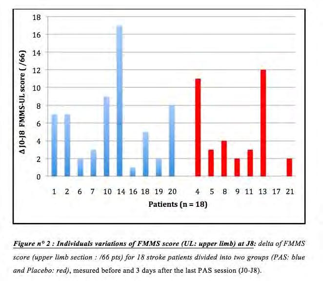
210 210
211 Poster Session I Therapeutic Applications I P 124 Behavioral and fmri changes associated with combined tdcs and cognitive rehabilitation in a case series of patients with mild cognitive impairment. *B. Hampstead 1,2, K. Gopinath 3 1 Emory University, Rehabilitation Medicine, Atlanta, United States 2 Atlanta VAMC RR&D Center of Excellence, Decatur, United States 3 Emory University, Radiology, Atlanta, United States A growing body of evidence supports the use of transcranial direct current stimulation (tdcs) for motor rehabilitation; however, the use of tdcs for cognitive rehabilitation has rarely been examined. Patients with mild cognitive impairment (MCI) demonstrate significant learning and memory deficits and are likely to develop Alzheimer s disease. Functional magnetic resonance imaging (fmri) typically demonstrates hypoactivation in MCI patients relative to healthy individuals, suggesting reduced neuronal functioning. tdcs provides a non-invasive method through which it may be possible to facilitate neuronal functioning. However, such facilitation may only reinforce the suboptimal networks that have emerged as a result of the disease process. We previously demonstrated that cognitive rehabilitation enhances learning and memory, alters the pattern of task-based activation, and increases connectivity in those with MCI. The primary question is whether the combined use of tdcs and cognitive rehabilitation can engage and reinforce adaptive brain networks more than either approach in isolation. Methods: We present a case series of MCI patients who were administered 5 consecutive days of tdcs (2mA for 20 minutes; 35cm2 Anode over FP1, Cathode on right deltoid). Patients underwent fmri during rest-state and task-based (memory encoding of object location associations) conditions before and after tdcs. One patient received only tdcs, while the other 2 received tdcs and cognitive rehabilitation (combined treatment). Behavioral change was measured using an object-location task in which the distance (in cm) between the recalled vs. actual object location was the dependent variable. Change in task-based activation used the contrast: (novel stimuli (post > pre) > repeated (post > pre)). Regions of interest were selected based on these changes and theoretical rationale and used as seed regions for reststate cross-correlation based functional connectivity. Results: Behaviorally, patients receiving combined treatment became more accurate (3-4cm improvement) whereas the tdcs only patient showed minimal change (~0.6cm improvement). fmri showed increased activation within task-relevant regions including the prefrontal, parietal, and inferior temporal cortices, bilaterally. Conversely, the tdcs only patient showed a pattern of increased activation that was restricted to the right superior temporal gyrus and reduced activation in the temporal poles bilaterally. Across all patients, increased activation seemed to be more common in sulcal depths, possibly reflecting the pooling of electrical current in these regions. Reduced activation was observed in the frontopolar cortex (i.e., the area immediately under the anode). Rest-state analyses consistently demonstrated more focused patterns of connectivity post- compared to pre-training in the combined treatment patients. These changes seemed to restore the normal differences between the attention and default mode networks in at least some areas (dorsolateral prefrontal cortex). The tdcs only patient showed substantial increases in connectivity that appeared non-specific and involved a number of both lateral and medial prefrontal and parietal cortices. Conclusions: Although caution is warranted in interpreting our findings, these cases provide behavioral and physiologic evidence suggesting the combination of tdcs and cognitive rehabilitation may facilitate learning and memory in patients with MCI. 211
212 Poster Session I Therapeutic Applications I P 125 Transorbital alternating current stimulation strengthens oscillatory activity and functional connectivity in patients with visual system damage: a resting-state EEG study. *M. Bola 1, C. Gall 1, C. Moewes 2, A. Fedorov 1, B. Sabel 1 1 University of Magdeburg, Institute of Medical Psychology, Magdeburg, Germany 2 University of Magdeburg, Department of Computer Science, Magdeburg, Germany Introduction: Non-invasive brain stimulation is an emerging treatment option for patients suffering from visual system damage and persistent visual field loss. Transorbital alternating current stimulation (tacs) applied daily for 10 days was shown to improve patients` detection abilities (Sabel et al., 2011) and subjective quality of vision (Gall et al., 2011). Further, 10 days tacs treatment progressively increases alpha band power in the resting state EEG of the treated subjects (Schmidt et al., 2012). However, immediate neurophysiological after-effects of tacs have not been studied so far. Objective: To investigate immediate neurophysiological effects of a single tacs session in patients with visual system damage. Methods: Optic neuropathy patients were randomly assigned to either placebo (n=7) or verum group (n=7). The stimulation was delivered with electrodes attached below and above each eye. Current parameters were individually adjusted: amplitude was set above the phosphene threshold, and frequencies ranging from 8 to 22Hz were used. Resting state eyes-closed EEG (20 electrodes acc. to system) was recorded immediately before and after the tacs. Power density and coherence were calculated. Coherence graphs, representing the same number (40) of strongest connections (Fig. 2), were created for each subject and characterized by graph measures indicating of the small-world structure. Changes in EEG parameters were correlated with the extent of visual system damage as assessed by perimetry. Results: One session of tacs: (i) increased power of delta, theta and beta bands at the occipital region; (ii) increased theta and beta bands coherence between occipital electrodes (Fig. 1); (iii) altered the structure of beta band coherence graphs, as indicated by decreased characteristic path length (Fig. 2). Coherence increase was more profound in subjects with less extensive damage (r=0.94; p=0.016). Conclusions: One session of tacs modulates brain activity and connectivity in patients with visual system damage. The neuromodulation includes increase of power (delta, theta, beta bands) and coherence (theta, beta), and results in a modification of coherence network topology (beta) towards more optimal smallworld pattern. These neurophysiological effects are stronger in subjects with mild optic nerve damage. Further, tacs induced neuromodulation seems to be non-specific, as power of frequencies not included in the stimulation protocol (delta, theta) also increased. Further studies need to elucidate how long these changes persist and how they relate to perceptual functioning and vision restoration. References: Gall, C. et al. (2011). Noninvasive transorbital alternating current stimulation improves subjective visual functioning and vision related quality of life in optic neuropathy. Brain Stimulation 4, Sabel, B. A. et al. (2011b). Non invasive alternating current stimulation improves vision in optic neuropathy. Restorative Neurology Neuroscience 29, Schmidt, S. et al. (2012). Progressive enhancement of alpha activity and visual function in patients with optic neuropathy: a twoweek repeated session alternating current stimulation study. Brain Stimulation, in press. Funding: The study was funded by the Otto-von-Guericke University, Magdeburg. 212

213 Figure 1. Coherence spectra before and after tacs session. Figure 2. Graphs representing beta band coherence networks. 213
214 Poster Session I Therapeutic Applications I P 126 Righthemispheric inhibitory rtms in patients with letfsided brain infarcts: effect on cerebral blood flow using PET and speech performance *A. Hartmann 1, I. Rubi-Fessen 1, W.- D. Heiß 2, L. Kracht 2, J. Kessler 2, T. Rommel 1 1 Rehanova clinic, neurology, Köln, Germany 2 mpi f neurologische Forschung, Köln, Germany Introduction: Several small studies suggest that non-invasive repetitive transcranial magnetic stimulation (rtms) of the non-dominant right Brodmann area might support recovery from aphasia in patients with leftsided stroke during conventional speech therapy.we evaluated the effect of this therapeutic regimen in patients with subacute stroke on cerebral blood flow (rcbf) and speech performance. Protocol: 21 subjects have been included in this randomized, double blind, placebo controlled study. The intervention group was treated daily for 2 weeks with rtms (1 Hz) for 20 min directly prior to speech therapy. The magnetic coil was placed over the right sided gyrus frontalis inferior, as indentified by T1 MRI. In the control group, the protocol was the same but the coil was placed over the vertex. Prior to the 2 weeks protocol, a PET (H 2 O 15 ) was performed during rest and during verb generation to calculate the rcbf change by this activation. The PET was repeated at the end of the protocol to identify a possible shift of cortical activation during verb generation. The Aachener Aphasia test (AAT) and the Amsterdam-Nijmegen Everyday Language Test (ANELT) were used for tests of the language performance. Results: CBF measurement revaled a significant shift of activated language related cortex towards the left hemisphere in the group treated with rtms compared to the control group. We calculated a significant difference in the pre- and posttreatment values of the global AAT scores between verum treated patients and control patients. The mean scores post treatment was significantly higher in the verum patients, predominantly in the subtest picture naming. Discussion: rtms with inhibitory effect on the rightsided homologous speech area plus speech therapy in patients with left sided brain infarcts and aphasia results in better recovery of language performance compared to patients who received speech therapy only. 214

215 Poster Session I Therapeutic Applications I P 127 Evaluation of outcomes from Transcranial Direct Current Stimulation (tdcs) for the treatment of chronic pain *H. Knotkova 1,2, A. Greenberg 1, Z. Leuschner 1, E. Soto 1, R. A. Cruciani 1,2 1 Beth Israel Medical Center, Institute For Non-invasive Brain Stimulation, Research Division, Department of Pain Medicine and Palliative Care, New York, United States 2 Albert Einstein College of Medicine, Department of Neurology, Bronx, United States Introduction: Transcranial direct current stimulation (tdcs) has been proposed as an alternative noninvasive technique with potential to alleviate pain in patients with various pain syndromes. Objective: To evaluate the clinical outcomes of five-day tdcs treatment in patients with chronic pain of various etiologies. Methods: This is a retrospective review of medical records of 100 patients who had chronic pain of different etiologies and were treated with tdcs between November 2008 and September The patients were divided in the following categories based on the etiology of their pain: neuropathic, central, nociceptive/somatic, nociceptive/visceral and headaches. Patients provided self-ratings of pain using the 11-point Numerical Rating Scale (NRS) before and after each tdcs treatment, and ratings of pain characteristics before and after the five-day treatment course. The chart review was approved by the Institutional Review Board (IRB) at BIMC. Transcranial direct current stimulation was delivered using a Phoresor II 850 PM unit in five 20-min sessions on 5 consecutive days, at 2mA, applying either anodal (atdcs) over the motor cortex or cathodal (c-tdcs) stimulation over the somatosensory cortex, using 2 saline-soaked sponge electrodes of size 25cm 2. Results: One hundred patients received 172 five-day treatment courses. The patient distribution per category was the following: 59 neuropathic, 28 nociceptive/somatic, 6 headache, 6 central pain and 1 nociceptive/visceral pain. Eighty patients received a-tdcs only, 14 patients had c-tdcs only, 6 patients received both. Of those receiving a-tdcs, 57 got 1 treatment cycle and 23 got between 2 and 5 cycles; 2 of 20 c-tdcs patients received multiple cycles (2 and 7 respectively). Pain intensity (mean, SD) before and after a-tdcs was 6.2±2.4 and 3.9±2.9 (n=86); before c-tdcs 6.8±2.2, after 4.2±3.7(n=20). Pain reduction 30% was observed in: i) Forty-four patients (76%) in the neuropathic pain group [with the highest number of responders among patients with facial pain, post-herpetic neuralgia and complex regional pain syndrome]; ii) 20 patients (77%) in the nociceptive/somatic category (n=26); 83% in the headache group (n=6); 20% in the central-pain group (n=5). Secondary benefits besides pain relief were: decreased intake of pain medication, mood and sleep improvement, improved mobility and function, itch relief. Durability of analgesic effects was up to 12 weeks and showed high inter-individual variability. Neither a-tdcs nor c-tdcs resulted in any serious adverse events (AEs). The most frequently reported non-serious AEs were: i) transient tingling/burning/itching under the electrode during the stimulation (reported in 27 (4.2%) of a-tdcs visits and 14 (7.9%) of c-tdcs visit); ii) transient headache after the stimulation:16 (2.5%) of a-tdcs, 2 (1.1%) of c-tdcs; iii) Fatigue: reported in 7 (1.1%) of a-tdcs, 0 of c-tdcs. Conclusion: The findings support the evidence of analgesic efficacy of tdcs in a diversity of chronic pain types. 215
216 Poster Session I Therapeutic Applications I P 128 Predicting behavioural response to TDCS in chronic motor stroke *J. O'Shea 1, M.- H. Boudrias 1, C. J. Stagg 1, V. Bachtiar 1, J. U. Blicher 1, H. Johansen-Berg 1 1 University of Oxford, Clinical Neurosciences, Oxford, United Kingdom Transcranial direct current stimulation (TDCS) of primary motor cortex (M1) can transiently improve paretic hand function in chronic stroke. However, responses are variable so there is incentive to try to improve efficacy or to predict response in individual patients. Both excitatory (Anodal) stimulation of ipsilesional M1 and inhibitory (Cathodal) stimulation of contralesional M1 can speed simple reaction time. Here we tested whether combining these two effects simultaneously, by using a bilateral M1-M1 electrode montage, would improve efficacy. We tested the physiological efficacy of Bilateral, Anodal or Cathodal TDCS in changing motor evoked potentials (MEPs) in the healthy brain and their behavioural efficacy in changing reaction times with the paretic hand in chronic stroke. In addition, we aimed to identify clinical or neurochemical predictors of patients behavioural response to TDCS. There were three main findings: 1) Unlike Anodal and Cathodal TDCS, Bilateral M1-M1 TDCS (1mA, 20 minutes) had no significant effect on MEPs in the healthy brain or on reaction time with the paretic hand in chronic stroke patients; 2) GABA levels in ipsilesional M1 predicted patients behavioural gains from Anodal TDCS; 3) Although patients were in the chronic phase, time since stroke (and its combination with Fugl-Meyer score) was a positive predictor of behavioural gain from Cathodal TDCS. These findings indicate the superiority of Anodal or Cathodal over Bilateral TDCS in changing motor corticospinal excitability in the healthy brain and in speeding reaction time in chronic stroke. The identified clinical and neurochemical markers of behavioural response should help to inform the optimization of TDCS delivery and to predict patient outcome variability in future TDCS intervention studies in chronic motor stroke. 216
217 Poster Session I Therapeutic Applications I P 129 Cognitive effects induced by cerebellar transcranial DC stimulation *R. Ferrucci 1, M. Vergari 1, F. Mameli 1, M. Fumagalli 1, M. Rosa 1, G. Giannicola 1, E. Scelzo 1,2, T. Bocci 1,3, S. Zago 1,2, A. Priori 1,2 1 Fondazione IRCCS Ca' Granda Ospedale Maggiore Policlinico di Milano, Milan, Italy 2 University of Milan, Milan, Italy 3 Universy of Pisa, University of Siena, Pisa, Siena, Italy Introduction: Neuroanatomical, neuroimaging, neurostimulation and clinical studies have substantially extended the role of the cerebellum beyond motor control to a considerably more complex regulation of cognitive and affective functions. Objectives: To assess the effect of cerebellar transcranial DC stimulation on human cognitive and affective functions. Materials & Methods: We enrolled a total of 55 healthy subjects (aged 19-49). In experiment 1 we evaluated the effect of cerebellar tdcs on working memory (1), in experiment 2 on emotion recognition (2), and in experiment 3 on procedural learning. Participants did the tasks before and 35 min after receiving 20- min (2mA) anodal/cathodal and sham cerebellar tdcs in a randomized order. Results: Experiment 1 showed that anodal or cathodal tdcs over the cerebellum impaired the practicedependent improvement in the reaction times in a working memory task (sham p=0.001; tdcs p=0.36). Experiment 2 demonstrated that anodal and cathodal cerebellar tdcs both significantly enhanced sensory processing in response to negative facial expressions (anodal tdcs p=0.0021; cathodal tdcs p=0.018). Finally, experiment 3 showed that anodal tdcs influenced implicit learning processes (anodal vs. sham p=0.04). Conclusion: Overall cerebellar tdcs changes tests of cognitive and affective functions, possibly through cerebellum-cerebral pathways. Although whether changes induced by cerebellar tdcs in cognition, emotional recognition and learning observed in our experiments can be behaviorally relevant remains to be clarified, cerebellar DC stimulation might be a window for modulating complex mental process. References: 1. Ferrucci R, Marceglia S, Vergari M, et al. Cerebellar transcranial direct current stimulation impairs the practice-dependent proficiency increase in working memory. J Cogn Neurosci 2008;20: Ferrucci R, Giannicola G, Rosa M, et al. Cerebellum and processing of negative facial emotions: Cerebellar transcranial DC stimulation specifically enhances the emotional recognition of facial anger and sadness. Cogn Emot 2012;26 (5):
218 Poster Session II Cognitive Neuroscience II P 130 The impact of dorsolateral prefrontal cortex in the control of heart rate variability investigated by repetitive transcranial magnetic stimulation *K. Sakreida 1,2, S. Gauggel 1 1 University Hospital of RWTH Aachen, Institute of Medical Psychology and Medical Sociology, Aachen, Germany 2 University Hospital of RWTH Aachen, Division of Clinical and Cognitive Neurosciences, Department of Neurology, Aachen, Germany The central autonomic network appears to support goal-directed behavior and adaptability since it serves as an integrated component of an internal regulation system through which the brain controls visceromotor, neuroendocrine, pain, and behavioral responses essential for survival. It includes predominantly a multiplicity of subcortical brain structures, but also medial prefrontal and insular cortex. There is also evidence on the impact of the dorsolateral prefrontal cortex (DLPFC) in central autonomic functions as it integrates sensorimotor input information and anticipations, and thus, regulates behavioural outcome. Moreover, behavioural inhibition is suggested to be a core function of DLPFC with emphasis on the right hemisphere. The aim of this study is to provide evidence on effects in heart rate variability by manipulating the cortical excitability of the DLPFC. For that reason neuro-navigated repetitive low frequency transcranial magnetic stimulation is applied over right and left DLPFC as well as right and left dorsolateral premotor cortex as control target sites. Electrocardiogram is recorded before, during, and after stimulation. Preliminary data point to a pronounced right-lateralized effect in cardiac autonomic balance during as compared to post rtms application. Certainly, this study will contribute to a better understanding of autonomic functions and the relationship to the frontal cortex. 218
219 Poster Session II Cognitive Neuroscience II P 131 Repetitive TMS over the left posterior parietal cortex affects the regulation of body sway *F. Hirschauer 1, W. Stadler 1, A. Geipel 1, J. Hermsdörfer 1, L. Johannsen 1 1 Technische Universität München, Munich, Germany Introduction: During upright stance, skin contact with an earth-fixed referent provides tactile feedback about own body sway relative to the contact location. In this situation body sway variability is reduced compared to standing without contact. Recent studies on the time course of sway before, during and after periods of intermittent touch contact indicated that the stabilisation of sway is a time-consuming integrative process. In addition, it has been suggested that dorsolateral prefrontal cortex is involved in the processing of tactile feedback for the control of body sway. Objectives: Using repetitive TMS, we aimed to investigate the influence of activity inhibition within the left posterior parietal cortex (PPC) as well as the left lateral prefrontal cortex (PFC) on the time course of sway stabilisation after the onset of finger tip contact as well as sway destabilisation after contact removal. We expected that PPC inhibition would disrupt the processing of tactile feedback for sway control indicated by more delayed and less strong sway reduction compared to PFC inhibition. Materials and methods: In two sessions, separated by a week, 5 adult participants received 20 minutes of 1 Hz repetitive TMS stimulation at 110% passive motor threshold over the left PPC (CP3) and lateral PFC (F3) respectively. Before and after each stimulation interval, blind-folded and ear-plugged participants stood quietly on a force plate with their right forearm as well as fingers of the right hand reaching forward. In this posture, the fingers were held at hip level slightly above a contact plate of which its vertical position was controlled by a linear motor. Body sway was assessed in terms of Centre-of-Pressure (CoP) motion and trunk kinematics in the antero-posterior direction. Within each of 6 trials of 120 s duration, 5 pairs of touch onset and removal were timed at random intervals by driving the contact plate upwards until contact was established, respectively downwards until contact was safely removed. The minimum time period between subsequent onsets and removals was 7 s. Time course of sway was evaluated across 3 s before and after each contact event. Results: As expected, sway reduction was attenuated as well as rate of sway stabilisation and destabilisation were lower after PPC inhibition. In addition, body sway was generally increased. In contrast, sway control with or without tactile feedback remained unaffected compared to both pre-stimulation baselines after lateral PFC stimulation. Conclusion: 1Hz repetitive TMS over the left PPC disrupts the sensorimotor control of body balance as well as the sensory reorganization following onset and removal of tactile body sway-related feedback from the finger tips of the right hand. 219
220 Poster Session II Cognitive Neuroscience II P 132 Modulation of encoding-related networks by intermittent TBS stimulation in healthy aging *D. Vidal-Pineiro 1, J. P. Martin-Trias 1, I. Mena-Sanchez 1, E. M. Arenaza-Urquijo 1, R. Sala-Llonch 1, N. Bargalló 2, D. Bartrés-Faz 1,3 1 Universitat de Barcelona, Dept Psiquiatria i Psicobiologia Clinica, Barcelona, Spain 2 Hospital Clínic de Barcelona, Radiology service, Barcelona, Spain 3 Institut d Investigacions Biomèdiques Agust Pi i Sunyer IDIBAPS, Barcelona, Spain Question: Memory dysfunction is one of the main cognitive hallmarks of aging. Repetitive Transcranial Magnetic Stimulation induces brain plasticity changes that may underlie its capacity to modulate associative memory in elders (1). Memory recall also depends on the manner in which stimuli are encoded (2) (i.e. the depth or shallowness of the processing) according to level of processing (LOP) theory (3). Our aim was to investigate the impact of a single session of excitatory intermittent theta burst stimulation (itbs) on behavioral outcomes and the expression of brain networks in healthy elders, related to an episodic memory task with two types of processing. Materials & Methods: After an initial MRI scanner and neuropsychological testing 21 healthy elder subjects (71.6yo.) were randomly assigned to Real (n=11) or Sham itbs group. Subjects underwent a baseline functional MRI session (Siemens 3T) followed by Real or Sham itbs (MagVenture, MagPro x100 Opt; 80% AMT; 600pulses). itbs was neuronavigated (Nexstim-NBS-eXimia) over the Left Inferior Frontal Gyrus (LIFG) (group coordinates from metanalysis (4)). Immediately after itbs, subjects realized a second equivalent MRI session. The memory task consisted in 8 24 block-design paradigm were subjects were instructed to try to remember words either using deep (i.e. semantic) or shallow (i.e. perceptual) encoding. Repeated stimuli were also presented for control purposes. After both MRI sessions participants performed a recognition memory test. Results: In a three way ANOVA, only main effects of LOP (p<0.001) were found on performance, being deep encoding more easily recognized, but unrelated to itbs modality (real or sham). For fmri results, we found a significant interaction in Encoding Deep> Repeated Deep in Left and Right occipital cortex and in Encoding Deep> Encoding Shallow contrast in the anterior LIFG (Figure 1) and in Left visual cortex indicating that Real itbs compared to Sham stimulation resulted in increased BOLD signal but only associated to deep encoding processes (positive slope differences). Furthermore, these areas corresponded to those related to deep memory encoding in our baseline analyses. Conclusion: While failing to observe a behavioral effect, itbs specifically modulated the expression of a brain network only when subjects engaged a cognitive process related to encoding at Deeper LOP. Within the general framework of the state-dependency postulates (5), we suggest that our results provide further first evidence, in a study combining neuroimaging and a behavioral paradigm in elder participants, of the importance of considering the interaction between the objective brain stimulation parameters with the functional state of the neuronal populations, to account for the brain responses to non-invasive brain stimulation studies in cognitive studies in aging population. 220
 Innocenti et al (2010). Neuroimage, 53(1): 325-30. (3) Craik & Lockhart (1972). J verb learn Verb behav, 11: 671-84. (4) Kim (2011). Neuroimage 54(3): 2446-61. (5) Silvanto et al (2008).")
221 Time*Stimulation interaction. Red-yellow areas represents an increased BOLD response after real itbs compared to sham (1) Solé-Padullés et al (2006). Cereb Cortex, 16 (10): (2) Innocenti et al (2010). Neuroimage, 53(1): (3) Craik & Lockhart (1972). J verb learn Verb behav, 11: (4) Kim (2011). Neuroimage 54(3): (5) Silvanto et al (2008). Trends Cogn Sci, 12(12):
222 Poster Session II Cognitive Neuroscience II P 133 Effects of transcranial direct-current stimulation (tdcs) on the recognition of emotional facial expressions in schizophrenic patients and healthy controls. *N. Klimm 1, B. Zwissler 1, N. Eder 1, F. Große Wentrup 2, L. Wolkenstein 2, C. Plewnia 1 1 University Hospital Tuebingen, Tübingen, Germany 2 University Tuebingen, Tübingen, Germany Introduction: Emotion recognition is a basic condition of successful social cognition and interaction. It is a prerequisite for correct attribution of mental states ( Theory of Mind ). Schizophrenic psychoses are associated with reduction of this ability and thereby characterised with deficits in social interactions. Less activation of the left inferior prefrontal cortex (ifc), an area which includes the motoric speech area (Broca Area) as well as mirror neuron activity (BA 44), was identified as a neurophysiological signature of this deficit in patients with schizophrenia. BA 44 is considered as a basic element of emotional recognition. Transcranial direct current stimulation (tdcs) is a non-invasive technology for temporary and polarity specific modulation of cortical activity and cognitive function. Objectives: This research was performed to assess the effects of tdcs on emotional recognition and possibly provide new treatment options for patients with reduction of this ability. Materials and Methods: In a double-blind, randomised, placebo controlled cross-over design, healthy subjects underwent either activity enhancing anodal (n=26) or activity reducing cathodal (n=24) tdcs (20 minutes, 1mA), while performing the reading the mind in the eyes (RME) test where subjects have to infer mental states from pictures that depicted others' eyes. The stimulating electrode was placed over the left ifc (EEG 10/20 system: F3) and the reference electrode supraorbital on the right. Results: There was neither an effect on accuracy of emotion recognition under anodal (F=0.173; p=0.681) and cathodal (F=0.453; p=0.508) stimulation nor an effect on recognition speed (anodal: F=0.029; p=0.866; cathodal: F=0.453; p=0.508). We found a trend for an interaction between stimulation order and stimulation condition on reaction time (the participants were faster in the second measurement - anodal: F=3.952; p=0.058 / cathodal: F=3.946; p=0.06). This trend was not found for the number of errors (anodal: F=0.285; p=0.598; cathodal: F=1.468; p=0.239). Conclusion: TDCS of the left ifc has no effect on accuracy of speed of emotional recognition in healthy subjects. However, interindividual differences in the underlying mechanisms of test performance and tdcs action may have interacted with the effect. First evidence for a reverse effect on emotional recognition in schizophrenic patients are analysed in current experiments. 222
223 Poster Session II Cognitive Neuroscience II P 134 Causal and directional information flow from visual to motor areas during resting-state revealed by a TMS-EEG co-registration *M. KAWASAKI 1, Y. Mizuno 1,2, K. Kitajo 1,2,3 1 RIKEN, BSI-TOYOTA Collaboration Center, Saitama, JAPAN, Japan 2 Tokyo University of Agriculture and Technology, Tokyo, Japan 3 PRESTO, Kawaguchi, Japan Our brain is spontaneously active and constructs a resting-state network even when there are no sensory inputs or no motor output responses. Such networks connect many distributed brain regions,howeverbutit is notwell known howclear about causal and directional information flowoccurswithin the resting-state network. Such causal information flow could be evaluated by perturbing electroencephalograph (EEG) oscillations using transcranial magnetic stimulation (TMS). A single-pulse TMS can reset the phase of ongoing cortical oscillations. Here we analyzed global propagation of phase resetting across different brain regions to elucidate causal information flow. More specifically, we applied single-pulse TMS to the restingstate brain and analyzed spatial spread of TMS-induced phase resetting in the human brain. Eleven right-handed subjects took part in this experiment. A single-pulse TMS was delivered to left primary motor cortex or visual cortex at second intervals during the eye-closed resting states. EachsSubject completed 4 separate sessions; 2 TMS intensities (95% and 50%; 100% means motor threshold) X 2 TMStarget locations (left primary motor cortex (C3) and visual cortex (Oz)). Each session consisted of 50 times of TMS applications. To confirm their arousal, they were asked to respondseby their right index finger when theyfeltsensedaweak flashlight which was sometimes induced by a whiteflashsquareon a computer displayduring inter-pulseinterval periods. EEGs were measured from 67 scalp electrodes. To elucidate the cortical activity freewithouterrorsfrom volume conduction, we applied current source density analysis. Next, we removedthe EEG epochs that would be affected bytms artifacts, using a linear interpolation. Finally, to identify the time-frequency (TF) phases, we applied wavelet transforms using Morlet s wavelets. TMS-inducedevokedphase resetting wasassessedcalculatedby phase locking values (PLV) at each electrode, time point and frequency. The time-frequency EEG results showed enhanced PLVforofboth the low and high frequency bands at the TMS target electrodes at the instant of TMS applications. In particular, the theta (4-8 Hz) PLV increased from the onset of TMS applications around the TMS target locations. Under visual area-targeting TMS, phase resetting (i.e. enhanced PLV) of the theta oscillations were transmitted from the visual areas to the motor areas (in particulartothe left motor area). UnderhighlargeTMS intensity, the left motor electrode showed the highest theta PLV from about 150 msec after the onset of TMS applications, whereas PLV at the visual electrode reached peaking values just when TMS was applied. UnderlowsmallTMS intensity, such regional transmissions of the phase resets decreased and disappeared. In contrast, the TMS to the left motor areas increased only the theta PLV in the left and central motor areas. High PLV was observed from about 100 msec after TMS applications. In contrast, the visual areas showedlowsmallplv. Similar to the visual area-targeting TMS, the regional transitions of the theta PLV disappeared when the TMS intensity waslowsmall. We succeeded in evaluating directional information flow in the resting state. Our results indicated the existence of potential networks from the sensory input regions to the motor output regions in the resting state. The sensory-motor mechanisms might be related totheaspontaneous preparation of perceptionaction coupling; e.g. seeing a visual stimulus, making a decision, and then responding by the motor output. 223
224 Poster Session II Cognitive Neuroscience II P 135 Bilateral transcranial direct current stimulation over the posterior parietal cortex enhances recognition memory in healthy individuals *Z. Turi 1, A. Pisoni 1,2, R. Almuth 1, A. Schacht 3, G. Ambrus 1,4, W. Paulus 1, A. Antal 1 1 Georg-August University of Göttingen, Department Clinical Neuropysiologie, Göttingen, Germany 2 University of Milano-Bicocca, Department of Psychology, Milano, Italy 3 Humboldt-Universität zu Berlin, Department of Psychology, Berlin, Germany 4 Georg-August University of Göttingen, Department of Medical Psychology and Medical Sociology, Göttingen, Germany There is emerging evidence from imaging studies that the parietal cortex plays an active role in recognition memory, however, its functional contribution is still under debate. To evaluate the potential role of the PPC (posterior parietal cortex) for episodic memory retrieval, 14 healthy participants received tdcs (transcranial direct current stimulation) over PPC by using a sham-controlled, counterbalanced, single-blinded study design. Participants were randomly assigned into two separate groups, where 7 participants received real tdcs (1.5 ma for 15 minutes) and 7 participants (matched on age, gender and years of education) received sham stimulation (1.5 ma for 30 seconds) during the recognition part of the word-list learning paradigm. In both groups, the anodal electrode was placed over the P3 and the cathodal electrode over the P4 (35 cm 2 each) by using the international 10/20 system. Accuracy and reaction times of recognition memory were measured immediately and 24 hours after the initial episodic memory encoding. We found that recognition performance for episodic memory was better during verum than during sham tdcs. This difference was marginally significant 24 hours later. No differences were found for RTs. Our results show that bilateral tdcs over the PPC significantly enhanced recognition memory performance during immediate recognition and possibly have an effect on post-learning processing. 224
225 Poster Session II Cognitive Neuroscience II P 136 The influence of academic stress on cortical plasticity M. Al Sawah 1, M. Rimawi 1, E. Chusid 1, *F. Battaglia 2 1 New York College of Podiatric Medicine, New York, United States 2 Seton hall University, Health and Medical Sciences, South Orange, NJ, United States Question: Medical education is perceived as stressful. The purpose of this study was to explore the possible effect of academic examination stress on cortical long-term potentiation (LTP)-like plasticity in a group of university students. Methods: The study included 9 healthy medical students (males: mean age 30.2+/- 4.3 SE) and was conducted during a major exam period (stressed) and 5 weeks later after the exams (non-stressed). Facilitatory associative plasticity was induced by paired associative stimulation (PAS) in the human motor cortex (PAS-25 protocol). In addition, students were required to fill the Perceived Stress Scale 10 (PSS) questionnaire. Results: Sixty minutes after PAS-25 induction protocol, students showed lower amounts of potentiation during the exam period compared with after the exams (p=0.039). The average PSS score significantly higher during the stressed period (P = 0.024). LTP-like plasticity showed an inverse correlation with the perceived stress. Conclusions: Academic stress appears to affect LTP-like plasticity in students. Deficit in attention and impairment of N-methyl-D-aspartate receptor-dependent neural plasticity associated with stress might underlie our findings. Larger scale studies are needed to address the interrelationship between LTP-like plasticity and academic performance. 225
226 Poster Session II Cognitive Neuroscience II P 137 High frequency rtms treatment prevents stress-induced memory impairment M. Cambiaghi 1, R. Crupi 1, J. Lildharrie 1, R. Sigh 1, E. Spina 2, *F. Battaglia 3 1 New York City College, New York, United States 2 University of Messina, Italy 3 Seton hall University, Health and Medical Sciences, South Orange, NJ, United States Question: Stress has a detrimental effect on memory. We investigated whether high-frequency rtms treatment is capable of reducing and/or preventing stress-induced memory impairment in the object recognition test mice. Methods: Adult male mice were divided into four groups. (1) sham + non-stressed; (2) rtms + nonstressed; (3) sham + stressed (4) rtms + stressed. The treatment consisted of either five days of 15 Hz rtms or Sham stimulation. One day after the last treatment mice underwent to acute restraint stress procedure (stressed). Non-stressed groups were housed for the same amount of time without any procedure. Memory was tested with the object recognition test. Results: Restraint stress induced memory impairment in the object recognition test. 15 Hz rtms treatment was able to prevent the memory impairment. Conclusions: Our results confirm that high frequency rtms treatment has memory enhancer effect in mice. In addition, we demonstrated that High frequency rtms treatment might reduce the detrimental effect of negative emotional experiences on memory 226
227 Poster Session II Cognitive Neuroscience II P 138 Right cathodal transcranial direct current stimulation over the prefrontal cortex improves implicit learning in healthy individuals *C. Saiote 1, D. Nemeth 2, K. Janacsek 2, Z. Turi 1, G. Ambrus 1, W. Paulus 1, A. Antal 1 1 Georg-August University Goettingen, Clinical Neurophysiology, Goettingen, Germany 2 Eötvos Loránd University, of Clinical Psychology and Addiction, Budapest, Hungary The fronto-striatal network is a key region in implicit sequence learning in the human brain. Previous studies suggested the functional involvement of the right prefrontal cortex, however, the exact contribution of this region in implicit learning and consolidation is still under debate. In the present study, we aimed at further exploring the putative role of the right dorsolateral prefrontal cortex (rdlpfc) by using transcranial direct current stimulation (tdcs) technique. In a sham-controlled, single-blinded, parallel group design, 20 participants received verum tdcs (1.0 ma for 10 minutes) and additional 20 participants (matched on age, gender and years of education) received sham stimulation (1.0 ma for 30 seconds) while performing the Alternating Serial Reaction Time (ASRT) task. Using this task we can measure sequence-specific and general skill improvement separately. In both groups, the cathodal electrode was placed over the rdlpfc with the anodal return electrode over Cz. (electrode size 35 cm^2 each). Accuracy and reaction time were measured in three consecutive ASRT sessions. Consolidation (second and third session) was examined 2 and 24 hours following the learning phase (first session). Results comparing sham (n=20) and cathodal (n=10) stimulation groups show a significant general improvement in task performance as well as sequence-specific learning for both groups. Additionally, cathodal stimulation caused a better general performance in the second and third sessions regarding both accuracy and reaction times, but not in the first session. These results suggest that cathodal stimulation of the rdlpfc causes greater general improvement in task performance only after consolidation but no alterations in sequence-specific learning. Further experiments are required to understand the specificity of the results regarding polarity and region used for stimulation. 227
228 Poster Session II Cognitive Neuroscience II P 140 Virtual lesions of the inferior parietal and prefrontal cortex alter implicit religiousness and spirituality in healthy individuals *C. Crescentini 1,2, S. M. Aglioti 2, F. Fabbro 1, C. Urgesi 1 1 University of Udine, Department of Human Sciences, Udine, Italy 2 University of Rome, Department of Psychology, Rome, Italy Introduction: Belief in a higher power constitutes a core feature of religiousness and spirituality (RS), two ubiquitous aspects of human experience and behaviour that have long been considered impervious to scientific investigation. Recently, however, cognitive neuroscience studies have shown an association between RS experiences and neural activity in fronto-parietal regions. Much less is known on whether activity in the above areas is causally involved in modulating RS experiences or just epiphenomenal to them. Objectives and Methods: Here we combined two-pulse (10 Hz) transcranial magnetic stimulation (TMS) with a novel, ad-hoc developed RS-related, implicit Association Test (IAT) to investigate whether implicit RS representations, although supposedly rather stable, can be modified in the short-term by a virtual lesion of inferior parietal lobe (IPL) and dorsolateral prefrontal cortex (DLPFC). A self-esteem (SE) IAT, focused on self-concepts non related to RS representations, was developed as control. Results: The results show that inhibiting the IPL induced a specific increase in RS. In contrast, interfering with the left DLPFC induced a marginal increase of difficulty in controlling automatic associations of the self with both RS and SE dimensions. Conclusions: Thus, suppression of neural activity in parietal regions seems causally involved in the specific induction of fast RS increase. Conversely, the DLPFC appears to contribute to RS in relation to attentional processes or activation of self-related concepts. 228
229 Poster Session II Cognitive Neuroscience II P 141 Global/Local Attention: Effects of tdcs on Brain Dynamics *C. Tesche 1, D. Stone 1 1 University of New Mexico, Psychology, Albuquerque, United States Introduction: Posterior parietal cortex (PPC) has been identified as a critical region for the control of spatial and non-spatial attention. Hierarchical letter stimuli (large letters composed of smaller letters) have been used to investigate the contribution of PPC to global/local processing of features in the visual field, and also to the switching of attention between the global and local level. Objectives: We utilized transcranial direct current stimulation (tdcs) and high-density EEG to characterize brain dynamics during global/local attentional switching. Materials & Methods: 128-channel EEG data was recorded from 14 healthy, right-handed adults who made motor responses to hierarchical letter stimuli. A cue before each block of 4-8 hierarchical stimuli directed attention to either global or local features. tdcs was applied over left posterior parietal cortex (electrode P3, forearm reference; 2 ma for 20 min) during separate anodal, cathodal and sham stimulation sessions. The EEG data were analyzed using Brainstorm (Tadel et al., 2011) to extract brain-based activation waveforms for regions defined in the BrainVisa Tzourio-Mazoyer Atlas. Time-frequency plots were computed for left and right superior and inferior parietal and mid occipital corteces. Results: Behavioral responses differed for the first stimuli following switching of attention between global/local features, but not for visual processing per se. Attentional shifts from local-to-global features were degraded for at least 20 minutes following anodal stimulation (Stone & Tesche, 2009). Practice effects were observed in the theta-band between baseline data and data recorded immediately after sham stimulation for both G/L cues to switch attention and for the first compound letter stimuli after the G/L cues. In left PPC, anodal and cathodal stimulation increased theta for global-to-local switches for the first hierarchical letters after a cue to switch, whereas anodal increased and cathodal decreased theta for localto-global switches. In right PPC, anodal tdcs increased and cathodal tdcs decreased theta for both global-to-local and local-to-global switches. Differential time-frequency activations were observed in left (but not right) mid occipital cortex following tdcs; anodal stimulation enhanced theta/alpha activity for local-toglobal, but not global-to-local, switches. Conclusion: The data suggest that tdcs of left PPC induces changes in theta-band activity in a distributed network which includes left and right PPC and occipital cortex, and that theta-band activity may be salient for PPC control of early ( ms) global/local processing. 229

230 Poster Session II Cognitive Neuroscience II P 142 Functional connectivity of the left somatosensory cortex and the putative mirror neuron system *N. Valchev 1, B. Ćurčić-Blake 1, N. Maurits 1 1 University of Groingen, Groningen, Netherlands Introduction: Several brain areas have been identified as active during action perception and execution (see Rizzolatti and Sinigaglia, 2010). This parieto-frontal system of regions has been referred to as the putative Mirror Neuron System (pmns). Primary somatosensory cortex (SI), has also been shown to be active during action perception and execution (see Keysers et al., 2010) but whether it is part of the pmns or a separate somatosensory network, is still not clear. To answer this question we chose to deliver inhibitory Transcranial Magnetic Stimulation (TMS) (the protocol used was continuous theta burst (ctbs, see Huang et al., 2005)over the left SI and then collect resting state functional magnetic resonance (RSfMRI) data from our subjects. Methods: 18 healthy volunteers participated in a three days study. On Day 1, subjects were scanned while observing and performing simple single-handed actions. The target area for the stimulation was selected as the part of SI active in both conditions (see figure 1 A). On the other two days in a randomized order subjects received ctbs or sham stimulation over the left SI. After stimulation participants were transported to the scanner and RSfMRI data was collected. Three different types of functional connectivity analysis were applied to the data from Day 2 and Day 3: 1) a seed-based correlational approach, 2) partial correlations between the nodes of the pmns and 3) independent component analysis (ICA). Results: The seed based correlational approach indicated a small decrease in the connectivity between the left SI and the dorsal premotor areas (dpm) after ctbs as compared to sham (cluster level corrected p(fwe) = 0.09; T = 4.47). Partial correlation analysis between the nodes of the pmns calculated separately for the data from the sham and ctbs sessions suggested a decrease in the connectivity between the stimulated region and the left dorsal premotor area (dpm), part of the pmns. ICA, which enables differentiation of independent functional networks, suggested a decrease in the synchronization of one network as a result of ctbs stimulation to SI (cluster level corrected p(fwe)=0.03; T=6.59) after ctbs. The change was localized in the same area of the left dpm as identified in the other two analyses (see figure 1 B). Conclusions: Taken together our results suggest that the effect of inhibitory stimulation over the left SI is not focal but visible in the functionally connected distal brain areas. ICA analysis indicates that SI is part of the same functional network which includes the regions of the pmns, but the seed based and partial correlation analyses show that this connection is disrupted by our stimulation, i.e. stimulation over SI does not have an effect on the synchronization of the network as a whole but causes a decrease of the connectivity between SI and dpm. Considering their co-activation during mental simulations of actions, we propose that SI also belongs to the pmns. Huang, Y.Z., Edwards, M.J., Rounis, E., Bhatia, K.P., and Rothwell, J.C. (2005). Theta burst stimulation of the human motor cortex. Neuron 45, Keysers, C., Kaas, J.H., and Gazzola, V. (2010). Somatosensation in social perception. Nat. Rev. Neurosci. 11, Rizzolatti, G., and Sinigaglia, C. (2010). The functional role of the parieto-frontal mirror circuit: interpretations and misinterpretations. Nat. Rev. Neurosci. 11,
231 Poster Session II Cognitive Neuroscience II P 143 The contents of a steady visual background have an effect on TMS-evoked EEG perturbation: natural scenes compared to scenes with man-made environments increase ERP slow negativity R. Rutiku 1, A. Einberg 1, K. Imanaka 2, *T. Bachmann 1 1 University of Tartu, Tallinn, Estonia 2 Tokyo Metropolitan University, Tokyo, Japan Transcranial magnetic stimulation (TMS) has been often used for studying causal relevance of certain cortical areas for visual perception and cognition. Typically, single-pulse or rtms has been applied to some brain location hypothesized to be involved in performing certain visual tasks with the purpose to interfere with information processing responsible for adequate performance in this main task. In the present experiment we used a different perspective: we explored whether the type of a steadily present visual background irrelevant for the invariant simple main task has an effect on EEG/ERP responses to the occipitally applied TMS pulses. If the answer would be affirmative, TMS-evoked EEG perturbations could potentially become useful as electrophysiological signatures of task-free visual information processing contents. The results showed that spatial frequency and brightness of the background did not cause significant differences in the TMS-evoked ERPs. However, when natural scenes including natural objects (plants, animals) were used as a task-irrelevant background parietally and frontally recorded ERPs showed more negativity from 100 ms up to 700 ms compared to when pictures with scenes consisting of man-made environments were presented. Future research should ascertain whether the effect is caused by some relatively low level visual image statistics associated with natural versus artificial scenes or by higher level visual-semantic image attributes. 231
232 Poster Session II Cognitive Neuroscience II P 144 Activity prior to motor cortex and the sensation of agency modulate brain responses to self-initiated sounds *J. Timm 1, I. SanMiguel 1, J. Keil 2, E. Schröger 1, M. Schönwiesner 3 1 University of Leipzig, Institute of Psychology, Leipzig, Germany 2 Charité Berlin, Department of Psychiatry and Psychotherapy, Berlin, Germany 3 University of Montreal, International Laboratory for Brain, Music and Sound Research (BRAMS), Montreal, Canada Introduction: One of the functions of the brain is to predict sensory consequences of our own actions. In auditory processing self-initiated sounds evoke a smaller auditory brain response than passive sound exposure of the same sound sequence [1]. Previous work suggests that this response attenuation reflects a predictive mechanism to differentiate the sensory consequences of one s own actions from other sensory input [2]. On a psychological level correctly predicted sensory feedback has been linked to a pre-reflective sensation of self-agency (feeling of coherence in action processing) [3] which is formed in premotor areas involved in movement planning [4]. Objectives: We addresses the question whether attenuation of brain responses to self-initiated sounds can be explained by brain activity leading up to, but not including, the motor cortex. Moreover, the relation between attenuated auditory brain responses to self-initiated sounds and the pre-reflective sensation of agency was investigated. Materials & Methods: Event-related brain potentials (ERPs) from the human scalp of 17 participants were recorded in response to sounds initiated by a button press. In one experimental condition, participants moved a finger to press the button voluntarily, whereas, in another condition, we initiated a similar finger movement involuntarily by stimulating the corresponding region of the primary motor cortex with transcranial magnetic stimulation (TMS). No sensation of agency was experienced for involuntary movements as no motor plans were available. Results: A portion of the brain response evoked by the sounds, the N1-P2 complex, was reduced in amplitude only following a voluntary, self-initiated, movement, but not following a movement initiated by motor cortex stimulation. Conclusion: Our findings demonstrate that sensory attenuation of brain responses to self-initiated sounds depends on a pre-reflective sensation of agency and on predictive mechanisms that operate prior to the activation of the primary motor cortex. The present results support the assumptions of an internal forwardmodel account [2]. References: 1. Schafer EW, Marcus MM (1973) Self-stimulation alters human sensory brain responses. Science 181: Wolpert DM, Ghahramani Z, Jordan MI (1995) An internal model for sensorimotor integration. Science 269: Synofzik M, Vosgerau G, Newen A (2008) Beyond the comparator model: a multifactorial two-step account of agency. Conscious Cogn 17: doi: /j.concog Yomogida Y, Sugiura M, Sassa Y, Wakusawa K, Sekiguchi A, et al. (2010) The neural basis of agency: an fmri study. Neuroimage 50: doi: /j.neuroimage Founding sources: This work was supported by the Erasmus Mundus Student Exchange Network in Auditory Cognitive Neuroscience and a Reinhart- Koselleck grant of the German Research Foundation (Deutsche Forschungsgemeinschaft, DFG, Project 375/20-1). 232
233 Poster Session II Cognitive Neuroscience II P 145 Modulating multimodal feature integration with transcranial direct current stimulation (tdcs) over the parietal and prefrontal cortices *S. Zmigrod 1,2, L. S. Colzato 1,2, B. Hommel 1,2 1 Leiden University, Cognitive Psychology, Leiden, Netherlands 2 Leiden Institute for Brain and Cognition, Leiden, Netherlands Understanding how the brain integrates features from diverse sensory modalities that are processed in distinct cortical regions calls for the examination of integration processes ( the binding problem ). Recent studies of feature-repetition effects demonstrated interactions across perceptual features from different modalities: repeating only some features of the multimodal stimuli hinders performance compared to repeating all or none of the features (Zmigrod, Spapé, & Hommel, 2009). These partial-repetition costs point to the existence of temporary memory traces of multimodal information (object file / event files). Here, we investigated whether brain stimulation with transcranial direct current stimulation (tdcs) influences the integration processes. In Experiment 1, tdcs was employed over the left dorsolateral prefrontal cortex (DLPFC) which is linked with cognitive control processes (Metuki, Sela, & Lavidor, 2012), while in Experiment 2, we applied tdcs over the right posterior parietal cortex (PPC) which is a multimodal region associated with sensory convergence of multisensory information (Bolognini et al., 2010). In different sessions, healthy participants received anodal, cathodal, or sham tdcs (2mA, 15 min) while performing the audio-visual event file task. The results show that there is no significant difference in terms of partial repetition costs between the stimulations over the left DLPFC, yet anodal stimulation over the right PPC eliminated the integration between auditory and visual features, as compared to cathodal and sham stimulation. In additional control experiments no such effect was found in the left PPC. These findings emphasize the role of the right posterior parietal cortex in multimodal feature integration. tdcs may be considered as an effective therapeutic modulation tool in cognitive rehabilitation, especially in populations where multimodal integration does not operate in the most resourceful manner, such as in older adults (Diaconescu, Hasher, & McIntosh, 2012) and in autistic children (Zmigrod et al., in press). 233
234 Poster Session II Cognitive Neuroscience II P 146 Motor resonance requires conscious perception but is modulated by unseen actions *A. Mattiassi 1, S. Mele 1,2, C. Urgesi 1,2 1 University of Udine, Human Sciences, Udine, Italy 2 IRCCS Eugenio Medeae, San Vito al Tagliamento (PN), Italy Evidence for automatic mirror-like activation of the motor cortex (motor resonance) during observation of others actions has been found by several studies. Motor resonance has also been found during observation of implied actions, i.e. static pictures depicting ongoing actions. Here we explored whether motor resonance with implied actions requires the perceptual awareness of the observer or is also triggered in conditions of reduced awareness. In order to test our hypothesis, we analyzed motor response to consciously and unconsciously perceived implied actions using a masked priming paradigm. We presented a sequence of 3 static stimuli depicting right hands: sample, masked prime and target stimulus. While the sample stimuli were always static hands, both prime and target stimuli could display a static hand, an index finger or a little finger abduction. The prime stimuli were presented for 53 ms and were forward and backward masked by a rotating masking stimulus, a procedure that allowed a relatively long lasting presentation of the prime while at the same time disrupting conscious perception: indeed, 17 out of 22 subjects reported they did not see the prime when the experimenter revealed the actual presentation structure after the experiment. A transcranial magnetic stimulation (TMS) pulse was delivered over the motor cortex 133 ms ( early ) or 307 ms ( late ) after the onset of the probe. Electromyography was used to record TMS motor evoked potentials (MEPs) from the muscles that corresponded to the driving muscle of the observed implied actions, first digital interosseus (FDI) and abductor digiti minimi (ADM). Analysis of the MEPs recorded in the 17 subjects reporting unawareness of the prime replicated the finding of a mirror-like increase in corticospinal excitability following observation of consciously perceived target hands (i.e., greater MEP amplitude during observation of the movement involving the recorded muscle as compared to observation of the static hand or of a movement not involving that muscle). Such facilitation was present only in the late TMS delay. Furthermore, no change in corticospinal excitability was found in response to implied action prime, suggesting that masked actions are not sufficient to evoke motor resonance. Importantly, however, we found a significant interaction between prime and target stimuli. In particular, the amplitude of MEPs recorded from a muscle during observation of a target showing the action driven by that muscle was lower when preceded by a prime showing an incongruent action with respect to when it was preceded by a prime showing a congruent action or a still hand. Crucially, no difference was found between MEPs recorded from the muscle during conscious observation of actions driven by that muscle but primed with an unconsciously perceived incongruent action and MEPs recorded during conscious observation of incongruent or neutral movements regardless of the prime. This finding suggests that the masked presentation of a movement that doesn t involve a certain muscle yields a somatotopic inhibition of the motor resonance in that muscle. Our results provide first time evidence that conscious perception of implied actions is not required for modulation of motor resonance, yet it is required for a direct mirror motor activation. Moreover, they shed additional light on how the cognitive system copes with a crowded social world and the needs for a fast, accurate and dynamic representation of others movements. 234
235 Poster Session II Cognitive Neuroscience II P 147 Impaired modulation of intracortical inhibition during Go-Stop task in schizophrenia *P. Lindberg 1, A. Saby 2,3, B. Crepon 2,3, M.- O. Krebs 2,3, R. Gaillard 2,3, B. Gueguen 2,3, I. Amado 2,3 1 Université Paris Descartes, CNRS UMR 8194, Paris, France 2 Hôpital Sainte Anne, Paris, France 3 Université Paris Descartes, Inserm U894, Paris, France Background: Impaired inhibition has been reported in patients with schizophrenia in a variety of cognitive and motor tasks. Recent transcranial magnetic stimulation studies performed during rest show reduced cortical inhibition in primary motor cortex (M1) at rest in schizophrenia patients, supporting the hypothesis of impaired GABA-ergic inhibition as an underlying mechanism. However, cortical excitability and intracortical inhibition in M1 are highly task-dependent, for example, inhibiting a planned motor response leads to reduced excitability and enhanced intracortical inhibition. Studies on task-dependent cortical excitability and inhibition are lacking in schizophrenia. The aim of this study was therefore to investigate cortical excitability and intracortical inhibition during voluntary motor inhibition in patients with schizophrenia. Methods: 25 stabilized patients with schizophrenia (DSMIV) and a group of 23 age-comparable healthy volunteers were included. We used a modified version of the Go-Stop task (Coxon et al, 2006) to study voluntary motor inhibition. This task consisted of moving the index finger in response to Go and Stop signals. In 30% of trials the subject received a visual cue to inhibit the prepared finger lift. Paired-pulse transcranial magnetic stimulation (TMS) was used to measure M1 excitability and short-latency intracortical inhibition (SICI%) during three phases of the task: (i) early without movement preparation (Early); (ii) late with preparation of finger lift (Late prep ); and (iii) late with inhibition of finger lift (Late inhib ). Motor evoked potentials (MEPs) were recorded from the first dorsal interosseous muscle. Results: Patients and control subjects performed equally on the voluntary inhibition task, with similar stop signal reaction times (mean SSRT controls 189±21ms and patients 186±31ms, P=0.6). TMS at rest revealed similar resting and active motor thresholds in patients and controls (P>0.9). The cortical silent period was shorter in the patients but did not differ significantly (mean controls 162±30ms and patients 147±61ms, P=0.36). During the Go-Stop task, cortical excitability was similar in patients and controls with a task-dependent modulation of MEPs (mean RMS MEPs: Early=477µV, Late prep =854µV, and Late inhib =531µV; effect of Phase, P<0.001). SICI% in patients was reduced compared to controls (effect of Group, F=4.5, P=0.02) with posthoc comparisons revealing a significant reduction in the Late inhib phase in patients compared to controls (P=0.02). In patients the SICI% during Late inhib correlated negatively with SSRT (R=-0.72, P<0.001) such that patients with lowest SICI% during Late inhib had longer SSRT. Conclusions: To our knowledge this is the first study on task-dependent modulation of cortical excitability and intracortical inhibition in patients with schizophrenia. We show that patients with schizophrenia have reduced SICI during voluntary inhibition of a prepared finger lift. Surprisingly, there were no differences found in task performance or in MEP amplitudes during the task. These findings show that schizophrenia patients are capable of inhibiting motor tasks despite having reduced intracortical inhibition. This suggests the use of compensatory mechanisms in voluntary motor inhibition in stabilized schizophrenia patients. 235

236 Poster Session II Cognitive Neuroscience II P 148 Anticipatory motor simulation of deceptive actions in soccer players. *S. Makris 1, C. Urgesi 1 1 University of Udine, Udine, Italy The ability to form anticipatory representations of ongoing actions is crucial for meaningful and effective interactions in dynamic environments. Previous research has shown that simulating and predicting the outcome of ongoing actions is modulated by motor expertise. In sports, elite athletes exhibit great ability in predicting other players' actions, mainly based on action body kinematics as well as visual body representations. Surprisingly, there has been little evidence so far on how motor experts such as athletes perceive and/or anticipate bluffing actions. A few studies applying temporal occlusion paradigms have shown that expert players are better in detecting deceptive actions compared to expert observers. A recent study by Tomeo et al (2012) in soccer players has found that the ability to detect deceptive actions relies on a complex mechanism of inhibitory and excitatory processes within the motor system. Furthermore, it was indicated that the balance between these processes depends on both visual and motor expertise. The present study has tried to identify the involvement and interactions between visual and motor areas concerning the detection of bluffing actions in soccer penalty kicks. We used the experimental paradigm described by Tomeo et al (2012), during which subjects (expert kickers, goalkeepers and novices) were judging the direction of the ball after a penalty kick, and we applied a repetitive TMS methodology in order to cause online disruption of two different regions; the dorsal premotor cortex (PMd) and the superior temporal sulcus (STS). Results have shown a strong modulation in the task perfromance between the sample groups caused by disruption of the STS area compared to control areas. Hence, new evidence is provided on how visual and motor expertise contributes to representing actions and detecting bluffing actions. 236
237 Poster Session II Cognitive Neuroscience II P 149 DLPFC is involved in perceptual decision-making but not metacognitive evaluation. *R. Rutiku 1, K. Peterson 1, T. Bachmann 1 1 University of Tartu, Tallinn, Estonia Growing evidence suggests that dorsolateral prefrontal cortex (DLPFC) plays a significant role in perceptual decision-making by integrating sensory evidence for different alternatives. This leads us to wonder whether the same information is used for metacognitive evaluation (e.g. confidence) of the decisions we make. After all, the two processes - decisions and their evaluations - are closely related under normal circumstances and thus it is possible that their underlying neural mechanisms are also widely overlapping. To clarify the role of DLPFC in perceptual decision-making and metacognitive evaluation we used off-line repetitive transcranial magnetic stimulation (rtms). Subjects completed a visual categorization task and gave confidence ratings for the decisions they made. We also introduced extra information into the system by priming the stimuli. Priming is well known to increase objective as well as subjective measures of performance. If DLPFC incorporates this extra information into its processes a differential effect of TMS can be expected for primed stimuli. The results show that TMS clearly affected categorization performance. Subjects had more difficulty categorizing the stimuli correctly after rtms to the right DLPFC. However, there was no effect of TMS for confidence ratings. The expected priming effect was only observed for repeated stimuli. Thus, our findings confirm that DLPFC is directly involved in perceptual decision-making. Since we did not find an effect of TMS on confidence ratings, our results suggest that metacognitive evaluation of decisions is a separate process which has its neurobiological correlates in other areas distinct from the DLPFC. 237
238 Poster Session II Cognitive Neuroscience II P 150 Online rtms over cerebellar vermis affects motion discrimination in healthy individuals *S. Casali 1,2, C. Renzi 1,2, E. D'Angelo 1, T. Vecchi 1,2, Z. Cattaneo 1,3 1 BCC, IRCCS Mondino, Pavia, Italy 2 University of Pavia, Psychology, Pavia, Italy 3 University of Milano-Bicocca, Psychology, Milano, Italy Patients with cerebellar lesions have been found to be impaired in visual motion discrimination. The present work investigated whether temporarily interfering with cerebellum activity with transcranial magnetic stimulation (TMS) affects visual motion discrimination in healthy young participants. Stimuli consisted of 100 white dots placed at a random position within an imaginary square subtending 4.3 x 4.3 of visual angle. In each trial, a proportion of dots moved coherently either leftward or rightward, whereas the others moved randomly within the square. Motion stimuli were visible for msec, and participants were asked to indicate as fast and as accurately as possible whether the dots moved leftward or rightward by left/right key pressing. Prior to the experimental session, the percentage of coherently and randomly moving dots was adjusted for each participant in order to obtain a stable performance around 75% of accuracy. TMS was applied on line during the task. In particular, triple-pulses TMS (20 Hz, 100% resting motor threshold) was applied after (13 msec post) the onset of the motion signal on either V1 or the cerebellar vermis. V1 was chosen as a stimulation site given previous TMS evidence indicating its involvement in motion detection. Sham (coil tilted 90 degrees) stimulation of V1 and vermis was used as control condition in addition to a baseline no-tms condition. Our results showed that TMS applied to V1 affected motion detection compared to sham TMS over V1 and to the no-tms condition, confirming previous evidence. Critically, TMS delivered over the cerebellar vermis also affected motion detection compared to the sham TMS vermis and to the no-tms conditions. Notably, vermis TMS did not affect a control orientation discrimination task, in which Gabor patches with different orientations were used. Our data are discussed in light of a possible specific role of the cerebellum in sensory prediction and in signal to noise segregation. 238
239 Poster Session II Cognitive Neuroscience II P 151 The right (but not the left) lateral occipital complex is causally implicated in visual mirror symmetry detection: an fmri-guided TMS study *S. Bona 1, A. Herbert 2, C. Toneatto 3, J. Silvanto 1, Z. Cattaneo 4 1 Brain Research Unit, Aalto University, Espoo, Finland 2 Rochester Institute of Technology, New York, United States 3 Cognition Psychology Neuroscience Lab, Pavia, Italy 4 Brain Connectivity Center, IRCCS Mondino, Pavia, Italy Despite the fact that bilateral mirror symmetry is an important characteristic of the visual world, few studies have investigated its neural basis. Here we addressed this issue by investigating whether the objectselective lateral occipital cortex (LO), a key brain region in object and shape processing, is causally involved in this kind of symmetry detection. Participants were asked to discriminate between symmetric and asymmetric dot patterns, while fmri-guided repetitive TMS was delivered online over either the left LO, the right LO or two control sites in the occipital cortex. We found that the application of TMS over the right LO slowed down symmetry judgments, whereas stimulation of the left LO, or of two control sites, had no impact. Our results thus indicate that the right (but not the left) LO is causally implicated in symmetry detection. This finding may relate to the previously reported right-hemisphere lateralization of holistic processing of stimuli, including faces for which symmetry is a critical cue. References: Julesz B & Chang J.J. (1979).Perception, 8: Sasaky Y et al.(2005).proc Natl Acad Sci U S A, 102: Sergent et al. (1992).Brain, 115:15-36 Tyler et al. (2005).Neuroimage, 24: Yovel et al. (2001).J Exp Psychol Hum Percept Perform,27: Figures will be presented shortly. Figure 1 Timeline of an experimental trial: Each trial started with a fixation cross appearing in the middle of the screen for 500 ms. The target stimulus was then presented for 75 ms and participants were instructed to indicate, as quickly as possible, whether the pattern was symmetric or asymmetric. A TMS train of 3 pulses (10 Hz) was given at stimulus onset. Figure 2 Activation peak of left and right LO respectively for a representative subject. The control sites (CL and CR) were localized such that the coil was moved up 2 centimeters dorsally from left and right LO respectively. 239
240 Poster Session II Cognitive Neuroscience II P 152 Phosphene threshold and 256 channels EEG in MCI patients as a measure of cognitive decline *C. Arcaro 1,2, A. Del Felice 2, S. F. Storti 3, G. Gambina 4, E. Broggio 4, P. Manganotti 1,2 1 IRCCS San CAMILLO-VENICE and UNIVERSITY OF VERONA, Neurophysiologic, Verona, Italy 2 University of Verona, AOUI, Department of Neurological, Neuropsychological, Morphological and Motor Sciences, Verona, Italy 3 University of Verona, AOUI, Department of Neurological, Neuropsychological, Morphological and Motor Sciences, Verona, Italy 4 Azienda Ospedaliera Universitaria Integrata, UOC Neurologia Centro Alzheimer e Disturbi Cognitivi, Verona, Italy Question: since phosphene has been considered as a measure of cortical excitability(brigo et al., 2012; Abrahamyan et al. 2011) and EEG is a measure of the brain rhythms development in MCI (Mild Cognitive Impairment) patients(babiloni at al.,2011), it has been described the correlation between motor and phosphene threshold compared with alpha posterior rhythms amplitude.the Aim of the study was to correlate the phosphene threshold with the alpha posterior rhythms in patients affected by MCI who were involved in a cognitive decline. Methods: a single pulse TMS stimulation (MX6000 Transcranial Magnetic Stimulator. ATES BIOMEDICA ITALY) was performed for each patient on the occipital right and left cortex. The frequency of stimulation was administered randomly. We marked stimulation sites on posterior regions as O1, O2 and OZ and P3,P4 positions. The Focal coil (figure of eight) was handled in a vertical position. Each patient was trained to say if he would have some bright sensation on his eyes i.e. phosphene. The basic stimulation was set at 30% of the simulator s output increasing it at rates of 10% until 100% considered as maximum. A resting state 256 channels EEG was performed in order to evaluate the alpha brain activity of each patient. Results: only 20% (5 patients) saw phopshenes. Two of them at low intensity (46% stimulator's output), 3 of them at high intensity (90-100% stimulator's output). All our controls presented a phosphene threshold of 60-70% with motor threshold of 40-50% showing always a difference of 20% in the stimulator's output between the former and latter.most of the patients presented a borderline pathological EEG dealing with decrease of alpha brain rhythms in occipital regions instead of a progressive spreading over the frontal areas, or in few cases an increase of theta rhythms over the temporal EEG derivations T3/T4;T5/T6. Conclusions: in MCI patients phosphene threshold can be considered as a measure of visual cortical excitability. Only a small percentage of patients were involved in phosphene (20%) and the major of them with a high intensity output stimulator's (60%), while the majority did not perceived any posphene. This finding is associated with a decrease in alpha rhythms and could be related to the degenerative involvement of cortical layers typical of such disease. These preliminary data suggest that changes in EEG rhythms associated with reduction of phosphene evoked by TMS could be a signature of cognitive decline. 240
241 Poster Session II Cognitive Neuroscience II P 153 Influence of transcranial slow oscillation current stimulation (tsos) on EEG, sleepiness and alertness *R. Weise 1, I. von Mengden 1, M. Glos 1, C. Garcia 1, T. Penzel 1 1 Charité - Universitätsmedizin Berlin, Interdisciplinary Center of Sleep Medicine, Berlin, Germany Introduction: Transcranial slow oscillation stimulation (tsos) has been shown to have an impact on EEG. It acts upon memory consolidation during sleep and on memory encoding during wakefulness (Marshall et al. 2006, Kirov et al. 2009). The goal of our study was to reproduce the stimulation protocol of Kirov et al. during daytime to investigate whether we could find effects on EEG, sleepiness and alertness. Methods: We performed a randomized, sham-controlled, double-blind cross over trial with 20 healthy individuals. Every subject was stimulated during 2 visits with a washout phase of at least 10 days in between. Subjects were stimulated during daytime and received either real or sham stimulation. We stimulated with anodal tsos (f=0.75hz, waveform: sinus, current: 250μA, DC offset: 130μA, current density: 13,8µA/cm 2, electrode positions: (F3)/(F4)-mastoids, stimulator: Neuroconn DC Stimulator Plus) and with sham mode provided by manufacturer. Group A (n=10) received only one stimulation per visit whereas group B (n=10) received 3. One stimulation session consisted of 5 blocks of stimulation, each with a length of 5min and a stimulation free interval of 1min after each block. Several cognitive tests were performed before and after stimulation (PVT, DSST, Digit Span, KSS). EEG was recorded before, during and after stimulation. The data were analyzed using Matlab, EEGLab and Fieldtrip. Results: In both groups neither in the cognitive performance tests nor in EEG power relevant significant differences between real and sham stimulation were found. We observed a slight, however non-significant increase in theta power in the stimulation free intervals as well for active as for sham stimulation. This effect was only of a short duration. Discussion: A possible reason for the lacking improvement of cognitive performance might be the stimulation during quiet wakefulness. It has been shown that the response to tsos depends on the brain state. Increased memory encoding due to tsos, for instance, can be reached by stimulation during learning. Effects of tsos are known to be short-lasting, maybe too short-lasting to have an impact on the cognitive tests performed afterwards. The slight increase of theta activity matches with previous studies. The lacking significance may be due to the stimulation with a lower current density. Furthermore we used the sham mode provided by the manufacturer of our stimulator, which applies an impedance check with a frequency of 2Hz. It might have an influence on EEG similar to our active stimulation. Further studies discarding these problems are necessary and might be able to reproduce results of previous studies. Acknowledgement: This study was funded by the European project HIVE within the 7 th framework program. References: Kirov R, Weiss C, Siebner HR, Born J, Marshall L. Slow oscillation electrical brain stimulation during waking promotes EEG theta activity and memory encoding. Proc. Natl. Acad. Sci. U.S.A. 2009;106(36): Marshall L, Helgadóttir H, Mölle M, Born J. Boosting slow oscillations during sleep potentiates memory. Nature. 2006;444(7119):

242 Poster Session II Cognitive Neuroscience II P 154 State dependent effects of transcranial alternating current stimulation of the motor system: what you think matters. *M. Feurra 1, P. Pasqualetti 2, G. Bianco 1, E. Santarnecchi 1, A. Rossi 1, S. Rossi 1 1 University of Siena, department of neurology and clinical neurophysiology, Siena, Italy 2 AFaR-Fatebenefratelli Association for Research, Medical Statistics & Information Technology, Isola Tiberina, Rome, Italy Introduction: Recent evidences suggested that imperceptible transcranial alternating current stimulation (tacs) resonates in a frequency-specific manner with the endogenous cortical oscillatory activity. Such a phenomenon causes behavioral consequences on perceptual, motor and cognitive tasks. In the motor system, 20 Hz tacs in beta range, coincident with the idling rhythm of the motor cortex at rest, increased corticospinal output.. Objectives: To assess how tacs modulates corticospinal excitability in a differential frequency dependent fashion during motor imagery and rest, we delivered transcranial oscillatory frequencies on the primary motor cortex ranging from theta to gamma band. Thus, we aimed to verify whether facilitatory tacs effects persisted during motor imagery, a cognitive task which desynchronizes the rolandic 20 Hz rhythm of quiescent motor areas, thereby becoming theoretically less susceptible to resonance effects of beta stimulation. Materials and Methods: Eighteen fully healthy right-handed volunteers (8 females, 10 males; mean age 32.2 ± 7.05 years)underwent fourteen different randomized conditions. Both for motor imagery and for resting condition, a basal 1 session (without tacs), tacs on the left motor cortex at 5 Hz (θ band), 10 Hz (α band), 20 Hz (β band), 40 Hz (γ band), as well as 20 Hz on the right parietal cortex (as a control for unspecific effects on cortical excitability) and a basal 2 session (again without tacs), were run. Each session of stimulation lasted minutes. TMS was applied over the sponge electrode used for tacs overlying the left M1. Corticospinal excitability changes during stimulation at different frequencies were indexed by motor evoked potentials (MEPs) through navigated Transcranial Magnetic Stimulation (TMS) of the primary motor cortex. MEPs were recorded from the right First Dorsal Interosseus. For motor imagery tasks, subjects were requested to visually imagine a thumb-index finger pinch grip with their right hand. Each TMS pulse was delivered 1-2 seconds after the initiation of the motor imagery task as well as for rest condition. Results: A General Estimation Equation (factors motor imagery and rest x frequency conditions) showed that the maximal increase of corticospinal excitability took place when tacs was applied at 5Hz and an additional slighter effect of 10 Hz tacs with subjects engaged in a motor imagery task. On the other hand, tacs at 20 Hz confirmed the maximal increase of corticospinal excitability with subjects at rest. Conclusions: On one hand results confirmed the frequency-dependence effects of tacs. On the other hand a state-dependent effect of tacs emerged. The MEPs increase in theta range stimulation during motor imagery may reflect reinforcement of working memory processes required to mentally elaborate and execute the task. We infer that tacs induces an entrainment effect by dragging the endogenous oscillatory activity to the one induced by stimulation. This indicates that human brain motor processes might be driven and promoted by application of external sinusoidal electrical forces. 242
243 Poster Session II Cognitive Neuroscience II P 155 Serial anodal tdcs over V1 induces long-term effects on colour discrimination in V4 measured in the unimpaired hemifield of patients with occipital stroke *R. Dargie 1, M. Olma 2, J. Behrens 2, A. Kraft 2, K. Irlbacher 2, M. Fahle 3, S. Brandt 2 1 The University of Edinburgh, Medical School, Edinburgh, United Kingdom 2 Charité Universitätsmedizin Berlin, Vision and Motor System Group, Klinik für Neurologie, Berlin, Germany 3 University of Bremen, Department of Human Neurobiology, Bremen, Germany Question: Little is known about the impact of tdcs on higher level visual functions such as colour perception. Previous studies have primarily investigated the effects of anodal and cathodal direct current stimulation over the occipital cortex on basic visual functions (Antal & Paulus, 2008). In a recent singlesession tdcs study in healthy young subjects, anodal stimulation was ineffective in transiently improving red/green discrimination (Costa et al. 2012). One possible explanation for this may be a ceiling effect of red/green discrimination in the healthy visual system. Given the high degree of plasticity in the sensory cortices of adults (Fahle & Poggio, 2002) we examined whether serial anodal tdcs can induce long-lasting improvements in colour discrimination in the unimpaired visual hemifield of patients with occipital stroke. Materials and Methods: Twelve chronic stroke patients with unilateral visual cortex lesions (mean age 54.0 years, 5 male) participated in a within-subject, sham-controlled, double-blind study. MRI-registered anodal (1.5 ma, 20 min.) or sham tdcs was applied on 5 consecutive days above the ipsilesional calcarine sulcus. Campimetric testing of age matched colour discrimination was performed in the unaffected hemifield before and after each stimulation session and at 14- and 28-day follow-up. After a 14-day interval, an identical stimulation block with the other stimulation parameter was performed. The visual task was a forced choice task involving binocular detection of red circular stimuli, presented for 200ms in one visual field quadrant at 5 offset from fixation against a green background. Results: The age matched overall deviation of colour discrimination was entered in a 2-way ANOVA. In order to control for assumable learning effects, a covariate was added to the ANOVA model. The repeated measures ANOVA attained significance for the factor stimulation (p=0.004) (stimulation*learning: p=0.006). The factor time reached significance by trend (p=0.065) (time*learning: p=0.181). No significant interaction was revealed for the factor stimulation*time (stimulation*time* learning). Due to a positive learning effect post hoc tests were performed within the first block. No significant difference was seen between the baselines of anodal and sham conditions (p=0.21). In the anodal condition colour discrimination improved compared to sham on day 5 (p=0.045) and at two (p=0.066) and four week (p=0.041) follow-up. Conclusion: Long-term modulation in colour perception following serial anodal tdcs may represent evidence of inducible long-term plasticity in distantly connected components of the visual system (V1 and V4) in patients with occipital stroke measured in the unimpaired hemifield. This builds on studies describing the immediate effects of tdcs in the visual system in healthy volunteers. The temporal dynamics of serial anodal tdcs seem to interact with learning processes, and may yield potential support for neuroplastic adaptation processes following cerebral lesions. Future studies should examine the long-term outcomes and dynamics of tdcs-induced neuromodulation. Antal A, Paulus W. Perception. 2008;37(3): Costa T et al. Front Psychiatry. 2012;3:78. Fahle M, Poggio T. (2002). Perceptual learning. Cambridge: The MIT Press. 243
244 Poster Session II Cognitive Neuroscience II P 156 Deficient speech motor preparation in stuttering N. E. Neef 1, T. N. L. Hoang 1, A. Neef 2, W. Paulus 1, *M. Sommer 1 1 University of Göttingen, Department of Clinical Neurophysiology, Göttingen, Germany 2 Bernstein Focus Neurotechnology, Göttingen, Germany Introduction: Persistent developmental stuttering (PDS) is a speech fluency disorder that affects 1% of the population, predominantly males. Its etiology is unclear and likely multifactoral, with contributions from intrinsic factors pertaining to brain anatomy and neurophysiology, particularly abnormal right-hemispheric lateralization of blood flow in motor and premotor areas. Clinically, a major feature of stuttering is the paroxysmal inability to smoothly sequence articulatory gestures, resulting in frustraneous partial repetitions of the initial gestures. Objectives: Attempting to further clarify the pathophysiology of the intermittent losses of motor control, we investigated changes in motor cortex excitability in the transition phase between articulatory gestures. We therefore studied speech-related modulation of excitability in the motor representation of the tongue. Materials & methods: Thirteen fluent speakers und thirteen adults who stutter were asked to build compound verbs with the verbal prefix auf. Single-pulse transcranial magnetic stimulation was applied over the primary motor cortex during the transition phase between a fixed labiodental articulatory configuration towards various articulatory configurations at five different latencies after transition onset. Bilateral electromyography was recorded from self-adhesive electrodes placed on the upper top of the tongue. Off-line, we extracted the motor evoked potential amplitudes and normalized these amplitudes to the individual baseline excitability during pronouncing of the prefix. Results: Fluent-speaking control subjects showed a gradual increase of motor evoked potential amplitudes before the target articulatory gesture in the left motor cortex, and to a much lesser extent after stimulation of the right motor cortex. By contrast, stuttering adults lacked any premovement facilitation in the left motor cortex, but showed some premovement facilitation in the right motor cortex Conclusion: For decades theories of speech production postulate that the selection and the execution of speech units arise from facilitation of target units & inhibition of others. We present the first measurement of motor unit facilitation during speaking in real time. Excitability tuning of layer V pyramidal cell populations is quantified by MEP recordings from the tongue. Such data can guide the much needed integration of psycholinguistic and motor control approaches to speech production at a neurobiological level. Excitability tuning was absent in adults afflicted with PDS, indicating that the internal, speech-related signals triggering motor cortex facilitation were insufficient in strength or timing. This corroborates a disconnection hypothesis of speech-related brain areas in individuals suffering from PDS. 244
245 Poster Session II Cognitive Neuroscience II and plasticity P 157 Anodal tdcs during skill learning preconditioned by cathodal tdcs improves motor memory a TMS study *M. Christova 1, D. Rafolt 2, S. Golaszewski 3, E. Gallasch 1 1 Instituite of Physiology, MedUni Graz, Graz, Austria 2 Medical University Vienna, Vienna, Austria 3 Christian Doppler Clinic Salzburg, Salzburg, Austria Question: Weak anodal tdcs applied to the primary motor cortex during motor learning is known to increase synaptic activity in specific cortical areas involved in learning and thus to improve motor performance and memory formation. According to the rules of homeostatic metaplasticity (Ziemann and Siebner, 2008) it has been proposed that decrease of the threshold for induction of synaptic plasticity by experimental lowering the neuronal activity before practice may also facilitate learning. The study aimed to examine if anodal transcranial direct current stimulation (atdcs) applied during learning preconditioned by cathodal tdcs (ctdcs) proves to be more effective then atdcs alone in order to boost performance gains and to extend retention of motor memory. Methods: To prove this hypothesis three matched study groups (n=12) were recruited: a group receiving 15min sham preconditioning stimulation before learning and atdcs during learning (atdcs), a group receiving 15 min preconditioning ctdcs before learning and atdcs during learning (c/atdcs) and a group receiving sham stimulation (sham). Grooved pegboard test (GPT) was employed as a learning paradigm. For evaluation of the learning and tdcs- induced effects, behavioural measures and transcranial magnetic stimulation (TMS) assessments were considered. Time to complete the GPT was evaluated across 4 blocks; task retention was tested after two weeks. Cortical excitability changes were assessed with single and paired-pulse TMS at baseline (T0), post preconditioning stimulation (T1), post training (T2) and 60min post training (T3). Results: Anodal tdcs applied to the motor cortex during execution of the grooved pegboard test improved significantly motor performance compared to the sham group. This effect was even enhanced when the motor cortex was preconditioned with ctdcs. Importantly in this case also the motor memory was improved as tested after two weeks. The observed effects correlated with changes in motor cortical excitability. Application of ctdcs induced decrease in motor evoked potential (MEP) amplitude and intracortical facilitation (ICF) and increase in intracortical inhibition (SICI). However application of atdcs resulted in increased MEP amplitude and ICF and decreased SICI. When atdcs was preceeded by 15 min ctdcs the facilitatory effects were retained for 1h. Conclusion: Decreased neuronal activity by ctdcs increase subsequent facilitation of learning-dependent plasticity resulting in improved performance gains and retention. The results provide better understanding on the ability to modulate motor learning and memory with non-invasive brain stimulation through gating and homeostatic metaplasticity. This holds potential for clinical applications, specifically for the development of learning-stimulation protocols. 245
246 Poster Session II Motor Learning and Plasticity II P 158 Neural correlates of Hebbian and anti-hebbian spike-timing-dependent plasticity in human: a TMS- EEG combined study *D. Veniero 1, V. Ponzo 1, C. Caltagirone 1,2, G. Koch 1,2 1 Santa Lucia Foundation IRCCS, Non-invasive Brain Stimulation Unit, Roma, Italy 2 University of Rome Tor Vergata, Department of Neuroscience, Rome, Italy Introduction: In a recent work by our group we used Transcranial Magnetic Stimulation (TMS) to repeatedly activate the connection between the posterior parietal cortex (PPC) and the primary motor cortex (M1) of the left dominant hemisphere with a paired associative stimulation (PAS) protocol. By varying inter-pulse interval and the activation of different M1 neuronal populations we were able to induce LTP or LTD-like effects as indexed by Motor Evoked Potentials (MEPs) amplitude modulation that followed either Hebbian or anti-hebbian temporal rules. Objectives: Our aim was to further investigate the effects induced by this cortico-cortical PAS protocol over EEG activity, by means of a TMS-EEG coregistration approach. Materials & methods: 13 volunteers underwent our novel PAS protocol. Paired stimuli were applied with PPC-TMS preceding (+5 ms) or following (-5 ms) M1 stimulation. In a third condition, the induced current direction over M1 was changed from postero-anterior (PA) to antero-posterior (AP) and the ISI was set at +5 ms. Eighty single pulse TMS were applied to M1 before and after PAS while continuously acquiring EEG from 21 electrodes and recording MEPs from the first dorsal interosseous muscle. We then analysed i) functional coupling though event-related coherence (ERcoh) for alpha (8-12Hz) and beta (13-30Hz) band ii) changes in M1 reactivity through TMS-Evoked Potentials (TEPs) and iii) MEPs amplitude. Results: When PPC preceded M1PA stimulation (+5 ms) MEPs amplitude significantly decreased (p=0.001), while there was a concurrent increase in ERcoh for the beta band, topographically restricted to P3-C3 pair (p=0.02), representing the closest electrodes to the stimulated regions. TEPs analysis indicated a decrease of those components forming a dipole over left centro-parietal sites peaking at about 13 and 20 ms. When PPC followed M1PA stimulation (-5 ms) an increase in MEPs amplitude was induced (p=0.01) with a concurrent ERcoh increase in the alpha band for P3-C3 pair (p=0.01). TEPs component were again modulated at 13 and 20 ms, with an increased amplitude after PAS administration. The AP condition led to opposite results: an increased MEP amplitude when PPC preceded M1AP stimulation (p Conclusions: These findings shed new light on the cortical mechanisms underlying antithetic learning rules in humans. We found that both post-synaptic long term potentiation or depression are always associated to an increased functional connectivity that exclusively upsurge between the stimulated areas. However, the sign of cortical plasticity in M1 is indexed by different brain oscillation activities in the alpha and beta bands. Finally distinct plastic changes differently modulate M1 reactivity as indexed by opposite changes of TEPs amplitude. 246
247 Poster Session II Motor Learning and Plasticity II P 159 D2 agonist administration restores impaired LTP-like cortical plasticity in AD patients *F. Di Lorenzo 1, A. Martorana 1,2, S. Bonnì 1, C. Caltagirone 1,2, G. Koch 1,2 1 IRCSS Santa Lucia, Non Invasive Brain Stimulation Unit, Roma, Italy 2 Università di Roma Tor Vergata, Dipartimento di Medicina dei Sistemi, Roma, Italy Introduction: In animal models of Alzheimer s disease (AD), amyloid beta fragments interfere with mechanisms of cortical plasticity such as long-term potentiation (LTP) and long-term depression (LTD) and with cholinergic transmission. In a recent study with theta burst stimulation (TBS) we showed that LTP-like cortical plasticity is impaired in AD patients, while LTD seems to be preserved. The neuromodulator dopamine has a strong role in neuroplasticity induction and modulation and has been recently invoked in the pathophysiology of AD. We recently showed that in AD patients the treatment with a subtype- 2 receptor dopaminergic agonist (D2) leads to an enhancement of short latency afferent inhibition (SLAI) that is a neurophysiological measure known to be under cholinergic control, but its effect on cortical plasticity has not yet been explored. Objectives: we aimed at investigating whether administration of a D2 agonist could modulate cortical plasticity induced with TBS over primary motor cortex (M1) in AD patients. Materials and methods: We tested the impact of two weeks administration of D2 agonist (rotigotine) on LTP/LTD-like effects induced respectively by means of intermittent (i-) and continuous (c-) TBS delivered over M1 in eight mild AD patients. Each patient was separately evaluated for itbs and ctbs plasticity induction before and after the two weeks treatment. In each session twenty MEPs were collected at the baseline and then, over the same hot-spot, at 1-5, 6-10, 11-15, and minutes after TBS protocols. We also investigated SLAI circuits before and after rotigotine administration. All patients underwent a comprehensive neuropsychological evaluation. Results: We found that at baseline AD patients showed an impaired LTP-like and a normal LTD-like cortical plasticity as assessed by itbs and ctbs respectively. The efficacy of SLAI circuits was also reduced. After two weeks of D2 agonist administration we observed a marked change in the itbs protocol effects, revealing that LTP-like plasticity was strikingly enhanced (p>0.05), while the ctbs protocol did not show similar remarkable modifications. As expected, SLAI was also partially restored by D2 agonist therapy, confirming our recent findings. Conclusions: These preliminary results increasingly highlight the role of dopamine in the pathophysiology of AD and in particular suggest that a dysfunction of D2-like receptors is involved in abnormal cortical plasticity in AD. Clinical studies are needed to better understand and identify the potential place of this class of drugs in AD treatment. 247
248 Poster Session II Motor Learning and Plasticity II P 160 The dormant potential of the non-dominant hemisphere: in teremispheric a symmetry of cortial plasticity *A. Scalise 1, V. Russo 1, A. Fontana 1, I. Pittaro-Cadore 1, S. Simeoni 1, G. L. Gigli 1 1 University of Udine Medical School, Neurology, Udine, Italy Introduction: Lateralization of manual dexterity is a striking example of interemispheric asymmetry in cortical organization. Transcranial magnetic stimulation (TMS) studies have documented handedness related functional asymmetries in corticomotor excitability. Particularly, TMS has been used to analyze movement related cortical plasticity. Motor evoked potentials (MEP) amplitude increases immediately after brief periods of exercise (post-exercise facilitation) and then increases again after a rest period of 15 min following a defined motor task (delayed facilitation). The post-exercise facilitation seems to be due to transient increase of motor cortex excitability. Objective: The delayed facilitation seems to reflect an intra-cortical synaptic reorganization consequent to motor tasks. In the present study, we compared changes in cortical excitability from respectively dominant (DH) and non dominant hemisphere (NDH) in both right-handed and left-handed subjects in function of a bimanual non-fatiguing motor task. We evaluated handedness related asymmetries. Material and Methods: As primary screening, the twenty-four enrolled subjects were classified as right or left handed by according to their description of the hand used for writing. Then, each subject completed the Edinburgh Handedness Inventory and was reclassified as a function of the handedness score. For each subject, the experimental protocol was repeated twice so that the cortical excitability could be separately assessed in each hemisphere. By mean of TMS, MEP amplitude, motor threshold, and silent period were assessed. Then subjects performed a bimanual motor task (regular repetitive opening-closing bilateral movements of the index finger onto the thumb). MEPs of the first dorsal interosseus were recorded before exercise (baseline condition), immediately after each exercise periods of 30, 60, 90 seconds (exercise conditions), and after 15 minutes of rest (rest condition). MEP amplitude elicited in each exercise conditions, and then in rest condition was compared with baseline, to evaluate the presence of postexercise facilitation and delayed facilitation. Hemispheric differences of MEP amplitude after the rest period were related to the handedness score. Results: Mean threshold intensity of TMS was significantly lower for DH compared to NDH. MEP amplitude was significantly increased in exercise conditions, independently of the hemisphere stimulated. However, in rest condition, only the NDH presented increased MEP amplitude, showing delayed-facilitation. Handedness scores and MEP asymmetry in rest condition were positively correlated. Conclusion: Delayed facilitation asymmetry was lateralized to NDH and varied as a linear function of handedness. These results suggest that cortical plasticity changes in the two hemispheres. Our data provide evidence that DH and NDH answer differently to motor requests and they differently recover after exercise. Bimanual motor task induces changes in cortical excitability differently in the two hemispheres, in favor of the non-dominant site. We speculate that NDH has a dormant potential that can be activated if necessary. The examination of the effects of a bimanual motor task on cortical excitability may be relevant to better understanding cortical plasticity processes and may provide new tools to study neurological disorders characterized by central fatigue and less capacity to recovery. Caramia MD, Scalise A, Gordon R, Michalewski HJ, Starr A. Delayed facilitation of motor cortical excitability following repetitive finger movements. Clin Neurophysiol Sep;111(9):
249 249
250 Poster Session II Motor Learning and Plasticity II P 161 State-dependency of tdcs-induced plasticity in the motor cortex *M. Bortoletto 1, M. C. Pellicciari 1, C. Miniussi 1,2 1 IRCCS San Giovanni di Dio Fatabenefratelli, Cognitive Neuroscience Unit, Brescia, Italy 2 University of Brescia, Department of Clinical and Experimental Sciences, Brescia, Italy Transcranial direct current stimulation (tdcs) is a non-invasive technique that can induce LTP/LTD-like plasticity in the stimulated area [1]. The electrical field generated by the stimulation modulates cortical activity according to polarity of the current: anodal stimulation (A-tDCS) has been shown to increase cortical excitability and cathodal stimulation (C-tDCS) to decrease cortical excitability [2-3]. In this study we have combined tdcs with motor practice in order to evaluate if tdcs may enhance plasticity and learning through gating mechanisms or may trigger compensatory mechanisms of metaplasticity [4-5]. To this aim, we ran two experiments: in the first experiment, the motor practice (MP) task consisted of fast thumb abduction movements (F-MP) of the left hand, which induce learning [6]. In the second experiment, the motor practice task consisted of slow thumb abduction movements (S-MP), which do not induce learning. In both experiments, six blocks of MP (corresponding to 20 minutes) were performed while tdcs was delivered at 1.5 ma (electrode surface 25 cm2; current density 0.06 ma/cm2) on contralateral M1. Moreover, two blocks with fast thumb abduction were performed, one before and one after tdcs application. Peak acceleration in the fast thumb abduction blocks was used as measure of performance. Results showed a general learning effect when participants performed the F-MP task. Moreover, learning rates varied across stimulation conditions. A-tDCS reduced performance compared to sham in the early phase of learning, and C-tDCS increased performance compared to A-tDCS in the latter phase of learning. When participants performed the S-MP task, performance improved after A-tDCS, whereas no learning effect was found in the sham stimulation condition. Therefore, our results highlight that tdcs-induced plasticity in the motor cortex is state-dependent: When applied during a MP task that does not induce learning, A-tDCS increased performance,. When applied during motor learning, A-tDCS interfered with performance, suggesting that the concurrent combination of tdcs with another plasticity-inducing protocol may trigger compensatory mechanisms of metaplasticity. References: 1. Fritsh B et al Neuron 66: Bindman et al J Physiol 172: Nitsche and Paulus J Neurophysiol 527: Ziemann and Siebner 2008 Brain Stim 1: Pozo R and Goda Y Neuron 66: Muellbacher et al 202. Nature 415:
251 Poster Session II Motor Learning and Plasticity II P 162 The effects of direct current stimulation on motor cortex excitability in children and adolescents *V. Moliadze 1, T. Schmanke 1, S. Bassüner 1, C. Freitag 1, M. Siniatchkin 1 1 J.W. Goethe University Hospital and Faculty of Medicine, Child and Adolescent Psychiatry, Psychosomatics and Psychotherapy, Frankfurt am Main, Germany Introduction: Transcranial direct current stimulation (tdcs) is a non-invasive technique for brain stimulation that has been frequently used to study fundamental mechanisms of neuronal plasticity. Successful application of this technique as a therapeutic tool for adult neurological and psychiatric diseases such as stroke, migraine, epilepsy, Parkinson s disease and major depression, may prompt us to use this technique in pediatric studies. Since the developing brain shows a greater capacity of brain plasticity, noninvasive brain stimulation might induce greater benefits in children than in adults. Thus, tdcs can provide insight into normal and aberrant developmental neurology and neurophysiology in children. So far, applications of the tdcs in the pediatric studies are not well developed and the effect of tdcs on the developing brain is unknown. The aim of our study is to investigate whether tdcs over the motor cortex has different effect in children and adults. Our special interest is related to correlations between the brain maturation (shown with EEG and MRI/fMRI) and effects as well as after-effects of the tdcs. Methods: 30 children and adolescents, aged between 10 and 18 years, will participate in the study. Parameters such as electrode size and position will take from those used in previous tdcs studies in adults (Nitsche & Paulus, 2000, 2001) Stimulation techniques: DC stimulation will deliver by a battery driven stimulator (NeuroConn GmbH, Ilmenau, Germany) through conductive-rubber electrodes. To detect changes in cortical excitability we will analyze Motor evoked potentials (MEPs) revealed by transcranial magnetic stimulation (TMS) and electroencephalogram (EEG)-power, after 10 minutes of DC stimulation by using 1 ma and sham stimulation over the primary motor cortex. Preliminary results: the effects of the anodal and cathodal as well as the sham tdcs were studied in 7 children. In all subjects, the tdcs was well tolerated. The anodal tdcs caused a clear increase in teh amplitude of MEPs. However, the cathodal tdcs did not exert an expected inhibitory effect. It seems likely that the modulatory inhibitory influence of tdcs is more difficult to elicit in children than in adults. Conclusion: Our study will provide new insight into the principles of tdcs stimulation and its therapeutic value in treatment of childhood neurological and psychiatric disorders. This may further refine the effective use of tdcs in pediatric population. 251
252 Poster Session II Motor Learning and Plasticity II P 163 Apraxia in Early Stage Parkinson s Disease: Preliminary Results of an Electrophysiological Study *A. Gunduz 1, M. E. Kiziltan 1, G. Kiziltan 1, S. Ertan 1, H. Apaydın 1, S. Ozekmekci 1 1 Istanbul University, Cerrahpasa School of Medicine, Department of Neurology, Istanbul, Turkey Introduction: Neurons firing during observation of movement are termed as mirror neurons which are suggested to play role in performing skilled movement and praxis functions. In monkeys, function of mirror neurons was reported to be preparation for movement. In humans, motor evoked potential (MEP) amplitudes get increased during observation of movement similar to performance of the same act 1. Based on the previous work which showed presence of apraxia in Parkinson s disease (PD) 2, we aimed to examine praxis function in PD using electrophysiological parameters in comparison to clinical examination battery. Patients and method: Three PD patients with apraxia and four PD patients without apraxia were included in the study. Presence of apraxia was determined using Mayo Clinic apraxia assessment test which was validated for Turkish population. Detailed neurological and cognitive examinations were performed. MEPs were recorded over dominant abductor policis brevis (APB) muscle after stimulation over C3 /C4 area using circular coil in 2 states: 1. resting, and 2. observation. Resting motor threshold, motor amplitudes and latencies during resting and observation, and change ratios of amplitudes and latencies were compared between patients with and without apraxia. Results: Resting motor thresholds as well as resting and observation latencies were lower, and resting and observation amplitudes were higher in the nonapraxia group (Table). However, none of them reached statistical significance. Latencies got shortened and amplitudes increased during observation significantly in the nonapraxia group compared to apraxia group. Table. Motor evoked potential parameters in PD groups Nonapraxia Apraxia p Motor threshold 65.0± ± Resting amplitude ± ± Resting latency 22.1± ± Observation ± ± amplitude Observation latency 22.1± ± Discussion: Cognitive symptoms are one of the nonmotor symptoms which are as frequent as motor symptoms in PD and recent studies showed development of isolated memory, visuo-spatial or executive deficits even in the early stages of disease. However, praxis functions are less commonly investigated in PD. Electrophysiological studies suggested that mirror neurons possibly exist in humans as MEP amplitudes gets increased during observation of movement similar to performance of the same act. We also observed a similar increase in nonapraxia PD patients. We think that absence of this pattern also provides additional support for apraxia in early stage PD. 1. Motor facilitation during action observation: a magnetic stimulation study.fadiga L, Fogassi L, Pavesi G, Rizzolatti G. J Neurophysiol Jun;73(6): Uluduz D, Ertürk O, Kenangil G, Ozekmekçi S, Ertan S, Apaydin H, Erginöz E.Apraxia in Parkinson's disease and multiple system atrophy.eur J Neurol Mar;17(3):
253 Poster Session II Motor Learning and Plasticity II P 164 Short-term immobilisation of the hand changes excitability and plasticity of the human motor cortex *K. Rosenkranz 1,2, J. Seibel 3, A. Kacar 4, J. Rothwell 2 1 University of Heidelberg, Medical Faculty Mannheim, Central Institute, Mannheim, Germany 2 UCL Institute of Neurology, Sobell Department, London, United Kingdom 3 University of Mannheim, School of Social Sciences, Mannheim, Germany 4 Clinical Centre Serbia, Insitute of Neurology, Belgrade, Serbia Introduction: Motorcortical excitability and plasticity in the human motor cortex can be modulated by interventions such as motor learning, sensory stimulation or brain stimulation 1-3, which increase input to the motor cortex. Whether reducing input to the motor cortex can change motorcortical excitability and plasticity is still largely unknown. Objectives: We tested the effect of 8 hours of immobilisation of the left wrist and thumb on motorcortical excitability and plasticity. Materials and methods: Excitability was tested by measuring recruitment of motor-evoked-potentials (input-output curve; IOcurve) as induced by transcranial magnetic stimulation and recorded in the abductor pollicis brevis (APB) muscle. Plasticity was examined using paired-associative stimulation (PAS) of the median nerve and motor cortex to induce long-term-potentiation (LTP)-like (interstimulus interval 25ms; PAS25) or long-term-depression (LTD)-like (interstimulus interval 10ms; PAS10) plasticity. Subjects were divided into two groups, consisting of 8 subjects each. Motorcortical excitability and plasticity, either LTP-like (PAS25 group) or LTD-like (PAS10 group), were tested in two sessions separated by at least 7 days: at baseline and after immobilisation of the left wrist and thumb in a splint. Results: After immobilisation, the slope of the IOcurve was significantly lower in both groups (p<0.01), indicating a decrease in motorcortical excitability; furthermore, the effect of PAS25 to increase the MEP amplitude was significantly enhanced, as was the effect of PAS10 to reduce it (both p<0.01), which indicates an increase of the modification range of synaptic plasticity after immobilisation. Interestingly, the change of the PAS10 effect after immobilisation was correlated to the change of IOcurve: subjects showing a strong decrease of the slope of the IOcurve had a weaker increase of the PAS10 effect, and vice versa (Pearson s r = ; p= 0.026). Conclusion: Reducing input to the motor cortex by short-term immobilisation induces interdependent changes of motorcortical excitability and plasticity. The increase of the synaptic modification range may reflect a homeostatic reaction to the concurrent reduction of motor excitability, which aims to re-establish or to stabilise the level of motorcortical excitability. References: 1.Stefan Ket al. Temporary occlusion of associative motor cortical plasticity by prior dynamic motor training.cercor2006;16(3): Rosenkranz K, Rothwell J. Differences between the effects of three plasticity inducing protocols on the organization of the human motor cortex. EurJNeurosci 2006; 23: Platz T, Rothwell JC.Brain stimulation and brain repair--rtms: from animal experiment to clinical trials--what do we know?restorneurolneurosci. 2010;28(4): Funding: Dystonia Medical Research Foundation, USA Action Medical Research, UK 253
254 Poster Session II Motor Learning and Plasticity II P 165 Transcranial electrical acceleration of motor skill learning: Direct comparison of different brain stimulation paradigms *G. Prichard 1,2, B. Fritsch 1, C. Weiller 1, *J. Reis 1 1 University of Freiburg, Dept. of Neurology, Freiburg, Germany 2 UCL, Institute for cognitive Neuroscience, London, Germany Introduction: Transcranial electrical stimulation (TES) has been shown to enhance motor learning. Several stimulation protocols are known to be effective, but have not yet been directly compared for practical motor learning. Question: Electrode placement and stimulation parameters were varied on a realistic motor task to allow a direct comparison of their efficacy to enhance complex motor skill learning. Methods: A novel, complex, continuous motor task was developed for the study: the tracing task. Participants traced over a series of template words and shapes on a graphics tablet with their nondominant (left) hand in a manner similar to handwriting while performance was evaluated by calculating how close the trace was to the template. The experiment took place over three days in order to measure both online and offline learning effects. Electrode montages and stimulation parameters varied as follows: transcranial direct current stimulation (tdcs) with anodal stimulation over the contralateral motor cortex and cathodal stimulation over either the ipsilateral motor cortex or ipsilateral supraorbital area; high frequency ( Hz) transcranial random noise stimulation (trns) over the supraorbital area and the contralateral motor cortex. These were compared with sham stimulation and trns of the supraorbital area and posterior temporal lobe (T6 of system) as a reference active stimulation region. TES was applied for 20 minutes with a current density of 60µA/cm2 for tdcs and trns. Results: TES of all types applied over the motor cortex improved motor skill acquisition over 2 days of training and a follow up compared to sham stimulation. In all groups, this effect was driven by large online effects within sessions, which developed faster in the tdcs groups compared to the trns group. In the active control experiment for region specificity (posterior temporal trns) trns was less effective and altered subcomponents of learning differently (offline stabilization). Conclusions: TES is effective at improving motor learning with motor cortex stimulation. trns is as effective as tdcs, and may be preferable as nonpolarised currents appear safer (demonstrated in deep brain stimulation) and don t have cathodal inhibition effects. 254
255 Poster Session II Motor Learning and Plasticity II P 166 Absence of Delayed Facilitation of Cortical Excitability at early stage of Multiple Sclerosis: Evidence of Altered Cortical Plasticity? *A. Scalise 1, I. Pittaro-Cadore 1, V. Russo 1, A. Fontana 1, S. Simeoni 1, G. L. Gigli 1 1 University of Udine Medical School, Neurology, Udine, Italy Introduction: Multiple Sclerosis (MS) is a chronic inflammatory disease of the central nervous system, characterized by demyelination and axonal degeneration. By means of Transcranial magnetic stimulation (TMS) MS has been studied. Disabilities correlate with alterations of TMS parameters such as prolonged central motor conduction time, prolonged motor evoked potentials (MEP) latencies and increased motor threshold (MT). TMS has also been used to explore movement-related cortical plasticity. In healthy subjects if exercise is non-exhausting, an early transient facilitation of MEPs can occur immediately after exercise (post-exercise facilitation) and also after 15 min of rest (delayed facilitation). Neural mechanisms underlying post-exercise facilitation seem to reside within motor cortex. The delayed facilitation may be functional evidence of intracortical synaptic reorganization consequent to repetitive motor tasks. Objective: We aimed to compare cortical excitability in patients at early stage of MS versus healthy controls. Material and Methods: In each subject TMS of non-dominant hemisphere was used to define MEP amplitude and motor threshold (MT). Then subjects performed 3 blocks (30-s, 60-s, and 90-s duration) of a bimanual motor task (exercise condition). Amplitude of MEPs elicited immediately after each block, and then after a 15-minute rest period were compared with baseline to evaluate the presence of post-exercise facilitation and delayed facilitation. Patients were not receiving any immunomodulatory or immunosuppressive therapy and their Expanded Disability Status Scale score was between Results: MT resulted significantly lower in patients compared to controls. Compared with baseline, controls had larger MEP amplitudes after 30 and SMP60 seconds of exercise (post-exercise facilitation) and also after the rest period (delayed facilitation). In contrast, MEP amplitudes in patients were not significantly different from baseline after any of the exercise conditions or following the rest period. Conclusion: Patients do not show the normal fluctuations of cortical excitability usually found after a bimanual finger motor task. These results suggest a modification in the central circuits and suppose a reduction or alteration in the cortical plasticity. The absence of postexercise facilitation and delayed facilitation in patients suggests a reduction or an alteration in neural plasticity that mediates functions such as learning and memory. We speculate that in SM patients capability to recover from functional impairments caused by demyelination is compromised. In patients at early stage of MS cortical excitability changes can occur before motor performances appear significantly altered. So, when patients present clinical symptoms and the disease becomes evident, cerebral mechanisms of compensation are already totally exploited, and possibility of further increasing performances are already lost. Like in chronic fatigue patients, and maybe in the majority of patients affected by central fatigue, impaired motor cortex plasticity may affect recovery of function in the motor system. Zeller D, Kampe K, et al Rapid-onset central motor plasticity in multiple sclerosis. Neurology Mar 2;74(9): Starr A, Scalise A, et al Motor cortex excitability in chronic fatigue sindrome. Clin Neurophysiol Nov;111(11): Caramia MD, Scalise A,et al Delayed facilitation of motor cortical excitability following ripetitive finger movements. Clin Neurophysiol 2000; 111:
256 256
257 Poster Session II Motor Learning and Plasticity II P 167 Attention dependent induction of synaptic plasticity in healthy controls and patients with Noonan syndrome *F. Mainberger 1, N. Jung 1, M. Zenker 2, I. Delvendahl 3, A. Brandt 3, L. Freudenberg 3, F. Heinen 4, V. Mall 1 1 TUM, München, Germany 2 University Hospital Erlangen and Institute of Human Genetics, University Hospital Magdeburg, Magdeburg, Germany 3 University Hospital Freiburg, Freiburg, Germany 4 Dr von Haunersches Kinderspital, Ludwig-Maximilians-University Munich, München, Germany Question: Noonan syndrome (NS; OMIM ) is a developmental disorder caused by activating mutations in various components of the RAS-MAPK pathway. Recent in vitro studies demonstrated impairment of synaptic plasticity caused by RAS-MAPK pathway hyperactivity. Induction of synaptic plasticity critically depends on the level of attention. We therefore studied the induction of synaptic plasticity in patients with NS and healthy volunteers under different conditions of attention using transcranial magnetic stimulation. Methods: We investigated 10 patients with NS and healthy controls (HC) using paired associative stimulation (PAS) with different attention levels (unspecific, visual and electrical attention control). Changes in motor evoked potential (MEP) amplitudes were assessed immediately after as well as 30 and 60 minutes after PAS. Results: We demonstrated before that MEP amplitudes of healthy controls significantly increased from 1.00 ± 0.17 to 1.74 ± 0.50 mv (p=0.001), which was not seen in patients with Noonan-Syndrome (0.88 ± 0.09 to 1.10 ± 0.48 mv, p=0.148) and there was a significant difference between both groups (p=0.003) when using an unspecific attention control. Under specific electrical attention control, MEP amplitudes decreased significantly in patients with NS, whereas a visual attention focus diminished synaptic plasticity in healthy controls. Conclusion: Our study provides evidence that synaptic plasticity is impaired in patients with NS, which is probably a consequence of constitutive activity of the RAS-MAPK pathway. The induction of synaptic plasticity in these patients critically depends on attention and results may have direct implications for learning and memory strategies in patients with a RAS-pathway disorder. 257
258 258
259 Poster Session II Motor Learning and Plasticity II P 168 TMS neurophysiology and interventional rtms in children with perinatal stroke: Safety and tolerability in the PLASTIC CHAMPS trial *T. Rajapakse 1, O. Kirton 1, J. Roe 2, A. Kirton 1 1 Alberta Children's Hospital Research Institute, Section of Pediatric Neurology, Calgary, Canada 2 Alberta Children's Hospital, Alberta Perinatal Stroke Project, Calgary, Canada Introduction/Question: Perinatal stroke causes hemiplegic cerebral palsy and lifelong disability. TMS can define neurophysiology and central therapeutic targets while rtms carries therapeutic potential in adult stroke but have not been applied to the more plastic developing brain. Ipsilateral projections from the unlesioned hemisphere to the affected hand are prominent in this population and the effects of nonlesioned inhibitory rtms are unknown. Methods: PLASTIC CHAMPS is a randomized, blinded, factorial clinical trial of rtms and constraint therapy (CIMT) to enhance upper extremity function in children (6-18 years) with perinatal stroke hemiparesis. Thirty five children (mean age 11.25y, 20 males) attended a 2-week intensive motor learning camp, randomized 1:1 to daily non-lesioned M1 inhibitory (1Hz, 1200 stimulations) rtms or sham. TMS neurophysiology mapping lasting minutes (baseline and 1 week post-camp) included RMT, AMT, stimulus response curves, isp, SICI, ICF, and IHI. Primary safety outcomes were decreased function in: (1) affected hand in children with ipsilateral projections (Assisting Hand Assessment (AHA), Melbourne Assessment (MA)) and (2) unaffected hand in all children (grip strength [GS], pinch strength [PS]). The Pediatric TMS Tolerability Evaluation was administered at 4 timepoints: 2 TMS neurophysiology sessions (pre and post camp) and 2 rtms treatments (days 1, 10). This measure characterizes any adverse events and subjectively ranks TMS against 7 common childhood experiences. Differences in hand function and tolerability scores were compared between rtms and sham and over time (paired t-tests, ANOVA). Results: TMS and rtms procedures were well tolerated with no serious adverse events and no drop-outs. Affected hand function in children with ispsilateral projections did not decrease between rtms and sham (AHA no change, MA increased). Unaffected hand function did not decrease with rtms (GS and PS same). TMS and rtms tolerability scores were favourable, scoring better than a long car ride on average. All side effects were mild, brief (minutes), and self-limiting with none requiring medication. Headache was common (43% during 1 st TMS session) but resolved with removal of the swim cap used for mapping. Headache rates decreased (20%) with the same protocol 3 weeks later. Headache was uncommon during rtms (11%) with tolerance (0% at 2 nd session) and comparable rates between rtms and sham. Other sideeffects (neck pain, tingling, nausea, presyncope) were infrequent (<5% on final TMS session). Conclusions: Non-invasive brain stimulation trials are safe and feasible in children with perinatal stroke. Inhibitory rtms over the non-lesioned M1 does not negatively affect normal hand function or affected hand function in children with ipsilateral projections. Headaches are common but mild and self-limiting. 259
260 Poster Session II Motor Learning and Plasticity II P 169 Is cortical excitability affected by sleep fragmentation? *A. Scalise 1, I. Pittaro-Cadore 1, A. Serafini 1, L. Fratticci 1, G. L. Gigli 1 1 University of Udine Medical School, Neurology, Udine, Italy Introduction: Sleep plays a critical role in modulating learning processes. Sleep loss and sleep disorders are crucial for sleep dependent plasticity. Sleep fragmentation (SF) is interruption of sleep continuity with frequent and transient arousals. SF and sleep deprivation (SD) are very similar in clinical manifestation, both are associated with increased sleepiness the following day, with impairment of daytime cognitive functions. Transcranial magnetic stimulation (TMS) studies showed that SD may alter neuronal excitability and synaptic communication in neuronal network implicated in cognition, learning, and brain plasticity. Alterations in movement-related cortical plasticity had been demonstrated; also, in Restless legs Syndrome (RLS), clinically characterized by markedly fragmented sleep. Objective: By the means of TMS, we evaluated effect of SF on cortical excitability. Material and Methods: In basal condition (BC), after a full night of spontaneous sleep, and again in fragmented condition (FC), after a fragmented night of sleep, MEP amplitude, motor threshold (MT), silent period (SP), and intracortical inhibition were assessed in healthy subjects. In both conditions each subject performed, also, a bimanual motor task (regular repetitive opening-closing bilateral movements of the index finger onto the thumb). MEPs of the first dorsal interosseus were recorded before exercise (baseline), immediately after each exercise periods of 30, 60, 90 seconds, and after 15 minutes of rest. MEP amplitude elicited immediately after each exercise, and then after rest was compared with baseline, to evaluate the presence of post-exercise facilitation and delayed facilitation. Before each experimental session, subjects were asked to report their alertness level (Stanford Sleepiness Scale - SSS). Results: MT and SSS were significantly increased in SF. Instead, no significant differences for MEP amplitude or SP or intracortical inhibition were found. In both conditions MEP amplitude was significantly larger than baseline immediately after 30-second and 60-second time periods, indicating the presence of post-exercise facilitation and then again after rest showing delayed facilitation. Comparing the two conditions at each time point we found no significant differences in MEP amplitude. Conclusion: SF produces significant disruption of nocturnal sleep, reduces daytime alertness, and increases sleepiness. In accordance with this, we observed a significant increase of SSS and of MT in FC. On the other side, SF was unable to modify both cortical inhibition and cortical plasticity. These results seem in contrast to TMS alterations observed in SD and RLS. A possible explanation of these apparent contradictions is that maybe SD and SF represent different phenomena that can depend on variously networks acting on motor cortex. SF seems to impair the restorative cognitive benefits of sleep via alterations in hippocampal synaptic plasticity, involving mechanisms different from altered in SD. Previously, in RLS patients we demonstrated alterations in movement related cortical plasticity that we did not found in SF. Although RLS involves also problems of partial SD, it is certainly primarily a problem of SF. We speculate that the contradiction between our SF data and our previous results in RLS may not be associated to SF, but related specifically to RLS pathophysiology. Scalise A, et al Absence of post-exercise and delayed facilitation of motor cortex excitability in restless legs syndrome: evidence of altered cortical plasticity? Sleep 2006;29(6):
261 261
262 Poster Session II Motor Learning and Plasticity II P 170 Characterization of Brain Plasticity in Diabetes Type II and Alzheimer s Disease Supports Link Between Both Diseases *L. Schilberg 1,2, N. Atkinson 1,2, E. Seligson 1,2, E. Gold 1,2, C. Freitas 1,2, I. Vidrin 1,2, A.- K. Brem 1,2, A. Pascual-Leone 1,2 1 Beth Israel Deaconess Medical Center, Neurology, Boston, United States 2 Harvard Medical School, Neurology, Boston, United States Introduction: Increased incidence of Alzheimer s disease (AD) in individuals with Diabetes Type II (DM2) suggests a possible pathophysiologic link. Support for such a link comes from several experimental animal models. Insulin deficiency and resistance are major factors in the pathogenesis of DM2 and appear to be involved in the neurodegenerative processes of AD by altering synaptic brain plasticity. Transcranial magnetic stimulation (TMS) can be used to examine mechanisms of plasticity, and may thus reveal abnormalities in patients with DM2 that might resemble those in patients with AD. Objectives: Evaluate cortical plasticity mechanisms in subjects with DM2 and compare them with those in patients with AD as assessed by TMS measures. Explore possible similarities in abnormal brain plasticity in order to investigate the link between DM2 and AD. Materials & Methods: To evaluate cortical brain plasticity we applied intermittent theta burst stimulation (itbs) at 80% of active motor threshold (amt) over the left primary motor cortex (M1) in patients with mild to moderate AD, DM2 and in healthy individuals. itbs consisted of a total of 600 pulses divided into 20 trains, each including bursts of 3 pulses at 50Hz repeated at 5Hz for 2 seconds and with an 8 second interval between trains. Motor evoked potentials (MEPs) of the right first dorsal interosseus muscle (FDI) were triggered by neuronavigated single pulse transcranial magnetic stimulation (sptms) over left M1 at 120% of resting motor threshold (rmt) before and after itbs. Changes in MEP amplitudes after itbs were examined as measures of long term potentiation (LTP) like plasticity. Results: Within the first 20 minutes following itbs baseline corrected MEPs were significantly larger for healthy subjects (1.32 ± 0.21mV) compared to patients with AD (0.92 ± 0.08mV; p=0.023) and DM2 (0.69 ± 0.15mV; p=0.007) (Fig.1). There was no significant difference between AD and DM2 (p=0.63). Interestingly, while itbs had almost no effect on AD patients it tended to have a paradoxically inhibitory effect in patients with DM2. Conclusion: The itbs-induced modulation of corticospinal excitability is impaired in both AD and DM2. This finding supports the notion of aberrant LTP-like mechanisms of plasticity in DM2 and AD, and suggests a pathophysiologic link between the two diseases. This link may account for the epidemiologically increased risk for patients with DM2 to develop AD. Further studies and longitudinal assessments are needed to explore such a link further, but in this context, TMS-methods appear promising and may become a valuable early biomarker for AD and individuals at risk. Acknowledgements: The study was sponsored and supported by the Harvard Catalyst, Nexstim, Neuronix Ltd., the National Institutes of Health, and the Berenson-Allen Foundation. 262
263 Poster Session II Motor Learning and Plasticity II P 171 Bihemispheric motor cortex stimulation in older adults induces modulations of resting state and task-related activity *R. Lindenberg 1, L. Nachtigall 1, M. Meinzer 1, M. M. Sieg 1, A. Flöel 1 1 Charité University Medicine, Neurology, Berlin, Germany Bihemispheric transcranial direct current stimulation ( dual tdcs) of primary motor cortices has been described to enhance motor learning in healthy subjects and to facilitate motor recovery after stroke. In order to investigate the neural correlates of its mode of action, we compared different tdcs montages in a group of healthy older adults in a cross-over design ( dual vs. anodal vs. sham ). 20 subjects (mean age 68.7±4.7 years, all right-handed) underwent tdcs and simultaneous MRI at 3T, including resting state fmri and a choice reaction time task. In the task, subjects were presented with different symbols in a randomized order and required to respond with button presses using either left or right index fingers. In both active stimulation conditions, the anode was placed over left primary motor cortex (M1). The cathode was positioned over right M1 ( dual ) or the contralateral supraorbital region ( anodal ). The current was constantly delivered during resting state and task-related fmri. Task-related fmri analysis was carried out with SPM8; resting state data were analyzed with Lipsia using low-frequency spectral Eigenvector Centrality Mapping (ECM). Task-specific analyses revealed differential effects of the two active tdcs conditions. Compared with anodal tdcs, dual tdcs yielded stronger activations in bilateral primary motor cortices when either the left or right index fingers were used. In the resting state analysis, ECM values in left prefrontal and cingulate cortices were higher in the dual condition as compared to sham. Anodal yielded higher ECM values in the left prefrontal as well as left ventral premotor cortex when compared with sham; lower values were found in right M1 and the left precuneus. Comparing the two active tdcs conditions, increased connectivity of the cingulate cortex and decreased connectivity in the right cerebellum was found under dual compared to anodal tdcs (all p<.001, Monte Carlo-corrected). In conclusion, tdcs induced specific changes of M1 activation in task-specific activations, dependent on the electrode set-up. As a complement, the resting state analysis demonstrated altered connectivity within a multimodal network including motor cortices as well as prefrontal regions, indicating that the previously documented stronger behavioral effects of dual as compared to anodal tdcs may not be merely mediated by a simple add-on effect of cathodal stimulation, but rather due to complex bihemispheric network modulations. 263
264 Poster Session II Motor Learning and Plasticity II P 172 Focal tdcs in Chronic Stroke patients: a pilot study of physiological effects using TMS and concurrent EEG D. Boratyn 1, *G. Ruffini 2, M. Cortes 1, A. Rykman 1, A. Medeiros 1, A. Pascual-Leone 3,4, D. Edwards 1 1 Burke Medical Research Institute, Non-invasive Brain Stimulation and Human Motor Control Laboratory, White Plains, NY, United States 2 Starlab Barcelona S.L, Barcelona, Spain 3 Institut Guttmann, Hospital de Neurorehabilitació, Universitat Autònoma de Barcelona, Barcelona, Spain 4 Beth Israel Deaconess Medical Center and Harvard Medical School, Berenson-Allen Center for Noninvasive Brain Stimulation, Boston, MA, United States Motor dysfunction is known to be a prominent residual impairment in stroke survivors. There is evidence supporting short-term behavioral changes correlated with electrophysiological changes using electric brain stimulation. Studies suggest that altering cortical excitability may prime the cortex for subsequent training and improve functional activity. Using Neuroelectrics Starstim novel multichannel wireless device, which allows for simultaneous electroencephalography (EEG), 3D accelerometry and transcranial direct current stimulation (tdcs) using relatively small, gelled, Ag/AgCl electrodes (1 cm diameter), we aimed to determine the effects of anodal stimulation tdcs on EEG and transcranial magnetic stimulation (TMS) response, as well as kinematic movement performance in chronic stroke survivors with residual motor deficit in the arm. Fifteen chronic stroke patients with hemiparesis following a first single unilateral lesion received 20 minutes of bilateral 1 ma anodal tdcs over the motor cortex of the lesioned hemisphere. Four neurophysiological and motor evaluations were conducted during the experiment: one prior to stimulation and three at different time points following stimulation. Evaluations consisted of a 5 minute EEG recording at rest and assessment of cortical excitability properties of the lesioned hemisphere using TMS. During the TMS evaluations, motor evoked potentials (MEP) were recorded via surface electromyography (EMG) electrodes both at rest and during maximal voluntary contraction. In addition, EEG was recorded during tdcs stimulation. We report results of MEPs, EEG, and motor behavior. We show, for the first time, that tdcs and EEG recording can be concurrently applied in stroke patients. Bilateral M1 stimulation using small Ag/AgCl electrodes is well tolerated and can augment corticospinal excitability in the affected hemisphere. In the literature, there is only one prior studying using concurrent EEG recording during cathodal tdcs in healthy subjects and two patients with epileptic encephalopathy (Faria et al., 2012). As far as we know, no studies have applied tdcs simultaneous with EEG recording in chronic stroke patients. We report the first study investigating feasibility and proof-of-concept of tdcs in 15 chronic stroke patients using EEG recording simultaneously with tdcs. With continuous EEG recording and neurophysiological data, we hope to gain insight into the mechanisms of biological responses to tdcs and the behavioral changes resulting from stimulation. 264
265 Poster Session II Motor Learning and Plasticity II P 173 Motor Skill Acquisition in Neurofibromatosis Type 1 Patients *M. Zimerman 1, M. Wessel 1, J. Timmermann 1, C. Gerloff 1, V.- F. Mautner 2, F. C. Hummel 1 1 University Medical Center Hamburg-Eppendorf, Neurology, Hamburg, Germany 2 University Medical Center Hamburg-Eppendorf, Neurofibromatoseambulanz, Department of Neurology, Hamburg, Germany Introduction: Neurofibromatosis type 1 (NF-1) is the most common single gene disorder affecting the human nervous system, with an estimated prevalence of two to three cases per 10,000 population. The disorder is inherited in an autosomal dominant manner with equal sex incidence. Cognitive deficits and academic learning difficulties are the most common neurological complication, and can be responsible for significant lifetime morbidity. Recent animal studies proposed increased GABA-mediated inhibition with concomitant deficits in long-term potentiation as a potential mechanism for the learning impairment in these patients. The aim of the present study was to investigate the acquisition of a new motor skill in a group of young NF-1 patients without any neurological impairment. Furthermore, we used double-pulse TMS (dptms) in order to determine NF1-related effects on inhibitory (GABAergic) and facilitatory (Glutamatergic) motorcortical circuits during the process of motor skill acquisition. Methods: In this Pilot study, a group of NF-1 patients without any cognitive or learning deficit and healthy controls participated in an explicit motor learning experiment (for details see Zimerman et al. 2012), composed of a sequential pressing of nine-elements sequence on a four-button electronic keyboard with the left hand. All participants attended five consecutive days of training and two follow-up measurements 10 and 20 days after training, where the task was re-evaluated. dptms measurements were performed at baseline, immediately after the training days and before the follow-up times. Additionally, factors that might potentially influence motor learning, such as level of attention, perception of fatigue or discomfort were evaluated. Results: Preliminary results suggested an impairment in motor skill acquisition in NF-1 patients (F=2.8; p=.05), driven mainly by a decrease on offline improvement in NF-1 compared to healthy subjects (T=-2.9; p=.03). In healthy subjects a reduction of intracortical inhibition in the motor cortex was apparent after the learning task compared to baseline, this was not the case for the NF-1 patients, who did not show any change due to learning. Furthermore, reduced intracortical facilitation was obvious in the patients. Conclusions: The present pilot study provides a first hint that the acquisition of a skill might be impaired in even clinically intact NF-1 patients. One potential mechanism that could at least in part contribute to explain these behavioural deficits observed in NF1-patients is alteration of intracortical circuits related to GABAergic and/or glutamatergic neurotransmission. Further experiments are necessary to support the present findings and to gather greater understanding of the pathophysiological mechanisms of NF1, an important basis to design appropriate interventions to support skill acquisition in these patients. 265
266 Poster Session II Motor Learning and Plasticity II P 174 Effects of Paired Associative Stimulation on Developmental Motor Plasticity in Children *O. Damji 1, J. Roe 2, S. Shinde 3, O. Kotsovsky 4, A. Kirton 5 1 University of Calgary, Neurosciences, Calgary, Canada 2 Mount Royal University, Health Sciences, Calgary, Canada 3 University of Calgary, Cellular, Molecular, and Microbial Biology, Calgary, Canada 4 Alberta Children's Hospital, Calgary Pediatric Stroke Program, Calgary, Canada 5 Alberta Children's Hospital, Department of Pediatrics and Clinical Neurosciences, Calgary, Canada Introduction: Transcranial magnetic stimulation (TMS) offers increasingly sophisticated means of assessing neurophysiology and neuroplasticity mechanisms but applications in children have been limited. Paired associative stimulation (PAS) is an advanced TMS method that pairs peripheral sensory stimulation with TMS primary motor cortex (M1) stimulation. PAS induces rapid, reversible and topographically specific increases in adult motor cortex excitability consistent with NMDAR-dependent long-term potentiation. PAS has not been studied in the more plastic brains of children. Objectives: Our aim wasto define the developmental profile of PAS in children. We hypothesized that rates of PAS MEP enhancement would be higher in children compared to adults. Methods: Healthy, right-handed children aged 6-18 years were recruited from the Alberta Perinatal Stroke Project (APSP) healthy control cohort and general population. Median nerve stimulation (300% sensory threshold) was delivered 25 ms prior to suprathreshold (1mV) left M1 stimulation (90 pairings, 7 minutes).difference in mean peak-to-peak amplitude of right APB motor evoked potentials (MEP) was the primary outcome. Forty single pulse TMS measures were obtained at baseline, immediately after, and 15, 30, 45, and 75 minutes post-pas. PAS effects were categorized as definitive (significantly elevated post- MEP at multiple timepoints), possible (single timepoint only) or no effect. Secondary outcomes included change in slope of stimulus response curve (SRC) post-pas and standard safety and tolerability evaluations. Results: Of 15 children (9 male, mean age 12yrs), 8 (53%) showed definitive positive PAS effects, 2 were possible, and 5 showed no MEP change following PAS (none showed a decrease). Despite this, mean SRC slope increased across the entire group (pre 5.43±1.09, post 6.50±1.14, p=0.05). Of the 8 children with definite PAS, maximal mean MEP increase was immediately post-pas in 5 and at 15 minutes in 3. PAS effect was not clearly associated with age (p=0.24). PAS procedures were well tolerated with no serious adverse events. Recorded tolerability scores were favorable, scoring better than a long car ride on average. Wrist pain was common (20%) but became quickly tolerated. Other side effects (headache, neck pain, tingling, nausea, presyncope) were infrequent (<7%) and mild. Conclusions: PAS paradigms appear safe and well tolerated in children. Frequency of positive PAS effects may be higher in children (>50%) than in adults. SRC curves may be more sensitive than mean MEP amplitude changes to PAS effects. PAS may provide new insights into mechanisms of developmental motor plasticity and inform therapeutic interventions in cerebral palsy and motor disorders of childhood. 266
267 Poster Session II Motor Learning and Plasticity II P 175 Modulation of Motor Cortex Excitability and Skilled Task Learning *S. Filipovic 1, M. Jelic 1, V. Stevanovic 1, A. Kacar 1,2, L. Konstantinovic 3,4, S. Milanovic 1 1 University of Belgrade Institute for Medical Research, Department of Neurophysiology, Beograd, Serbia 2 Klinicki Centra Srbije Neuroloska Klinika, Beograd, Serbia 3 Hospital for Rehabilitation "Dr M. Zotovic", Beograd, Serbia 4 University of Belgrade School of Medicine, Department of Rehabilitation, Beograd, Serbia Question: Primary motor cortex (M1) is the principal cortical motor output and thus should be involved in motor learning. Theta burst stimulation (TBS) provides a mean for a non-invasive transient modulation of cortical activity and excitability of the targeted cortical area. In this study we aimed to check whether changes in M1 excitability induced by facilitatory and inhibitory TBS protocols can affect learning of a skilled movement with non-dominant hand. Methods: 30 right-handed healthy subjects (mean age 25±7y, 12 women) participated in the study. They were divided into three experimental groups (10 subject each) according to the intervention they had: 1) facilitatory, intermittent TBS (itbs) protocol- the itbs group; 2) inhibitory, continuous TBS (ctbs) protocol - the ctbs group; and 3) placebo, sham itbs protocol - Placebo group. During itbs protocol, short bursts of 50Hz stimulation (3 pulses of 80% amt) were applied at 5Hz in 2s trains every 10s, for a total of 600 pulses. The ctbs protocol was the same except that bursts were applied continuously (600 pulses in total). The sham itbs protocol was the same as the itbs protocol, but a placebo coil was used. Motor cortex hand area for the non-dominant side was targeted. Motor cortex excitability was assessed by measuring motor evoked potentials (MEP) from the first dorsal interosseus muscle (single pulse TMS at 120% rmt). Motor performance was evaluated using Purdue peg-board (PPB) test and simple reaction time (RT) at 3 time-points: before (B), immediately after (T0), and at 30 (T30) minutes following TBS intervention. Results: For MEP results (Figure 1), ANOVA showed significant effects of factor Group - post-hoc pairwise analyses showed as significant differences between all groups. However, significant was Group Time interaction as well: Following placebo, MEPs remained unchanged in comparison to time B; following itbs, MEPs increased significantly at T0, but then returned towards the baseline at T30; in contrast, following ctbs, MEPs were significantly reduced at both T0 and T30. For PPB results (Figure 2), ANOVA showed significant effects of both factors, Group and Time. Post-hoc pair-wise analyses showed that ctbs differed significantly from both itbs and Placebo groups, while the latter two did not differ. However, Group Time interaction was significant as well: Following placebo, number of pegs positioned on PBB test increased significantly at T0 in comparison to B, and continued with significant increase towards T30; following itbs results on PBB test also increased significantly at T0, but subsequently remained at the same level up to T30 (although difference between Placebo and itbs at T30 was not significant); following ctbs, the number of pegs did not change or decreased slightly at T0, only to show significant increase afterwards towards T30 (but still significantly below Placebo). RT did not change following either of the stimulation procedures. Conclusion: Results suggest that changes in M1 excitability are of only partial functional significance for early phases of motor learning in healthy people. Decrease of excitability can considerably reduce efficiency of learning for up to 30min. In contrast, increase of excitability seems not able to bring any additional improvement over one that is seen spontaneously and may even have mild disruptive effect. Alternatively, achieved increase of M1 excitability may have been below the threshold for inducing learning changes. 267
268 268
269 Poster Session II Motor Learning and Plasticity II P 176 Evidence for high-fidelity timing dependent synaptic plasticity of human motor cortex *R. Cash 1,2, F. L. Mastaglia 2, G. W. Thickbroom 2 1 Toronto Western Research Institute, Neurology, Toronto, Canada 2 Australian Neuro-Muscular Research Institute and Centre for Neuromuscular and Neurological Disorders, University of Western Australia, Perth, Australia A single transcranial magnetic stimulation (TMS) pulse typically evokes a short series of spikes in corticospinal neurons (known as indirect (I)-waves) which are thought to arise from transynaptic input. Delivering a second pulse at inter-pulse intervals (IPIs) corresponding to the timing of these I-waves leads to a facilitation of the response, and if stimulus pairs are delivered repeatedly, a persistent LTP-like increase in excitability can occur. This has been demonstrated at an IPI of 1.5 ms, which corresponds to the first I-wave interval, in an intervention referred to as ITMS (I-wave TMS), and it has been argued that this may have similarities with timing dependent plasticity models. Consequently we hypothesised that if the second stimulus is delivered so as not to coincide with I-wave timing it should lead to LTD. We performed a cross-over study in ten subjects in which TMS doublets were timed to coincide (1.5 ms IPI, ITMS 1.5 ) or not coincide (2 ms IPI, ITMS 2 ) with I-wave firing. Single pulse motor evoked potential (MEP) amplitude, resting motor threshold (RMT) and short-interval cortical inhibition (SICI) were measured from the first dorsal interosseous (FDI) muscle. After ITMS 1.5 corticomotor excitability was increased by around ~170% for 15 minutes (p<0.05) and returned to baseline by 20 minutes. Increasing the IPI by just 500µs to 2 ms reversed the after-effect and MEP amplitude was significantly reduced (-35%, p<0.05) for 15 minutes before returning to baseline. This reduction was not associated with an increase in SICI, suggesting a reduction in excitatory transmission rather than an increase in inhibitory efficacy. RMT also remained unchanged suggesting that these changes were not due to changes in membrane excitability. Amplitude-matching ITMS 2 did not modulate excitability. The results are consistent with timing-dependent synaptic LTP/D-like effects, and suggest that there are plasticity mechanisms operating in the human motor cortex with a temporal resolution of the order of a few hundreds of microseconds. 269
270 Poster Session II Motor Learning and Plasticity II P 177 The modulation of human spinal plasticity using theta burst stimulation *K.- L. Yeh 1, Y.- Z. Huang 2,1 1 Chang Gung Unuversity, medicine, Taoyuan, Taiwan 2 Chang Gung Memorial Hospital, neurology, Taoyuan, Taiwan Introduction: Plasticity is an intrinsic reaction to adapt to environmental pressures, physiologic changes, and experiences. Although plasticity is most studied in brain, it is also evident in the spinal cord. Spinal plasticity is associated with the functional recovery of several neurological disorders. However, spinal plasticity may also lead to sequelae of neurological disorders, e.g. spasticity, which is one of the most common sequelae of SCIs and strokes. Hence, it is worthwhile developing non-invasive protocols to modulate plasticity in the human spinal cord for exploring the underlying mechanism and the therapeutic approach of relevant disorders. Objectives: The aim of the study was to develop electrical stimulation protocols based on theta burst stimulation (TBS) for the induction of spinal plasticity in humans. Materials & Methods: We tested electrical median nerve stimulation in the pattern of continuous TBS (spinal ctbs) at different stimulus intensity and duration on eight healthy subjects. The ratio of the amplitude of H-reflex to maximum M wave (H/M ratio) was measured before and after spinal ctbs for its effect on spinal plasticity. Moreover, short-interval intracortical inhibition and intracortical facilitation (SICI/ICF) and spinal reciprocal inhibition (RI) were tested to clarify the underlying mechanisms of spinal ctbs applied to the median nerve. Results: At 90% of H-reflex threshold, spinal ctbs for 80 seconds (ctbs1200) significantly suppressed H/M ratio for 45 min or more, while spinal ctbs for 40 seconds (ctbs600) only showed inhibition at 30 min after the end of spinal ctbs. Increasing the stimulus intensity to 110% threshold did not enhance the suppression effect, but, in contrast, produced a facilitatory effect. On the other hand, spinal ctbs at 80% threshold may be too weak to activate the spinal circuits to produce any effect on the H/M ratio. Maximum M wave remained unchanged after all tested spinal ctbs protocols, suggesting that the excitability of the anterior horn cell and its axon was not modified by spinal ctbs. The MEP size, SICI/ICF and RI were not changed by ctbs1200 at 90% threshold when the H/M ratio was suppressed further confirmed that the effect of spinal ctbs was very likely happening at the monosynapse of H-reflex at the spinal level. Conclusion: Spinal ctbs has shown the ability to modulate the amount of H-reflex at the spinal level. The modulation effect is very likely due the change of the efficiency of the monosynapse within the loop though plasticity-like mechanism. Moreover, the effect of spinal ctbs depends on the stimulus intensity and duration. In the future, we expect to apply the current protocol to patients with, for instance, spasticity to evaluate plasticity phenomena in the spinal cord and to test the therapeutic potential of the protocol. Acknowledge: This work was supported by National Institutes of Health and National Science Council of Taiwan and Chang Gung Memorial Hospital. We would like to thank Su-Juan Lin for the assistance in conducting the experiments. 270
271 Poster Session II Motor Learning and Plasticity II P 178 Reversal of motor learning-related effects in humans *M. Bologna 1, L. Rocchi 1, L. Marsili 1, A. Nardella 1, P. Livoti 1, A. Conte 1, M. Kojovic 2, J. C. Rothwell 3, A. Beradelli 1 1 Sapienza University of Rome, Neurology and Psychiatry, Rome, Italy 2 University of Ljubljana, Department of Neurology, Slovenia, 3 University College of London, Sobell Department of Neurology and Psychiatry, London, United Kingdom The induction of long-term potentiation (LTP) like plasticity in the human primary motor cortex (M1) using repetitive Transcranial Magnetic Stimulation can be reversed (depotentiated) if another intervention, by itself ineffective in inducing plastic changes (for example a short form of continuous theta burst stimulation - ctbs150) is delivered immediately after an experimentally inducing plasticity protocol. We here investigated if it is also possible to depotentiate the LTP-like after-effects of motor learning in the M1 and the behavioural outcome of learning (i.e. motor learning retention) using the ctbs150 as a depotentiation protocol. We used a well-characterized motor training task in which behavioural improvement is known to be associated with LTP-like plasticity changes of M1, as measured by increased M1 excitability after motor training. Eleven healthy participants were given 15 learning blocks (15 movements each) to maximize the initial acceleration of ballistic finger movements. Participants improved their motor performance during the training task and, as expected, this behavioural improvement was associated with increased M1 excitability. We found that the motor learning-related LTP-like effects were reversed if ctbs150 was given immediately after the end of the training. A control experiment confirmed that ctbs150 alone did not modify the M1 excitability. The analysis of the behavioural data showed that ctbs150 did not modify the motor-learning related behavioural outcome, supporting the hypothesis that M1 is not involved in motor retention. The present study demonstrate that is possible to reverse, i.e. depotentiate, motor learning-related LTP-like effects in the human M1 but this effect does not have a behavioural counterpart. A similar approach could be adopted to investigate if it is also possible to reverse LTP-like effect of more complex forms of motor learning, or to reverse abnormal cortical excitability associated with inappropriate motor learning in pathological conditions. 271
272 Poster Session II Motor Learning and Plasticity II P 179 Predicting and correcting the influence of pre-innervation on motor cortex excitability *R. Bathe-Peters 1, S. Schmidt 1, R. Fleischmann 1, M. Rönnefarth 1, S. A. Brandt 1 1 Charité - Universitätsmedizin Berlin, Department of Neurology, Berlin, Germany Introduction: Measures of cortico-spinal excitability (CSE) in studies with transcranial magnetic stimulation (TMS) are confounded by physical and physiological covariates, such as coil location, tilt, orientation and pre-innervation (Julkunen et al. 2009). Consequently, inter-trial-reliability is significantly reduced (Darling et al. 2006). Here, the core idea was to model CSE estimates independent of potential confounders. We introduce multilinear regression as our tool to partition the influence of confounders such as preinnervation, typically controlled for by audio-visual feedback. Hypothetically, variability of the corrected data is associated with the experimental conditions devoid of confounding effects. We investigate and validate this suggestion in previous data collected under differential conditions of focused motor attention arguably confounded by pre-innervation (Bathe-Peters et al., DGKN 2011). Methods: 8 healthy, brain stimulation-naive volunteers participated in the study. A modified simple reaction time paradigm cued motor attention to the dominant abductor pollicis brevis (APB), first dorsal interosseus (FDI) and abductor digiti minimi (ADM) (CUE 1-3) muscles. Single TMS-pulses were delivered at four latencies (LAT 1-4) relative to the cue onset always over the predefined FDI hot spot. Pre-innervation was quantified prior to each MEP trigger (100 to 0 ms) by the area under the curve in the respective electromyogram (EMG) trace. MEP amplitudes were subjected to stepwise multilinear regression analysis with pre-innervation and interactions of cue, EMG and latency conditions as possible predictors. Results: Inclusion of pre-innervation as a predictor yielded a reduction in variance of about 5%. A strong positive effect of motor attention persisted in interaction with stimulation 50 ms prior to expected cortical movement execution: beta values ([95% confidence intervals]) were approximately ([240.0; 455.5]) for APB, ([148.4; 300.0]) for FDI and ([160.8; 416.7]) for ADM, always p < Moreover, negative values were observed in false-cued conditions. Conclusions: Utilizing a multilinear model we first identified, and secondly corrected for pre-innervation as a potential confounder in measures of CSE. So far, audio-visual feedback has been the typical method for control; now, we offer a novel procedure which quantifies and corrects for confounders based on their predictive validity. Stepwise regression, that involves an iterative algorithm to find the best possible model, suggested pre-innervation as a predictor of MEP size in every single subject. Thus, residual variability can be attributed to the experimental conditions. Therefore, we confirm with additional evidence that the functional characteristics of the primary motor representation are dynamic and predicted by the allocation of motor attention. 272
273 Poster Session II Motor Learning and Plasticity II P 180 Bilateral Sequential Motor Cortex Stimulation Improves Skilled Task Learning *M. Jelic 1, V. Stevanovic 1, S. Milanovic 1, S. Filipovic 1 1 University of Belgrade Institute for Medical Research, Department of Neurophysiology, Beograd, Serbia Objective: Increase of excitability and activation of the primary motor cortex (M1) can be achieved either by direct M1 stimulation with an excitatory TMS protocol or by stimulation of contralateral M1 with an inhibitory TMS protocol. The aim of the study was to check whether bilateral sequential stimulation of the primary motor cortex (M1) with both types of protocols, excitatory one over target M1 and inhibitory one over contralateral M1, would be able to improve performance on a skilled task more than unilateral stimulations could do. Methods: 40 right-handed healthy subjects (mean age 25.7±2.4y, 16 women) participated in the study. They were divided into 4 matched experimental groups (10 subject each) according to the intervention they had: 1) facilitatory, intermittent TBS (itbs) protocol over target M1 - the itbst group; 2) inhibitory, continuous TBS (ctbs) protocol over contralateral M1 - the ctbsc group; 3) contralateral ctbs followed by itbs over target M1 - the ctbsc/itbst group, and 4) placebo, sham itbs protocol - Placebo group. During itbs protocol, short bursts of 50Hz stimulation (3 pulses of 80% amt) were applied at 5Hz in 2s trains every 10s, for a total of 600 pulses. The ctbs protocol was the same except that bursts were applied continuously (600 pulses in total). The sham itbs protocol was the same as the itbs protocol, but a placebo coil was used. Learning was evaluated by comparing scores on Purdue peg-board (PPB) test (number of pegs positioned by hand contralateral to target M1), before (B) and immediately after (T0) experimental interventions. Results: In all groups, the PPB test scores increased at T0 in comparison to B showing clear learning effect (Fig. 1). However, ANOVA showed significant Group effect (F (3,36) =4.53, p=0.008) for relative increase in number of pegs positioned on PBB test at T0. Post-hoc t-tests showed as significantly different from placebo only increase following bilateral sequential ctbsc/itbst protocol. Conclusion: Neither direct stimulation of the target M1 nor indirect stimulation through release from contralateral M1 inhibition (by inhibiting contralateral M1) were able to increase motor skill learning above the level seen with placebo. Only bilateral sequential stimulation consisting of inhibition of contralateral M1 followed by stimulation of target M1 was able to significantly improve learning of a skilled motor task in healthy subjects. The findings may have implications for use of TMS neuromodulatory methods in neurorehabilitation. 273
274 Poster Session II Motor Learning and Plasticity II P 181 Impact of offline transcranial direct current stimulation on consolidation of motor sequence learning in healthy elderly subjects *M. Wegscheider 1, J.- J. Rumpf 1, C. Fricke 1, D. Weise 1, J. Classen 1 1 University of Leipzig, Department of Neurology, Leipzig, Germany Objective: Stroke is a major cause of chronic disability in older adults, with higher level motor function often adversely affected. The rate of motor skill acquisition does not seem to differ between young and older adults, but the consolidation process seems to be impaired in the latter group. Studies investigating young and healthy subjects found that online application of transcranial direct current stimulation during motor training improves performance and consolidation of the trained task. Several studies on stroke patients suggested beneficial effects of online tdcs on motor rehabilitation. We investigated whether enhancing excitability in the primary motor cortex (M1) and premotor cortex (PMC) via anodal tdcs immediately after motor training improves consolidation of motor sequence learning in healthy elderly subjects. Methods: 26 healthy elderly subjects (mean age 65.4 y, 16 female) were included. Anodal tdcs (15 min., 1 ma, 35 cm 2 electrodes) or sham-tdcs was applied over left M1 or PMC immediately after completion of the explicit motor sequence training. The motor sequence training consisted of 14 blocks with 25 s rests between blocks. Subjects were instructed to perform a five element sequence ( , 1 = Dig. II, 4 = Dig. V) with their right hand as fast and accurate as possible. The sequence was practiced 12 times per block. Subjects were retested (4 blocks) 8 hours (in the evening) and 22 hours (next morning) post training and speed (times per correct sequence) was evaluated. Results: The ANOVA over both retests revealed a main effect for group regarding improvement of performance compared to the end of training (last 4 blocks) (F = , p = 0.001). Post hoc t-tests show that this effect is driven by the improved performance of the M1 group (M1 vs. SHAM: p = 0.001, M1 vs. PMC: p = 0.006, SHAM vs. PMC: p = 1.000). No interaction of group * time of retest was found. Conclusions: Offline application of anodal tdcs over M1 post training improves consolidation of motor sequence learning in elderly subjects until at least 22 h post training. Further studies are needed to investigate the potential benefit of offline-tdcs in motor learning in healthy elderly and stroke patients. 274
275 Poster Session II Motor Learning and Plasticity II P 182 Normal lateral inhibition mechanism during sensory-motor plasticity in dystonia *C. Terranova 1, V. Rizzo 1, F. Morgante 1, P. Girlanda 1, A. Quartarone 1 1 University of Messina, Department of Neuroscience, Psychiatry and Anaesthesiological Sciences, Messina, Italy Object: To explore if sensory information is processed and integrated during sensory-motor plasticity phenomena by using lateral inhibition mechanisms in normal humans and in patients with dystonia. Background: Several evidence suggest that lateral inhibition is a system within sensory-motor cortex operating during the acquisition of new motor tasks in order to select the appropriate muscle sequence to be stored within the final motor engram. This mechanism is thought to be lost in dystonia and this should explain the development of redundant motor memories which could culminate in overflow phenomena and overt dystonia. Methods: We have used transcranial magnetic stimulation to explore lateral inhibition during sensorymotor plasticity in 12 dystonic patients (7 focal hand dystonia, 5 cranial dystonia) and in 8 healthy subjects. In particular we looked at motor evoked potential (MEP) facilitation, in the abductor pollicis brevis (APB) and abductor digiti minimi (ADM), obtained after 5 Hz repetitive paired associative stimulation after median (PAS M), ulnar nerve stimulation (PAS U) and median+ulnar nerve (PAS MU) stimulation. In this way we evaluated the ratio MU/M+Ux100 (lateral inhibition index). Moreover, we evaluated the lateral inhibition index (LI index). This parameter is easily obtained by using the following formula: ratio MU/(M+U) x 100. MU is the MEP facilitation measured after PAS with simultaneous stimulation of median and ulnar and M+U is the amount of MEP facilitation, after PAS, induced from stimulation of the individual nerves. Results: Our data confirmed that patients with dystonia had two main abnormalities: first the amount of facilitation was larger than normal subjects; second and more important the spatial specificity was lost. A three-factorial ANOVA demonstrated a significant time x group x conditioning interaction (F = 7.9; p = 0.005). Lateral inhibition index was similar (about 50%) in healthy subjects and dystonic patients. Conclusions: These data suggest that lateral inhibition is normal in dystonia during sensory-motor plasticity. Another mechanism could contribute to the formation of motor memories with redundant information, which could culminate in overt dystonia. 275
276 Poster Session II Motor Learning and Plasticity II P 183 Long term depression: a study with rapid-rate PAS *R. Maggio 1, C. Mastroeni 1, M. Caffarelli 1, T. Brizzi 1, V. Rizzo 1, P. Girlanda 1, A. Quartarone 1 1 University of Messina, Department of Neurosciences, Psychiatry and Anaesthesiological Sciences, Messina, Italy Introduction: In a previous paper we showed that sub-motor threshold 5Hz rens of the right median nerve when synchronized with sub-motor threshold 5Hz rtms of the left M1 at a constant interval of 25 ms for 2 minutes caused a somatotopically specific increase in cortical excitability (5Hz PAS LTP ). Objective: In this study we used a similar protocol with a different interstimulus interval (ISI) to evaluate if an asynchronous paired stimulation could cause a somatotopically specific reduction of motor cortical excitability (5Hz PAS LTD ). We also study the effect of 5Hz PAS LTD on intracortical paired-pulse excitability and sensorimotor intracortical inhibition. Material and Methods: 5Hz rpas consisted of 600 pairs of stimuli which were continuously delivered to the left M1 at a rate of 5 Hz for 2 min in 20 healthy volunteers. Each pair of stimuli consisted of an electrical conditioning stimulus given to the right median nerve followed by a biphasic transcranial magnetic stimulus given to the left M1 controlling APB at 15 ms interval. Before and after rpas (T0-T30-T60), we measured the amplitude of MEP, intracortical inhibition (ICI) and facilitation (ICF), short (SAI 20) and long latency (LAI 200 ms) afferent inhibition. We also recorded the MEP amplitudes at rest from first dorsal interosseus (FDI) and extensor carpi radialis (ECR) muscle at baseline and up to 1 h after 5Hz rpas15ms conditioning. Results: The 5Hz PAS LTD protocol caused a significant and somatotopically specific decrease in mean MEP amplitudes in the APB muscle that lasted for at least 1 h which was paralleled by a reduction of SAI. Conclusions: These findings show that 2 min of 5Hz rpas at 15 ms can induce a long-lasting and somatotopically specific reduction in the excitability of the corticospinal output from the stimulated M1 for 60 min. 276
277 Poster Session II Motor Learning and Plasticity II P 184 Exercise-induced Strengthening of Inter-digital Connections in Musicians *S. Y. Kang 1,2, M. Hallett 3, Y. H. Sohn 1 1 Yonsei University College of Medicine, Department of Neurology and Brain Research Institute, Seoul, Korea, Republic of 2 Hallym University College of Medicine, Department of Neurology, Dongtan Sacred Heart Hospital, Hwaseong Si, Korea, Republic of 3 National Institutes of Health, Human Motor Control Section, NINDS, Bethesda, United States Objective: To investigate whether finger exercise affects surround inhibition in professional musicians as it was previously observed in non-musicians, we performed a transcranial magnetic stimulation (TMS) study in 13 healthy right-handed professional musicians. Methods: TMS was set to be triggered by self-initiated flexion of the index finger at 3 ms after electromyography onset (self-triggered TMS). Motor evoked potentials (MEPs) of the abductor digiti minimi (ADM) were measured before and at 0, 10, 20 and 30 min after single (little finger abduction) and dual (both index finger flexion and little finger abduction) exercise at 0.5 Hz for 30 min. Results: Control and self-triggered MEPs were not different between the two exercise sessions. MEP enhancements were significantly greater in self-triggered TMS than control TMS after single exercise as well as dual exercise. Conclusion: This result demonstrates that MEP enhancement in self-triggered TMS was comparable between two exercise sessions in professional musicians, a result different from that observed in healthy non-musicians. Enhanced self-triggered MEPs after isolated finger exercise suggest that inter-digital cortical connections are strengthened in musicians, presumably due to previous musical training. Interdigital cortical connections are strengthened in musicians and are not differently modulated by different types of short-term finger exercise. 277
278 Poster Session II Multimodal Approaches P 185 Functional imaging of the M1 representation of the tongue, the hand and the foot in patients with eloquently localized intracerebral tumors. Discripancies and validity of navigated TMS and functional MRI results when compared to direct cortical stimulation. *C. Weiß 1,2, C. Nettekoven 2, V. Neuschmelting 1, A. Eisenbeis 1, K. J. Langen 3, R. Goldbrunner 1, C. Grefkes 2,4 1 University of Cologne, Neurosurgery, Köln, Germany 2 Max Planck Institute, for Neurological Research, Köln, Germany 3 Research Center Juelich, Neuroscience and Medicine, Juelich, Germany 4 University of Cologne, Neurology, Köln, Germany Intruduction: Functional magnetic resonance imaging (fmri) and neuronavigated transcranial magnetic stimulation (ntms) may be the most commonly used techniques for non-invasive mapping of the primary motor cortex (M1). The results of both techniques differ to a certain extent. However, which mapping technique displays a more realistic map of the M1 representation still remains unclear, especially when dealing with the tongue / face representation and under pathological conditions such us presence of a brain tumor adjacent to the M1 representation. We, therefore, evaluated both methods in patients presenting with intracerebral tumors adjacent to the cortical M1 representation or associated pyramidal fiber tracks, comparing the presurgical imaging results to the gold standard of monpolar direct cortical stimulation (DCS). Material and Methods: To date, 20 patients with primary brain tumors and cerebral metastases adjacent to the M1 representation and associated fiber tracks were prospectively investigated by anatomical MRI including DTI, fmri and ntms. NTMS and fmri measurements were performed recording the following muscle MEPs and the corresponding movement paradigms: Abductor pollicis brevis (APB) / thumb abduction, plantaris medialis (PM) / toe flexion, anterior lateral tongue (LT) / tongue movements side-toside. Morever, monopolar direct cortical stimulation (mdcs) was performed intraoperatively, applying a train of five on the hotspots of respective M1 representation, according to the fmri and ntms results (N = 8 patients with reliable results, according to DCS quality and registration mismatch). Euclidean distances (ED) and standard deviations (SD) between DCS and fmri / ntms coordinates were calculated. For analyses, iplannet (Brainlab), MRICron, FSL, Matlab, Excel and SPSS (PASW 18) were used. For comparisons between means, Student s T-test was computed. Results: EDs between fmri and DCS hotspots were within the same range (APB: 14.2 mm +/- 6.4 mm SD; PM: 11.6 mm +/- 9.2 mm SD; LT: 10.4 mm +/- 4.4 mm SD) as the EDs between ntms and DCS hotspots (13.7 mm +/- 6.0 mm SD; PM: 12.6 mm +/- 2.9 mm SD; LT: 10.2 mm +/- 6.8 mm SD). However, matching between ntms / fmri and DCS results differed remarkably in some patients. Thus, combining both presurgical mapping techniques revealed lower EDs (APB: 10.9 mm +/- 4.9 mm SD; PM: 8.1 mm +/- 4.3 mm SD; LT: 9.2 +/- 5.4 mm SD). Even when examining patients with symptomatic epilepsy, no side effects of ntms were observed. Conclusion: Overall, our preliminary results reveal no significant difference between both methods. Further analysis and recruiting a higher number of suitable patients may show which presurgical functional mapping technique amounts to the highest validity -compared to DCS- in specific subsets of patients. Preliminary results let suggest that clinical factors such as anticonvulsant drugs, motor deficits and alertness may considerably affect the results of both mapping techniques. Which presurgical functional mapping technique -fmri or ntms- may be eligible for certain patients, however, still remains to be further inverstigated. 278
279 Poster Session II Multimodal Approaches P 186 Inter- and intra-individual differences in the attentional modulation of the somatosensory steady state signal *D. Goltz 1,2, C. Gundlach 1, T. Nierhaus 1, A. Villringer 1, M. Müller 2 1 MPI für Kognitions- und Neurowissenschaften, Leipzig, Germany 2 Universität Leipzig, Psychologie, Leipzig, Germany Introduction: In electroencephalography (EEG) research, frequency-tagging has become an important measure for investigating sustained attention in the field of vision, audition and both (cf. Keitel et al. 2011; Saupe et al. 2009; Müller et al. 2003). Frequency-tagged stimuli elicit the steady state evoked potential (SSEP), which is an oscillatory brain response of the same frequency as the driving frequency. Crucially, paying attention to such a stimulus causes an increase in SSEP amplitude compared to when the respective stimulus had to be ignored. In the somatosensory domain however, attentional modulation of the steady state signal seems to be highly variable across and within subjects; i.e. there is either an increased or decreased amplitude when attention is paid to a vibrotactile stimulus. Interestingly, despite this opposing pattern behavioral performance showed that participants seemed to perform the task correctly. The present study wanted to shed light on possible factors causing these inter- and intra-individual differences. Methods & Hypotheses: We conducted a simultaneous EEG and functional magnetic resonance imaging (fmri) study because both methods provide us with different markers of attention and brain states at different time scales. We expected to find a direct relationship between the attentional modulation seen in the EEG and fmri signal. Participants with an increased amplitude of the EEG steady state signal should show an increased BOLD response in primary somatosensory cortex compared to participants with a decreased amplitude. Besides differences in pre-stimulus EEG oscillations (e.g. mu-alpha or mu-beta) and resting state fmri measures might account for inter and intra-individual alterations in attention effects. Conclusion: The present study allows the direct comparisons of EEG, fmri and behavioral attention markers and thereby paves the road for multi-method approaches in sustained somatosensory attention research. References: Keitel C, Schröger E, Saupe K, Müller MM (2011) Sustained selective intermodal attention modulates processing of language-like stimuli. Experimental Brain Research 213 (2-3): doi:doi /s Müller MM, Malinowski P, Gruber T, Hillyard SA (2003) Sustained division of the attentional spotlight. Nature 424 (6946): doi:doi /Nature01812 Saupe K, Schröger E, Andersen SK, Müller MM (2009) Neural mechanisms of intermodal sustained selective attention with concurrently presented auditory and visual stimuli. Front Hum Neurosci 3. doi:artn 58 Doi /Neuro
280 Poster Session II Multimodal Approaches P 187 Tinnitus-related neuroplastic alterations in the motor cortex *M. Schecklmann 1, E. Frank 1, T. Kleinjung 2, M. Landgrebe 3, B. Langguth 1 1 University of Regensburg, Department of Psychiatry, Regensburg, Germany 2 University of Zurich, Department of Otorhinolaryngology, Zurich, Switzerland 3 Social Foundation Bamberg, Department of Psychiatry, Psychosomatic Medicine and Psychotherapy, Bamberg, Germany Introduction: Chronic tinnitus is a brain network disorder with involvement of auditory and non-auditory areas motivating the clinical use of repetitive transcranial magnetic stimulation (rtms). Several small studies indicated that motor cortex excitability is altered in tinnitus. Furthermore, motor cortex excitability was changed according to treatment response of rtms of auditory cortex. Objectives: Here, we aimed to investigate motor cortex excitability before and after rtms of auditory cortex. Materials & Methods: We investigated 231 patients with chronic tinnitus and 120 healthy controls in a retrospective manner pooling data from different studies. Variables of interest were resting motor threshold (RMT), short-interval intra-cortical inhibition (SICI), intracortical facilitation (ICF), and cortical silent period (CSP). 118 patients were tested twice - at the first and last day of treatment with temporal oriented rtms. Treatment response was defined as five-point change in tinnitus questionnaire. Results: Tinnitus was associated with increased motor inhibition (SICI, CSP) and facilitation (ICF). Successful treatment response was associated with normalisation of increased SICI. Conclusion: Our data suggest that altered SICI may reflect a state parameter, whereas CSP and ICF may rather mirror a trait-like predisposing factor of tinnitus. 280
281 Poster Session II Multimodal Approaches P 188 Pharmacological characterization of TMS-evoked EEG responses by positive modulators at the GABA-A receptor *I. Premoli 1, D. Rivolta 2, N. Perales Castellanos 3, C. Zipser 1, T. Heidegger 1, F. Müller-Dalhlhaus 1, U. Ziemann 4,5 1 Goethe University, Department of Neurology, Frankfurt, Germany 2 Max Planck Institute for Brain Research, Department of Neurophysiology, Frankfurt, Germany 3 Universidad Politécnica de Madrid, Centre for Biomedical Technology, Madrid, Spain 4 Eberhard-Karls-University Tübingen, Department of Neurology and Stroke, Tuebingen, Germany 5 Hertie Institute for Clinical Brain Research, Tuebingen, Germany Introduction: The combined use of Transcranial Magnetic Stimulation (TMS) and Electroencephalography (EEG) enables a direct investigation of cortical-evoked responses in humans. The physiology of the peaks characterizing the TMS-evoked EEG responses has not been clearly elucidated yet. EEG is able to detect fast-inhibitory post synaptic potentials (fipsps, < 50 ms), which are linked to GABA-A receptor activity. Thus, the early TMS-evoked potentials (TEPs) could be generated and/or mediated by GABA-A receptors. Objectives: In the present study we characterized the pharmaco-physiology of TEPs in healthy volunteers by using two GABA-A receptors positive modulators: alprazolam and zolpidem. The main aim of the study was to test to which extent early TEPs are affected by fast inhibitory neurotransmission through the GABA- A receptor. Materials & Methods: Twenty healthy subjects participated in a pseudorandomized, placebo-controlled, double-blind crossover design, using a single oral dose of alprazolam (1 mg), a classical benzodiazepine preferentially binding on alpha1, alpha2, alpha3 and alpha5 subunit bearing subtypes of the GABA-A receptor, and zolpidem (10 mg), which mainly binds at the alpha1 subtype. TEPs were recorded by TMS- EEG before and 90 min after drug administration and the effects of the drugs on the TEP amplitudes were statistically evaluated. Results: A non parametric cluster-based permutation analysis of amplitudes at channel-level was run for placebo, alprazolam and zolpidem administration. Both alprazolam and zolpidem, increased the amplitude of the negative potential at 45 ms after stimulation (N45). Moreover, a strong positive correlation was found between their modulation on the N45 magnitude. In addition, alprazolam but not zolpidem decreased the amplitudes of the later TEPs, in particular, the negative at 100 ms (N100) and the positive at 180 ms (P180). Conclusion: We provide first-time evidence that the N45 potential may reflect a biological marker of alpha1 containing GABA-A receptor activity. The broader modulation of several TEP components by alprazolam could be explained by its wider pharmacological profile, as it is responsible for the activation of several subtypes of GABA-A receptors. 281
282 Poster Session II Multimodal Approaches P 189 The extent of perifocal edema in function associated motor fibers correlates to the primary motor deficit in patients with M1-adjacent intracerebral tumors. A volumetric analysis of presurgically acquired data based on MRI, FET-PET and navigated TMS on primary motor functions *V. Neuschmelting 1, A. Eisenbeis 1, C. Grefkes 2,3, K.- J. Langen 4, R. Goldbrunner 1, W. Carolin 1,2 1 University Hospital Cologne, Department of Neurosurgery, Cologne, Germany 2 Max-Planck-Institute for Neurological Research, Cologne, Germany 3 University Hospital Cologne, Department of Neurology, Cologne, Germany 4 Research Center Jülich, Institute of Neuroscience and Medicine, Jülich, Germany Question: Many patients with brain tumors adjacent to the primary motor (M1) region initially present with transient or permanent motor deficits. Factors like compression by tumor mass, tumor infiltration, as well as the extent of the perifocal edema might contribute to the motor deficit. In order to assess the relevance of these factors, we analyzed the spatial relationship between M1 cortical representation assessed with navigated transcranial magnetic stimulation (ntms) and the anatomical and metabolic extension of tumors in the M1 region in patients prior to surgery. Methods: To date, 21 patients with M1-adjacent tumors (glioblastoma (n=14), anaplastic astrocytoma (n=1), carcinoma metastasis (n=4), anaplastic ependymoma (n=1), lymphoma (n=1)) underwent an anatomical MRI protocol including a T2-weighted sequence, a gadolinium-contrast-enhanced sequence (GCE) and diffused tensor imaging as well as 18F-fluoroethyl-L-tyrosine (FET) PET for presurgical planning. Cortical M1 representations for the hand, foot and tongue were assessed by ntms within 7 days. Functional fiber tracking of the corticospinal tract (CST) consisted of the following processing steps: (1) setting the seed points defined by the coordinate revealing the highest motor evoked potential (MEP) when stimulated by ntms at 110% of the resting motor threshold and the surrounding 5 mm, according to the registration and E-field calculation mismatch and (2) a seed volume at the midbrain level. Total volumes and intersection volumes of the CST and the anatomically defined areas were expressed in cm 3 +/- standard deviation (SD). Results: 15 out of the 21 patients had a primary motor deficit recorded in either the upper or lower limb, the facial muscles or initially presented with a hemiparesis, 6 patients had not developed any deficit prior to or at admission to surgery. Neither the GCE (p =.96) nor the FET (p =.45) tumor volume statistically differed between the group of patients with a motor deficit (GCE: / cm 3 ; FET: 9.6 +/- 5.3 cm 3, n=15) compared to one without a motor deficit (GCE: / cm 3 ; 6.4 +/- 6.5 cm 3, n=6). The mean perifocal edema volume based on the T2-weighted MRI data was slightly greater in the motor deficit group (64.2 +/ cm 3 ) than in the group of patients without any motor deficit (43.4 +/ cm 3 ) but did again not pass the statistical threshold (p =.37). However, the mean relative volume of functional fibers intersected by the T2-weighted edema volume was statistically greater in the primary motor deficit group (16.1 +/ %) than in the one without motor deficits (0.1 +/- 0.0 %). By contrast, the mean relative volume of cortical M1 representation intersected by the edematous area did not statistically differ between the two groups (p =.09). Nor the mean relative volume of functional fibers or functional motor cortex intersected by GCE or PET tumor volumes were statistically different between the two groups. Likewise, neither the mean absolute volume of supratentorial functional fibers nor of the cortical M1 representation volumes differed between the two groups. Conclusion: The study implicates that neither direct tumor infiltration of the M1 region or the CST nor the tumor volume nor the extension of perifocal edema in total may be the main causes for the primary motor deficit of brain tumor patients at initial presentation. In contrast, the extent of the perifocal edema affecting CST fibers seems to be responsible for motor deficits prior to surgery. 282
283 Poster Session II Multimodal Approaches P 190 Role of the cerebellum and the supplementary motor area in anticipatory postural adjustments in humans. *A. Richard 1,2, M.- L. Welter 1,3, S. Meunier 1,3 1 Centre de Recherche de l'institut du Cerveau et de la Moelle épinière (CR-ICM), Paris, France 2 Université Pierre et Marie Curie (UPMC), Paris, France 3 INSERM UMR_S975, CNRS UMR_7225, GHU Pitié-Salpêtrière, Paris, France Introduction: Understanding of the pathophysiology of gait and balance is an issue of public health, both in the elderly and patients with neurological disease, due to the increased morbidity and mortality associated to these disorders. Functional imaging studies obtained in healthy subjects who imagine to be standing or walking (Jahn & al, 2008; Snijders & al, 2011) or in a cortical activity study during induction of destabilization (Nadeau & al, 2007) suggest the supplementary motor area (SMA), the posterior parietal cortex, the dorsolateral prefrontal cortex, the basal ganglia, the cerebellum and the midbrain are involved in postural control and locomotion in humans. Patients with a lesion of the cerebellum or SMA have problems with balance and walking such as walking called ataxic. Objectives: We studied the role of the cerebellum and SMA in postural control during the initiation step in 10 healthy subjects before and after functional inactivation (using inhibitory repetitive transcranial magnetic stimulation, rtms) of the cerebellum or SMA. Materials & methods: The initiation step was studied using a force platform on which the subject stood upright. Reflective markers were positioned at the joints of both legs; their position was monitored in real time using infrared cameras (VICON system). The electromyographic activity of the Soleus and Tibialis anterior muscles of both legs was recorded. The rtms, delivered as a theta burst stimulation (Huang & al, 2005) was used to functionally inactivate the cerebellum or the SMA. The stimulation protocol consisted in conditions: functional inactivation of the cerebellum, functional inactivation of the SMA, and sham stimulation applied either on the SMA or on the cerebellum. Results: In spontaneous walking condition, inhibition of the SMA induced a significant increase in the duration of anticipatory postural adjustments (0.47 ± 0.06 sec vs 0.51 ± 0.05 sec, p<0.02). In fast walking condition, functional inactivation of the cerebellum resulted in a significant increase in step length (69.3 ± 10.5 cm vs 72.3 ± 11.2 cm, p<0.03), execution velocity of the step, the fall of the center of gravity (-0.25 ± 0.08 m/s vs ± 0.08 m/s, p<0.04). Mediolateral displacement of the center of pressure (6.3 ± 3.3 cm vs 5.5 ± 2.4 cm, p<0.01) and step width (21.0 ± 7.6 cm vs 19.8 ± 6.1 cm, p< 0.02) were also significantly reduced. Conclusion: The preliminary data obtained in our study suggest that the SMA and the cerebellum have a different role in the initiation of walking, with a predominant control of the preparatory phase for the SMA and motor coordination in the implementation phase for the cerebellum. References: Huang Y.Z. (2005) Theta Burst Stimulation of the Human Motor Cortex. Neuron; 45: Jahn K. (2008) Imaging human supraspinal locomotor centers in brainstem and cerebellum. Neuroimage. Jan 15;39(2): Nadeau SE. (2007) Gait apraxia: further clues to localization. Eur. Neurol. 58(3):142-5 Snijders AH. (2011) Gait-related cerebral alterations in patients with Parkinson s disease with freezing of gait. Brain. 134(Pt 1):
284 Poster Session II Multimodal Approaches P 191 Modulation of the functional connectivity between the cerebellum and the cortico-striatal loops *T. Popa 1, A. Messe 2, W. Francx 3,4, S. Meunier 5, J. Doyon 4, H. Benali 2, G. Marrelec 2 1 Institut du Cerveau et de la Moelle epiniere (ICM), Centre de NeuroImagerie de Recherche (CENIR), Paris, France 2 INSERM UMR-S 678/UPMC, Laboratoire d'imagerie Fonctionnelle (LIF), Paris, France 3 University of Maastricht, Cognitive Sciences, Maastricht, Netherlands 4 University of Montreal, Functional Neuroimaging Unit, Montreal, Canada 5 Institut du Cerveau et de la Moelle epiniere (ICM), Paris, France Question: Artificial modulation of the cerebellar output with repetitive transcranial magnetic stimulation could directly influence the plastic response of the motor cortex that depends on the peripheral input [1]. Here we investigate the change of the resting-state functional connectivity between the cerebellum, the basal ganglia, the thalamus, and the cortical motor areas before and after the inhibitory rtms or sham stimulation of the right cerebellar hemisphere. Methods: The resting state fmri of seventeen right-handed healthy subjects (age 26.6 ± 9 years, range: 18-53) was acquired twice at three time points around two types of intervention. The intervention types were either real or sham continuous theta-burst stimulation (ctbs; [2]) of the right cerebellar hemisphere, delivered in two different days. The scanning time-points for each day were: baseline, before the intervention, and at 10min and 60min after the end of the intervention. The stimulation was aimed at the VIII lobule of the right cerebellar hemisphere [3] and delivered at 90% of the active motor threshold. The MRI data was preprocessed with SPM5 [4] and time series of the signal in selected regions of interest were extracted. The regions of interest were selected in FSLview [5] as 4mm-radius spheres around specific coordinates previously defined for circuits involved in motor-skill learning, which are both distinct and connected [6]: the cerebello-thalamo-cortical (CTC) loop and the striatio-thalamo-cortical (STC) loop. The integration within a circuit was defined as I x = -1/2ln R x and between circuits as I x/y = 1/2ln( R x. R y / R xy ), where R x is the correlation matrix of network X and. stands for the determinant function [7]. The integration was computed for each subject, each stimulation type, each time-point, and every combination of regions/circuits. Results: We have found that despite the lack of effect on the integration of the whole motor network, there were significant changes within and between sub-networks: an increase in integration from baseline to 10 min after the intervention between the left cortico-cerebellar and the left and right cortico-striatal loops, as well as within the cerebello-thalamo-striatal loops bilaterally. All parameters returned to baseline levels at 60 min after the intervention. Conclusions: This demonstrates that inhibitory cerebellar stimulation is actively influencing the connectivity within the motor loops, involving both cortical and subcortical structures. It suggests that the stimulation of one area can potentially change the flow of information throughout the brain, and that the inhibition of cerebellar cortex in particular can enhance the strength of the communication between the cortical motor areas and the basal ganglia. References: [1] Popa et al, Cereb Cortex, 2012 [2] Huang et al., Neuron 2005 [3] Popa et al, Brain Stim, 2010 [4] [5] [6] Doyon et al, Behav Brain Res, 2009 [7] Merrelec et al, Med Image Anal,
285 Poster Session II Multimodal Approaches P 192 Transcranial direct current stimulation modulates functional connectivity within and between motor cortices *B. Sehm 1, J. Kipping 1, A. Schäfer 1, A. Villringer 1, P. Ragert 1 1 Max Planck Institute for Human Cognitive and Brain Sciences, Neurology, Leipzig, Germany Introduction: tdcs over the primary sensorimotor cortex (SM1) has been shown to induce changes in motor performance and learning. Recent studies indicate that tdcs is capable of modulating neural network properties within the whole brain. Objectives: To investigate the temporal evolution of online tdcs effects on functional connectivity within and between the stimulated sensorimotor cortices. Materials & methods: Two different tdcs montages were investigated: (i) unilateral tdcs (anode over right SM1, cathode over contralateral supraorbital region) and (ii) bilateral tdcs (anode over right and cathode over left SM1). In a randomized single-blinded crossover design, 12 healthy subjects underwent functional magnetic resonance imaging (fmri) at rest before, during and after bilateral, unilateral or sham tdcs at rest. Seed-based analysis was used to investigate tdcs-induced changes in functional connectivity between SM1 and interconnected areas. Results: Both uni- and bilateral tdcs, induced dynamic and non-linear changes in functional connectivity of both SM1 and interconnected brain areas. More specifically, tdcs induced decreases in functional connectivity between both SM1 as compared to sham in both conditions. This effect was more prominent during bilateral tdcs as compared to unilateral tdcs. Furthermore, only during bilateral tdcs, an increase in intracortical connectivity within right M1 was observed. Conclusion: Our results provide evidence that depending on the electrode montage, tdcs acts upon a modulation of either intracortical and/or interhemispheric processing of SM1. 285
286 Poster Session II Multimodal Approaches P 193 Excitability modulation of the motor system induced by transcranial direct current stimulation. *M. C. Pellicciari 1, D. Brignani 1, C. Miniussi 1,2 1 IRCCS The Saint John of God- Fatebenefratelli, Cognitive Neuroscience Section, Brescia, Italy 2 University of Brescia, Dept of Clinical and Experimental Sciences, Neuroscience Section, Brescia, Italy Question: Transcranial direct current stimulation (tdcs) is a non-invasive brain stimulation technique that modulates cortical excitability and activity in a polarity-dependent way. In the human motor system, such cortical modulations are inferred through changes in the amplitude of motor evoked potentials (MEPs). To directly evaluate tdcs-induced changes at the cortical level, we investigated polarity-dependent tdcsinduced effects on the motor system, evaluating changes in MEPs, TMS-evoked potentials (TEPs) and in the EEG oscillatory activity. Methods: Sixteen young healthy right-handed subjects participated in this study. Two experimental sessions were performed for each subject in randomized order: anodal and cathodal tdcs (a- and c-tdcs). The EEG activity was recorded from 10 scalp electrodes while EMG activity was recorded from the right FDI. Corticospinal excitability and cortical reactivity were investigated through the recording of MEPs and TEPs, whereas the cortical state was evaluated through the acquisition of the EEG activity. All the measures were collected before tdcs, immediately and 30 min after tdcs, to evaluate short and longlasting tdcs effects. The tdcs was applied for 13 min (1mA) over the left primary motor cortex. The TEP- MEP block consisted of 100 TMS pulses (intensity of 110% of the RMT), delivered with a random inter stimulus interval of 2-4 s. The EEG block consisted of 3 min of recording during a resting state. To determine the tdcs induced changes in the cortical evoked potentials, a local mean field power analysis was computed. To characterize tdcs-induced changes in cortical oscillatory activity, the EEG power density was estimated by means of the Fast Fourier transform. Results: The application of a-tdcs and c-tdcs over M1 induced respectively a short-term increase and decrease of MEPs amplitude. The MEPs changes persisted 30 min after a-tdcs but not after c-tdcs. The TEPs analysis highlighted short term changes induced by tdcs polarities. Particularly, we found a significant pattern of topographically specific and current-dependent changes. A-tDCS and c-tdcs induced consistent differences in cortical reactivity only on the stimulated area. The long lasting changes partially overlapped those observed in the short-term analyses. Finally, the EEG frequency analysis revealed a significant main effect only in the theta and alpha bands, suggesting a general increase in their power density after tdcs. These changes were reduced 30 min after stimulation. Conclusions: A-tDCS over primary motor cortex induced an enhancement of corticospinal excitability, whereas c-tdcs produced an excitability reduction. More interestingly, the cortical reactivity resulted increased after anodal stimulation whereas cathodal stimulation produced a decrease over the stimulated area. These cortical reactivity changes lasted for at least 30 min. Moreover, a general increase in the power density of theta and alpha frequencies was also present over all scalp sites for both the stimulation polarities. These results shows direct evidence that tdcs induces polarity-dependent changes on brain activity at cortical level. 286
287 Poster Session II Multimodal Approaches P 194 The EEG Correlates of the TMS Induced EMG Silent Period in Humans *F. Farzan 1, M. S. Barr 2, S. Hoppenbrouwers 3, P. B. Fitzgerald 4, R. Chen 5, A. Pascual-Leone 6,1, Z. J. Daskalakis 2 1 Harvard Medical School/BIDMC, Boston, United States 2 University of Toronto/CAMH, Toronto, Canada 3 Helmholtz Institute, Utrecht University, Department of Experimental Psychology, Utrecht, Netherlands 4 Monash Alfred Psychiatry Research Centre, The Alfred and Monash University Central Clinical School, Victoria, Australia 5 University of Toronto/Toronto Western Research Institute, Division of Neurology, Toronto, Canada 6 Institut Universitari de Neurorehabilitació Guttmann, Badalona, United States Application of magnetic or electrical stimulation to the motor cortex can result in a period of electromyography (EMG) silence in a tonically active peripheral muscle. This period of EMG silence is referred to as the silent period (SP). The duration of SP shows intersubject variability and reflects the integrity of the cortical and corticospinal pathways. A non-invasive technique for assessing the duration of SP is the combination of Transcranial Magnetic Stimulation (TMS) with EMG. Utilizing TMS-EMG, several studies have reported on the shortening or lengthening of SP in neuropsychiatric disorders such as schizophrenia, bipolar disorder, depression, obsessive compulsive disorder, epilepsy, Parkinson s disease, and stroke. However, cortical, corticospinal and peripheral components are difficult to disentangle from EMG alone. Here, we use the multimodal neuroimaging technique of TMS-EMG combined with concurrent electroencephalography (EEG) recording to further examine the cortical origin of SP and the cortical oscillatory activity that underlies SP genesis. We demonstrate that the duration of SP is related to the temporal characteristics of the cortical reactivity and the power of low frequency cortical oscillations (1-15Hz) in both local and remote areas ipsilateral and contralateral to the stimulation site. We illustrate that, compared to EMG, the EEG indices of the SP provide additional information about the brain dynamics and propose that the EEG measures of SP may be used in future clinical and research investigations to more precisely delineate the mechanisms underlying inhibitory impairments. 287
288 Poster Session II Multimodal Approaches P 195 TMS/EEG responses in epilepsy patients *E. ter Braack 1, I. Silva Santos 1,2, C. Eertman 3, M. van Putten 1,3 1 University of Twente, Clinical Neurophysiology, Enschede, Netherlands 2 New University of Lisbon, Faculty of Sciences and Technology, Lisbon, Portugal 3 Medisch Spectrum Twente, Clinical Neurophysiology, Enschede, Netherlands Introduction: Diagnosing epilepsy is often time-consuming, partially due to the limited sensitivity of the routine electroencephalogram (EEG). Therefore, there is a need for additional diagnostic measures. There is usually a small brain area responsible for the seizure onset, although it cannot always be localized. Transcranial magnetic stimulation (TMS) enables quantification of the brain s excitability. Previous studies have shown an increased excitability in epilepsy patients (Badawy 2010). When TMS is applied while recording EEG, a characteristic waveform - the TMS evoked potential (TEP) - is induced in the EEG. A previous study showed that TEP consists of an early part, which is always present, and a late part, that was present in 9 out of 11 epilepsy patients, and not in healthy subjects (Valentin 2008). Objectives: To investigate late TEP responses and the spread of induced activity over the cortex in healthy subjects and epilepsy patients. Materials and Methods: TMS/EEG was recorded in healthy controls and adult epilepsy patients using a Magstim Rapid 2 stimulator and a 64-channel EEG amplifier (ANT Neuro, Enschede). TMS was targeted at the left and right motor cortex. We administered 75 pulses at an intensity of 110% motor threshold for both targets. The TEP was obtained by averaging over all TMS pulses, and the baseline power was then subtracted from the late response power. The values for all trials before and after the TMS pulse were then compared using a student t-test. Increases in power of >1 µv in the 9 electrodes surrounding the stimulation point, with a significance level of p<0.01, were regarded as a late response. Results: At present, 18 healthy subjects (11 males, mean age 28 years) and 10 epilepsy patients (3 males, mean age 24 years) have been included. Nine patients were taking anti-epileptic drugs. In all healthy controls and epilepsy patients we found an early TEP, and three patients and five healthy subjects showed a late response. Conclusion: Initial results show that the late responses are not sufficient to reliably differentiate between healthy subjects and epilepsy patients. We are currently analysing the activity spread data. In addition, more patient measurements have been scheduled. Badawy et al, Ann Neurol 2010;67:64-73 Valentin et al, Epilepsia 2008;49(3):
289 Poster Session II Multimodal Approaches P 196 Brain stem reflex abnormalities in patients with multiple sclerosis *F. Deriu 1, G. Pilurzi 1,2, I. Magnano 2, F. Ginatempo 1, M. P. Cabboi 2, G. M. Pes 2, M. Conti 2 1 University of Sassari, Biomedical Sciences, Sassari, Italy 2 University of Sassari, Clinical and Experimental Medicine, Sassari, Italy Introduction: Patients with multiple sclerosis (MS) often exhibit a brainstem (BS) involvement, which is sometimes undetected by conventional investigation. Recently, the vestibulocollic reflex (VCR) has been widely used in MS to assess vestibulospinal pathways. Besides VCR, other myogenic potentials can be used to explore BS circuits. Among these, the trigeminocollic reflex (TCR) has never been systematically studied in MS, while the vestibulomasseteric (VMR) and acousticmasseteric (AMR) reflexes have never been investigated in neurological diseases. Objectives: To perform a comprehensive evaluation of VMR, AMR, VCR and TCR in MS and compare frequency of abnormalities with those detected in controls; to correlate BSR data to those obtained from clinical examination, multimodal evoked potentials (EP) and conventional neuroimaging (MRI) assessment. Methods: Sixty patients (33.3±8.3 years old) with diagnosis of relapsing-remitting MS and 60 age- and sex matched controls were studied. All participants underwent clinical examination and BSR recording. MS underwent additional mep and MRI assessment. Group differences were tested with χ² test and Mann- Whitney U test. Spearman s rank correlation coefficient was used for correlation analysis. Results: Patients had a mean illness duration of 8.2±6.4 years and EDSS score of 1.78±1.10 (with EDSS=0 in 15.3%). Neurological examination showed symptoms and/or signs of BS involvement in 37.3% of cases. The frequency of altered BSR was significantly different (p= ) between controls and patients. In patients, the false negative fraction was 10% and the true positive fraction of having at least one, two, three or four altered reflexes was 90.0%, 73.3%, 50.0% and 15.0% respectively. The distribution of reflex alteration in the altered group of patients was the following: VMR and AMR were altered in 66.7% of cases, TCR in 63.3% and VCR in 31.7% of patients. As for the pattern of alteration, ranked as absence, delay and delay plus absence, the most represented alteration was the reflex absence. Overall, meps revealed BS abnormalities in 82.8% of patients. As for the pattern of EP alterations, absence was significantly (p<0.01) more frequent than delay. MRI detected BS lesions in 71.7% of patients (midbrain 53.3%, pons 95.3% and medulla 60.5%). Alterations of the whole BSR set correlated with: global EDSS score (p=0006), pyramidal signs and/or symptoms (p=0.01); alterations of the III-IV peak interval of the Brainstem Auditory EP (BAEP) (p=0.003) and of the p14 wave of the median Somatosensory Potential (msep) (p=0.003); lesion load in the whole BS (0=0.0001), pons (p=0.004) and medulla (p=0.018). AMR and VMR significantly correlated with III-IV BAEP peak interval (p=0.001) and msep p14 wave (p=0.007). Conclusions: BSR-mEP correlations suggest that the combined use of this reflex battery may allow studying the BS level involved, with distinction of the medullo-spinal and medullo-pontine regions. The sensitivity of BSR set is comparable to that of mep. The combination of these neurophysiological methods showed a high performance in spotting BS dysfunctions which were not clinically evident. The combined assessment of VMR, AMR, VCR and TCR may provide painless, easy to perform and low cost additional functional evaluation of BS integrity that may better assist conventional testing. This research was founded by Fondazione Italiana Sclerosi Multipla (FISM 2011/R/17) 289
290 Poster Session II Multimodal Approaches P 197 Frequency-specific directed connectivity and information flow in large-scale synchrony networks in the human brain - A TMS-EEG manipulative approach - *K. Kitajo 1,2,3, Y. Nakagawa 2,3, T. Kitahara 4,3, M. Kawasaki 2,3 1 JST, PRESTO, Kawaguchi, Japan 2 RIKEN Brain Science Institute, Lab for Advanced Brain Signal Processing, Wako, Japan 3 RIKEN Brain Science Institute, BSI-Toyota Collaboration Center, Rhythm-based Brain Information Processing Unit, Wako, Japan 4 Keio University, Graduate School of Science and Technology, Yokohama, Japan Synchronous neural oscillations are important in mediating information processing in the brain. To our knowledge, no study, however, has succeeded in analyzing directed connectivity and information flow in large-scale synchrony networks in humans. We therefore propose a novel manipulative approach for estimating frequency-specific network connectivity and information flow. We analyzed how single-shot TMS-modulated phase dynamics of ongoing oscillations at one cortical area propagates to the rest of the brain using phase resetting and amplitude measures in combination with an information flow measure, namely, transfer entropy. Using TMS-compatible electroencephalography(eeg) amplifiers, we recorded TMS-modulated ongoing brain activity while participants (N=26 in total) sit on a chair with their eyes closed or eyes open fixating a gray cross. We targeted single-pulse TMS to the left primary motor cortex (95% motor threshold) or the visual cortex (95% phosphene threshold). First, we computed the instantaneous phase and amplitude of the filtered EEG signals. Next we calculated the phase locking values across trials as a measure for TMSinduced phase resetting at each electrode at different frequencies. We analyzed spatial propagation of TMS-induced phase resetting and amplitude changes. Finally, we estimated directed information flow between EEG phase signals and amplitude signals by transfer entropy (Schreiber 2000). We found prominent phase resetting of ongoing oscillations elicited by TMS. The phase resetting propagated from the stimulated area to the rest of the brain at various frequency bands. The spatiotemporal patterns of propagation of phase resetting were frequency-specific and state-dependent (Fig. 1). Specifically we found more prominent global propagation of phase resetting in the eyes-open condition than in the eyes-closed condition most prominently at the 10 Hz alpha band in both motor and visual area targeted experiments. Amplitude changes were also frequency-specific and state-dependent. Significant increase of directed information flow from the stimulated area to the rest of the brain estimated by transfer entropy was also observed in the phase dynamics in all experimental conditions. We propose that this manipulative approach by TMS/EEG in combination with the transfer entropy method is good for estimating directed connectivity and information flow in the cortical and/or thalamocortical synchrony networks in the intact human brain. Since propagation of TMS-induced phase resetting is a robust and causal phenomenon, we can adjust and evaluate parameters of causality measures such as transfer entropy and Granger causality, which can indirectly assess directed connectivity and information flow from time series data. The results indicate that alpha synchrony networks are more globally connected in eyes-open condition than in eyes-closed condition, which is not obvious from indirect methods such as phase synchronization analyses of ongoing EEG activity, presumably associated with differences in gating of visual information processing. References: Schreiber, Physical Review Letters, 85, ,
. Red points in the stats headmaps show significant changes in PLV (eyes open-closed; permutation test p<0.")
291 PLV at various times before and after TMS at 10Hz for eyes-open and eyes-closed conditions in the visual area targeted experiment (N=10). Red points in the stats headmaps show significant changes in PLV (eyes open-closed; permutation test p<0.05 FDR corrected) 291
292 Poster Session II Multimodal Approaches P 198 Relationship between functional connectivity and interhemispheric inhibition in older adults *A. Rosen 1, J. Stephens 1, G. Glover 2, K. Main 1, M. Boakye 3, P. Fraser 1, J.- C. Lamy 4 1 Palo Alto VAHCS, Psychiatry, Palo Alto, United States 2 Stanford University, Radiology, Stanford, United States 3 Unversity of Louisville, Neurosurgery, Louisville, KY, United States 4 University Paris, Descartes, Paris, United States Question: Brain hemisphere co-activation or selection through inhibition of the contralateral hemisphere is implicit in the hemispheric asymmetry reduction in older adults (HAROLD) model and functional connectivity (FC) and interhemispheric inhibition (IHI) have been applied to operationalize these concepts. FC and IHI have never been directly compared in older adults. FC between motor cortices is demonstrated when fmri signal intensity is correlated between homologous motor cortices even in subjects at rest (Biswal, Van Kylen, & Hyde, 1997). IHI is demonstrated and reflects the degree to which the hand contraction evoked from a TMS pulse over motor cortex is inhibited when proceeded by a conditioning pulse in the opposite hemisphere. IHI is also believed to be mediated by the corpus callosum and has been previously related to IHI (Wahl et al., 2007). Older adults have previously been found to have decreases in long (40 ms interpulse interval) but not short (10 ms) IHI and under conditions related to the contraction state of the muscle (Talelli, Ewas, Waddingham, Rothwell, & Ward, 2008). Methods: Resting state FC, resting muscle IHI, and diffusion tensor imaging (DTI) were collected in 16 older adults. IHI was measured with the right motor cortex (left hand) as the target. DTI was analyzed by placing a seed voxel in the motor hand area (based on structural MRI) and performing tractography projecting to the callosum and measuring fractional anisotropy. Long IHI was correlated with FC and it was also correlated with FA. Results: Older adults demonstrated a significant correlation between long, IHI and FC (r=-0.53, p<.05). There was also a correlation between long IHI and FA but this only significant with a unidirectional test in the expected direction such that higher FA was associated with stronger IHI (r=-0.44,p<.05). Conclusions: Resting IHI is related to resting state FC and callosum integrity in older adults. Implications for the HAROLD model and stroke-related functional compensation will be discussed. References: Biswal, B. B., Van Kylen, J., & Hyde, J. S. (1997). Simultaneous assessment of flow and BOLD signals in resting-state functional connectivity maps. NMR in Biomedicine, 10(4-5), Talelli, P., Ewas, A., Waddingham, W., Rothwell, J. C., & Ward, N. S. (2008). Neural correlates of age-related changes in cortical neurophysiology. Neuroimage., 40(4), Wahl, M., Lauterbach-Soon, B., Hattingen, E., Jung, P., Singer, O., Volz, S. et al. (2007). Human Motor Corpus Callosum: Topography, Somatotopy, and Link between Microstructure and Function. Journal of Neuroscience, 27(45),
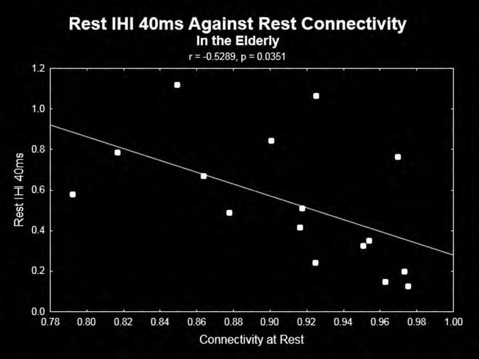
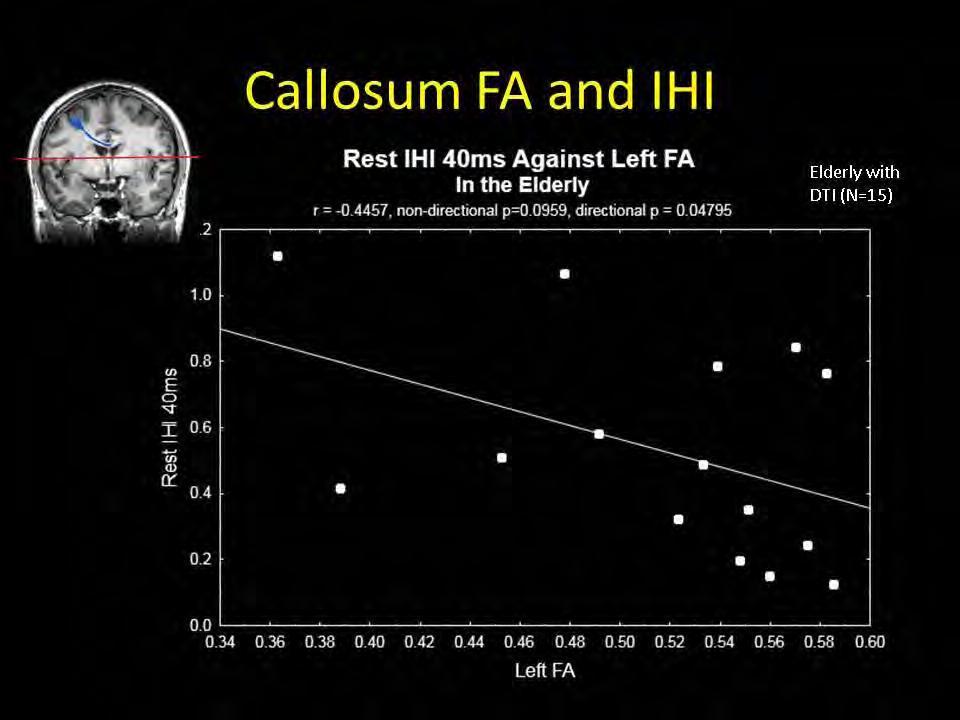
293 293

294 Poster Session II Multimodal Approaches P 199 Prefrontal transcranial direct current stimulation (tdcs) changes negative symptoms and functional connectivity MRI (fcmri) in a single case of treatment-resistant schizophrenia *D. Keeser 1, M. Kupka 2, U. Palm 1, J. Blautzik 2, O. Pogarell 1, B. Ertl-Wagner 2, H. Reichard 1, N. Müller 1, P. Falkai 1, F. Padberg 1 1 Ludwig-Maximilians-University, Department of Psychiatry and Psychotherapy, Munich, Germany 2 Ludwig-Maximilian University, Institute for Clinical Radiology, Munich, Germany, Germany Background: Transcranial Direct Current Stimulation (tdcs) has been associated with improvement in affective disorders. However, little is known about the influence oft tdcs on treatment-resistant schizophrenia with predominant negative symptoms. In this case study we looked on the effects of prefrontal tdcs on schizophrenic symptoms and functional MRI connectivity (fcmri) changes. Methods: The 19year old patient was diagnosed with paranoid schizophrenia (DSM-IV: ). He was treated with olanzapine 20 mg/day over 8 weeks without relevant psychopathological changes of paranoia and avolition. The patient underwent 10 tdcs sessions with the anode over the left DLPFC and the cathode over the right supraorbital region using a stimulation intensity of 2 ma and a stimulation duration of 20 min (Eldith Stimulator, electrode size: 7x5 cm 2 ).The following clinical ratings and tests were used at baseline, after 5 an 10 tdcs treatments: PANSS, SANS, CDSS, SOPT, TMT. fcmri was recorded at baseline, immediately after one tdcs sesion and after 10 days of tdcs (3.0 Tesla standard clinical scanner (Achieva TX, Philips Healthcare)). Results: After two weeks of tdcs treatment there was a considerable change in psychopathology. PANSS total score declined by 29%, SANS total score declined by 28%, Subjective feelings of depression (CDSS) were reduced by 82%. There was a pronounced improvement in the Trail Making Test (TMT) and the Self Ordered Pointing Task (SOPT). The fcmri in the subgenual region was significantly reduced immediately after tdcs stimulation and this effect was even more extended after 10 days of tdcs (p<0.0005, uncorrected) for the Default Mode Network (DMN). For the frontal-parietal network (FPN) the frontal and parietal connectivity was increased (p<0.0005, uncorrected). In the executive control network we found increased activity in the posterior cingulate gyrus (p<0.0001), increased bilateral insular corex activation (p<0.0005, uncorrected) and reduced cingulate cortex activations (p<0.0005). Conclusion: The significant fcmri changes are consistent to the clinical and cognitive improvements of the patient. tdcs of the dorsolateral prefrontal cortex seems to be a promising tool for the treatment of negative symptoms in schizophrenia. However, larger studies are needed for investigating the role of the specific effects of tdcs on the symptom variety in schizophrenia. 294
295 Poster Session II Multimodal Approaches P 200 Coupling of the sensorimotor cortical activation explored by blood-oxygenation-level-dependent signal navigated and traditional magnetic evoked potential recordings *M.- K. Lu 1,2, J.- R. Duann 1, C.- M. Chen 3, C.- H. Tsai 1,2 1 China Medical University, Taichung, Taiwan 2 China Medical University Hospital, Neurology, Taichung, Taiwan 3 China Medical University Hospital, Radiology, Taichung, Taiwan Introduction: The blood-oxygenation-level-dependent (BOLD) signal in functional magnetic resonance imaging (fmri) reflects associated hemodynamics for a specific motor task. Motor evoked potentials (MEPs) represent the excitability of the motor cortex. Recently the MEP recording can be visually guided by the BOLD signal in fmri. However, the two methods may represent diverse physiological mechanisms 1,2. Objectives: To study the coupling of the sensorimotor cortical activation observed in fmri BOLD signals and measured by traditional MEP recordings. Materials & Methods: Ten healthy, right-handed subjects (7 men, mean age: 34.6 ± 7.6 years) participated in this study. Functional imaging was acquired by using a 3.0T GE MR scanner with a single-shot EPI sequence. The subjects were requested to perform simple, repetitive abduction movement at right index finger while they saw a go-signal appeared on the projection screen and to take a rest during no-go signal periods. Transcranial magnetic stimulation (TMS) was delivered through a focal figure-of-eight stimulating coil. The coil was held tangential to the scalp with the handle pointing backwards and ~45 away from the midline. Twenty single TMS trials were measured with a randomly varying intertrial interval ranging from s at four coil positions. The electrophysiological optimal (EO) position was determined as the site where TMS at a slightly suprathreshold intensity produced consistently the largest MEPs in right first dorsal interosseus (FDI). The site was marked on the scalp to assure a constant placement of the coil throughout this session. The other three positions were determined by a frameless MR-navigation system (Visor TM ). The maximal BOLD signals at left primary motor cortex (BM1), left primary sensory cortex (BS1) and the lateral margin of the BOLD activity (BL) were targeted by the guidance of the navigation system. Two TMS intensities which were adjusted to produce MEPs of about 0.5 mv (low intensity) and 1 mv (high intensity) in peak-to-peak amplitude in the right FDI muscle were tested. In total 8 recording sessions including 4 positions and 2 intensities were completed by a randomized order for each subject. The mean MEP amplitude of each session was analyzed by a two-way repeated measures analysis of variance (rmanova) with the within-subject factors of POSITION and INTENSITY. Results: All subjects demonstrated a clear BOLD signal in the left sensorimotor cortex with response to the right finger movement. RmANOVA of the MEP amplitudes revealed significant main effects of POSITION and INTENSITY, and a significant interaction between POSITION and INTENSITY. The post-hoc analysis revealed a significant MEP difference between the four positions except EO-BM1 and BS1-BL at high intensity stimulation (all p<0.01). At low intensity stimulation, the MEP difference is significant only at the positions of EO-BL and BM1-BL (both p<0.02). Conclusion: Findings suggest that the coupling of the BOLD signals and the MEPs can be TMS-intensity dependent. A distinct localization of the BOLD signal in M1 or S1 by the TMS-navigation system may significantly vary the MEP findings under a high TMS-intensity condition. References: Kriváneková L,Baudrexel S,Bliem B,Ziemann U. Relation of brain stimulation induced changes in MEP amplitude and BOLD signal. Brain Stimul 2012 [Epub ahead of print] Antal A, Polania R, Schmidt-Samoa C, Dechent P, Paulus W. Transcranial direct current stimulation over the primary motor cortex during fmri. Neuroimage 2011;55:
296 Poster Session II Multimodal Approaches P 201 The value of simultaneous TMS-EEG recordings for diagnostic purposes in epilepsy patients *D. Klooster 1, B. Kleine 1, M. Munneke 2, H. van Dijk 1, D. Stegeman 2, M. Zwarts 1 1 Kempenhaeghe, Clinical Physics, Heeze, Netherlands 2 Donders Institute for Brain, Cognition and Behaviour, Radboud University Nijmegen Medical Centre, Neurology/Clinical Neurophysiology, Nijmegen, Netherlands Introduction: Epilepsy is one of the most common neurological diseases. The diagnosis of epilepsy is currently based on clinical history together with EEG recordings. However, low sensitivity is a major disadvantage of the standard EEG recording. Therefore, patients with unclassified spells often undergo multiple EEG registrations before a diagnosis and treatment plan can be made. To increase the sensitivity of the EEG, it would be beneficial to be able to provoke the occurrence of epileptiform activity in the EEG. Transcranial magnetic stimulation (TMS) is a non-invasive method that can be used to stimulate parts of the human brain. It is now possible to record TMS and EEG simultaneously. In this way, the changes in brain activity induced by TMS can be recorded with EEG. Objectives: We are going to investigate the possibility to induce epileptiform activity with TMS in patients with epilepsy. We expect to see abnormal discharges on TMS-EEG and we expect that these findings are of diagnostic and localizing value for epilepsy patients. Materials and methods: 10 healthy volunteers and 30 patients (10 focal epilepsy, 10 generalized epilepsy, 10 cortical dysplasia patients) will be studied. Single pulse TMS will be performed at an intensity of the resting motor threshold. In total, 16 pulses will be delivered at the standard electrode positions according to the system. After eight stimuli at each position, the EEG responses are averaged. If no response is seen, the stimulation intensity will be increased. Results and conclusion: Preliminary results and conclusions will be presented at the conference. 296
297 Poster Session II Multimodal Approaches P 202 Time-frequency analysis of short-lasting modulation of EEG induced by intracortical and transcallosal paired TMS over motor areas *P. Manganotti 1,2, E. Formaggio 2, S. F. Storti 1, A. Zamboni 1, A. Del Felice 1, G. Toffolo 3 1 University of Verona, Neurological Sciences, Verona, Italy 2 IRCCS, Venice, Italy 3 University of Padua, Department of Information Engineering, Padova, Italy Dynamic changes in spontaneous electroencephalogram (EEG) rhythms can be seen to occur with a high rate of variability. An innovative method to study brain function is by triggering oscillatory brain activity with transcranial magnetic stimulation (TMS). Methods: EEG-TMS coregistration was performed on five healthy subjects during a 1-day experimental session that involved four steps: baseline acquisition, unconditioned single-pulse TMS, intracortical inhibition (ICI, 3 ms) paired-pulse TMS, and transcallosal stimulation over left and right primary motor cortex (M1). A time-frequency analysis based on the wavelet method was used to characterize rapid modifications of oscillatory EEG rhythms induced by TMS. Results: Single, paired, and transcallosal TMS applied on the sensorimotor areas induced rapid desynchronization over the frontal and central-parietal electrodes mainly in the alpha and beta bands, followed by a rebound of synchronization, and rapid synchronization of delta and theta activity. Conclusions: Wavelet analysis after a perturbation approach is a novel way to investigate modulation of oscillatory brain activity. The main findings are consistent with the concept that the human motor system may be based on networklike oscillatory cortical activity and might be modulated by single, paired, and transcallosal magnetic pulses applied to M1, suggesting a phenomenon of fast brain activity resetting and triggering of slow activity. 297
298 Poster Session II Multimodal Approaches P 203 A Historical Developement of Transcranial Electrical Stimulation: Dose developement from 1900 to contemporary apporaches *B. Guleyupoglu 1, M. Bikson 1, F. Fregni 2, P. Schestatsky 2 1 The City College of New York, Biomedical Engineering, New York, United States 2 Harvard Medical School, Boston, United States Transcranial Electrical Stimulation (TES) encompasses all forms of non-invasive current application to the brain in regards to research and clinical applications. Approaches to TES have evolved in both terminology and dosage over the past 100 years of research. We outline the dose and historical development of TES since 1900 through modern approaches. Contemporary transcranial Pulsed Current Stimulation (tpcs) approaches such as Cranial Electrotherapy Stimulation (CES) and NeuroElectric Therapy (NET) descended from Electrosleep (ES) while others like Transcutaneous Cranial Electrical Stimulation (TCES), and Limoge, descended from Electroanesthesia (EA). Contemporary approaches such as transcranial Direct Current Stimulation (tdcs) and transcranial Alternating Current Stimulation (tacs) have historical analogues. We consider the role of seminal conferences and medical device regulations in the evolution of techniques and terminology. Select commercial devices and brands are noted for context. We clarify and disambiguate TES terminology including varied conventions across countries, and non-tes techniques. Understanding the roots of contemporary TES approaches in comparable approaches spanning decades, as well as the resurgence and abandonment of past techniques, may inform ongoing TES research. 298
299 Poster Session II Novel Techniques P 204 Additive effect of repetitive transcranial magnetic stimulation and anticonvulsants *V. Kistsen 1, V. Evstigneev 1, B. Dubovik 2 1 Belarusian Medical Academy of Postgraduate Education, Minsk, Belarus 2 Belarusian State Medical University, Minsk, Belarus Objective: The aim of our study was to reveal of rtms and antiepileptic drugs (AEDs) interaction effects. Methods: The comparative analysis of anticonvulsive effects of rtms combination with different AEDs (carbamazepine, valproic acid, topiramate and gabapentin) was performed. The experimental data was obtained at maximal electroshock test (MEST) on Wistar rats which took AED in minimal effective doses. RTMS with 1 Hz frequency and difference intensities was performed by circular coil (Neuro-MS, Russia) at pike concentration of AEDs time. Absence of maximal tonic hind limb extension (MTHLE) and all seizure phases durations were estimated. Results of combined therapy groups were conferred with monotherapy (AED or rtms only) and control group (n=10 in each group). Results: RTMS has additive effect concerning MTHLE absence when assign with carbamazepine in ED 40 in 90% (р=0.01) and topiramate in ED 30 in 80% animals (р=0.035). Tonic phase duration significant shorten practically in all rtms+aeds groups, particular with topiramate (p=0.002), but had not potentiating effect with gabapentin (p=0.12). RTMS shortened phase of clonic seizures with body turning-over reflex loss and total seizure duration for all AEDs (p<0.05). Period after turning-over reflex recovery corrected by rtms in carbamazepine group only (p=0.04). Limbs pedaling phenomenon reduced in rtms+carbamazepine (p=0.01) and rtms+valproic acid (p=0.001) groups. Conclusions: Thereby, rtms has an additive effect with AEDs. Magnetic stimulation causes the most important anticonvulsive effects when arrange with carbamazepine and topiramate. Results of this study can to be a reason to include low-frequency rtms in epilepsy treatment with low AEDs dose what can deliver patients from negative side effects. 299
300 Poster Session II Novel Techniques P 205 Cortico-conus motor conduction time (CCCT) *H. Matsumoto 1, R. Hanajima 2, Y. Shirota 2, M. Hamada 2, Y. Terao 2, S. Ohminami 2, T. Furubayashi 3, S. Nakatani- Enomoto 3, Y. Ugawa 3 1 Japanese Red Cross Medical Center, Neurology, Tokyo, Japan 2 University of Tokyo, Neurology, Tokyo, Japan 3 Fukushima Medical University, Neurology, Fukushima, Japan Objective: To measure the conduction time from the motor cortex to the conus medullaris (cortico-conus motor conduction time, CCCT) for leg muscles using magnetic stimulation. Methods: Motor evoked potentials (MEPs) were recorded from right tibialis anterior muscle in 100 healthy volunteers. To activate spinal nerves at the most proximal cauda equina level or at the conus medullaris level, magnetic stimulation was performed using a MATS coil. Transcranial magnetic stimulation of the motor cortex was also conducted to measure the cortical latency for the target muscle. To obtain the CCCT, the latency of MEPs to conus stimulation (conus latency) was subtracted from the cortical latency. Results: MATS coil stimulation evoked reproducible MEPs in all subjects, yielding CCCT data for all studied tibialis anterior muscles. Conclusions: MATS coil stimulation provides CCCT data for healthy subjects. This novel method is useful for evaluation of corticospinal tract conduction for leg muscles because no peripheral component affects the CCCT. References related to MATS coil stimulation Matsumoto H, Octaviana F, Hanajima R, Terao Y, Yugeta A, Hamada M, Inomata-Terada S, Nakatani-Enomoto S, Tsuji S, Ugawa Y. Magnetic lumbosacral motor root stimulation with a flat, large round coil. Clin Neurophysiol 2009;120: Matsumoto H, Octaviana F, Terao Y, Hanajima R, Yugeta A, Hamada M, Inomata-Terada S, Nakatani-Enomoto S, Tsuji S, Ugawa Y. Magnetic stimulation of the cauda equina in the spinal canal with a flat, large round coil. J Neurol Sci 2009;284: Matsumoto H, Hanajima R, Shirota Y, Hamada M, Terao Y, Ohminami S, Furubayashi T, Nakatani-Enomoto S, Ugawa Y. Corticoconus motor conduction time (CCCT) for leg muscles. Clin Neurophysiol 2010;121: Matsumoto H, Shirota Y, Ugawa Y. Magnetic augmented translumbosacral stimulation coil stimulation method for accurate evaluation of corticospinal tract function in peripheral neuropathy. Neurol India 2010;58: Matsumoto H, Hanajima R, Terao Y, Yugeta A, Hamada M, Shirota Y, Ohminami S, Nakatani-Enomoto S, Tsuji S, Ugawa Y. Prominent cauda equina involvement in patients with chronic inflammatory demyelinating polyradiculoneuropathy. J Neurol Sci 2010:290: Matsumoto H, Hanajima R, Terao Y, Yugeta A, Hamada M, Shirota Y, Ohminami S, Nakatani-Enomoto S, Tsuji S, Ugawa Y. Cauda equina conduction time in patients with acquired demyelinating polyneuropathy. Clin Neurophysiol 2010;121:S171. Matsumoto H, Shimizu T, Okabe S, Konoma Y, Takahashi T, Hirakawa-Yamada M, Igeta Y, Hashida H. Recurrent spinal cord attacks in a patient with a limited form of neuromyelitis optica. Intern Med. 2011;50: Matsumoto H, Hanajima R, Terao Y, Hashida H, Ugawa Y. Neurophysiological analysis of the cauda equina in POEMS syndrome. Neurol Sci (in press). Matsumoto H, Konoma Y, Shimizu T, Okabe S, Shirota Y, Hanajima R, Terao Y, Ugawa Y. Aging influences central motor conduction less than peripheral motor conduction: A transcranial magnetic stimulation study. Muscle Nerve (in press). Tokushige S, Sonoo T, Maekawa R, Shirota Y, Hanajima R, Terao Y, Matsumoto H, Hossain MA, Sakai N, Shiio Y. Isolated pyramidal tract impairment in the central nervous system of adult-onset Krabbe disease with novel mutations in the GALC gene. Brain Dev (in press). 300
301 Poster Session II Novel Techniques P 206 Monitoring short term effects of repetitive transcranial magnetic stimulation (TMS) by TMS-evoked potentials in children *S. Bender 1 1 University of Technology, Dresden, Child and Adolescent Psychiatry, Dresden, Germany Question: Repetitive transcranial magnetic stimulation (rtms) allows non-invasive stimulation of the human brain. However, no suitable marker has yet been established to monitor the immediate rtms effects on cortical areas in children. TMS-evoked EEG potentials (TEPs) could present a well-suited marker for real-time monitoring. Monitoring is particularly important in children where only few data about rtms effects and safety are currently available. Methods: In a single-blind sham-controlled study, twenty-five school-aged children with ADHD received subthreshold 1Hz-rTMS to the primary motor cortex. The TMS-evoked N100 was measured by 64-channel- EEG pre, during and post rtms, and compared to sham stimulation as an intraindividual control condition. Results: TMS-evoked N100 amplitude decr eased during 1Hz-rTMS and, at the group level, reached a stable plateau after approximately 500 pulses. N100 amplitude to supra-threshold single pulses post rtms confirmed the amplitude reduction in comparison to the pre-rtms level while sham stimulation had no influence. EEG source analysis indicated that the TMS-evoked N100 change reflected rtms effects in the stimulated motor cortex. Amplitude changes in TMS-evoked N100 and MEPs (pre versus post 1Hz-rTMS) correlated significantly, but this correlation was also found for pre versus post sham stimulation. Conclusions: The TMS-evoked N100 represents a promising candidate marker to monitor rtms effects on cortical excitability in children with ADHD. TMS-evoked N100 can be employed to monitor real-time effects of TMS for subthreshold intensities. Though TMS-evoked N100 was a more sensitive parameter for rtmsspecific changes than MEPs in our sample, further studies are necessary to demonstrate whether clinical rtms effects can be predicted from rtms-induced changes in TMS-evoked N100 amplitude and to clarify the relationship between rtms-induced changes in TMS-evoked N100 and MEP amplitudes. The TMSevoked N100 amplitude reduction after 1Hz-rTMS could either reflect a globally decreased cortical response to the TMS pulse or a specific decrease in inhibition. 301
302 Poster Session II Novel Techniques P 207 Reliability of an automated protocol versus manual interpreters in analysing cortical silent period *P. Julkunen 1, E. Kallioniemi 1, L. Säisänen 1, M. Könönen 1 1 Kuopio University Hospital, Kuopio, Finland Introduction: Cortical silent period (csp) is measured after shortly interrupting active muscle contraction with transcranial magnetic stimulation (TMS) [1]. The csp is a measure of cortical inhibition and representing interneuron inhibitory effect at excited motor cortical areas. Several pathological conditions and pharmacological manipulations induce changes to csp duration. In addition, csp has exhibited prognostic value e.g., during stroke recovery [2]. It has been suggested that input-output characteristics of csp be determined for thorough assessment of inhibitory interneurons [3,4]. These characteristics are commonly analyzed manually from measured electromyography (EMG) signal. However, to avoid interinterpreter effects in csp interpretation and detection, as well as to allow quick measurement in real-time, an automatic analysis routine would be preferable. Methods: We reanalyzed previously manually analyzed csps [5] of the right hand of 55 healthy subjects (27 male, 28 female, age range: 23-80) using a novel automatic routine. Five csps were induced at 120% of the resting motor threshold (rmt) focused on the left M1. Furthermore, we recruited one female (age 28) subject for whom the csps were induced with several stimulation intensities (SIs), and those csps were analyzed manually by two of the authors as well as using the automatic routine. In the automatic routine, we computed the first time-derivative of the single-trial EMG signal (smoothed), and detected longest interval with lower than 15µV difference between consecutive samples. Results: We found that 99% of all csps were identified correctly by the automated routine. There was a good agreement between the csp durations analyzed manually and automatically (ICC=0.972, p<0.001, Figure). Based on ANOVA, the automatic and manual routine did not exhibit significant differences, while the between-subject effect was significant (p<0.001). When studying the effects of SI and analysis type, we found SI had an effect on the csp duration (p<0.001), but not the analysis type (manual or automatic). Between two interpreters, the agreement in manually analyzed csp durations was excellent (ICC=0.990, p<0.001, 95%CI for the difference: 12 ms), as it was between the automatic and manual analysis (mean of two interpreters) (ICC=0.990, p<0.001, 95%CI for the difference: 12 ms). The difference between interpreters was similar to that between manual and automated analysis (Figure). Conclusions: Use of automatic csp detection may enable new mapping modality based on csp duration, e.g. in patients with lowered cortical excitability, the csp having a lower threshold than motor evoked potential [3]. Also, automatic analysis routine will allow for a quick assessment of input-output curve for the csp improving its applicability. References: [1]Fuhr 1991; [2]Curra 2002; [3]Werhahn 2007; [4]Kimiskidis 2005; [5]Säisänen 2008; [6]Julkunen Figure: The agreement between the manually and automatically analyzed csp durations (left). The scatter plot shows the analyzed csps with both techniques for 55 subjects, 5 csp trials each. The csp durations measured at different SIs from one subject were analyzed manually by two interpreters and automatically. Those csps were constructed to a threshold curve [6]. The agreement between the interpreters and the automatic routine was very similar (right). The rmt is indicated with a vertical dotted line. 302
303 Poster Session II Novel Techniques P 208 A K-Rb hybrid Optically Pumped Atomic Magnetometer toward Ultra-low field Multimodal MRI Systems *T. Kobayashi 1, Y. Ito 1, M. Ohnishi 1, K. Mamada 1, T. Oida 1 1 Kyoto University, Department of Electrical Engineering, Kyoto, Japan Introduction: To contribute for improvement of longevity and quality of human life, we have been developing optically pumped atomic magnetometers (OPAMs) to measure tiny biomagnetic fields and MRIs. In recent years, OPAMs have reached sensitivities comparable to and even surpassing those of magnetometers based on super-conducting quantum interference devices (SQUIDs) 1. In addition, OPAMs have the intrinsic advantage of not requiring cryogenic cooling. Meanwhile, MRI is one of the most useful diagnostic imaging modalities, which enables visualization of the anatomy and function of the human. However, the conventional high magnetic field scanner has some limitations such as high cost and risk for patients with metal implants. To acquire MRIs without these limitations, ultra-low filed (ULF) MRI systems attracted attention in recent years. Objective: We have developed an OPAM using a hybrid cell of K and Rb atoms 2 toward ULF multimodal MRI systems. The objective of this study is to investigate the optimal properties of the OPAM theoretically comparing with experiment results. Materials & Methods: The sensor head of the OPAM used in this study was a cubic Pyrex glass cell, whose size was 3 x 3 x 3 cm 3 and within which K and Rb atoms were enclosed with He and N 2 as buffer gases at a ratio of 10 to 1 and at a total pressure of 150 kpa at room temperature. The Rb atoms were spin-polarized by a circularly-polarized pump beam and the spin polarization was transferred to the K atoms by spin exchange collisions. The spin polarization rotated around the external magnetic field orthogonal to both the pump and probe beams. Here, the plane of a linearly-polarized probe beam penetrating the group of spin-polarized atoms is rotated by Faraday effect. We applied a 100 Hz sinusoidal magnetic field of 48 pt as a test signal and examined sensitivities of the OPAM Results: We theoretically investigated the properties of the hybrid OPAM and considered the adequacy of the properties comparing with experiment results. We have found that the experimental results agree well with the theoretical values. The optimum density ratio of K and Rb atoms was expected to be for more sensitive magnetometers. We could measure of human magnetocardiograms (MCGs) and plan to measure MR signals using the hybrid OPAM. Conclusion: We developed a K-Rb hybrid OPAM and examined its sensitivities. Results shown in this study demonstrate the feasibility of the OPAM as a magnetic sensor toward ULF multimodal MRI systems. This work is partly supported by Grant-in-Aid for Researches (No & ) and the Innovative Techno-Hub for Integrated Medical Bio-imaging of the Project for Developing Innovation Systems, from MEXT, Japan. References: [1] D. Budker, and M.V. Romalis, Nature Phys., vol.3, pp , [2] Y. Ito, et al., IEEE Trans. Magn., vol.47, no.10, pp ,
304 Poster Session II Novel Techniques P 209 Transcranial random noise stimulation: A new approach to stimulating the brain *K.- A. Ho 1,2, J. Taylor 3,4, C. Loo 1,2,5 1 University of New South Wales, School of Psychiatry, Sydney, Australia 2 Black Dog Institute, Sydney, Australia 3 University of New South Wales, School of Medical Sciences, Sydney, Australia 4 Neuroscience Research Australia, Sydney, Australia 5 St George Hospital, South Eastern Sydney Health, Australia Introduction: Transcranial random noise stimulation (trns) is a neuromodulatory technique that involves the delivery of a bi-directional, randomly oscillating current. Introduction of a positive DC offset to the stimulation can produce a polarity-specific randomly oscillating current that produces effects similar to that of transcranial direct current stimulation (tdcs). It is thought that trns modulates cortical excitability by interfering with the ongoing neural oscillations in the cortex. In contrast to using a direct current, trns may avoid the homeostatic neural mechanisms associated with repeated stimulation sessions. This may be an advantage in clinical treatment protocols which seek to induce cumulative neuroplastic changes over multiple sessions. To date, there has only been one reported use of trns with a positive DC offset for the treatment of depression. Findings were promising, suggesting therapeutic potential for this form of stimulation (Chan et al., 2012). Objectives: The present study aimed to elucidate the clinical potential of trns with a positive offset by examining its effects on motor cortical excitability in healthy participants. We aimed to examine the effect of 2 ma trns + 1 milliamp (ma) offset for 10 min on cortical excitability by using single pulse transcranial magnetic stimulation (TMS). This was compared to four other transcranial electrical stimulation (tes) conditions as follows: 2 ma trns without an offset, 1 ma tdcs, 2 ma tdcs and sham stimulation. Materials & methods: Fifteen healthy participants will be tested across five sessions in a within-subjects design. One form of tes was tested at each session, the order of which was randomised for each participant. tes was applied to the left motor cortex. Sets of 20 motor evoked potentials (MEPs) were elicited in the right first dorsal interosseus (FDI) muscle using single-pulse TMS before and up to 90 min after tes. Peak-to-peak amplitude of each MEP was measured. The mean amplitude of all responses after tes was calculated and normalised to the baseline amplitude for each subject. Results: Results from the first 10 participants show mean (sd) post-tes normalised MEPs as follows: 2 ma trns + 1 ma offset, 1.44 (0.38); 2 ma trns, 1.01 (0.11); 1 ma tdcs, 1.24 (0.15); 2 ma tdcs, 1.30 (0.18) and sham, 1.06 (0.13). Results from all 15 participants will be subsequently presented. Conclusion: This is the first empirical study examining the effect of trns with an offset on cortical excitability. Preliminary results suggest that trns with an offset leads to an increase in cortical excitability similar to that produced by tdcs. Further, trns with an offset appears to be more effective than trns without an offset in producing changes in cortical excitability. References: Chan, H. N., Alonzo, A., Martin, D. M., Player, M., Mitchell, P. B., Sachdev, P., & Loo, C. K. (2012). Treatment of major depressive disorder by transcranial random noise stimulation: case report of a novel treatment. Biological Psychiatry, 72(4), e9-e10. Funding Sources: KAH is supported by an Australian Post-Graduate Award and University of New South Wales Brain Sciences Top-Up Scholarship. 304
 on the brain depends on the")
305 Poster Session II Novel Techniques P 210 An automatic, computer-controlled TMS-coil calibrator *J. O. Nieminen 1, L. M. Koponen 1, R. J. Ilmoniemi 1 1 Aalto University, Department of Biomedical Engineering and Computational Science, Espoo, Finland The effect of transcranial magnetic stimulation (TMS) on the brain depends on the focality of the induced electric field (E-field). However, with commercial TMS coils, it is typically not known precisely how the E- field behaves as a function of distance from the coil. Our aim was to develop an automatic, computercontrolled calibrator for measuring the E-field induced by TMS coils. Such an instrument would allow comparing different coils and help in ensuring that an investigator knows the properties of the E-field. In the spherical head model, the triangle construction, where two radial wires extending from the origin are connected with a short tangential path, allows straightforward computation or measurement of the E-field induced by an external coil. Thus, we constructed two orthogonal triangular loops (radial edges 70 mm, tangential path 5 mm, i.e., 4.1 degrees) using 0.15-mm-thick copper wire. To reach sufficient precision, we used additive manufacturing for making a coil former for the wires. The orientation of the triangles, which defines the measurement point, is controlled by two servo motors, which rotate the triangles about the origin. This allows for measuring the induced E-field on the surface of a hemisphere: the voltage induced in the triangular loops by a TMS coil is proportional to the tangential components of the E-field that would be induced at the position of the tangential edges in a spherically symmetric conductor. The calibrator is shown in the figure. The measurement, which is operated by a LabView program, proceeds as follows: We connect a TMS coil in series with a signal generator and apply a sinusoidal current time-locked to the measurement to produce an alternating magnetic field. The time-varying magnetic field induces a voltage in the triangles which is amplified and fed to a National Instruments data-acquisition (DAQ) system connected to a laptop. The DAQ system is also used to control the movement of the servo motors so that the measurement points cover the surface of a hemisphere with uniform density (typically, 1000 points). To minimize the time needed for the servo movement, we traverse through the measurement points in an order that is an approximate solution to a travelling salesman problem. After the measurement completes, we analyze the acquired data with a Mathematica program: By fitting a sinusoid to the data measured at each point, we can extract the amplitude and direction of the induced E-field. Finally, we visualize the field and extract some measures, e.g., the maximum amplitude or the focality of the field. We have used the calibrator to measure the E-field produced by a home-made coil. The measurement results agreed well with the calculated field values. We will use the device for characterizing several commercially available TMS coils. In summary, the developed calibrator gives a simple means to measure the E-field produced by any TMS coil. Furthermore, by changing the triangle module, the same instrument can measure the E-field at different depths. The measurement of the spatial profile of the induced E-field of TMS coils helps in ensuring that an investigator knows the properties of the E-field. 305

, which defines the given stimulus. This is known as the forward problem, and there exist no general analytic solution for it.")
, where we split the volume into small polyhedrons, and the boundary element method (BEM), where we divide the system")

306 Poster Session II Novel Techniques P 211 Efficient Method for Computing the TMS-Induced Electric Field in Spherically Symmetric Head Models, the Triangle Construction *L. M. Koponen 1, J. O. Nieminen 1, R. J. Ilmoniemi 1 1 Aalto University School of Science, Department of Biomedical Engineering and Computational Science, Espoo, Finland In transcranial magnetic stimulation (TMS), we are interested in calculating the TMS-induced electric field (E-field), which defines the given stimulus. This is known as the forward problem, and there exist no general analytic solution for it. This is because the electric field is dictated by Maxwell's equations, which are partial differential equations. Thus, for non-trivial boundary conditions, numerical methods are needed. General methods for solving these equations are the finite element method (FEM), where we split the volume into small polyhedrons, and the boundary element method (BEM), where we divide the system into uniform-conductance regions and split the formed boundaries into small polygons. Both of these methods can be applied for nearly arbitrary geometries but are hard to parallelize. However, often one can simplify the analysis by using a spherically symmetric head model. For example, if one is to assess the general properties of a stimulator coil, using a more complicated head model will just increase the computational cost and complicate the analysis of the results. Also when the head can be approximated being locally spherical, the spherical symmetric head model allows a significant simplification in the computations. This is because the spherical symmetry of the conducting medium allows us to compute an analytic solution for the forward problem. We will present an efficient method for computing the induced E-field. The model works for spherically symmetric head models and is based on the triangle construction, (R. J. Ilmoniemi, M. S. Hämäläinen, and J. Knuutila, The forward and inverse problems in the spherical model, in Biomagnetism: Applications & Theory, eds. H. Weinberg, G. Stroink, and T. Katila, Pergamon Press, Amsterdam 1985, pp ); see Figure. Devices based on the triangle construction have been used to calibrate magnetoencephalography (MEG) and TMS systems. Now, to obtain the two tangential components (the radial component is always zero) of the induced E-field in a point, one simply has to calculate the mutual inductance between the TMS coil and two perpendicular triangular loops with one corner at the sphere center and the opposite edge tangential to the spherical surface. For the mutual-inductance calculations, the TMS coil can be modeled as a set of current-carrying filaments. Then, the mutual inductance between the coil and the triangle loops can be calculated using analytic formulas. Using the triangle construction, we developed an O(n) time and memory algorithm for the TMS forward problem, where n is the number of points where the induced E-field is computed. This is a considerable improvement over O(M 2 ) for the BEM or O(N) for the FEM, where M is the number of surface elements, and N is the number of volume elements. (For FEM, we need much more 3D volume elements than points in our construction because we have to sample also the volume surrounding the system. For BEM, we need to sample all the 2D surfaces, also the uninteresting ones. Thus, for a small region of interest, N>>M>>n.) The developed algorithm is very simple compared to those for BEM or FEM as it does not require any tessellation. Also, because in our model the computation for the field in a point does not depend on the field in the other points, the algorithm is embarrassingly parallel, i.e., trivial to parallelize with minimal overhead. Figure: An illustration of the triangle construction and the induced E-field of a figure-of-eight coil calculated using the developed algorithm. 306
307 Poster Session II Novel Techniques P 212 A novel high sensitivity MR-device for combined TMS/fMRI experiments *L. Navarro de Lara 1, C. Windischberger 1,2, J. Sieg 1,2, E. Moser 1,2, E. Laistler 1,2 1 Medical University Vienna, Center for Medical Physics ans Biomedical Engineering/ MR Center of Excellence, Vienna, Austria 2 Medical University of Vienna, MR Centre of Excellence, Vienna, Austria Introduction: Studies with simultaneous functional magnetic resonance imaging (fmri) and transcranial magnetic stimulation (TMS) have become increasingly popular as the combination of these techniques allows investigation of TMS effects on the brain and could help answer unresolved questions concerning causality in fmri [1]. So far, experiments are being performed using rather large, standard MRI head coils, with the TMS positioned between head and MR coil, resulting in poor sensitivity for the MRI experiment, while also preventing visualization of activation spots directly below the TMS focus. Objective: To gain sensitivity for combined TMS and fmri experiments, we developed a dedicated, slim MR coil on a curved, form-fitted surface that can be placed between TMS and the head. This way, the TMS is close enough to the brain to achieve efficient stimulation, and the MR coil can provide far higher sensitivity than in the conventional arrangement due to its proximity to the head. This way, the TMS coil itself does not cause artifacts in the fmri maps. Materials and methods: This dedicated receive coil is designed for 3 T MR systems, with a field of view of 15 cm and a target depth of cm. It consists of seven hexagonally arranged loop elements of 6 cm diameter each. To decouple adjacent elements, loops were overlapped. The array is placed on a spherical support with a curvature to fit the average human head. An illustration of the device positioned for investigations in the dorsolateral prefrontal cortex is shown in Fig. 1. All elements of the MR coil are passively and actively detunable for decoupling from the MR system's body coil during signal transmission. A second stage matching network [2] connects the coil elements to low noise preamplifiers, additionally improving mutual decoupling between array elements. Preamplifiers are not placed directly on the coil, minimizing coil thickness to assure efficient TMS stimulation, and avoiding damage to the preamplifiers by TMS pulses. Results: Mutual coupling between elements of the MR coil is below -9 db for all channels. Additional -20 db decoupling was achieved by preamplifier decoupling, minimizing noise correlation between elements, and thus, enabling parallel imaging with low g-factors. The dedicated MR coil is well isolated from the transmitting MR body coil, proven by field distortions well below 5%. This avoids image artifacts and ensures patient safety. A gradient echo image of a phantom acquired with one of the seven elements is shown in Fig. 2, demonstrating the high sensitivity of the basic elements in cortical areas. Conclusion: A dedicated 7-channel MR coil array for simultaneous fmri/tms measurements is presented. Individual coil elements were efficiently decoupled from each other and from the transmit body coil. Due to the substantial gain in SNR and good isolation between channels, parallel imaging can also be implemented to speed up TMS-fMRI measurements. This novel hardware development will boost sensitivity of fmri in combined fmri/tms experiments, enabling fast and high-resolution fmri while efficiently and safety stimulating the brain with TMS. Acknowledgements: This work was funded by the Austrian BMWFJ, FFG Project Nr
![References: [1]](/docs-images/76/73180322/images/308-0.jpg "Kobayashi K, 2003, The")
![Lancet, 2:145-156 [2]](/docs-images/76/73180322/images/308-1.jpg "Reykowski S.")
308 References: [1] Kobayashi K, 2003, The Lancet, 2: [2] Reykowski S.M, 1995, MRM,
309 Poster Session II Novel Techniques P 213 Repetitive Transcranial Magnetic Stimulation (rtms) in Organic Personality and Behavioral Disorder, secondary to Suicide attempt by Hanging. *N. Kumar 1, S. Jha 1 1 All India Institute of Medical sciences (AIIMS), Psychiatry, New Delhi, India Background: Hanging is a frequently used method of suicide in many countries (1). Hanging is a particularly lethal mode of suicide with an estimated fatality rate of over 70 %.( 2,3,4).In cases of hanging reduced blood supply and deficiency of available oxygen alter brain perfusion which may causes hypoxia or anoxia that may lead to behavioral abnormality. Single-Photon Emission Computed Tomography (SPECT) is a valuable tool to investigate regional cerebral blood flow (rcbf). repetitive Transcranial Magnetic Stimulation (rtms) is a relatively new noninvasive technique that can modulate cortical function that may translate into behavioral alteration, depending upon the site of stimulation. In our rtms clinic we observed remarkable improvement in a person suffering from post hanging organic personality disorder after rtms intervention. We report a case of suicide attempt of an unmarried 27 year old female who attempted suicide by hanging when she was 21 years of age but rescued by the family members with marked behavioral changes after the suicide attempt. Post-rescue the person became unconscious and subsequently developed abnormal behavior characterized by irrelevant talking and laughing, markedly decreased self care, memory impairment and incontinence with severe socio-occupational dysfunction. The person was diagnosed with organic personality and behavior disorder as per ICD -10 diagnostic criteria and prescribed oral Phenytoin and Risperidone without much improvement.since there was no other alternative treatment the rtms technique was discussed with the caregivers. After the informed consent the person underwent SPECT. The SPECT showed hypo perfusion in left inferior frontal cortex which corresponds to left Orbito Frontal Cortex (OFC) area. Method: The person was assessed at baseline with Neuropsychiatric Rating Scale (NRS), Clinical Global Impression (CGI), Global Assessment of Functioning (GAF) and Mini-Mental Status Examination (MMSE). After baseline assessment the person was administered rtms (Frequency: 10 Hz, Motor Threshhold: 100%, Power: 53%, Duration: 10 seconds. Total trains: 20. Total pulse: 2000). Subsequently the patient was assessed with similar set of instruments after the fifth session and 15 th rtms session to observe the outcome. Results: The comparison of NRS from baseline and end of rtms session reflected decrease in severity from from severe to moderat. The CGI score improved remarkably from 13 to 09 at the end of rtms session. The MMSE score improved from 13/30 to 21/30.Similarly there was remarkable improvement in the GAF score after the rtms intervention. Conclusions: Hanging leads to hypoxic brain damage that may leads to behavioral abnormality. This is the first Case to the best of our knowledge reflecting therapeutic use of rtms technique in organic personality changes secondary to suicidal attempt by hanging. The observation shows that role of rtms technique may be explored further in such behavioral abnormality secondary to organic brain changes. References: Farmer R, Rohde J. Effect of availability and acceptability of lethal instruments on suicide mortality. An analysis of some international data. Acta Psychiatr Scand, 1980;62: Simounet C, Bourgeois M. Suicides and attempted suicides by hanging. Ann Med Psychol 1992;150: Aufderheide TP, Aprahamian C, Mateer JR et al. Emergency airway management in hanging victims. Ann Emerg Med 1994; 24: Luke JL, Reay DT, Eisele JW, Bonnell HJ. Correlation of circumstances with pathological findings in asphyxial deaths by hanging: a prospective study of 61 cases from Seattle, WA. J Forensic Sci 1985;30:
310 Poster Session II Novel Techniques P 214 Visualization of transcranial magnetic stimulation effects by voltage-sensitive dye imaging *V. Kozyrev 1, S. Rekauzke 1, U. T. Eysel 2, D. Jancke 1 1 Ruhr-Universität Bochum, Neuroinformatics, Bochum, Germany 2 Ruhr-Universität Bochum, Neurophysiology, Bochum, Germany Transcranial magnetic stimulation (TMS) induces electrical currents in the brain which stimulate the neural tissue. Despite the widespread use of TMS, the neuronal mechanisms of TMS-induced activity are not well understood. Here we introduce a novel method of imaging TMS-evoked activity in-vivo in a cortical patch (~0.5 cm^2) stained by a voltage-sensitive dye (VSD). Molecules of the VSD, presumably bound to neuronal membranes, transduce changes of the membrane potential into an optical signal. This signal, originating from large neuronal populations, is recorded by a video camera at high temporal (5 ms) and spatial (~50 mkm) resolution, allowing the measurement of dynamics and spatial spread of a TMS pulse. We used VSD imaging to monitor activity induced in primary visual cortex of anesthetized adult cats by single and repetitive (rtms) pulses as well as alteration of neuronal function during and beyond the stimulation period. We observed a gradual build-up of ongoing cortical activity in response to each magnetic pulse within 10 Hz rtms trains. We hypothesize that this indicates the evolution of an excitatory cortical state that may facilitate plastic reorganization of orientation map layout. We compared the dynamics of evoked responses to oriented gratings before and after a protocol of 10 Hz rtms trains presented during min. First, a transient suppression within the rise time of responses, typically referred to as deceleration-acceleration notch in VSD recordings (see Sharon & Grinvald, 2002), was significantly diminished indicating weakened inhibition. Second, responses to non-preferred orientations increased while modulation depth, as a global measure of orientation selectivity, was unaltered. Third, preliminary data showed shifts in the balance of represented orientations within the map. Altogether, these findings imply that TMS-induced cortical reorganization processes are accompanied by increased overall plateau levels of nonspecific activity and reduced intracortical inhibition. Our results demonstrate that combining TMS with VSD imaging allows artifact-free visualization of TMS-induced activity in the animal brain and provides a powerful method to study plasticity of the functional cortical architecture. The project is funded by the Deutsche Forschungsgemeinschaft, SFB
311 Poster Session II Novel Techniques P 215 A method for online correction of artifacts in EEG signals during transcranial Electrical Stimulation *F. Schlegelmilch 1, K. Schellhorn 1, P. Stein 1 1 neuroconn GmbH, Ilmenau, Germany Introduction: Simultaneous recording of EEG during transcranial electrical stimulation (tes) is a noninvasive and painless method to evaluate the modulation of cortical oscillatory brain activity and cerebral plasticity. Latest investigations in neuroscience with transcranial direct current (tdcs), alternating current (tacs) or random noise current (trns) provide a wide area of research topics (Miniussi et al. 2012, Thut et al. 2011). The distortion of the EEG signals during tes makes it impossible to evaluate the EEG signals. Therefore methods for artifact correction are necessary to reconstruct the underlying EEG signal during tes. Objectives: The objective of this paper is to demonstrate a method for online artifact correction of EEG signals during tes. The measured distorted EEG signals are modeled as a linear superposition of the current artifact signals and raw EEG. A linear regression model can be used for subtraction of linear scaled current artifacts from the measured EEG. Therefore, an independent reference signal from the electrical stimulator has to be recorded simultaneously with the EEG signals. Materials and Methods: Two low noise, analogue signals were derived from the electrical stimulator (DC Stimulator PLUS, neuroconn GmbH, Ilmenau) as reference signals. The galvanic isolated signals (amplitude +/- 40mV) were fed into the EEG amplifier (full-band DC-EEG amplifier NEURO PRAX ) together with EEG, EOG and ECG signals. The galvanic isolation of the reference signals ensures the electrical safety for the patient. Sinusoidal stimulation in the alpha-band of the EEG (10 Hz) was performed over P3 and P4 locations using easycap s Ag/AgCl ring electrodes (current peak-to-peak amplitude: 1000uA). A short-time learning period (about 10 s) was used to calculate the scaling factor of the regression model. Online correction was tested with eyes-closed condition (generation of alpha-waves). Results: Figure 1 shows the uncorrected EEG, EOG, ECG signals and the two reference signals (tacs1, tacs2). Beside the ECG, all EEG and EOG signals are strongly distorted by current artifacts. By comparing the EEG signals at one time-stamp (e.g. 12:16:22), the current artifacts (amplitude from 5000uV to 30000uV) show different phase and polarity between the EEG channels. This has to be considered by the online correction method. Figure 2 shows the same time window of all corrected signals (compare the ECG signals and time stamps of figure 1 and 2). Blinking eyes artifacts, DC shifts of frontopolar signals as well as alpha-eeg are clearly visible in the EEG signals during eyes-closed condition. All signal parts which are phase-synchronized to the 10Hz sinusoidal stimulation, will be eliminated by the correction procedure. However, alpha-waves are still visible in T4, Pz, O1 and O2. Conclusions. Artifacts in EEG signals during single-channel tes can be easily corrected using reference signals from the tes device. This new method was implemented into neurconn's tes-eeg products. References: Miniussi, C., Brignani, D., Pellicciari, M.C. Combining Transcranial Electrical Stimulation With Electroencephalography: A Multimodal Approach. Clinical EEG and Neuroscience 2012; 43(3): Thut, G., Schyns, P.G., Gross, J. Entrainment of perceptually relevant brain oscillations by non-invasive rhythmic stimulation of the human brain. Frontiers in Psychology 2011; Vol. 2, Article
. Artifact amplitudes ranges from 5000uV to 30000uV.")
 and sinusoidal stimulation (10 Hz, 1000uApp) over P3")
312 Figure 1: Uncorrected EEG signals during sinusoidal stimulation (10 Hz). Artifact amplitudes ranges from 5000uV to 30000uV. Figure 2: Online corrected EEG signals during eyes-closed condition (alpha-waves) and sinusoidal stimulation (10 Hz, 1000uApp) over P3 and P4. 312
313 Poster Session II Novel Techniques P 216 Impairment of triad conditioned facilitation in amyotrophic lateral sclerosis *S. J. Groiss 1,2, H. Mochizuki 1,3, S. Nakatani-Enomoto 1, K. Otani 4, Y. Ugawa 1 1 Fukushima Medical University, Neurology, Fukushima, Japan 2 Heinrich-Heine University, Neurology / Clinical Neuroscience, Düsseldorf, Japan 3 University of Miyazaki, Neurology, Miyazaki, Japan 4 Fukushima Medical University, Orthopedic Surgery, Fukushima, Japan Introduction: Triad conditioned facilitation induced by transcranial magnetic stimulation (TMS) in healthy individuals has been proposed to reflect the intrinsic 40 Hz piper rhythm of the motor cortex. However, it is unclear if triad conditioned facilitation is influenced by degeneration of the motor cortex. Objective: Amyotrophic lateral sclerosis (ALS) is characterized among others by degeneration of the motor cortex. We therefore hypothesized that triad conditioned facilitation may be impaired in ALS patients. Moreover, we hypothesized that this triad conditioning paradigm may be helpful to differentiate ALS from cervical myelopathy, an important differential diagnosis. Material and Methods: In 10 patients with ALS and 9 patients with cervical myelopathy serving as disease control, we compared single pulse conditioned intracortical inhibition (ICI), intracortical facilitation (ICF) and also triad conditioned facilitation at various interstimulus intervals (ISI) between 3 ms and 50 ms. Results: There were no differences in ICI and ICF between both groups. Both groups also showed similar triad conditioned facilitation at 10 ms ISI. Patients with cervical myelopathy showed normal triad conditioned facilitation at 25 ms ISI comparable to healthy subjects. In contrast ALS patients showed no triad conditioned facilitation at 25 ms. Conclusion: The absence of triad conditioned facilitation at 25 ms ISI in ALS patients may represent changes of the intrinsic rhythm of the motor cortex probably caused by cortical degeneration. Triad conditioned TMS may be a valuable tool to differentiate ALS patients from patients with cervical myelopathy. 313
314 Poster Session II Novel Techniques P 217 Analysis of cortical reorganization after stroke S. Kuznetsova 1, *N. Skachkova 1 1 State Institution «Institute of Gerontology of NAMS of Ukraine», neurological, Kiev, Ukraine Introduction: Transcranial magnetic stimulation (TMS) is a valuable non-invasive method for investigation functional changes of the motor cortex after stroke [1-3]. Objectives: The purpose of this study was to assess the excitability of the motor cortex of affected hemisphere (AH) and unaffected hemisphere (UH) in patients with stroke. Materials and methods: The study involved 48 patients with cerebral hemispheric ischemic stroke in subacute period (mean age - 66,15±1,53 years). Single-pulse TMS was performed to evaluate motor evoked potential (MEP) and resting motor threshold (rmt) with figure-of-eight coil Cool-B65 connected to magnetic stimulator MagPro R100 (Medtronic A/S, Denmark). Results: The MEP from AH was not obtained even with facilitation in 27,08% patients. The MEP from AH was registered with facilitation in 16,67% patients. The significantly smaller MEP amplitude and the significantly higher rmt elicited from AH than that of MEP amplitude and rmt elicited from UH were observed in 31,25% patients. Only the significantly higher rmt elicited from AH than that rmt elicited from UH was occurred in 25% patients. Based on TMS results it is useful to distinguish four degrees of reducing excitability of the motor cortex of AH of post-stroke patients: rough, severe, moderate and mild. Conclusion: MEP and rmt are informative criteria for defining the functional state of the motor cortex and features of cortical reorganization after stroke. The graduation of reducing excitability of the motor cortex of AH is proposed for use in clinical practice and could be considered in evaluation the prognosis after stroke. Acknowledgement: This work is supported by the National Academy of Medical Sciences of Ukraine. References: Groppa S. A practical guide to diagnostic transcranial magnetic stimulation: report of an IFCN committee / S. Groppa, A. Oliviero, A. Eisen, A. Quartarone, L.G. Cohen, V. Mall, A. Kaelin-Lang, T. Mima, S. Rossi, G.W. Thickbroom, P.M. Rossini, U. Ziemann, J. Valls-Solé, H.R. Siebner // Clin. Neurophysiol Vol Hendricks H.T. Motor evoked potentials in predicting recovery from upper extremity paralysis after acute stroke / H.T. Hendricks, J.W. Pasman, J. van Limbeek, M.J. Zwarts // Cerebrovasc. Dis Vol Nascimbeni A. Motor evoked potentials: prognostic value in motor recovery after stroke. / A. Nascimbeni, A. Gaffuri, P. Imazio // Funct. Neurol Vol

315 Poster Session II Novel Techniques P 218 Optimized small animal transcranial magnetic stimulators using distributed coils *G. Crevecoeur 1, N. De Geeter 1, L. Dupré 1 1 Ghent University, Electrical Energy, Systems and Automation, Ghent, Belgium Question: The precise impact of TMS on the neural pathways and the mechanisms of action remain unknown. Performing studies of different coil configurations in human beings is restricted due to ethical reasons and it is difficult to gather statistically significant data of large human study groups. Therefore, to explore TMS in a systematic and flexible way, miniaturization of TMS for rodent brain studies is a complementary addition to the human studies. Current stimulators for small animal studies lack a high degree of focus of electric field. More optimal designs are thus needed. Methods: We aim at numerically optimizing a distributed planar coil array that can generate a focal electric field in rodent brains. The distributed coil array consists of NxN coils distributed in a rectangular grid (34mmx34mm). Since it is practically difficult to activate each coil with a different current, we aim at the activation of the coil array with a single current by placing the different coils in series connection. Since only a single current is flowing through the coil array, the geometrical configuration, e.g. radii and number of turns, of the different coils needs to be optimized. The numerical optimization is performed using a genetic algorithm [1] on the basis of the calculated electric fields. The electric fields are calculated starting from the magnetic fields. Further details on the electrical field solver can be found in [2]. The rat head is modeled as four concentric ellipsoids representing the tissues scalp, bone, cerebrospinal fluid (CSF) and brain. The minor and major axes of the inner layer brain are 14 mm and 28 mm long and the other layers are 1 mm and 2 mm thick near the minor and major axes respectively, similar to the rat s real dimensions. Results: We performed simulations onto a rectangular coil array with N=2 and N=4. We observed that N=2 yields a limited amount of degrees of freedom so to excite a focal volume of high electric field. N>4 is practically very difficult to implement because the radii of the coils would become too small. The optimal spatial electric field distribution in the sagittal plane is given in the figure here below for the N=4 distributed coil array (right). As a comparison, a non-optimized electric field distribution (left) is also given, illustrating the impact of the coil configuration parameters. In this configuration we fixed the outer radii of the coils to be 4mm, so that the coils can be placed within the rectangular grid, as well as the current direction in the different coils. The current directions were chosen similar to the current directions in the figure-8 coils. The optimization parameters were here the number of turns. About 500 genetic algorithm generations were needed for the optimization which resulted in approximately 8h computation time. Conclusions: An optimized small animal TMS is presented that enables focal stimulation by activating localized high electric fields. Simulations were performed on a simplified rat model and can be extended towards more realistic head models of rats. Moreover, the degrees of freedom in the design can be extended by including unknown radii of the coils. The numerically designed TMS can be deployed for further in-depth in-vivo studies that evaluate the impact of stimulation using neuro-imaging techniques. [1] G. Crevecoeur, et al. (2010) IEEE Trans. Magn. 46: [2] N. De Geeter, et al. (2012) Phys. Med. Biol. 57:
316 Poster Session II Novel Techniques P 219 Navigated repeated transcranial magnetic stimulation in stroke rehabilitation (randomized blind sham-controlled study), Preliminary results: safety and tolerability M. Piradov 1, L. Chernikova 1, M. Tanashyan 1, A. Kadykov 1, *A. Chervyakov 1, M. Nazarova 1, V. Gnezditsky 1, R. Konovalov 1, N. Savitskaya 1, P. Fedin 1, A. Suslin 1, M. Glebov 1, L. Dobrynina 1 1 Research center of neurology Russian academy of medical science, Neurorehabilitation, Moscow, Russian Federation Stroke is one of the most disabling diseases of the nervous system. Search for new approaches to stroke rehabilitation is an important clinical challenge. Among several noninvasive brain stimulation techniques repetitive transcranial magnetic stimulation (rtms) demonstrated beneficial effect for motor recovery after stroke. Now there are two main therapeutic strategies of brain stimulation for motor rehabilitation in stroke patients: up-regulation of excitability of the primary motor cortex (M1) of the affected hemisphere (HS) with high-frequency stimulation and/or inhibition of the M1 of the unaffected HS with low-frequency stimulation. The transcranial magnetic stimulation with MRI navigation (ntms) permits to take into account individual brain anatomy and to repeat stimulation focally. So, we supposed that for rtms of the primary motor areas for stroke rehabilitation neuronavigation also may improve the results. The randomized blind shamcontrolled study of repetitive navigated TMS of primary motor cortex for motor stroke rehabilitation was started. Design: We plan to include 100 patients in this research. Primary Outcome Measures: evidence of clinically definite ischemic stroke confirmed by CT or MRI; emergence of epileptic seizures. Secondary Outcome Measures: evaluation of the clinical condition of the patients. Clinical condition of patients including motor deficit was assessed with a battery of scales: Fugl-Meyer scale, Ashworth scale, Perry scale, test with 10 meters walking, Bartel index, Renkin scale. Patients were randomly assigned to one of the four groups in the study: 1. Experimental: Low-frequency: 1 Hz, 100% Motor threshold (МТ), 20 minutes, unaffected hemisphere. 2. Experimental: High frequency stimulation: 10 Hz, 80% MT, 2 seconds - stimulation, 58 seconds - rest. - 8 session; affected hemisphere. 3. Experimental: Both hemispheric stimulation: low-frequency to unaffected hemisphere than high-frequency to affect hemisphere. 4. Sham stimulation group: standard treatment and sham transcranial magnetic stimulation. Patients were not aware of the stimulation regimen. Inclusion Criteria: stroke from 8 days to 3 years in a pool of carotid arteries. NIHSS from 5 to 20 points. Rankin scale at most 3. Exclusion Criteria: implanted pacemaker, intracardiac catheters, electronic pumps, pregnancy or possibility of pregnancy in women of reproductive age; presence of metallic elements or implants in the head region; epilepsy or seizures in anamnesis. It has been recruited 15 patients until now: 3 patients in the sham group; 6 patients in the first group and 6 in the second experimental group. Mean age was 56.6±9.7 years. Results: Headache appeared at 5 patients after stimulation which held itself. Four patients had an increase of epileptiform discharges on EEG after 10 sessions. Secondary generalized epileptic seizure occurred in 1 patient during single pulse diagnostic TMS (3 months after the stroke in the middle cerebral artery region). Preliminary results show the relief of the post-stroke spasticity and pain after high-frequency stimulation of the affected M1. Motor improvement was demonstrated for low-frequency stimulation of the unaffected M1. Conclusion: Repeated ntms is safe and effective add-method in motor post-stroke rehabilitation, but continuous study and forming protocols are necessary to validate this method. ClinicalTrials.gov Identifier: NCT Key words: navigated transcranial magnetic stimulation, stroke, rehabilitation. 316
317 Poster Session II Novel Techniques P 220 Why do we need methods for removing artifacts from TMS-evoked EEG data? *J. C. Hernandez-Pavon 1,2, J. Metsomaa 1,2, M. Stenroos 1, J. Sarvas 1, R. Ilmoniemi 1,2 1 Aalto University, Department of Biomedical Engineering and Computational Science, Espoo, Finland, Finland 2 BioMag Laboratory, HUS Medical Imaging Center, Helsinki University Central Hospital, Helsinki, Finland, Finland Transcranial magnetic stimulation (TMS) combined with electroencephalography (EEG) is a powerful tool for studying cortical excitability and connectivity. In this study, TMS-EEG is used to study different brain areas. Despite the fact that there are many ways to reduce the artifacts from the EEG signals, it is still very challenging to record EEG data free from artifacts, especially when lateral areas are stimulated, e.g., language areas, or visual areas. The problems that produce the artifacts are described in detail. In this presentation I will present two approaches to deal with large artifacts. One of the approaches consists of developing methods to remove artifacts by using independent component analysis (ICA). The second approach presents methods for suppressing the artifacts rather than removing them. The methods are tested with real and simulated data. The results show that these techniques are promising in removing and suppressing artifacts, allowing one to study artifactual areas of the human brain with TMS-EEG. These methods combined with source localization open possibilities for studying functional connectivity and brain mapping of artifactual areas. 317
318 Poster Session II Novel Techniques P 221 Subject specific finite element models predict cortical excitation volumes generated by transcranial magnetic stimulation. *A. Opitz 1,2, W. Legon 2, A. Rowlands 2, W. Bickel 2, W. Paulus 1, W. Tyler 2 1 Clinical Neurophysiology Göttingen, Göttingen, Germany 2 Virginia Tech Carilion Research Institute, Roanoke, Roanoke, VA,, United States Introduction: TMS is now widely used in research and holds therapeutic promise. Nevertheless there remains a high degree of variability in the effects reported across subjects, studies and treatment paradigms. Recently, computational approaches using realistic finite element models (FEM) (Opitz, Windhoff, Heidemann, Turner, & Thielscher, 2011;Thielscher, Opitz, & Windhoff, 2011;Windhoff, Opitz, & Thielscher, 2011) of the brain have enabled more accurate estimations of the electric fields generated during TMS protocols. These models help to differentiate between interindividual TMS variability due to gyral folding patterns and other confounds such as subject-specific conductivity anisotropy. Objectives: Validation approaches of FEM-simulated electric fields induced by TMS by actual physiological responses, such as motor evoked potential (MEP) amplitudes are scarce. Here, we compared computational predictions with MR-guided TMS motor-mapping and fmri. Furthermore, predictions of stimulated areas are compared between realistic finite element models and currently used projection approaches widely used in neuronavigation systems. Materials & Methods: Anatomical voxel based, diffusion weighted imaging (DTI) and functional MRI during voluntary finger movement was conducted on all subjects. An individual FEM model including conductivity anisotropy derived from DTI data was generated for each subject. We measured MRI-guided TMS-evoked MEP from four different muscles (FDI, ADM, ECR, FCR). The motor cortex was mapped at different locations with 1 cm spacing using 5x5 cm grids centered on the subject s motor hot-spot for a given muscle. At each stimulus site, we evoked MEPs using two different TMS coil orientations (45 to midline and 90 to midline). For each coil position, FEM simulations of the TMS-induced electric field were generated and compared with TMS-evoked MEP amplitudes. In addition a theoretical comparison between different methods to determine stimulated cortical areas was conducted. Results: FEM simulations reliably predicted motor areas as stimulation target independent of the given TMS orientations in a subject specific manner, although MEP amplitude maps differed significantly in spatial extent and amplitude across coil orientations. MEP amplitudes at different grid points were highly correlated with the perpendicular component of the electric field strength in regions with strong BOLD activations during voluntary finger movement. A similar relationship was found for the electric field component in direction of WM fiber bundles in M1. Realistic finite element simulations produce more robust results in indicating stimulated areas in comparison to projection approaches. Conclusion: We conclude that taking subject-specific electric field distributions into account can improve our understanding of the variability of physiological measurements obtained during various TMS protocols. Furthermore, combining fmri with FEM simulations may enhance the precision of targeting of brain circuits such as the dlpfc, which do not exhibit immediate behavioral responses to TMS. Opitz, A., Windhoff, M., Heidemann, R. M., Turner, R., & Thielscher, A. (2011). How the brain tissue shapes the electric field induced by transcranial magnetic stimulation. Neuroimage, 58(3) Thielscher, Axel, Opitz, Alexander, & Windhoff, Mirko. (2011). Impact of the gyral geometry on the electric field induced by transcranial magnetic stimulation. NeuroImage, 54(1) Windhoff, M., Opitz, A., & Thielscher, A. (2011). Electric field calculations in brain stimulation based on finite elements: An optimized processing pipeline for the generation and usage of accurate individual head models. Hum Brain Mapp. 318
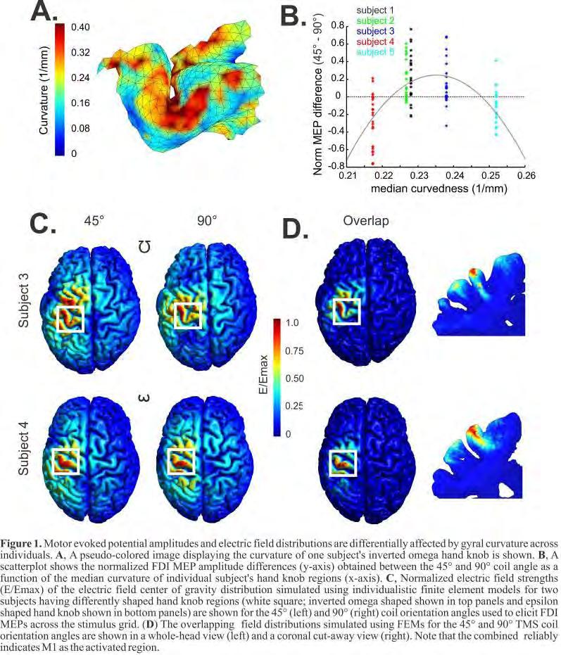
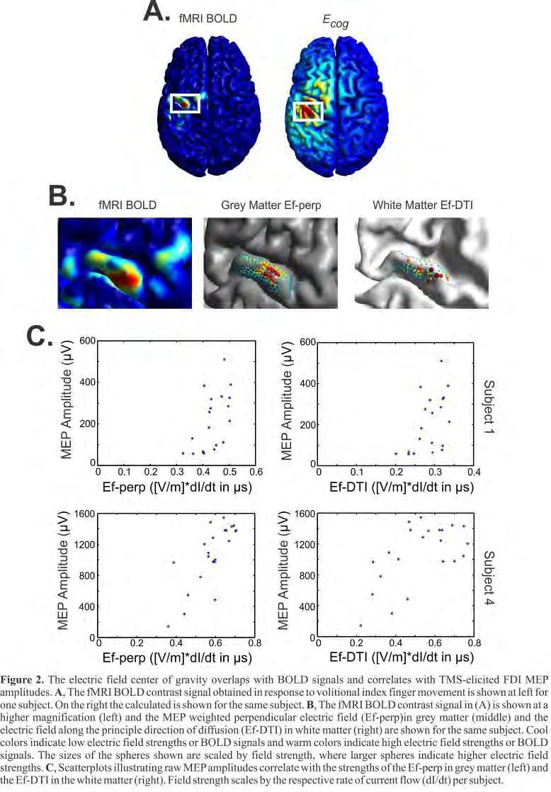
319 319
320 Poster Session II Novel Techniques P 222 Studying the dynamics of the TMS-EEG signal *T. Mutanen 1,2, J. O. Nieminen 1,2, R. J. Ilmoniemi 1,2 1 Aalto University School of Science, Department of Biomedical Engineering and Computational Science, Espoo, Finland 2 BioMag Laboratory, HUSLAB, Helsinki University Central Hospital, Helsinki, Finland Introduction: Combined transcranial magnetic stimulation (TMS) and electroencephalography (EEG) has proven to be a useful tool when probing the effective connectivity. However, the TMS-evoked responses seem to vary significantly from trial to another. This is partially due to the constantly changing underlying brain state, which is likely to affect the evoked response. Thus, it is important to understand the relationship between the current brain state dynamics and the evoked TMS-EEG responses. This work concentrates on the non-linear dynamic behaviour of EEG signal before and after TMS stimuli. Objectives: Our objective is to study the effects of TMS on the dynamics of the brain state as well as the effects of the current brain state dynamics on the TMS-evoked responses. Materials & methods: In the analysis, 16 different TMS-EEG data sets, where the stimuli had been delivered on the left primary motor cortex with 100% motor threshold intensity. The Nexstim eximia magnetic stimulator and TMS-compatible EEG system were used to collect the data. It is known that the trajectory of a system in the state space can reveal some vital properties of the underlying dynamics. In this work, we studied the projection of the state-space trajectory on to the EEG signal space using 16 EEG channels confining the stimulation area. The data analysis consisted of computation of three commonly known non-linear measures for trial-level data: quantitative recurrence analysis, Lyapunov stability, and correlation dimension. In short, the recurrence analysis measures the distances between the state vectors defining the state of the system as a function of time. Lyapunov stability is used to describe the chaotic properties of the system, whereas correlation dimension measures the complexity of the underlying system. In the figure one can see a schematic picture of the hypothesis behind the present work. The post-synaptic currents (A) define accurately the electric state of the brain. The brain state advances spontaneously in the state space (red line). However, TMS might shift the system rapidly to a certain subset and also affect the later motion of the trajectory (green line). We can observe the projection of the trajectory on the EEG signal space (dotted lines) spanned by channels ch i and ch j. Results: The mean distance between the state vectors right before and right after the stimulus was greater than the mean distance separating state vectors during spontaneous activity. Furthermore, the results indicated that the state vector was moving faster during approximately 300-ms long period after the stimulus than during spontaneous activity. The results also showed some evidence that the correlation dimension might decrease as an effect of TMS. With the present data available the Lyapunov-stability analysis did not show clear results. Conclusion: The results indicate that the artificial activity created by TMS is propagating faster in the system than the spontaneous activity. This might be due to higher local free energy close to the stimulation site, which the system tries to minimize as fast as possible. Furthermore, it seems that TMS actually does shift the brain state to a slightly different subset in the brain state space. 320
321 Poster Session II Novel Techniques P 223 A multielectrode cap for transcranial direct current stimulation (tdcs) *E. Rossi 1,2, E. Della Torre 3, L. Rossi 3, A. Priori 1,4,3 1 University of Milan, Department of Medical-Surgical Pathophysiology and Transplants, Milan, Italy 2 Politecnico di Milano, Department of Bioengineering, Milan, Italy 3 Newronika srl, Milan, Italy 4 Fondazione IRCCS Cà Granda - Ospedale Maggiore di Milano, Clinical Center for Neurotechnology, Neurostimulation & Movement Disorders, Milan, Italy Introduction: Many electrode montages for transcranial direct current stimulation (tdcs) are described in literature: attention must be paid on the correct positioning of the electrodes and on their geometry to obtain reproducible results. Also, to make tdcs even more simple and accessible to a large number of subjects, the electrode positioning should be easy for patients and their caregivers. Therefore, to guarantee safety during tdcs treatment any electrode system should protect the users from i) incorrect placement of the electrodes over the scalp, ii) wrong connection of the electrodes to the DC stimulator, and should ensure the proper pressure over the electrode to warrant the optimal impedance. Objective: Design a system for correct and easily reproducible placement of tdcs electrode. Materials and methods: From literature review and our experience in the clinic we have defined the design requirements for the new system. The system has to be composed by: a cap, an armband and a set of electrodes. Based on the numerical simulation of the current distribution near the electrodes, we designed a new electrode geometry to avoid hotspots preventing dangerous current density and the resulting tissue damage. Results: The cap is equipped with electrodes disposed according to the International System and has an innovative structure characterized by three lines of force. This structure guarantee the correct strain over the entire scalp s surface and the necessary pressure on the electrode under the electrical connections for the correct contact s impedance. The shape of the electrode has not angles or vertex: this geometry avoids the insurance of hotspots. Moreover the electrode is made of inert materials, without any metal components and the internal sponge of the electrode is able to absorb a large quantity of saline solution. The cap has two locking system. With the first locking system the clinicians can block the electrode s anchorages on the cap in placement different from the montage dedicated to the patient. With the second locking system the doctor can block the electrical connection of the cap in order to avoid the connection of the DC stimulator s cable to the cap in a wrong placement without the underlying electrode. Conclusion: We present a stimulating cap for tdcs that promises to be easy to use, to allow reproducible electrode placement and to avoid mistakes in stimulation polarity. Conflict of interest: Elena Rossi is supported by Dote ricerca : FSE, Regione Lombardia and Newronika srl., a spin-off company of the Fondazione IRCCS Ca Granda Ospedale Maggiore Policlinico and the Università degli Studi di Milano. Ernesto Della Torre is an employee in Newronika s.r.l., a spin-off company of the Fondazione IRCCS Ca Granda Ospedale Maggiore Policlinico and of the Università degli Studi di Milano. Alberto Priori and Lorenzo Rossi are stakeholders in Newronika s.r.l., a spin-off company of the Fondazione IRCCS Ca Granda Ospedale Maggiore Policlinico and of the Università degli Studi di Milano. 321
322 Poster Session II Novel Techniques P 224 Cerebellar transcranial direct current stimulation: a computer-based modeling study *M. Parazzini 1, E. Rossi 2,3, R. Ferrucci 2,4, P. Ravazzani 1, A. Priori 2,4 1 Consiglio Nazionale delle Ricerche CNR, Istituto di Ingegneria Biomedica ISIB, Milano, Italy 2 Università degli Studi di Milano, Dipartimento di Fisiopatologia medico-chirurgica e dei Trapianti, Milano, Italy 3 Politecnico di Milano, Dipartimento di Bioingegneria, Milano, Italy 4 Fondazione IRCCS Ca Granda Ospedale Maggiore Policlinico, Centro Clinico per la Neurostimolazione, le Neurotecnologie ed i Disordini del Movimento, Milano, Italy Introduction: Transcranial direct current stimulation over the cerebellum (or cerebellar tdcs) modulates motor and cognitive brain circuits [Ferrucci et al., 2008; 2012]. There are however still open questions on cerebellar tdcs. One of these is the amount and the spatial distribution of the electrical current that crosses the skull and reaches the cerebellum. Objective: To evaluate the electric field (E) and the current density (J) generated by cerebellar transcranial DC stimulation in different brain structures using electromagnetics computational techniques applied to a realistic human model. Materials and methods: The computation was conducted using the simulation platform SEMCAD X (Schmid & Partner Engineering AG, Zurich, Switzerland) solving the Laplace equation. One realistic human model of the Virtual Family [Christ et al., 2010] was used. The model is based on high resolution magnetic resonance images of an healthy volunteer (a 26-years-old female adult model Ella ). The model consists of up to 77 different tissues, the dielectric properties of which were assigned on the basis of the data at low frequency [Gabriel et al., 1996]. The active electrode was placed on the scalp over the cerebellum and the reference over the right arm. The electrodes were modeled as sponge material, consisting of a rectangular pad conductors placed within a sponge. The stimulating current was 2 ma. To assess how much the results depend on the exact electrode placement, the active electrode was moved longitudinally and laterally of ± 0.5 cm. Results: The peak values of E and J in the cerebellum are higher than the ones in the cortex of 56% and of 73% respectively. The percentage of volume of the cortex where the amplitude of E is greater than 70% of the peak found in the cerebellum it is much less than 1%. Varying the position of the active electrode of ± 0.5 cm longitudinally and laterally, the maximum variation of the peak values of E and J in the cortex and in the cerebellum is of 4%. Conclusion: The higher amplitudes of the electric field (and/or the current density) occur mainly in the cerebellum with a spread toward the occipital region of the cortex. Data, moreover, show that small changes in the positioning of the active electrode result in a small effect on the induced field amplitudes in all the brain structures here analyzed. The model shows that the cerebellum is the main structure involved by cerebellar transcranial DC stimulation in humans and the current spread to other structures is negligible, probably without any physiological relevance. References: Ferrucci, R. et al., Cerebellum and processing of negative facial emotions: Cerebellar transcranial DC stimulation specifically enhances the emotional recognition of facial anger and sadness. Cogn Emot 26(5), Ferrucci, R. et al., Cerebellar transcranial direct current stimulation impairs the practice-dependent proficiency increase in working memory. J Cogn Neurosci 20, Christ, A., et al., The Virtual Family-development of surface-based anatomical models of two adults and two children for dosimetric simulations. Phys Med Biol 55, N23-N38. Gabriel, S. et al., The dielectric properties of biological tissues: II. Measurements in the frequency range 10 Hz to 20 GHz. Phys Med Biol 41,
323 Poster Session II Novel Techniques P 225 Computational models of current densities in transcutaneous spinal direct current stimulation (tsdcs) *M. Parazzini 1, E. Rossi 2,3, P. Ravazzani 1, A. Priori 2,4 1 Consiglio Nazionale delle Ricerche CNR, Istituto di Ingegneria Biomedica ISIB, Milano, Italy 2 Università degli Studi di Milano, Dipartimento di Fisiopatologia medico-chirurgica e dei Trapianti, Milano, Italy 3 Politecnico di Milano, Dipartimento di Bioingegneria, Milano, Italy 4 Fondazione IRCCS Ca Granda Ospedale Maggiore Policlinico, Centro Clinico per la Neurostimolazione, le Neurotecnologie ed i Disordini del Movimento, Milano, Italy Introduction: Non-invasive transcutaneous spinal direct current stimulation (tsdcs) induces changes in spinal cord function in humans [Cogiamanian et al., 2012]. The physiological mechanisms underlying these changes however remain speculative. A relevant issue to better understand this innovative non-invasive neuromodulation technique is the estimation of the spatial distribution of the current density in the spinal cord during tsdcs. Objective: To evaluate the current density (J) induced by dorsal tsdcs in the spinal cord using electromagnetics computational techniques applied to realistic human models of different age and gender. Materials and methods: The computation was conducted using the simulation platform SEMCAD X (Schmid & Partner Engineering AG, Zurich, Switzerland) solving the Laplace equation. Three realistic human models of the Virtual Family [Christ et al., 2010] were used. The models are based on high resolution magnetic resonance images of healthy volunteers (a 26-years-old female adult model Ella ; a 34 years-old male adult model Duke ; an 11 years old female adolescent model Billie ). The models consist of up to 77 different tissues, the dielectric properties of which were assigned on the basis of the data at low frequency [Gabriel et al., 1996]. In this preliminary study three electrode montages were modeled in which the anode was always over the spinal process of the tenth thoracic vertebra and the cathode was placed: i) above the right deltoid ii) over the umbilicus; iii) over Cz. The injected current was 3 ma. The electrodes were conductors within rectangular sponges. Results: Preliminary data indicates that in the dorsal spinal cord below the stimulating electrode the peaks of J across the models are of 0.02 A/m 2, 0.03 A/m 2 and 0.06 A/m 2 for the three electrode montages, respectively. Conclusion: The electric field generated by tsdcs reaches the spinal cord at physiologically effective strength. References: Cogiamanian F, Ardolino G, Vergari M, Ferrucci R, Ciocca M, Scelzo E, Barbieri S, Priori A. Transcutaneous Spinal Direct Current Stimulation. Front Psychiatry. 2012;3:63. Epub 2012 Jul 4. Christ, A., Kainz, W., Hahn, E.G., et al., The Virtual Family-development of surface-based anatomical models of two adults and two children for dosimetric simulations. Phys Med Biol 55, N23-N38 Gabriel, S., Lau, R.W., Gabriel C The dielectric properties of biological tissues: II. Measurements in the frequency range 10 Hz to 20 GHz. Phys Med Biol 41,
324 Poster Session II Novel Techniques P 226 Robot guided positioning of a magnetic coil for rtms over the cortex in patients with cerebral lesions *A. Hartmann 1, R. Kalis 1,2, L. Matthäus 3, F. Weber 4, T. Rommel 1 1 rehanova clinic, neurology, Köln, Germany 2 eemagine medical imaging solutions, Berlin, Germany 3 neuro b.v., ant, Enschede, Netherlands 4 Krankenhaus Köln-Merheim, Neurochirurgie, Köln, Germany Introduction: rtms with narrow focus coils is able to stimulate or inhibit depolarization in circumscribed cerebral areas. However, it is difficult to position the coil over the skull precisely above the target gyrus. Orientation on the EEG system or calculation of bone markers of the skull in MRI imaging with transfer of the data to the skull of the living patient has been used to position the coil manually. These procedures are rather imprecise. We here present a system which coordinates the MRI imaging of the brain with an infrared camera system guided robot to position the coil directly over the region of interest (ROI). Protocol: 5 patients have been included into this protocol. A T1-MRI of the brain was used to identify the ROI, here the precentral motor area of the hand (handknob). The sedantary patient was placed into a relaxing chair and small passive sphere markers (head reference markers) were fixed to the head by a headband. These skull markers were tracked by an infrared camera system positioned in front of and above the patient s head in a distance of about 1.3 m and correlated to the cerebral MRI imaging. A robot system ( Smart Move R, ANT) with a 6 joint metal arm allowing positioning of the coil with 6 degrees of freedom (DoF) was placed beside the patient. The magnetic coil (8-shaped) was fixed to the robot arm. The infrared camera system identified the position of the magnetic coil and the patient s head by the passive sphere markers fixed to the magnetic coil and the patient. The MRI data were stored into a PC and the surface image of the brain was coordinated with the position of the patient s skull and of the magnetic coil. By this technique, it was possible to identify the ROI of the cortex and direct the coil precisely by the robot arm over the skull area of the underlying ROI. Results: Immediate correct position of the coil manoeuvred by the robot arm over the ROI was successfully controlled by rtms of the precentral hand knob and surface electromyography of the contralateral hand muscles. Summary: It is possible to manoeuvre precisely a magnetic coil for rtms over brain tissue of interest using a robot arm by coordinating the cortical surface MRI of the patient with the skull and the magnetic coil. Registration of the positions is achieved by an infrared camera system. The robot arm is able to follow the head during limited movements. 324
325 Poster Session II Novel Techniques P 227 Ultramicroscopy (UM) in Neurobiology *N. Jährling 1,2,3, S. Saghafi 1,2, K. Becker 1,2, C. P. Mauch 1,2,4, H.- U. Dodt 1,2 1 TU Vienna, Bioelectronics, Vienna, Austria 2 Center for Brain Research, Vienna, Austria 3 Carl-von-Ossietzky Universität Oldenburg, Dept. Neurobiology, Oldenburg, Austria 4 MPI for Psychiastry, Munich, Germany UM is a prime example for an interdisciplinary research field invention. It enables researchers to obtain high-resolution 3D-reconstructions of organ systems in different animal models. Knowledge about the interconnections of the neuronal network to the vascular system is often essential neurobiological research. Ultramicroscopy (UM) is a light sheet based fluorescence microscopy. This novel bioimaging technique allows the 3D-visualization of cm-sized biological specimens withμm-resolution [1,3]. Artifacts of conventional microscopy such as distortion of the tissue are avoided. It is due to optical sectioning instead of mechanical sclicing [2,3]. In UM, a specimen is illuminated by a thin sheet of laser light, formed by one or more cylindrical lenses [2,3]. To generate a light sheet distribution in the standard UM, we basically employ a single cylindrical lens placed in front of a variable rectangular slit aperture [2,3]. Lectinis are proteins that bind to sugar complexes, which are attached to proteins and lipids. We employed an approach using fluorescent conjugated lectins during the transcardial perfusion of mice to contrast the endothelium building up the vascular system [3,4].Biological samples are prepared chemically to become as transparent as possible [3]. In this study the architecture of the blood vessel system of whole organs of animal models is visualized [3,4]. By combining light sheet based UM with lectin-labelling, 3D reconstructions of vascular structures of the mouse spinal cord can be generated. Alteration in optics, morphological analysis of neurobiological disorders, multiple labeling, improving histology of clearing procedure, the analysis of brain tumors are subjects of interests are subjects of interests in our future work. [1] Dodt et al., (Nature methods 2007) [2] Jährling & Saghafi, (Elektrotechnik & Informationstechnik, Springer 2011) [3] Jährling (Dissertation Dr.rer.nat. 2011, University Oldenburg, Supervisor: Reto Weiler) [4] Jährling et al., (Organogenesis2009) 325
326 Poster Session II Novel Techniques P 228 Repetitive transorbital alternating current stimulation (rtacs) alpha activity enhancement in patients with visual field deficits: a prospective, randomized, blinded, controlled, multicenter study. *A. Mante 1, M. Rönnefarth 1, R. Bathe-Peters 1, R. Fleischmann 1, G. Ambrus 2, C. Gall 3, A. Fedorov 3, B. A. Sabel 3, S. Schmidt 1 1 Charité-Universitätsmedizin Berlin, Department of Neurology, Berlin, Germany 2 Universitätsmedizin Göttingen, Department of Clinical Neurophysiology, Göttingen, Germany 3 Otto-von-Guericke University of Magdeburg, Medical Faculty, Institute of Medical Psychology, Magdeburg, Germany Introduction: Retrospective studies suggested that rtacs simulates neuronal activity and results in neuroplasticity (Fedorov et al., 2011). Prospective, randomized, controlled studies in patients with prechiasmatic optic nerve damage showed that rtacs reduces visual field deficits, enhances vision-related Quality of Life (Gall et al., 2011) and entrains rhythmic brain activity (Schmidt et al., 2012). This is understood to reflect periodic enforcing (Zaehle et al., 2010) associated with the activation of residual vision resulting in restoration of visual field deficits (Sabel et al., 2011). Objectives: The objective of the present prospective multicenter study is to provide evidence that alpha power gain over occipital electrodes is a bio-marker that can predict visual field size improvement. Materials & methods: 80 patients with pre-chiasmatic partial optic nerve damage were randomized prior to inclusion in the sham-stimulation controlled rtacs trial (RCT). The stimulation group (n=44) received rtacs for min/day on 10 consecutive days. Treatment parameters were defined daily 1) by subject specific amperage when patients have an obvious perception of phosphenes, 2) the strongest alpha frequency over occipital electrodes (lower cut-off) and 3) the flicker-fusion frequency (upper cut-off). The control group (n=36) received subliminal stimulation. Electroencephalographic (EEG) power spectra were assessed on each of the 10 days over the occipital cortex. The size of visual fields was measured before and after treatment with high resolution perimetry (HRP). Data analysis was non-parametric regression to account for the heterogeneous patient-population. Results: The EEG data shows a systematic enhancement of alpha-power after stimulation (p <.05). This effect is stronger in the treatment group as compared with the placebo group (p <.05). Enhanced alphapower was associated with reduced visual field deficits (p <.05). Clinical results are reported in an complementary abstract. Discussion & Conclusion: The results confirm that rtacs entrains alpha-oscillatory brain activity and show that changes in brain-rhythms is associated with changes in the visual field size. Thus, EEG alphaoscillations seem to provide a bio-marker that can predict adaptive processes in a visual system with a prechiasmatic lesion. Future studies should continue to address the optimization of rtacs as well as the stratification by etiology prior to clinical application in individuals or small populations. Acknowledgements: The study was co-funded by the EBS Technologies GmbH Bibliography: Fedorov, A., Jobke, S., Bersnev, V., Chibisova, A., Chibisova, Y., Gall, C. & Sabel, B. A. (2011). Restoration of vision after optic nerve lesions with noninvasive transorbital alternating current stimulation: a clinical observational study. Brain Stimul 4, Gall, C., Sgorzaly, S., Schmidt, S., Brandt, S., Fedorov, A. & Sabel, B. A. (2011). Noninvasive transorbital alternating current stimulation improves subjective visual functioning and vision-related quality of life in optic neuropathy. Brain Stimul 4, Sabel, B. A., Henrich-Noack, P., Fedorov, A. & Gall, C. (2011). Vision restoration after brain and retina damage: the "residual vision activation theory". Prog Brain Res 192, Schmidt, S., Mante, A., Ronnefarth, M., Fleischmann, R., Gall, C. & Brandt, S. A. (2012). Progressive enhancement of alpha activity and visual function in patients with optic neuropathy: A two-week repeated session alternating current stimulation study. Brain Stimul. Zaehle, T., Rach, S. & Herrmann, C. S. (2010). Transcranial alternating current stimulation enhances individual alpha activity in human EEG. PLoS One 5, e
327 Poster Session II Novel Techniques P 229 Transcranial Laser Stimulation - A New Method for Non-Invasive Modulation of Cortical Excitability *S. Filipovic 1, M. Jelic 1, A. Jeremic 2, V. Stevanovic 1, S. Milanovic 1, L. Konstantinovic 2,3 1 University of Belgrade Institute for Medical Research, Department of Neurophysiology, Beograd, Serbia 2 Hospital for Rehabilitation "Dr M. Zotovic", Beograd, Serbia 3 University of Belgrade School of Medicine, Department of Rehabilitation, Beograd, Serbia Question: Near infrared low-level laser (nirlll) irradiation penetrates scalp and skull. It was shown to be able to reduce damage from experimentally induced stroke in animals and to improve memory in middleaged mice. It was also reported to improve the outcome in human cases of acute stroke. In this study we evaluated whether transcranial application of the nirlll (transcranial laser stimulation - TLS) can modulate the excitability of the motor cortex (M1) as measured by transcranial magnetic stimulation (TMS). Methods: In the 1 st part of the study, motor evoked potentials (MEPs) from the right abductor pollicis brevis (APB) muscle, elicited by single-pulse TMS applied to the left M1 (intensity 120% of the resting motor threshold, rmt) were measured at baseline and every 5 min (up to 30 min) after the TLS, in 18 healthy subjects. For TLS, the nirlll (wavelength 905 nm, pulse frequency 3 khz, power density of 50 mw/cm2, single dose of 3 J/cm 2 ) was applied for 60 sec over a circular area 3 cm in diameter cantered at left M1 APB hot-spot. In the 2 nd part of the study, the MEPs were recorded in the same way in 6 subjects who were found to have very high rmt and poor response to TLS in the Exp1. The TLS parameters were the same except pulse frequency that was increased to 5 khz which increased total nirlll delivered (single dose of 4.5 J/cm 2 ). All applied dosed were within the recommended safety limits for human application of nirlll in physical medicine. The study was conducted in accordance with the Declaration of Helsinki and was approved by the local ethics committee. Results: In the 1 st part, the most prominent MEP suppression was within time-window min following TLS (Fig. 1A). Total of 12 subjects (66.7%) had MEP sizes below baseline level in at least 4 out of 7 (i.e. >50%) post-tls time points - Responders. ANOVA and post-hoc pair-wise analyses showed significant reduction of post-tls MEP sizes related to baseline in Responders (Fig. 1B). The only difference between Responders and Non-Responders was in rmt (54.9±5.8, and 63.2±8.7, respectively; t (16) =2.41, p=0.029). For all subjects together, average post-tls MEP size correlated significantly with rmt (Spearman R=0.499, p=0.035). In the 2 nd part, with higher intensity of TLS, 5 subjects were Responders, 4 of them who were Non-Responders in the 1 st part, and 1 who was Responder from the beginning but had much larger MEP suppression in the 2 nd part. Average post-tls MEP sizes in these 5 Responders dropped significantly (t (4) =5.105, p=007) in comparison to the 1 st part. Conclusions: The M1 excitability was found to be reduced after TLS in dose dependent manner. These findings may give insight into the mechanisms of nirlll effects in the human cerebral cortex, also suggesting more suitable applications of TLS in clinical settings. 327
 at each time point; vertical lines are standard errors (SE). Figure 2.")

328 Figure 1. Results of the 1 st part. A. Shaded segments of the bars represent number of participants with below baseline MEP sizes at each time point. B. Responders sub-group: mean change in MEP sizes (relative to baseline) at each time point; vertical lines are standard errors (SE). Figure 2. Comparison of average post-tls relative MEP sizes (relative to baseline) between 1 st and 2 nd part. First 4 subjects were non-responders in the 1 st part and became responders in the 2 nd. Subject 5 was responder in the 1 st part, but with relatively high motor threshold and low level of overall MEP size suppression - in 2 nd part his MEP size became considerably more suppressed. Subject 6 was nonresponder in both parts. 328
329 Poster Session II Novel Techniques P 230 Third generation controllable pulse parameter transcranial magnetic stimulation device *A. Peterchev 1, D. Murphy 1 1 Duke University, Psychiatry & Behavioral Sciences, Durham, United States Introduction: Commercially available transcranial magnetic stimulation (TMS) devices induce electric field (E-field) pulses with damped cosine shape and provide very limited control over the pulse parameters. Furthermore, conventional TMS devices consisting of a single power stage cannot produce rapid bursts (< 10 ms inter pulse interval) with equal or increasing strength since the energy on the capacitor diminishes from pulse to pulse due to circuit losses, and the time between the pulses is too short for the capacitor charger to supply the lost charge. Consequently, conventional TMS topologies require combining the output of multiple power stages to generate paradigms such as paired-pulse and quadripulse stimulation. Addressing these limitations we have developed a third generation controllable pulse parameter TMS device (ctms3) that allows extensive adjustment of the pulse parameters such as the pulse width, number and shape of phases, and directionality (ratio of positive to negative phase amplitude), as well as enables the generation of powerful pulse bursts. Objectives: To summarize the ctms3 device capabilities and compare it to conventional TMS and other ctms devices. Materials & methods: The coil current and electric field were measured with a Rogowski current sensor and a search coil, respectively. Results: ctms3 uses a circuit topology with two energy storage capacitors and four controllable switches. It can generate rapid-rate (<= 50 Hz) trains of mono/bi/polyphasic near-rectangular electric field pulses with adjustable amplitude (maximum capacitor voltages of 1 kv and 2.6 kv), pulse width (phase duration of 10 us to over 200 us), and directionality (positive/negative phase amplitude ratio range of 1-52). In contrast to earlier ctms devices, in ctms3 the pulse phases can assume 4 possible electric field levels, proportional to either of the two capacitor voltages, the capacitor voltage difference, or 0. Thus, ctms3 can produce waveforms with a staircase shape that can approximate conventional sinusoidal current pulses (Fig. 1). To counteract the loss of capacitor charge in rapid pulse bursts, the pulse width of the second and subsequent pulses can be increased, allowing their stimulation strength to be equal or even higher than the first pulse (Fig. 2). In Fig. 2, the voltage on the two energy storage capacitors is reduced by 2% and 17%, respectively, after the first pulse, but the second pulse is 67% longer than the first pulse. Consequently, the strength of the second pulse, as measured by modeled neural membrane depolarization (membrane time constant = 196 us) is 45% higher than the first pulse. Thus, paired-pulse paradigms, where the second pulse can have higher strength than the first pulse, can be implemented. Similarly, increasing the pulse width in a rapid burst of pulses can enable quadripulse stimulation. Conclusions: ctms3 offers flexibility of pulse shape adjustment unparalleled in other available TMS devices. The combination of high energy efficiency of the near rectangular pulses, pulse energy recovery, and pulse width control allow the generation of rapid bursts of pulses with equal or even increasing strength that can implement paired-pulse and quadripulse stimulation. These features could extend the capabilities of TMS as a research and diagnostic tool, and could enable optimization of the stimulus in therapeutic interventions. 329
330 Figure 1: Staircase pulse example. Figure 2: Paired pulse example. 330
331 Poster Session II Novel Techniques P 231 A physiological characterization of biphasic transcranial magnetic stimulation *I. Delvendahl 1, N. Gattinger 2, T. Berger 3, B. Gleich 2, H. Siebner 4, V. Mall 5 1 ENI, Göttingen, Germany 2 IMETUM, Munich, Germany 3 University Medical Centre, Freiburg, Germany 4 Danish Research Center for Magnetic Resonance, Copenhagen, Denmark 5 Technical University, Munich, Germany Question: In transcranial magnetic stimulation (TMS), two pulse waveforms are mainly used to study cortical function. While monophasic stimuli are optimal to measure cortical excitability, the biphasic pulse waveform is increasingly used to induce cortical plasticity and to treat neurological as well as psychiatric illnesses. Methods: We used a novel stimulation device (flextms) to study variable pulse configurations in young healthy volunteers. Primary outcome measure was cortical resting motor threshold (RMT). We determined the strength-duration relationship for single-pulse biphasic TMS using pulse durations between 120 and 400 µs. To further characterize the biphasic stimulus, we systematically varied amplitude and current direction of the first and second part of the stimulus waveform. Results: The strength-duration relation has a parabolic shape for biphasic stimulation with a minimum at a pulse length of approximately 240 µs. Varying the amplitude of the part inducing a posterior-anterior oriented current considerably raised RMT, while changes of the part which induces an anterior-posterior current had little effect Conclusion: Our results clarify the role of pulse duration in biphasic TMS. The part inducing a posterioranterior current in the brain contributes most to the effect of biphasic stimulation. These findings have important methodological implications and contribute to the understanding of the physiology of TMS. 331
332 Poster Session II Novel Techniques P 232 Leaving the beaten track of TMS waveform restrictions: Concepts and prototype for a convertible stimulator that can generate almost every type of existing and future waveform S. Götz 1,2, F. Helling 3, *T. Weyh 3,2 1 Duke University, Durham, United States 2 TU München, Munich, Germany 3 Universität der Bundeswehr München, Neubiberg, Germany Introduction: Waveforms are an important issue in magnetic stimulation. They provide the key to an important characteristic of neurons: their distinctive nonlinear dynamics. Different waveforms seem to have different activation sites and may allow selective stimulation. This is reflected for example by their different corticospinal D- and I-wave patterns (2). However, available pulse types are very restricted. In essence, the only established waveforms are monophasic and biphasic pulses. In addition to selective stimulation and neuromodulation, higher efficiency (1) could enable highly focal small coils to overcome thermal issues and provide high repetition rates. Objectives: Current devices can only provide very distinct pulses for technological reasons. Even in alternative, more flexible approaches, the underlying circuitry is limited to a certain class of stimuli. Among these stimuli, only a subclass does not change the device state of charge and can therefore be generated repetitively. Our objective was to conceive and design a technology that is able to generate almost any waveform. Accordingly, the selection of waveforms would no longer depend on device capabilities and technology, but merely follow the user s needs. Methods: For this aim, we had to abandon all traditional concepts and designs for pulse generation in magnetic stimulation as well as other fields of science and engineering. We developed novel topologies that can handle the special requirements of magnetic stimulation pulses, namely high voltage, high current and high speed. The winning approach uses a dynamical reconfiguration of small energy storages: Metaphorically speaking, it uses a high number of small-voltage batteries that can be combined in any parallel and series connection dynamically changeable for every time step so as to exactly control the coil voltage accurately to the curve outlined by the desired waveform. The key components of the technology are no longer expensive high-voltage semiconductors, but inexpensive mass-produced devices used in consumer electronics. We designed a control strategy for the distributed concept and implemented a prototype for characterization of the concept. Results: The system can generate almost any waveform, both existing and potential future pulse shapes. All waveforms can be generated with a recovery of the pulse energy from the coil, which has been known from biphasic stimulators. We achieved this even for classical monophasic waveforms, which can except for rare research devices (3) provide high pulse repetition rates, although being preferred. The high losses prevent that. Furthermore, the generation of waveforms is not done by any mechanical reconfiguration, but controlled dynamically. Therefore, the system can even fundamentally change the waveform from pulse to pulse in a theta-burst or double-pulse protocol. Conclusion: The generation of arbitrary waveforms is possible and has several advantages also for classical waveforms. Among others, this includes the ability to use high repetition rates for almost every waveform and profit from cost-effective mass-produced components. References: (1) S.M. Goetz et al. (2012). IEEE EMBC 2012, doi: /embc (2) V. Di Lazzaro et al. (2004). Clin Neurophys, 115(2): doi: /j.clinph (3) M. Schmid, T. Weyh, B.-U. Meyer (1993). Biomed Eng, 38(12): doi: /bmte
333 Poster Session II Novel Techniques P 233 Robot-assisted and image-guided randomized TMS mapping of human and primate hand muscle *M. Hewitt 1, J. Meincke 1, R. Ahlert 2, H. Scherberger 2, D. Liebetanz 1 1 University of Göttingen, Clinical Neurophysiology, Göttingen, Germany 2 German Primate Center, Göttingen, Germany Introduction: Coil positioning is a crucial factor for transcranial magnetic stimulation (TMS). Manual positioning of flat TMS coil against the curved head allows for errors in translation and rotation. As scalp markings are an ineffective way to ensure consistency in coil positioning, image-guided neuronavigation is used to reduce errors. However, as manual neuronavigation increases time for coil positioning it is less suitable when the coil has to be moved between pulses during TMS mapping. The use of a robot for coil positioning may overcome these drawbacks of manual neuronavigation. Objectives: We aimed at assessing the practicability and efficacy of the using a robot for a neuronavigated TMS mapping of the cortical representation of the first dorsal interosseus (FDI) of humans and rhesus monkey. Materials & methods: For FDI mapping, 12 supra-threshold stimuli (120% RMT) were applied in a randomized order to each grid point of a 7x7 grid with a spacing of 10 mm in 8 subjects (mean age 26 y, 6 male) with a Magstim 200 stimulator and a standard 70 mm figure-8 coil. Surface EMG was recorded from contralateral FDI. An Adept Viper s850 robotic arm moved the coil to each grid point on a MRI-based head model in a random order. An IR camera obtained actual positional data of the head. This allowed tracking of head movements and respective online movement compensation by the robot. Centers of gravity were projected onto the brain surface and co-registered to an MNI152 T1 1mm brain template. A similar setup was used for FDI mapping in a male adult rhesus monkey on a 4x5 grid with 5 mm spacing. TMS was applied with a Magstim 25 mm figure-8 coil. The mapping was performed during multiple sessions during which the monkey s head was attached to a frame with frontal and occipital head posts. Relaxation of FDI was rewarded with water after each pulse. Results: TMS Mapping sessions took about min in each subject with a pulse interval of 8 s. Preparations lasted about 20 min. A closely spaced TMS representation of FDI could be identified in each subject within the precentral gyrus. The shape of the representation area resembled ovals with a mean maximum MEP amplitude of 1.35 mv (SD: 0.52). Mean stereotaxic coordinates of the centers of gravity were (-36, -12, 67) in MNI152 space. In the animal experiment, the monkey adapted to the setup within 4 weeks. The preparation time for each session was about 10 min. Across all sessions muscle relaxation was present during 64 % of the pulses. The pulse interval was 13.4 s. Four repetitions of a complete grid could be recorded in a single session of about 45 min duration. MEPs from contralateral FDI were elicited from a highly defined elliptical area (1 x 1.5cm) within the precentral gyrus. Conclusion: Due to the combination of fast high-precision coil positioning and online head tracking, robotassisted and image-guided TMS is exquisitely suited to perform randomized TMS mappings in the human with acceptable preparation times. In addition, this method allows for precise TMS mapping of the considerably smaller rhesus brain where high-precision coil positioning with a high inter-session reliability is required for multiple session mappings with grid sizes in the millimeter scale. 333

334 Figure: TMS representation of the FDI in human (A) and monkey (B). Blue dots represent stimulation sites, the black dot the center of gravity. Colors indicate average MEP amplitudes from maximum (red; A: 1.85mV, B: 1.22mV) to minimum (blue; A: 0.01mV, B: 0.03mV). 334
Trans-spinal direct current stimulation: a novel tool to promote plasticity in humans
 Trans-spinal direct current stimulation: a novel tool to promote plasticity in humans Jean-Charles Lamy, PhD Brain and Spine Institute, Paris 1 Background Grecco et al., J Neuroresto, 2015 2 Background:
Neurophysiological Basis of TMS Workshop
 Neurophysiological Basis of TMS Workshop Programme 31st March - 3rd April 2017 Sobell Department Institute of Neurology University College London 33 Queen Square London WC1N 3BG Brought to you by 31 March
Non-therapeutic and investigational uses of non-invasive brain stimulation
 Non-therapeutic and investigational uses of non-invasive brain stimulation Robert Chen, MA, MBBChir, MSc, FRCPC Catherine Manson Chair in Movement Disorders Professor of Medicine (Neurology), University
Introduction to TMS Transcranial Magnetic Stimulation
 Introduction to TMS Transcranial Magnetic Stimulation Lisa Koski, PhD, Clin Psy TMS Neurorehabilitation Lab Royal Victoria Hospital 2009-12-14 BIC Seminar, MNI Overview History, basic principles, instrumentation
Transcranial Magnetic Stimulation
 Transcranial Magnetic Stimulation Session 4 Virtual Lesion Approach I Alexandra Reichenbach MPI for Biological Cybernetics Tübingen, Germany Today s Schedule Virtual Lesion Approach : Study Design Rationale
AdvAnced TMS. Research with PowerMAG Products and Application Booklet
 AdvAnced TMS Research with PowerMAG Products and Application Booklet Table of ConTenTs Introduction p. 04 Legend p. 06 Applications» navigated TMS p. 08» clinical Research p. 10» Multi-Modal TMS p. 12»
Water immersion modulates sensory and motor cortical excitability
 Water immersion modulates sensory and motor cortical excitability Daisuke Sato, PhD Department of Health and Sports Niigata University of Health and Welfare Topics Neurophysiological changes during water
Primary motor cortical metaplasticity induced by priming over the supplementary motor area
 J Physiol 587.20 (2009) pp 4845 4862 4845 Primary motor cortical metaplasticity induced by priming over the supplementary motor area Masashi Hamada 1, Ritsuko Hanajima 1, Yasuo Terao 1,ShingoOkabe 1, Setsu
Cortical Map Plasticity. Gerald Finnerty Dept Basic and Clinical Neuroscience
 Cortical Map Plasticity Gerald Finnerty Dept Basic and Clinical Neuroscience Learning Objectives Be able to: 1. Describe the characteristics of a cortical map 2. Appreciate that the term plasticity is
Combining tdcs and fmri. OHMB Teaching Course, Hamburg June 8, Andrea Antal
 Andrea Antal Department of Clinical Neurophysiology Georg-August University Goettingen Combining tdcs and fmri OHMB Teaching Course, Hamburg June 8, 2014 Classical Biomarkers for measuring human neuroplasticity
Paired-Pulse TMS to one Brain Region. Joyce Gomes-Osman Research Fellow Berenson-Allen Center for Non-Invasive Stimulation LEASE DO NOT COPY
 Paired-Pulse TMS to one Brain Region Joyce Gomes-Osman Research Fellow Berenson-Allen Center for Non-Invasive Stimulation Paired-Pulse Paradigms Sequential pulses applied to the same cortical region Variable
Final Report. Title of Project: Quantifying and measuring cortical reorganisation and excitability with post-stroke Wii-based Movement Therapy
 Final Report Author: Dr Penelope McNulty Qualification: PhD Institution: Neuroscience Research Australia Date: 26 th August, 2015 Title of Project: Quantifying and measuring cortical reorganisation and
The basics of tdcs: Marom Bikson. The City College of New York of CUNY
 The basics of tdcs: Technology and Mechanism Marom Bikson Lucas Parra, Jacek Dmochowski,Asif Rahman, Niranjan Khadka, Mark Jackson, Dennis Truong, Belen Lafon, Gregory Kronberg, Devin Adair, Nigel Gebodh
STRUCTURAL ORGANIZATION OF THE NERVOUS SYSTEM
 STRUCTURAL ORGANIZATION OF THE NERVOUS SYSTEM STRUCTURAL ORGANIZATION OF THE BRAIN The central nervous system (CNS), consisting of the brain and spinal cord, receives input from sensory neurons and directs
Summary of my talk. Cerebellum means little brain but a huge neural resource. Studying the cerebellum in. Chris Miall
 Studying the cerebellum in sensory motor control Chris Miall Behavioural Brain Sciences School of Psychology University of Birmingham Summary of my talk Cerebellum means little brain but a huge neural
Neural Correlates of Human Cognitive Function:
 Neural Correlates of Human Cognitive Function: A Comparison of Electrophysiological and Other Neuroimaging Approaches Leun J. Otten Institute of Cognitive Neuroscience & Department of Psychology University
Development of a New Rehabilitation System Based on a Brain-Computer Interface Using Near-Infrared Spectroscopy
 Development of a New Rehabilitation System Based on a Brain-Computer Interface Using Near-Infrared Spectroscopy Takafumi Nagaoka, Kaoru Sakatani, Takayuki Awano, Noriaki Yokose, Tatsuya Hoshino, Yoshihiro
Neurosoft TMS. Transcranial Magnetic Stimulator DIAGNOSTICS REHABILITATION TREATMENT STIMULATION. of motor disorders after the stroke
 Neurosoft TMS Transcranial Magnetic Stimulator DIAGNOSTICS REHABILITATION TREATMENT of corticospinal pathways pathology of motor disorders after the stroke of depression and Parkinson s disease STIMULATION
TMS: Full Board or Expedited?
 TMS: Full Board or Expedited? Transcranial Magnetic Stimulation: - Neurostimulation or neuromodulation technique based on the principle of electro-magnetic induction of an electric field in the brain.
MOTOR EVOKED POTENTIALS AND TRANSCUTANEOUS MAGNETO-ELECTRICAL NERVE STIMULATION
 MOTOR EVOKED POTENTIAS AND TRANSCUTANEOUS MAGNETO-EECTRICA NERVE STIMUATION Hongguang iu, in Zhou 1 and Dazong Jiang Xian Jiaotong University, Xian, People s Republic of China 1 Shanxi Normal University,
Recovery mechanisms from aphasia
 Recovery mechanisms from aphasia Dr. Michal Ben-Shachar 977 Acquired language and reading impairments 1 Research questions Which brain systems can support recovery from aphasia? Which compensatory route
Functional neuroplasticity after stroke: clinical implications and future directions
 Functional neuroplasticity after stroke: clinical implications and future directions Dr. Michael R. Borich, PT, DPT, PhD Division of Physical Therapy, Department of Rehabilitation Medicine Emory University
NEURO-MS TMS. Diagnostic Monophasic Magnetic Stimulator
 NEURO-MS Diagnostic Monophasic Magnetic Stimulator Diagnostics of neurological disorders Powerful monophasic stimulus Ergonomic and lightweight coils of different shapes and sizes Configurations for single
TREATMENT-SPECIFIC ABNORMAL SYNAPTIC PLASTICITY IN EARLY PARKINSON S DISEASE
 TREATMENT-SPECIFIC ABNORMAL SYNAPTIC PLASTICITY IN EARLY PARKINSON S DISEASE Angel Lago-Rodriguez 1, Binith Cheeran 2 and Miguel Fernández-Del-Olmo 3 1. Prism Lab, Behavioural Brain Sciences, School of
Resistant Against De-depression: LTD-Like Plasticity in the Human Motor Cortex Induced by Spaced ctbs
 Cerebral Cortex July 2015;25:1724 1734 doi:10.1093/cercor/bht353 Advance Access publication January 31, 2014 Resistant Against De-depression: LTD-Like Plasticity in the Human Motor Cortex Induced by Spaced
The Contribution of TMS-EEG Coregistration in the Exploration of the Human Connectome
 IRCCS San Giovanni di Dio Fatebenefratelli The Contribution of TMS-EEG Coregistration in the Exploration of the Human Connectome Marta Bortoletto Carlo Miniussi Cognitive Neuroscience Section, The Saint
NEUROPLASTICITY. Implications for rehabilitation. Genevieve Kennedy
 NEUROPLASTICITY Implications for rehabilitation Genevieve Kennedy Outline What is neuroplasticity? Evidence Impact on stroke recovery and rehabilitation Human brain Human brain is the most complex and
PsychoBrain. 31 st January Dr Christos Pliatsikas. Lecturer in Psycholinguistics in Bi-/Multilinguals University of Reading
 PsychoBrain 31 st January 2018 Dr Christos Pliatsikas Lecturer in Psycholinguistics in Bi-/Multilinguals University of Reading By the end of today s lecture you will understand Structure and function of
Movimento volontario dell'arto superiore analisi, perturbazione, ottimizzazione
 Movimento volontario dell'arto superiore analisi, perturbazione, ottimizzazione Antonio Currà UOS Neurologia Universitaria, Osp. A. Fiorini, Terracina UOC Neuroriabilitazione ICOT, Latina, Dir. Prof. F.
Oscillations: From Neuron to MEG
 Oscillations: From Neuron to MEG Educational Symposium, MEG UK 2014, Nottingham, Jan 8th 2014 Krish Singh CUBRIC, School of Psychology Cardiff University What are we trying to achieve? Bridge the gap from
Statement on Repetitive Transcranial Magnetic Stimulation for Depression. Position statement CERT03/17
 Statement on Repetitive Transcranial Magnetic Stimulation for Depression Position statement CERT03/17 Approved by the Royal College of Psychiatrists, Committee on ECT and Related Treatments: February 2017
Naoyuki Takeuchi, MD, PhD 1, Takeo Tada, MD, PhD 2, Masahiko Toshima, MD 3, Yuichiro Matsuo, MD 1 and Katsunori Ikoma, MD, PhD 1 ORIGINAL REPORT
 J Rehabil Med 2009; 41: 1049 1054 ORIGINAL REPORT REPETITIVE TRANSCRANIAL MAGNETIC STIMULATION OVER BILATERAL HEMISPHERES ENHANCES MOTOR FUNCTION AND TRAINING EFFECT OF PARETIC HAND IN PATIENTS AFTER STROKE
Neuro-MS/D Transcranial Magnetic Stimulator
 Neuro-MS/D Transcranial Magnetic Stimulator 20 Hz stimulation with 100% intensity Peak magnetic field - up to 4 T High-performance cooling: up to 10 000 pulses during one session Neuro-MS.NET software
Biomarkers in Schizophrenia
 Biomarkers in Schizophrenia David A. Lewis, MD Translational Neuroscience Program Department of Psychiatry NIMH Conte Center for the Neuroscience of Mental Disorders University of Pittsburgh Disease Process
Repetitive transcranial magnetic stimulation or transcranial direct current stimulation?
 Brain Stimulation (2009) 2, 241 5 www.brainstimjrnl.com Repetitive transcranial magnetic stimulation or transcranial direct current stimulation? Alberto Priori a, Mark Hallett b, John C. Rothwell c a Dipartimento
Using Transcranial magnetic stimulation to improve our understanding of Transverse Myelitis
 Using Transcranial magnetic stimulation to improve our understanding of Transverse Myelitis Kathy Zackowski, PhD, OTR Kennedy Krieger Institute Johns Hopkins University School of Medicine TMS (transcranial
The Nervous System. Neuron 01/12/2011. The Synapse: The Processor
 The Nervous System Neuron Nucleus Cell body Dendrites they are part of the cell body of a neuron that collect chemical and electrical signals from other neurons at synapses and convert them into electrical
Clinical Applications of tdcs: past, present and future
 Clinical Applications of tdcs: past, present and future Felipe Fregni, MD, PhD, MPH, MMSc, MEd Spaulding Neuromodulation Center Spaulding Rehabilitation Hospital Massachusetts General Hospital Harvard
Brain Stimulation. Comparing Cortical Plasticity Induced by Conventional and High-Definition 4 1 Ring tdcs: A Neurophysiological Study
 Brain Stimulation 6 (2013) 644e648 Contents lists available at SciVerse ScienceDirect Brain Stimulation journal homepage: www.brainstimjrnl.com Original Articles Comparing Cortical Plasticity Induced by
Practical. Paired-pulse on two brain regions
 Practical Paired-pulse on two brain regions Paula Davila Pérez, MD Berenson-Allen Center for Noninvasive Brain Stimulation Beth Israel Deaconess Medical Center Harvard Medical School Plans for the afternoon
The role of amplitude, phase, and rhythmicity of neural oscillations in top-down control of cognition
 The role of amplitude, phase, and rhythmicity of neural oscillations in top-down control of cognition Chair: Jason Samaha, University of Wisconsin-Madison Co-Chair: Ali Mazaheri, University of Birmingham
SUPPLEMENTARY INFORMATION. Supplementary Figure 1
 SUPPLEMENTARY INFORMATION Supplementary Figure 1 The supralinear events evoked in CA3 pyramidal cells fulfill the criteria for NMDA spikes, exhibiting a threshold, sensitivity to NMDAR blockade, and all-or-none
The Three Pearls DOSE FUNCTION MOTIVATION
 The Three Pearls DOSE FUNCTION MOTIVATION Barriers to Evidence-Based Neurorehabilitation No placebo pill for training therapy Blinded studies often impossible Outcome measures for movement, language, and
MagPro by MagVenture. Versatility in Magnetic Stimulation
 MagPro by MagVenture Versatility in Magnetic Stimulation MagPro A proven Record of Innovation With 7 different magnetic stimulators and 27 different coils, the MagPro line from MagVenture provides the
biological psychology, p. 40 The study of the nervous system, especially the brain. neuroscience, p. 40
 biological psychology, p. 40 The specialized branch of psychology that studies the relationship between behavior and bodily processes and system; also called biopsychology or psychobiology. neuroscience,
Computational & Systems Neuroscience Symposium
 Keynote Speaker: Mikhail Rabinovich Biocircuits Institute University of California, San Diego Sequential information coding in the brain: binding, chunking and episodic memory dynamics Sequential information
Københavns Universitet
 university of copenhagen Københavns Universitet Interference in ballistic motor learning: specificity and role of sensory error signals Lundbye-Jensen, Jesper; Petersen, Tue Hvass; Rothwell, John C; Nielsen,
An Overview of BMIs. Luca Rossini. Workshop on Brain Machine Interfaces for Space Applications
 An Overview of BMIs Luca Rossini Workshop on Brain Machine Interfaces for Space Applications European Space Research and Technology Centre, European Space Agency Noordvijk, 30 th November 2009 Definition
Myers Psychology for AP*
 Myers Psychology for AP* David G. Myers PowerPoint Presentation Slides by Kent Korek Germantown High School Worth Publishers, 2010 *AP is a trademark registered and/or owned by the College Board, which
CONTENTS. Foreword George H. Kraft. Henry L. Lew
 EVOKED POTENTIALS Foreword George H. Kraft xi Preface Henry L. Lew xiii Overview of Artifact Reduction and Removal in Evoked Potential and Event-Related Potential Recordings 1 Martin R. Ford, Stephen Sands,
Can brain stimulation help with relearning movement after stroke?
 stroke.org.uk Final report summary Can brain stimulation help with relearning movement after stroke? The effect of transcranial direct current stimulation on motor learning after stroke PROJECT CODE: TSA
Neurophysiology of systems
 Neurophysiology of systems Motor cortex (voluntary movements) Dana Cohen, Room 410, tel: 7138 danacoh@gmail.com Voluntary movements vs. reflexes Same stimulus yields a different movement depending on context
Transcranial Magnetic Stimulation for the Treatment of Depression
 Transcranial Magnetic Stimulation for the Treatment of Depression Paul E. Holtzheimer, MD Associate Professor Departments of Psychiatry and Surgery Geisel School of Medicine at Dartmouth Dartmouth-Hitchcock
Functional Connectivity and the Neurophysics of EEG. Ramesh Srinivasan Department of Cognitive Sciences University of California, Irvine
 Functional Connectivity and the Neurophysics of EEG Ramesh Srinivasan Department of Cognitive Sciences University of California, Irvine Outline Introduce the use of EEG coherence to assess functional connectivity
Differential modulation of intracortical inhibition in human motor cortex during selective activation of an intrinsic hand muscle
 J Physiol (2003), 550.3, pp. 933 946 DOI: 10.1113/jphysiol.2003.042606 The Physiological Society 2003 www.jphysiol.org Differential modulation of intracortical inhibition in human motor cortex during selective
MagPro by MagVenture. Versatility in Magnetic Stimulation
 MagPro by MagVenture Versatility in Magnetic Stimulation MagPro A proven record of innovation With 7 different magnetic stimulators and 33 different coils, the MagPro line from MagVenture provides the
Modeling Depolarization Induced Suppression of Inhibition in Pyramidal Neurons
 Modeling Depolarization Induced Suppression of Inhibition in Pyramidal Neurons Peter Osseward, Uri Magaram Department of Neuroscience University of California, San Diego La Jolla, CA 92092 possewar@ucsd.edu
Effect of Transcranial Magnetic Stimulation(TMS) on Visual Search Task
 on Visual Search Task") INTERNATIONAL JOURNAL OF APPLIED BIOMEDICAL ENGINEERING VOL.2, NO.2 2009 1 Effect of Transcranial Magnetic Stimulation(TMS) on Visual Search Task K. Iramina, Guest member ABSTRACT Transcranial magnetic
INTERNATIONAL JOURNAL OF APPLIED BIOMEDICAL ENGINEERING VOL.2, NO.2 2009 1 Effect of Transcranial Magnetic Stimulation(TMS) on Visual Search Task K. Iramina, Guest member ABSTRACT Transcranial magnetic
The role of non-invasive brain stimulation in neurorehabilitation of poststroke
 Case Report http://www.alliedacademies.org/journal-brain-neurology/ The role of non-invasive brain stimulation in neurorehabilitation of poststroke dysphagia. Meysam Amidfar*, Hadis Jalainejad Fasa University
The Journal of Physiology Neuroscience
 J Physiol 591.7 (2013) pp 1987 2000 1987 The Journal of Physiology Neuroscience Partially non-linear stimulation intensity-dependent effects of direct current stimulation on motor cortex excitability in
Intrinsic Signal Optical Imaging
 Intrinsic Signal Optical Imaging Introduction Intrinsic signal optical imaging (ISOI) is a technique used to map dynamics in single cells, brain slices and even and most importantly entire mammalian brains.
Timing and the cerebellum (and the VOR) Neurophysiology of systems 2010
 Neurophysiology of systems 2010") Timing and the cerebellum (and the VOR) Neurophysiology of systems 2010 Asymmetry in learning in the reverse direction Full recovery from UP using DOWN: initial return to naïve values within 10 minutes,
Timing and the cerebellum (and the VOR) Neurophysiology of systems 2010 Asymmetry in learning in the reverse direction Full recovery from UP using DOWN: initial return to naïve values within 10 minutes,
Modulation of single motor unit discharges using magnetic stimulation of the motor cortex in incomplete spinal cord injury
 1 SHORT REPORT Division of Neuroscience and Psychological Medicine, Imperial College School of Medicine, Charing Cross Hospital, London W 8RF, UK H C Smith NJDavey D W Maskill P H Ellaway National Spinal
Compound Action Potential, CAP
 Stimulus Strength UNIVERSITY OF JORDAN FACULTY OF MEDICINE DEPARTMENT OF PHYSIOLOGY & BIOCHEMISTRY INTRODUCTION TO NEUROPHYSIOLOGY Spring, 2013 Textbook of Medical Physiology by: Guyton & Hall, 12 th edition
We are IntechOpen, the world s leading publisher of Open Access books Built by scientists, for scientists. International authors and editors
 We are IntechOpen, the world s leading publisher of Open Access books Built by scientists, for scientists 3,900 116,000 120M Open access books available International authors and editors Downloads Our
Research Perspectives in Clinical Neurophysiology
 Research Perspectives in Clinical Neurophysiology A position paper of the EC-IFCN (European Chapter of the International Federation of Clinical Neurophysiology) representing ~ 8000 Clinical Neurophysiologists
fmri (functional MRI)
") Lesion fmri (functional MRI) Electroencephalogram (EEG) Brainstem CT (computed tomography) Scan Medulla PET (positron emission tomography) Scan Reticular Formation MRI (magnetic resonance imaging) Thalamus
Lesion fmri (functional MRI) Electroencephalogram (EEG) Brainstem CT (computed tomography) Scan Medulla PET (positron emission tomography) Scan Reticular Formation MRI (magnetic resonance imaging) Thalamus
Selective bias in temporal bisection task by number exposition
 Selective bias in temporal bisection task by number exposition Carmelo M. Vicario¹ ¹ Dipartimento di Psicologia, Università Roma la Sapienza, via dei Marsi 78, Roma, Italy Key words: number- time- spatial
CASE 49. What type of memory is available for conscious retrieval? Which part of the brain stores semantic (factual) memories?
 memories?") CASE 49 A 43-year-old woman is brought to her primary care physician by her family because of concerns about her forgetfulness. The patient has a history of Down syndrome but no other medical problems.
CASE 49 A 43-year-old woman is brought to her primary care physician by her family because of concerns about her forgetfulness. The patient has a history of Down syndrome but no other medical problems.
Neurons! John A. White Dept. of Bioengineering
 Neurons! John A. White Dept. of Bioengineering john.white@utah.edu What makes neurons different from cardiomyocytes? Morphological polarity Transport systems Shape and function of action potentials Neuronal
BIPN 140 Problem Set 6
 BIPN 140 Problem Set 6 1) The hippocampus is a cortical structure in the medial portion of the temporal lobe (medial temporal lobe in primates. a) What is the main function of the hippocampus? The hippocampus
Acetylcholine (ACh) Action potential. Agonists. Drugs that enhance the actions of neurotransmitters.
 Action potential. Agonists. Drugs that enhance the actions of neurotransmitters.") Acetylcholine (ACh) The neurotransmitter responsible for motor control at the junction between nerves and muscles; also involved in mental processes such as learning, memory, sleeping, and dreaming. (See
Acetylcholine (ACh) The neurotransmitter responsible for motor control at the junction between nerves and muscles; also involved in mental processes such as learning, memory, sleeping, and dreaming. (See
Transcranial Magnetic Stimulation
 Transcranial Magnetic Stimulation Scientific evidence in major depression and schizophrenia C.W. Slotema Parnassia Bavo Group The Hague, the Netherlands Faraday s law (1831) Electrical current magnetic
Psychology in Your Life
 Sarah Grison Todd Heatherton Michael Gazzaniga Psychology in Your Life SECOND EDITION Chapter 2 The Role of Biology in Psychology 1 2016 W. W. Norton & Company, Inc. 2.1 How Do Our Nervous Systems Affect
BIPN 140 Problem Set 6
 BIPN 140 Problem Set 6 1) Hippocampus is a cortical structure in the medial portion of the temporal lobe (medial temporal lobe in primates. a) What is the main function of the hippocampus? The hippocampus
Physiology Unit 2 CONSCIOUSNESS, THE BRAIN AND BEHAVIOR
 Physiology Unit 2 CONSCIOUSNESS, THE BRAIN AND BEHAVIOR In Physiology Today What the Brain Does The nervous system determines states of consciousness and produces complex behaviors Any given neuron may
The neurolinguistic toolbox Jonathan R. Brennan. Introduction to Neurolinguistics, LSA2017 1
 The neurolinguistic toolbox Jonathan R. Brennan Introduction to Neurolinguistics, LSA2017 1 Psycholinguistics / Neurolinguistics Happy Hour!!! Tuesdays 7/11, 7/18, 7/25 5:30-6:30 PM @ the Boone Center
tdcs in Clinical Disorders
 HBM Educational course Brain Stimulation: Past, Present and Future Hamburg, June 8th, 2014 tdcs in Clinical Disorders Agnes Flöel NeuroCure Clinical Research Center, Neurology, & Center for Stroke Research
Reduced dynamic range to tune the sensory-motor coupling on the left, at least in males who stutter
 Updated Perspectives on the Neural Bases of Stuttering: Sensory & Motor Mechanisms Underlying Dysfluent Speech Reduced dynamic range to tune the sensory-motor coupling on the left, at least in males who
Therapeutic Uses of Noninvasive Brain Stimulation Current & Developing
 Therapeutic Uses of Noninvasive Brain Stimulation Current & Developing Alvaro Pascual-Leone, MD, PhD Berenson-Allen Center for Noninvasive Brain Stimulation Harvard Catalyst Beth Israel Deaconess Medical
Bursting dynamics in the brain. Jaeseung Jeong, Department of Biosystems, KAIST
 Bursting dynamics in the brain Jaeseung Jeong, Department of Biosystems, KAIST Tonic and phasic activity A neuron is said to exhibit a tonic activity when it fires a series of single action potentials
CS/NEUR125 Brains, Minds, and Machines. Due: Friday, April 14
 CS/NEUR125 Brains, Minds, and Machines Assignment 5: Neural mechanisms of object-based attention Due: Friday, April 14 This Assignment is a guided reading of the 2014 paper, Neural Mechanisms of Object-Based
Intraoperative Monitoring: Role in Epilepsy Based Tumor Surgery December 2, 2012
 Intraoperative Monitoring: Role in Epilepsy Based Tumor Surgery December 2, 2012 Aatif M. Husain, M.D. Duke University and Veterans Affairs Medical Centers, Durham, NC American Epilepsy Society Annual
Clinically Available Optical Topography System
 Clinically Available Optical Topography System Clinically Available Optical Topography System 18 Fumio Kawaguchi Noriyoshi Ichikawa Noriyuki Fujiwara Yûichi Yamashita Shingo Kawasaki OVERVIEW: Progress
Neuro-MS/D DIAGNOSTICS REHABILITATION TREATMENT STIMULATION. Transcranial Magnetic Stimulator. of motor disorders after the stroke
 Neuro-MS/D Transcranial Magnetic Stimulator DIAGNOSTICS of corticospinal pathway pathology REHABILITATION of motor disorders after the stroke TREATMENT of depression and Parkinson s disease STIMULATION
CYTOARCHITECTURE OF CEREBRAL CORTEX
 BASICS OF NEUROBIOLOGY CYTOARCHITECTURE OF CEREBRAL CORTEX ZSOLT LIPOSITS 1 CELLULAR COMPOSITION OF THE CEREBRAL CORTEX THE CEREBRAL CORTEX CONSISTS OF THE ARCHICORTEX (HIPPOCAMPAL FORMA- TION), PALEOCORTEX
TMS Disruption of Time Encoding in Human Primary Visual Cortex Molly Bryan Beauchamp Lab
 TMS Disruption of Time Encoding in Human Primary Visual Cortex Molly Bryan Beauchamp Lab This report details my summer research project for the REU Theoretical and Computational Neuroscience program as
EDWARDS RESEARCH GRANTS AND SUPPORT
 EDWARDS RESEARCH GRANTS AND SUPPORT Edwards served as the principal investigator or co-investigator on a number of National and International grants and commissions since 2009 with accumulated earning
Memory Systems II How Stored: Engram and LTP. Reading: BCP Chapter 25
 Memory Systems II How Stored: Engram and LTP Reading: BCP Chapter 25 Memory Systems Learning is the acquisition of new knowledge or skills. Memory is the retention of learned information. Many different
Introduction to Computational Neuroscience
 Introduction to Computational Neuroscience Lecture 7: Network models Lesson Title 1 Introduction 2 Structure and Function of the NS 3 Windows to the Brain 4 Data analysis 5 Data analysis II 6 Single neuron
Neural Basis of Motor Control
 Neural Basis of Motor Control Central Nervous System Skeletal muscles are controlled by the CNS which consists of the brain and spinal cord. Determines which muscles will contract When How fast To what
Patients with disorders of consciousness: how to treat them?
 Patients with disorders of consciousness: how to treat them? Aurore THIBAUT PhD Student Coma Science Group LUCA meeting February 25 th 2015 Pharmacological treatments Amantadine Giacino (2012) 184 TBI
Copyright 2002 American Academy of Neurology. Volume 58(8) 23 April 2002 pp
 23 April 2002 pp") Copyright 2002 American Academy of Neurology Volume 58(8) 23 April 2002 pp 1288-1290 Improved executive functioning following repetitive transcranial magnetic stimulation [Brief Communications] Moser,
Copyright 2002 American Academy of Neurology Volume 58(8) 23 April 2002 pp 1288-1290 Improved executive functioning following repetitive transcranial magnetic stimulation [Brief Communications] Moser,
Neural basis of conceptual knowledge: insights from noninvasive
 Neural basis of conceptual knowledge: insights from noninvasive brain stimulation JSPS Fellowship 2013 Bridge Fellowship 2015 Gorana Pobric School of Psychological Sciences University of Manchester Neuroscience
What is Repetitive Transcranial Magnetic Stimulation?
 rtms for Refractory Depression: Findings and Future Jonathan Downar, MD PhD Asst Professor, Dept of Psychiatry University of Toronto, Canada Co-Director, rtms Clinic Toronto Western Hospital University
Constraint Induced Movement Therapy (CI or. is a form of rehabilitation therapy that improves upper
 Janeane Jackson What is CIMT? Constraint Induced Movement Therapy (CI or CIMT)- Is based on research done by Edward Taub and is a form of rehabilitation therapy that improves upper extremity function in
Analysis of in-vivo extracellular recordings. Ryan Morrill Bootcamp 9/10/2014
 Analysis of in-vivo extracellular recordings Ryan Morrill Bootcamp 9/10/2014 Goals for the lecture Be able to: Conceptually understand some of the analysis and jargon encountered in a typical (sensory)
Neuroimaging. BIE601 Advanced Biological Engineering Dr. Boonserm Kaewkamnerdpong Biological Engineering Program, KMUTT. Human Brain Mapping
 11/8/2013 Neuroimaging N i i BIE601 Advanced Biological Engineering Dr. Boonserm Kaewkamnerdpong Biological Engineering Program, KMUTT 2 Human Brain Mapping H Human m n brain br in m mapping ppin can nb
Homework Week 2. PreLab 2 HW #2 Synapses (Page 1 in the HW Section)
") Homework Week 2 Due in Lab PreLab 2 HW #2 Synapses (Page 1 in the HW Section) Reminders No class next Monday Quiz 1 is @ 5:30pm on Tuesday, 1/22/13 Study guide posted under Study Aids section of website
Homework Week 2 Due in Lab PreLab 2 HW #2 Synapses (Page 1 in the HW Section) Reminders No class next Monday Quiz 1 is @ 5:30pm on Tuesday, 1/22/13 Study guide posted under Study Aids section of website
Physiology of synapses and receptors
 Physiology of synapses and receptors Dr Syed Shahid Habib Professor & Consultant Clinical Neurophysiology Dept. of Physiology College of Medicine & KKUH King Saud University REMEMBER These handouts will