|
|
|
- Leonard Allen
- 5 years ago
- Views:
Transcription
1
2
3
4
5

6 INTRAUTERINE DEVICE = IUD
7
8
9
10

11 INTRAUTERINE DEVICE = IUD
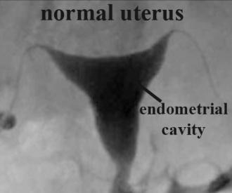
12 CONGENITAL DISORDERS


13 Pyometra = pyometrea is a uterine infection, it is accumulation of purulent material in the uterine cavity. Ultrasound is usually the initial form of imaging. CT and MRI scanning may be required for the diagnosis and assessment of perforated pyometra. Doppler scanning is helpful in detecting blood flow changes when pyometra complicates endometrial cancer. Pneumoperitoneum on plain X-ray (subdiaphragm free gas) or CT scan shows evidence of spontaneous perforation.


 in the right adnexa. U = uterus.")
 shows diffuse enlargement of the uterus (U) with small")
14 = is a bacterial infection that results in collections of pus in the body. Pelvic actinomycosis involving the adnexa and uterus in a 68-year-old woman. (a) Axial contrast-enhanced CT image shows a mixed solid and cystic mass (arrows) in the right adnexa. U = uterus. (b) Axial contrast-enhanced CT image obtained at a lower level than (a) shows diffuse enlargement of the uterus (U) with small abscesses (white arrows). Note the perirectal soft-tissue infiltrations (black arrows) and the intrauterine device (arrowhead).
15 ENDOMETRIOSIS =is defined as the presence of functional endometrial glands and stroma outside the uterine cavity. Transvaginal ultrasonography (US) is the first imaging technique used to diagnose endometriosis and remains the most accessible technique. Transvaginal US is used in identifying deep endometriosis, especially in detecting lesions of the rectal wall and retrocervical space. However, the accuracy of transvaginal US in the detection of some deep endometriotic lesions may vary depending on the location of the lesions and the experience of the operator. MRI imaging is a noninvasive imaging method with high spatial resolution that allows multiplanar evaluation and good tissue characterization, but without the use of ionizing radiation or iodinated contrast agents. It is highly accurate in the diagnosis of infiltrating extraperitoneal endometriosis and allows the identification of lesions that are hidden by adhesions and the evaluation of subperitoneal lesion extension. MR imaging also possesses a huge advantage over other imaging modalities in that it allows a complete survey of the anterior and posterior compartments of the pelvis to be made with a single study.
, prevesical space (outlined in white), vesicouterine pouch (outlined in red), and")

16 Normal anatomy of the anterior compartment. Sagittal T2-weighted MR image shows the bladder (*), prevesical space (outlined in white), vesicouterine pouch (outlined in red), and vesicovaginal septum (outlined in yellow) Endometriotic involvement of the posterior pelvic compartment in a 29-year-old woman with hypermenorrhea and dysmenorrhea. Sagittal T2-weighted MR images show an irregular hypointense mass (arrowhead) that extends from the posterior cervix inferiorly to the vaginal fornix. Note the presence of a subserous leiomyoma in the anterior uterine wall (black arrow )

17 LEIOMYOMA=is a benign smooth muscle tumor that very rarely becomes cancer

 and submucosal")
18 Multiple intramural leiomyomas in a 50-year-old woman. Axial (a) and sagittal (b) T2-weighted images show multiple intramural (IM) and submucosal (SM) leiomyomas with decreased signal intensity.
19
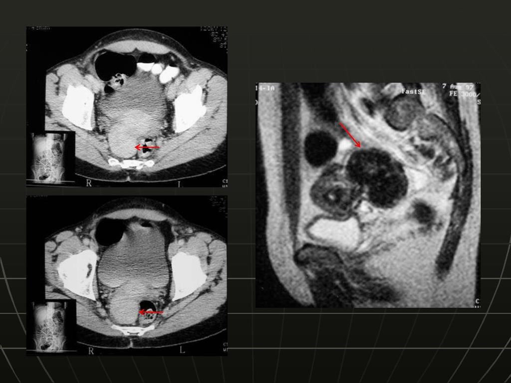
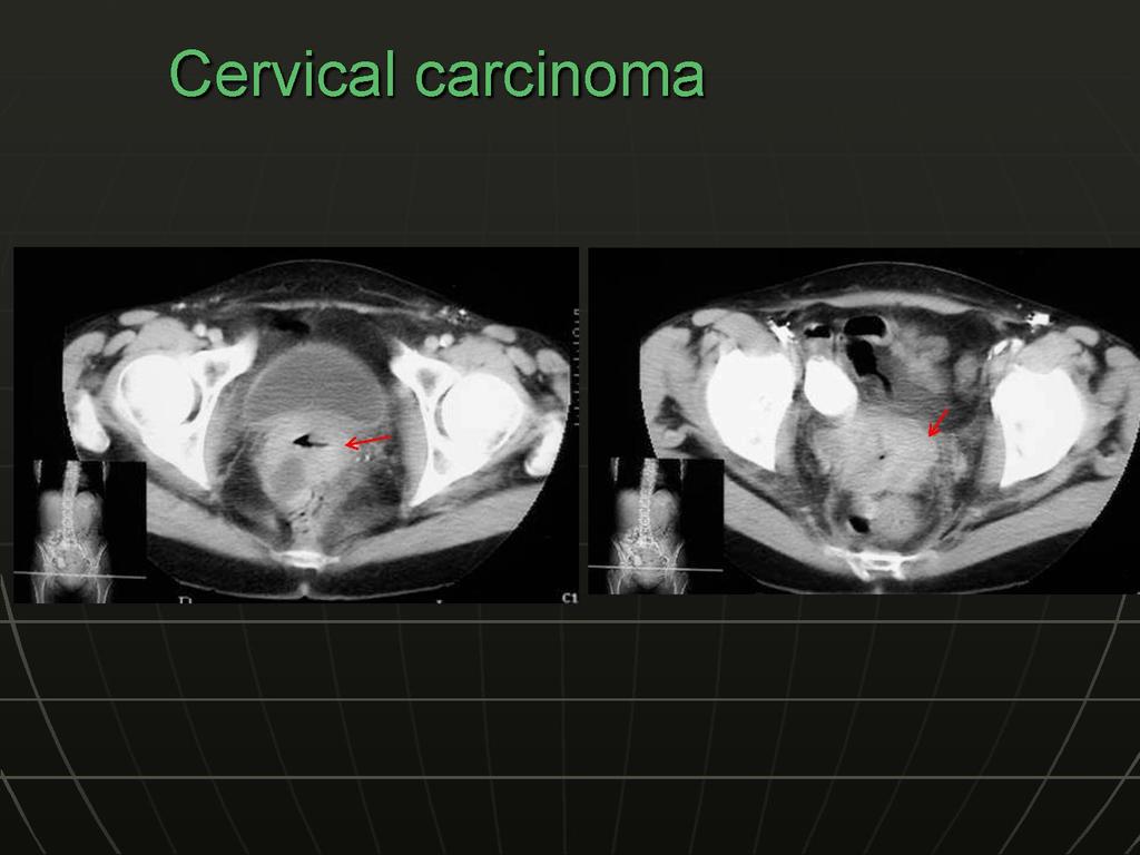
20 Cervical carcinoma is the third most common gynecologic malignancy, with an average patient age at onset of 45 years. The International Federation of Gynecology and Obstetrics (FIGO) staging system is used for standardization of treatment results. Magnetic resonance (MR) imaging is accepted as optimal for evaluation of the main prognostic factors and selection of treatment strategy. MR imaging examination obviates the use of invasive procedures such as cystoscopy and proctoscopy
21 TABLE 1. Correlation between FIGO Staging, MR Imaging Staging, and Treatment of Cervical Carcinoma
22


23 Cervical carcinoma. Sagittal T2-weighted MR image reveals a small, posterior cervical carcinoma (arrow) Exophytic cervical carcinoma. Sagittal T2- weighted MR image demonstrates a large, exophytic cervical mass protruding into the posterior vaginal fornix (arrow)
24
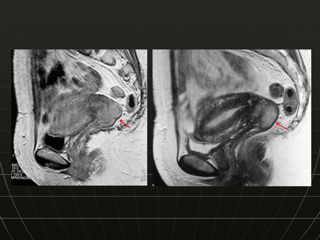
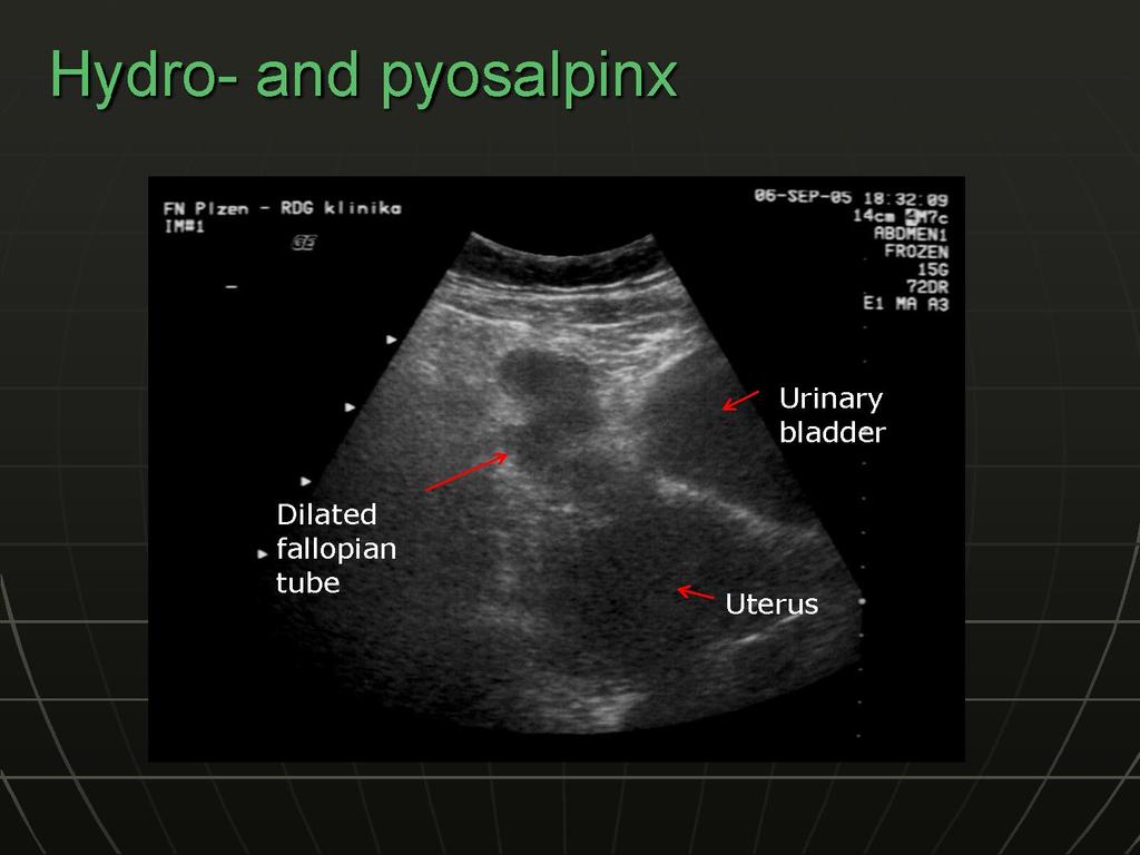
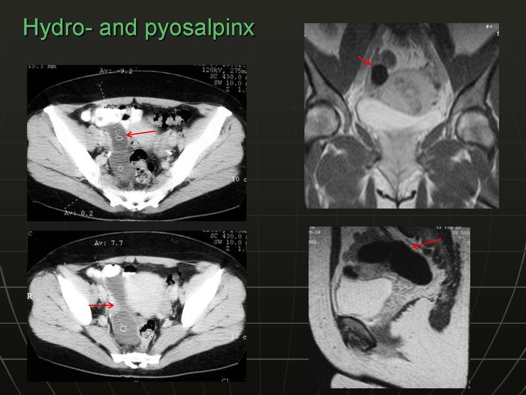
25 Hydrosalpinx occurs when a distally blocked fallopian tube fills with fluid. The blocked tube may be substantially distended Serous fluid, hemorrhage, or pus may accumulate within the tube, depending on the cause of the obstruction A fallopian tube that is filled and distended with blood is referred to as hematosalpinx, and a tube filled with pus is referred to as pyosalpinx. NOTE: On MR images, both hydrosalpinx and pyosalpinx appear as dilated, fluid-filled, tubular structures. With pyosalpinx, the wall of a dilated fallopian tube may be thickened, and it has variable signal intensity on T1-weighted images and heterogeneous signal intensity on T2-weighted images. However, pyosalpinx often cannot be reliably differentiated from hydrosalpinx on MR images
26
27

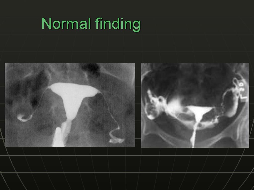
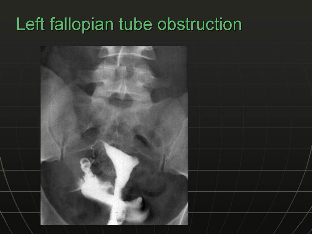
28 Radiography shows dilatation of the ampullary portion of the left fallopian tube, a finding that is consistent with a hydrosalpinx. The right fallopian tube is abruptly cut off, a finding that is consistent with previous tubal ligation.
29
30
31
 infiltrating adjacent myometrium to more than 50% of its")
, which is continuous with the")
32 Endometrial carcinoma infiltrating whole thickness of myometrium (stage IC). Sagittal image shows hyperintense endometrial neoplasm (arrows) infiltrating adjacent myometrium to more than 50% of its thickness Normal uterine zonal anatomy. Sagittal image shows the normal uterine zonal anatomy. The endometrium is surrounded by the homogeneous low-signal-intensity junctional zone (arrow), which is continuous with the fibrous cervical stroma. The myometrium (arrowhead) has intermediate signal intensity
33

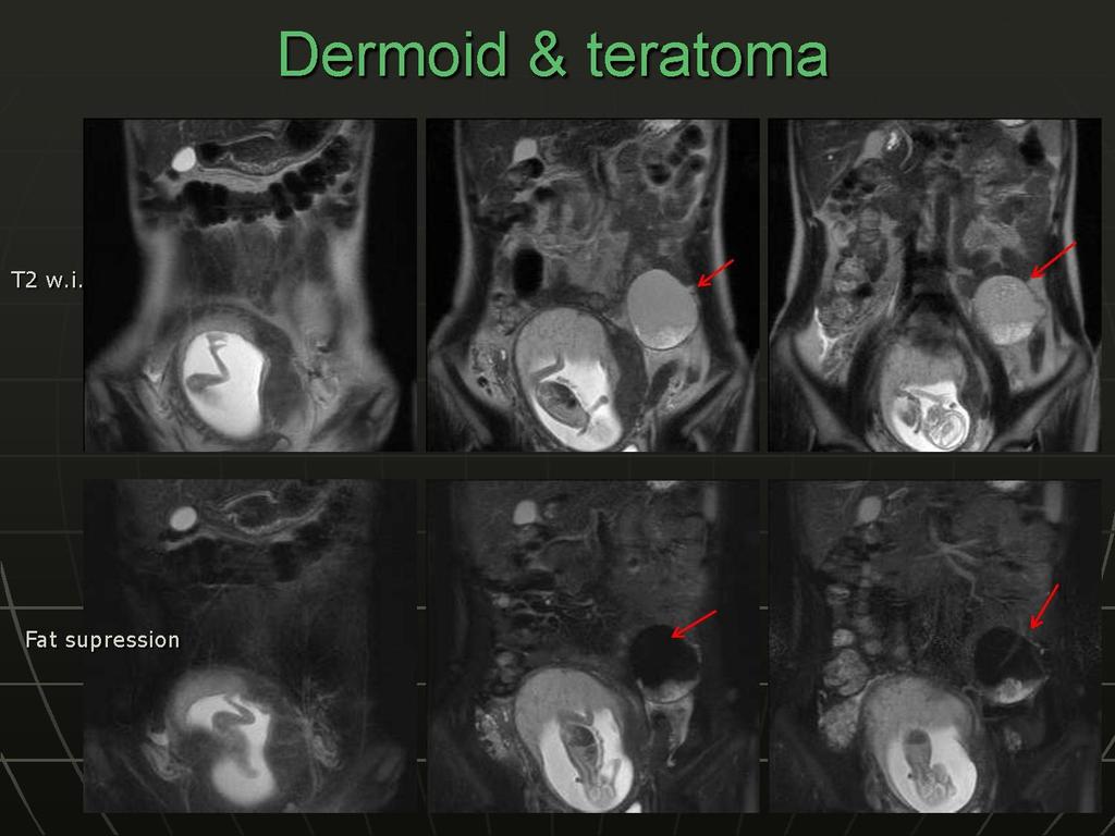
34 The diagnosis of teratoma or dermoid at CT and MR imaging is fairly straightforward because these modalities are more sensitive for fat
35
36
37
38
39
40
41
42
43
44
45
46
JMSCR Vol 05 Issue 06 Page June 2017
 www.jmscr.igmpublication.org Impact Factor 5.84 Index Copernicus Value: 83.27 ISSN (e)-2347-176x ISSN (p) 2455-0450 DOI: https://dx.doi.org/10.18535/jmscr/v5i6.29 MRI in Clinically Suspected Uterine and
Endometriosis - MRI findings with anatomic-pathologic correlation
 Endometriosis - MRI findings with anatomic-pathologic correlation Poster No.: C-2551 Congress: ECR 2015 Type: Educational Exhibit Authors: E. Matos, A. T. Almeida, A. Sanches; Vila Nova de Gaia/PT Keywords:
Value of MRI in Characterizing Adnexal Masses
 The Journal of Obstetrics and Gynecology of India (July August 2015) 65(4):259 266 DOI 10.1007/s13224-015-0730-9 PHOTO ESSAY Value of MRI in Characterizing Adnexal Masses Alpana Karnik 1 Raina Anil Tembey
What is endometrial cancer?
 Uterine cancer What is endometrial cancer? Endometrial cancer is the growth of abnormal cells in the lining of the uterus. The lining is called the endometrium. Endometrial cancer usually occurs in women
Accuracy of transvaginal ultrasound and magnetic resonance imaging in diagnosis and extension of pelvic endometriosis
 Accuracy of transvaginal ultrasound and magnetic resonance imaging in diagnosis and extension of pelvic endometriosis A.Salem, Kh. Fakhfakh, S. Mehiri, Y. Ben Brahim, F. Ben Amara, H. Rajhi, R. Hamza,
Endometrial Stromal Sarcoma
 May 26, 2011 By Sushila Ladumor, MD [1] Endometrial stromal sarcoma (ESS) is a rare malignant tumor of the endometrium, occurring in the age group of 40-50 years. History The 50-year-old, female patient
CLINICAL PRESENTATION AND RADIOLOGY QUIZ QUESTION
 Donald L. Renfrew, MD Radiology Associates of the Fox Valley, 333 N. Commercial Street, Suite 100, Neenah, WI 54956 8/20/2011 Radiology Quiz of the Week # 34 Page 1 CLINICAL PRESENTATION AND RADIOLOGY
Usual and unusual endometriosis locations. an MRI based approach
 Usual and unusual endometriosis locations. an MRI based approach Poster No.: C-1950 Congress: ECR 2014 Type: Educational Exhibit Authors: E. E. Martin, M. I. BOLAÑO VEGA, D. L. PINEDA, S. JARUFFE, E. P.
ENDOMETRIOSIS AS A COMMON CAUSE OF PELVIC PAIN
 ENDOMETRIOSIS AS A COMMON CAUSE OF PELVIC PAIN M.Basta Nikolić, S. Stojanović, O. Nikolić, T. Mrđanin, D. Donat, V. Žigić Center for Radiology, Clinical Center of Vojvodina Novi Sad Chronic pelvic pain
Malignant Transformation of Endometriosis: Magnetic Resonance Imaging Aspects
 Malignant Transformation of Endometriosis: Magnetic Resonance Imaging Aspects Poster No.: C-0084 Congress: ECR 2014 Type: Scientific Exhibit Authors: E. A. Yukhno, I. Trofimenko, G. Trufanov; St. Petersburg/RU
Malignant Transformation of Endometriosis: Magnetic Resonance Imaging Aspects
 Malignant Transformation of Endometriosis: Magnetic Resonance Imaging Aspects Poster No.: C-0084 Congress: ECR 2014 Type: Scientific Exhibit Authors: E. A. Yukhno, I. Trofimenko, G. Trufanov; St. Petersburg/RU
A Practical Approach to Adnexal Masses
 A Practical Approach to Adnexal Masses Darcy J. Wolfman, MD Section Chief of Genitourinary Imaging American Institute for Radiologic Pathology Clinical Associate Johns Hopkins Community Radiology Division
Bilateral Primary Fallopian Tube Carcinoma: Findings on Sequential MRI
 Hosokawa et al. MRI in Fallopian Tube Carcinoma Women s Imaging Case Report WOMEN S IMAGING Chisa Hosokawa 1 Mitsuo Tsubakimoto 2 Yuichi Inoue 3 Tetsuo Nakamura 2 Hosokawa C, Tsubakimoto M, Inoue Y, Nakamura
Genitourinary Imaging Pictorial Essay
 rown et al. MRI of the Female Pelvis Genitourinary Imaging Pictorial Essay Downloaded from www.ajronline.org by 37.44.202.41 on 12/17/17 from IP address 37.44.202.41. Copyright RRS. For personal use only;
Please complete prior to the webinar. HOSPITAL REGISTRY WEBINAR FEMALE REPRODUCTIVE SYSTEM EXERCISES CASE 1: FEMALE REPRODUCTIVE
 Please complete prior to the webinar. HOSPITAL REGISTRY WEBINAR FEMALE REPRODUCTIVE SYSTEM EXERCISES PHYSICAL EXAMINATION CASE 1: FEMALE REPRODUCTIVE 3/5 Patient presents through the emergency room with
Adenomyosis by myometrial Invasion of endometriosis: Comparison with typical adenomyosis
 Adenomyosis by myometrial Invasion of endometriosis: Comparison with typical adenomyosis Poster No.: C-1294 Congress: ECR 2010 Type: Scientific Exhibit Topic: Genitourinary Authors: S. Moon, H. K. Lim,
Endometrial line thickness in different conditions.
 Endometrial line thickness in different conditions 1 Endometrial thickens in response to Rising estrogen levels during the menstrual cycle and then shedding endometrial at the times of menses 2 The thickens
Characterizing Adnexal Masses: Pearls and Pitfalls 20 th Annual Summer Practicum SCBT-MR Jackson Hole August 11, 2010
 Characterizing Adnexal Masses: Pearls and Pitfalls 20 th Annual Summer Practicum SCBT-MR Jackson Hole August 11, 2010 Evan S. Siegelman MD University of Pennsylvania Medical Center Adnexal Masses: Pearls
ADENOMYOSIS CHRONIC PELVIC PAIN IN WOMEN IMAGING CHRONIC PELVIC PAIN IN WOMEN CHRONIC PELVIC PAIN IN WOMEN ADENOMYOSIS: PATHOLOGY ADENOMYOSIS
 CHRONIC PELVIC PAIN IN WOMEN IMAGING CHRONIC PELVIC PAIN IN WOMEN MOSTAFA ATRI, MD Dipl. Epid. UNIVERSITY OF TORONTO Non-menstrual pain of 6 months Prevalence 15%: 18-50 years of age 10-40% of gynecology
Female Genital Tract Lab. Dr. Nisreen Abu Shahin Assistant Professor of Pathology University of Jordan
 Female Genital Tract Lab Dr. Nisreen Abu Shahin Assistant Professor of Pathology University of Jordan Ovarian Pathology A 20-year-old female presented with vague left pelvic pain. Pelvic exam revealed
Carcinoma della cervice uterina
 Carcinoma della cervice uterina 16/01/2018 Carpi Dott. Matteo Generali U.O. Ginecologia Epidemiology Second most common malignancy in women worlwide CASI: 493.293 DECESSI: 273.505 In Italy, 3000-3400 new
The many faces of Endometriosis
 The many faces of Endometriosis Beryl Benacerraf M.D Harvard Medical School What is Endometriosis? Endometriosis is defined as the presence of normal endometrial tissue occurring outside of the endometrial
Current staging of endometrial carcinoma with MR imaging
 Current staging of endometrial carcinoma with MR imaging Poster No.: C-1436 Congress: ECR 2015 Type: Educational Exhibit Authors: M. Magalhaes, H. Donato, C. B. Marques, P. Gomes, F. Caseiro Alves; Coimbra/PT
MR Imaging of the Adnexal Masses: A Review
 Page54 Review of Literature NJR 2011;1(1):54 60; Available online at www.nranepal.org MR Imaging of the Adnexal Masses: A Review I Ahmad 1, S Kirmani 1, M Rashid 2, K Ahmad 3 1 Department of Radiodiagnosis,
Radiological assessment of infertility: A pictorial review
 Radiological assessment of infertility: A pictorial review Poster No.: C-1681 Congress: ECR 2015 Type: Educational Exhibit Authors: J. P. Walsh, N. Healy, M. O'sullivan, S. Harte, M. T. Knox; Dublin/ IE
Pelvic Pain: Overlooked
 EDUCATION EXHIBIT 3 Pelvic Pain: Overlooked and Underdiagnosed Gynecologic Conditions 1 CME FEATURE See accompanying test at http:// www.rsna.org /education /rg_cme.html LEARNING OBJECTIVES FOR TEST 1
CELL AND TISSUE INJURY COURSE-II PATHOLOGY LABORATORY. PATHOLOGY of MASS LESIONS and TISSUE DEFECTS -MACROSCOPY Assoc. Professor Rengin Ahıskalı
 CELL AND TISSUE INJURY COURSE-II PATHOLOGY LABORATORY PATHOLOGY of MASS LESIONS and TISSUE DEFECTS -MACROSCOPY Assoc. Professor Rengin Ahıskalı M1 - RENAL TUBERCULOSIS cavitary areas caseous necrosis fibrous
MRI of Endometriosis with Pre and Post- Operative Correlation
 MRI of Endometriosis with Pre and Post- Operative Correlation Shannon P. Sheedy, Candice A. Bookwalter, Wendaline M. VanBuren Mayo Clinic Rochester, Department of Radiology SCBT-MR - Nashville, TN September
Deep Pelvic Endometriosis: MR Imaging for Diagnosis and Prediction of Extension of Disease 1
 Marc Bazot, MD Emile Darai, MD, PhD Roula Hourani, MD Isabelle Thomassin, MD Annie Cortez, MD Serge Uzan, MD Jean-Noël Buy, MD Index terms: Bladder, diseases, 83.3192 Bladder, MR, 83.121411, 83.121412,
Outline - MRI - CT - US. - Combinations of imaging modalities for treatment planning
 Imaging Outline - MRI - CT - US - Combinations of imaging modalities for treatment planning Imaging Part 1: MRI MRI for cervical cancer high soft tissue contrast multiplanar imaging MRI anatomy: the normal
Category Term Definition Comments 1 Major Categories 1a
 Working Lexicon Categories, Terms & Definitions Category Term Definition Comments 1 Major Categories 1a Physiologic Category (consistent with normal ovarian physiology) Follicle Simple 3 cm in premenopausal
Deep pelvic endometriosis: MR imaging with laparoscopic and histologic correlation
 Deep pelvic endometriosis: MR imaging with laparoscopic and histologic correlation Poster No.: C-0372 Congress: ECR 2012 Type: Scientific Exhibit Authors: S. Gispert; Barcelona/ES DOI: 10.1594/ecr2012/C-0372
The new FIGO classification in endometrial carcinoma
 The new FIGO classification in endometrial carcinoma Poster No.: C-1073 Congress: ECR 2012 Type: Educational Exhibit Authors: A. IGLESIAS CASTAÑON, M. Arias Gonzales, J. Mañas Uxó, 1 2 1 2 2 2 B. NIETO
Page # 1. Endometrium. Cellular Components. Anatomical Regions. Management of SIL Thomas C. Wright, Jr. Most common diseases:
 Endometrium Pathology of the Endometrium Thomas C. Wright Columbia University, New York, NY Most common diseases: Abnormal uterine bleeding Inflammatory conditions Benign neoplasms Endometrial cancer Anatomical
Essentials of Clinical MR, 2 nd edition. 73. Urinary Bladder and Male Pelvis
 73. Urinary Bladder and Male Pelvis Urinary bladder carcinoma is best locally staged with MRI. It is important however to note that a thickened wall (> 5 mm) is a non-specific finding seen in an underfilled
Normal endometrium: A, proliferative. B, secretory.
 Normal endometrium: A, proliferative. B, secretory. Nội mạc tử cung Nội mạc tử cung Cyclic changes in endometrium.. Approximate relationship of useful microscopic changes. Arias-Stella reaction in endometrial
Case 9539 Endometriosis in the canal of Nuck
 Case 9539 Endometriosis in the canal of Nuck Monteiro V, Cunha TM Section: Genital (Female) Imaging Published: 2011, Sep. 27 Patient: 26 year(s), female Authors' Institution V Monteiro 1 TM Cunha 2 1 Unidade
Pelvic Angiogram - Male
 Pelvic Angiogram - Male Common iliac artery Internal iliac artery Lateral sacral artery Iliolumbar artery Posterior trunk of internal iliac artery Superior gluteal artery Internal pudendal artery External
Fallopian tube carcinoma: pearls and pitfalls
 Fallopian tube carcinoma: pearls and pitfalls Poster No.: C-0543 Congress: ECR 2013 Type: Educational Exhibit Authors: C. N. Tentugal, T. M. Cunha, A. Félix ; Portimão/PT, Lisbon/PT Keywords: Cancer, elearning,
Pitfalls in the CT diagnosis of appendicitis
 The British Journal of Radiology, 77 (2004), 792 799 DOI: 10.1259/bjr/95663370 E 2004 The British Institute of Radiology Pictorial review Pitfalls in the CT diagnosis of appendicitis 1 C D LEVINE, 2 O
Female pelvic MRI for infertility: Radiological findings in a cohort of patients referred by a fertility specialist.
 Female pelvic MRI for infertility: Radiological findings in a cohort of patients referred by a fertility specialist. Poster No.: C-0684 Congress: ECR 2016 Type: Scientific Exhibit Authors: S. Saha, S.
Pitfall in differentiation of hemorrhagic vs. fatty lesions in female pelvis using fat saturated...
 IOSR Journal of Dental and Medical Sciences (IOSR-JDMS) e-issn: 2279-0853, p-issn: 2279-0861.Volume 14, Issue 3 Ver. V (Mar. 2015), PP 86-90 www.iosrjournals.org Pitfall in Differentiation of Hemorrhagic
PELVIC INFLAMMATORY DISEASE (PID) SALIM ABDUL-RAZAK (INTERN RADIOGRAPHER) TAMALE TEACHING HOSPITAL
 SALIM ABDUL-RAZAK (INTERN RADIOGRAPHER) TAMALE TEACHING HOSPITAL") PELVIC INFLAMMATORY DISEASE (PID) SALIM ABDUL-RAZAK (INTERN RADIOGRAPHER) TAMALE TEACHING HOSPITAL OBJECTIVES Definition of PID Prevalence rate of PID Causes of PID Symptoms of PID Risk factors Investigations
PELVIC INFLAMMATORY DISEASE (PID) SALIM ABDUL-RAZAK (INTERN RADIOGRAPHER) TAMALE TEACHING HOSPITAL OBJECTIVES Definition of PID Prevalence rate of PID Causes of PID Symptoms of PID Risk factors Investigations
MRI in Cervix and Endometrial Cancer
 28th Congress of the Hungarian Society of Radiologists RCR Session Budapest June 2016 MRI in Cervix and Endometrial Cancer DrSarah Swift St James s University Hospital Leeds, UK Objectives Cervix and endometrial
Incidental Esophageal Findings on Chest CT. Amira Hussien, MD, Elliot Fishman, MD, Bouchra Younes, MD, Ahmed Hatw. Johns Hopkins Medical Institution
 Incidental Esophageal Findings on Chest CT Amira Hussien, MD, Elliot Fishman, MD, ouchra Younes, MD, Ahmed Hatw. Johns Hopkins Medical Institution I have nothing to disclose. DISCLOSURE INTRODUCTION Although
Erratum to: Fertility-sparing for young patients with gynecologic cancer: How MRI can guide patient selection prior to conservative management
 Abdominal Radiology ª Springer Science+Business Media, LLC 2017 Published online: 11 November 2017 Abdom Radiol (2017) 42:2966 2973 DOI: 10.1007/s00261-017-1205-5 Erratum to: Fertility-sparing for young
Basic Training Programme. 16 Februrary 2018, ROTTERDAM. Pre and Post-Course Test Answers
 Basic Training Programme 16 Februrary 2018, ROTTERDAM Pre and Post-Course Test Answers Your details: Name: Conference registration number/ BT delegate number: Email address: Are you already performing
My Patient Has Pelvic Pain. David A. Kenny DO
 My Patient Has Pelvic Pain David A. Kenny DO Definition Of apparent pelvic origin Present most of the time for at least six months Severe enough to cause functional disability Requiring surgical or medical
CLINICAL PRESENTATION AND RADIOLOGY QUIZ QUESTION
 Donald L. Renfrew, MD Radiology Associates of the Fox Valley, 333 N. Commercial Street, Suite 100, Neenah, WI 54956 2/12/2011 Radiology Quiz of the Week # 7 Page 1 CLINICAL PRESENTATION AND RADIOLOGY QUIZ
Case Fibrothecoma of the ovary
 Case 10646 Fibrothecoma of the ovary Elisa Melo Abreu, Teresa Margarida Cunha Section: Genital (Female) Imaging Published: 2015, Jan. 2 Patient: 70 year(s), female Authors' Institution Department of Radiology,
Journal of Medical Imaging and Radiation Oncology
 Journal of Medical Imaging and Radiation Oncology 61 (2017) 767 773 MEDICAL IMAGING PICTORIAL ESSAY MRI findings in deep infiltrating endometriosis: A pictorial essay Anitha L Thalluri, 1 Steven Knox 1,2,3
Deep pelvic endometriosis: MR evaluation
 Deep pelvic endometriosis: MR evaluation Poster No.: C-2074 Congress: ECR 2012 Type: Educational Exhibit Authors: R. J. Méndez, J. Barrera Ortega; Madrid/ES Keywords: Abdomen, Genital / Reproductive system
Assessment of adnexal masses. Ultrasound workup of adnexal masses. symptoms. symptoms. Age. Serum tumor markers 10/1/2018
 Assessment of adnexal masses Ultrasound workup of adnexal masses Kevin Robinson, DO Department of Radiology Michigan State University October 4, 2018 Patients symptoms Age Menstrual status Serum tumor
PROFESSIONAL SKILLS 1 3RD YEAR SEMESTER 6 RADIOGRAPHY. THE URINARY SYSTEM Uz. Fatema shmus aldeen Tel
 PROFESSIONAL SKILLS 1 3RD YEAR SEMESTER 6 RADIOGRAPHY THE URINARY SYSTEM Uz. Fatema shmus aldeen Tel. 0925111552 Professional skills-2 THE URINARY SYSTEM The urinary system (review anatomy and physiology)
Ovarian Lesion Benign vs Malignant?
 Ovarian Lesion Benign vs Malignant? Michele Keenan 1,2 Bernice Dunne 2 Mary Moran 1 Therese Herlihy 1 1. Radiography and Diagnostic Imaging, School of Medicine, University College Dublin, Ireland 2. Midland
MR imaging diagnosis of dilated fallopian tubes
 MR imaging diagnosis of dilated fallopian tubes Poster No.: C-314 Congress: ECR 2009 Type: Educational Exhibit Topic: Genitourinary Authors: P. Papadopoulou, N. Michailidis, I. Kalaitzoglou, A. Haritanti,
Laparoscopy and Hysteroscopy
 AMERICAN SOCIETY FOR REPRODUCTIVE MEDICINE Laparoscopy and Hysteroscopy A Guide for Patients PATIENT INFORMATION SERIES Published by the American Society for Reproductive Medicine under the direction of
MPH Quiz. 1. How many primaries are present based on this pathology report? 2. What rule is this based on?
 MPH Quiz Case 1 Surgical Pathology from hysterectomy performed July 11, 2007 Final Diagnosis: Uterus, resection: Endometrioid adenocarcinoma, Grade 1 involving most of endometrium, myometrial invasion
Disclosure. Acknowledgement. What is the Best Workup for Rectal Cancer Staging: US/MRI/PET? Rectal cancer imaging. None
 What is the Best Workup for Rectal Cancer Staging: US/MRI/PET? Zhen Jane Wang, MD Assistant Professor in Residence UC SF Department of Radiology Disclosure None Acknowledgement Hueylan Chern, MD, Department
American Journal of Oral Medicine and Radiology
 American Journal of Oral Medicine and Radiology e - ISSN - XXXX-XXXX ISSN - 2394-7721 Journal homepage: www.mcmed.us/journal/ajomr ULTRASONOGRAPHIC EVALUATION OF ADNEXAL MASSES Nageswar Rao* Professor,
MR Findings of Extrauterine Mu llerian Adenosarcoma Associated with Deep Pelvic Endometriosis 1
 MR Findings of Extrauterine Mullerian Adenosarcoma Associated with Deep Pelvic Endometriosis 1 Dae Kun Oh, M.D., Chan Kyo Kim, M.D., Byung Kwan Park, M.D., Ji Young Kim, M.D. 2 Extrauterine mu llerian
Question Bank III - BHMS
 Question Bank III - BHMS Sub:- Ob/Gy -Paper-II 1. Give the definition of Puberty. 2. Enumerate five important physical changes evident during puberty. 3. Write down the vaginal changes during puberty.
Case 1. Gynaecology Case Presentation. Objectives. Disclosures 22/10/ year old female Clinical history: Assess right ovarian cyst
 Gynaecology Case Presentation Organ Imaging 2016 University of Toronto Sarah Johnson 39 year old female Clinical history: Assess right ovarian cyst Clinically diagnosed endometriosis Started fertility
2D and 3D MR imaging in the assessment of Fallopian tube features
 2D and 3D MR imaging in the assessment of Fallopian tube features Poster No.: C-1292 Congress: ECR 2010 Type: Topic: Scientific Exhibit Genitourinary Authors: J. Takahama, S. Kitano, N. Marugami, A. Takahashi,
PALM-COEIN: Your AUB Counseling Guide
 PALM-COEIN: Your AUB Counseling Guide 10 million+ Treat the cause, not the symptom In the U.S, more than 10 million women between the ages of 35 and 49 are affected by AUB 1 Diagnosis Cause Structural
Chapter 100 Gynecologic Disorders
 Chapter 100 Gynecologic Disorders Episode Overview: 1. Describe the presentation and RF for Adnexal torsion 2. List the imaging findings of adnexal torsion (US vs CT) 3. What is the management of adnexal
Top Tips for Gynaecological Ultrasound. Catherine Kirkpatrick Consultant Sonographer Dublin Oct 2018
 Top Tips for Gynaecological Ultrasound Catherine Kirkpatrick Consultant Sonographer Dublin Oct 2018 We can all scan a pelvis so what can we do to improve? Uterus, endometrium and ovaries, got it covered!
Staging and Treatment Update for Gynecologic Malignancies
 Staging and Treatment Update for Gynecologic Malignancies Bunja Rungruang, MD Medical College of Georgia No disclosures 4 th most common new cases of cancer in women 5 th and 6 th leading cancer deaths
We are IntechOpen, the world s leading publisher of Open Access books Built by scientists, for scientists. International authors and editors
 We are IntechOpen, the world s leading publisher of Open Access books Built by scientists, for scientists 3,500 108,500 1.7 M Open access books available International authors and editors Downloads Our
بسم هللا الرحمن الرحيم. Prof soha Talaat
 بسم هللا الرحمن الرحيم Ovarian tumors The leading indication for gynecologic surgery. Preoperative characterization of complex solid and cystic adnexal masses is crucial for informing patients about possible
Magnetic Resonance Imaging in Carcinoma of Cervix
 IOSR Journal of Dental and Medical Sciences (IOSR-JDMS) e-issn: 2279-0853, p-issn: 2279-0861.Volume 17, Issue 9 Ver. 3 (September. 2018), PP 23-29 www.iosrjournals.org Dr. Mahak Sood, Dr. Ankita Boricha,
Magnetic resonance imaging characteristics of deep endometriosis
 Human Reproduction vol.14 no.4 pp.1080 1086, 1999 Magnetic resonance imaging characteristics of deep endometriosis Karen Kinkel 1,2,4, Charles Chapron 3, Corinne Balleyguier 1, Xavier Fritel 3, Jean-Bernard
Pelvic Applications of Diffusion Magnetic Resonance Images
 Pelvic Applications of Diffusion Magnetic Resonance Images Antonio C. Coutinho Jr, MD a,b, *, Arun Krishnaraj, MD, MPH c, Cintia E. Pires, MD a, Leonardo K. Bittencourt, MD a,d,e, Alexander R. Guimarães,
Role of pelvic MRI in detection and characterization of uterine leiomyoma
 Role of pelvic MRI in detection and characterization of uterine leiomyoma Poster No.: C-1197 Congress: ECR 2016 Type: Educational Exhibit Authors: N. zouari, A. Ben Miled, E. KOULIBALY SANOU, S. Zaouali,
Endometriosis د. نجمه محمود كلية الطب جامعة بغداد فرع النسائية والتوليد
 Endometriosis د. نجمه محمود كلية الطب جامعة بغداد فرع النسائية والتوليد Objectives:- To know what is endometriosis The sites where it occur To explain its itiology & pathogenesis To know the clinical features
Advances in transvaginal ultrasound scanning and their clinical application
 Advances in transvaginal ultrasound scanning and their clinical application Bill Smith, Head of Ultrasound CDS Clinical Diagnostic Services, London, UK Introduction Transvaginal ultrasound scanning (TVS)
JMSCR Vol 3 Issue 9 Page September 2015
 www.jmscr.igmpublication.org Impact Factor 3.79 ISSN (e)-2347-176x DOI: http://dx.doi.org/10.18535/jmscr/v3i9.66 MR Evaluation of Isolated Fallopian Tubal Torsion, Rare Cause of Lower Abdominal Pain in
A Case Report Hydronephrosis and Hydrodureter due to Ureteral Deep Infiltrating Endometriosis mimic Ureteral Stricture Suryamanggala SI 1, Satria ML 2
 A Case Report Hydronephrosis and Hydrodureter due to Ureteral Deep Infiltrating Endometriosis mimic Ureteral Stricture Suryamanggala SI 1, Satria ML 2 1 Departement of Obstetric and Gynecology Faculty
Radiology of hepatobiliary diseases
 GI cycle - Lecture 14 436 Teams Radiology of hepatobiliary diseases Objectives 1. To Interpret plan x-ray radiograph of abdomen with common pathologies. 2. To know the common pathologies presentation.
Isolated Torsion of the Distal Part of the Fallopian Tube in a Premenarcheal 12 Year Old Girl: A Case Report
 Tohoku J. Exp. Med., 2004, Torsion 202, 239-243 of Fallopian Tube in a 12 Year Old Virgin Girl 239 Isolated Torsion of the Distal Part of the Fallopian Tube in a Premenarcheal 12 Year Old Girl: A Case
Imaging in gastric cancer
 Imaging in gastric cancer Gastric cancer remains a deadly disease because of late diagnosis. Adenocarcinoma represents 90% of malignant tumors. Diagnosis is based on endoscopic examination with biopsies.
IN THE NAME OF GOD POV: CYSTIC OVARIAN LESION
 IN THE NAME OF GOD POV: CYSTIC OVARIAN LESION CASE 1 20 years old girl with AUB and pelvic pain from 2 weeks ago Impression :Simple unilocular 6 cm ovarian cyst Next step? Almost certainly benign so FU
FDG-PET Findings in an Ovarian Endometrioma: A Case Report
 FDG-PET Findings in an Ovarian Endometrioma: A Case Report Jia-Huei Lin 1, Victor Chit-kheng Kok 2, Jian-Chiou Su 3 1 Department of Nuclear medicine, Kuang Tien General Hospital, Sha-Lu, Taichung, Taiwan
Salivary ultrasound. Dr T J Beale Royal National Throat Nose & Ear and UCLH Hospitals London UK
 Salivary ultrasound Dr T J Beale Royal National Throat Nose & Ear and UCLH Hospitals London UK Two main groups of patients with presenting symptoms of: Obstructive or chronic inflammatory symptoms (salivary
Key imaging features of acute gynaecological emergencies.
 Key imaging features of acute gynaecological emergencies. Poster No.: C-2200 Congress: ECR 2014 Type: Educational Exhibit Authors: Ó. Roche, N. Bharwani, A. G. Rockall; London/UK Keywords: Acute, Complications,
Correlation of Endometrial Thickness with the Histopathological Pattern of Endometrium in Postmenopausal Bleeding
 DOI 10.1007/s13224-014-0627-z ORIGINAL ARTICLE Correlation of Endometrial Thickness with the Histopathological Pattern of Endometrium in Postmenopausal Bleeding Singh Pushpa Dwivedi Pooja Mendiratta Shweta
Endometrioma With Calcification Simulating a Dermoid on Sonography
 Case Report Endometrioma With Calcification Simulating a Dermoid on Sonography Kiran A. Jain, MD Several investigators have explored the sonographic diagnostic criteria of endometriomas. Endometriomas
Deep infiltrating endometriosis MR imaging with surgical correlation
 Review rticle Deep infiltrating endometriosis MR imaging with surgical correlation Xue Tang 1, Rennan Ling 1, Jingshan Gong 1, Dongdong Mei 1, Yan Luo 1, Minge Li 2, Jianmin Xu 1, Liguo Ma 2 1 Department
One of the commonest gynecological cancers,especially in white Americans.
 Gynaecology Dr. Rozhan Lecture 6 CARCINOMA OF THE ENDOMETRIUM One of the commonest gynecological cancers,especially in white Americans. It is a disease of postmenopausal women with a peak incidence in
reproductive organs. Malignant neoplasms. 4. Inflammatory disorders of female reproductive organs 2 5. Infertility. Family planning.
 Thematic plan of lectures module iii. Diseases of female reproductive system. Family planning. Topic No of hours 1. Disturbances of menstrual function. Neuroendocrinological 2 syndromes in gynecology 1.
11/10/2015. Prostate cancer in the U.S. Multi-parametric MRI of Prostate Diagnosis and Treatment Planning. NIH estimates for 2015.
 Multi-parametric MRI of Prostate Diagnosis and Treatment Planning Temel Tirkes, M.D. Associate Professor of Radiology Director, Genitourinary Radiology Indiana University School of Medicine Department
A Tale of Two Ovaries: Cross-Sectional Imaging Spectrum of Ovarian Emergencies
 A Tale of Two Ovaries: Cross-Sectional Imaging Spectrum of Ovarian Emergencies Presenting author A J Baxi Co-authors A M Nagar, MBBS D Rajderkar, MD V Ojili, MD Contact:ojili@uthscsa.edu Disclaimer: We
Role of imaging in RCC. Ultrasonography. Solid lesion. Cystic RCC. Solid RCC 31/08/60. From Diagnosis to Treatment: the Radiologist Perspective
 Role of imaging in RCC From Diagnosis to Treatment: the Radiologist Perspective Diagnosis Staging Follow up Imaging modalities Limitations and pitfalls Duangkamon Prapruttam, MD Department of Therapeutic
2/24/19. Myometrial evaluation. Size Echotexture. Homogeneous Heterogeneous. Adenomyosis Fibroids. Adenomyosis. MUSA guidelines
 Content Adenomyosis and MUSA guidelines for myometrial disorders Adenomyosis MUSA guidelines Dr Lufee Wong FRANZCOG, MPH, DDU Recommended reporting guidelines Fibroids Adenomyosis Myometrial evaluation
Ultrasound - Pelvis. What is Pelvic Ultrasound Imaging?
 Scan for mobile link. Ultrasound - Pelvis Ultrasound imaging of the pelvis uses sound waves to produce pictures of the structures and organs in the lower abdomen and pelvis. There are three types of pelvic
Laparoscopy-Hysteroscopy
 Laparoscopy-Hysteroscopy Patient Information Laparoscopy The laparoscope, a surgical instrument similar to a telescope, is inserted through a small incision (cut) in the belly button during laparoscopy.
7/2/15. Approach to imaging of pelvic pain. Acute pelvic pain. Chronic pelvic pain. Dysmenorrhea
 I have no financial rela.onships to disclose. Approach to imaging of pelvic pain To emphasize the importance of ultrasound as the imaging modality of choice for the most commonly presen6ng diagnoses in
Diffusion-weighted MR imaging for Diagnosis of Uterine Leiomyomas
 Diffusion-weighted MR imaging for Diagnosis of Uterine Leiomyomas Poster No.: C-0111 Congress: ECR 2015 Type: Scientific Exhibit Authors: A. Er 1, G. Pekindil 2, M. Gök 3, A. R. Kandiloglu 2, A. G. Tamay
A Rare Case of Invasive Squamous Cell Carcinoma of Cervix Extending to Endometrium and Right Fallopian Tube
 A Rare Case of Invasive Squamous Cell Carcinoma of Cervix Extending to Endometrium and Right Fallopian Tube Kate Madhuri S 1, Gulhane Sushma R 2, Mane Sheetal V 3 1 Professor and Head, 2 Specialist cum