Interesting Cases in Gynecologic Pathology. Michael Ward, MD Surgical Pathology Fellow University of Utah Health Sciences Center Salt Lake City, UT
|
|
|
- Cory McCoy
- 5 years ago
- Views:
Transcription

1 Interesting Cases in Gynecologic Pathology Michael Ward, MD Surgical Pathology Fellow University of Utah Health Sciences Center Salt Lake City, UT
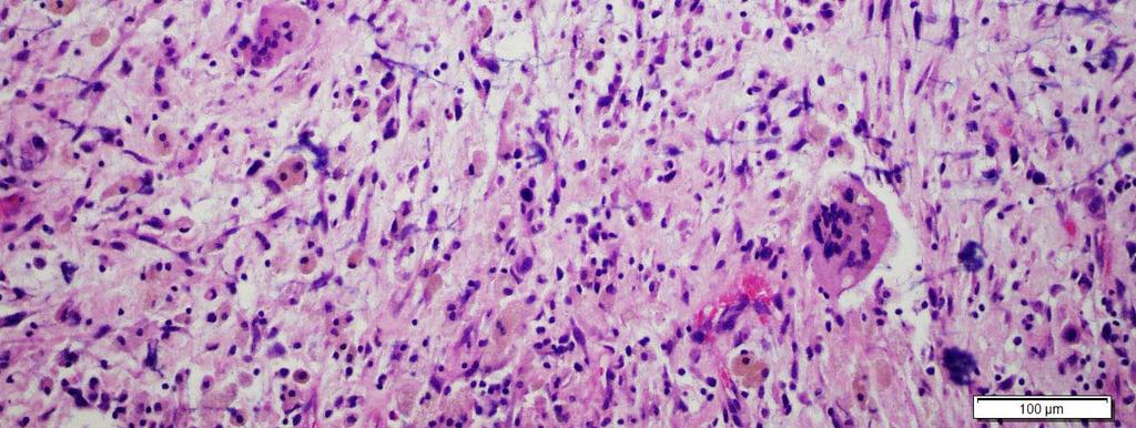
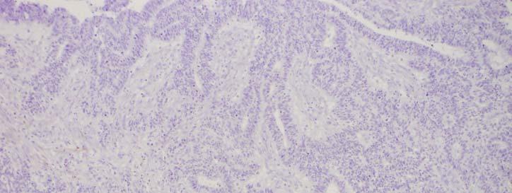
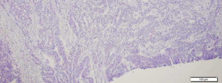
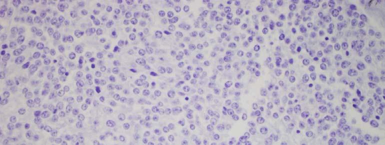
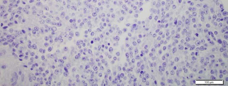
2 Case 1 History: 50 year old woman with a uterine mass Hysterectomy and BSO 11 cm yellow-white, gelatinous tumor

3 Case 1 Uterine Mass

4 Case 1 Uterine Mass

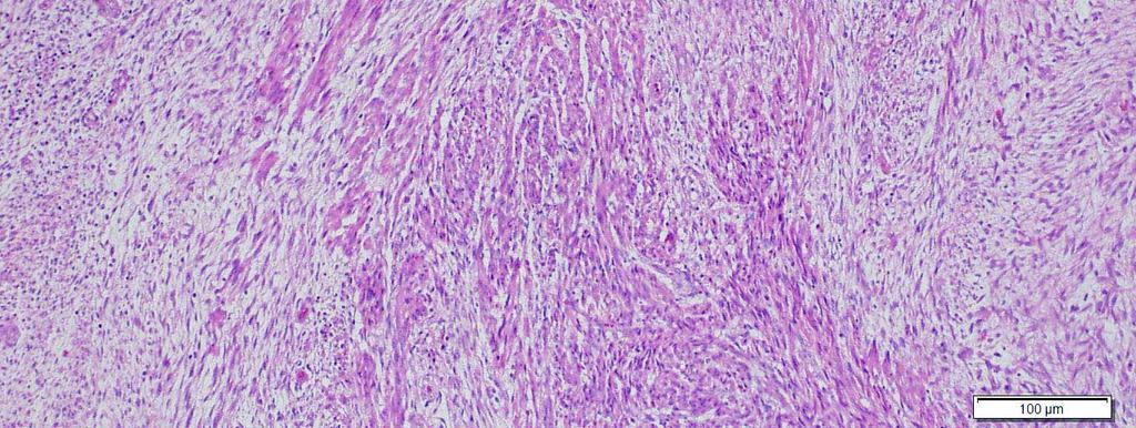
5 Case 1 Uterine Mass

6 Case 1 Uterine Mass

7 Case 1 Uterine Mass

8 Case 1 Uterine Mass

9 Case 1 Uterine Mass
10 Case 1- Differential Diagnosis Myxoid leiomyosarcoma Inflammatory myofibroblastic tumor Leiomyoma with degeneration or hydropic change Myxoid endometrial stromal sarcoma
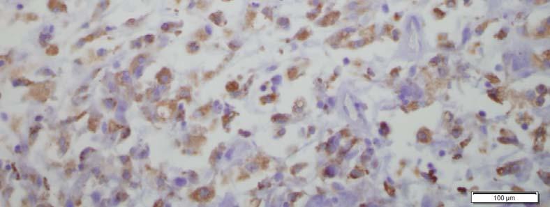
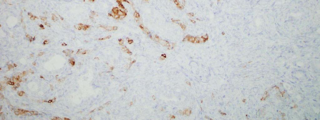
11 Case 1- Immunohistochemistry Smooth Muscle Actin (+) ALK (-)
12 Case 1- Final Diagnosis - MYXOID LEIOMYOSARCOMA Dr. Esther Oliva, MGH, Boston, MA
13 Case 1- Myxoid Leiomyosarcoma A rare and distinct malignant tumor
14 Case 1- Myxoid Leiomyosarcoma A rare and distinct malignant tumor: Doesn t conform to the conventional leiomyosarcoma diagnostic features Worse prognosis Good recent paper : Carlos Parra-Herran et al, Myxoid Leiomyosarcoma of the Uterus A Clinicopatholgic Analysis of 30 Cases With Reappraisal of Its Distinction From Other Uterine Myxoid Mesenchymal Neoplasma, (2016).
")

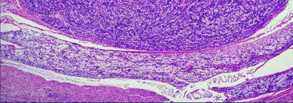
15 Case 1- Myxoid Leiomyosarcoma Gross Features: Large (average size 11 cm) Gelatinous, mucoid or myxoid Infiltrative growth pattern Parra-Herran et al, (2016)
 Variable necrosis Variable atypia What s Not Variable?")
16 Case 1- Myxoid Leiomyosarcoma Histology: Spindle cell tumor arranged in a myxoid matrix Variable mitotic activity (may be as low as 2/10hpf) Variable necrosis Variable atypia What s Not Variable? Infiltrative border Retained positivity for at least 1 smooth muscle marker

17 Case 1- Myxoid Leiomyosarcoma Parra-Herran et al, (2016)

18 Case 1- Myxoid Leiomyosarcoma Parra-Herran et al, (2016)
19 Case 1- Myxoid Leiomyosarcoma Immunohistochemistry: SMA, Desmin are most sensitive CD10 (+) in 66% KI-67 of little utility Alcian Blue stains myxoid LMS (negative in edema/hydropic change)
20 Case 1- Myxoid Leiomyosarcoma Treatment: Surgery Chemo/radiation have little effect Prognosis: Poor prognosis Worse than conventional LMS

21 Practical Classification of Smooth Muscle Tumors with Typical Spindle Cell Differentiation Diagnosis Geographic Tumor Necrosis Mitotic Rate (per 10 HPF) Atypia Leiomyosarcoma Present Any Present or absent Leiomyosarcoma Absent >10 Moderate to severe STUMP Absent >15 Absent Atypical Leiomyoma Absent <10 Moderate to severe Leiomyoma with increased mites Absent <15 Absent Table adapted from textbook: Crum et Al, Diagnostic Gynecologic and Obstetric Path, 2011
22 Case 1- Myxoid Leiomyosarcoma Diagnostic Features: 1. >50% myxoid matrix 2. Infiltrative border, plus any one of the following: 2 or more mites /10 hpf Unequivocal coagulative tumor necrosis Moderate to severe nuclear atypia

23 Strange things growing on walls
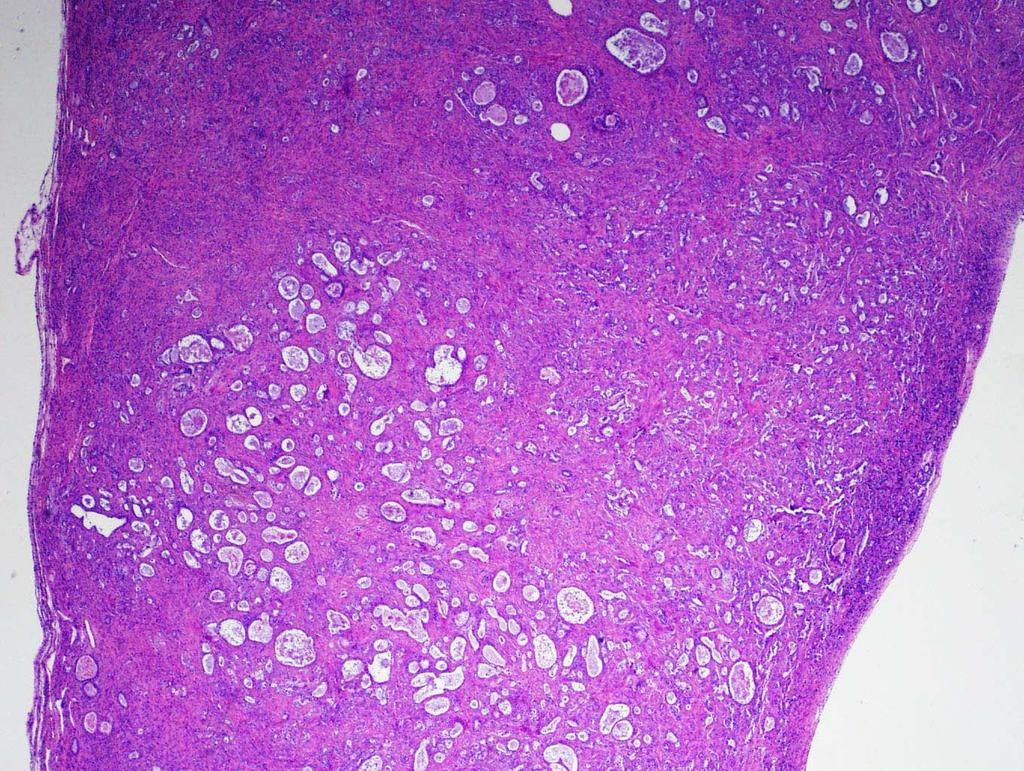
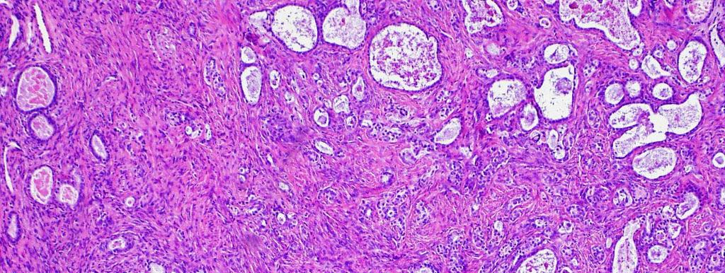
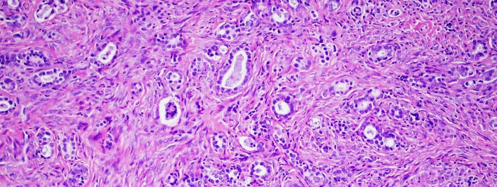
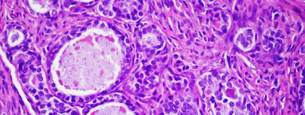
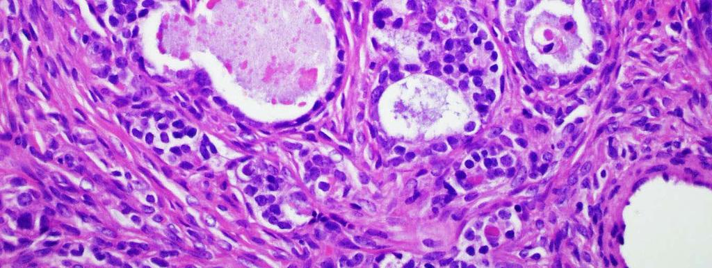
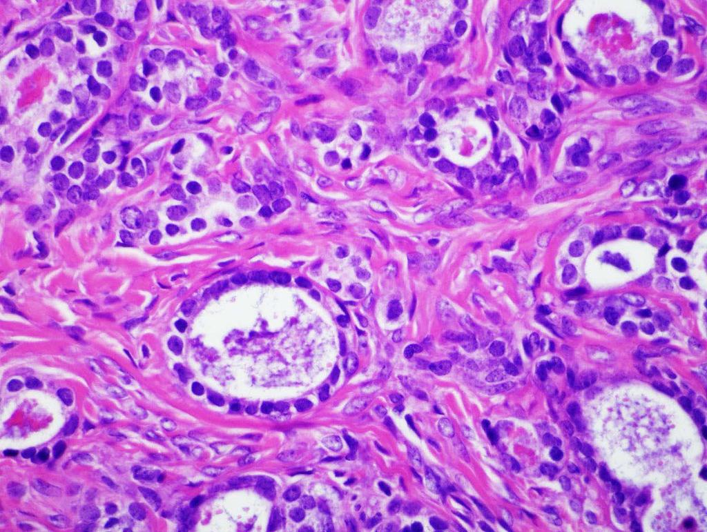
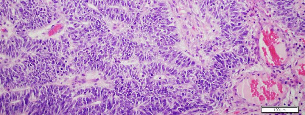
24 Case 2 History: 74 year old woman, cystic intra abdominal mass Hysterectomy and BSO 35 cm cystic ovarian mass 12.0 cm mass involving the fallopian tube

25 Case 2 Ovarian Cyst with Nodule

26 Case 2 Ovarian Cyst with Nodule

27 Case 2 Ovarian Cyst with Nodule


28 Case 2 Ovarian Cyst with Nodule
29 Case 2 Ovarian Cyst with Nodule


30 Case 2 Ovarian Cyst with Nodule

31 Case 2 Ovarian Cyst with Nodule

32 Case 2 Ovarian Cyst with Nodule
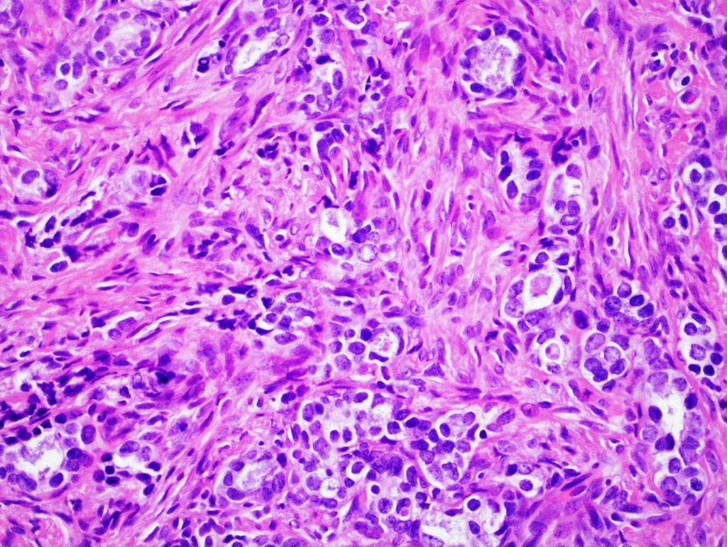
33 Case 2 - Differential Diagnosis Sarcoma Carcinoma Metastasis Focus of anaplastic carcinoma Lymphoma Sarcoma-like mural nodule
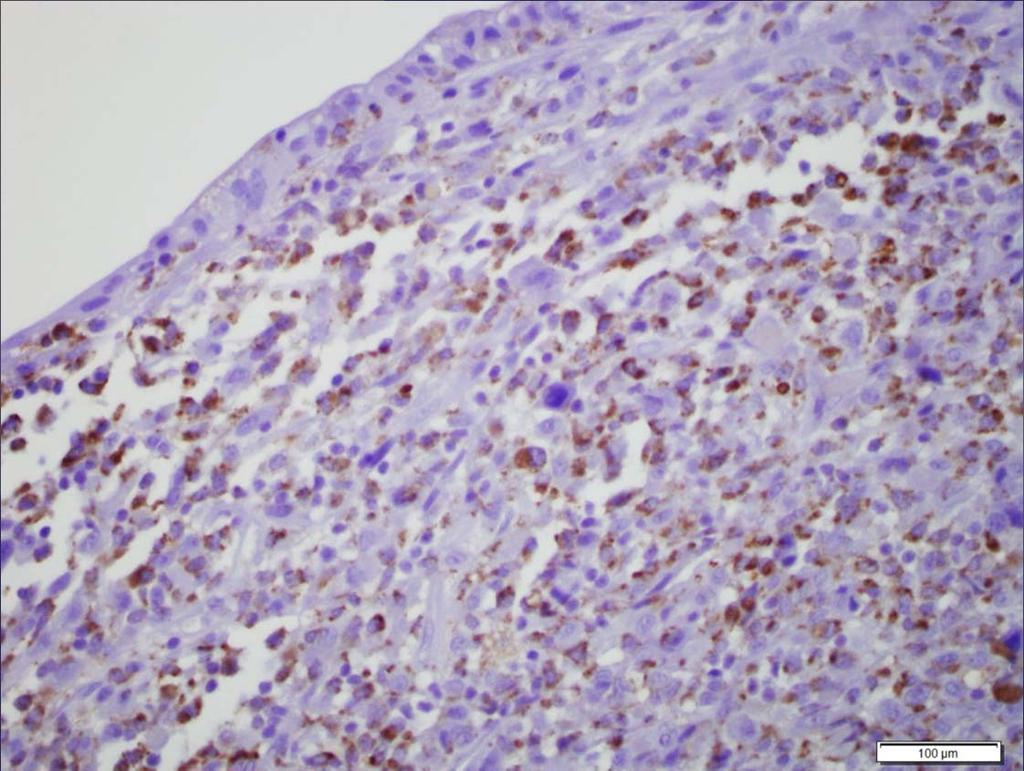
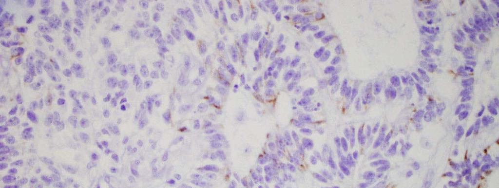
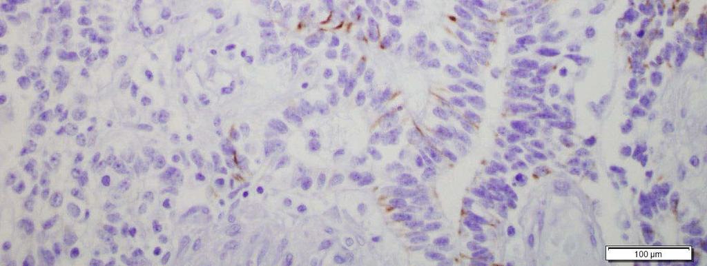
34 CD 68
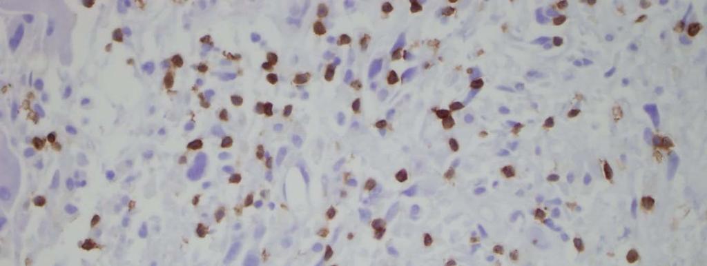
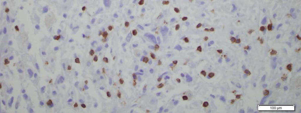
35 CD 3
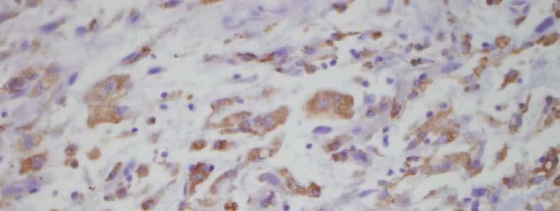
36 AE1/3
37 Case 2 - Immunohistochemistry CD68 (+) CK7 (+ patchy) AE1/3 (+ patchy) ER (-) CD10 (-) CD3 (+ small scattered lymphocytes) CD20 (-)
38 Case 2 - Final Diagnosis - MUCINOUS CYSTADENOMA WITH A SARCOMA-LIKE MURAL NODULE Dr. Robert Young, MGH, Boston


39 Case 2- Mural Nodules Strange things can be found growing in the wall of cystic ovarian neoplasms Which ones are benign? Which ones are more problematic?
40 Case 2- Mural Nodules General Features: Well demarcated nodule or plaque in the wall of an ovarian cyst Variable appearance: Yellow, pink, red, hemorrhagic, necrotic Single or multiple 1 to 20 cm Benign or malignant
41 Case 2- Mural Nodules Types of mural nodules: Benign: Sarcoma-like mural nodule Malignant: Sarcomatous mural nodule Mural nodule with anaplastic carcinoma
42 Case 2- Mural Nodules Benign: Younger Smaller (<6cm) Sharply demarcated Heterogeneous cell population (giant cells, spindle cells and inflammation) Malignant: Older Larger Infiltrative/vascular invasion Uniform cell population (spindled or carcinoma)
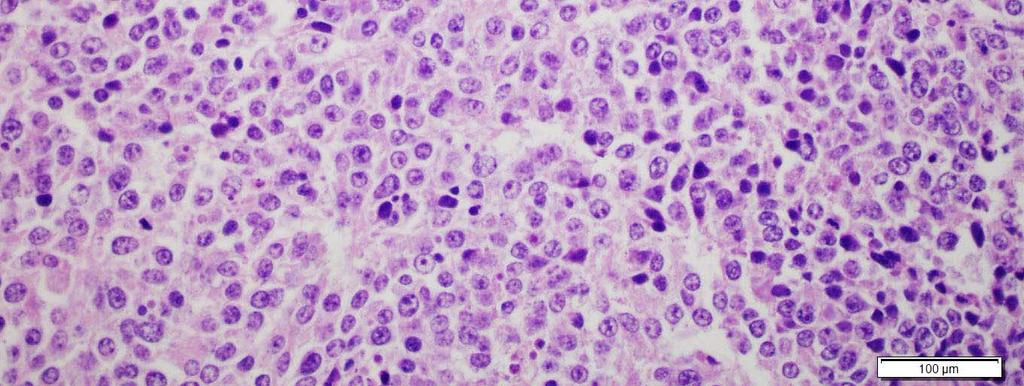
43 Case 2- Sarcoma-Like Mural Nodule Histology: Shows mix of MNGCs, spindle cells and inflammation WITH significant pleomorphism, mites, atypical mites
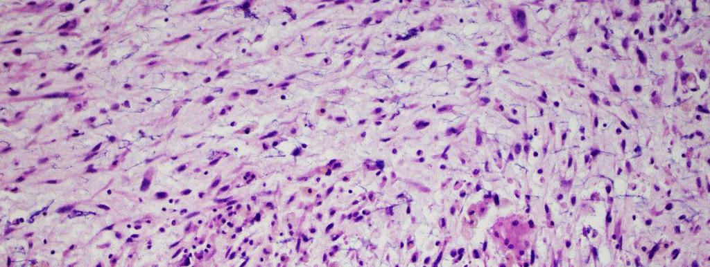
44 Case 2 Sarcoma-Like Mural Nodule
45 Case 2- Sarcoma-Like Mural Nodule Immunohistochemistry: Cytokeratin Negative or patchy in sarcoma-like mural nodules Strong and diffuse in anaplastic carcinoma

46 Case 2- Sarcoma-Like Mural Nodule

47 Case 2- Sarcoma-Like Mural Nodules Prithwijit et al, (2014)

48 Sarcomatous nodule Hitoshi et al, (2013)

49 Anaplastic Mucinous Carcinoma Prat:


50 Cueva do los cristales, Chihuaua, Mexico
51 Case 3 History: 62 year old woman with pain and vaginal bleeding Anemic Ultrasound showed fibroid uterus Hysterectomy Simple ovarian cyst Endometriosis

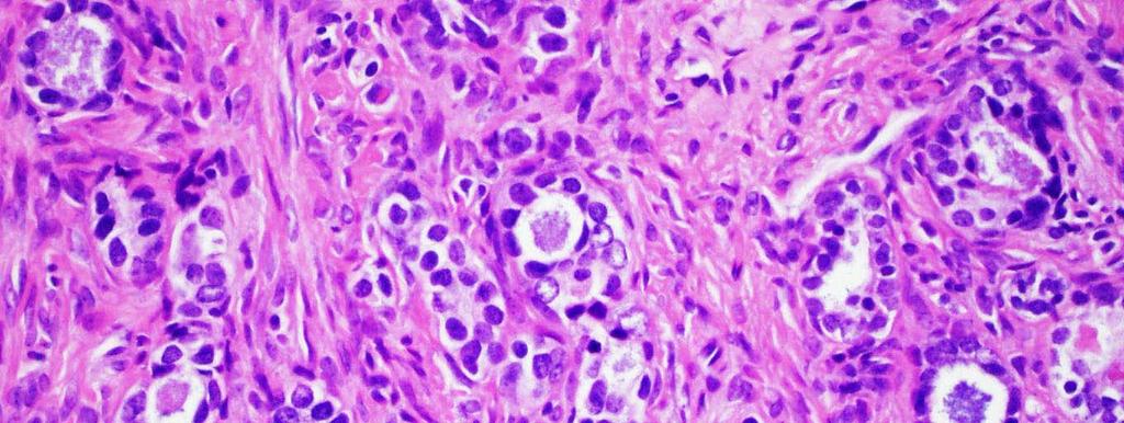
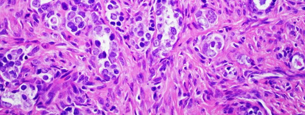
52 Case 3 Ovarian Cyst
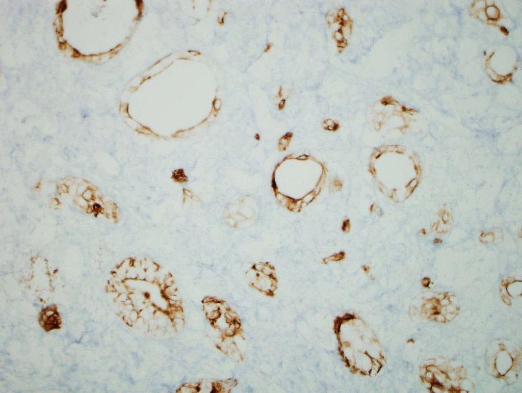
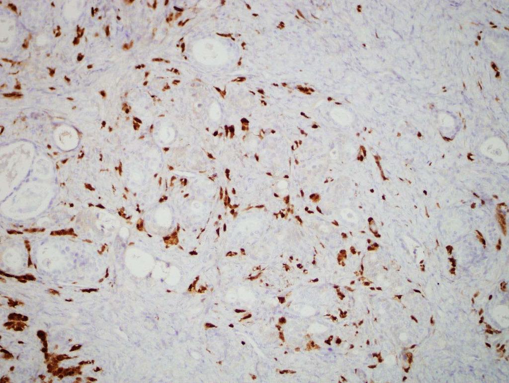
53 Case 3 Endometriosis
54 Case 3

55 Case 3

56 Case 3
57 Case 3
58 Case 3
59 Case 3
60 Case 3
61 Case 3 - Differential Diagnosis Carcinoma Metastasis Sex cord stromal tumor Mesonephric remnants Adenomatoid tumor Monodermal teratoma Struma ovarii Carcinoid
62 CK7 (+)
63 Calretinin

64 Inhibin (Focally +)
65 Other Stains: PAX 8 (weak focal +) CD10 (-) ER (-) Chromogranin (-) Synaptophysin (-) TTF-1 (-)
66 Case 3 - Final Diagnosis In my opinion, the diagnosis here is endometrioid adenocarcinoma, grade 1 of 3, arising out of the background of a mucinous cystadenoma. The mucinous cystadenoma is of mullerian mucinous type, and of course that cell type is very closely related to endometriosis. As is so typical, there is a background of endometriosis. That a carcinoma arising on the background of a mucinous cystadenoma should be endometrioid is no surprise. I think one has to call it endometrioid because there is simply no mucinous nature to the low-grade carcinoma Dr. Robert Young, MGH, Boston
67 Case 3 - Mucinous Ovarian Tumors General Features: Second most common epithelial-stromal neoplasm of the ovary 80% benign 10% borderline 10% malignant
68 Case 3 - Mucinous Ovarian Tumors Clinical Features: Age at presentation Benign tumors - younger women Borderline and malignant tumors - average 50 s Commonly accompanied by hormone manifestations (androgen or estrogen) CA-125 is less sensitive for mucinous tumors
69 Case 3 - Mucinous Ovarian Tumors Other common cyst wall findings: 5% have a mature teratoma component Not uncommonly associated with Brenner tumor

70 Case 3 Ovarian Cyst with Brenner Tumor

71 Case 3 Ovarian Cyst with Brenner Tumor
72 Case 3 - Endometrioid Ovarian Tumors General Features: Benign endometrioid tumors of the ovary are rare Second most common ovarian malignancy after serous
73 Case 3 - Endometrioid Ovarian Tumors Clinical Features: Postmenopausal Mid 50 s Discovered at earlier stage than serous (stage for stage, same prognosis) Frequently associated with endometriosis Often bilateral
74 Case 3 - Endometrioid Ovarian Tumors Classic Histology: Back to back glands, fibrotic stroma Stratified epithelial cells Squamous morules May be villous or papillary Grading in ovary same as for uterine tumors

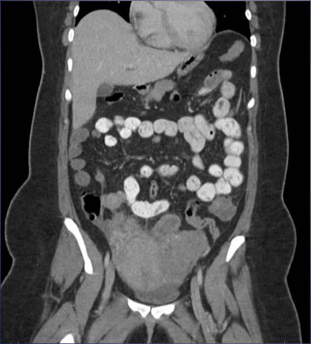
75 Case 3 - Endometrioid Ovarian Tumors Classic Endometrioid Histology FIGO grade I FIGO grade II FIGO grade III Malpica A et al. Modern Pathol 2016


76 Case 3 - Endometrioid Ovarian Tumors Endometrioid carcinoma with villoglandular pattern Malpica A et al. Modern Pathol 2016
77 Case 3 - Endometrioid Ovarian Tumors Variant Histology: Secretory Oxyphilic Ciliated Balloon Like Spindle Cell Mimics Clear cell Sex cord stromal Sertoli-leydig * In each, finding a focus of typical endometrioid carcinoma facilitates a correct interpretation

78 Case 3 - Endometrioid Ovarian Tumors Endometrioid carcinoma with secretory changes Malpica A et al. Modern Pathol 2016


79 Case 3 - Endometrioid Ovarian Tumors Endometrioid carcinoma with spindle cells Malpica A et al. Modern Pathol 2016


80 Case 3 - Endometrioid Ovarian Tumors Endometrioid carcinoma with clear cell changes Malpica A et al. Modern Pathol 2016


81 Case 3 - Endometrioid Ovarian Tumors Endometrioid carcinoma with sex cord stromal pattern Malpica A et al. Modern Pathol 2016

82 Case 3 - Endometrioid Ovarian Tumors Endometrioid carcinoma with microglandular pattern Malpica A et al. Modern Pathol 2016
83
84 Case 4 History: 30 year old woman 10 month history of heavy vaginal bleeding Endometrial biopsy at outside hospital

85 Case 4 - CT Scan

86 Case 4 Uterine Mass

87 Case 4 Uterine Mass

88 Case 4 Uterine Mass


89 Case 4 Uterine Mass

90 Case 4 Uterine Mass

91 Case 4 Uterine Mass
92 Case 4 - Differential Diagnosis Endometrioid Adenocarcinoma Carcinosarcoma Metastasis Sex Cord Stromal Tumor Ewing's/PNET Small Cell Rhabdomyosarcoma Germ Cell Tumor
93 Cam 5.2
94 AE1/3
95 Stains: CAM5.2 Patchy (+) Vimentin (+) AE1/3 (-) CK7 (-) PAX8 (-) ER (-) Chromogranin (-) Synaptophysin (-) Desmin (-) Myf4 (-) Inhibin (-) AFP (-) CD99 (-) FLI1 (-) EWSR1 FISH (-)
96 Case 4 - Diagnosis - HIGH GRADE ADENOCARCINOMA

97 Case 4 - History Cont. One month later, increasing abdominal pain Rapid recurrence and peritoneal carcinomatosis Urgently started on carboplatin/paclitaxel Showed little response
98 Case 4 - History cont. Pathology sent for consultation: Dr. Robert Young offered another diagnosis and 2 weeks later her chemo was changed to VIP (etoposide/ifosfamide/cisplatin) 6 Cycles, near complete response
99 Case 4 - Final Diagnosis I feel this is a primitive neuroectodermal tumor (PNET) of course we use the term primitive neuroectodermal tumor for the central-type morphology one sees in the ovary and uterus more commonly than one sees in the peripheral (Ewing s) variant. Dr. Robert Young, MGH, Boston, MA
100 Case 4 - Uterine PNET General Features: Small round blue cell tumors Ewing family of tumors (a spectrum) Characteristic translocations t(11:22) EWSR1/FLI1 Ewing < > PNET -small round cell morphology - Neural differentiation PNET further Divided into: Central-type PNET Peripheral-type PNET
101 Case 4 - Uterine PNET Peripheral-type PNET / Ewing Sarcoma Usually have EWSR1 translocations Neural crest derived Occur outside the CNS Central-type PNET / Neuroblastoma Usually don t have EWSR1 translocations Develop from / Involve CNS Most uterine and ovarian tumors are cpnet
102 Case 4 Uterine PNET Clinical Features: Rare in the Gyn tract Ovary most common, then uterus Postmenopausal or adolescent women, abnormal vaginal bleeding and uterine mass Presents in advanced stage, rapidly progressive Associated with VHL disease
103 Case 4 PNET Histology: EWING and PNET have similar morphology Uniform, small round cells, powdery chromatin, small nucleoli May have rosettes, fibrillary background Numerous mites, necrosis Pure form or admixed with other uterine tumors
104 Case 4 Uterine PNET IHC: CD99 ( usually +) (7/9 + in case series) FLI-1 (usually +) Vimentin (+) May express keratins, chromogranin, synaptophysin, NSE and S100 FISH: Usually negative for EWSR1 rearrangements
105 Case 4 Uterine PNET Prognosis: Very few case reports of uterine PNET Poor prognostic factors Older age Higher stage Tumor >8 cm Poor chemo response Absence of EWSR1 translocation
106 Case 4 Uterine PNET Treatment: Patients without mets at presentation respond well to intense multi-modal treatment Case reports of long disease free survival with platinum based chemo and etoposide
107 Case 4 Uterine PNET Take Home: Rapid diagnosis important Keep in mind that Ovarian and Uterine PNET tends to be of the central type No EWSR1 translocation May not express CD99 or FLI1 Follow up on our patient: - Currently, no evidence of disease

108 Thank You Witches Finger, Carsbad Caverns, NM
109 References: Neoplasms, M., Parra-herran, C., Schoolmeester, J. K., Yuan, L., Cin, P. D., Fletcher, C. D.,... & Nucci, M. R. (2016). Myxoid Leiomyosarcoma of the Uterus A Clinicopathologic Analysis of 30 Cases and Review of the Literature With Reappraisal of Its Distinction From Other Uterine Myxoid. Crum, C. P., Lee, K. R., & Nucci, M. R. (2011). Diagnostic gynecologic and obstetric pathology. Elsevier Health Sciences. Elizalde, C. R., Yagüe, A., Fernandez, J., Dieste, P., Puente, M. J., & Hernandez, J. (2016). Primitive neuroectodermal tumor of the uterus. Gynecologic Oncology Reports, 18, Bagué, S., Rodríguez, I. M., & Prat, J. (2002). Sarcoma-like mural nodules in mucinous cystic tumors of the ovary revisited: a clinicopathologic analysis of 10 additional cases. The American journal of surgical pathology, 26(11), Malpica, A. (2016). How to approach the many faces of endometrioid carcinoma. Modern Pathology, 29, S29-S44. IMAGES: Ghosh, Prithwijit, Kaushik Saha, and Sourav Bhowmik. "Sarcoma-like mural nodule in a borderline mucinous tumor of the ovary: A rare entity." Journal of mid-life health 5.4 (2014): 192. Yamazaki, Hitoshi, et al. "Ovarian mucinous cystic tumor of borderline malignancy with a mural nodule of anaplastic spindle cell carcinoma: a case report." Journal of ovarian research 6.1 (2013): 1. Anaplastic Mucinous Carcinoma: Prof. Jaime Prat, Hospital de la Santa Creu i Sant Pau, Autonomous University of Barcelona, Spain
Mody. AIS vs. Invasive Adenocarcinoma of the Cervix
 Common Problems in Gynecologic Pathology Michael T. Deavers, M.D. Houston Methodist Hospital, Houston, Texas Common Problems in Gynecologic Pathology Adenocarcinoma in-situ (AIS) of the Cervix vs. Invasive
05/07/2018. Types of challenges. Challenging cases in uterine pathology. Case 1 ` 65 year old female Post menopausal bleeding Uterine Polyp
 Types of challenges Challenging cases in uterine pathology Nafisa Wilkinson Gynaecological Pathologist UCLH London Lack of complete history often, NO clinical history at all! Cases from other centres often
Dr Sanjiv Manek Oxford. Oxford Pathology Course 2010 for FRCPath Illustration-Cellular Pathology. Oxford Radcliffe NHS Trust
 Dr Sanjiv Manek Oxford Oxford Pathology Course 2010 for FRCPath Illustration-Cellular Pathology. Oxford Radcliffe NHS Trust Ovarian Endometrial Vulvo-vaginal Cervical Illustration-Cellular Pathology. Oxford
Normal endometrium: A, proliferative. B, secretory.
 Normal endometrium: A, proliferative. B, secretory. Nội mạc tử cung Nội mạc tử cung Cyclic changes in endometrium.. Approximate relationship of useful microscopic changes. Arias-Stella reaction in endometrial
Ovarian Clear Cell Carcinoma
 Ovarian Clear Cell Carcinoma Rouba Ali-Fehmi, MD Professor of Pathology The Karmanos Cancer Institute, Wayne State University School of Medicine 50 year old woman with chief complaint of shortness of breath
Female Genital Tract Lab. Dr. Nisreen Abu Shahin Assistant Professor of Pathology University of Jordan
 Female Genital Tract Lab Dr. Nisreen Abu Shahin Assistant Professor of Pathology University of Jordan Ovarian Pathology A 20-year-old female presented with vague left pelvic pain. Pelvic exam revealed
Diagnostically Challenging Cases in Gynecologic Pathology
 Diagnostically Challenging Cases in Gynecologic Pathology Eric C. Huang, M.D., Ph.D. Department of Pathology and Laboratory Medicine University of California, Davis Medical Center Case 1 Presentation 38
Ovarian mucinous borderline tumor accompanied by LGESS with myxoid change: a case report and literature review
 https://doi.org/10.1186/s40001-017-0295-4 European Journal of Medical Research CASE REPORT Open Access Ovarian mucinous borderline tumor accompanied by LGESS with myxoid change: a case report and literature
Tumores de células pequeñas, redondas y azules: diagnóstico diferencial cuando el tiempo apremia
 Tumores de células pequeñas, redondas y azules: diagnóstico diferencial cuando el tiempo apremia Sílvia Bagué Servei de Patologia Hospital de Sant Pau Barcelona Soft tissue sarcomas Heterogeneous group
The role of immunohistochemistry in surgical pathology of the uterine corpus and cervix
 The role of immunohistochemistry in surgical pathology of the uterine corpus and cervix Prof. Ben Davidson, MD PhD Department of Pathology, Norwegian Radium Hospital, Oslo University Hospital, Oslo, Norway
64 YO lady THBSO for prolapse At gross : A 3 cm endometrial polyp in the fundus
 Case 6 64 YO lady THBSO for prolapse At gross : A 3 cm endometrial polyp in the fundus Numerous irregular, large glands with leaf-like pattern Large glands with broad-based papillary infolding into the
3 cell types in the normal ovary
 Ovarian tumors 3 cell types in the normal ovary Surface (coelomic epithelium) the origin of the great majority of ovarian tumors (neoplasms) 90% of malignant ovarian tumors Totipotent germ cells Sex cord-stromal
Disclosure. Case. Mixed Tumors of the Uterine Corpus and Cervix. I have nothing to disclose
 Mixed Tumors of the Uterine Corpus and Cervix Marisa R. Nucci, M.D. Division of Women s and Perinatal Pathology Department of Pathology Brigham and Women s Hospital Boston, MA UCSF Current Issues in Anatomic
Mucinous Tumors of the Ovary Beirut, Lebanon. Anaís Malpica, M.D. Professor Department of Pathology
 Mucinous Tumors of the Ovary Beirut, Lebanon Anaís Malpica, M.D. Professor Department of Pathology Primary Mucinous Tumors of the Ovary Cystadenoma Borderline (Tumor of Low Malignant Potential/Atypical
Problem 1: Differential of Neuroendocrine Carcinoma 3/23/2017. Disclosure of Relevant Financial Relationships
 Differential of Neuroendocrine Carcinoma Alain C. Borczuk,MD Weill Cornell Medicine Disclosure of Relevant Financial Relationships USCAP requires that all faculty in a position to influence or control
Diagnostic problems in uterine smooth muscle tumors
 Diagnostic problems in uterine smooth muscle tumors Marina Kos Ljudevit Jurak Clinical Department of Pathology, Clinical Hospital Center Sestre milosrdnice, Zagreb Institute of Pathology, University of
Endometrial Stromal Tumors
 Endometrial Stromal Tumors WHO Categories: Endometrial Stromal Nodule (ESN) Endometrial Stromal Sarcoma, low grade (LGESS) Endometrial Stromal Sarcoma, high grade (HGESS) Undifferentiated Uterine Sarcoma
Pathology of Ovarian Tumours. Dr. Jyothi Ranganathan MD ( Path) AFMC Pune PDCC (Cytopathology) PGI Chandigarh
 AFMC Pune PDCC (Cytopathology) PGI Chandigarh") Pathology of Ovarian Tumours Dr. Jyothi Ranganathan MD ( Path) AFMC Pune PDCC (Cytopathology) PGI Chandigarh Outline Incidence Risk factors Classification Pathology of tumours Tumour markers Prevention
Pathology of Ovarian Tumours Dr. Jyothi Ranganathan MD ( Path) AFMC Pune PDCC (Cytopathology) PGI Chandigarh Outline Incidence Risk factors Classification Pathology of tumours Tumour markers Prevention
of 20 to 80 and subsequently declines [2].
![of 20 to 80 and subsequently declines [2].](/thumbs/80/81450506.jpg "of 20 to 80 and subsequently declines [2].") - - According to the 2014 World Health Organization (WHO) classification and tumor morphology, primary ovarian tumors are subdivided into three categories: epithelial (60%), germ cell (30%), and sex-cord
- - According to the 2014 World Health Organization (WHO) classification and tumor morphology, primary ovarian tumors are subdivided into three categories: epithelial (60%), germ cell (30%), and sex-cord
Atypical Hyperplasia/EIN
 EIN Atypical Hyperplasia/EIN Based on scientific and diagnostic advances, in 2014 the WHO moved that the precursor lesion for endometrioid carcinoma be atypical hyperplasia/ein, rather than what was previously
Presenter: Yeh-Han Wang M.D.
 Korea-Taiwan-Japan Joint Meeting for Gynecological Pathology Mini-lecture Female Adnexal Tumor of Probable Wolffian Origin (FATWO) in Taiwan: A Small Case Series and Literature Review Presenter: Yeh-Han
6/5/2010. Outline of Talk. Endometrial Alterations That Mimic Cancer & Vice Versa: Metaplastic / reactive changes. Problems in Biopsies/Curettages
 Outline of Talk Endometrial Alterations That Mimic Cancer & Vice Versa: Problems in Biopsies/Curettages Metaplastic / reactive changes Mucinous change Microglandular hyperplasia-like change Squamous metaplasia
ESS: Pathologic Insights
 GEIS XVI INTERNATIONAL SYMPOSIUM Seville 4th October 2018 ESS: Pathologic Insights Sílvia Bagué The Royal Marsden Hospital London (United Kingdom) I have no conflicts of interest Endometrial stromal sarcoma
Pathology of the female genital tract
 Pathology of the female genital tract Common illnesses of the female genital tract Before menarche Developmental anomalies Tumors (ovarial teratoma) Amenorrhea Fertile years PCOS, ovarian cysts Endometriosis
Case 2. Dr. Sathima Natarajan M.D. Kaiser Permanente Medical Center Sunset
 Case 2 Dr. Sathima Natarajan M.D. Kaiser Permanente Medical Center Sunset History 24 year old male presented with a 3 day history of right flank pain, sharp in nature Denies fever, chills, hematuria or
International Society of Gynecological Pathologists Symposium 2007
 International Society of Gynecological Pathologists Symposium 2007 Anais Malpica, M.D. Department of Pathology The University of Texas M.D. Anderson Cancer Center Grading of Ovarian Cancer Histologic grade
Classification (1) Classification (3) Classification (2) Spindle cell lesions. Spindle cell lesions of bladder (Mills et al.
 Classification (3) Classification (2) Spindle cell lesions. Spindle cell lesions of bladder (Mills et al.") Non-epithelial tumours and nonepithelial tumour-like lesions of the bladder Dr Jonathan H Shanks The Christie NHS Foundation Trust, Manchester, UK Classification (1) Myofibroblastic proliferations and
Non-epithelial tumours and nonepithelial tumour-like lesions of the bladder Dr Jonathan H Shanks The Christie NHS Foundation Trust, Manchester, UK Classification (1) Myofibroblastic proliferations and
Uterine Mesenchymal Tumors from a Gynaecological Point of View: A Mini-Review
 EC Gynaecology Special Issue - 2017 Uterine Mesenchymal Tumors from a Gynaecological Point of View: A Mini-Review Mini Review Dr. Huseyin Aydogmus, Dr. Servet Gencdal, Dr. Nihan Gencdal and Dr. Serpil
Current Concept in Ovarian Carcinoma: Pathology Perspectives
 Current Concept in Ovarian Carcinoma: Pathology Perspectives Rouba Ali-Fehmi, MD Professor of Pathology The Karmanos Cancer Institute, Wayne State University School of Medicine Current Concept in Ovarian
Salivary Glands 3/7/2017
 Salivary Glands 3/7/2017 Goals and objectives Focus on the entities unique to H&N Common board type facts Information for your future practice Salivary Glands Salivary Glands Major gland. Paratid. Submandibular.
What really matters When and Why. Pathology of Uterine Mesenchymal Lesions. Nafisa Wilkinson London
 What really matters When and Why Pathology of Uterine Mesenchymal Lesions Nafisa Wilkinson London Patient centred approach immunohistochemistry Histological diagnosis Next generation sequencing Genetic
4/12/2018. MUSC Pathology Symposium Kiawah Island April 18, Jesse K. McKenney, MD
 MUSC Pathology Symposium Kiawah Island April 18, 2018 Jesse K. McKenney, MD 1 Urothelial Carcinoma with Alternative Differentiation 2 Urothelial Carcinoma with Alternative Differentiation Recognition as
Note: The cause of testicular neoplasms remains unknown
 - In the 15- to 34-year-old age group, they are the most common tumors of men. - Tumors of the testis are a heterogeneous group of neoplasms that include: I. Germ cell tumors : 95%; all are malignant.
3 cell types in the normal ovary
 Ovarian tumors 3 cell types in the normal ovary Surface (coelomic epithelium) the origin of the great majority of ovarian tumors 90% of malignant ovarian tumors Totipotent germ cells Sex cord-stromal cells
3/24/2017. Disclosure of Relevant Financial Relationships. Mixed Epithelial Endometrial Carcinoma. ISGyP Endometrial Cancer Project
 Disclosure of Relevant Financial Relationships USCAP requires that all planners (Education Committee) in a position to influence or control the content of CME disclose any relevant financial relationship
3/25/2019. Rare uterine cancers ~3% Leiomyosarcoma Carcinosarcoma (MMMT) Endometrial Stromal Sarcomas Aggressive tumors High Mortality Rates
 Endometrial Stromal Sarcomas Aggressive tumors High Mortality Rates") J. Anthony Rakowski D.O., F.A.C.O.O.G. MSU SCS Board Review Coarse Rare uterine cancers ~3% Leiomyosarcoma Carcinosarcoma (MMMT) Endometrial Stromal Sarcomas Aggressive tumors High Mortality Rates Signs
J. Anthony Rakowski D.O., F.A.C.O.O.G. MSU SCS Board Review Coarse Rare uterine cancers ~3% Leiomyosarcoma Carcinosarcoma (MMMT) Endometrial Stromal Sarcomas Aggressive tumors High Mortality Rates Signs
ENODMETRIAL CARCINOMA: SPECIAL & NOT SO SPECIAL VARIANTS
 ENODMETRIAL CARCINOMA: SPECIAL & NOT SO SPECIAL VARIANTS Pacific Northwest Society of Pathologists Vancouver, B.C. September 26, 2015 Teri A. Longacre, M.D. longacre@stanford.edu Stanford University, Stanford,
An Overview of Genital Stromal Tumors
 An Overview of Genital Stromal Tumors By Konstantinos Linos MD, FCAP, FASDP Bone, Soft Tissue and Dermatopathology Assistant Professor of Pathology Dartmouth-Hitchcock Medical Center Geisel School of Medicine
Lung Tumor Cases: Common Problems and Helpful Hints
 Lung Tumor Cases: Common Problems and Helpful Hints Brandon T. Larsen, MD, PhD Senior Associate Consultant Department of Laboratory Medicine and Pathology Mayo Clinic Arizona Arizona Society of Pathologists
International Journal of Case Reports in Medicine
 International Journal of Case Reports in Medicine Vol. 2013 (2013), Article ID 665097, 28 minipages. DOI:10.5171/2013.665097 www.ibimapublishing.com Copyright 2013 Hemalatha A. L., Varna I, Deepthi B.
Case E1. Female aged 63 years Right Nephrectomy Two separate tumours Section of each tumour
 Case E1 Female aged 63 years Right Nephrectomy Two separate tumours Section of each tumour Tumour 1 Upper pole tumour 28mm macro diameter Circumscribed Friable cut surface Tumour 2 Middle pole Part solid
Annual report of the Committee on Gynecologic Oncology, the Japan Society of Obstetrics and Gynecology
 bs_bs_banner doi:10.1111/jog.12596 J. Obstet. Gynaecol. Res. Vol. 41, No. 2: 167 177, February 2015 Annual report of the Committee on Gynecologic Oncology, the Japan Society of Obstetrics and Gynecology
Endometrial Stromal Sarcoma
 May 26, 2011 By Sushila Ladumor, MD [1] Endometrial stromal sarcoma (ESS) is a rare malignant tumor of the endometrium, occurring in the age group of 40-50 years. History The 50-year-old, female patient
5 Mousa Al-Abbadi. Ola Al-juneidi & Obada Zalat. Ahmad Al-Tarefe
 5 Mousa Al-Abbadi Ola Al-juneidi & Obada Zalat Ahmad Al-Tarefe Abnormal Uterine Bleeding (AUB) AUB is a very common scenario or symptom where women complain of menorrhagia (heavy and/or for long periods),
57th Annual HSCP Spring Symposium 4/16/2016
 An Unusual Malignant Spindle Cell Lesion to Involve the Breast Erinn Downs-Kelly, D.O. Associate Professor of Pathology University of Utah & ARUP Laboratories No disclosures Case 39 y/o female with no
When Immunostains Can Get You in Trouble: Gynecologic Pathology p16: Panacea or Pandora s Box?
 When Immunostains Can Get You in Trouble: Gynecologic Pathology p16: Panacea or Pandora s Box? Teri A. Longacre, MD Stanford Medicine Stanford California pi6 in Gynecologic Pathology: Panacea or Pandora
Endometrial Metaplasia, Hyperplasia & Other Cancer Mimics: a Consultant s Experience
 Endometrial Metaplasia, Hyperplasia & Other Cancer Mimics: a Consultant s Experience Pacific Northwest Society of Pathologists Vancouver, B.C. September 26, 2015 Teri A. Longacre, M.D. longacre@stanford.edu
How to Recognize Gynecologic Cancer Cells from Pelvic Washing and Ascetic Specimens
 How to Recognize Gynecologic Cancer Cells from Pelvic Washing and Ascetic Specimens Wenxin Zheng, M.D. Professor of Pathology and Gynecology University of Arizona zhengw@email.arizona.edu http://www.zheng.gynpath.medicine.arizona.edu/index.html
CASO 1. Xavier Matias-Guiu Hospital Universitari Arnau de Vilanova, Universitat de Lleida, IRBLLEIDA.
 CASO 1 Xavier Matias-Guiu Hospital Universitari Arnau de Vilanova, Universitat de Lleida, IRBLLEIDA. ASSESSMENT OF PRIMARY ORIGIN IN PATIENTS WITH A PREVIOUS HISTORY OF CANCER Primary Tumor T1 T5 T6 T13
Immunohistochemistry in Bone and Soft Tissue Tumors. Sahar Rassi Zankoul, MD
 Immunohistochemistry in Bone and Soft Tissue Tumors Sahar Rassi Zankoul, MD Introduction Bone tumors represent a wide variety of tumors of various origins and malignant potentials. These different tumor
Icd 10 ovarian stroma
 Icd 10 ovarian stroma Struma ovarii; Micrograph of a struma ovarii. Characteristic thyroid follicles are seen on the right, and ovarian stroma on the left. H&E stain. Classification and. Free, official
What s (new) and Important in Reporting of Uterine Cancers Katherine Vroobel The Royal Marsden
 and Important in Reporting of Uterine Cancers Katherine Vroobel The Royal Marsden") What s (new) and Important in Reporting of Uterine Cancers Katherine Vroobel The Royal Marsden Maastricht Pathology 2018 Wednesday 20 th June Endometrioid adenocarcinoma High grade carcinomas (common)
What s (new) and Important in Reporting of Uterine Cancers Katherine Vroobel The Royal Marsden Maastricht Pathology 2018 Wednesday 20 th June Endometrioid adenocarcinoma High grade carcinomas (common)
Case Report Clinicopathologic study of endometrial dedifferentiated endometrioid adenocarcinoma: a case report
 Int J Clin Exp Pathol 2012;5(1):77-82 www.ijcep.com /ISSN: 1936-2625/IJCEP1111016 Case Report Clinicopathologic study of endometrial dedifferentiated endometrioid adenocarcinoma: a case report Yan Shen
The Diagnostic Challenges of Low Grade and High Grade Tubo-Ovarian Serous Carcinomas. W Glenn McCluggage Belfast, Northern Ireland
 The Diagnostic Challenges of Low Grade and High Grade Tubo-Ovarian Serous Carcinomas W Glenn McCluggage Belfast, Northern Ireland Enterprise Interest None OVARIAN SEROUS CARCINOMA (OSC) RECENT DEVELOPMENTS
Page # 1. Endometrium. Cellular Components. Anatomical Regions. Management of SIL Thomas C. Wright, Jr. Most common diseases:
 Endometrium Pathology of the Endometrium Thomas C. Wright Columbia University, New York, NY Most common diseases: Abnormal uterine bleeding Inflammatory conditions Benign neoplasms Endometrial cancer Anatomical
Applications of IHC. Determination of the primary site in metastatic tumors of unknown origin
 Applications of IHC Determination of the primary site in metastatic tumors of unknown origin Classification of tumors that appear 'undifferentiated' by standard light microscopy Precise classification
HISTOLOGICAL TYPES OF UTERINE CANCER IN THE DR. SALVATOR VUIA CLINICAL OBSTETRICS AND GYNECOLOGY HOSPITAL ARAD DURING THE PERIOD
 HISTOLOGICAL TYPES OF UTERINE CANCER IN THE DR. SALVATOR VUIA CLINICAL OBSTETRICS AND GYNECOLOGY HOSPITAL ARAD DURING THE 2000-2009 PERIOD Gheorghe Furău 1), Voicu Daşcău 1), Cristian Furău 1), Lucian
Respiratory Tract Cytology
 Respiratory Tract Cytology 40 th European Congress of Cytology Liverpool, UK Momin T. Siddiqui M.D. Professor of Pathology and Laboratory Medicine Director of Cytopathology Emory University Hospital, Atlanta,
NEOPLASIA-I CANCER. Nam Deuk Kim, Ph.D.
 NEOPLASIA-I CANCER Nam Deuk Kim, Ph.D. 1 2 Tumor in the hieroglyphics of the Edwin Smith papyrus (1,600 B.C., Breasted s translation 1930) 3 War on Cancer (National Cancer Act, 1971) 4 Cancer Acts in Korea
Gynecologic Evening Specialty Conference. Karuna Garg, MD University of California San Francisco
 Gynecologic Evening Specialty Conference Karuna Garg, MD University of California San Francisco Disclosure of Relevant Financial Relationships The USCAP requires that anyone in a position to influence
Special slide seminar
 Special slide seminar Tomáš Rozkoš The Fingerland Department of Pathology Charles University Medical Faculty and Faculty Hospital in Hradec Králové Czech Republic Case history, 33 years old resistance
Gross appearance of peritoneal cysts. They have a thin, translucent wall and contain a clear fluid.
 Gross appearance of peritoneal cysts. They have a thin, translucent wall and contain a clear fluid. So-called multicystic benign mesothelioma. A, Gross appearance. So-called multicystic benign mesothelioma.
أملس عضلي غرن = Leiomyosarcoma. Leiomyosarcoma 1 / 5
 Leiomyosarcoma 1 / 5 EPIDEMIOLOGY Exact incidence is unknown, but older studies suggest that leiomyosarcomas comprise approximately 3 percent of soft-tissue sarcomas. Superficial leiomyosarcoma occurs
Adenocarcinoma of the Cervix
 Question 1. Each of the following statements about cervical adenocarcinoma is true except: Adenocarcinoma of the Cervix SAMS a) A majority of women with cervical adenocarcinoma have stage I tumors at diagnosis.
OVARIES. MLS Basic histological diagnosis MLS HIST 422 Semester 8- batch 7 L13 Dr: Ali Eltayb.
 OVARIES MLS Basic histological diagnosis MLS HIST 422 Semester 8- batch 7 L13 Dr: Ali Eltayb. OBJECTIVES Recognize different disease of ovaries Classify ovarian cyst Describe the pathogenesis, morphology
Gynaecological Malignancies
 Gynaecological Malignancies Dr Rodney Itaki Lecturer Anatomical Pathology Discipline University of Papua New Guinea Division of Pathology School of Medicine & Health Sciences Overview Genital tract tumors
Scotland and Northern Ireland EQA Scheme. Circulation 46
 Scotland and Northern Ireland EQA Scheme Circulation 46 Special Educational Cases E1 and E2 Presented by Dr K Robertson Case E1 Female 42 year old with heavy menstrual and intermenstrual bleeding. IUS
MPH Quiz. 1. How many primaries are present based on this pathology report? 2. What rule is this based on?
 MPH Quiz Case 1 Surgical Pathology from hysterectomy performed July 11, 2007 Final Diagnosis: Uterus, resection: Endometrioid adenocarcinoma, Grade 1 involving most of endometrium, myometrial invasion
ACCME/Disclosures ALK FUSION-POSITIVE MESENCHYMAL TUMORS. Tumor types with ALK rearrangements. Anaplastic Lymphoma Kinase. Jason L.
 Companion Meeting of the International Society of Bone and Soft Tissue Pathology The Evolving Concept of Mesenchymal Tumors ALK FUSION-POSITIVE MESENCHYMAL TUMORS Jason L. Hornick, MD, PhD March 13, 2016
EQA circulation 35 educational cases. Dr. A Graham Aberdeen Royal Infirmary
 EQA circulation 35 educational cases Dr. A Graham Aberdeen Royal Infirmary Case E1 Female 52 Polypoid mass right side of cervix, adjacent to os 70 Biphasic lesion 4 No answer 3 Prolapsed tube 2 Endometriosis
Annual report of Gynecologic Oncology Committee, Japan Society of Obstetrics and Gynecology, 2013
 bs_bs_banner doi:10.1111/jog.12360 J. Obstet. Gynaecol. Res. Vol. 40, No. 2: 338 348, February 2014 Annual report of Gynecologic Oncology Committee, Japan Society of Obstetrics and Gynecology, 2013 Daisuke
Desmoplastic Melanoma R/O BCC. Clinical Information. 74 y.o. man with lesion on left side of neck r/o BCC
 R/O BCC Sabine Kohler, M.D. Professor of Pathology and Dermatology Dermatopathology Service Stanford University School of Medicine Clinical Information 74 y.o. man with lesion on left side of neck r/o
CyclinD1 Positive High-Grade Endometrial Stromal Sarcoma: A Fascinating Entity!
 Case Report DOI: 10.21276/APALM.1530 CyclinD1 Positive High-Grade Endometrial Stromal Sarcoma: A Fascinating Entity! Divya Shelly*, Imtiaz Ahmed, Sampath K. Srinivasagowda and Reena Bharadwaj Department
Characterizing Adnexal Masses: Pearls and Pitfalls 20 th Annual Summer Practicum SCBT-MR Jackson Hole August 11, 2010
 Characterizing Adnexal Masses: Pearls and Pitfalls 20 th Annual Summer Practicum SCBT-MR Jackson Hole August 11, 2010 Evan S. Siegelman MD University of Pennsylvania Medical Center Adnexal Masses: Pearls
CASE 4 21/07/2017. Ectopic Prostatic Tissue in Cervix. Female 31. LLETZ for borderline nuclear abnormalities
 Female 31 CASE 4 LLETZ for borderline nuclear abnormalities PSA Ectopic Prostatic Tissue in Cervix AJSP 2006;30;209-215 usually incidental microscopic finding usually in ectocervical stroma? developmental
Male Genital Cancers in the US in Frequency of Types
 Germ Cell Tumors of the Testis Pathology, Immunohistochemistry, and the Often Confusing Appearance of Their Metastases Charles Zaloudek, MD Department of Pathology UCSF Male Genital Cancers in the US in
Endometrial adenocarcinoma icd 10 code
 Endometrial adenocarcinoma icd 10 code Gogamz Menu Cancer of the endometrium, adenocarcinoma ;. (mucous membrane that lines the endometrial cavity). ICD - 10 -CM C54.1 is grouped within. ICD-10 -CM Diagnosis
Enterprise Interest Nothing to declare
 Enterprise Interest Nothing to declare Diagnoses one would not like to miss in soft tissue pathology early in your career Marta Sbaraglia, MD Department of Pathology Hospital of Treviso University of Padua
Trophoblastic tumors
 Trophoblastic tumors Uterus tumor course Oslo, 21-22/1/16 Prof. Ben Davidson, MD PhD Department of Pathology, Norwegian Radium Hospital, Oslo University Hospital, Oslo, Norway Cases 45 38 39 4 Case 45
Mu ath M.A. Rjoub Supervised by: Dr. Huda Zahawi, FRCPath. King Abdullah University Hospital )KAUH(
KAUH(") Mu ath M.A. Rjoub Supervised by: Dr. Huda Zahawi, FRCPath. King Abdullah University Hospital )KAUH( Clinical History A 56 year old single female, presented complaining of postmenopausal bleeding. She underwent
Mu ath M.A. Rjoub Supervised by: Dr. Huda Zahawi, FRCPath. King Abdullah University Hospital )KAUH( Clinical History A 56 year old single female, presented complaining of postmenopausal bleeding. She underwent
Disclosure. Relevant Financial Relationship(s) None. Off Label Usage None MFMER slide-1
 None. Off Label Usage None MFMER slide-1") Disclosure Relevant Financial Relationship(s) None Off Label Usage None 2013 MFMER slide-1 Case Presentation A 43 year old male, with partial nephrectomy for a right kidney mass 2013 MFMER slide-2 2013
Disclosure Relevant Financial Relationship(s) None Off Label Usage None 2013 MFMER slide-1 Case Presentation A 43 year old male, with partial nephrectomy for a right kidney mass 2013 MFMER slide-2 2013
7 Mousa. Obada Zalat. Mohammad Badi
 7 Mousa Obada Zalat Mohammad Badi Tumors of the ovaries Last lecture we talked about surface epithelial tumors of the ovaries (the most common type). But there are many other types of tumors of germ cell
Icd 10 uterine leiomyosarcoma
 Icd 10 uterine leiomyosarcoma Free, official coding info for 2018 ICD - 10 -CM C55 - includes detailed rules, notes, synonyms, ICD -9-CM conversion, index and annotation. Histologically confirmed nonresectable
Case Report Serous Ovarian Carcinoma Recurring as Malignant Mixed Mullerian Tumor
 Case Reports in Obstetrics and Gynecology Volume 2015, Article ID 612824, 5 pages http://dx.doi.org/10.1155/2015/612824 Case Report Serous Ovarian Carcinoma Recurring as Malignant Mixed Mullerian Tumor
Papillary Lesions of the Breast A Practical Approach to Diagnosis. (Arch Pathol Lab Med. 2016;140: ; doi: /arpa.
 Papillary Lesions of the Breast A Practical Approach to Diagnosis (Arch Pathol Lab Med. 2016;140:1052 1059; doi: 10.5858/arpa.2016-0219-RA) Papillary lesions of the breast Span the spectrum of benign,
Wendy L Frankel. Chair and Distinguished Professor
 1 Wendy L Frankel Chair and Distinguished Professor Case 1 59 y/o woman Abdominal pain No personal or family history of cancer History of colon polyps Colonoscopy Polypoid rectosigmoid mass Biopsy 3 4
Case Report Ovarian Seromucinous Borderline Tumor and Clear Cell Carcinoma: An Unusual Combination
 Case Reports in Obstetrics and Gynecology Volume 2015, Article ID 690891, 5 pages http://dx.doi.org/10.1155/2015/690891 Case Report Ovarian Seromucinous Borderline Tumor and Clear Cell Carcinoma: An Unusual
Case year female. Routine Pap smear
 Case 1 57 year female Routine Pap smear Diagnosis? 1. Atypical glandular cells of unknown significance (AGUS) 2. Endocervical AIS 3. Endocervical adenocarcinoma 4. Endometrial adenocarcinoma 5. Adenocarcinoma
Icd 10 uterine leiomyosarcoma
 Icd 10 uterine leiomyosarcoma Search Aetna considers myomectomy or hysterectomy using power morcellation experimental and investigational for the removal of uterine fibroids because its safety and. Free,
Case Report Ovarian mucinous cystic tumor with sarcoma-like mural nodules and multifocal anaplastic carcinoma: a case report
 Int J Clin Exp Pathol 2013;6(8):1688-1692 www.ijcep.com /ISSN:1936-2625/IJCEP1306008 Case Report Ovarian mucinous cystic tumor with sarcoma-like mural nodules and multifocal anaplastic carcinoma: a case
Pathology Mystery and Surprise
 Pathology Mystery and Surprise Tim Smith, MD Director Anatomic Pathology Medical University of South Carolina Disclosures No conflicts to declare Some problem cases Kidney tumor Scalp tumor Bladder tumor
Newer soft tissue entities
 Newer soft tissue entities Examples among fibroblastic tumors Turku, May 6, 2010 Markku Miettinen, M.D. AFIP, Washington, DC Fibroblastic neoplasms Solitary fibrous tumor /Hemangiopericytoma Low-grade
Small (and large) Blue Cell Tumors of the Skull Base
 Blue Cell Tumors of the Skull Base") Small (and large) Blue Cell Tumors of the Skull Base Jennifer L. Hunt, MD, MEd Aubrey J. Hough Jr, MD, Endowed Professor of Pathology Chair of Pathology and Laboratory Medicine University of Arkansas for
Small (and large) Blue Cell Tumors of the Skull Base Jennifer L. Hunt, MD, MEd Aubrey J. Hough Jr, MD, Endowed Professor of Pathology Chair of Pathology and Laboratory Medicine University of Arkansas for
Tinh hoàn
 Tinh hoàn Tinh hoàn Tinh hoàn Tiền liệt tuyến Tiền liệt tuyến Mào tinh hoàn Mào tinh hoàn Túi tinh Túi tinh Túi tinh Túi tinh So-called cystadenoma of seminal vesicle. Gross appearance of granulomatous
Case Presentation. Maha Akkawi, MD, Fatima Obeidat, MD, Tariq Aladily, MD. Department of Pathology Jordan University Hospital Amman, Jordan
 Case Presentation Maha Akkawi, MD, Fatima Obeidat, MD, Tariq Aladily, MD Department of Pathology Jordan University Hospital Amman, Jordan The 25th Annual Congress of the ADIAP The 8/11/2013 1 5th International
Cytology and Surgical Pathology of Gynecologic Neoplasms
 Cytology and Surgical Pathology of Gynecologic Neoplasms Current Clinical Pathology ANTONIO GIORDANO, MD, PHD SERIES EDITOR For further titles published in this series, go to http://www.springer.com/springer/series/7632
Brief History. Identification : Past History : HTN without regular treatment.
 Brief History Identification : Name : 陳 x - Admission : 94/10/06 Gender : male Age : 75 y/o Chief Complaint : Urinary difficulty for months. Past History : HTN without regular treatment. Brief History
Neoplasms of the Canine, Feline and Lemur Liver:
 Neoplasms of the Canine, Feline and Lemur Liver: Classification and Prognosis Annual Seminar of the French Society of Veterinary Pathology John M. Cullen VMD PhD DACVP North Carolina State University Primary
UTERINE SARCOMAS CURRENT THERAPEUTIC OPTIONS
 Review Journal of Translational Medicine and Research, volume 19, no. 1-2, 2014 UTERINE SARCOMAS CURRENT THERAPEUTIC OPTIONS N. Bacalbaæa 1, A. Traistaru 2, I. Bãlescu 3 1 Carol Davila University of Medicine
Department of Pathology, Royal Group of Hospitals Trust, Belfast, Northern Ireland.
 UTERINE ADENOSARCOMA W Glenn McCluggage Department of Pathology, Royal Group of Hospitals Trust, Belfast, Northern Ireland. Definition of Adenosarcoma: A mixed tumor composed of benign neoplastic glandular