Chapter 14. The Brain Meninges and Cerebral Spinal Fluid
|
|
|
- Ralf Jefferson
- 5 years ago
- Views:
Transcription
1 Chapter 14 The Brain Meninges and Cerebral Spinal Fluid
2 Meninges of the Brain Skull Brain: Blood vessel Pia mater Gray matter White matter Dura mater: Periosteal layer Meningeal layer Arachnoid villus Arachnoid mater Subdural space Subarachnoid space Superior sagittal sinus Falx cerebri (in longitudinal fissure only)
3
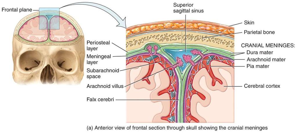
4 Meninges three connective tissue membranes that envelop the brain lies between the nervous tissue and bone similar layers cover the spinal cord, dura mater arachnoid mater pia mater protect the brain and provide structural framework for its arteries and veins
5 Dura Mater Located in cranial cavity // 2 separate layers outer periosteal (periosteal layer) equivalent to periosteum of cranial bones inner meningeal (meningeal layer) continues into vertebral canal and forms dural sac around spinal cord cranial dura mater is pressed closely against cranial bones no epidural space (differ from around spinal cord) attached only around foramen magnum, sella turcica, the crista galli, and sutures of the skull Two layers separated by in specific areas called dural sinuses collect blood circulating through brain
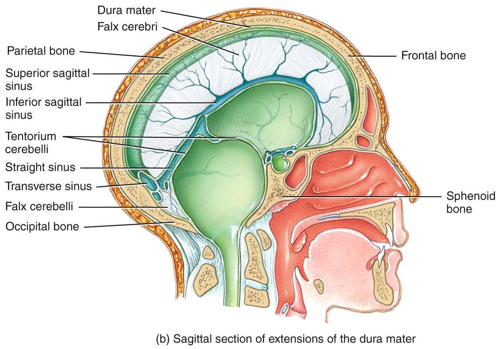
6 Dura Mater Inward folds extend between three parts of the brain falx cerebri separates the two cerebral hemispheres tentorium cerebelli separates cerebrum from cerebellum falx cerebelli separates the right and left halves of cerebellum
7 Arachnoid & Pia Mater of the Meninges arachnoid mater and pia mater are similar to those in the spinal cord arachnoid mater transparent membrane over brain surface subarachnoid space separates it from pia mater below subdural space separates it from dura mater above in some places pia mater very thin membrane that follows contours of brain, even dipping into sulci not usually visible without a microscope

8 Meninges of the Brain Skull Brain: Blood vessel Pia mater Gray matter White matter Dura mater: Periosteal layer Meningeal layer Arachnoid villus Arachnoid mater Subdural space Subarachnoid space Superior sagittal sinus Falx cerebri (in longitudinal fissure only)
9 Meningitis inflammation of the meninges serious disease of infancy & childhood especially between 3 months and 2 years of age always treated as medical emergency // death follows as soon as three hours after symptoms caused by bacterial and virus invasion of the CNS by way of the nose and throat pia mater and arachnoid are most often affected
10 Meningitis bacterial meningitis can cause swelling the brain, enlarging the ventricles, and cause hemorrhage signs include high fever, stiff neck, drowsiness, and intense headache and may progress to coma death within hours of onset diagnosed by examining the CSF for bacteria lumbar puncture (spinal tap) draws fluid from subarachnoid space between two lumbar vertebrae

 Lateral view (b)")
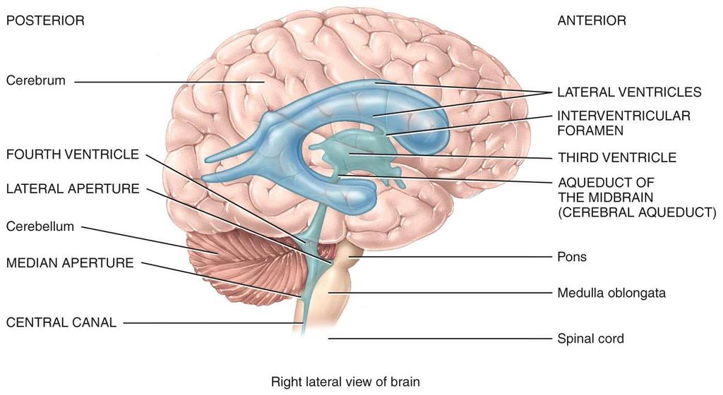
11 Brain Ventricles Copyright The McGraw-Hill Companies, Inc. Permission required for reproduction or display. Caudal Rostral Lateral ventricles Interventricular foramen Third ventricle Cerebral aqueduct Fourth ventricle Lateral aperture Median aperture Central canal Cerebrum Lateral ventricle Interventricular foramen Third ventricle Cerebral aqueduct Fourth ventricle Lateral aperture Median aperture (a) Lateral view (b) Anterior view
12 Functions of CSF buoyancy allows brain to attain considerable size without being impaired by its own weight if it rested heavily on floor of cranium, the pressure would kill the nervous tissue protection protects the brain from striking the cranium when the head is jolted shaken child syndrome and concussions do occur from severe jolting chemical stability flow of CSF rinses away metabolic wastes from nervous tissue and homeostatically regulates its chemical environment
13
 Caudate nucleus Septum pellucidum Sulcus Gyrus Insula Lateral ventricle Thalamus Choroid plexus Occipital lobe Corpus callosum (posterior part) Longitudinal fissure (c) Caudal")
14 Ventricles of the Brain Rostral (anterior) Longitudinal fissure Frontal lobe Gray matter (cortex) White matter Lateral ventricle Temporal lobe Third ventricle Lateral sulcus Corpus callosum (anterior part) Caudate nucleus Septum pellucidum Sulcus Gyrus Insula Lateral ventricle Thalamus Choroid plexus Occipital lobe Corpus callosum (posterior part) Longitudinal fissure (c) Caudal (posterior)
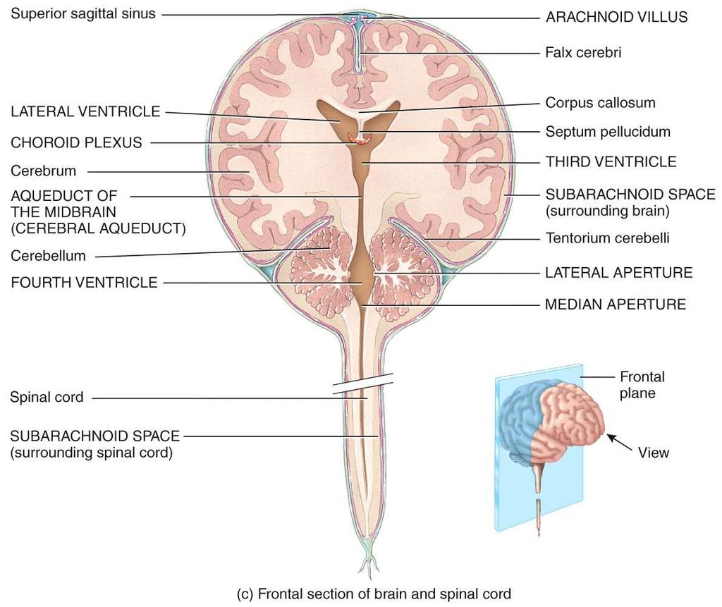
15 Ventricles and Cerebrospinal Fluid ventricles four internal chambers within the brain two lateral ventricles one in each cerebral hemisphere interventricular foramen - a tiny pore that connects to third ventricle third ventricle - single narrow medial space beneath corpus callosum cerebral aqueduct runs through midbrain and connects third and fourth ventricle fourth ventricle small triangular chamber between pons and cerebellum // connects to central canal runs down through spinal cord One medial and two lateral appentures // allow CSF to enter subarachnoid space
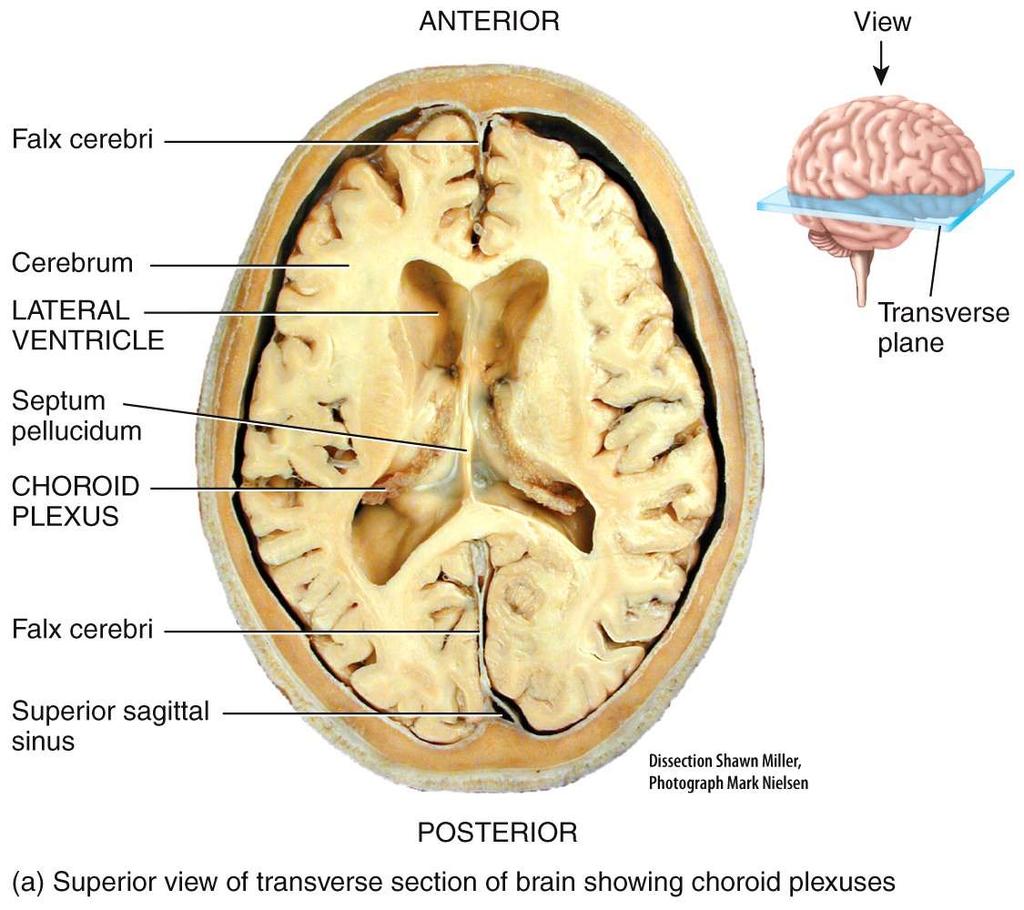
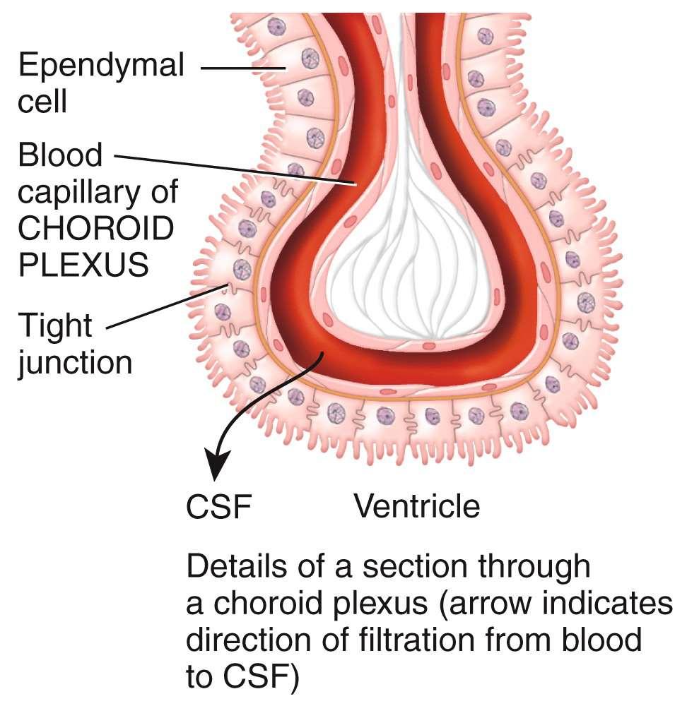
16 Ventricles and Cerebrospinal Fluid choroid plexus spongy mass of blood capillaries on the ceiling of each ventricle ependyma neuroglia that lines the ventricles and covers the choroid plexus // produces 30% of the cerebrospinal fluid
17
18
19
20
21 Cerebrospinal Fluid (CSF) clear, colorless liquid that fills the ventricles and canals of CNS // also bathes brains external surface brain produces and absorbs 500 ml/day ml normally present at one time 40% formed in subarachnoid space external to brain 30% by the general ependymal lining of the brain ventricles 30% by the choroid plexuses /// Note: Choroid Plexuses creates the Blood CSF Barrier
22
23
24 Cerebrospinal Fluid (CSF) production begins with the filtration of blood plasma through the capillaries of the brain ependymal cells modify the filtrate /// so CSF has more sodium and chloride than plasma, but less potassium, calcium, glucose, and very little protein
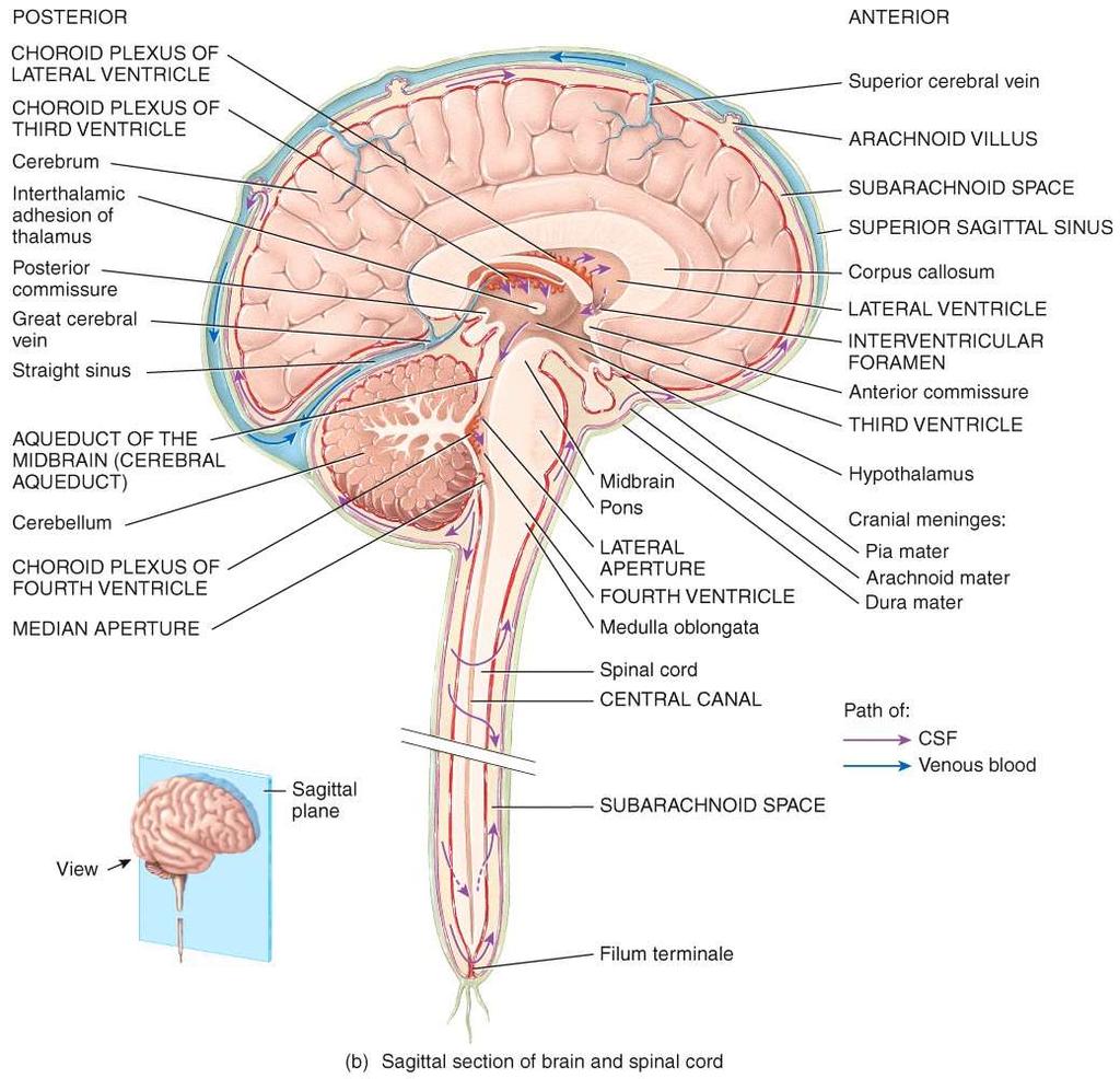
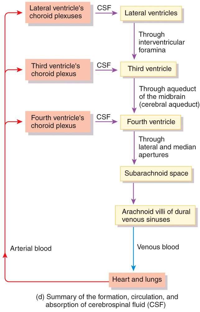
25 Cerebrospinal Fluid (CSF) Circulation CSF continually flows through and around the CNS driven by its own pressure, beating of ependymal cilia, and pulsations of the brain produced by each heartbeat CSF secreted in lateral ventricles flows through interventricular foramina into third ventricle then down the cerebral aqueduct into the fourth ventricle third and fourth ventricles add more CSF along the way
26 Cerebrospinal Fluid (CSF) Circulation small amount of CSF fills the central canal of the spinal cord all escapes through three pores median aperture and two lateral apertures leads into subarachnoid space of brain and spinal cord surface CSF is reabsorbed by arachnoid villi cauliflower-shaped extension of the arachnoid meninx // protrudes through dura mater into superior sagittal sinus // CSF penetrates the walls of the villi and mixes with the blood in the sinus
27 Flow of Cerebrospinal Fluid 8 Arachnoid villus Superior sagittal sinus Arachnoid mater 1 2 CSF is secreted by choroid plexus in each lateral ventricle. CSF flows through Interventricular foramina into third ventricle. 2 1 Subarachnoid space Dura mater Choroid plexus 3 Choroid plexus in third ventricle adds more CSF. 3 Third ventricle 4 CSF flows down cerebral aqueduct to fourth ventricle. 4 7 Cerebral aqueduct Lateralaper ture 5 Choroid plexus in fourth ventricle adds more CSF. Fourth ventricle 6 CSF flows out two lateral apertures and one median aperture CSF fills subarachnoid space and bathes external surfaces of brain and spinal cord. 7 Median aperture 8 At arachnoid villi, CSF is reabsorbed into venous blood of dural venous sinuses. Centralcanal of spinal cord Subarachnoid space of spinal cord
28
29 Blood Supply to the Brain brain is only 2% of the adult body weight, and receives 15% of the blood // 750 ml/min neurons have a high demand for ATP, and therefore, oxygen and glucose, so a constant supply of blood is critical to the nervous system 10 second interruption of blood flow may cause loss of consciousness 1 2 minute interruption can cause significant impairment of neural function 4 minutes with out blood causes irreversible brain damage
30 Brain Barrier Systems blood is also a source of antibodies, macrophages, bacterial toxins, and other harmful agents Blood brain barrier system strictly regulates what substances can get from the bloodstream into the tissue fluid of the brain two separate groups of capillaries which represent potential points of entry for substances into the brain tissue must be guarded: blood capillaries throughout the brain tissue capillaries of the choroid plexus
31 Blood Brain Barrier System protects blood capillaries throughout brain tissue consists of tight junctions between endothelial cells that form the capillary walls astrocytes reach out and contact capillaries with their perivascular feet induce the endothelial cells to form tight junctions that completely seal off gaps between them anything leaving the blood must pass through the cells, and not between them endothelial cells can exclude harmful substances from passing to the brain tissue while allowing necessary ones to pass
32 Blood CSF Barrier System blood-csf barrier another type of barrier designed to protects the brain at the level of the choroid plexus form tight junctions between the ependymal cells tight junctions are absent from ependymal cells elsewhere important to allow exchange between brain tissue and CSF Note: no CSF-brain barrier
33 Brain Barrier System blood barrier system is highly permeable to water, glucose, and lipid-soluble substances such as oxygen, carbon dioxide, alcohol, caffeine, nicotine, and anesthetics slightly permeable to sodium, potassium, chloride, and the waste products urea and creatinine obstacle for delivering medications such as antibiotics and cancer drugs trauma and inflammation can damage BBS and allow pathogens to enter brain tissue
34 Designed Breakdown of the Brain Barrier System Circumventricular organs (CVOs) places in the third and fourth ventricles where the blood brain barrier is absent blood has direct access to the brain enables the brain to monitor and respond to fluctuations in blood glucose, ph, osmolarity, and other variables CVOs afford a route for invasion by the human immunodeficiency virus (HIV)
35
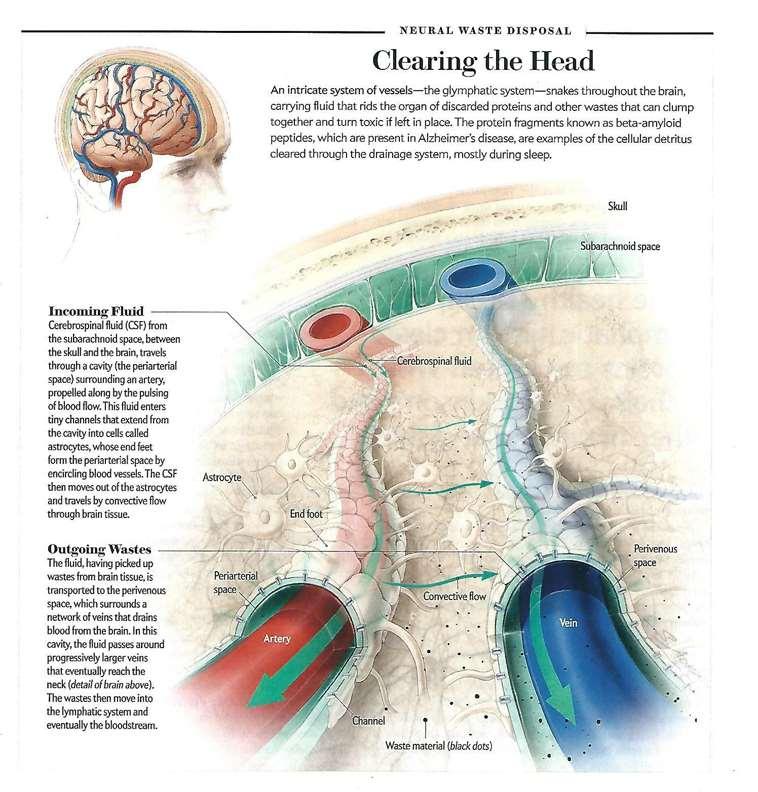
36 Recent Research Findings 1. Glymphatic discovered in brain tissue 2. Drainage system formed by astrocytes. These glial cells form a perivascular space around arteries and veins within the brain. This drainage system is most active when the animal is sleeping. 3. Implicated in removal of proteins which were discarded by cells through normal cellular metabolism. 4. We know many different neurodegenerative diseases exhibit a build up of old proteins which disrupts normal brain function. 5. This is also an example how basic research opens new ways to study pathophysiology. See Archival Articles / Brain / review article Brain Drain see next slide
37
Brain Meninges, Ventricles and CSF
 Brain Meninges, Ventricles and CSF Lecture Objectives Describe the arrangement of the meninges and their relationship to brain and spinal cord. Explain the occurrence of epidural, subdural and subarachnoid
Central Nervous System - Brain & Cranial Nerves. Chapter 14 Part A
 Central Nervous System - Brain & Cranial Nerves Chapter 14 Part A Central Nervous System Central nervous system (CNS) is responsible for: Receiving impulses from receptors Integrating information Sending
The Nervous System. PowerPoint Lecture Slides C H A P T E R 7. Prepared by Patty Bostwick-Taylor, Florence-Darlington Technical College
 PowerPoint Lecture Slides Prepared by Patty Bostwick-Taylor, Florence-Darlington Technical College C H A P T E R 7 The Nervous System NERVOUS SYSTEM OVERVIEW Essential Question: What are the primary functions
Meninges and Ventricles
 Meninges and Ventricles Irene Yu, class of 2019 LEARNING OBJECTIVES Describe the meningeal layers, the dural infolds, and the spaces they create. Name the contents of the subarachnoid space. Describe the
Brain ميهاربا لض اف دمح ا د The Meninges 1- Dura Mater of the Brain endosteal layer does not extend meningeal layer falx cerebri tentorium cerebelli
 .احمد د فاضل ابراهيم Lecture 15 Brain The Meninges Three protective membranes or meninges surround the brain in the skull: the dura mater, the arachnoid mater, and the pia mater 1- Dura Mater of the Brain
Ventricles, CSF & Meninges. Steven McLoon Department of Neuroscience University of Minnesota
 Ventricles, CSF & Meninges Steven McLoon Department of Neuroscience University of Minnesota 1 Coffee Hour Thursday (Sept 14) 8:30-9:30am Surdyk s Café in Northrop Auditorium Stop by for a minute or an
Anatomy and Physiology (Bio 220) The Brain Chapter 14 and select portions of Chapter 16
 The Brain Chapter 14 and select portions of Chapter 16") Anatomy and Physiology (Bio 220) The Brain Chapter 14 and select portions of Chapter 16 I. Introduction A. Appearance 1. physical 2. weight 3. relative weight B. Major parts of the brain 1. cerebrum 2.
Anatomy and Physiology (Bio 220) The Brain Chapter 14 and select portions of Chapter 16 I. Introduction A. Appearance 1. physical 2. weight 3. relative weight B. Major parts of the brain 1. cerebrum 2.
CSF. Cerebrospinal Fluid(CSF) System
 System") Cerebrospinal Fluid(CSF) System By the end of the lecture, students must be able to describe Physiological Anatomy of CSF Compartments Composition Formation Circulation Reabsorption CSF Pressure Functions
Cerebrospinal Fluid(CSF) System By the end of the lecture, students must be able to describe Physiological Anatomy of CSF Compartments Composition Formation Circulation Reabsorption CSF Pressure Functions
Organization of The Nervous System PROF. MOUSAED ALFAYEZ & DR. SANAA ALSHAARAWY
 Organization of The Nervous System PROF. MOUSAED ALFAYEZ & DR. SANAA ALSHAARAWY Objectives At the end of the lecture, the students should be able to: List the parts of the nervous system. List the function
Central Nervous System (CNS) -> brain and spinal cord. Major Divisions of the nervous system:
 -> brain and spinal cord. Major Divisions of the nervous system:") Central Nervous System (CNS) -> brain and spinal cord Major Divisions of the nervous system: Afferent (sensory input) -> cell bodies outside of the central nervous system (CNS), carry info into the CNS
Central Nervous System (CNS) -> brain and spinal cord Major Divisions of the nervous system: Afferent (sensory input) -> cell bodies outside of the central nervous system (CNS), carry info into the CNS
Central Nervous System: Part 2
 Central Nervous System: Part 2 1. Meninges 2. CSF 3. Spinal Cord and Spinal Nerves Explain spinal cord anatomy, including gray and white matter and meninges (give the general functions of this organ).
Chapter 18: The Brain & Cranial Nerves. Origin of the Brain
 Chapter 18: The Brain & Cranial Nerves BIO 218 Fall 2015 Origin of the Brain The brain originates from a structure called the neural tube, which arises during a developmental stage called neurulation.
Organization of The Nervous System PROF. SAEED ABUEL MAKAREM
 Organization of The Nervous System PROF. SAEED ABUEL MAKAREM Objectives By the end of the lecture, you should be able to: List the parts of the nervous system. List the function of the nervous system.
ACTIVITY 7: NERVOUS SYSTEM HISTOLOGY, BRAIN, CRANIAL NERVES
 ACTIVITY 7: NERVOUS SYSTEM HISTOLOGY, BRAIN, CRANIAL NERVES LABORATORY OBJECTIVES: 1. Histology: Identify structures indicated on three different slides or images of nervous system tissue. These images
Histology of the CNS
 Histology of the CNS Lecture Objectives Describe the histology of the cerebral cortex layers. Describe the histological features of the cerebellum; layers and cells of cerebellar cortex. Describe the elements
The dura is sensitive to stretching, which produces the sensation of headache.
 Dural Nerve Supply Branches of the trigeminal, vagus, and first three cervical nerves and branches from the sympathetic system pass to the dura. Numerous sensory endings are in the dura. The dura is sensitive
Department of Cognitive Science UCSD
 Department of Cognitive Science UCSD Verse 1: Neocortex, frontal lobe, Brain stem, brain stem, Hippocampus, neural node, Right hemisphere, Pons and cortex visual, Brain stem, brain stem, Sylvian fissure,
TRANSVERSE SECTION PLANE Scalp 2. Cranium. 13. Superior sagittal sinus
 TRANSVERSE SECTION PLANE 1 1. Scalp 2. Cranium 3. Superior sagittal sinus 4. Dura mater 5. Falx cerebri 6. Frontal lobes of the cerebrum 7. Middle meningeal artery 8. Cortex, grey matter 9. Cerebral vessels
Nsci 2100: Human Neuroanatomy Examination 1
 Name KEY Lab Section Nsci 2100: Human Neuroanatomy Examination 1 On this page, write your name and lab section. On your scantron answer sheet, enter your name (last name, space, first name), internet ID
Sheep Brain Dissection
 Sheep Brain Dissection Mammalian brains have many features in common. Human brains may not be available, so sheep brains often are dissected as an aid to understanding the mammalian brain since he general
The CNS Part II pg
 The CNS Part II pg. 455-474 Protection of the Brain Objectives Describe how the meninges, cerebrospinal fluid, and the blood brain barrier protect the CNS. Explain how Cerebrospinal fluid is formed, and
Principles of Anatomy and Physiology
 Principles of Anatomy and Physiology 14 th Edition CHAPTER 14 The Brain and Cranial Nerves Introduction The purpose of the chapter is to: 1. Understand how the brain is organized, protected, and supplied
HEAD AND NECK IMAGING. James Chen (MS IV)
") HEAD AND NECK IMAGING James Chen (MS IV) Anatomy Course Johns Hopkins School of Medicine Sept. 27, 2011 OBJECTIVES Introduce cross sectional imaging of head and neck Computed tomography (CT) Review head
HEAD AND NECK IMAGING James Chen (MS IV) Anatomy Course Johns Hopkins School of Medicine Sept. 27, 2011 OBJECTIVES Introduce cross sectional imaging of head and neck Computed tomography (CT) Review head
Cerebral hemisphere. Parietal Frontal Occipital Temporal
 Cerebral hemisphere Sulcus / Fissure Central Precental gyrus Postcentral gyrus Lateral (cerebral) Parieto-occipital Cerebral cortex Frontal lobe Parietal lobe Temporal lobe Insula Amygdala Hippocampus
Page. Ch 11 A CNS. This set. Major Landmarks: Brain size is proportional to body size only and can be divided into three major portions;
 1 BIO 211: ANATOMY & PHYSIOLOGY I 1 Ch 11 A CNS This set Ch 11 B Notes: PNS Somatic ANS Ch 11 C ANS Dr. Dr. Lawrence G. G. Altman www.lawrencegaltman.com Some illustrations are courtesy of McGraw-Hill.
Chapter 13 Brain and Cranial Nerves
 Chapter 13 Brain and Cranial Nerves 13-1 Brain and Cranial Nerves Brain Part of CNS contained in cranial cavity Control center for many of body s functions Much like a complex computer but more Parts of
8.3 The Central Nervous System. SBI4U Ms. Ho-Lau
 8.3 The Central Nervous System SBI4U Ms. Ho-Lau The Central Nervous System the structural and functional centre for the entire nervous system the site of neural integration and processing The Central
Blood Supply of the CNS
 Blood Supply of the CNS Lecture Objectives Describe the four arteries supplying the CNS. Follow up each artery to its destination. Describe the circle of Willis and its branches. Discuss the principle
Introduction and Basic structural organization of the nervous system
 Introduction and Basic structural organization of the nervous system **the slides are in bold and the book is in red Done by : razan krishan & marah marahleh INTRODUCTION The nervous system, along with
Embryonic Brain Development
 Chapter 14 The Brain and Cranial Nerves Largest organ in the body? Brain functions in sensations, memory, emotions, decision making, behavior 19-1 19-2 Embryonic Brain Development Principal Parts of the
Slide 1. Slide 2. Slide 3. Tomography vs Topography. Computed Tomography (CT): A simplified Topographical review of the Brain. Learning Objective
: A simplified Topographical review of the Brain. Learning Objective") Slide 1 Computed Tomography (CT): A simplified Topographical review of the Brain Jon Wheiler, ACNP-BC Slide 2 Tomography vs Topography Tomography: A technique for displaying a representation of a cross
Slide 1 Computed Tomography (CT): A simplified Topographical review of the Brain Jon Wheiler, ACNP-BC Slide 2 Tomography vs Topography Tomography: A technique for displaying a representation of a cross
Superior View of the Skull (Norma Verticalis) Anteriorly the frontal bone articulates with the two parietal bones AT THE CORONAL SUTURE
 Anteriorly the frontal bone articulates with the two parietal bones AT THE CORONAL SUTURE") Superior View of the Skull (Norma Verticalis) Anteriorly the frontal bone articulates with the two parietal bones AT THE CORONAL SUTURE 1 The two parietal bones articulate in the midline AT THE SAGITTAL
Superior View of the Skull (Norma Verticalis) Anteriorly the frontal bone articulates with the two parietal bones AT THE CORONAL SUTURE 1 The two parietal bones articulate in the midline AT THE SAGITTAL
BRAIN PART I (A & B): VENTRICLES & MENINGES
: VENTRICLES & MENINGES") BRAIN PART I (A & B): VENTRICLES & MENINGES Cranial Meninges Cranial meninges are continuous with spinal meninges Dura mater: inner layer (meningeal layer) outer layer (endosteal layer) fused to periosteum
BRAIN PART I (A & B): VENTRICLES & MENINGES Cranial Meninges Cranial meninges are continuous with spinal meninges Dura mater: inner layer (meningeal layer) outer layer (endosteal layer) fused to periosteum
Chapter 3. Structure and Function of the Nervous System. Copyright (c) Allyn and Bacon 2004
 Allyn and Bacon 2004") Chapter 3 Structure and Function of the Nervous System 1 Basic Features of the Nervous System Neuraxis: An imaginary line drawn through the center of the length of the central nervous system, from the
Chapter 3 Structure and Function of the Nervous System 1 Basic Features of the Nervous System Neuraxis: An imaginary line drawn through the center of the length of the central nervous system, from the
b. The groove between the two crests is called 2. The neural folds move toward each other & the fuse to create a
 Chapter 13: Brain and Cranial Nerves I. Development of the CNS A. The CNS begins as a flat plate called the B. The process proceeds as: 1. The lateral sides of the become elevated as waves called a. The
Cranial Cavity REFERENCES: OBJECTIVES OSTEOLOGY. Stephen A. Gudas, PT, PhD
 Stephen A. Gudas, PT, PhD Cranial Cavity REFERENCES: Moore and Agur, Essential Clinical Anatomy (ECA), 3rd ed., pp. 496 498; 500 507; 512 514 Grant s Atlas 12 th ed., Figs 7.6; 7.19 7.30. Grant s Dissector
Student Lab #: Date. Lab: Gross Anatomy of Brain Sheep Brain Dissection Organ System: Nervous Subdivision: CNS (Central Nervous System)
") Lab: Gross Anatomy of Brain Sheep Brain Dissection Organ System: Nervous Subdivision: CNS (Central Nervous System) Student Lab #: Date 1 Objectives: 1. Learn the main components making up a motor neuron.
Lab: Gross Anatomy of Brain Sheep Brain Dissection Organ System: Nervous Subdivision: CNS (Central Nervous System) Student Lab #: Date 1 Objectives: 1. Learn the main components making up a motor neuron.
Unit Three. The brain includes: cerebrum, diencephalon, brain stem, & cerebellum. The brain lies within the cranial cavity of the skull.
 Human Anatomy & Physiology 11 Divisions of the Nervous System Karen W. Smith, Instructor Unit Three BRAIN & SPINAL CORD Refer to the following URLs. Be sure to study these along with your book. http://www.sirinet.net/~jgjohnso/nervous.html
Neurology study of the nervous system. nervous & endocrine systems work together to maintain homeostasis
 Nervous System Neurology study of the nervous system nervous & endocrine systems work together to maintain homeostasis Nervous System works very fast Uses electrical signals called nerve impulses Short-lived
Anatomy Lecture Notes Chapter 13
 I. embryonic development of the CNS A. neurulation is the formation of the CNS in the embryo invagination of dorsal ectoderm (outer layer of embryo cells) this process is induced (caused) by the notochord
NOTES CHAPTER 9 (Brief) The Nervous System LECTURE NOTES
 The Nervous System LECTURE NOTES") NOTES CHAPTER 9 (Brief) The Nervous System LECTURE NOTES I. Divisions of the Nervous System two major divisions A. Central Nervous System (CNS) 1. brain 2. spinal cord B. Peripheral Nervous System (PNS)
NOTES CHAPTER 9 (Brief) The Nervous System LECTURE NOTES I. Divisions of the Nervous System two major divisions A. Central Nervous System (CNS) 1. brain 2. spinal cord B. Peripheral Nervous System (PNS)
Spinal Cord and Properties of Cerebrospinal Fluid: Options for Drug Delivery. SMA Foundation New York
 Spinal Cord and Properties of Cerebrospinal Fluid: Options for Drug Delivery New York Why Do We Need to Know about the Spinal Cord Anatomy and Properties of Cerebrospinal Fluid? SMA therapeutics need to
The Nervous System PART C. PowerPoint Lecture Slide Presentation by Patty Bostwick-Taylor, Florence-Darlington Technical College
 PowerPoint Lecture Slide Presentation by Patty Bostwick-Taylor, Florence-Darlington Technical College The Nervous System 7 PART C Protection of the Central Nervous System Scalp and skin Skull and vertebral
Cranial cavity. Dr. Heba Kalbouneh Assistant Professor of Anatomy and Histology
 Cranial cavity Dr. Heba Kalbouneh Assistant Professor of Anatomy and Histology Cerebrum Cerebral hemispheres The Meninges The brain in the skull is surrounded by three membranes or meninges: 1-THE DURA
SPINAL CORD AND PROPERTIES OF CEREBROSPINAL FLUID: OPTIONS FOR DRUG DELIVERY
 SPINAL CORD AND PROPERTIES OF CEREBROSPINAL FLUID: OPTIONS FOR DRUG DELIVERY WHY DO WE NEED TO KNOW ABOUT THE SPINAL CORD ANATOMY AND PROPERTIES OF CEREBROSPINAL FLUID? SMA therapeutics need to reach cells
The neurvous system senses, interprets, and responds to changes in the environment. Two types of cells makes this possible:
 NERVOUS SYSTEM The neurvous system senses, interprets, and responds to changes in the environment. Two types of cells makes this possible: the neuron and the supporting cells ("glial cells"). Neuron Neurons
Chapter 13 Lecture Outline *
 Anatomy and Physiology, Seventh Edition Rod R. Seeley Idaho State University Trent D. Stephens Idaho State University Philip Tate Phoenix College Chapter 13 Lecture Outline * *See PowerPoint Image Slides
CENTRAL NERVOUS SYSTEM
 Student Name CHAPTER 13 CENTRAL NERVOUS SYSTEM Approximately one hundred billion neurons make up the brain. Everything we are and everything we hope to become are centered in this structure, which is about
Characteristic features of CNS pathology. By: Shifaa AlQa qa
 Characteristic features of CNS pathology By: Shifaa AlQa qa Normal brain: - The neocortex (gray matter): six layers: outer plexiform, outer granular, outer pyramidal, inner granular, inner pyramidal, polymorphous
Chapter 2. Central Nervous System; the brain and spinal cord
 Chapter 2 Central Nervous System; the brain and spinal cord CNS 1. Topography; - what are the main components of the brain - how do you recognize them? 2. The location of the major functional areas of
ACTIVITY 7: NERVOUS SYSTEM HISTOLOGY, BRAIN, CRANIAL NERVES NERVOUS SYSTEM TISSUES: HISTOLOGY SLIDES
 ACTIVITY 7: NERVOUS SYSTEM HISTOLOGY, BRAIN, CRANIAL NERVES OBJECTIVES: 1) How to get ready: Read Chapter 14 & 15 McKinley et al., Human Anatomy, 4e. All text references are for this textbook. Read dissection
I. Anatomy of the Brain A. Cranial Meninges and Ventricles of the Brain 1. Meninges a. Dura mater 1) Endosteal/Periosteal Layer - Outer 2) Meningeal
 Endosteal/Periosteal Layer - Outer 2) Meningeal") I. Anatomy of the Brain A. Cranial Meninges and Ventricles of the Brain 1. Meninges a. Dura mater 1) Endosteal/Periosteal Layer - Outer 2) Meningeal Layer - Inner 3) Falx cerebri a) Superior sagittal sinus
I. Anatomy of the Brain A. Cranial Meninges and Ventricles of the Brain 1. Meninges a. Dura mater 1) Endosteal/Periosteal Layer - Outer 2) Meningeal Layer - Inner 3) Falx cerebri a) Superior sagittal sinus
Introduction to the Organization of the Brain 98% of the body s neural tissue is in the brain
 Introduction to the Organization of the Brain 98% of the body s neural tissue is in the brain An avg. human brain weighs 1.4 kg (3lbs.) and has a volume of 1200 cc brains average about 10% bigger than
Central nervous system (CNS): brain and spinal cord Collections of cell body and dendrites (grey matter) are called nuclei/nucleus Nucleus can also
: brain and spinal cord Collections of cell body and dendrites (grey matter) are called nuclei/nucleus Nucleus can also") Chapter 3 Part 1 Orientation Directions in the nervous system are described relatively to the neuraxis An imaginary line drawn through the center of the length of the central nervous system, from the bottom
Chapter 3 Part 1 Orientation Directions in the nervous system are described relatively to the neuraxis An imaginary line drawn through the center of the length of the central nervous system, from the bottom
The Nervous System PART B
 7 The Nervous System PART B PowerPoint Lecture Slide Presentation by Jerry L. Cook, Sam Houston University ESSENTIALS OF HUMAN ANATOMY & PHYSIOLOGY EIGHTH EDITION ELAINE N. MARIEB Central Nervous System
MENTAL HOSPITAL PHONE MENU
 If you have low self-esteem, please hang up. Our operators are too busy to talk with you. MENTAL HOSPITAL PHONE MENU Hello and thank you for calling The State Mental Hospital. Please select from the following
Cranial cavity. Dr. Heba Kalbouneh Associate Professor of Anatomy and Histology
 Cranial cavity Dr. Heba Kalbouneh Associate Professor of Anatomy and Histology The Meninges The brain in the skull is surrounded by three membranes or meninges: 1-DURA MATER 2-ARACHNOID MATER 3-PIA MATER
Brain and Cranial Nerves (Ch. 15) Human Anatomy lecture. caudal = toward the spinal cord)
 Human Anatomy lecture. caudal = toward the spinal cord)") Insight: Some cranial nerve disorders Brain and Cranial Nerves (Ch. 15) Human Anatomy lecture I. Overview (Directional terms: rostral = toward the forehead caudal = toward the spinal cord) A. 3 Major parts
Insight: Some cranial nerve disorders Brain and Cranial Nerves (Ch. 15) Human Anatomy lecture I. Overview (Directional terms: rostral = toward the forehead caudal = toward the spinal cord) A. 3 Major parts
M555 Medical Neuroscience Blood Flow in CNS Meninges Blood Brain Barrier CSF
 M555 Medical Neuroscience Blood Flow in CNS Meninges Blood Brain Barrier CSF Arterial Blood Flow to CNS approximately % of what goes wrong within the skull that produces neurological deficits is vascular
stored information, making decisions, and taking action. 1. It is also the center for intellect, emotions, behavior, and memory.
 Chapter 14 - Outline I. INTRODUCTION A. The brain is the center for registering sensations, correlating them with one another and with stored information, making decisions, and taking action. 1. It is
Classical CNS Disease Patterns
 Classical CNS Disease Patterns Inflammatory Traumatic In response to the trauma of having his head bashed in GM would have experienced some of these features. NOT TWO LITTLE PEENY WEENY I CM LACERATIONS.
Unit 12a: The Nervous System The Brain. MDL231 Principle of Anatomy
 Unit 12a: The Nervous System The Brain MDL231 Principle of Anatomy The Brain - Overview Cerebrum T PP H midbrain Cerebellum pons m.o. Brain stem medulla oblongata (M.O.) pons midbrain (mesencephalon) Diencephalon
Dissection of the Sheep Brain
 Dissection of the Sheep Brain Laboratory Objectives After completing this lab, you should be able to: 1. Identify the main structures in the sheep brain and to compare them with those of the human brain.
Chapter 5: Fetal Central Nervous System 71
 71 Chapter 5 Fetal Central Nervous System Embryology NEURULATION begins with the formation of the neural plate, the neural folds and their ultimate fusion and closure as the NEURAL TUBE. NEURAL PLATE -
CNS consists of brain and spinal cord Cephalization Evolutionary development of rostral (anterior) portion of CNS Increased number of neurons in head
 portion of CNS Increased number of neurons in head") CNS consists of brain and spinal cord Cephalization Evolutionary development of rostral (anterior) portion of CNS Increased number of neurons in head Highest level reached in human brain 1 Mostly to orient
CNS consists of brain and spinal cord Cephalization Evolutionary development of rostral (anterior) portion of CNS Increased number of neurons in head Highest level reached in human brain 1 Mostly to orient
Human Anatomy and Physiology I Laboratory
 Human Anatomy and Physiology I Laboratory Histology of Nervous Tissue and The Spinal Cord This lab involves two laboratory exercises: 1) Histology of Nervous Tissue, and 2) Spinal Cord, Spinal Nerves,
Principles Arteries & Veins of the CNS LO14
 Principles Arteries & Veins of the CNS LO14 14. Identify (on cadaver specimens, models and diagrams) and name the principal arteries and veins of the CNS: Why is it important to understand blood supply
Unit 18: Cranial Cavity and Contents
 Unit 18: Cranial Cavity and Contents Dissection Instructions: The calvaria is to be removed without damage to the dura mater which is attached to the inner surface of the calvaria. Cut through the outer
Nervous system. Dr. Rawaa Salim Hameed
 Nervous system Dr. Rawaa Salim Hameed Central nervous system (CNS) CNS consists of the brain (cerebrum, cerebellum, and brainstem) and spinal cord CNS is covered by connective tissue layers, the meninges
Development of Brain Stem, Cerebellum and Cerebrum
 Development of Brain Stem, Cerebellum and Cerebrum The neural tube cranial to the 4th pair of somites develop into the brain. 3 dilatations and 2 flexures form at the cephalic end of the neural tube during
Anatomy Lab (1) Theoretical Part. Page (2 A) Page (2B)
 Theoretical Part. Page (2 A) Page (2B)") Anatomy Lab (1) This sheet only includes the extra notes for the lab handout regarding the theoretical part, as for the practical part it includes everything the doctor mentioned. Theoretical Part Page
Anatomy Lab (1) This sheet only includes the extra notes for the lab handout regarding the theoretical part, as for the practical part it includes everything the doctor mentioned. Theoretical Part Page
BIOL Dissection of the Sheep and Human Brain
 BIOL 2401 Dissection of the Sheep and Human Brain Laboratory Objectives After completing this lab, you should be able to: Identify the main structures in the sheep brain and to compare them with those
Biological Bases of Behavior. 3: Structure of the Nervous System
 Biological Bases of Behavior 3: Structure of the Nervous System Neuroanatomy Terms The neuraxis is an imaginary line drawn through the spinal cord up to the front of the brain Anatomical directions are
Central Nervous System. Dr. Gary Mumaugh
 Central Nervous System Dr. Gary Mumaugh COVERINGS OF THE BRAIN AND SPINAL CORD Two protective coverings Outer covering is bone; cranial bones encase the brain and vertebrae encase the spinal cord Inner
Neurotransmitter: dopamine. Physiology of additive drugs. Dopamine and reward. Neurotransmitter: dopamine
 Physiology of additive drugs Cocaine, methamphetamine, marijuana, and opiates influence the neurotransmitter dopamine. Neurotransmitter: dopamine Dopamine - a neurotransmitter associated with several functions,
The Brain. Brain. Spinal Cord. Cauda Equina
 The Brain Brain Spinal Cord Cauda Equina The Brain Ventricles- cavities in the brain filled with cerebrospinal fluid connected to the subarachnoid space- fluid filled space surrounding the brain Brain
Superior View of the Skull (Norma Verticalis) Anteriorly the frontal bone articulates with the two parietal bones AT THE CORONAL SUTURE
 Anteriorly the frontal bone articulates with the two parietal bones AT THE CORONAL SUTURE") Superior View of the Skull (Norma Verticalis) Anteriorly the frontal bone articulates with the two parietal bones AT THE CORONAL SUTURE 1 The two parietal bones articulate in the midline AT THE SAGITTAL
Superior View of the Skull (Norma Verticalis) Anteriorly the frontal bone articulates with the two parietal bones AT THE CORONAL SUTURE 1 The two parietal bones articulate in the midline AT THE SAGITTAL
CHAPTER 13&14: The Central Nervous System. Anatomy of the CNS
 CHAPTER 13&14: The Central Nervous System Anatomy of the CNS in human consists of brain and spinal cord as stated earlier neurons have little support from their extracellular matrix and depend on glial
GUIDELINES. Module 2 Content module 11. brain. The formation and ways of cerebrospinal fluid circulation. Medical1,2,3,4, military.
 Bogomolets National Medical University Department of human anatomy GUIDELINES Academic Subject Matter HUMAN ANATOMY Module 2 Content module 11 Theme of the lesson The meninges of spinal cord and brain.
Chapter 14: The Brain and Cranial Nerves. Copyright 2009, John Wiley & Sons, Inc.
 Chapter 14: The Brain and Cranial Nerves Development of the Brain Three to four-week embryo: prosencephalon, mesencephalon and rhombencephalon. Five-week embryo: telencephalon (cerebrum), diencephalon
PARA210 SUMMARY Hyperglycaemia (DKA & HHS) Brain & Nervous System Anatomy & Physiology Degenerative Neurological Disorders
 Brain & Nervous System Anatomy & Physiology Degenerative Neurological Disorders") PARA210 SUMMARY Page Topic 01-03 Diabetes Mellitus 04-05 Hyperglycaemia (DKA & HHS) 06-13 Toxicology 14-18 12 Lead ECG 19-21 Brain & Nervous System Anatomy & Physiology 22-24 Degenerative Neurological
PARA210 SUMMARY Page Topic 01-03 Diabetes Mellitus 04-05 Hyperglycaemia (DKA & HHS) 06-13 Toxicology 14-18 12 Lead ECG 19-21 Brain & Nervous System Anatomy & Physiology 22-24 Degenerative Neurological
BIO 115 Anatomy & Physiology II Practice Assignment 4: The Nervous System & The Senses This is not a required assignment but it is recommended.
 BIO 115 Anatomy & Physiology II Practice Assignment 4: The Nervous System & The Senses This is not a required assignment but it is recommended. 1. This figure depicts a typical neuron. What structures
Somatic Nervous Systems. III. Autonomic Nervous System. Parasympathetic Nervous System. Sympathetic Nervous Systems
 7/21/2014 Outline Nervous System - PNS and CNS I. II. Two Parts of the Nervous System Central Nervous System vs Peripheral Nervous System Peripheral Nervous System A. B. Brain and Spinal Cord III. Autonomic
meninges Outermost layer of the meninge dura mater arachnoid mater pia mater membranes located between bone and soft tissue of the nervous system
 membranes located between bone and soft tissue of the nervous system meninges Outermost layer of the meninge dura mater middle layer of the meninges, contains no blood vessels arachnoid mater Innermost
Announcement. Danny to schedule a time if you are interested.
 Announcement If you need more experiments to participate in, contact Danny Sanchez (dsanchez@ucsd.edu) make sure to tell him that you are from LIGN171, so he will let me know about your credit (1 point).
Human Anatomy. Brain and Cranial Nerves
 Human Anatomy Brain and Cranial Nerves 1 Brain and Cranial Nerves An adult brain weighs between 1.35 and 1.4 kilograms (kg) (around 3 pounds) and has a volume of about 1200 cubic centimeters (cc). Brain
A&P 1 Brain & Cranial Nerves Guide - Lab Exercises
 A&P 1 Brain & Cranial Nerves Guide - Lab Exercises Please make sure you read the entire set of instructions on Dissection the Sheep Brain before beginning to cut. Also, please do not forget to go over
3/15/17. Outline. Nervous System - PNS and CNS. Two Parts of the Nervous System
 Nervous System - PNS and CNS Bio 105 Outline I. Central Nervous System vs Peripheral Nervous System II. Peripheral Nervous System A. Autonomic Nervous Systems B. Somatic Nervous Systems III. Autonomic
The Brain and Cranial Nerves Student Objectives
 The Brain and Cranial Nerves Student Objectives Chapter 14 Textbook and Laboratory Manual Name the major regions of the brain and describe their functions Name the ventricles of the brain and describe
Nervous System - PNS and CNS. Bio 105
 Nervous System - PNS and CNS Bio 105 Outline I. Central Nervous System vs Peripheral Nervous System II. Peripheral Nervous System A. Autonomic Nervous Systems B. Somatic Nervous Systems III. Autonomic
Medical Neuroscience Tutorial Notes
 Medical Neuroscience Tutorial Notes Blood Supply to the Brain MAP TO NEUROSCIENCE CORE CONCEPTS 1 NCC1. The brain is the body's most complex organ. LEARNING OBJECTIVES After study of the assigned learning
Neuroanatomy. Assistant Professor of Anatomy Faculty of Medicine The University of Jordan Dr Maha ELBeltagy
 Neuroanatomy Dr. Maha ELBeltagy Assistant Professor of Anatomy Faculty of Medicine The University of Jordan 2018 Development of the Central Nervous System Development of the nervous system Development
Blood Brain Barrier (BBB)
") Cerebral Blood Flow, Cerebral Spinal Fluid, and Brain Metabolism Part Two Guyton Chapter 61 Morgan & Mikhail, 4 th ed, Chapter 25 (or Morgan & Mikhail 5 th ed, Chapter 26) Blood Brain Barrier (BBB) Cerebral
Cerebral Blood Flow, Cerebral Spinal Fluid, and Brain Metabolism Part Two Guyton Chapter 61 Morgan & Mikhail, 4 th ed, Chapter 25 (or Morgan & Mikhail 5 th ed, Chapter 26) Blood Brain Barrier (BBB) Cerebral
SOME BASIC TERMINOLOGY CNS: Central Nervous System: Brain + Spinal Cord
 SOME BASIC TERMINOLOGY CNS: Central Nervous System: Brain + Spinal Cord CEREBROSPINAL FLUID (CSF): The fluid filling the ventricles, cerebral aqueduct, central canal, and subarachnoid space. It is a filtrate
ANATOMY & PHYSIOLOGY DISSECTION OF THE SHEEP BRAIN LAB GROUP:
 ANATOMY & PHYSIOLOGY DISSECTION OF THE SHEEP BRAIN LAB GROUP: Introduction The purpose of the sheep brain dissection is to familiarize you with the three dimensional structure of the brain and teach you
Chapter 10 The Nervous System: The Brain and Cranial Nerves
 Chapter 10 The Nervous System: The Brain and Cranial Nerves Copyright 2015 Wolters Kluwer Health Lippincott Williams & Wilkins Overview Key Terms aphasia corpus callosum meninges basal nuclei diencephalon
Blood supply to the brain Blood brain barrier isolates neural tissue from general circulation
 The Brain and Cranial Nerves Objectives Name the major regions of the brain and describe their functions. Discuss the formation, circulation, and functions of the CSF. List the main components of the medulla
Bellringer: The central nervous system is comprised of: What is the name of the outermost layer of the brain? a. Brain. b.
 Bellringer: The central is comprised of: a. Brain b. Spinal cord c. Sensory receptors d. Both a and b What is the name of the outermost layer of the brain? a. Pia mater b. Dura mater c. Arachnoid d. Pons
Chapter 9. Nervous System
 Chapter 9 Nervous System Central Nervous System (CNS) vs. Peripheral Nervous System(PNS) CNS Brain Spinal cord PNS Peripheral nerves connecting CNS to the body Cranial nerves Spinal nerves Neurons transmit
Anatomy of the Brain > 1. Figure 1. Eight bones form the skull and fourteen bones form the face.
 Anatomy of the Brain Overview The human brain is an amazing three-pound organ that controls all functions of the body, interprets information from the outside world, and embodies the essence of the mind