Dural Arteriovenous Malformations and Fistulae (DAVM S DAVF S)
|
|
|
- Erin Merritt
- 5 years ago
- Views:
Transcription

1 Jorge Guedes Campos NEUROIMAGING DEPARTMENT HOSPITAL SANTA MARIA UNIVERSITY OF LISBON PORTUGAL
2 DEFINITION region of arteriovenous shunting confined to a leaflet of packymeninges often adjacent to a major dural sinus 10 to 15% of all AVM s older population; female predominance


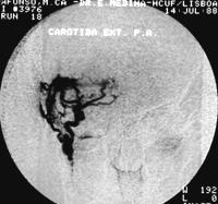
3 ANGIO-ARCHITECTURE meningeal arterial feeders related to the location pial supply and transosseous extracranial arterial collaterals can be recruited nidus or single hole fistula venous drainage type Lateral sinus DAVM


4 trauma surgery vascular diseases tumor infection hormonal effects ETIOLOGIC FACTORS congenital origin Congenital middle cranial fossa DAVM (right paracavernous sinus) SILAN 25th 2008 SIMI - CANCÚN Buenos Aires - MÉXICO 2016


5 Unlike pial AVM S regarded as congenital lesions the pathogenesis of DAVM s is controversial with arguments for congenital and acquired etiology Importance of venous thrombosis PATHOPHYSIOLOGY SILAN 25th 2008 SIMI - CANCÚN Buenos Aires - MÉXICO 2016 Left Cavernous sinus DAVM type II (arterial pedicles from left ICA and ECA, thrombosis of both ophtalmic veins and inferior petrosal sinus)


6 SILAN th SIMI BUENOS Buenos AIRES Aires ARGENTINA Dural Arteriovenous Malformations and Fistulae Location / Topography cavernous sinus transverse-sigmoid sinus superior sagital sinus tentorial anterior fossa CLASSIFICATION Unusual deep straight sinus / vein of Galen middle cranial fossa torcula and posterior fossa Superior sagital sinus DAVM type III

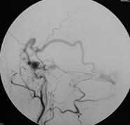
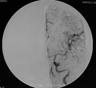
7 CLASSIFICATION Venous Drainage pattern (Djindjian) I into a sinus with normal direction of flow (Cognard) II into a sinus with reflux to cortical veins III into a cortical vein with retrograde flow IIa only into sinus II b only into cortical veins IIa+b into sinus and cortical veins IV presence of venous lake V into perimedullary veins Right lateral sinus DAVM type IV High hemorrhagic risk


8 CLINICAL SPECTRUM Clinical presentation is related to the venous drainage pattern, flow, topography and rarely arterial symptons Right lateral sinus DAVM type III Anterior fossa SAH 63% ICH 50% SDH 25% Cavernous sinus Proptosis 83% CNP 44% Bruit 42% Tentorium SAH 80% ICH 60% CNS 42% DAVM s in children high flow cardiac disorders Lateral sinus Bruit 70% Headache 40% ICH 15% CNS 13%


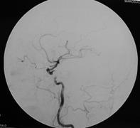
9 Dynamic nature of DAVM S Agressive clinical course CLINICAL SPECTRUM risk of hemorrhage focal neurological symptons hydrocephalus and papilledema visual loss intractable pain dementia Left lateral sinus DAVM type II
 DAVM itself is rarely detected Epiphenomena may")

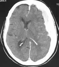
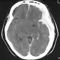
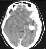
10 IMAGING Plain Skull Films Increased vascular markings or bone density CT (nonenhanced, enhanced) DAVM itself is rarely detected Epiphenomena may be detected thrombosed sinus dilated veins hemorrhage (acute) hydrocephalus Cavernous dural AV Fistulae (Enhanced CT dilated supra-ophtalmic veins)


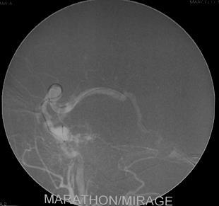
11 IMAGING MRI plain and Gad-enhanced same problems as CT in demonstrating DAVM Angio-MR Arterial / Venous is important Angiography Indispensable to diagnose and to evaluate a DAVM in order to plan the treatment Superior sagital sinus DAVM feeding arteries nidus venous drainage and functional hemodynamic analysis


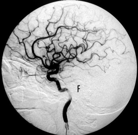
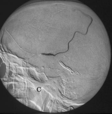
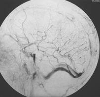
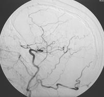
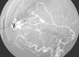
12 INTRA-ARTERIAL EMBOLIZATION PARTICLES - PVA proximal meningeal arteries supplying cranial nerves in the skull base low flow shunts low risk patients Low number of arterial feeders Frequently in cavernous sinus, lateral sinus type I/II shunts Rare in other locations Left cavernous sinus DAVM type I

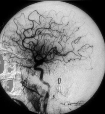
13 INTRA-ARTERIAL EMBOLIZATION Left cavernous sinus DAVM type I SILAN 25th 2008 SIMI - CANCÚN Buenos Aires - MÉXICO 2016 Intra-arterial embolization - particles PVA left acessory meningeal and ascending pharingeal arteries

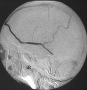
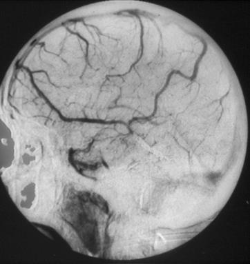
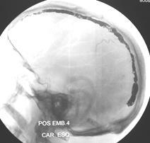
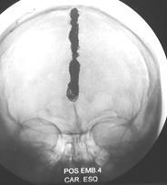
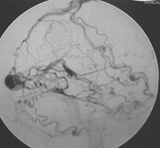
14 INTRA-ARTERIAL EMBOLIZATION Left cavernous sinus DAVM type I Left external carotid artery Post-Emb


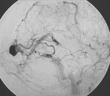
15 INTRA-ARTERIAL EMBOLIZATION GLUE - NBCA distal meningeal arteries high risk patients high flow fistula Low number of arterial feeders Frequently superior sagital sinus, lateral sinus, posterior fossa type II / III / IV shunts Also tentorial e anterior fossa Posterior fossa DAVM type III


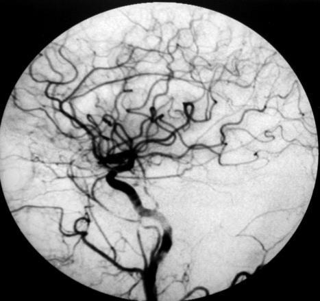
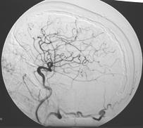
16 INTRA-ARTERIAL EMBOLIZATION Posterior fossa DAVM type III Intra-arterial embolization with GLUE left posterior meningeal artery


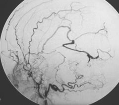
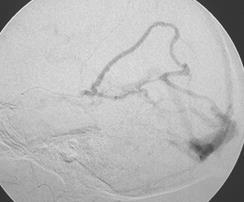
17 INTRA-ARTERIAL EMBOLIZATION Posterior fossa DAVM type III Left vertebral artery Post-Emb


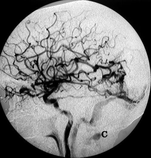
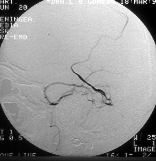
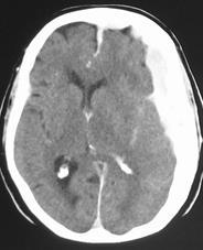
18 INTRA-ARTERIAL EMBOLIZATION Right lateral sinus DAVM type III Right temporal hematoma Intra-arterial embolization with GLUE of left middle meningeal and occipital arteries Post Emb


19 INTRA-ARTERIAL EMBOLIZATION Superior sagital sinus DAVM type IV epilepsy Intra-arterial embolization with GLUE right and left middle meningeal arteries and right occipital artery


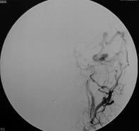
20 INTRA-ARTERIAL EMBOLIZATION Multiple shunts high and low flow feeders different type of arterial feeders PARTICLES PVA + GLUE - NBCA Frequently lateral sinus and cavernous sinus type I / II Right cavernous sinus DAVM type II


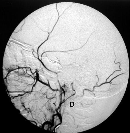
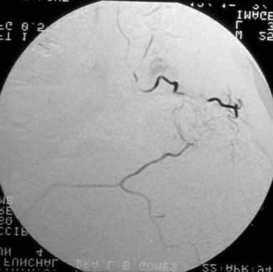
21 INTRA-ARTERIAL EMBOLIZATION Right cavernous sinus DAVM type II Distal internal maxilary artery with PVA Middle meningeal artery with COIL and GLUE

22 INTRA-ARTERIAL EMBOLIZATION Right cavernous sinus DAVM type II Distal internal maxilary artery with PVA Middle meningeal artery with COIL and GLUE Post Emb


23 INTRA-ARTERIAL EMBOLIZATION ONYX / SQUID / PHIL impossible microcatheterisation of some arterial feeders alternative to venous approach type I / II complex DAVF S with multiple AV shunts and high number of arterial feeders type III-IV lesions Male, 60 years, vertigo + right hemiparaesthaesia Frequently lateral sinus, superior sagital sinus and tentorial Tentorial DAVF type III CT - AngioCT


24 INTRA-ARTERIAL EMBOLIZATION ONYX right middle meningeal artery PRE EMB Right and left internal carotid cavernous meningeal branches Right external carotid middle meningeal, ascending pharingeal and occipital arteries Tentorial DAVF Type III Right distal middle meningeal artery ONYX CAST


25 INTRA-ARTERIAL EMBOLIZATION Tentorial DAVF type III ONYX ONYX CAST POST EMB Control angiogram Right internal and external carotid Left internal carotid
26 INTRA-ARTERIAL EMBOLIZATION Right lateral sinus DAVM type II 2007 right lateral sinus thrombosis 2009 Headache + right bruit + seizures right and left middle meningeal and occipital arteries; right posterior auricular artery; left posterior meningeal artery; right meningo-tentorial artery ONYX PRE EMB SILAN th SIMI BUENOS Buenos AIRES Aires ARGENTINA

27 INTRA-ARTERIAL EMBOLIZATION Right lateral sinus DAVM type II PRE EMB

28 INTRA-ARTERIAL EMBOLIZATION Right middle meningeal artery microcatheter and ONYX cast (5.2 cc)

29 INTRA-ARTERIAL EMBOLIZATION Right lateral sinus DAVM type II Multiple shunts 5 months follow-up arterio-venous shunt exclusion

30 INTRA-ARTERIAL EMBOLIZATION DAVM type II torcular region 72 y with dementia right and left middle meningeal and occipital arteries exclusion of both lateral sinus ONYX PRE-EMB right and left external carotid angiograms

31 INTRA-ARTERIAL EMBOLIZATION DAVM type II torcular region Left middle meningeal artery ONYX cast final result

32 INTRA-ARTERIAL EMBOLIZATION DAVM type II torcular region Follow-up right and left ext. carotid angiograms Arterio-venous shunt exclusion clinical improvement


33 INTRA-ARTERIAL EMBOLIZATION 60 y. o. Cerebellar hematoma SQUID DAVF type IV falx cerebelli right and left middle meningeals, ascending pharyngeals and occipitals Right meningotentorial branch of right ACI


34 INTRA-ARTERIAL EMBOLIZATION DAVF type IV falx cerebelli right and left middle meningeals, ascending pharyngeals and occipitals Right meningotentorial branch of right ACI SQUID injection left and right middle meningeals and left ascending pharyngeal
 type I / II")


35 INTRA-VENOUS EMBOLIZATION SINUS OCCLUSION (Coils) type I / II multiple shunts high number of arterial feeders alternative to intra-arterial embolization with Glue / ONYX / SQUID / PHIL or PVA frequently cavernous sinus and lateral sinus Left cavernous sinus DAVM type I


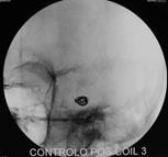
36 INTRA-VENOUS EMBOLIZATION Left cavernous sinus DAVM type I Coils - Venous approach
37 INTRA-VENOUS EMBOLIZATION Left cavernous sinus DAVM type I Post Emb

38 INTRA-VENOUS EMBOLIZATION Right lateral sinus DAVM type I


39 INTRA-VENOUS EMBOLIZATION Right lateral sinus DAVM type I Coils - Venous approach


40 INTRA-VENOUS EMBOLIZATION Right lateral sinus DAVM type I Right carotid and vertebral arteries POST EMB

41 INTRA-VENOUS EMBOLIZATION DRAINAGE VEIN OCCLUSION (Coils) type III / IV DAVF impossible microcatheterization of arterial feeders alternative to intra-arterial embolization GLUE, ONYX, SQUID and PHIL Left lateral sinus DAVM type III
42 INTRA-VENOUS EMBOLIZATION Left lateral sinus DAVM type III Left carotid angiogram POST EMB


43 UNUSUAL INTRAVENOUS EMBOLIZATION Superior Longitudinal Sinus Occlusion Male, 74 years, right parietal haemorragic stroke + dementia Superior sagital sinus DAVF type II Multiple shunts arterial feeders: Left anterior and posterior meningeal, right and left superficial temporal and occipital arteries (transosseous branches)


44 UNUSUAL INTRAVENOUS EMBOLIZATION Superior Longitudinal Sinus Occlusion with Coils Pre-emb angiogram - DAVF type II Functional exclusion of the SLS
45 UNUSUAL INTRAVENOUS EMBOLIZATION Superior Longitudinal Sinus Occlusion with Coils Left Right Pos-emb angiogram


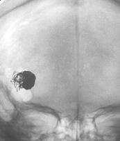
46 UNUSUAL INTRAVENOUS EMBOLIZATION Direct Percutaneous Venous Punction - Coil Embolization Female, 60 years, lytic occipital skull lesion + dementia Right lateral sinus DAVF type IV Arterial feeders: Right middle meningeal and right occipital arteries Venous hipertension pattern
47 UNUSUAL INTRAVENOUS EMBOLIZATION Direct Percutaneous Venous Punction - Coil Embolization Direct Punction Pre Emb Post Emb

")
48 INTRA-ARTERIAL EMBOLIZATION Intra-arterial embolization and Radiosurgery Post. Embolization residual high risk shunts Problematic surgery (craniotomy) Frequently tentorial


49 INTRA-ARTERIAL EMBOLIZATION Intra-arterial embolization and Radiosurgery Male 46 y.o. Headache and Bruit ++ Right DAVF type III right middle meningeal, right occipital, meningeal branches of petrous and cavernous segm. of right ICA 5 previous intra-arterial embolizations and PVA 2007/2008 Residual shunt Post Radiosurgery Exclusion of the shunt


, left anterior")
50 INTRA-ARTERIAL EMBOLIZATION Residual shunt Low surgical risk Intra-arterial embolization and Surgery Frequently anterior fossa Male 51 y.o. SAH DAVF type IV left anterior fossa 2 previous intra-arterial embolizations left middle meningeal (PVA), left anterior ethmoidal (Glue)


 Post-surgery follow")
51 INTRA-ARTERIAL EMBOLIZATION Intra-arterial embolization and Surgery (craniotomy) DAVF type IV left anterior fossa left middle meningeal (PVA), left anterior ethmoidal (GLUE) Post-surgery follow up Exclusion of the shunt
52 ISCHEMIA ENDOVASCULAR SURGERY RISKS AND COMPLICATIONS cerebral embolism anastomosis ECA ICA / ECA VA cranial nerves palsy HEMORRHAGE intracranial hemorrhage arterial rupture or venous outflow occlusion without complete embolization of the AV shunt transformation of a benign DAVM (type I/II) into a high risk lesion (type III/IV)
53 ENDOVASCULAR SURGERY RISKS AND COMPLICATIONS Right temporal fossa DAVM type IV Venous approach
54 ENDOVASCULAR SURGERY RISKS AND COMPLICATIONS Right temporal fossa DAVM type IV Post Emb Subdural hematoma after embolization
55 Hospital Santa Maria University of Lisbon Endovascular embolization - RESULTS Cavernous sinus 51 lateral sinus 42 SLS 11 Skul base / posterior fossa patients tentorial 4 anterior fossa 1 temporal fossa 4 COMPLETE ARTERIOVENOUS SHUNT EXCLUSION 87 % (6 patients remain in treatment and 5 abandoned the therapeutic protocol) Clinical improvement or cure 97 %
56 Hospital Santa Maria University of Lisbon Endovascular embolization - RESULTS 119 patients CLINICAL COMPLICATIONS 2% 1 right internal carotid embolism 1 left subdural hematoma (venous approach) MORTALITY 0 %
57 ENDOVASCULAR SURGERY CONCLUSIONS Endovascular surgery is the first treatment modality in DAVF with excelent clinical results and arteriovenous shunt occlusion. Presents a low morbidity and mortality. We should avoid a dogmatic attitude and according to the location and angioarchitecture of the lesion make use of different therapeutic techniques venous and arterial approach; different embolic materials (PVA particles, Coils, GLUE and ONYX / SQUID / PHIL).
58 CONCLUSIONS ENDOVASCULAR SURGERY The therapeutic options and results depend mainly: detailed angioarchitecture and functional haemodynamic analysis clinical criteria some cases need emergent therapy personal experience
Brain Arteriovenous Malformations Endovascular Therapy and Associated Therapeutic Protocols Jorge Guedes Cabral de Campos
 Endovascular Therapy and Associated Therapeutic Protocols Jorge Guedes Cabral de Campos Neuroradiology Department Hospital de Santa Maria University of Lisbon CEREBRAL AVM CLINICAL / EPIDEMIOLOGY Brain
A.J. Hauer Intracranial dural arteriovenous fistulae
 A.J. Hauer 27-06-2018 Intracranial dural arteriovenous fistulae Dural arteriovenous fistulae (davfs) epidemiology Pathological anastomoses (within the dural leaflets) between meningeal arteries and dural
Vascular Malformations
 Vascular Malformations LTC Robert Shih Chief of Neuroradiology Walter Reed Medical Center Special thanks to LTC Alice Smith (retired) Disclosures: None. This presentation reflects the personal views of
Intracranial dural arteriovenous fistulas (DAVFs) with retrograde
 with retrograde") ORIGINAL RESEARCH W.J. van Rooij M. Sluzewski G.N. Beute Dural Arteriovenous Fistulas with Cortical Venous Drainage: Incidence, Clinical Presentation, and Treatment BACKGROUND AND PURPOSE: Our purpose
ORIGINAL RESEARCH W.J. van Rooij M. Sluzewski G.N. Beute Dural Arteriovenous Fistulas with Cortical Venous Drainage: Incidence, Clinical Presentation, and Treatment BACKGROUND AND PURPOSE: Our purpose
HEAD/NECK VESSELS. Objectives
 Objectives Arterial Supply to Head and Neck Arteries to Head Surrounding Brain Common carotid arteries Arteries to Head Surrounding Brain External carotid arteries Arteries to Head Surrounding Brain External
Transverse-Sigmoid Sinus Dural Arteriovenous Malformations
 Transverse-Sigmoid Sinus Dural Arteriovenous Malformations Kenan I. Amautovic, M.D., and Ali F. Krisht, M.D. '-...--- Learning Objectives: After reading this article, the participant should: 1. Have an
Untangling Cerebral Dural Arteriovenous Fistulas
 Untangling Cerebral Dural Arteriovenous Fistulas Bradley A. Gross, MD Assistant Professor, Dept of Neurosurgery, University of Pittsburgh September 2017 davfs Definition Clinical Presentation Natural History
Tentorial Dural Arteriovenous Fistulas: A Single-Center Cohort of 12 Patients
 Journal of Cerebrovascular and Endovascular Neurosurgery pissn 2234-8565, eissn 2287-3139, http://dx.doi.org/10.7461/jcen.2017.19.4.284 Original Article Tentorial Dural Arteriovenous Fistulas: A Single-Center
Differences between CS-DAVF and TCCF to reveal and redefine CS-DAVF
 Pan et al. Chinese Neurosurgical Journal (2018) 4:26 https://doi.org/10.1186/s41016-018-0121-z CHINESE MEDICAL ASSOCIATION COMMENTARY Differences between CS-DAVF and TCCF to reveal and redefine CS-DAVF
Three Cases of Dural Arteriovenous Fistula of the Anterior Condylar Vein within the Hypoglossal Canal
 AJNR Am J Neuroradiol 20:2016 2020, November/December 1999 Case Report Three Cases of Dural Arteriovenous Fistula of the Anterior Condylar Vein within the Hypoglossal Canal Robert Ernst, Robert Bulas,
CASE OF THE WEEK PROFESSOR YASSER METWALLY
 CLINICAL PICTURE CLINICAL PICTURE: CASE OF THE WEEK PROFESSOR YASSER METWALLY A 29 years old male patients presented with proptosis, ecchymoses of the left eye with both subjective and objective bruit
Complex dural arteriovenous fistulas. Results of combined endovascular and neurosurgical treatment in 16 patients
 J Neurosurg 71:352-358,1989 Complex dural arteriovenous fistulas Results of combined endovascular and neurosurgical treatment in 16 patients STANLEY L. BARNWELL, M.D., PH.D., VAN V. HALBACH, M.D., RANDALL
Dural arteriovenous fistulas (DAVFs) are abnormal. Long-term angiographic results of endovascularly cured intracranial dural arteriovenous fistulas
 are abnormal. Long-term angiographic results of endovascularly cured intracranial dural arteriovenous fistulas") clinical article J Neurosurg 124:1123 1127, 2016 Long-term angiographic results of endovascularly cured intracranial dural arteriovenous fistulas Sudheer Ambekar, MD, Brandon G. Gaynor, MD, Eric C. Peterson,
clinical article J Neurosurg 124:1123 1127, 2016 Long-term angiographic results of endovascularly cured intracranial dural arteriovenous fistulas Sudheer Ambekar, MD, Brandon G. Gaynor, MD, Eric C. Peterson,
Spontaneous Closure of Dural Arteriovenous Fistulas: Report of Three Cases and Review of the Literature
 AJNR Am J Neuroradiol 22:992 996, May 2001 Case Report Spontaneous Closure of Dural Arteriovenous Fistulas: Report of Three Cases and Review of the Literature Alain Luciani, Emmanuel Houdart, Charbel Mounayer,
Cerebrovascular Malformations in the Elderly Indications for Treatment
 Cerebrovascular Malformations in the Elderly Indications for Treatment Johanna T. Fifi, MD, FAHA, FSVIN Director of Endovascular Ischemic Stroke Assistant Professor of Neurology, Neurosurgery, and Radiology
Dural arteriovenous fistulas (DAVFs) are acquired abnormal
 are acquired abnormal") ORIGINAL RESEARCH R.G. Nogueira G. Dabus J.D. Rabinov C.J. Eskey C.S. Ogilvy J.A. Hirsch J.C. Pryor Preliminary Experience with Onyx Embolization for the Treatment of Intracranial Dural Arteriovenous Fistulas
ORIGINAL RESEARCH R.G. Nogueira G. Dabus J.D. Rabinov C.J. Eskey C.S. Ogilvy J.A. Hirsch J.C. Pryor Preliminary Experience with Onyx Embolization for the Treatment of Intracranial Dural Arteriovenous Fistulas
North Oaks Trauma Symposium Friday, November 3, 2017
 Traumatic Intracranial Hemorrhage Aaron C. Sigler, DO, MS Neurosurgery Tulane Neurosciences None Disclosures Overview Anatomy Epidural hematoma Subdural hematoma Cerebral contusions Outline Traumatic ICH
Principles Arteries & Veins of the CNS LO14
 Principles Arteries & Veins of the CNS LO14 14. Identify (on cadaver specimens, models and diagrams) and name the principal arteries and veins of the CNS: Why is it important to understand blood supply
BACKGROUND AND PURPOSE:
 AJNR Am J Neuroradiol 26:1715 1722, August 2005 Dural Sinus Compartment in Dural Arteriovenous Shunts: A New Angioarchitectural Feature Allowing Superselective Transvenous Dural Sinus Occlusion Treatment
Brain AVM with Accompanying Venous Aneurysm with Intracerebral and Intraventricular Hemorrhage
 Cronicon OPEN ACCESS EC PAEDIATRICS Case Report Brain AVM with Accompanying Venous Aneurysm with Intracerebral and Intraventricular Hemorrhage Dimitrios Panagopoulos* Neurosurgical Department, University
Dural arteriovenous fistulas of the cavernous sinus - clinical case and treatment
 166 Chiriac et al Dural arteriovenous fistulas of the cavernous sinus Dural arteriovenous fistulas of the cavernous sinus - clinical case and treatment A. Chiriac, N. Dobrin*, St.M. Iencean, I. Poeata
Neurosurgical decision making in structural lesions causing stroke. Dr Rakesh Ranjan MS, MCh, Dip NB (Neurosurgery)
") Neurosurgical decision making in structural lesions causing stroke Dr Rakesh Ranjan MS, MCh, Dip NB (Neurosurgery) Subarachnoid Hemorrhage Every year, an estimated 30,000 people in the United States experience
Neurosurgical decision making in structural lesions causing stroke Dr Rakesh Ranjan MS, MCh, Dip NB (Neurosurgery) Subarachnoid Hemorrhage Every year, an estimated 30,000 people in the United States experience
Endovascular Treatment of Cerebral Arteriovenous Malformations. Bs. Nguyễn Ngọc Pi Doanh- Bs Đặng Ngọc Dũng Khoa Ngoại Thần Kinh
 Endovascular Treatment of Cerebral Arteriovenous Malformations Bs. Nguyễn Ngọc Pi Doanh- Bs Đặng Ngọc Dũng Khoa Ngoại Thần Kinh Stroke Vascular Malformations of the Brain Epidemiology: - Incidence: 0.1%,
Combining endovascular and neurosurgical treatments of high-risk dural arteriovenous fistulas in the lateral sinus and the confluence of the sinuses
 J Neurosurg 90:289 299, 1999 Combining endovascular and neurosurgical treatments of high-risk dural arteriovenous fistulas in the lateral sinus and the confluence of the sinuses KATSUYA GOTO, M.D., PH.D.,
Transvenous Embolization of Cavernous Sinus Dural Arteriovenous Fistulas with Shunts Involving the Laterocavernous Sinus
 Journal of Neuroendovascular Therapy 2017; 11: 1 7 Online November 9, 2016 DOI: 10.5797/jnet.oa.2016-0062 Transvenous Embolization of Cavernous Sinus Dural Arteriovenous Fistulas with Shunts Involving
Brain Meninges, Ventricles and CSF
 Brain Meninges, Ventricles and CSF Lecture Objectives Describe the arrangement of the meninges and their relationship to brain and spinal cord. Explain the occurrence of epidural, subdural and subarachnoid
Dural Arteriovenous Fistulas: the value of Carotid Ultrasonography.
 Dural Arteriovenous Fistulas: the value of Carotid Ultrasonography. Poster No.: C-2199 Congress: ECR 2014 Type: Educational Exhibit Authors: J. P. Filipe, T. Parreira, C. Andrade, R. Santos, E. Azevedo;
Cranial dural arteriovenous fistula: transarterial Onyx embolization experience and technical nuances
 Division of Neurological Surgery, Barrow Neurological Institute, St. Joseph s Hospital and Medical Center, Phoenix, Arizona, USA Correspondence to Dr C G McDougall, Division of Neurological Surgery, Barrow
Treatment of Dural Arteriovenous Malformations Involving the Superior Sagittal Sinus
 337 Treatment of Dural Arteriovenous Malformations Involving the Superior Sagittal Sinus Van V. Halbach1 Randall T. Higashida 1 Grant B. Hieshima 1 Mark Rosenblum 2 Les Cahan 3 We report the diagnosis
PEER-REVIEW REPORTS. various signs and symptoms such as headaches, tinnitus, bruit, neurologic deficits, venous hypertensive encephalopathy with
 Multimodality Treatment of Intracranial Dural Arteriovenous Fistulas in the Onyx Era: a Single Center Experience Sabareesh K. Natarajan 1, Basavaraj Ghodke 1,2, Louis J. Kim 1,2, Danial K. Hallam 1,2,
A Case of Carotid-Cavernous Fistula
 A Case of Carotid-Cavernous Fistula By : Mohamed Elkhawaga 2 nd Year Resident of Ophthalmology Alexandria University A 19 year old male patient came to our outpatient clinic, complaining of : -Severe conjunctival
What Is an Arteriovenous malformation (AVM)?
?") American Society of Neuroradiology What Is an Arteriovenous malformation (AVM)? From the Cerebrovascular Imaging and Intervention Committee of the American Heart Association Cardiovascular Council Randall
American Society of Neuroradiology What Is an Arteriovenous malformation (AVM)? From the Cerebrovascular Imaging and Intervention Committee of the American Heart Association Cardiovascular Council Randall
The superior ophthalmic vein approach for the treatment of carotid-cavernous fistulas: our first experience
 230 Chiriac et al - Superior ophthalmic vein approach The superior ophthalmic vein approach for the treatment of carotid-cavernous fistulas: our first experience A. Chiriac, N. Dobrin 1, Georgiana Ion
Dural arteriovenous fistula discovered in patient presenting with recent head trauma
 ISSN 1507-6164 DOI: 10.12659/AJCR.889610 Received: 2013.07.25 Accepted: 2013.08.08 Published: 2013.10.28 Dural arteriovenous fistula discovered in patient presenting with recent head trauma Authors Contribution:
A Shunt of the Diploic Vein of the Orbital Roof Accompanying a Cavernous Sinus Dural Arteriovenous Fistula: A Case Report
 Journal of Neuroendovascular Therapy 2018; 12: 38 42 Online September 11, 2017 DOI: 10.5797/jnet.cr.2017-0056 A Shunt of the Diploic Vein of the Orbital Roof Accompanying a Cavernous Sinus Dural Arteriovenous
Combining endovascular and neurosurgical treatments of high-risk dural arteriovenous fistulas in the lateral sinus and the confluence of the sinuses
 Neurosurg Focus 5 (4):Article 10, 1998 Combining endovascular and neurosurgical treatments of high-risk dural arteriovenous fistulas in the lateral sinus and the confluence of the sinuses Katsuya Goto,
Posterior fossa veins: Embryology, anatomy, variations and pathology
 Posterior fossa veins: Embryology, anatomy, variations and pathology Poster No.: C-2668 Congress: ECR 2010 Type: Educational Exhibit Topic: Neuro Authors: S. Nair, D. B. Sarkar, J. J. Bhattacharya, M.
Venous Stroke with Intracranial Dural Arteriovenous Fistula
 Case Reports 24 Venous Stroke with Intracranial Dural Arteriovenous Fistula Tzung-Wen Chiang 1,2, Shung-Lon Lai 1, Leang-Kai Chang 2, Yung-Yee Chang 1, Min-Yu Lan 1, Yeh-Lin Kuo 3, Chen-Chung Lu 3, and
Spontaneous occlusion of a cerebral arteriovenous malformation after subtotal endovascular embolisation
 206 Chiriac et al Spontaneous occlusion of a cerebral arteriovenous malformation Spontaneous occlusion of a cerebral arteriovenous malformation after subtotal endovascular embolisation A. Chiriac, N. Dobrin*,
Navigation-guided Burr Hole Aspiration Surgery for Acute Cerebellar Infarction
 FPⅣ-1 Navigation-guided Burr Hole Aspiration Surgery for Acute Cerebellar Infarction Eun-Sung Park, Dae-Won Kim, Sung-Don Kang Department of Neurosurgery, School of Medicine, Wonkwang University, Iksan,
Selective disconnection of cortical venous reflux as treatment for cranial dural arteriovenous fistulas
 J Neurosurg 101:31 35, 2004 Selective disconnection of cortical venous reflux as treatment for cranial dural arteriovenous fistulas J. MARC C. VAN DIJK, M.D., PH.D., KAREL G. TERBRUGGE, M.D., ROBERT A.
Pediatric Neurointervention: Vein of Galen Malformations
 Pediatric Neurointervention: Vein of Galen Malformations Johanna T. Fifi, M.D. Assistant Professor of Neurology, Neurosurgery, and Radiology Icahn School of Medicine at Mount Sinai November 9 th, 2014
General Data. Gender: Female Birthday and age: 1932/11/03, 73 y/o Occupation: house keeper Date of Admission: 2005/03/30
 General Data Gender: Female Birthday and age: 1932/11/03, 73 y/o Occupation: house keeper Date of Admission: 2005/03/30 Chief Complain Dizziness and light headache for recent 1 year. Present illness Hypertension
[(PHY-3a) Initials of MD reviewing films] [(PHY-3b) Initials of 2 nd opinion MD]
![[(PHY-3a) Initials of MD reviewing films] [(PHY-3b) Initials of 2 nd opinion MD]](/thumbs/89/98619893.jpg "[(PHY-3a) Initials of MD reviewing films] [(PHY-3b) Initials of 2 nd opinion MD]") 2015 PHYSICIAN SIGN-OFF (1) STUDY NO (PHY-1) CASE, PER PHYSICIAN REVIEW 1=yes 2=no [strictly meets case definition] (PHY-1a) CASE, IN PHYSICIAN S OPINION 1=yes 2=no (PHY-2) (PHY-3) [based on all available
2015 PHYSICIAN SIGN-OFF (1) STUDY NO (PHY-1) CASE, PER PHYSICIAN REVIEW 1=yes 2=no [strictly meets case definition] (PHY-1a) CASE, IN PHYSICIAN S OPINION 1=yes 2=no (PHY-2) (PHY-3) [based on all available
Interventions in the Management of Acute Stroke. Dr Md Shafiqul Islam Associate Professor Neurosurgery Dhaka Medical College Hospital
 Interventions in the Management of Acute Stroke Dr Md Shafiqul Islam Associate Professor Neurosurgery Dhaka Medical College Hospital Acute stroke intervention Number of stroke patients increasing day by
PTA 106 Unit 1 Lecture 3
 PTA 106 Unit 1 Lecture 3 The Basics Arteries: Carry blood away from the heart toward tissues. They typically have thicker vessels walls to handle increased pressure. Contain internal and external elastic
Dilemma in Imaging Diagnosis, Endovascular Management and Complications
 Vascular anomaly at the craniocervical junction presenting with sub arachnoid hemorrhage: Dilemma in Imaging Diagnosis, Endovascular Management and Complications Ajeet 1* 1. Department of Radiology, St
7/5/2016. Neonatal high-output cardiac failure. Case 1 POSTNATAL STRATEGIES FOR CEREBRAL ATERIOVENOUS MALFORMATIONS
 John Deveikis, M.D. POSTNATAL STRATEGIES FOR CEREBRAL ATERIOVENOUS MALFORMATIONS JULY, 2016 Neonatal high-output cardiac failure Tachypnea, tachycardia, hypotension, failure to thrive When congenital heart
Dural arteriovenous fistulas (DAVFs) are
 are") TOPIC DURAL FISTULAS DURAL FISTULAS Advances in Surgical Approaches to Dural Fistulas Patrick P. Youssef, MD Albert Jess Schuette, MD C. Michael Cawley, MD Daniel L. Barrow, MD Department of Neurosurgery,
TOPIC DURAL FISTULAS DURAL FISTULAS Advances in Surgical Approaches to Dural Fistulas Patrick P. Youssef, MD Albert Jess Schuette, MD C. Michael Cawley, MD Daniel L. Barrow, MD Department of Neurosurgery,
Case Log Mapping Update: April 2018 Review Committee for Neurological Surgery
 Case Log Mapping Update: April 2018 Review Committee for Neurological Surgery The Review Committee has made the following changes to the CPT code mappings: The following previously untracked CPT codes
Vascular and Parameningeal Infections of the Head and Neck
 Vascular and Parameningeal Infections of the Head and Neck Kevin B. Laupland, MD, MSc, FRCPC Associate Professor Departments of Medicine, Critical Care Medicine, Pathology and Laboratory Medicine, and
Borden type3 superior sagittal sinus dural arteriovenous fistula, angiographic features and endovascular treatment.
 Borden type3 superior sagittal sinus dural arteriovenous fistula, angiographic features and endovascular treatment. Poster No.: C-1939 Congress: ECR 2013 Type: Scientific Exhibit Authors: T. Dotsu 1, H.
INTERVENTIONAL NEURORADIOLOGY
 iii48 INTERVENTIONAL NEURORADIOLOGY c CEREBRAL Correspondence to: Dr Shelley Renowden, Department of Neuroradiology, Frenchay Hospital, Bristol BS16 1LE, UK; Shelley.Renowden@ north-bristol.swest.nhs.uk
INSTITUTE OF NEUROSURGERY & DEPARTMENT OF PICU
 CEREBRAL BYPASS An Innovative Treatment for Arteritis INSTITUTE OF NEUROSURGERY & DEPARTMENT OF PICU CASE 1 q 1 year old girl -recurrent seizure, right side limb weakness, excessive cry and irritability.
EMBOLIZATION OF ARTERIOVENOUS FISTULA AFTER RADIOSURGERY FOR MULTIPLE CEREBRAL ARTERIOVENOUS MALFORMATIONS
 Arteriovenous fistula after radiosurgery for multiple CAVM EMBOLIZATION OF ARTERIOVENOUS FISTULA AFTER RADIOSURGERY FOR MULTIPLE CEREBRAL ARTERIOVENOUS MALFORMATIONS Chao-Bao Luo, Wan-Yuo Guo, Michael
Dural Arteriovenous Fistulas of the Posterior Fossa Draining into Subarachnoid Veins
 Dural Arteriovenous Fistulas of the Posterior Fossa Draining into Subarachnoid Veins Laurent Pierot, 1 Jacques Chiras, 1 Jean-Frans:ois Meder, 2 Michele Rose, 1 Maurice Rivierez, 3 and Claude Marsault
MASSIVE EPISTAXIS IN A NEONATE: A SYMPTOM OF VEIN OF GALEN MALFORMATION!
 CASE REPORT MASSIVE EPISTAXIS IN A NEONATE: A SYMPTOM OF VEIN OF GALEN MALFORMATION! Shagufta Wahab 1, Rizwan Ahmad Khan 2, Manjari Thapa Manger 3 1. Radiodiagnosis, Aligarh Muslim University, Aligarh,
Advanced Vascular Imaging: Pulsatile Tinnitus. Disclosures. Pulsatile Tinnitus: Differential Diagnosis. Pulsatile Tinnitus
 Advanced Vascular Imaging: Pulsatile Tinnitus Patrick Turski MD, Zach Clark MD, Tabby Kennedy MD The Objectives of this presentation are to: Review the differential diagnosis of pulsatile tinnitus Discuss
Diagnosis of Subarachnoid Hemorrhage (SAH) and Non- Aneurysmal Causes
 and Non- Aneurysmal Causes") Diagnosis of Subarachnoid Hemorrhage (SAH) and Non- Aneurysmal Causes By Sheila Smith, MD Swedish Medical Center 1 Disclosures I have no disclosures 2 Course Objectives Review significance and differential
Diagnosis of Subarachnoid Hemorrhage (SAH) and Non- Aneurysmal Causes By Sheila Smith, MD Swedish Medical Center 1 Disclosures I have no disclosures 2 Course Objectives Review significance and differential
Transvenous Embolization of Dural Fistulas Involving the Transverse and Sigmoid Sinuses
 385 Transvenous Embolization of Dural Fistulas Involving the Transverse and Sigmoid Sinuses Van V. Halbach 1 Randall T. Higashida 1 Grant B. Hieshima 1 C. Mark Mehringer 2 Carl W. Hardin 1 Received February
Original Article Pial arteriovenous fistulas: two pediatric cases and a literature review
 Int J Clin Exp Med 2016;9(5):7855-7862 www.ijcem.com /ISSN:1940-5901/IJCEM0019732 Original Article Pial arteriovenous fistulas: two pediatric cases and a literature review Lei Feng *, Yunzhen Liu *, Jun
Vascular Malformations of the Brain: A Review of Imaging Features and Risks
 Vascular Malformations of the Brain: A Review of Imaging Features and Risks Comprehensive Neuroradiology: Best Practices October 27-30, 2016 Sudhakar R. Satti, MD Associate Director Neurointerventional
Endovascular Treatment of Cavernous Sinus Dural Arteriovenous Fistulas by Direct Puncture of Facial Vein
 J Radiol Sci 2011; 36: 129-134 Endovascular Treatment of Cavernous Sinus Dural Arteriovenous Fistulas by Direct Puncture of Facial Vein Shih-Wei Hsu 1 Yeh-Lin Kuo 1 Min-Hsiung Cheng 2 Department of Diagnostic
Therapeutic Embolization of the Dural Arteriovenous Malformation Involving the Jugular Bulb
 J Korean Med Sci 2001; 16: 527-31 ISSN 1011-8934 Copyright The Korean Academy of Medical Sciences Therapeutic Embolization of the Dural Arteriovenous Malformation Involving the Jugular Bulb Pulsatile tinnitus
Enhancement of Cranial US: Utility of Supplementary Acoustic Windows and Doppler Harriet J. Paltiel, MD
 Enhancement of Cranial US: Utility of Supplementary Acoustic Windows and Doppler Harriet J. Paltiel, MD Boston Children s Hospital Harvard Medical School None Disclosures Conventional US Anterior fontanelle
An Onyx tunnel: reconstructive transvenous balloon-assisted Onyx embolization for dural arteriovenous fistula of the transverse-sigmoid sinus
 TECHNICAL NOTE J Neurosurg 129:922 927, 2018 An Onyx tunnel: reconstructive transvenous balloon-assisted Onyx embolization for dural arteriovenous fistula of the transverse-sigmoid sinus *Mena G. Kerolus,
Retrospective analytical six months study of vascular abnormalities of brain
 International Journal of Advances in Medicine http://www.ijmedicine.com pissn 2349-3925 eissn 2349-3933 Research Article DOI: http://dx.doi.org/10.18203/2349-3933.ijam20160185 Retrospective analytical
NEURO IMAGING 2. Dr. Said Huwaijah Chairman of radiology Dep, Damascus Univercity
 NEURO IMAGING 2 Dr. Said Huwaijah Chairman of radiology Dep, Damascus Univercity I. EPIDURAL HEMATOMA (EDH) LOCATION Seventy to seventy-five percent occur in temporoparietal region. CAUSE Most likely caused
Blood Supply of the CNS
 Blood Supply of the CNS Lecture Objectives Describe the four arteries supplying the CNS. Follow up each artery to its destination. Describe the circle of Willis and its branches. Discuss the principle
Fatal progression of posttraumatic dural arteriovenous fistulas refractory to multimodal therapy
 See the Letter to the Editor and the Response in this issue in Neurosurgical Forum, pp 858 861. J Neurosurg 94:831 835, 2001 Fatal progression of posttraumatic dural arteriovenous fistulas refractory to
Dural Arteriovenous Fistula Following Translabyrinthine Resection of Cerebellopontine Angle Tumors: Report of Two Cases
 Dural Arteriovenous Fistula Following Translabyrinthine Resection of Cerebellopontine Angle Tumors: Report of Two Cases Peter M.M.C. Li, M.D., 1 Nancy J. Fischbein, M.D., 1,2 Huy M. Do, M.D., 2,3 and Nikolas
Diagnosis and Management of AVM in the Pregnant Patient
 Diagnosis and Management of AVM in the Pregnant Patient Wade Cooper, D.O. University of Michigan Assistant Professor Departments of Neurology & Anesthesiology Disclosures Wade Cooper - None Developmental
Meninges and Ventricles
 Meninges and Ventricles Irene Yu, class of 2019 LEARNING OBJECTIVES Describe the meningeal layers, the dural infolds, and the spaces they create. Name the contents of the subarachnoid space. Describe the
Sinus Venous Thrombosis
 Sinus Venous Thrombosis Joseph J Gemmete, MD FACR, FSIR, FAHA Professor Departments of Radiology and Neurosurgery University of Michigan Hospitals Ann Arbor, MI Outline Introduction Medical Treatment Options
Modern treatment of brain arteriovenous malformation
 ORIGINAL RESEARCH W.J. van Rooij M. Sluzewski G.N. Beute Brain AVM Embolization with Onyx BACKGROUND AND PURPOSE: To report the initial experience by using a new liquid embolic agent (Onyx) for embolization
Overview of Cerebrovascular Malformations
 Overview of Cerebrovascular Malformations Pursuit of Neurovascular Excellence 8 th annual Barbara Albani, MD Chief, Neurointerventional Surgery Christiana Care Health Systems Newark, DE Financial Disclosures
Supratentorial cerebral arteriovenous malformations : a clinical analysis
 Original article: Supratentorial cerebral arteriovenous malformations : a clinical analysis Dr. Rajneesh Gour 1, Dr. S. N. Ghosh 2, Dr. Sumit Deb 3 1Dept.Of Surgery,Chirayu Medical College & Research Centre,
Dural arteriovenous fistulas (DAVFs) are relatively
 are relatively") case report J Neurosurg 122:1208 1213, 2015 Torcular dural arteriovenous fistula treated via stent placement and angioplasty in the affected straight and transverse sinuses: case report Shigeki Takada,
case report J Neurosurg 122:1208 1213, 2015 Torcular dural arteriovenous fistula treated via stent placement and angioplasty in the affected straight and transverse sinuses: case report Shigeki Takada,
OBJECTIVES. At the end of the lecture, students should be able to: List the cerebral arteries.
 DR JAMILA EL MEDANY OBJECTIVES At the end of the lecture, students should be able to: List the cerebral arteries. Describe the cerebral arterial supply regarding the origin, distribution and branches.
Spontaneous Obliteration of Pial Arteriovenous Malformations: A Review of 27 Cases
 AJNR Am J Neuroradiol :, March 00 Spontaneous Obliteration of Pial Arteriovenous Malformations: A Review of ases Maneesh. Patel, Timothy J. Hodgson, Andras A. Kemeny, and David M. Forster BAKGROUND AND
Brain ميهاربا لض اف دمح ا د The Meninges 1- Dura Mater of the Brain endosteal layer does not extend meningeal layer falx cerebri tentorium cerebelli
 .احمد د فاضل ابراهيم Lecture 15 Brain The Meninges Three protective membranes or meninges surround the brain in the skull: the dura mater, the arachnoid mater, and the pia mater 1- Dura Mater of the Brain
AVM and AV fistula. Effective diagnostic imaging for spinal arteriovenous fistula
 FPⅤ-1 Effective diagnostic imaging for spinal arteriovenous fistula Ryohei Miyazaki, Hidetoshi Murata, Mitsuru Sato, Takahiro Tanaka, Nobutaka Shimizu, Jun Suenaga, Tetsuya Yamamoto Yokohama City University
Dural Arteriovenous Fistula in Children: Endovascular Treatment and Outcomes in Seven Cases
 AJNR Am J Neuroradiol 22:1217 1225, June/July 2001 Dural Arteriovenous Fistula in Children: Endovascular Treatment and Outcomes in Seven Cases Patricia K. Kincaid, Gary R. Duckwiler, Y. Pierre Gobin, and
A Case of Curable Dementia Treated by Effective Endovascular Embolization for Dural Arteriovenous Fistula
 Published online: April 16, 2014 1662 680X/14/0061 0116$39.50/0 This is an Open Access article licensed under the terms of the Creative Commons Attribution-NonCommercial 3.0 Unported license (CC BY-NC)
The central nervous system
 Sectc.qxd 29/06/99 09:42 Page 81 Section C The central nervous system CNS haemorrhage Subarachnoid haemorrhage Cerebral infarction Brain atrophy Ring enhancing lesions MRI of the pituitary Multiple sclerosis
Endovascular Embolization of Intracranial Dural AV Fistula- Benign?
 Endovascular Embolization of Intracranial Dural AV Fistula- Benign? Suneeta Gollapudy, M.D. Assistant Professor of Anesthesiology Director, Division of Neuroanesthesia Director, Division of PACU Rotation(
Treatment of Superior Sagittal Sinus Dural Arteriovenous Fistula by Transarterial Multiple Balloon-assisted Onyx Embolization: A Case Report
 DOI: 10.5797/jnet.tn.2016-0136 Treatment of Superior Sagittal Sinus Dural Arteriovenous Fistula by Transarterial Multiple Balloon-assisted Onyx Embolization: A Case Report Shunsuke Yamashita, 1 Atsushi
Dural arteriovenous fistulas (DAVFs) constitute 10% to
 constitute 10% to") Color Doppler Flow Imaging of the Superior Ophthalmic Vein in Dural Arteriovenous Fistulas Shoichiro Kawaguchi, MD; Toshisuke Sakaki, MD; Ryunosuke Uranishi, MD Background and Purpose This article evaluates
Color Doppler Flow Imaging of the Superior Ophthalmic Vein in Dural Arteriovenous Fistulas Shoichiro Kawaguchi, MD; Toshisuke Sakaki, MD; Ryunosuke Uranishi, MD Background and Purpose This article evaluates
Contents. 1 Embryological and Anatomical Introduction... 1
 1 Embryological and Anatomical Introduction.... 1 1.1 Preliminary Remarks.................... 1 1.2 Leptomeninges....................... 21 1.3 Subpial Space........................ 22 1.3.1 Anatomy...........................
T HE blood supply of cerebral arteriovenous malformations is often extensive
 NOVEMBER, 1974 ROENTGENOGRAPHIC ANALYSIS OF ARTERIOVENOUS MALFORMATIONS OF THE OCCIPITAL LOBE* By B. TODD TROOST, M.D.,t and THOMAS H. NEWTON, M.D4 T HE blood supply of cerebral arteriovenous malformations
Cranial Cavity REFERENCES: OBJECTIVES OSTEOLOGY. Stephen A. Gudas, PT, PhD
 Stephen A. Gudas, PT, PhD Cranial Cavity REFERENCES: Moore and Agur, Essential Clinical Anatomy (ECA), 3rd ed., pp. 496 498; 500 507; 512 514 Grant s Atlas 12 th ed., Figs 7.6; 7.19 7.30. Grant s Dissector
Dementia Resulting from Dural Arteriovenous Fistulas: The Pathologic Findings of Venous Hypertensive Encephalopathy
 AJNR Am J Neuroradiol 19:1267 1273, August 1998 Dementia Resulting from Dural Arteriovenous Fistulas: The Pathologic Findings of Venous Hypertensive Encephalopathy Robert W. Hurst, Linda J. Bagley, Steven
Comparison of Five Major Recent Endovascular Treatment Trials
 Comparison of Five Major Recent Endovascular Treatment Trials Sample size 500 # sites 70 (100 planned) 316 (500 planned) 196 (833 estimated) 206 (690 planned) 16 10 22 39 4 Treatment contrasts Baseline
Transfemoral Venous Embolization of Vein of Galen Malformations
 643 Transfemoral Venous Embolization of Vein of Galen Malformations Christopher F. Dowd 1 Van V. Halbach 1 2 Stanley L. Barnwell 1 2 Randall T. Higashida 1 2 MichaelS. B. Edwards 2 3 Grant B. Hieshima
Vascular malformations: Venous malformations anomalous veins drain normal brain tissue for 65% of all cases 2.5%. was 0, 3% per year
 Vascular malformations: 1. Venous malformations: congenital venous anomalies pathologically characterised by anomalous veins (thickened and hyalinised walls) separated by normal brain. These anatomically
TCD AND VASOSPASM SAH
 CURRENT TREATMENT FOR CEREBRAL ANEURYSMS TCD AND VASOSPASM SAH Michigan Sonographers Society 2 Nd Annual Fall Vascular Conference Larry N. Raber RVT-RDMS Clinical Manager General Ultrasound-Neurovascular
DIAGNOSTIC NEURORADIOLOGY
 Neuroradiology (2008) 50:1013 1023 DOI 10.1007/s00234-008-0433-3 DIAGNOSTIC NEURORADIOLOGY The anterior medullary anterior pontomesencephalic venous system and its bridging veins communicating to the dural
Case Conference: Neuroradiology. Case 1: Tumor Case 1: 22yo F w/ HA and prior Seizures
 Case Conference: Neuroradiology Case 1: 22yo F w/ HA and prior Seizures David E. Rex, MD, PhD Stanford University Hospital Department of Radiology Case 1: Tumor Most likely gangiloglioma, oligodendroglioma,
Unit 18: Cranial Cavity and Contents
 Unit 18: Cranial Cavity and Contents Dissection Instructions: The calvaria is to be removed without damage to the dura mater which is attached to the inner surface of the calvaria. Cut through the outer
Intracranial venous sector thrombectomy with endovascular thromboaspiration system
 Intracranial venous sector thrombectomy with endovascular thromboaspiration system Poster No.: C-1191 Congress: ECR 2015 Type: Scientific Exhibit Authors: D. D. J. De la Rosa Porras, E. Castro Reyes, J.