When Immunostains Can Get You Into Trouble (and how they can help you out): Neuroendocrine Neoplasms
|
|
|
- Hope Heath
- 5 years ago
- Views:
Transcription
1 When Immunostains Can Get You Into Trouble (and how they can help you out): Neuroendocrine Neoplasms Arthur Purdy Stout Society March 5, 2017 David S. Klimstra, MD Chairman, Department of Pathology James Ewing Alumni Chair of Pathology Attending Pathologist Memorial Sloan Kettering Cancer Center Professor of Pathology and Laboratory Medicine Weill Cornell Medical College
2 Disclosure Statement Dr. Klimstra receives royalty payments from Up To Date and the American Registry of Pathology PET-CT


3 Neuroendocrine Neoplasms Diverse but related groups of tumors Lung, thymus, pancreas, GI tract, other sites Characteristic pathologic features Immunohistochemical evidence of neuroendocrine differentiation (chromogranin / synaptophysin / CD56) Range of biological aggressiveness Can be either well differentiated tumors or poorly differentiated carcinomas


4 Differentiation: Extent of resemblance of the cells of a neoplasm to their normal cellular counterparts Usually closely linked to grade (for NETs)
5 Differentiation: Immunohistochemistry Chromogranin A Synaptophysin


6 Well Differentiated vs. Poorly Differentiated Neuroendocrine Neoplasms Two different families Both share neuroendocrine differentiation Can be difficult to distinguish Fundamentally different Cell of origin Relationship to non-ne neoplasia Genetic background Clinical aggressiveness Treatment
7 Grading of Pulmonary Neuroendocrine Neoplasms Low Grade Carcinoid Tumor Intermediate Grade Atypical Carcinoid Tumor High Grade Small Cell Carcinoma Large Cell Neuroendocrine Carcinoma
8 WHO 2010 Grading of GEP-NETs Grade Mitoses Ki-67 Index G1 < 2 / 10 H.P.F. < 3% G / 10 H.P.F. 3-20% G3 > 20 / 10 H.P.F. > 20% Poorly Differentiated (High Grade ) Neuroendocrine Carcinoma


9 Pancreatic NETs: Overall Survival by Grade Rindi et al., J Natl Cancer Inst 2012; 104: 764
10 Terminology for Neuroendocrine Neoplasms: WHO 2010 Well Differentiated NETs Well differentiated NET (pancreas, GI tract, etc.) Carcinoid tumor (lung, thymus) Poorly Differentiated NECs Small cell carcinoma Large cell neuroendocrine carcinoma Mixed neuroendocrine carcinoma (with component of adenocarcinoma, squamous cell carcinoma, etc.)
11 Use of Immunohistochemistry in Neuroendocrine Neoplasms Diagnosis (recognition of neuroendocrine differentiation) Delineation of primary site PET-CT Determination of grade, classification, prognosis

12 Recognition of Neuroendocrine Differentiation PET-CT
13 Recognition of Neuroendocrine Differentiation PET-CT
14 Recognition of Neuroendocrine Differentiation: Immunohistochemical Markers Conventional markers Chromogranin A PET-CT Synaptophysin CD56 (neural cell adhesion molecular / NCAM) Neuron specific enolase (NSE) CD57 / Leu7 PGP9.5 Novel markers Synaptic vesicle protein 2 (SV2) Achaete-scute complex homolog (MASH1) Insulinoma-associated protein 1 (INSM1) Neuroendocrine secretory protein 55 (NESP55)
15 Large Cell Neuroendocrine Carcinoma Chromogranin
16 Sensitivity of Chromogranin A % Negative (n) Pulmonary carcinoid tumor 3% (368) Duodenal NET 7% (61) Ileal NET 4% (51) Pancreatic NET 17% (108) Thymic carcinoid tumor 19% (95) Pheochromocytoma 1% (182) Pulmonary small cell carcinoma 57% (596) Pulmonary large cell NE carcinoma 37% (252) Source: Immunoquery
17 Sensitivity of Synaptophysin % Negative (n) Pulmonary carcinoid tumor 2% (333) Pulmonary atypical carcinoid tumor 10% (115) Rectal NET 4% (28) Ileal NET 2% (58) Pancreatic NET 1% (75) Thymic carcinoid tumor 21% (101) Pheochromocytoma 2% (188) Pulmonary small cell carcinoma 25% (97) Pulmonary large cell NE carcinoma 17% (268) Source: Immunoquery


18 Small Bowel Tumor with Mesenteric Deposits


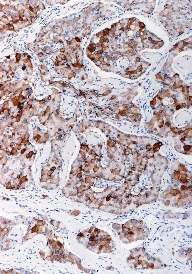
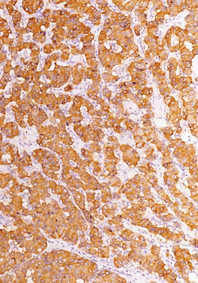
19 Tumor positive with somatostatin receptor scintigraphy Chromogranin Synaptophysin CD56
20 Specificity of Chromogranin and Synaptophysin % Chromogranin Positive (n) Breast ductal carcinoma 2% (287) % Synaptophysin Positive (n) Breast colloid carcinoma 18% (112) 41% (105) Pulmonary adenocarcinoma 2% (689) 11% (689) Pulmonary squamous cell carcinoma 2% (586) 4% (584) GIST 1% (88) Adrenal cortical carcinoma 2% (81) 63% (269) Renal cell carcinoma 2% (379) Clear cell sarcoma 25% (59) Melanoma 11% (114) Source: Immunoquery
21 Specificity of CD56 for Neuroendocrine Neoplasms Source: Immunoquery % Positive (n) Lung adenocarcinoma 3% (639) Lung squamous cell carcinoma 9% (520) Renal cell carcinoma 17% (455) Pancreatic solid pseudopapillary neoplasm 98% (152) Adrenal cortical carcinoma 88% (49) Melanoma 7% (130) Adult granulosa cell tumor 100% (40) Synovial sarcoma 51% (68) Rhabdomyosarcoma 76% (34) Granular cell tumor 95% (58) Glioma 36% (148) Dendritic cell tumor 94% (164) Nk T-cell lymphoma 74% (267) Chloroma 27% (62)
22 Immunohistochemical Staining for the Diagnosis of Well Differentiated Neuroendocrine (Carcinoid) Tumors Sensitivity For most primary sites, chromogranin and synaptophysin are highly sensitive When used in combination, ~95% positive Specificity PET-CT Certain specific non-ne neoplasms stain predictably 2-5% idiosyncratic staining of other neoplasms Is it necessary? Specific differential diagnoses Metastatic disease What about histologically typical primary tumors?


 23.")
23 (Am J Surg Pathol 2010;34: ) Are immunohistochemical stains for general neuroendocrine markers mandated as necessary in all cases? Agree strongly 23.53% 4 Agree with minor reservation 11.76% 2 Agree with major reservation 0% 0 Disagree with minor reservation (disagree mildly) 11.76% 2 Disagree with major reservation (disagree moderately) 23.53% 4 Disagree strongly 29.41% 5 Totals 100% 17 NO AGREEMENT
24 Immunohistochemical Staining for the Diagnosis of Poorly Differentiated Neuroendocrine Carcinomas Small cell carcinoma NOT mandated when classic morphologic findings are present Consider ruling out alternatives (e.g., basaloid squamous cell carcinoma, spindle cell carcinoid tumor, etc.) Chromogranin or synaptophysin positive in ~75% of cases Large cell neuroendocrine carcinoma NE marker expression required for diagnosis Must be positive in 100% of cases (by definition) Which makers? Thoracic vs. gastroenteropancreatic How strongly / diffusely positive? WHO 2015: The diagnosis of LCNEC requires immunohistochemistry for confirmation of neuroendocrine differentiation. In decreasing order of frequency, NCAM/CD56 stains % of LCNEC cases, followed by chromogranin A in 80 85%, and synaptophysin in 50 60%. NCAM/CD56 needs a note of caution because of its lower specificity for neuroendocrine differentiation in lung cancer, but it is the most sensitive marker in the appropriate morphological context of a neuroendocrine neoplasm. Chromogranin A and synaptophysin are the most reliable stains for diagnostic accuracy in distinguishing LCNEC from non-neuroendocrine tumours, and one positive marker is enough if the staining is clear-cut.

25 Large Cell Neuroendocrine Carcinoma Chr Syn CD56

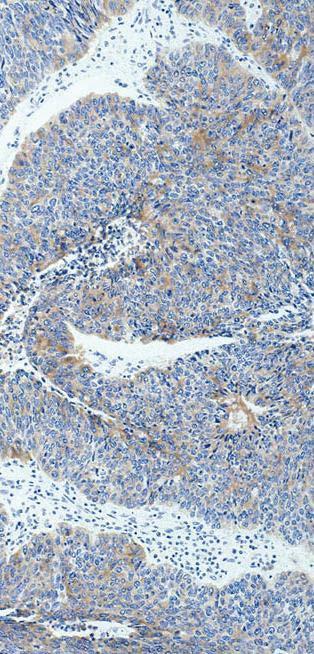
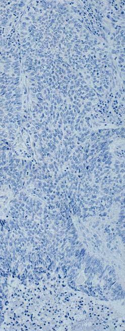
26 Large Cell Neuroendocrine Carcinoma Chr Syn CD56
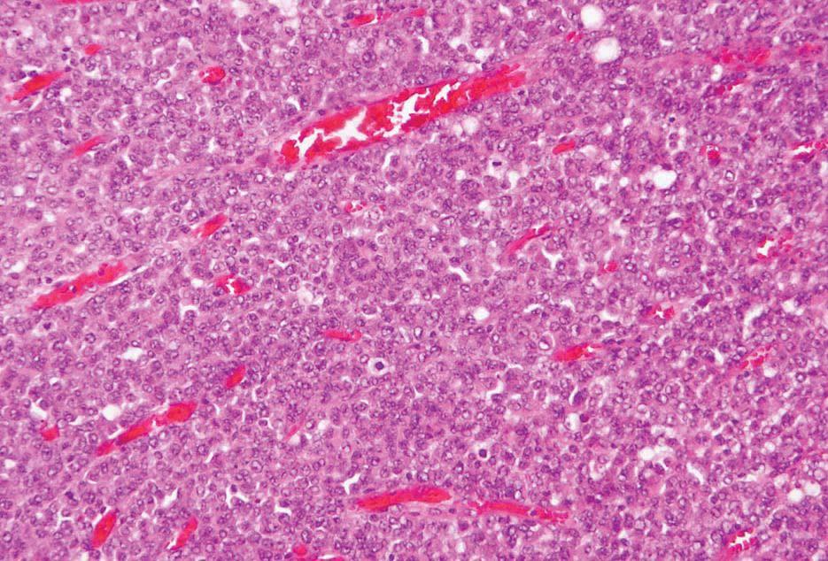
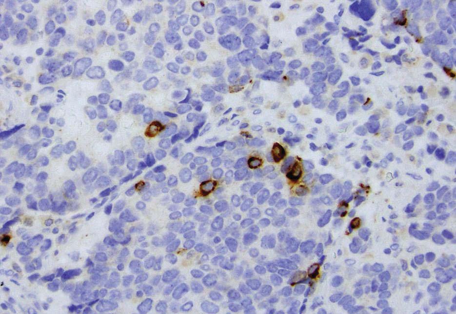
27 Chromogranin
28 Large Cell Lung Carcinoma Large Cell Undifferentiated Carcinoma Large Cell Neuroendocrine Carcinoma Large Cell Carcinoma with Neuroendocrine Differentiation Large Cell Carcinoma with Neuroendocrine Morphology


29 Large Cell Carcinoma with Neuroendocrine Morphology Chr, Synapto, CD56


30 Large Cell Carcinoma with Neuroendocrine Differentiation Chromogranin

31 Morphology vs. Immunohistochemistry in Large Cell Carcinoma
32 Neuroendocrine Differentiation in NSCLCs: Clinical Implications Found by IHC in 10-35% of NSCLCs Reports of both better and worse prognosis for NSCLC-ND vs. other NSCLCs Proposal that NSCLC-ND may respond better to chemotherapy Treatment of LCNEC with platinumbased chemotherapy
33 Genomic subgroups in LCNEC Gene alterations typical of: SCLC Adeno SCLC SCLC/SqCC Number of altered genes per case TP53 78% RB1 40% KRAS 22% STK11 40% MYCL 7% MYCN 2% IRS2 4% SOX2 11% FGFR1 4% PTEN 4% MEN1 SCLC-like NSCLC-like (predominantly adeno-like) Carcinoidlike total Loss loss total Gain amp Mutation total mut Loss Gain Missense mutation Truncating mutation Loss by IHC/WT gene

")
 2 (8%) Morphology strongly NSCLC-like")


34 Morphology of LCNEC subsets TP53 78% RB1 40% KRAS 22% STK11 40% SCLC-like NSCLC-like Morphology: N S S N S S i i S S S N i S S i N N N N S S N N N S i N N N N N N N N i N N N N N N S SC-like 9 (50%) 4 (16%) NSC-like 5 (28%) 19 (76%) Intermed/mixed 4 (22%) 2 (8%) Morphology strongly NSCLC-like SCLC-like molecular profile Rb IHC While morphology corresponds to molecular type in most cases in 35% of cases morphology is discordant or indeterminate?? Is clinical behavior predicted by genotype, phenotype or a combination?? (clinical outcomes with molecular correlates currently under investigation)
35 Combined Neuroendocrine Carcinomas At least 30% of both neuroendocrine and exocrine components Adenocarcinoma most common ( MANEC ) Also squamous, pancreatic acinar, other exocrine types Neuroendocrine component usually poorly differentiated; small cell carcinoma or LCNEC Lung, colon, pancreas, gallbladder, etc. Various combinations Biphasic Waxing and waning Amphicrine Aggressive biology; evolving genomic data; treatment as small cell carcinoma (?)
36 Mixed Adenocarcinoma Neuroendocrine Carcinoma Synaptophysin
37 Mixed Adenocarcinoma Neuroendocrine Carcinoma Chromogranin

38 Pancreas Mixed Acinar Neuroendocrine Carcinoma Chromogranin Chymotrypsin

39 Synaptophysin Chromogranin
40 Retinoblastoma Protein

41 Poorly Differentiated Neuroendocrine Carcinoma Retinoblastoma Protein
42 Adenocarcinoma with Neuroendocrine Differentiation Morphologically adenocarcinoma Neuroendocrine component <30% Neuroendocrine differentiation detected incidentally Focal NE differentiation: no prognostic impact Role of IHC for NE markers???
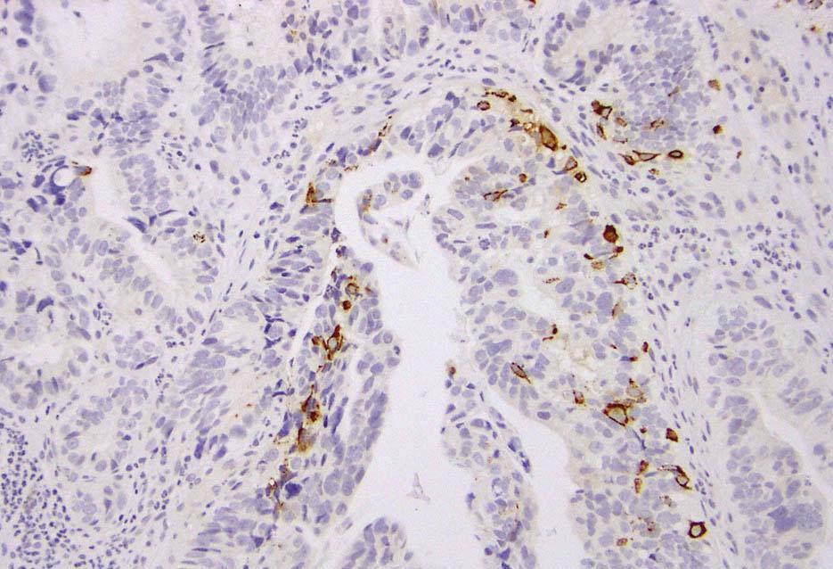
43 Chromogranin
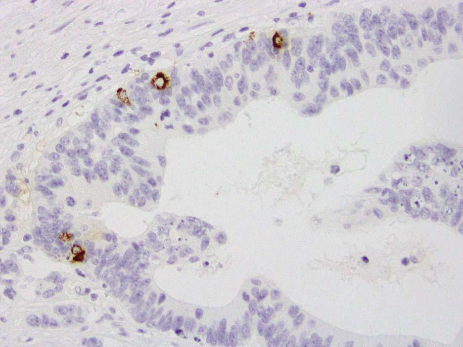
44 Chromogranin
45 Neuroendocrine Differentiation in Carcinomas: Treatment Implications Small cell carcinoma (lung or extrapulmonary) Platinum + etoposide Large cell neuroendocrine carcinoma Commonly treated like small cell carcinoma Few compelling studies; no randomized trials Carcinoma with neuroendocrine morphology / differentiation / features / minor elements / etc. Who know????
46 Neuroendocrine Neoplasms: Determination of Grade, Classification, and Prognosis
47 WHO 2010 Grading of GEP-NETs Grade Mitoses Ki-67 Index G1 < 2 / 10 H.P.F. < 3% G / 10 H.P.F. 3-20% G3 > 20 / 10 H.P.F. > 20% Poorly Differentiated (High Grade ) Neuroendocrine Carcinoma
48 ENETS/WHO Grading of GEP-NETs: Provisions Count mitoses in 50 high power fields Assess Ki67 based on counting 2000 (500) cells Assess Ki67 in hot spots If mitotic rate and Ki67 are discordant, assign higher grade
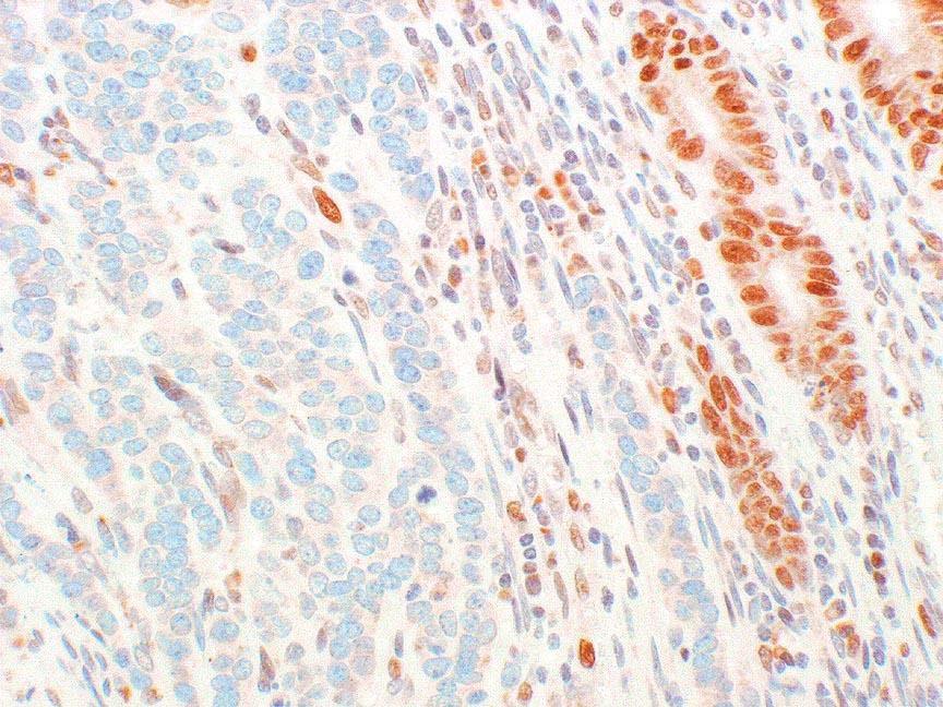
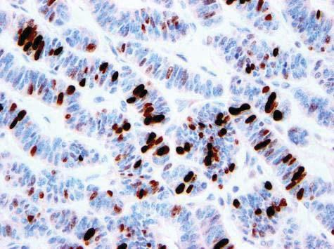
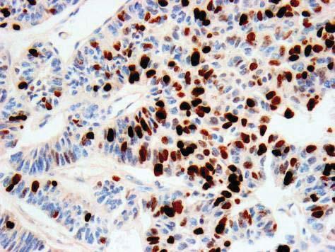
49 Ki67
50 Issues with Grading GEP-NETs Ki67 assessment Intratumoral and intertumoral heterogeneity Discordance between Ki67 and mitotic rate Distinction of well differentiated vs. poorly differentiated neoplasms

 Eyeballed estimate")
51 Ki67 Labeling Index of NETs Strong predictor of prognosis Basis for grading systems Correlates well with mitotic index Sharp separation of well and poorly differentiated neuroendocrine neoplasms Methods of Assessment Manual counting (2000 cells per ENETS) Eyeballed estimate Digital image analysis


52 Digital Image Analysis for Ki67 Quantification Ki67% = 1.7
53 Correlation between Digital Image Analysis and Manual Cell Count (2000 cells) ICC, 0.981; CI, Tang et al. Am J Surg Pathol 2012; 36:
54 Consistency of Ki67 Determination by Digital Image Analysis, Manual Cell Counting, and Eyeballed Estimate Image Analysis vs. Manual Counting Image Analysis vs. Eyeballed Estimate (Mean of 20 observers) Eyeballed Estimate Interobserver (n=20) Intraclass Correlation (ICC) 95% Confidence Interval Tang et al. Am J Surg Pathol 2012; 36:

55 Courtesy of Dr. Laura H. Tang Determining the Ki67 Labeling Index of NETs: How We Do It


56 Phosphohistone H3 (PHH3): an Immunohistochemical Marker of Mitoses Close correlation between phosphorylation on histone h3 and mitotic chromosome condensation Highlights mitotic figures Distinguishes from pyknotic nuclei More sensitive and specific than h&e counting Allows hot spot detection Potential for automated counting Percent of cells in mitoses, versus number per unit area Tsuta et al. am j clin pathol 2011; 136: Fung et al. acta cytol 2013; 57: Yang et al. am j surg pathol 2015; 39: 13-24
57 Ki67 Ki67 Heterogeneity in PanNETs


58 Heterogeneity of Ki67 Labeling in NETs: Impact on Prognostic Significance of Grading Ki67 on virtual biopsies and on whole sections Virtual biopsy TMA 45 resected hepatic metastases of WD NETs Yang et al., Am J Surg Pathol 2011; 35:853-60
59 Heterogeneity of Ki67 Labeling in NETs: Impact on Prognostic Significance of Grading 47% of cases with G1 vs. G2 heterogeneity Define grade based on highest Ki67 on whole sections: G2 identified in 48% of core biopsies (3 cores) G2 identified in 35% of core biopsies (1 core) Predictive value of G1 on core biopsy: 65% (3 cores); 59% (1 core)
60 Survival based on Ki67 Labeling Core Biopsies OS DFS PFS Three core Cum. Survival G2 1 1 p< p=0.002 p< G Time Cum. Survival G2 G Time Cum. Survival G2 G Time Single core Cum. Survival G Time Cum. Survival 1 p= p< p< G G2 G Time Cum. Survival G2 G Time

 Ki-67: 15%")
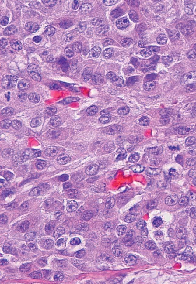
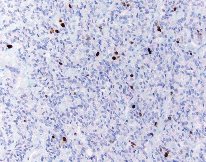
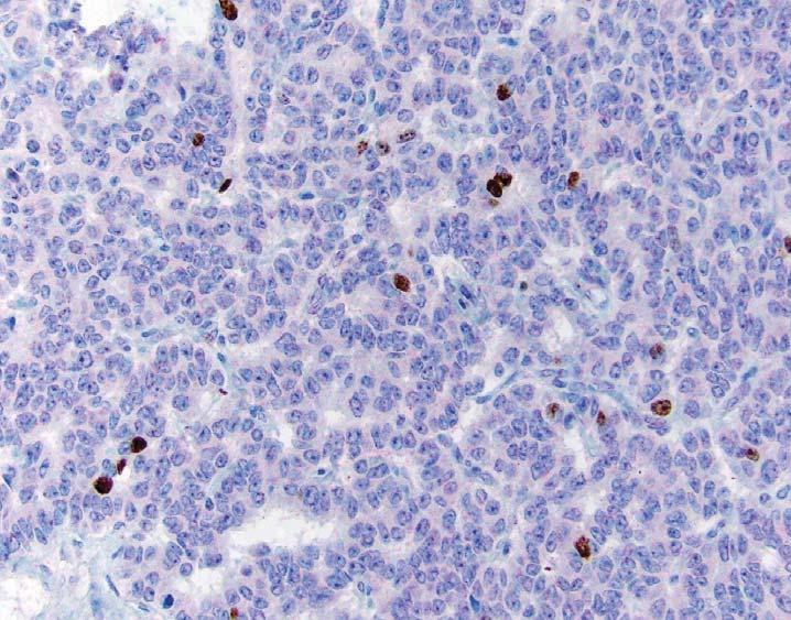
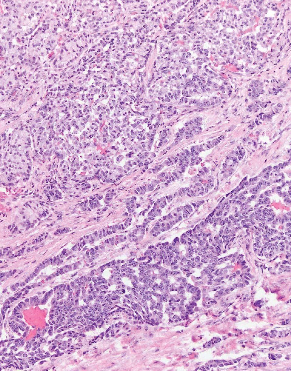
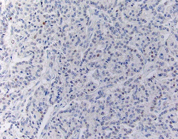
61 Ki67 and Mitotic Rate Discordance in PanNETs Mitotic rate: <1 per 10 hpf (G1) Ki-67: 15% positive (G2)
62 Ki67 and Mitotic Rate Discordance in PanNETs 297 WD PanNETs with Ki-67 data ( ) 36% discordance 264 Mitotic G1 33 Mitotic G2 165 Ki-67 G1 99 Ki-67 G2 8 Ki-67 G1 25 Ki-67 G2 McCall et al., Am J Surg Pathol 2013; 37:
63 Ki67 and Mitotic Rate Discordance in PanNETs 297 WD PanNETs with Ki-67 data ( ) 36% discordance 264 Mitotic G1 33 Mitotic G2 165 Ki-67 G1 99 Ki-67 G2 8 Ki-67 G1 25 Ki-67 G2 McCall et al., Am J Surg Pathol 2013; 37:
64 Ki-67 G2/mitotic G1 PanNETs have decreased overall survival Percentage Surviving K1M1 K2M1 p < Survival in Years
65 Ki-67 G2/mitotic G1 PanNETs are not significantly different from concordant G2 Percentage Surviving K2M1 K2M2 p = Survival in Years
66 What about G2 / G3 discordance?? (well differentiated vs. poorly differentiated)
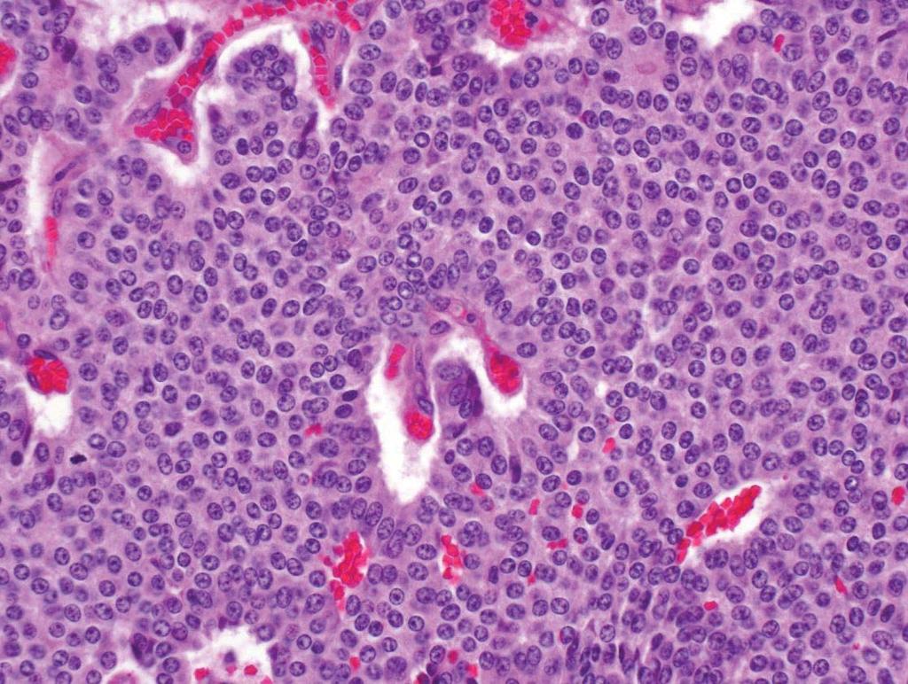
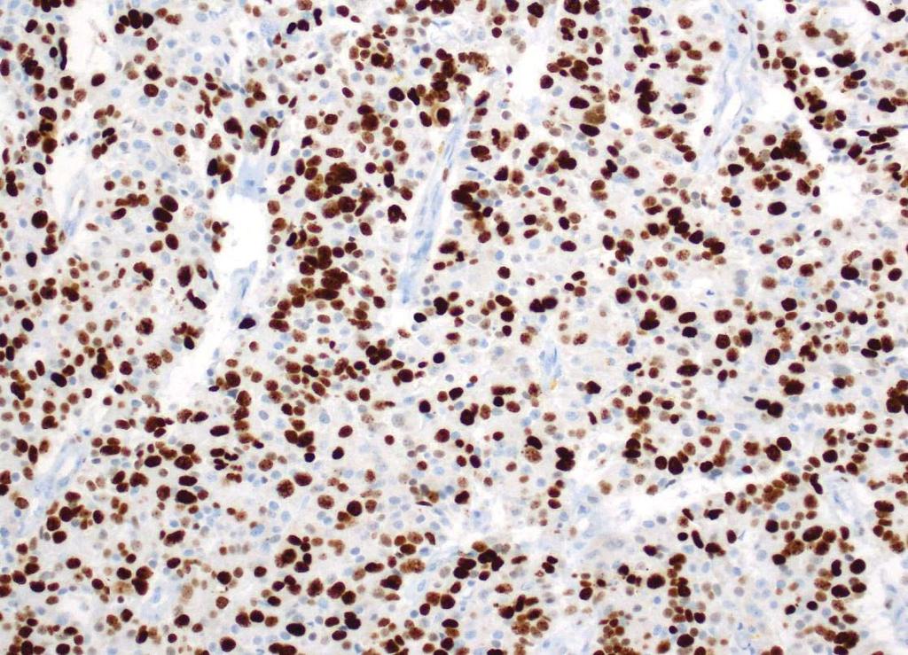
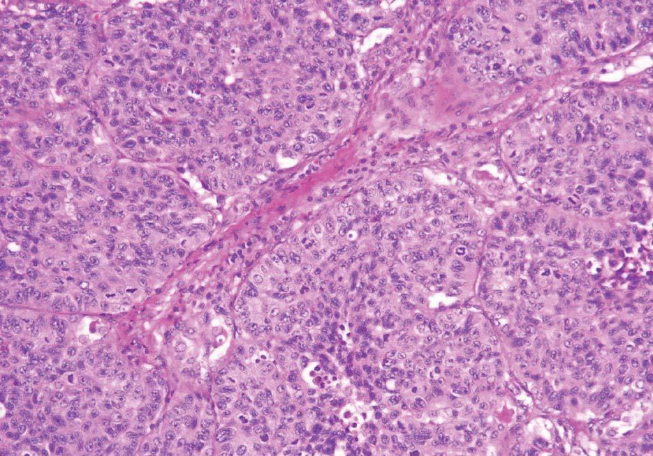
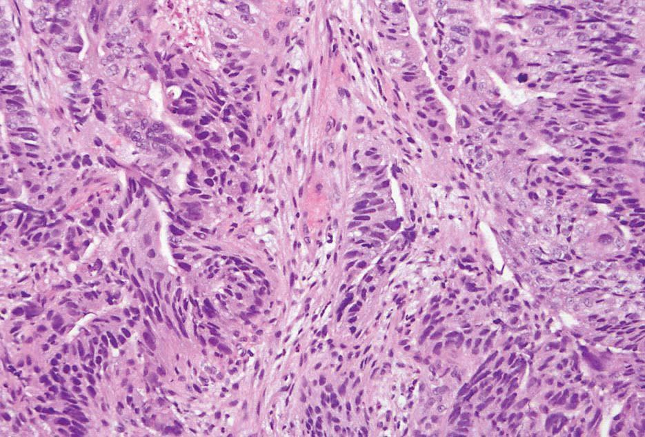
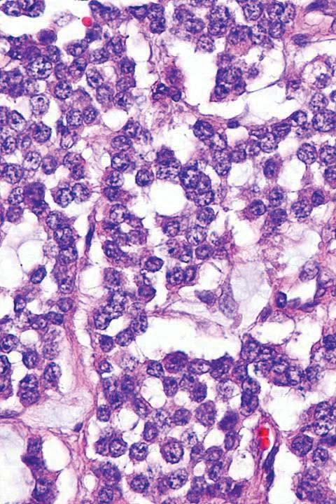
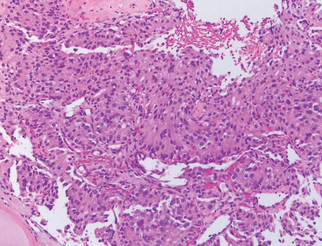
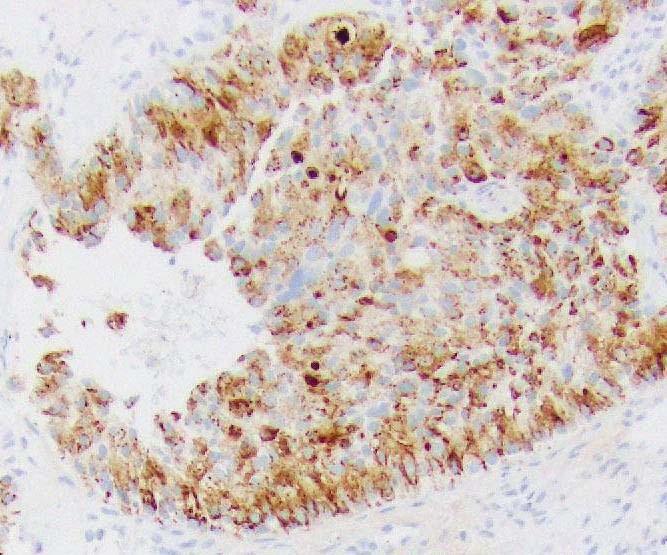
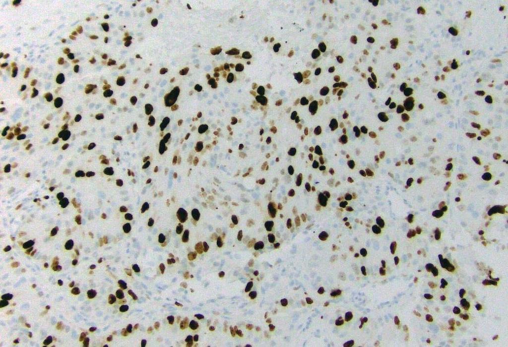
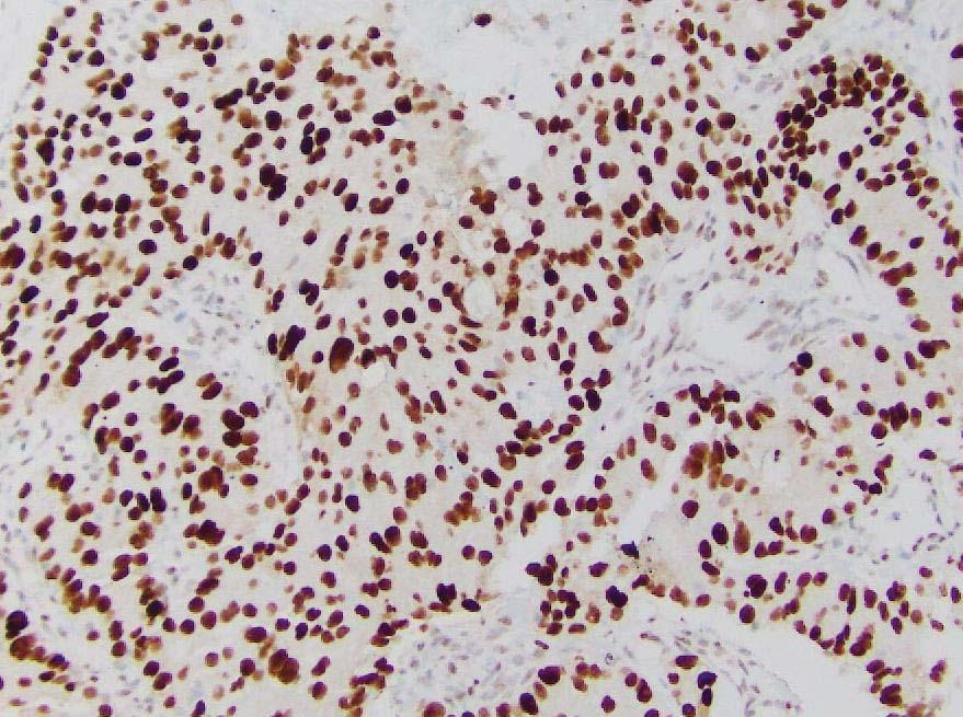
67 Well Differentiated PanNET Mitotic rate = 8 / 10 HPF Mitotic rate = 12 / 10 HPF Ki67 = 45% Ki67 = 55%


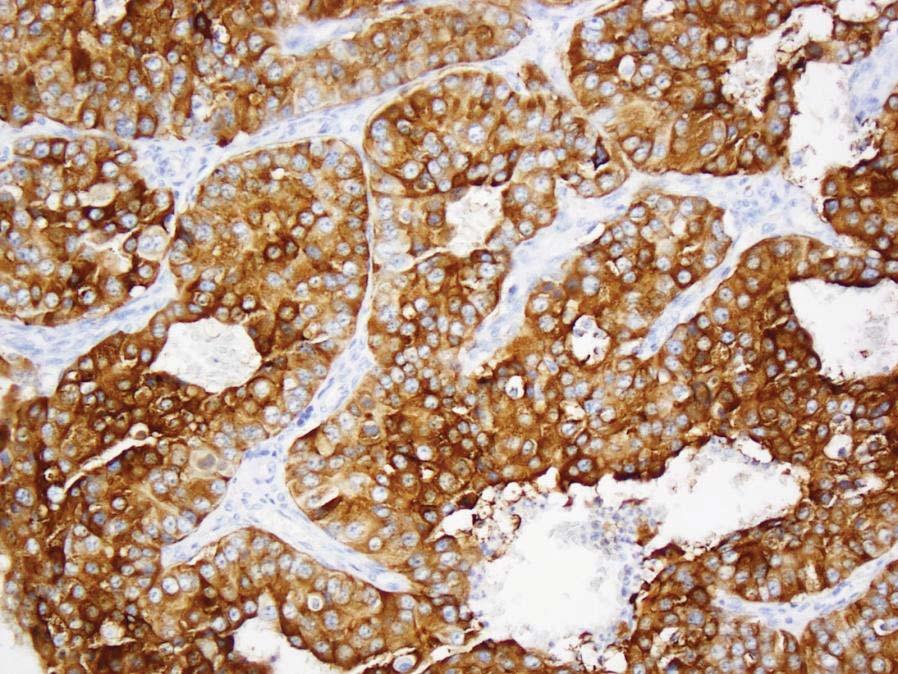
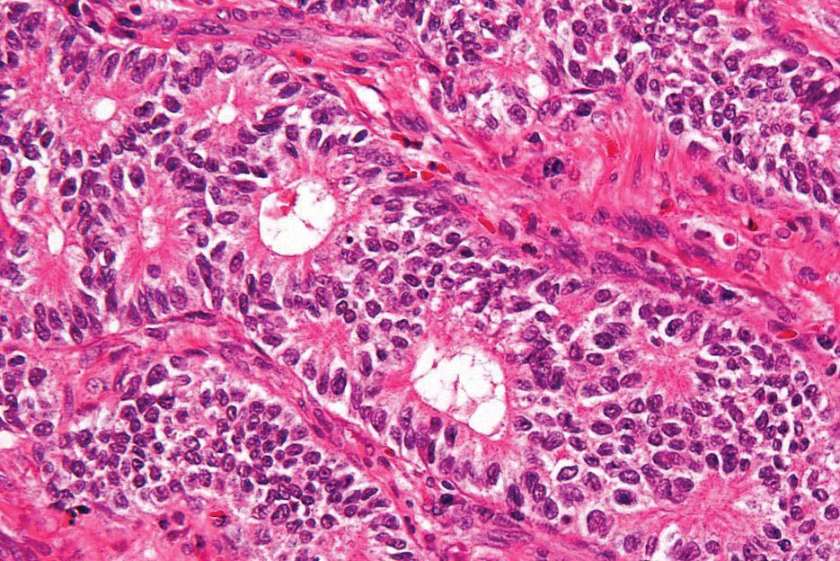
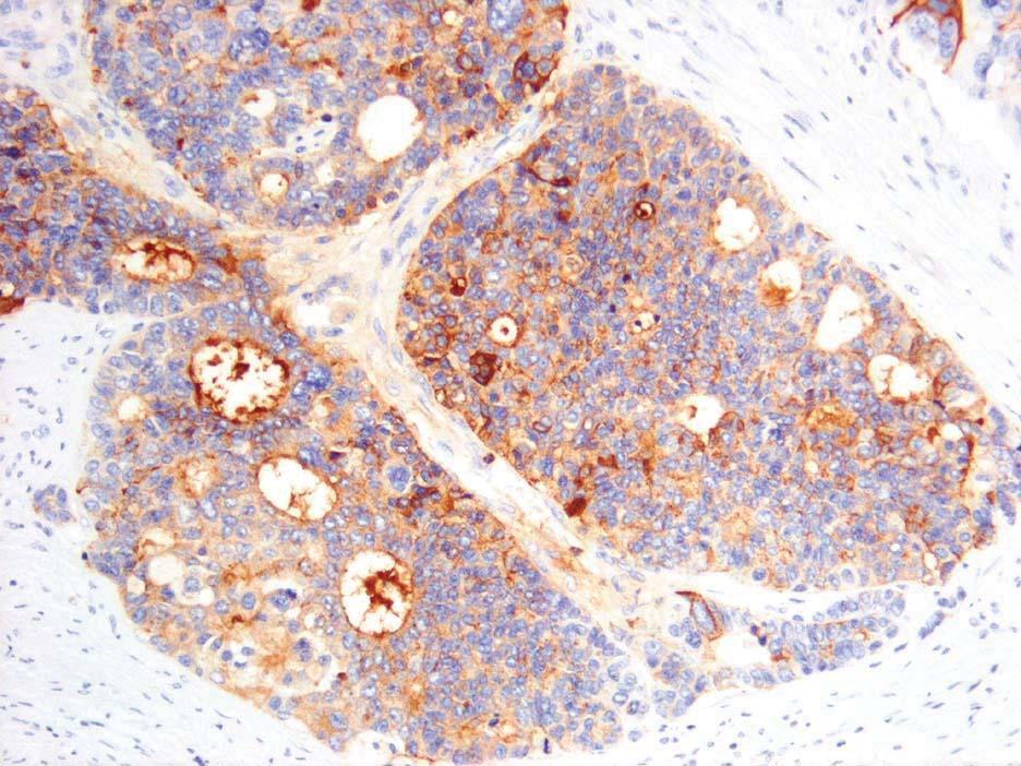
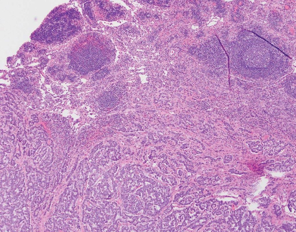
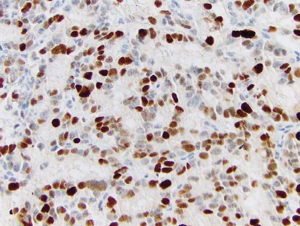
68 Poorly Differentiated Neuroendocrine Carcinoma Chromogranin
69 Are all G3 Neuroendocrine Neoplasms the Same? NO! Small cell carcinoma vs. Large cell NE carcinoma Large cell NE carcinoma vs. G3 well differentiated NET NEC G3 vs. NET G3


 NE")
70 WD NE Tumor PD NE Carcinoma Carcinoma Stable Disease NE Tumor Lower Grade NE Carcinoma High Grade Grade Progression High Grade Disease Progression Rapid Disease Progression, Death Two Pathways to the Development of High Grade (G3) NE Neoplasms


71 Whole Exome Sequencing of PanNETs: Three Mountains 1. MEN1 inactivation 44% Previously known 2. DAXX/ATRX mutations 43% DAXX = death-domain-associated protein, Chr 6p ATRX = α thalassemia/mental retardation syndrome X-linked Together form a complex Both required for H3.3 incorporation in telomeres Mutually exclusive 3. mtor pathway 15% PTEN 7.3% TSC2 8.8% PIK3CA 1.4% ATRX Jiao et al. Science 2011; 331:
72 PanNET vs. Ductal Adenocarcinoma Genes PanNET Adenocarcinoma KRAS 0% 100% TP53 4% 75% CDKN2A 0% 95% SMAD4 0% 55% MEN1 44% 0% DAXX, ATRX 43% 0% Genes in mtor pathway 15% 1% Jones et al., Science 2008; 321: Jiao et al., Science 2011; 331: 1199.
73 PanNET vs. Ductal Adenocarcinoma Genes PanNET Adenocarcinoma KRAS 0% 100% TP53 4% 75% CDKN2A 0% 95% SMAD4 0% 55% MEN1 44% 0% DAXX, ATRX 43% 0% Genes in mtor pathway 15% 1% Jones et al., Science 2008; 321: Jiao et al., Science 2011; 331: 1199.

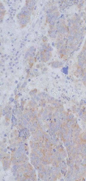
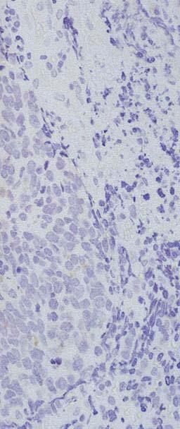
74 Genetics of Poorly Differentiated Neuroendocrine Carcinoma of Pancreas Gene Small Cell Large Cell NEC W.D. PanNET Ductal ACa Small Cell Lung CA KRAS 25% 33% 0% >90% 0-10% p16 11% 50% 0% 80-95% 0-10% p53 100% 90% 4% 75% 80% Smad4 0% 10% 0% 55% 0% Rb 89% 50% 0% 13% 90% DAXX/ATRX 0% 0% 45% 0% Rb Yachida et al., Am J Surg Pathol 2012; 36: 173 Large Cell Neuroendocrine Carcinoma
75 Basturk et al., Am J SurgPathol 2015; 39:
76 Predictive and prognostic factors for treatment and survival in 305 patients with advanced gastrointestinal neuroendocrine carcinoma (WHO G3) Reviewed clinical data on advanced stage G3 NECs, Ki67 > 20% 252 patients received chemotherapy (platinumbased) Median survival = 11 mos. Response rate = 31% Stable disease rate = 33% Ki67 < 55% predicted a lower response rate (15% vs 42%, p < 0.001) Ki67 < 55% predicted a better survival (14 vs 10 months, P < 0.001) Sorbye et al., Ann Oncol 2013; 24:
77 Conclusion: Some G3 NETs with Ki % may be well differentiated biologically!! ( Well Differentiated NET with an Elevated Proliferative Rate or Well Differentiated NET, G3 )
78 Grading of Pancreatic Neuroendocrine Neoplasms (WHO 2017) Well differentiated NE tumor* Grade Mitoses Ki-67 Index G1 <2 / 10 HPF </= 2% G / 10 HPF 3-20% G3** >20 / 10 HPF >20% *Organoid architecture, well differentiated cytology, absence of nonneuroendocrine carcinoma components, may have components of G1 or G2, usually strong immunoexpression of general NE markers **mitoses usually <20/HPF; Ki 67 >20% but usually <50% Poorly differentiated NE carcinoma* Grade Mitoses Ki-67 Index G3** >20 / 10 HPF >20% *Small cell carcinoma and large cell NE carcinoma; less organoid architecture, classic cytology of small cell and large cell NE CA, absence of G1 or G2 NE components, may have nonneuroendocrine carcinoma components, less diffuse immunoexpression of general NE markers **mitoses >20/10 HPF; Ki67 >20% and usually >50%
79 G1 G2 G3 G3 WDNET PDNEC Ki67%
 from G3")
80 How to distinguish G3 NEC (esp. large cell NE carcinoma) from G3 NET?


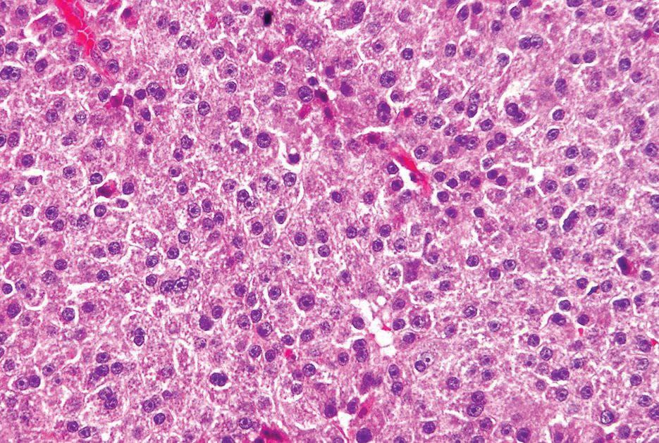
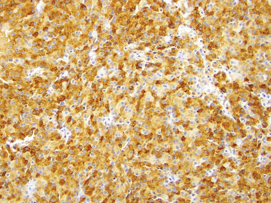
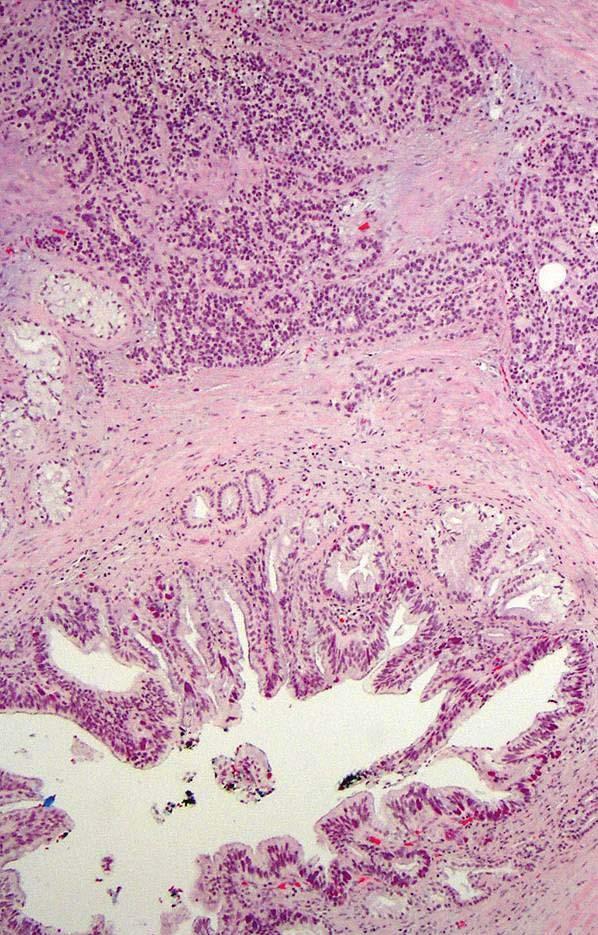
81 Pancreatic G3 NE Neoplasms Large Cell NEC G3 NET
82 How to distinguish G3 NEC (esp. large cell NE carcinoma) from G3 NET? Clinical clues History of well differentiated NET? Octreotide scan positive? FDG-PET positive? Morphologic clues Lower grade component? Non-neuroendocrine component? Mitotic rate? Molecular clues Status of TP53, RB1, DAXX, ATRX, MEN1
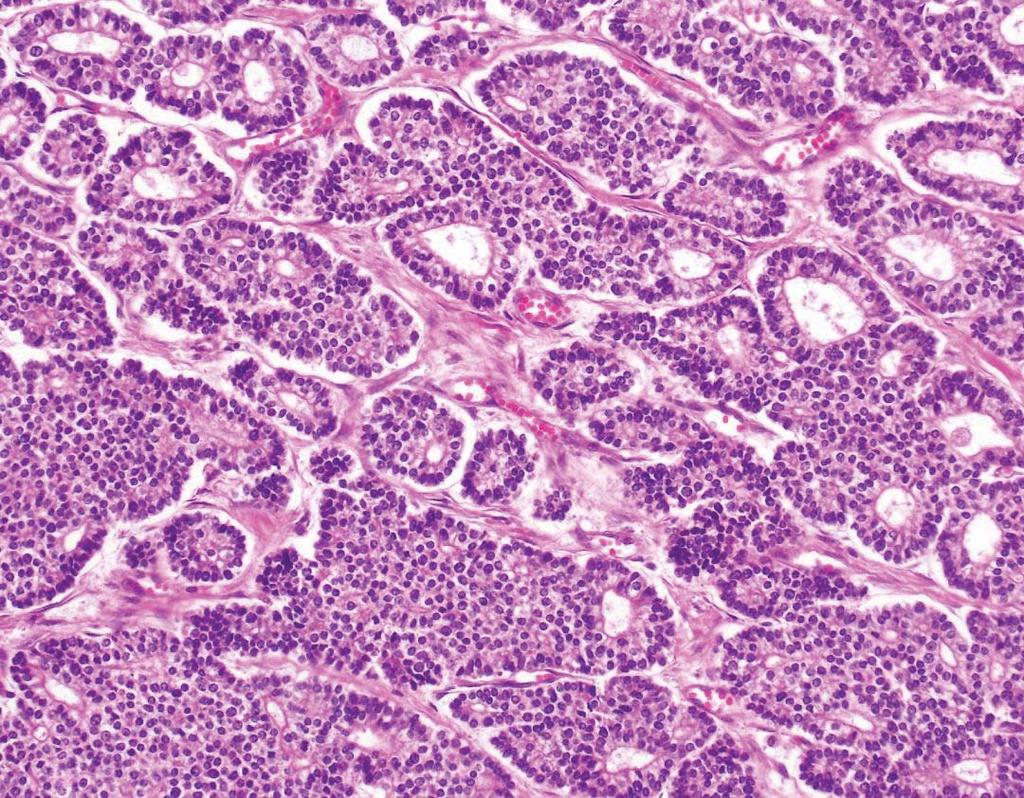
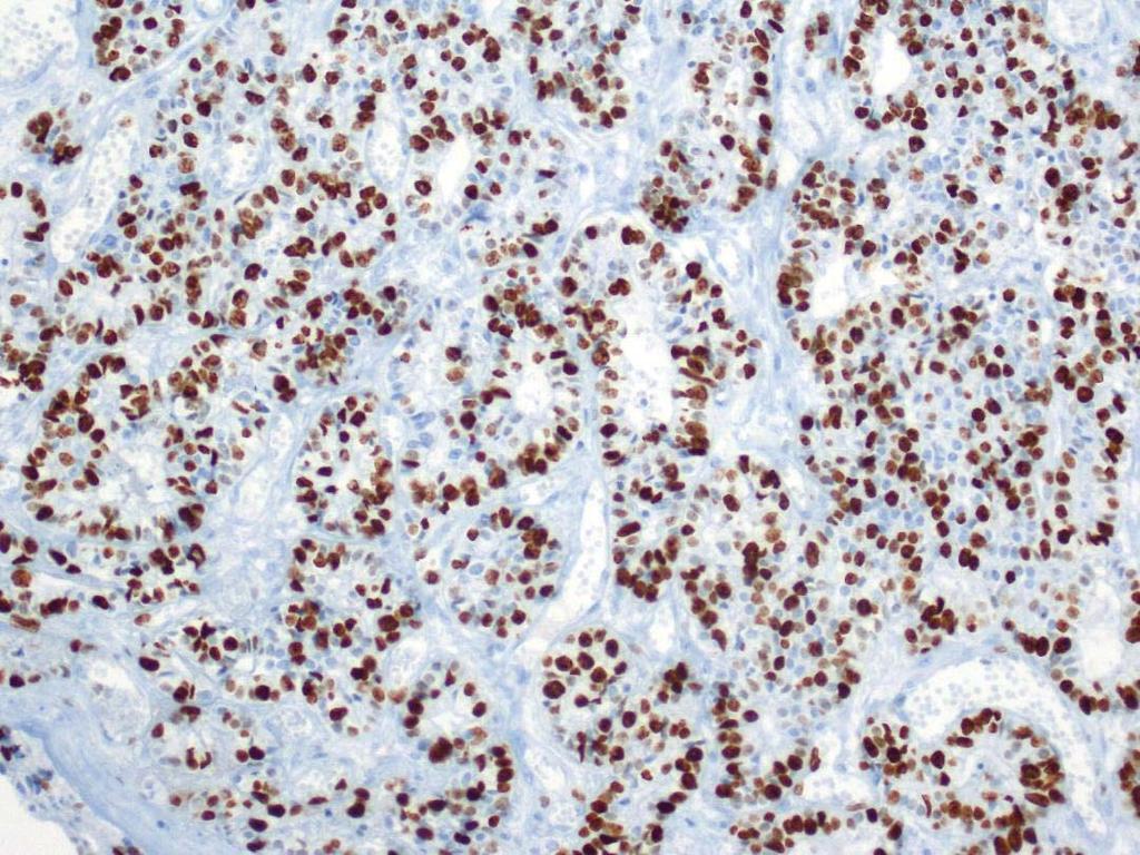
83 Ki67 Ki67 Heterogeneity in PanNETs
84

85 Mitosis <1/10 HPF Mitosis 13/10 HPF

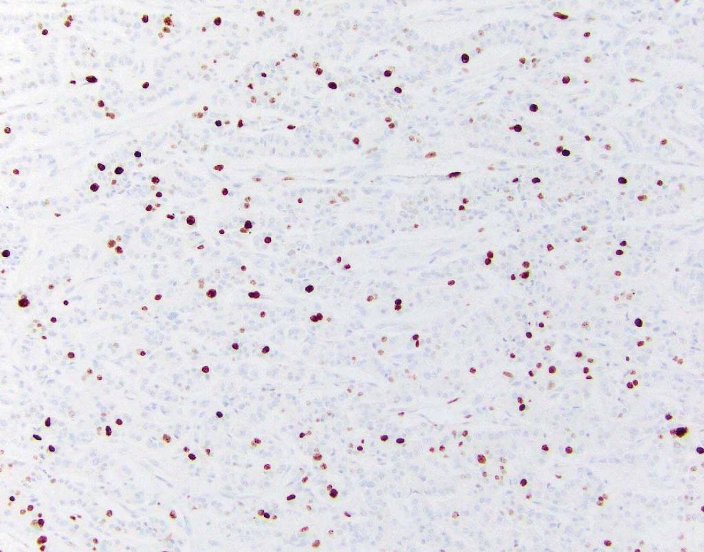
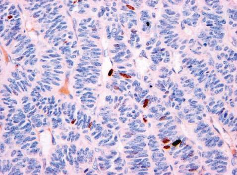
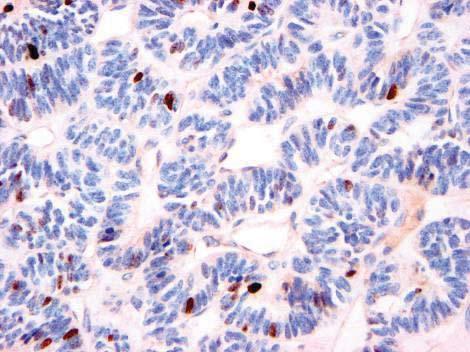
86 Ki67 = 2% G1 Ki67 = 45% G3
87 Progression of Low Grade to High Grade Neuroendocrine Tumors Fits idea of general concept of neoplastic progression Sites Pancreas 21, small bowel 6, bile duct 2, rectum 2 Progression is NOT to poorly differentiated NEC Rarely (?ever) gives rise to small cell carcinoma Present in primary tumor OR upon disease progression Biological behavior: Tang et al., Clin Cancer Res 2016; 22: 1011
88 n=35 n=21 n=329 Tang et al., Clin Cancer Res 2016; 22: 1011

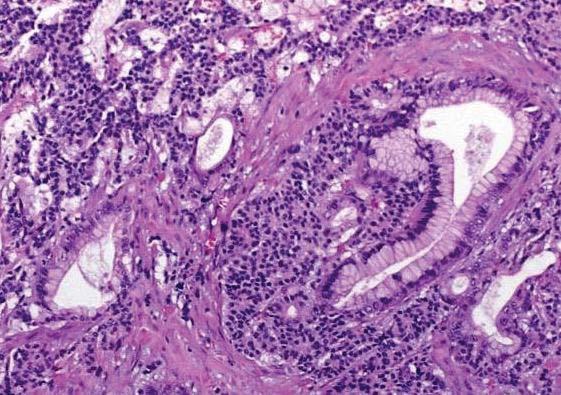
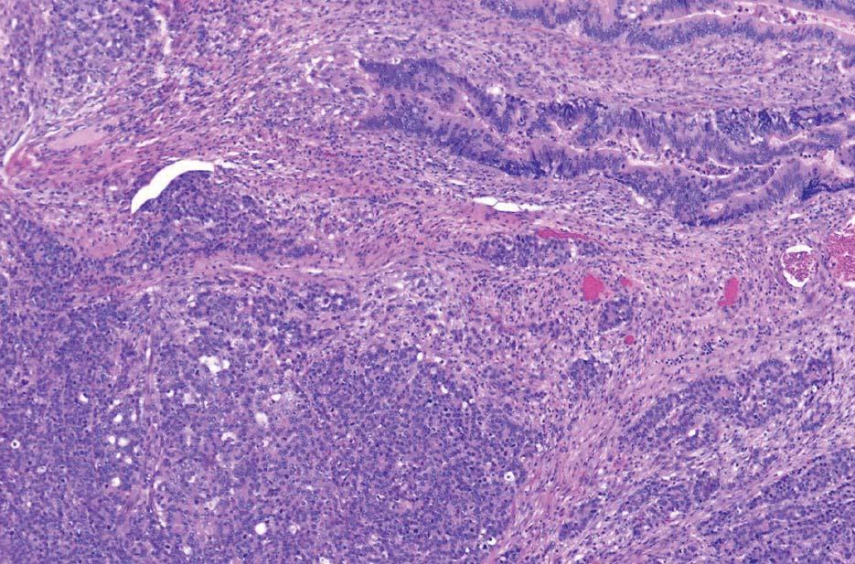
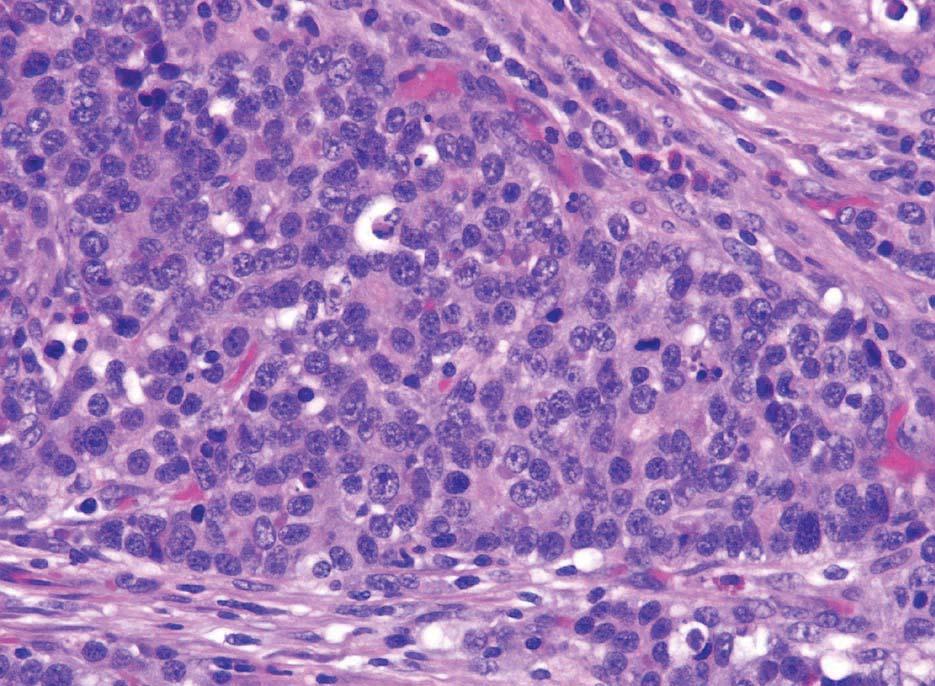
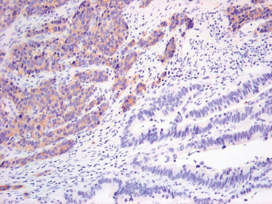
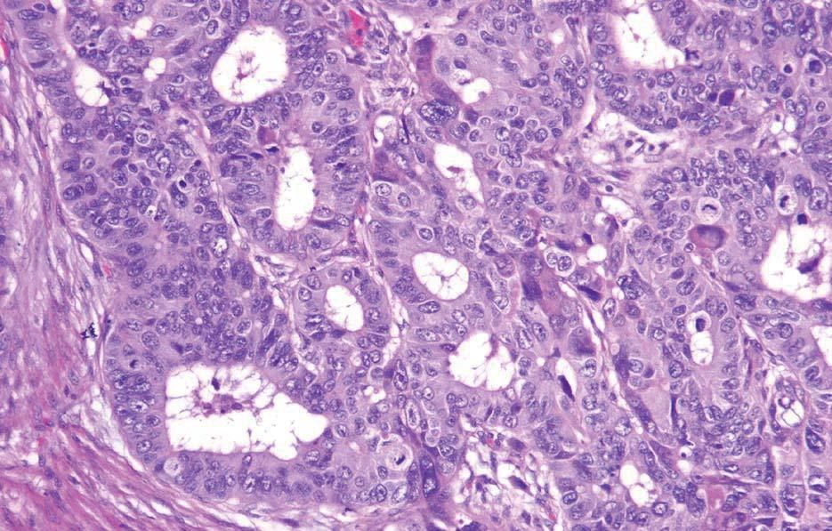
89 Mixed Ductal Neuroendocrine Carcinoma of Pancreas

90 Tubular GI Tract: Poorly Differentiated NEC


91 Ampulla of Vater: Small Cell Carcinoma
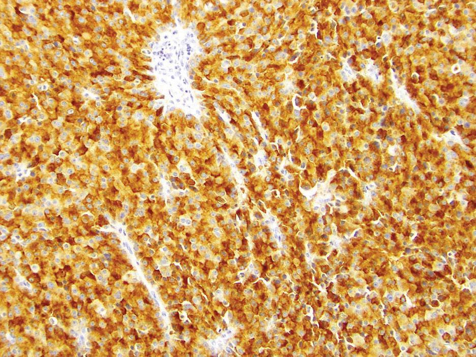
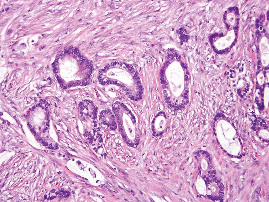
92 Well Differentiated PanNETs (G1-3) Exhibit a Different Molecular Phenotype from Poorly Differentiated NECs (G3) WD- PanNET TP53 RB1 SMAD4 DAXX / ATRX MEN1 4% % 44% PD-PanNEC 56% 72%?? (adeno, 55%) 0 0 Jiao et al. Science 2011; 331: 1199 Yachida et al., Am J Surg Pathol 2012; 36: 173

93 PD-NEC PD-NEC p53 Rb PD-NEC WD-NET SMAD4 DAXX Tang et al., Am J Surg Pathol 2016; 40: 1192
94 Morphologic Assessment of High Grade Pancreatic Neuroendocrine Neoplasms Tang et al., Am J Surg Pathol 2016; 40: 1192 Consensus Reviewer 1 Reviewer 2 Reviewer 3 Specimen Type WD-NET WD-NET WD-NET WD-NET Resection WD-NET WD-NET WD-NET WD-NET Resection WD-NET WD-NET WD-NET WD-NET Resection WD-NET WD-NET WD-NET WD-NET Resection WD-NET WD-NET WD-NET WD-NET Resection WD-NET WD-NET WD-NET WD-NET Resection Ambiguous WD-NET Uncertain WD-NET Biopsy Ambiguous WD-NET WD-NET Uncertain Resection Ambiguous Uncertain WD-NET WD-NET Biopsy Ambiguous WD-NET WD-NET Uncertain Resection Ambiguous WD-NET WD-NET Uncertain Resection Ambiguous WD-NET WD-NET Uncertain Resection Ambiguous WD-NET WD-NET Uncertain Biopsy Ambiguous WD-NET WD-NET PD-NET-LCC Resection Ambiguous WD-NET WD-NET PD-NET-LCC Biopsy Ambiguous Uncertain Uncertain Uncertain Biopsy Ambiguous Uncertain Uncertain PD-NEC-SCC Resection Ambiguous PD-NEC-SCC Uncertain PD-NEC-SCC Resection PD-NEC-LCC PD-NEC-LCC PD-NEC-LCC PD-NEC-LCC Resection Ambiguous Uncertain Uncertain Uncertain Biopsy PD-NEC-LCC PD-NEC-LCC PD-NEC-LCC PD-NEC-LCC Resection PD-NEC-LCC PD-NEC-LCC PD-NEC-LCC PD-NEC-LCC Resection PD-NEC-SCC PD-NEC-SCC PD-NEC-SCC PD-NEC-SCC Resection PD-NEC-SCC PD-NEC-SCC PD-NEC-SCC PD-NEC-SCC Resection PD-NEC-SCC PD-NEC-SCC PD-NEC-SCC PD-NEC-SCC Resection PD-NEC PD-NEC-LCC PD-NEC-SCC PD-NEC-LCC Resection PD-NEC PD-NEC-SCC PD-NEC-SCC PD-NEC-LCC Resection Ambiguous WD-NET PD-NEC-LCC PD-NEC-LCC Resection Ambiguous PD-NEC-LCC PD-NEC-LCC Uncertain Resection Ambiguous Uncertain Uncertain PD-NEC-SCC Resection Ambiguous Uncertain PD-NEC-SCC Uncertain Biopsy Ambiguous Uncertain PD-NEC-LCC Uncertain Biopsy Ambiguous Uncertain PD-NEC-LCC PD-NEC-LCC Resection
95 Classification of High Grade Pancreatic Neuroendocrine Neoplasms by Secondary Evidence Initial Consensus Tang et al., Am J Surg Pathol 2016; 40: 1192 Immunohistochemical Abnormalities Other Histologic Components Confirmed Classification WD-NET G1/G2 WD-NET WD-NET WD-NET DAXX G1/G2 WD-NET WD-NET WD-NET ATRX G1/G2 WD-NET WD-NET WD-NET G1/G2 WD-NET WD-NET WD-NET DAXX G1/G2 WD-NET WD-NET WD-NET G1/G2 WD-NET WD-NET Ambiguous G1/G2 WD-NET WD-NET Ambiguous G1/G2 WD-NET WD-NET Ambiguous DAXX G1/G2 WD-NET WD-NET Ambiguous ATRX G1/G2 WD-NET WD-NET Ambiguous DAXX G1/G2 WD-NET WD-NET Ambiguous G1/G2 WD-NET WD-NET Ambiguous ATRX WD-NET Ambiguous DAXX G1/G2 WD-NET WD-NET Ambiguous DAXX G1/G2 WD-NET WD-NET Ambiguous G1/G2 WD-NET WD-NET Ambiguous G1/G2 WD-NET WD-NET Ambiguous G1/G2 WD-NET WD-NET Ambiguous G1/G2 WD-NET WD-NET Ambiguous p53/rb PD-NEC Ambiguous p53/smad4 Ductal adenocarcinoma PD-NEC Ambiguous p53/rb PD-NEC Ambiguous p53/rb PD-NEC Ambiguous p53 PD-NEC Ambiguous Undetermined PD-NEC-LCC DAXX G1/G2 WD-NET WD-NET PD-NEC-LCC Rb PD-NEC PD-NEC-LCC Ductal adenocarcinoma PD-NEC PD-NEC-SCC p53 Ductal adenocarcinoma PD-NEC PD-NEC-SCC Rb PD-NEC PD-NEC-SCC p53/rb Ductal adenocarcinoma PD-NEC PD-NEC Rb PD-NEC PD-NEC p53 PD-NEC
96 Disease Specific Survival of High Grade (G3) Pancreatic Neuroendocrine Neoplasms 100 Percent survival WD-NET PD-NEC p< (N=20) (N=12) Months Tang et al., Am J Surg Pathol 2016; 40: 1192


97 Sequencing of Pancreatic Neuroendocrine Neoplasms at MSKCC
98 Chromogranin A Ki67 (45%) p53

99 Ki67 p53 Ki67 p53
100 p53
101 Distinction of G3 NEC from G3 NET: Practical Issues Primary site Pancreas Most common DAXX/ATRX, MEN1 Other GI / pulmonary NETs WD G3 NETs uncommon Formal WHO classification pending p53, Rb, associated exocrine elements for PD Morphology for WD Role of Ki67 >50% = usually PD NEC <50% = either WD NET or PD NEC Role of mitotic rate <20 per 10 HPF = WD NET >20 per 10 HPF = PD NEC
102 Use of Immunohistochemistry in Neuroendocrine Neoplasms: Conclusions IHC is needed for neuroendocrine neoplasm diagnosis, classification, and grading Limitations exist in interpretation and significance PET-CT Exercise pragmatism, not nihilism IHC is just one tool in the diagnostic arsenal use morphology, clinical findings, molecular data, and common sense!
3/23/2017. Differentiation: Differentiation: Immunohistochemistry. Well Differentiated vs. Poorly Differentiated Neuroendocrine Neoplasms
 Disclosure Statement When Immunostains Can Get You Into Trouble (and how they can help you out): Neuroendocrine Neoplasms Arthur Purdy Stout Society March 5, 2017 Dr. Klimstra receives royalty payments
Pancreatic Cancer: The ABCs of the AJCC and WHO
 Pancreatic Cancer: The ABCs of the AJCC and WHO Aatur D. Singhi, MD PhD Assistant Professor University of Pittsburgh Medical Center Department of Pathology singhiad@upmc.edu Case presentation Objectives
3/22/2017. Disclosure of Relevant Financial Relationships. Ki-67 in Pancreatic Neuroendocrine Neoplasms According to WHO 2017.
 Disclosure of Relevant Financial Relationships Ki-67 in Pancreatic Neuroendocrine Neoplasms According to WHO 2017. USCAP requires that all planners (Education Committee) in a position to influence or control
LUNG CANCER PATHOLOGY: UPDATE ON NEUROENDOCRINE LUNG TUMORS
 LUNG CANCER PATHOLOGY: UPDATE ON NEUROENDOCRINE LUNG TUMORS William D. Travis, M.D. Attending Thoracic Pathologist Memorial Sloan Kettering Cancer Center New York, NY PULMONARY NE TUMORS CLASSIFICATION
Neuroendocrine tumors of GI and Pancreatobiliary tracts. N. Volkan Adsay, MD
 Neuroendocrine tumors of GI and Pancreatobiliary tracts N. Volkan Adsay, MD New (2017) WHO WHO 2017 (endocrine book; for pancreas) WHO 2017 (endocrine book; for pancreas) PD-NE ca WD-NE Tumor Intended
Neuroendocrine neoplasms of the lung
 Neuroendocrine neoplasms of the lung M Papotti, L Righi, & M Volante University of Turin at San Luigi Hospital TORINO NETs OF THE LUNG Menu - Spectrum of NE lung tumors - CARCINOID TUMORS - SCLC /LCNEC
Neuroendocrine Lung Tumors Myers
 Diagnosis and Classification of Neuroendocrine Lung Tumors Jeffrey L. Myers, M.D. A. James French Professor Director, Anatomic Pathology & MLabs University of Michigan, Ann Arbor, MI myerjeff@umich.edu
Problem 1: Differential of Neuroendocrine Carcinoma 3/23/2017. Disclosure of Relevant Financial Relationships
 Differential of Neuroendocrine Carcinoma Alain C. Borczuk,MD Weill Cornell Medicine Disclosure of Relevant Financial Relationships USCAP requires that all faculty in a position to influence or control
Gastrointestinal Neuroendocrine Tumors: A Closer Look at the Characteristics of These Diverse Tumors
 Gastrointestinal Neuroendocrine Tumors: A Closer Look at the Characteristics of These Diverse Tumors Jaume Capdevila, MD, PhD Vall d'hebron University Hospital Vall d'hebron Institute of Oncology (VHIO)
Difficult Diagnoses and Controversial Entities in Neoplastic Lung
 Difficult Diagnoses and Controversial Entities in Neoplastic Lung Lynette M. Sholl, M.D. Associate Pathologist, Brigham and Women s Hospital Chief, Pulmonary Pathology Service Associate Professor, Harvard
LUNG CANCER. pathology & molecular biology. Izidor Kern University Clinic Golnik, Slovenia
 LUNG CANCER pathology & molecular biology Izidor Kern University Clinic Golnik, Slovenia 1 Pathology and epidemiology Small biopsy & cytology SCLC 14% NSCC NOS 4% 70% 60% 50% 63% 62% 61% 62% 59% 54% 51%
Insulinoma-associated protein (INSM1) is a sensitive and specific marker for lung neuroendocrine tumors in cytologic and surgical specimens
 is a sensitive and specific marker for lung neuroendocrine tumors in cytologic and surgical specimens") Insulinoma-associated protein (INSM1) is a sensitive and specific marker for lung neuroendocrine tumors in cytologic and surgical specimens Kartik Viswanathan, M.D., Ph.D New York Presbyterian - Weill
Insulinoma-associated protein (INSM1) is a sensitive and specific marker for lung neuroendocrine tumors in cytologic and surgical specimens Kartik Viswanathan, M.D., Ph.D New York Presbyterian - Weill
Objectives. Terminology 03/11/2013. Pitfalls in the diagnosis of Gastroenteropancreatic Neuroendocrine Tumors. Pathology Update 2013
 Pitfalls in the diagnosis of Gastroenteropancreatic Neuroendocrine Tumors Pathology Update 2013 Ozgur Mete, MD Consultant in Endocrine Pathology, Department of Pathology, University Health Network Assistant
Pancreatic Cytopathology: The Solid Neoplasms
 Pancreatic Cytopathology: The Solid Neoplasms Syed Z. Ali, M.D. Professor of Pathology and Radiology Director of Cytopathology The Johns Hopkins Hospital Baltimore, Maryland Pancreatic Cytopathology: Past,
SCOPE TODAYS SESSION. Case 1: Case 2. Basic Theory Stuff: Heavy Stuff. Basic Questions. Basic Questions
 MONDAY TEACHING SCOPE TODAYS SESSION Case 1: Basic Questions Case 2 Basic Questions Basic Theory Stuff: AJCC TNM + Stage Group for Carcinoid of the Appendix Management of Carcinoid of the Appendix (NCCN)
Hot of the press. Γρηγόριος Καλτσάς MD FRCP Καθηγητής Παθολογίας Ενδοκρινολογίας ΕΚΠΑ
 Hot of the press Γρηγόριος Καλτσάς MD FRCP Καθηγητής Παθολογίας Ενδοκρινολογίας ΕΚΠΑ Outline Diagnostic developments Histopathology Molecular Therapeutic developments Results on PRRT Telotristat in carcinoid
Prognostic factors and treatment of gastroenteropancreatic G3 neuroendocrine carcinomas.
 Prognostic factors and treatment of gastroenteropancreatic G3 neuroendocrine carcinomas. Halfdan Sorbye Medical Oncologist Professor, MD Dept of Oncology Haukeland Univ Hospital Bergen, Norway Gastroenteropancreatic
Lung neuroendocrine tumors: pathological characteristics
 Review Article Lung neuroendocrine tumors: pathological characteristics Luisella Righi 1, Gaia Gatti 1, Marco Volante 1, Mauro Papotti 2 1 Department of Oncology, San Luigi Hospital, Orbassano, Italy;
Small cell neuroendocrine carcinoma icd 10
 Small cell neuroendocrine carcinoma icd 10 1-10-2017 Free, official coding info for 2018 ICD - 10 -CM C34.90 - includes detailed rules, notes, synonyms, ICD -9- crosswalks, DRG. In most series, LCLC's
Respiratory Tract Cytology
 Respiratory Tract Cytology 40 th European Congress of Cytology Liverpool, UK Momin T. Siddiqui M.D. Professor of Pathology and Laboratory Medicine Director of Cytopathology Emory University Hospital, Atlanta,
Enterprise Interest Nothing to declare
 Enterprise Interest Nothing to declare Update of mixed tumours of the GI tract, the pancreas and the liver Introduction to the concept of mixed tumours and clinical implication Jean-Yves SCOAZEC Surgical
Non Small Cell Lung Cancer Histopathology ד"ר יהודית זנדבנק
 Non Small Cell Lung Cancer Histopathology ד"ר יהודית זנדבנק 26.06.09 Lecture outlines WHO histological classification Macro/Micro assessment Early diagnosis Minimal pathology Main subtypes SCC, AdCa, LCLC
The clinically challenging entity of liver metastasis from tumors of unknown primary
 The clinically challenging entity of liver metastasis from tumors of unknown primary Xuchen Zhang, MD, PhD Associate Professor of Pathology Department of Pathology Yale University School of Medicine Liver
Colon and Rectum: 2018 Solid Tumor Rules
 2018 SEER Solid Tumor Manual 2018 KCR SPRING TRAINING Colon and Rectum: 2018 Solid Tumor Rules 1 Colon and Rectum Solid Tumor Rules Separate sections for: Introduction Changes from 2007 MP/H rules Equivalent
Greater Manchester and Cheshire HPB Unit Guidelines for the Assessment & Management of Hepatobiliary and Pancreatic Disease Chapter 14
 Greater Manchester and Cheshire HPB Unit Guidelines for the Assessment & Management of Hepatobiliary and Pancreatic Disease Chapter 14 Contents 14. Neuroendocrine Tumours 161 14.1. Diagnostic algorithm
Histology: Its Influence on Therapeutic Decision Making
 Histology: Its Influence on Therapeutic Decision Making Mark A. Socinski, MD Professor of Medicine and Thoracic Surgery Director, Lung Cancer Section, Division of Hematology/Oncology Co-Director, UPMC
Genetics of Pancreatic Neuroendocrine Tumors Saturday March 2, 2013
 Genetics of Pancreatic Neuroendocrine Tumors Saturday March 2, 2013 Ralph H. Hruban, M.D. Professor of Pathology and Oncology The Sol Goldman Pancreatic Cancer Research Center The Johns Hopkins Medical
WHO e TNM: Importanza della classificazione nell approccio terapeutico
 WHO e TNM: Importanza della classificazione nell approccio terapeutico Marco Volante Mauro Papotti Dipartimento di Scienze Cliniche e Biologiche Ospedale San Luigi Orbassano, Torino Rare tumors Heterogeneous
PRINCESS MARGARET CANCER CENTRE CLINICAL PRACTICE GUIDELINES
 PRINCESS MARGARET CANCER CENTRE CLINICAL PRACTICE GUIDELINES GASTROINTESTINAL NEUROENDOCRINE GASTRO-ENTERO-PANCREATIC TUMOURS GI Site Group Neuroendocrine gastro-entero-pancreatic tumours Authors: Dr.
COLON AND RECTUM SOLID TUMOR RULES ABSTRACTORS TRAINING
 COLON AND RECTUM SOLID TUMOR RULES ABSTRACTORS TRAINING COLON AND RECTUM SOLID TUMOR RULES Separate sections for: Introduction Changes from 2007 MP/H rules Equivalent Terms Terms that are NOT Equivalent
CRITICAL ANALYSIS OF NEN GUIDELINES. G Pentheroudakis Associate Professsor of Oncology Medical School, University of Ioannina Chair, ESMO Guidelines
 CRITICAL ANALYSIS OF NEN GUIDELINES G Pentheroudakis Associate Professsor of Oncology Medical School, University of Ioannina Chair, ESMO Guidelines DISCLOSURES NO CONFLICTS OF INTEREST TO DECLARE UPDATED
Disclosure of Relevant Financial Relationships
 Disclosure of Relevant Financial Relationships USCAP requires that all faculty in a position to influence or control the content of CME disclose any relevant financial relationship WITH COMMERCIAL INTERESTS
NET und NEC. Endoscopic and oncologic therapy
 NET und NEC Endoscopic and oncologic therapy Classification well-differentiated NET - G1 and G2 - carcinoid poorly-differentiated NEC - G3 - like SCLC well differentiated NET G3 -> elevated proliferation
Appendix 4: WHO Classification of Tumours of the pancreas 17
 S3.01 The WHO histological tumour type must be recorded. CS3.01a The histological type of the tumour should be recorded based on the current WHO classification 17 (refer to Appendices 4-7). Appendix 4:
Applications of IHC. Determination of the primary site in metastatic tumors of unknown origin
 Applications of IHC Determination of the primary site in metastatic tumors of unknown origin Classification of tumors that appear 'undifferentiated' by standard light microscopy Precise classification
THYMIC CARCINOMAS AN UPDATE
 THYMIC CARCINOMAS AN UPDATE Mark R. Wick, M.D. University of Virginia Medical Center Charlottesville, VA CARCINOMA OF THE THYMUS General Clinical Features No apparent gender predilection Age range of 35-75
ORIGINAL ARTICLE. Am J Surg Pathol Volume 00, Number 00,
 ORIGINAL ARTICLE The High-grade (WHO G3) Pancreatic Neuroendocrine Tumor Category Is Morphologically and Biologically Heterogenous and Includes Both Well Differentiated and Poorly Differentiated Neoplasms
NO DISCLOSURES. 1. Incipient neoplasia: Dysplasia/Tis NEUROENDOCRINE TUMORS OF THE GI AND PANCREATOBILIARY TRACT
 3002: CYTO- HISTOLOGY OF NEUROENDOCRINE NEOPLASMS OF LUNG, GASTROINTESTINAL TRACT AND PANCREAS Momin Siddiqui, MD, FIAC Professor Divisional Director and Chief of Cytopathology Emory University Hospital
Oberndofer 1907 Illeal Serotonin Secreting Tumor Carcinoid (Karzinoide)
") GEP-NET Adel K. El-Naggar, M.D., Ph.D. The University of Texas MD Anderson Cancer Center, Houston, Texas Oberndofer 1907 Illeal Serotonin Secreting Tumor Carcinoid (Karzinoide) 1 Histogenesis 16 different
GEP-NET Adel K. El-Naggar, M.D., Ph.D. The University of Texas MD Anderson Cancer Center, Houston, Texas Oberndofer 1907 Illeal Serotonin Secreting Tumor Carcinoid (Karzinoide) 1 Histogenesis 16 different
EUS FNA NEUROENDOCRINE TUMORS. A. Ginès Endocopy Unit Hospital Cínic. Barcelona (Spain)
") EUS FNA NEUROENDOCRINE TUMORS A. Ginès Endocopy Unit Hospital Cínic. Barcelona (Spain) GI NEUROENDOCRINE TUMORS GENERAL CONCEPTS Rare neoplasms arising from the neuroendocrine cells of the GI tract Include:
EUS FNA NEUROENDOCRINE TUMORS A. Ginès Endocopy Unit Hospital Cínic. Barcelona (Spain) GI NEUROENDOCRINE TUMORS GENERAL CONCEPTS Rare neoplasms arising from the neuroendocrine cells of the GI tract Include:
GOBLET CELL CARCINOID. Hanlin L. Wang, MD, PhD University of California Los Angeles
 GOBLET CELL CARCINOID Hanlin L. Wang, MD, PhD University of California Los Angeles Disclosure of Relevant Financial Relationships USCAP requires that all planners (Education Committee) in a position to
GOBLET CELL CARCINOID
 GOBLET CELL CARCINOID Hanlin L. Wang, MD, PhD University of California Los Angeles Disclosure of Relevant Financial Relationships USCAP requires that all planners (Education Committee) in a position to
PNET 3/7/2015. GI and Pancreatic NETs. The Postgraduate Course in Breast and Endocrine Surgery. Decision Tree. GI and Pancreatic NETs.
 GI and Pancreatic NETs The Postgraduate Course in Breast and Endocrine Surgery Disclosures Ipsen NET Advisory Board Marines Memorial Club and Hotel San Francisco, CA Eric K Nakakura San Francisco, CA March
Enterprise Interest. Pfizer, Roche, BMS, MSD, Novartis
 Enterprise Interest Pfizer, Roche, BMS, MSD, Novartis Beyond the WHO 2015 classification of lung neuroendocrine tumours Genomics of lung NETs & identification of biomarkers for prognosis and therapy Prof.
Immunohistochemical Profile of Lung Tumors in Image Guided Biopsies
 Original Article DOI: 10.21276/APALM.1342 Immunohistochemical Profile of Lung Tumors in Image Guided Biopsies T. Pavithra 1 *, A. Dhanalakshmi 1, C. Lalitha 1, K.B. Lavanya 1 and S. Shifa 2 Department
Lung Neoplasia II Resection specimens Pathobasic. Lukas Bubendorf Pathology
 Lung Neoplasia II Resection specimens Pathobasic Lukas Bubendorf Pathology Agenda Preneoplastic lesions Histological subtypes of lung cancer Histological patterns of AC Cells of origin and characteristic
EBUS-TBNA Diagnosis and Staging of Lung Cancer
 EBUS-TBNA Diagnosis and Staging of Lung Cancer Nirag Jhala MD, MIAC Professor of Pathology and Lab Med. Director of Anatomic Pathology and Cytopathology Lewis Katz School of Medicine@ Temple University
Cytological Sub-classification of Lung Cancer: Morphologic and Molecular Characteristics. Mercè Jordà, University of Miami
 Cytological Sub-classification of Lung Cancer: Morphologic and Molecular Characteristics Mercè Jordà, University of Miami Mortality Lung cancer is the most frequent cause of cancer incidence and mortality
Impact of immunostaining of pulmonary and mediastinal cytology
 Impact of immunostaining of pulmonary and mediastinal cytology Harman Sekhon MD, PhD Director of Cytopathology Head of Ottawa-site Ontario Tumour Bank June 20, 2014 Disclaimer Pfizer: Honorarium-Advisory
NET ΠΝΕΥΜΟΝΑ: τι νεότερο / νέες μελέτες
 NETMASTERCLASS 2017: an interactive workshop NET ΠΝΕΥΜΟΝΑ: τι νεότερο / νέες μελέτες Νικόλαος Τσουκαλάς MD, MSc, PhD Ογκολόγος - Παθολόγος, MSc Βιοπληροφορική Επιμελητής Α, Ογκολογικό Τμήμα Νοσηλευτικό
Cancers of unknown primary : Knowing the unknown. Prof. Ahmed Hossain Professor of Medicine SSMC
 Cancers of unknown primary : Knowing the unknown Prof. Ahmed Hossain Professor of Medicine SSMC Definition Cancers of unknown primary site (CUPs) Represent a heterogeneous group of metastatic tumours,
NEUROENDOCRINE DIFFERENTIATION IN EPITHELIAL TUMORS Marco Volante
 NEUROENDOCRINE DIFFERENTIATION IN EPITHELIAL TUMORS Marco Volante University of Turin, San Luigi Hospital, Orbassano, Turin, Italy marco.volante@unito.it pure NE tum..a grey zone pure non-ne ca. 0% NE
I. Diagnosis of the cancer type in CUP
 Latest Research: USA I. Diagnosis of the cancer type in CUP II. Outcomes of site-specific therapy of the cancer type in CUP a. Prospective clinical trial b. Retrospective clinical trials 1 Latest Research:
Neuroendocrine Tumors: Just the Basics. George Fisher, MD PhD
 Neuroendocrine Tumors: Just the Basics George Fisher, MD PhD Topics that we will not discuss Some types of lung cancer: Small cell neuroendocrine lung cancer Large cell neuroendocrine lung cancer Some
5/21/2018. An Update on Pancreas Neoplasms. Arief Suriawinata, M.D. Lines of Differentiation in Pancreatic Neoplasms
 An Update on Pancreas Neoplasms Arief Suriawinata, M.D. Professor of Pathology and Laboratory Medicine Geisel School of Medicine at Dartmouth Department of Pathology and Laboratory Medicine Dartmouth-Hitchcock
HOW TO GET THE MOST INFORMATION FROM A TUMOR BIOPSY
 HOW TO GET THE MOST INFORMATION FROM A TUMOR BIOPSY 7 TH Annual New York Lung Cancer Symposium Saturday, November 10, 2012 William D. Travis, M.D. Attending Thoracic Pathologist Memorial Sloan Kettering
Select problems in cystic pancreatic lesions
 Disclosure Select problems in cystic pancreatic lesions Five Prime Therapeutics shareholder Adicet Bio shareholder Bristol-Meyer Squibb advisory board grace.kim@ucsf.edu Pancreatic cystic lesions Intraductal
Basaloid Carcinoma of the Lung: A Really Dismal Histologic Variant?
 Carcinoma of the Lung: A Really Dismal Histologic Variant? Dae Joon Kim, MD, Kil Dong Kim, MD, Dong Hwan Shin, MD, Jae Y Ro, MD, and Kyung Young Chung, MD Departments of Thoracic and Cardiovascular Surgery,
Immunohistochemistry in Bone and Soft Tissue Tumors. Sahar Rassi Zankoul, MD
 Immunohistochemistry in Bone and Soft Tissue Tumors Sahar Rassi Zankoul, MD Introduction Bone tumors represent a wide variety of tumors of various origins and malignant potentials. These different tumor
Chibueze Onyemkpa 1, Alan Davis 1, Michael McLeod 1, Tolutope Oyasiji 1,2. Original Article
 Original Article Typical carcinoids, goblet cell carcinoids, mixed adenoneuroendocrine carcinomas, neuroendocrine carcinomas and adenocarcinomas of the appendix: a comparative analysis of survival profile
Next generation image analysis for immunohistochemistry quantitation
 Next generation image analysis for immunohistochemistry quantitation Ben Vainer Department of Pathology, Rigshospitalet University of Copenhagen Medical Center Men are only so good as their technical developments
Disclosure of Relevant Financial Relationships NON-SMALL CELL LUNG CANCER: 70% PRESENT IN ADVANCED STAGE
 MORPHOLOGY AND MOLECULAR TESTING IN NON-SMALL CELL OF LUNG NEW FRONTIEIRS IN CYTOPATHOLOGY PRACTICE American Society for Cytopathology San Antonio, Texas Sunday March 5, 2017 Disclosure of Relevant Financial
The 2015 World Health Organization Classification for Lung Adenocarcinomas: A Practical Approach
 The 2015 World Health Organization Classification for Lung Adenocarcinomas: A Practical Approach Dr. Carol Farver Director, Pulmonary Pathology Pathology and Laboratory Medicine Institute Objectives Discuss
Small cell lung cancer and large cell neuroendocrine tumours interobserver variability. Michael den Bakker Erasmus MC
 Small cell lung cancer and large cell neuroendocrine tumours interobserver variability Michael den Bakker Erasmus MC PPC London 2007 WEB presentation Pulmonary pathology Club presentation, London, November
CODING TUMOUR MORPHOLOGY. Otto Visser
 CODING TUMOUR MORPHOLOGY Otto Visser INTRODUCTION The morphology describes the tissue of the tumour closest to normal tissue Well differentiated tumours are closest to normal Undifferentiated tumours show
performed to help sway the clinician in what the appropriate diagnosis is, which can substantially alter the treatment of management.
 Hello, I am Maura Polansky at the University of Texas MD Anderson Cancer Center. I am a Physician Assistant in the Department of Gastrointestinal Medical Oncology and the Program Director for Physician
Fast, automated, precise
 Thermo Scientific B R A H M S / NSE Immunodiagnostic Assays Fast, automated, precise Neuroendocrine tumor markers on KRYPTOR Systems First and only fully automated CgA assay worldwide Shortest time to
Human Papillomavirus and Head and Neck Cancer. Ed Stelow, MD
 Human Papillomavirus and Head and Neck Cancer Ed Stelow, MD No conflict of interest Declaration Cancer 1974 Lancet Oncol 2016; 17: e477-8 JAMA 1984; 252: 1857 JAMA 1988;259(13):1943-1944 Clin Cancer Res
Imaging of Neuroendocrine Metastases
 Imaging of Neuroendocrine Metastases Aoife Kilcoyne, Shaunagh McDermott, Colin McCarthy,Manuel Patino, Dushyant Sahani, Michael Blake Abdominal Imaging Division Massachusetts General Hospital Disclosure
Radiology Pathology Conference
 Radiology Pathology Conference Nadia F. Yusaf, M.D. PGY-3 1/29/2010 Presentation material is for education purposes only. All rights reserved. 2010 URMC Radiology Page 1 of 90 Case 1 60 year- old man presents
OUTLINE OUTLINE 25/04/2018. Massimo Milione MD, PhD. General Features and Classifications. Ki67 role?
 25/04/2018 Massimo Milione MD, PhD Department of Pathology and Laboratory Medicine Fondazione IRCCS Istituto Nazionale Tumori Milano- Italy massimo.milione@istitutotumori.mi.it OUTLINE General Features
Diagnostic & Predictive Immunohistochemistry in Lung Carcinomas
 Diagnostic & Predictive Immunohistochemistry in Lung Carcinomas Lynette M. Sholl, M.D. Associate Pathologist, Brigham and Women s Hospital Associate Professor, Harvard Medical School Boston, MA Disclosures
5/1/2009. Squamous Dysplasia/CIS AAH DIPNECH. Adenocarcinoma
 Pathological Assessment of Diagnostic Specimens Keith Kerr Department of Pathology Aberdeen University Medical School Aberdeen Royal Infirmary Foresterhill, Aberdeen, Scotland, UK Tumours of the Lung:
APPENDIX 5 PATHOLOGY 1. Handling and gross examination of gastrointestinal and pancreatic NETs
 APPENDIX 5 PATHOLOGY 1. Handling and gross examination of gastrointestinal and pancreatic NETs Specimen handling and gross examination should be performed according to the Royal College of Pathologists
NeuroEndocrine Tumors Diagnostic and therapeutic challenges: introduction
 NeuroEndocrine Tumors Diagnostic and therapeutic challenges: introduction Prof Eric Van Cutsem, MD, PhD Gastroenterology/Digestive Oncology Leuven, Belgium Eric.VanCutsem@uzleuven.be Diagnostic & therapeutic
Neuroendocrine Tumor of Unknown Primary Accompanied with Stomach Adenocarcinoma
 J Gastric Cancer 2011;11(4):234-238 http://dx.doi.org/10.5230/jgc.2011.11.4.234 Case Report Neuroendocrine Tumor of Unknown Primary Accompanied with Stomach Adenocarcinoma Ho-Yeun Kim, Sung-Il Choi 1,
Color Codes Pathology and Genetics Medicine and Clinical Pathology Surgery Imaging
 Saturday, November 5, 2005 8:30-10:30 a. m. Poorly Differentiated Endocrine Carcinomas Chairman: E. Van Cutsem, Leuven, Belgium 9:00-9:30 a. m. Working Group Sessions Pathology and Genetics Group leaders:
The Impact of Adjuvant Chemotherapy in Pulmonary Large Cell Neuroendocrine Carcinoma (LCNC)
") The Impact of Adjuvant Chemotherapy in Pulmonary Large Cell Neuroendocrine Carcinoma (LCNC) Disclosure None Background Torino, Italy LCNC Rare tumor (2% to 3% all resected primary lung cancers) Preoperative
The Impact of Adjuvant Chemotherapy in Pulmonary Large Cell Neuroendocrine Carcinoma (LCNC) Disclosure None Background Torino, Italy LCNC Rare tumor (2% to 3% all resected primary lung cancers) Preoperative
MEDICAL MANAGEMENT OF METASTATIC GEP-NET
 MEDICAL MANAGEMENT OF METASTATIC GEP-NET Jeremy Kortmansky, MD Associate Professor of Clinical Medicine Yale Cancer Center DISCLOSURES: NONE Introduction Gastrointestinal and pancreatic neuroendocrine
Case year old female presented with asymmetric enlargement of the left lobe of the thyroid
 Case 4 22 year old female presented with asymmetric enlargement of the left lobe of the thyroid gland. No information available relative to a prior fine needle aspiration biopsy. A left lobectomy was performed.
Objectives. Salivary Gland FNA: The Milan System. Role of Salivary Gland FNA 04/26/2018
 Salivary Gland FNA: The Milan System Dr. Jennifer Brainard Section Head Cytopathology Cleveland Clinic Objectives Introduce the Milan System for reporting salivary gland cytopathology Define cytologic
Case 4 Diagnosis 2/21/2011 TGB
 Case 4 22 year old female presented with asymmetric enlargement of the left lobe of the thyroid gland. No information available relative to a prior fine needle aspiration biopsy. A left lobectomy was performed.
INTRADUCTAL LESIONS OF THE PROSTATE. Jonathan I. Epstein
 INTRADUCTAL LESIONS OF THE PROSTATE Jonathan I. Epstein Topics Prostatic intraepithelial neoplasia (PIN) Intraductal adenocarcinoma (IDC-P) Intraductal urothelial carcinoma Ductal adenocarcinoma High Prostatic
Immunohistochemical classification of the unknown primary tumour (UPT) Part I. Prof. Mogens Vyberg NordiQC Institute of Pathology Aalborg, Denmark
 Part I. Prof. Mogens Vyberg NordiQC Institute of Pathology Aalborg, Denmark") Immunohistochemical classification of the unknown primary tumour (UPT) Part I Prof. Mogens Vyberg NordiQC Institute of Pathology Aalborg, Denmark Tumours of unknown origin: Histology Brain tumour - biopsy
Immunohistochemical classification of the unknown primary tumour (UPT) Part I Prof. Mogens Vyberg NordiQC Institute of Pathology Aalborg, Denmark Tumours of unknown origin: Histology Brain tumour - biopsy
Neoplasms Of The Lung: Based On The Proceedings Of The 57th Annual Anatomic Pathology Slide Seminar Of The American Society Of Clinical Pathologists
 Neoplasms Of The Lung: Based On The Proceedings Of The 57th Annual Anatomic Pathology Slide Seminar Of The American Society Of Clinical Pathologists By David H. Dail;Samuel P. Hammar;Darryl Carter If you
Histopathological diagnosis of CUP
 Histopathological diagnosis of CUP Dr Karin Oien karin.oien@glasgow.ac.uk Disclosure slide Dr Karin Oien has no financial interests in any company mentioned in this presentation. Dr Karin Oien is conducting
Adenocarcinoma of the pancreas
 Adenocarcinoma of the pancreas SEMINARS IN DIAGNOSTIC PATHOLOGY 31 (2014) 443 451 Ralph H.Hruban, MD, David S. Klimstra, MD Paola Parente Anatomia Patologica Casa Sollievo della Sofferenza San Giovanni
3/22/2017. Disclosure of Relevant Financial Relationships. Disclosure of Relevant Financial Relationships. Grading G1. Grading. Ki67 index V.
 Disclosure of Relevant Financial Relationships USCAP requires that all planners (Education Committee) in a position to influence or control the content of CME disclose any relevant financial relationship
Updates in Urologic Pathology WHO Made Those Changes?! Peyman Tavassoli Pathology Department BC Cancer Agency
 Updates in Urologic Pathology WHO Made Those Changes?! Peyman Tavassoli Pathology Department BC Cancer Agency World Health Organization Available in Feb 2016 Frame work for reporting Major contributing
Carcinoid Tumors: The Beginning and End. Surgical Oncology Update 2011 Chris Baliski MD, FRCS BC Cancer Agency, CSI October 21, 2011
 Carcinoid Tumors: The Beginning and End Surgical Oncology Update 2011 Chris Baliski MD, FRCS BC Cancer Agency, CSI October 21, 2011 1 st described by Oberndofer(1907) Karzinoide = cancer like Arise from
What the oncologist needs to know from the pathologist?
 Grade 3 GEP Neuroendocrine Neoplasms: from pathology to the clinic and back What the oncologist needs to know from the pathologist? Marianne Pavel Friedrich Alexander Universität Erlangen-Nürnberg 30th
Surgical Management of Neuroendocrine Tumors of the Gut. Richard Hodin MD Professor of Surgery Massachusetts General Hospital Harvard Medical School
 Surgical Management of Neuroendocrine Tumors of the Gut Richard Hodin MD Professor of Surgery Massachusetts General Hospital Harvard Medical School Sites of GI Carcinoid Tumors Small intestine 44% Rectum
Case Report Mixed Large Cell Neuroendocrine Carcinoma and Adenocarcinoma with Spindle Cell and Clear Cell Features in the Extrahepatic Bile Duct
 Case Reports in Pathology, Article ID 347949, 4 pages http://dx.doi.org/10.1155/2014/347949 Case Report Mixed Large Cell Neuroendocrine Carcinoma and Adenocarcinoma with Spindle Cell and Clear Cell Features
Mesothelioma: diagnostic challenges from a pathological perspective. Naseema Vorajee August 2016
 Mesothelioma: diagnostic challenges from a pathological perspective Naseema Vorajee August 2016 Naseema.vorajee@nhls.ac.za Pleural diseases (whether neoplastic, reactive or infective) may have similar
Developments in small cell lung cancer G. Giaccone, MD PhD Chief, Medical Oncology Branch and Affiliates National Cancer Institute Bethesda MD USA
 Developments in small cell lung cancer G. Giaccone, MD PhD Chief, Medical Oncology Branch and Affiliates National Cancer Institute Bethesda MD USA Geneva April 20, 2012 Neuroendocrine tumors of lung Typical
Gastric and Oesophageal Neuroendocrine tumours. Dr Tim Bracey, Consultant Pathologist MBChB PhD MRCS FRCPath
 Gastric and Oesophageal Neuroendocrine tumours Dr Tim Bracey, Consultant Pathologist MBChB PhD MRCS FRCPath Intestinal (and BO) endocrine cells in crypt bases NE cell (granules towards vessels) Paneth
Large-Cell Neuroendocrine Carcinoma of the Lung
 Accurate classification of this malignancy will help to guide management decisions regarding resection and adjuvant therapy. Manus Island Tree Snail_2281. Photograph courtesy of Henry Domke, MD. www.henrydomke.com.
Neoplasias Quisticas del Páncreas
 SEAP -Aproximación Práctica a la Patología Gastrointestinal- Madrid, 26 de mayo, 2006 Neoplasias Quisticas del Páncreas Gregory Y. Lauwers, M.D. Director, Service Massachusetts General Hospital Harvard
The PET-NET Study 2016 CNETS Grant Award
 The PET-NET Study 2016 CNETS Grant Award CANM Meeting April 21, 2017 Hagen Kennecke, MD, MHA, FRCPC Medical Oncology, BC Cancer Agency Associate Professor, University of British Columbia Raja Ampat, Indonesia
Update on 2015 WHO Classification of Lung Adenocarcinoma 1/3/ Mayo Foundation for Medical Education and Research. All rights reserved.
 1 Our speaker for this program is Dr. Anja Roden, an associate professor of Laboratory Medicine and Pathology at Mayo Clinic as well as consultant in the Anatomic Pathology Laboratory and co-director of