The Genetics of Myoepithelial Tumors: salivary glands, soft tissue and bone
|
|
|
- Helen Little
- 5 years ago
- Views:
Transcription

1 The Genetics of Myoepithelial Tumors: salivary glands, soft tissue and bone Cristina Antonescu, MD Memorial Sloan-Kettering Cancer Center, New York
2 Nothing to declare Disclosure
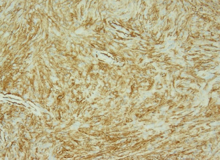
3 Spectrum of Myoepithelial (ME) Tumors Family of tumors with variable terminology based on anatomic location: pleomorphic adenoma (salivary gland) benign mixed tumor (skin) soft tissue myoepithelioma Definition: immunohistochemical evidence of myoepithelial differentiation (EMA/CK, S100/calponin) Uncertain if any/all have a common pathogenesis


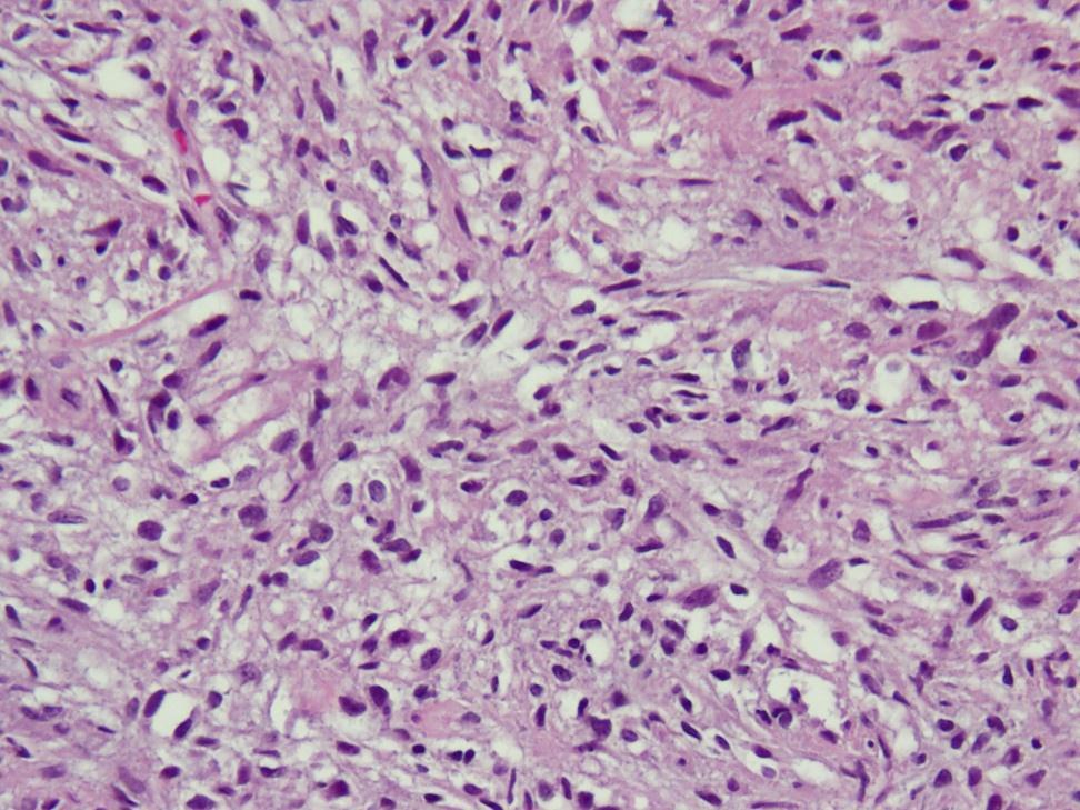
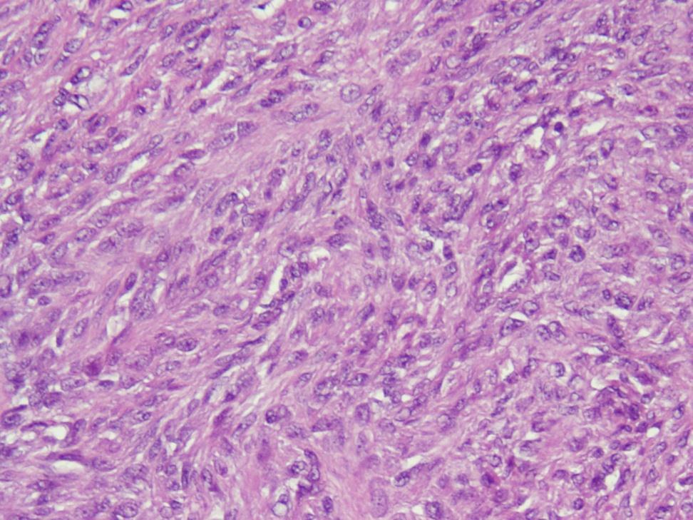
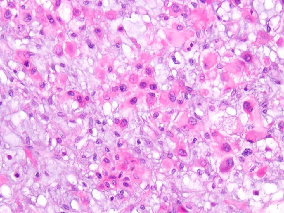
4 Altered Myoepithelial Cells Neoplastic Conditions High morphologic plasticity: - Spindle shaped or myoid cells - Hyalin or plasmacytoid cells - Clear cells Pleomorphic Adenoma, Parotid


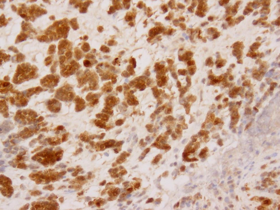
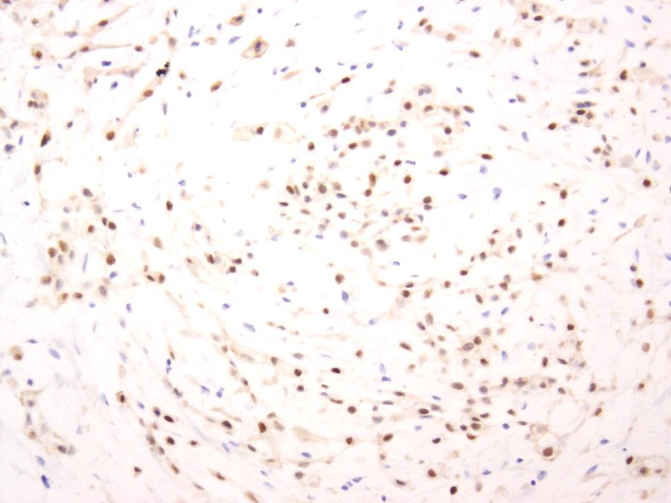
5 Salivary Gland Myoepithelial Tumors - Immunoprofile Hybrid epithelial and mesenchymal properties AE1:AE3 S100 Calponin

6 Hornick JL et al.. Am J Surg Path 2003
7 Soft Tissue ME Tumors Genetics Few reports of EWSR1 gene rearrangement one case report each, the fusion partner was identified as being either PBX1 or ZNF444
8 Towards a Molecular Classification of ME Tumors Systematic molecular analysis of a large spectrum of ME tumors, including a variety of anatomic locations, age groups and risk of malignancy To investigate the pathogenetic relationship with the salivary gland counterpart (PLAG1 & HMGA2 rearrangements in pleomorphic adenoma) Unifying concept of ME tumors within soft tissues, bone, and visceral locations
9 Molecular Study of ME Tumors 66 ME tumors with confirmed diagnosis and adequate material for molecular analysis 47 Soft Tissue 7 Cutaneous 6 Bone 6 Visceral (lung) Age distribution: 1-75 years; 15 children Gender distribution: 36 females/30 males Antonescu CR Genes Chromosomes Cancer 2010

10 EWSR1 Rearrangement in 45% of ME tumors Common presentations: Deep soft tissue Malignant histology Pediatric: 8/15 Bone: 5/6 Visceral: 4/6 EWSR1 Telomeric-Centromeric
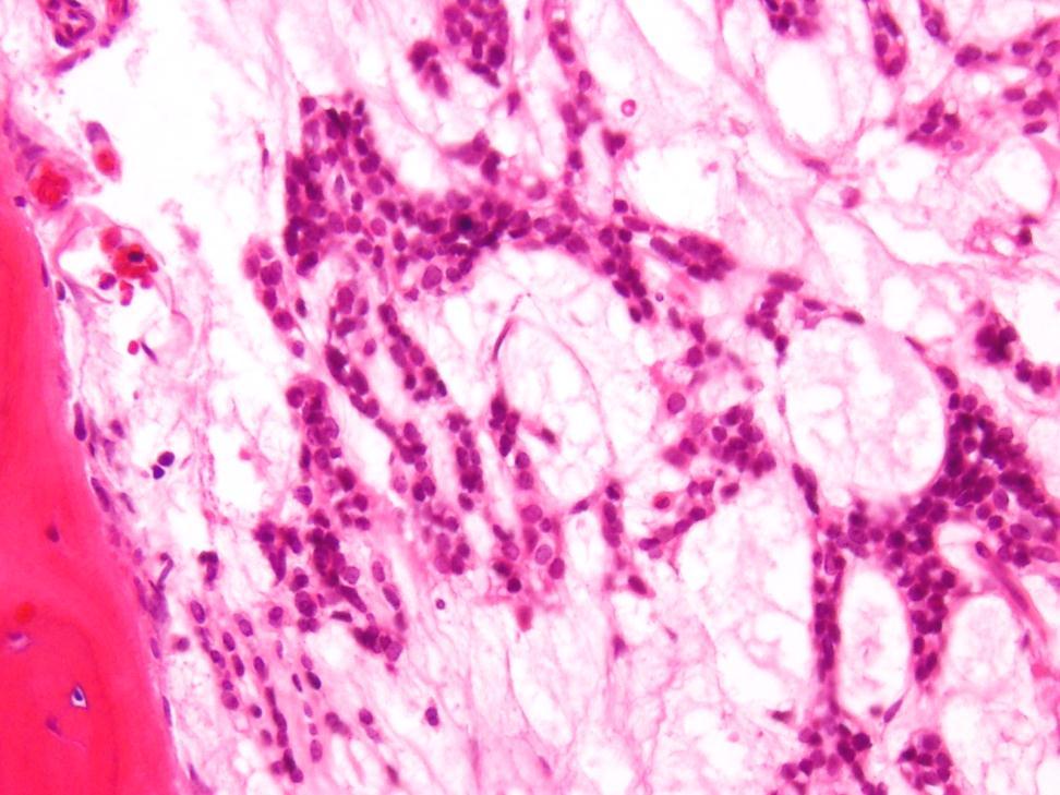
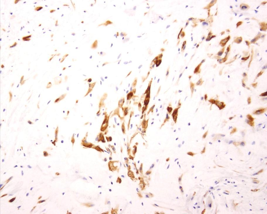
11 ME Tumors with EWSR1 gene rearrangement Histologic patterns: 1. Small blue cell phenotype 2. Epithelioid or rhabdoid histology with eosinophilic or clear cytoplasm 3. Spindle or ovoid appearance in a prominent sclerotic stroma 4. Lack ductal, squamous, or matrix differentiation


12 ME tumor with EWSR1 rearrangement 1. Small blue cell phenotype 53yF, R index finger, superficial IHC: s100 & EMA positive


13 ME tumor with EWSR1 rearrangement 2. Epithelioid morphology with clear or eosinophilic cytoplasm 20yM, Left foot, subcutaneous, benign S100 positive, EMA negative, CK positive


14 ME tumor with EWSR1 rearrangement 3. Sclerotic background, mixed spindle & epithelioid cells 32yM, subcutaneous R ankle, benign S100 & EMA positive


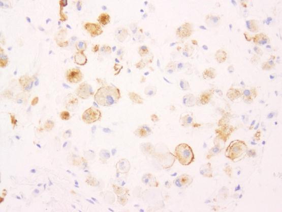
15 EWSR1 -POU5F1 Fusion in ME Tumors - Children & young adults - Deep soft tissue extremities - Clear cell morphology EWSR1 EXON6 3 RACE &RT-PCR POU5F1 EXON2 POU5F1 Telomeric/ Centromeric S100 EMA Antonescu CR Genes Chromosomes Cancer 2010


16 EWSR1-POU5F1 fusion positive Nested, diffuse clear cell changes 34 yr F, wrist, deep, malignant IHC: s100 & EMA positive; OCT4 negative
17 POU5F1 (a.k.a OCT3/4) FISH encodes a transcription factor which binds to the octamer motif (ATGCAAAT) present in the promoter or enhancer regions of target genes. is essential for keeping germ cells and embryonic stem cells in an immature and pluripotent status POU5F1 reactivation has been found to be implicated in human cancer: germ cell tumors (OCT3/4 IHC marker), bladder tumors


")
 S100, EMA/CK PBX1")
18 EWSR1-PBX1 positive ME Tumors - Age range years - Location: 3 soft tissue, 1 bone, 1 lung - Morphology: sclerotic, bland (fibromatosis-like) or clear cell changes - IHC: (+) S100, EMA/CK PBX1 break-apart


19 C EWSR1-T ZNF444 EWSR1-ZNF444 Positive ME Tumor 64yF, Lung S100 positive, EMA negative
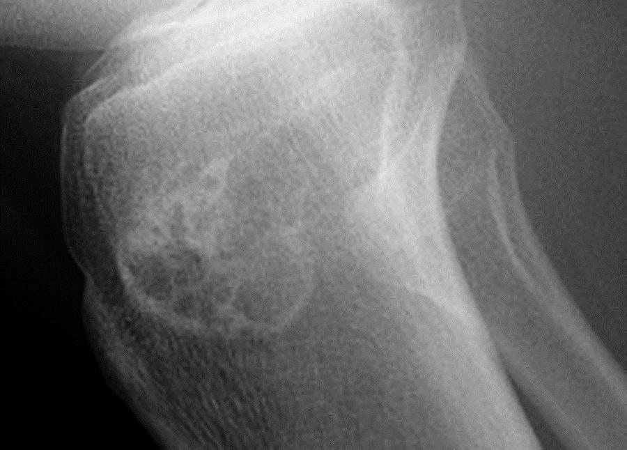
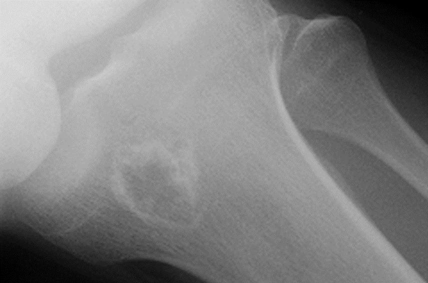
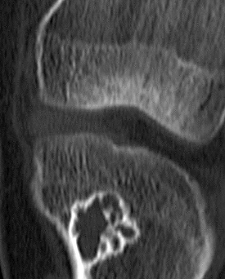
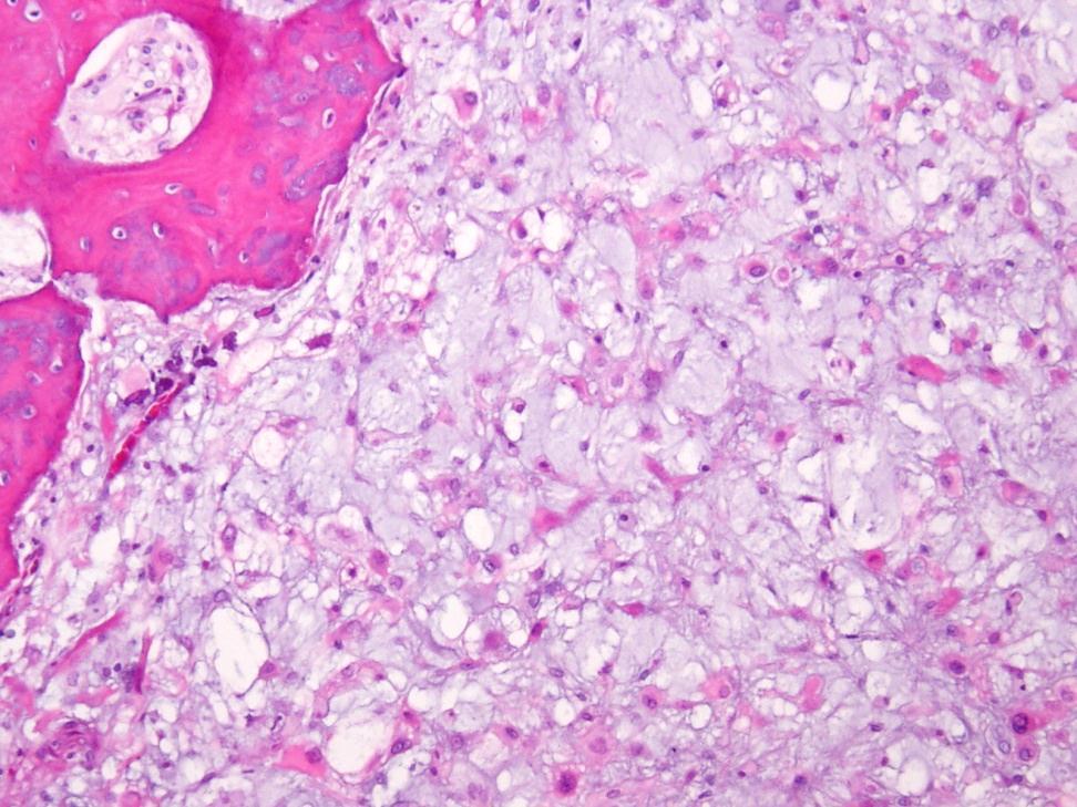
20 Intra-Osseous Myoepithelial Tumors
21 Intra-Osseous Myoepithelial Tumors 5/6 showed EWSR1 rearrangement Location: Vertebral body (L1) Long bones of extremity (humerus, fibula, prox femur) Pelvic Bones Morphology: epithelioid, clear cell, spindle Malignancy: 5 benign/1 malignant

22 ME tumor with EWSR1 rearrangement 45 yf, vertebral body/l1 S100 protein (+)


23 49yM, Iliac bone lesion long history followed for presumed FD CT MRI, Axial T1, fat saturation Courtesy Petur Nielsen, MGH


24 Intra-Osseous Myoepithelial Tumor Positive for EWSR1 & PBX1 gene rearrangements by FISH


25 26yM R Prox Tibia T1 T2 2007
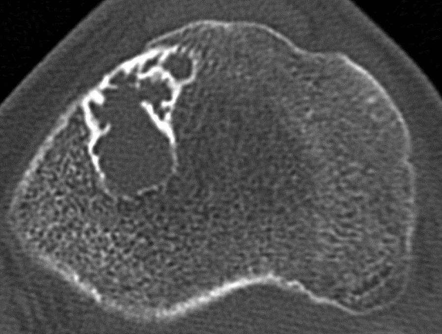
26 2011
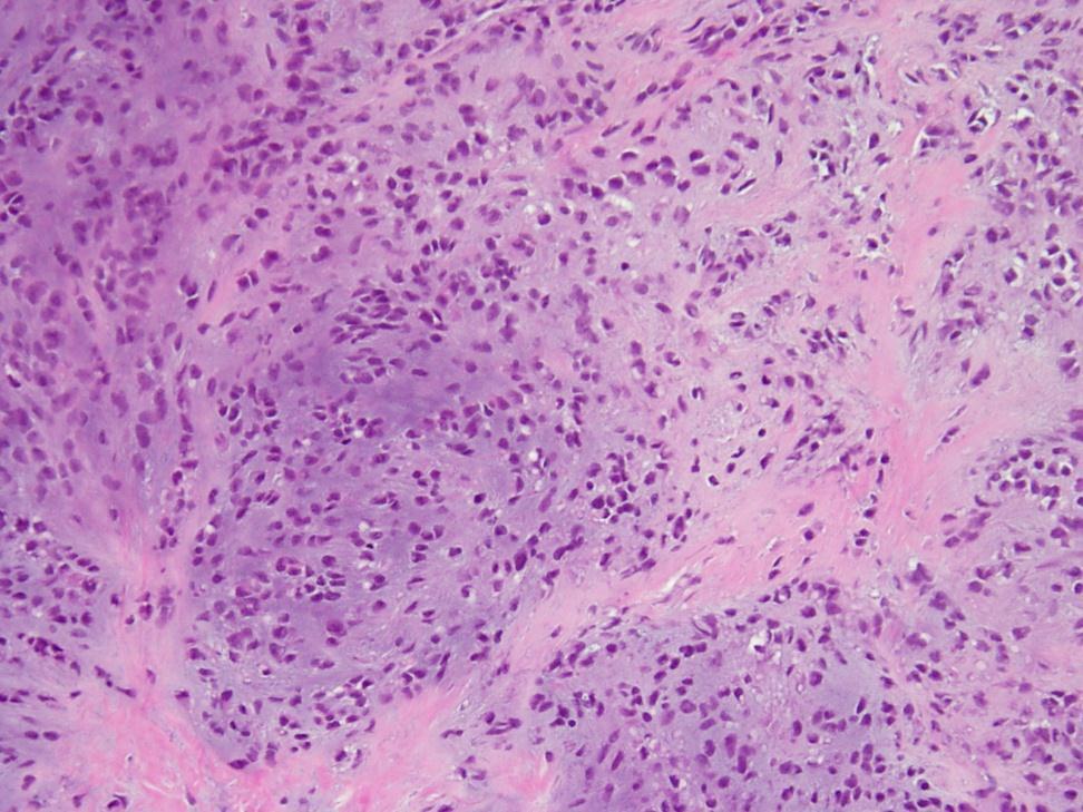
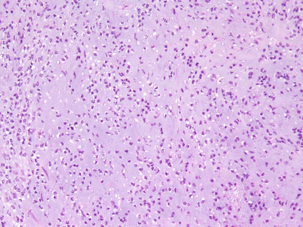
27 Benign Intra-Osseous Myoepithelial Tumor


28 S100 protein EMA HHF35 EWSR1
29 Visceral ME Tumors Lung ME Tumors (n=6) 4/6 positive for EWSR1 rearrangement 1 EWSR1-PBX1 1 EWSR1-ZNF444 1/2 FUS rearranged Breast ME tumors (n=5) all negative


30 EWSR1 negative ME tumors Common presentation: superficial cutaneous benign with ductal, squamous or cartilage matrix formation No difference in IHC profile Relationship with Myoepithelial Tumors of Salivary Gland?
31 PLAG1 Rearrangements in Cutaneous and Soft Tissue ME tumors/mixed Tumors Benign myoepithelial tumors with prominent tubulo-ductal differentiation Lack EWSR1 gene rearrangement Neoplasms with genuine salivary gland-like morphology, so-called soft tissue/cutaneous mixed tumors, are genetically related to their salivary gland counterpart Bahrami A, Genes Chromosome Cancer 2012 Antonescu CR, Modern Pathology 2012

32 PLAG1 Overexpression and Gene Rearrangement in Skin/Soft Tissue Mixed Tumors PLAG1 Bahrami Genes Chromosome Cancer 2012
33 Saliv Gland Tumors containing Myoepithelial cells Benign Myoepithelioma Pleomorphic Adenoma Malignant Myoepithelial Carcinoma (de novo or expleomorphic adenoma) Epi-Myoepithelial Carcinoma


34 Myoepithelial Carcinoma Ex- Pleomorphic Adenoma Capsule Pleomorphic adenoma Myoepithelial carcinoma


35 PLAG1 gene alterations in salivary gland pleomorphic adenoma and carcinoma ex-pleomorphic adenoma CK8-18/PLAG1 Calponin/PLAG1 Martins et al. Mod Path, 2005
36 PLAG1 Rearrangement in Myoepithelial and Ductal Epithelial Cells of Pleomorphic Adenoma increasing evidence that epithelial and myoepithelial cell populations share phenotypical and genotypical characteristics supporting the 'modified myoepithelial cell model Unified histogenesis: a single pluripotent cell, capable of differentiation into a variety of somatic phenotypes
37 Criteria for Malignancy Saliv Gland MET Infiltrative destructive growth, beyond the tumor capsule into adjacent salivary gland or soft tissue is the most widely accepted criterion Multinodular growth pattern with tongue-like processes (the most prevalent and characteristic pattern Hypercellular periphery and central hypocellular myxoid matrix and/or necrosis Sheet-like pattern
38 Criteria for Malignancy Saliv Gland MET cytologic atypia and mitotic rate have been reported to be useful, without firm guidelines for reaching a diagnosis of myoepithelial carcinoma (No specific mitotic rate cutoff exists for making this distinction, however, one series reported >7MF/10 HPF was diagnostic of malignancy) Nagao T et al. Cancer 1998
39 Criteria for Malignancy ST MET Most morphologically benign or low-grade behave in a benign fashion, with a low, but unpredictable, risk for local recurrence (approximately 20%). MET with at least moderate cytologic atypia are clinically malignant. Hornick JL et al. AJSP 2003
40 DIFFERENTIAL DIAGNOSIS
41 Chordoma Periphericum CK19 EMA T-Brachyury EWSR1 negative (N=2)


42 Ossifying Fibromyxoid Tumor (OFT) S100+ only in 70% of cases Desmin+ focally in up to 20% CK, SMA rarely + S-100


43 Ossifying Fibromyxoid Tumor (OFT) No recurrent cytogenetic abnormalities Ossifying fibromyxoid tumor: modified myoepithelial cell tumor? Min KW et al. Ultrastruct Pathol 2005
 CHN (NOR1) on 9q22 fusions: t(9;22)(q22;q12) EWSR1 t(9;17)(q22;q11) TAF2N t(9;15)(q22;q21) TCF12 t(3;9)(q11;q22)")
44 Extraskeletal Myxoid Chondrosarcoma Clinical: adults, extremity More diffusely myxoid IHC: non-contributory (weak/negative S100, EMA) CHN (NOR1) on 9q22 fusions: t(9;22)(q22;q12) EWSR1 t(9;17)(q22;q11) TAF2N t(9;15)(q22;q21) TCF12 t(3;9)(q11;q22) TFG


45 Ewing Sarcoma and other Primitive Sarcomas RT-PCR EWSR1-Fli1 O13 FISH: No info RE the gene partner >18 in-frame chimeric transcripts
46 Summary EWSR1 gene rearrangement is a common genetic event in soft tissue, bone and visceral ME tumors, particularly in the pediatric and young adults. EWSR1-POU5F1 fusion is present 8-10% of ME tumors, involving the extremity deep soft tissues, occurring in children or young adults and displaying a prominent clear cell morphology EWSR1-PBX1 detected in ME tumors with sclerosing and fibromatosis -like morphology
47 Summary EWSR1 fusion negative ME tumors are more often benign, cutaneous or superficially located and display ductal or cartilage differentiation. PLAG1 gene rearrangements are noted in 1/3 of tumors lacking EWSR1, which are typically benign and have prominent tubulo-ductal differentiation (i.e. mixed tumors)
48 Acknowledgements MSKCC, Pathology Nora Katabi Bill Travis Brigham & Women, Boston Christopher Fletcher Paola Dal Cin Mass General Hospital G. Petur Nielsen Andrew Rosenberg MSKCC, Lab Lei Zhang Peter Sung UHN, Toronto Ilan Weinreb CHOP Bruce Pawel
SOFT TISSUE TUMOR PATHOLOGY: AN UPDATE
 SOFT TISSUE TUMOR PATHOLOGY: AN UPDATE Jason L. Hornick, MD, PhD July 18, 2013 Department of Pathology Brigham and Women s Hospital Harvard Medical School Boston, MA, USA I have no disclosures. New Soft
Myxo-inflammatory Fibroblastic sarcoma
 AKA Myxo-inflammatory Fibroblastic sarcoma Acral Myxoinflammatory fibroblastic sarcomaam.j.surg.path1998; 22; 911-924 Inflammatory myxoid tumour of soft parts with bizarre giant cells [Pathol.Res.Pract.
Disclosure of Relevant Financial Relationships
 Ewing and Ewing like sarcomas Using Genetic Signatures in Refining Small Blue Round Cell Tumor Classification Cristina Antonescu, MD Department of Pathology Disclosure of Relevant Financial Relationships
Disclosure of Relevant Financial Relationships
 Surgical Pathology Companion Meeting Case 5: Locally Recurrent Chest wall Mass Cristina Antonescu, MD Department of Pathology, Memorial Sloan Kettering Cancer Center Disclosure of Relevant Financial Relationships
Update in Salivary Gland Pathology. Benjamin L. Witt University of Utah/ARUP Laboratories February 9, 2016
 Update in Salivary Gland Pathology Benjamin L. Witt University of Utah/ARUP Laboratories February 9, 2016 Objectives Review the different appearances of a selection of salivary gland tumor types Establish
ACCME/Disclosures ALK FUSION-POSITIVE MESENCHYMAL TUMORS. Tumor types with ALK rearrangements. Anaplastic Lymphoma Kinase. Jason L.
 Companion Meeting of the International Society of Bone and Soft Tissue Pathology The Evolving Concept of Mesenchymal Tumors ALK FUSION-POSITIVE MESENCHYMAL TUMORS Jason L. Hornick, MD, PhD March 13, 2016
Tumores de células pequeñas, redondas y azules: diagnóstico diferencial cuando el tiempo apremia
 Tumores de células pequeñas, redondas y azules: diagnóstico diferencial cuando el tiempo apremia Sílvia Bagué Servei de Patologia Hospital de Sant Pau Barcelona Soft tissue sarcomas Heterogeneous group
Financial disclosures
 Cutaneous Mesenchymal Neoplasms with EWSR1 Rearrangement By Konstantinos Linos MD, FCAP, FASDP Bone, Soft Tissue and Dermatopathology Assistant Professor of Pathology Dartmouth-Hitchc Geisel School of
Newer soft tissue entities
 Newer soft tissue entities Examples among fibroblastic tumors Turku, May 6, 2010 Markku Miettinen, M.D. AFIP, Washington, DC Fibroblastic neoplasms Solitary fibrous tumor /Hemangiopericytoma Low-grade
Salivary Gland FNA ATYPICAL : Criteria and Controversies
 Salivary Gland FNA ATYPICAL : Criteria and Controversies W.C. Faquin, M.D., Ph.D. Director, Head and Neck Pathology Massachusetts General Hospital Massachusetts Eye and Ear Infirmary Harvard Medical School
Disclosures. An update on ancillary techniques in the diagnosis of soft tissue tumors. Ancillary techniques. Introduction
 Disclosures An update on ancillary techniques in the diagnosis of soft tissue tumors. I have nothing to disclose. Andrew Horvai, MD, PhD Clinical Professor, Pathology Introduction Ancillary techniques
Cutaneous Mesenchymal Neoplasms with EWSR1 Rearrangement
 Cutaneous Mesenchymal Neoplasms with EWSR1 Rearrangement By Konstantinos Linos MD, FCAP, FASDP Bone, Soft Tissue and Dermatopathology Assistant Professor of Pathology Dartmouth-Hitchcock Medical Center
BSD 2015 Case 19. Female 21. Nodule on forehead. The best diagnosis is:
 BSD 2015 Case 19 Female 21. Nodule on forehead. The best diagnosis is: A. mixed tumour of skin B. porocarcinoma C. nodular hidradenoma D. metastatic adenocarcinoma BSD 2015 Case 19 Female 21 Nodule on
21/07/2017. Hobnail endothelial cells are not the same as epithelioid endothelial cells
 UPDATE IN CUTANEOUS VASCULAR S DERMATOPATHOLOGY SESSION BELFAST PATHOLOGY JUNE 21/2017 Dr E Calonje St John s Institute of Dermatology, London, United Kingdom THE FAMILY OF VASCULAR S WITH EPITHELIOID
Soft Tissue High Grade Myoepithelial Carcinoma With Round Cell Morphology: Report Of A Newly Described Entity With EWSR1 Gene Rearrangement
 Soft Tissue High Grade Myoepithelial Carcinoma With Round Cell Morphology: Report Of A Newly Described Entity With EWSR1 Gene Rearrangement Abstract M. El-Kabany, R. Al-Abdulghani, A. E. Ali, I. M. Francis,
3/27/2017. Disclosure of Relevant Financial Relationships
 Ophthalmic Pathology Evening Specialty Conference USCAP 2017 5 th March, 2017 Mukul K. Divatia, MD Assistant Professor Department of Pathology & Genomic Medicine Weill Cornell Medical College Houston Methodist
1/10/2018. Soft Tissue Tumors Showing Melanocytic Differentiation. Overview. Desmoplastic/ Spindle Cell Melanoma
 2016 MFMER slide-1 2016 MFMER slide-2 2016 MFMER slide-3 Soft Tissue Tumors Showing Melanocytic Differentiation Andrew L. Folpe, M.D. Professor of Laboratory Medicine and Pathology Mayo Clinic, Rochester,
A 60-year old Man with Left Jaw Mass. Simon Chiosea, MD University of Pittsburgh medical Center 3/15/2016
 ACCME/Disclosures The USCAP requires that anyone in a position to influence or control the content of CME disclose any relevant financial relationship WITH COMMERCIAL INTERESTS which they or their spouse/partner
Financial disclosures
 Mesenchymal Neoplasms with Melanocytic Differentiation By Konstantinos Linos MD, FCAP, FASDP Bone, Soft Tissue and Dermatopathology Assistant Professor of Pathology Dartmouth-Hitchcock Medical Center Geisel
Surgical Pathology Evening Specialty Conference USCAP 2015
 Surgical Pathology Evening Specialty Conference USCAP 2015 John R. Goldblum, M.D. Chairman, Department of Pathology, Cleveland Clinic Professor of Pathology, Cleveland Clinic Lerner College of Medicine
Molecular Diagnosis of Soft Tissue Tumors: Avoid Pitfalls
 Molecular Diagnosis of Soft Tissue Tumors: Avoid Pitfalls Cristina Antonescu, MD Department of Pathology Memorial Sloan-Kettering Cancer Center, New York Overview I. When should we rely on the help of
A 25 year old female with a palpable mass in the right lower quadrant of her abdomen
 May 2016 A 25 year old female with a palpable mass in the right lower quadrant of her abdomen Contributed by: Paul Ndekwe, MD, Resident Physician, Indiana University School of Department of Pathology and
ESS: Pathologic Insights
 GEIS XVI INTERNATIONAL SYMPOSIUM Seville 4th October 2018 ESS: Pathologic Insights Sílvia Bagué The Royal Marsden Hospital London (United Kingdom) I have no conflicts of interest Endometrial stromal sarcoma
Enterprise Interest Nothing to declare
 Enterprise Interest Nothing to declare Diagnoses one would not like to miss in soft tissue pathology early in your career Marta Sbaraglia, MD Department of Pathology Hospital of Treviso University of Padua
Case Report Clinicopathological features of primary myoepithelial carcinoma inside oblique eminence of cuboid bone: a case report
 Int J Clin Exp Med 2018;11(6):6327-6331 www.ijcem.com /ISSN:1940-5901/IJCEM0068496 Case Report Clinicopathological features of primary myoepithelial carcinoma inside oblique eminence of cuboid bone: a
Evening Specialty Conference Bone and Soft Tissue Pathology. Diagnostic pitfalls in bone and soft tissue pathology
 Evening Specialty Conference Bone and Soft Tissue Pathology. Case 1 Elizabeth G Demicco, MD, PhD Mount Sinai Hospital, New York Disclosure of Relevant Financial Relationships USCAP requires that all planners
Lung Tumor Cases: Common Problems and Helpful Hints
 Lung Tumor Cases: Common Problems and Helpful Hints Brandon T. Larsen, MD, PhD Senior Associate Consultant Department of Laboratory Medicine and Pathology Mayo Clinic Arizona Arizona Society of Pathologists
Objectives. Salivary Gland FNA: The Milan System. Role of Salivary Gland FNA 04/26/2018
 Salivary Gland FNA: The Milan System Dr. Jennifer Brainard Section Head Cytopathology Cleveland Clinic Objectives Introduce the Milan System for reporting salivary gland cytopathology Define cytologic
Case 2. Dr. Sathima Natarajan M.D. Kaiser Permanente Medical Center Sunset
 Case 2 Dr. Sathima Natarajan M.D. Kaiser Permanente Medical Center Sunset History 24 year old male presented with a 3 day history of right flank pain, sharp in nature Denies fever, chills, hematuria or
An unusual superficial small round cell sarcoma
 An unusual superficial small round cell sarcoma - Diagnostic problems - Differential diagnosis Antonio Llombart Bosch Isidro Machado Dep. Pathology Univ. Medical School Valencia, Institute of Oncology
Disclosures. An update on ancillary techniques in the diagnosis of soft tissue tumors. Ancillary techniques. Introduction
 Disclosures An update on ancillary techniques in the diagnosis of soft tissue tumors. I have nothing to disclose. Andrew Horvai, MD, PhD Clinical Professor, Pathology Introduction Ancillary techniques
Slide Seminar Spanish Society of Pathology
 Slide Seminar Spanish Society of Pathology John R. Goldblum, M.D. Chairman, Department of Anatomic Pathology Cleveland Clinic Professor of Pathology Cleveland Clinic Lerner College of Medicine 1921 Original
Slide seminar. Asist. Prof. Jože Pižem, MD, PhD Institute of Pathology Medical Faculty, University of Ljubljana
 Slide seminar Asist. Prof. Jože Pižem, MD, PhD Institute of Pathology Medical Faculty, University of Ljubljana Case 5 A 57-year-old man with a dermal/subcutaneous lesion on the scalp, which was interpreted
57th Annual HSCP Spring Symposium 4/16/2016
 An Unusual Malignant Spindle Cell Lesion to Involve the Breast Erinn Downs-Kelly, D.O. Associate Professor of Pathology University of Utah & ARUP Laboratories No disclosures Case 39 y/o female with no
04/09/2018. Salivary Gland Pathology in the Molecular Era Old Friends, Old Foes, & New Acquaintances
 Salivary Gland Pathology in the Molecular Era Old Friends, Old Foes, & New Acquaintances Jennifer L. Hunt, MD, MEd Aubrey J. Hough Jr, MD, Endowed Professor of Pathology Chair of Pathology and Laboratory
AANP Diagnostic Slide Session Case 5
 AANP Diagnostic Slide Session Case 5 E. Kelly S. Mrachek, M.D. Neuropathology Fellow University of Virginia M. Beatriz S. Lopes, M.D., Ph.D. Neuropathology Program Director University of Virginia Disclosures:
Financial disclosures
 An update on immunohistochemical markers in mesenchymal neoplasms By Konstantinos Linos MD, FCAP, FASDP Assistant Professor of Pathology Geisel School of Medicine at Dartmouth Dartmouth-Hitchcock Medical
USCAP 2012: Companion Meeting of the AAOOP. Update on lacrimal gland neoplasms: Molecular pathology of interest
 USCAP 2012: Companion Meeting of the AAOOP Vancouver BC, Canada, March 17, 2012 Update on lacrimal gland neoplasms: Molecular pathology of interest Valerie A. White MD, MHSc, FRCPC Department of Pathology
Update On Lipomatous Tumors: Old Standbys and New Concepts
 Update On Lipomatous Tumors: Old Standbys and New Concepts John R. Goldblum, M.D. Chairman, Department of Anatomic Pathology Cleveland Clinic Professor of Pathology Cleveland Clinic Lerner College of Medicine
Case Report A SMARCB1-deficient vulvar neoplasm with prominent myxoid stroma: report of a case showing ERG and FLI1 expression
 Int J Clin Exp Pathol 2015;8(6):7526-7532 www.ijcep.com /ISSN:1936-2625/IJCEP0008225 Case Report A SMARCB1-deficient vulvar neoplasm with prominent myxoid stroma: report of a case showing ERG and FLI1
The Relevance of Cytologic Atypia in Cutaneous Neural Tumors
 The Relevance of Cytologic Atypia in Cutaneous Neural Tumors Recent Findings - New Developments New Problems Zsolt B. Argenyi, M.D. Professor of Pathology & Dermatology Director of Dermatopathology Department
Original Article Primary malignant mixed tumor of bone: a case report
 Int J Clin Exp Pathol 2015;8(7):8433-8437 www.ijcep.com /ISSN:1936-2625/IJCEP0007490 Original Article Primary malignant mixed tumor of bone: a case report Zhansan Su 1, Zhi Li 1, Baoan Liu 2 1 Department
Differential Diagnosis of Oral Masses. Palatal Lesions
 Differential Diagnosis of Oral Masses Palatal Lesions Palatal Masses Periapical Abscess Torus Palatinus Mucocele Lymphoid Hyperplasia Adenomatous Hyperplasia Benign Salivary Neoplasms Malignant Salivary
Primary Myoepithelioma of the Testis - A Case Report -
 The Korean Journal of Pathology 2011; 45(S1): S20-24 DOI: 10.4132/KoreanJPathol.2011.45.S1.S20 Primary Myoepithelioma of the Testis - A Case Report - Seong Muk Jeong 1 Jung Hee Lee 1 Won Young Park 1 Na
Special slide seminar
 Special slide seminar Tomáš Rozkoš The Fingerland Department of Pathology Charles University Medical Faculty and Faculty Hospital in Hradec Králové Czech Republic Case history, 33 years old resistance
Update on Cutaneous Mesenchymal Tumors. Thomas Brenn
 Update on Cutaneous Mesenchymal Tumors Thomas Brenn Cutaneous Mesenchymal Tumours Wide morphological and biological spectrum Myofibroblastic, smooth muscle, neural, vascular, apidocytic, undifferentiated;
Lesions Mimicking Adenoid Cystic Carcinoma. Diagnostic Problems in Salivary Gland Pathology An Update 5/29/2009
 Diagnostic Problems in Salivary Gland Pathology An Update Lesions Mimicking Adenoid Cystic Carcinoma Stacey E. Mills, M.D. W.S. Royster Professor of Pathology Director of Surgical and Cytopathology University
PLEOMORPHIC ADENOMA ( BENIGN MIXED TUMOR )
") ( BENIGN MIXED TUMOR ) Grossly, the tumor is freely movable, solid, sometimes lobulated and occasionally cystic. If recurrent, multinodular masses are common. Histologically, within a fibrous capsule,
( BENIGN MIXED TUMOR ) Grossly, the tumor is freely movable, solid, sometimes lobulated and occasionally cystic. If recurrent, multinodular masses are common. Histologically, within a fibrous capsule,
Review of the AP Part II Practical Examination. Dr David Clift Co Chief Examiner
 Review of the AP Part II Practical Examination Dr David Clift Co Chief Examiner General Remarks The part II practical examination involved 15 cases which were presented with sufficient clinical data to
Keywords solitary fibrous tumor, dedifferentiation, dedifferentiated solitary fibrous tumor, STAT6, GRIA2, cytokeratin, rhabdomyosarcomatous
 758452IJSXXX10.1177/1066896918758452International Journal of Surgical PathologyCreytens et al research-article2018 Pitfalls in Pathology Multifocal Cytokeratin Expression in a Dedifferentiated Solitary
Klinisch belang van chromosomale translocatie detectie in sarcomen
 Translocations in sarcomas Klinisch belang van chromosomale translocatie detectie in sarcomen Judith V.M.G. Bovée, M.D., Ph.D. Department of Pathology Leiden University Medical Center RNA binding DNA binding
Pleomorphic Adenoma of Parotid in a Young Patient. Key words: Pleomophic, Adenoma, Salivary Glands, Parotid Neoplasms, Neoplasm Recurrence.
 JOURNAL OF CASE REPORTS 2013;3(1):142-147 Pleomorphic Adenoma of Parotid in a Young Patient Sangeeta R. Patankar, Megha A. Meshram, Vasant Shewale 1, Gokul S. From the Department of Oral Pathology & Microbiology
Disclosure of Relevant Financial Relationships
 Evening Specialty Conference - Genitourinary Pathology Case 2 Disclosure of Relevant Financial Relationships Sean R Williamson, MD Henry Ford Health System, Detroit, MI @Williamson_SR USCAP requires that
An Overview of Genital Stromal Tumors
 An Overview of Genital Stromal Tumors By Konstantinos Linos MD, FCAP, FASDP Bone, Soft Tissue and Dermatopathology Assistant Professor of Pathology Dartmouth-Hitchcock Medical Center Geisel School of Medicine
Immunohistochemistry in Bone and Soft Tissue Tumors. Sahar Rassi Zankoul, MD
 Immunohistochemistry in Bone and Soft Tissue Tumors Sahar Rassi Zankoul, MD Introduction Bone tumors represent a wide variety of tumors of various origins and malignant potentials. These different tumor
Case 1. Disclosure. Imaging. Clinical history 5/10/2016. USCAP 2016 Annual Meeting Evening Specialty Conference Bone and Soft tissue Pathology
 Disclosure Dr. Agaram has nothing to disclose Case 1 Narsi Agaram, MBBS USCAP 2016 Annual Meeting Evening Specialty Conference Bone and Soft tissue Pathology Clinical history Imaging 1998 A three month
Salivary Glands 3/7/2017
 Salivary Glands 3/7/2017 Goals and objectives Focus on the entities unique to H&N Common board type facts Information for your future practice Salivary Glands Salivary Glands Major gland. Paratid. Submandibular.
Biopsy Interpretation of Spindle cell proliferations of the Serosa
 Biopsy Interpretation of Spindle cell proliferations of the Serosa Richard Attanoos, Cardiff. U.K. Disclosure of Relevant Financial Relationships USCAP requires that all planners (Education Committee)
Classification (1) Classification (3) Classification (2) Spindle cell lesions. Spindle cell lesions of bladder (Mills et al.
 Classification (3) Classification (2) Spindle cell lesions. Spindle cell lesions of bladder (Mills et al.") Non-epithelial tumours and nonepithelial tumour-like lesions of the bladder Dr Jonathan H Shanks The Christie NHS Foundation Trust, Manchester, UK Classification (1) Myofibroblastic proliferations and
Non-epithelial tumours and nonepithelial tumour-like lesions of the bladder Dr Jonathan H Shanks The Christie NHS Foundation Trust, Manchester, UK Classification (1) Myofibroblastic proliferations and
Self assessment case. Dr Saleem Taibjee Dorset County Hospital, Dorchester
 Self assessment case Dr Saleem Taibjee saleemtaibjee@gmail.com Dorset County Hospital, Dorchester Clinical details 34-year-old man: Shave excision Skin tag / papilloma left thigh The best diagnosis is:
Selected Pseudomalignant Soft Tissue Tumors of the Skin and Subcutis
 Selected Pseudomalignant Soft Tissue Tumors of the Skin and Subcutis Andrew L. Folpe, M.D. Professor of Laboratory Medicine and Pathology Mayo Clinic, Rochester, MN folpe.andrew@mayo.edu 2016 MFMER slide-1
PRELIMINARY CYTOLOGIC DIAGNOSIS: Suspicious for Acinic Cell Carcinoma. Cell Block: Immunohistochemical Studies CYTOLOGIC DIAGNOSIS:
 1 PRELIMINARY CYTOLOGIC DIAGNOSIS: Suspicious for Acinic Cell Carcinoma. Cell Block: Immunohistochemical Studies GCDFP-15 S-100 CYTOLOGIC DIAGNOSIS: Consistent with mammary analogue secretory carcinoma.
Molecular pathology in soft tissue tumors. Sylvia Höller Pathologie
 Molecular pathology in soft tissue tumors Sylvia Höller Pathologie When do we perform molecular testing? Morphology and IHC are not clearly fitting with an entity some translocations are entity specific
ARIZONA SOCIETY OF PATHOLOGISTS 13 TH APRIL 2013 HEAD AND NECK CYTOPATHOLOGY. F ZAHRA ALY, MD, PhD
 ARIZONA SOCIETY OF PATHOLOGISTS 13 TH APRIL 2013 HEAD AND NECK CYTOPATHOLOGY F ZAHRA ALY, MD, PhD The main areas sites amenable for cytopathology include lymph nodes, thyroid, major salivary glands especially
No financial or other disclosures
 Case 2014-5 Esther N. Bit-Ivan, DO Northwestern University Jason Wang, MD Jason Park, MD Korgun Koral, MD Children s Medical Center Charles Timmons, MD Veena Rajaram, MD No financial or other disclosures
59 yo male with past medical history of prostate carcinoma, presented with upper abdominal pain
 December 2016 59 yo male with past medical history of prostate carcinoma, presented with upper abdominal pain Contributed by: Divya Sharma, MD. Fellow, Gastrointestinal Pathology, Department of Pathology
Oncocytic-Appearing Salivary Gland Tumors. Oncocytic, Cystic, Mucinous, and High Grade Salivary Gland Tumors SALIVARY GLAND FNA: PART II
 William C. Faquin, MD, PhD Professor of Pathology Harvard Medical School Director of Head and Neck Pathology Massachusetts Eye and Ear Massachusetts General Hospital SALIVARY GLAND FNA: PART II Oncocytic,
Los Angeles Society Of Pathologists Dr. Shobha Castelino Prabhu
 Los Angeles Society Of Pathologists Dr. Shobha Castelino Prabhu Loma Linda University Medical Center June 12, 2007 CASE 1 76 year-old gentleman Status post right parotidectomy 1 year ago for a rare tumor
Recent Advances In Select Round Cell Sarcomas
 Recent Advances In Select Round Cell Sarcomas Rajiv M. Patel, M.D. Associate Professor of Pathology & Dermatology University of Michigan, Ann Arbor, MI rajivpat@med.umich.edu 1 Translocation Associated
Cellular Neurothekeoma
 Cellular Neurothekeoma Scott W Binder, MD Pritzker Professor of Pathology & Dermatology Sr. Vice Chair Director, Pathology Clinical Services Chief, Dermatopathology Geffen/UCLA School of Medicine Clinical
Case 27 Male 42. Painless, static, well-circumscribed, subcutaneous nodule right lower leg,?lipoma. The best diagnosis is:
 Case 27 Male 42. Painless, static, well-circumscribed, subcutaneous nodule right lower leg,?lipoma. The best diagnosis is: A. Angiosarcoma B. Haemangiopericytoma C.Myopericytoma D.Myofibroma E. Angioleiomyoma
Case 1 10/2/17. Myxoid Soft Tissue Tumors & Tumor-like Lesions. Myxofibro- or Fibromyxo-?: Myxoid Soft Tissue Tumours We Are All Mixed Up About
 Myxoid Soft Tissue Tumors & Tumor-like Lesions Myxofibro- or Fibromyxo-?: Myxoid Soft Tissue Tumours We Are All Mixed Up About Rajiv M. Patel, M.D. RCPA NZ ASM 2017 (4:15-5:00pm, Saturday, 23-09-17) Heterogenous
What is New in the 2015 WHO Lung Cancer Classification? Zhaolin Xu, MD, FRCPC, FCAP
 What is New in the 2015 WHO Lung Cancer Classification? Zhaolin Xu, MD, FRCPC, FCAP Professor, Dept of Pathology, Dalhousie University, Canada Pulmonary Pathologist and Cytopathologist, QEII HSC Senior
Mody. AIS vs. Invasive Adenocarcinoma of the Cervix
 Common Problems in Gynecologic Pathology Michael T. Deavers, M.D. Houston Methodist Hospital, Houston, Texas Common Problems in Gynecologic Pathology Adenocarcinoma in-situ (AIS) of the Cervix vs. Invasive
MYOEPITHELIOMA OF SOFT TISSUE AND BONE CRISTINA ANTONESCU, MD. Memorial Sloan-Kettering Cancer Center, New York, NY
 MYOEPITHELIOMA OF SOFT TISSUE AND BONE CRISTINA ANTONESCU, MD Memorial Sloan-Kettering Cancer Center, New York, NY The spectrum of myoepithelial (ME) tumors represents a family of lesions with variable
Papillary Lesions of the breast
 Papillary Lesions of the breast Emad Rakha Professor of Breast Pathology The University of Nottingham Papillary lesions of the breast are a heterogeneous group of disease, which are characterised by neoplastic
USCAP 2011: ASDP companion meeting. Steven D. Billings 1
 USCAP 2011: ASDP companion meeting. Steven D. Billings (billins@ccf.org) 1 Spindle cell tumors that make you say, Oh $*&%! This lecture will focus on examples of cutaneous tumors that present particular
The Enigmatic Spitz Lesion
 The Enigmatic Spitz Lesion The Dawn of Spitz S Spitz Sophie Spitz Melanomas of Childhood ; Am J Pathol 1948 1910-1956 13 children (18 mo - 12 yrs) 12/13 had a benign clinical course Sophie Spitz Born 1910
Pathology Mystery and Surprise
 Pathology Mystery and Surprise Tim Smith, MD Director Anatomic Pathology Medical University of South Carolina Disclosures No conflicts to declare Some problem cases Kidney tumor Scalp tumor Bladder tumor
WHO 2017: New Classification of Head and Neck Tumours
 WHO 2017: New Classification of Head and Neck Tumours Ilmo Leivo Department of Pathology University of Turku & Turku University Hospital FINLAND Suomen IAP Aulanko 18.05.2018 WHO 2017 New entities Simplification
CASE year old male with a PET avid nodule in the left adrenal gland
 CASE 1 55 year old male with a PET avid nodule in the left adrenal gland Case 1 Adrenal gland parenchyma partly replaced by a spindle cell tumour with mild nuclear pleomorphism Atypical mitoses present
Pancreas. Atrophy, acinar cell. Pathogenesis: Diagnostic key features:
 Pancreas Atrophy, acinar cell Pathogenesis: Decrease in number and/or size of acinar cells may be due to spontaneous or experimentally induced degenerative changes, apoptosis, or a sequel of chronic inflammation.
3/27/2017. Disclosure of Relevant Financial Relationships. Papilloma???
 Management of Papillary Lesions Diagnosed at Rad Path Concordant Core Biopsy (CNB) Disclosure of Relevant Financial Relationships USCAP requires that all planners (Education Committee) in a position to
Spindle Cell Lesions Of The Breast. Emad Rakha Professor of Breast Pathology and Consultant Pathologist
 Spindle Cell Lesions Of The Breast Emad Rakha Professor of Breast Pathology and Consultant Pathologist * SCLs comprise a wide spectrum of diseases, ranging from reactive processes to aggressive malignant
Spinal Cord Compression caused by Metastatic Epithelial Myoepithelial Carcinoma of the Parotid Gland
 Spinal Cord Compression caused by Metastatic Epithelial Myoepithelial Carcinoma of the Parotid Gland Pages with reference to book, From 249 To 250 Irshad N. Soomro,Akber S. Hussainy,Rashida Ahmed,Sheema
Synonyms. Nephrogenic metaplasia Mesonephric adenoma
 Nephrogenic Adenoma Synonyms Nephrogenic metaplasia Mesonephric adenoma Definition Benign epithelial lesion of urinary tract with tubular, glandular, papillary growth pattern Most frequently in the urinary
Salivary gland neoplasms: an update 29th Annual Meeting of Arab Division of the International Academy of Pathology MUSCAT, OMAN 2017
 Salivary gland neoplasms: an update 29th Annual Meeting of Arab Division of the International Academy of Pathology MUSCAT, OMAN 2017 Dr Mary Toner Consultant Pathologist St James Hospital Trinity College
Notice of Faculty Disclosure
 Mesenchymal Tumors of the Vulva: Old, New, Something(s) Different Napa Valley Conference Pathology Education Partners Inc May 15, 2018 Teri A. Longacre, M.D. longacre@stanford.edu Stanford University,
Review. Myoepithelial tumor of soft tissue and bone: A current perspective. Histology and Histopathology
 Histol Histopathol (2017) 32: 861-877 http://www.hh.um.es Histology and Histopathology From Cell Biology to Tissue Engineering Review Myoepithelial tumor of soft tissue and bone: A current perspective
Neuroendocrine neoplasms of the lung
 Neuroendocrine neoplasms of the lung M Papotti, L Righi, & M Volante University of Turin at San Luigi Hospital TORINO NETs OF THE LUNG Menu - Spectrum of NE lung tumors - CARCINOID TUMORS - SCLC /LCNEC
4/12/2018. MUSC Pathology Symposium Kiawah Island April 18, Jesse K. McKenney, MD
 MUSC Pathology Symposium Kiawah Island April 18, 2018 Jesse K. McKenney, MD 1 Urothelial Carcinoma with Alternative Differentiation 2 Urothelial Carcinoma with Alternative Differentiation Recognition as
Pathology of Sarcoma ELEANOR CHEN, MD, PHD, ASSISTANT PROFESSOR DEPARTMENT OF PATHOLOGY UNIVERSITY OF WASHINGTON
 Pathology of Sarcoma ELEANOR CHEN, MD, PHD, ASSISTANT PROFESSOR DEPARTMENT OF PATHOLOGY UNIVERSITY OF WASHINGTON Presentation outline Background and epidemiology of sarcomas Sarcoma classification Sarcoma
CASE REPORT Benign epithelioid peripheral nerve sheath tumour resembling schwannoma
 Malaysian J Pathol 2014; 36(3) : 217 221 CASE REPORT Benign epithelioid peripheral nerve sheath tumour resembling schwannoma Thejasvi KRISHNAMURTHY MD and SR NIVEDITHA MD, DNB Department of Pathology,
Pulmonary Salivary Gland Type Tumors With Features of Malignant Mixed Tumor (Carcinoma Ex Pleomorphic Adenoma) A Clinicopathologic Study of Five Cases
 A Clinicopathologic Study of Five Cases") Anatomic Pathology / Pulmonary Salivary Gland Type Tumors Pulmonary Salivary Gland Type Tumors With Features of Malignant Mixed Tumor (Carcinoma Ex Pleomorphic Adenoma) A Clinicopathologic Study of Five
Anatomic Pathology / Pulmonary Salivary Gland Type Tumors Pulmonary Salivary Gland Type Tumors With Features of Malignant Mixed Tumor (Carcinoma Ex Pleomorphic Adenoma) A Clinicopathologic Study of Five
DIAGNOSTIC SLIDE SEMINAR: PART 1 RENAL TUMOUR BIOPSY CASES
 DIAGNOSTIC SLIDE SEMINAR: PART 1 RENAL TUMOUR BIOPSY CASES Dr. Andrew J. Evans MD, PhD, FACP, FRCPC Consultant in Genitourinary Pathology University Health Network, Toronto, ON Case 1 43 year-old female,
Mammary analogue secretory carcinoma of salivary gland A case report of new entity
 Case Report Mammary analogue secretory carcinoma of salivary gland A case report of new entity Vaibhav Bhika Bari 1*, Sandhya Unmesh Bholay 2 1 Assistant Professor, 2 Associate Professor Rajiv Gandhi Medical
Part 1. Slides 1-38, Rita Alaggio Soft tissue tumors Trondheim 14. mars 2013
 Part 1 Slides 1-38, Rita Alaggio Soft tissue tumors Trondheim 14. mars 2013 Pediatric Pathology Soft Tissue Tumors AN UPDATE Rita Alaggio Azienda Ospedaliera Università di Padova Soft Tissue Tumors More
Myoepithelial Carcinoma: A Rare Case Report with Review
 Review article: Myoepithelial Carcinoma: A Rare Case Report with Review Dr. Shilpa Parikh 1, *Dr. Bhavik Mavani 2, Dr. Jigna Shah 3,Dr.Sheetal Sharma 4 1 Professor, Dept of Oral Medicine & Radiology, Govt.
Enterprise Interest None
 Enterprise Interest None B3 lesions of the breast What are they at surgery? Case 4 Edi Brogi MD PhD Attending Pathologist - Director of Breast Pathology Memorial Sloan Kettering Cancer Center New York
Gross appearance of nodular hyperplasia in material obtained from suprapubic prostatectomy. Note the multinodular appearance and the admixture of
 Tiền liệt tuyến Tiền liệt tuyến Gross appearance of nodular hyperplasia in material obtained from suprapubic prostatectomy. Note the multinodular appearance and the admixture of solid and microcystic areas.