Chapter 4: Single Photon Emission Computed. Tomography (SPECT)
|
|
|
- Peregrine Fleming
- 5 years ago
- Views:
Transcription
1 Chapter 4: Single Photon Emission Computed Tomography (SPECT) NPRE 435, Principles of Imaging with Ionizing Radiation, Fall 2018
2 Contents Emission Tomography (ET) for Nuclear Medicine Applications Introduction and basic principle of Emission Tomography Early developments Generation of radio-nuclides for ET. Detector technologies for ET System design considerations Other Related Imaging Applications Coded aperture imaging Compton Imaging NPRE 435, Principles of Imaging with Ionizing Radiation, Fall 2018
3 Traditional Definition of Nuclear Medicine NUCLEAR MEDICINE SHOWS PHYSIOLOGY Whereas RADIOLOGY SHOWS ANATOMY NPRE 435, Principles of Imaging with Ionizing Radiation, Fall 2018


4 Traditional Definition of Nuclear Medicine NUCLEAR MEDICINE PHYSIOLOGY DIAGNOSIS NON-IMAGING IMAGING THERAPY Emission Tomography (ET) SPECT PET NPRE 435, Principles of Imaging with Ionizing Radiation, Fall 2018
5 What is Emission Tomography? A branch of medical imaging that encompasses two main modalities single photon emission computed tomography (SPECT) and positron emission tomography (PET) It uses radioactive materials to image properties of body s physiology, such as glucose metabolism, blood flow, receptor concentrations. ET is categorized as functional imaging techniques to distinguish it from methods such as X-ray CT that principally depict the body architectural structure (anatomy). NPRE 435, Principles of Imaging with Ionizing Radiation, Fall 2018
6 What is the tracer principle? The Tracer Principle Appropriately chosen radioactive compounds participate in an organism s physiological processes in the same way as non-radioactive materials. These compounds can be detected through the detecting of their radiation signatures, such as gamma rays. Two major attributes Because one can detect even minute quantities of radioactive material, the tracer principle can be used to measure molecular concentrations with a tremendous sensitivity. Tracer measurements are noninvasive the concentration of tracer is deduced from the number of gamma rays detected.
7 Emission Tomography Drug is labeled with radioisotopes that emit gamma rays. Drug localizes in patient according to metabolic properties of that drug. Trace (pico-molar) quantities of drug are sufficient. Radiation dose fairly small (<1 rem). Drug Distributes in Body


8 Emission tomography and its place in the matrix of molecular imaging technologies 1. New technological approaches (detectors, system designs etc.) for better balance between spatial resolution and sensitivity ICE-SPECT, Lai, PMB, CZT-PET, Groll et al, TRPMS, SPEM 2. Imaging through broadband EM radiation MRC-SPECT-I, Liang Cai, NIM, 2015 MRC-SPECT-II, Lai, PMB 2018 XFET/XLET 3. Non-traditional image formation techniques: XFCT/XLCT, stimulated emission tomography etc. Meng, TNS, 2013, G. Fu, Med Phys 2014, Groll, TNS, Radiation-activable (nano-) material for therapeutic delivery and imaging From S. R. Meikle et al, PMB Vol. 50, Topical Review, 2005.


9 Single Photon Emission Computed Tomography (SPECT) Siemens Symbia SPECT/CT Philips Precedence SPECT/CT
10 Single Photon Emission Computed Tomography (SPECT) Collimator in front of the detector to select gamma rays from certain directions only Collimator Pinhole Rotated around the object for collecting multiple projections Coded Aperture Compton


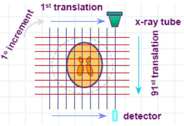
11 Single Photon Emission Computed Tomography (SPECT)
12 Chapter 3: Radioactivity What would be an ideal gamma ray emitter for nuclear imaging? Reasonably penetrative Half-life comparable to the biological process we are trying to visualize Easy radiochemistry Biologically safe Clean NPRE 435, Radiological Imaging, Fall 2018 Radiation Sources and Interactions
13 Radionuclides are Produced in a Nuclear Reactor or a Cyclotron Reactor - target bombarded with neutrons Cyclotron - target bombarded with charged particles Diagnosis uses radionuclides from both Therapy radionuclides produced in a nuclear reactor PET radionuclides produced in a cyclotron NPRE 435, Principles of Imaging with Ionizing Radiation, Fall 2018
14 Metastable Nuclear States and Gamma Ray Emission
15 Chapter 3: Radioactivity Metastable Nuclear States and Gamma Ray Emission NPRE 435, Radiological Imaging, Fall 2018 Radiation Sources and Interactions
16 Tc-99m Generator NPRE 435, Principles of Imaging with Ionizing Radiation, Fall 2018
17 Early Developments Radionuclides for diagnostic purpose 1935 Hevesy : radiophosphorous for metabolic studies in rats 1949 Cassen : rectilinear scanner consisting of a calcium tungstate crystal coupled to a photomultiplier tube for I-131 uptake in thyroid with ¼ resolution 1957 Anger : stationary area detector. scintillation camera (Anger or gamma camera) consisting of a large-area sodium iodide crystal coupled to an array of PM tubes NPRE 435, Principles of Imaging with Ionizing Radiation, Fall 2018
18 Early Development Radionuclides for diagnostic purpose cont. Early 1960s Kuhl & Edwards : idea of transverse section tomography (cf. X-ray CT), unsatisfactory results because of without the aid of computers & reconstruction algorithms 1977 Keyes & Jaszczak : SPECT (Single-photon emission computed tomography), improved image contrast compared to stationary cameras 1970s : PET (positron emmision tomography), large number of scintillation detectors surrounding the patient, unique capability to study metabolic function in humans. NPRE 435, Principles of Imaging with Ionizing Radiation, Fall 2018
19 DISA Applications (1) BRAIN Tc99m/I123: brain dopamine receptor imaging (Parkinson s disease), epilepsy, depression Tc99m/Tl201: differentiation in radiation necrosis and recurrent glioma THYROID/PARATHYROID Tc99m/I123 or Tl201: simultaneous thyroid/parathyroid imaging INFECTION Tc99m/In111: Infective endocarditis (111In white blood cells (occult infections) and 99mTc perfusion imaging) I123/Tl201: myocardial SPECT with Tl-201 and I-123 MIBG for myocarditis Tc99m/In111: osteomyelitis (bone infection),infected pelvic pressure ulcer, infection in the feet of diabetic patients
20 DISA Applications (2) MYOCARDIAL VIABILITY (INFARCTION) In111/Tl201: dual-isotope SPECT with indium 111-monoclonal antimyosin antibodies and thallium-201 can estimate infarct size and percentage myocardium infarcted MYOCARDIAL PERFUSION Tc99m/Tl201: simultaneous Tc-99m stress and Tl-201 rest MYOCARDIAL FATTY ACID METABOLISM Tc99m/I123: simultaneous perfusion (Tc99m tetrofosmin) and fatty acid metabolism (I123-BMIPP) MYOCARDIAL INNERVATION Tc99m/I123: simultaneous I-123 mibg innervation and Tc99m resting perfusion Tc99m/I123/Tl201: rest perfusion status with 201Tl, stress perfusion status with a 99mTc labeled agent, innervation status with 123I-MIBG

21 What do MPI images look like? In a typical nuclear cardiac imaging exam, the physician reviews: Static Summed Perfusion Images Dynamic Gated Images Perfusion Images are viewed in three orientations: SA Short Axis VLA Vertical Long Axis HLA - Horizontal Long Axis


22 Example of Dual-Isotope SPECT Imaging This slide is based on the work by Dr. Eric Frey, at Johns Hopkins University Fig. 1. Sample energy spectra of Tl-201 and Tc-99m. Fig. 2. Sample energy spectra of Tc-99m and I-123. Typical dual-isotope SPECT imaging: rest/stress myocardial perfusion imaging using Tc-99m and Tl-201 labeled agents and brain imaging using Tc-99m and I- 123 labeled isotopes.

. The patient presented with symptoms of stable atypical angina.")
23 Tc-99m Sestamibi Myocardial Perfusion Imaging (MPI) Exercise stress Tc-99m Sestamibi single day myocardial perfusion SPECT images of the female patient with a significant (80%) distal left main coronary artery disease. Classic features of the high-risk scan are present: severe partially reversible perfusion defect, involvement of the LAD and LCX territory, visual transient ischemic dilation and abnormal TID ratio (1.21). The patient presented with symptoms of stable atypical angina. No significant ECG or hemodynamic changes were noted during the stress portion of the test.
 and posterior (b) 131I scans show focal activity in the right suprarenal region.")
24 I-131 Whole Body Imaging Differentiation between malignancy and benign changes with SPECT/CT in a patient with thyroid cancer who underwent 131I whole-body imaging to assess for residual recurrent disease. Anterior(a) and posterior (b) 131I scans show focal activity in the right suprarenal region. Coronal (c) and axial (d) SPECT/CT images show that the uptake is located in the renal collecting system (arrow),consistent with physiologic urinary activity, and not recurrence of disease.
. (A) Age 10, 3.01 GBq; (B) age 11, 2.95 GBq; (C) age 13, 4.")
 showed the presence of radioiodine avid bilateral pulmonary metastases (<5 mm maximum diameter on computed tomography) and very small, low-grade lower")
25 Case Report: Management of metastatic thyroid cancer in pregnancy: risk and uncertainty, by Christopher W Rowe et al., Endocrinology, Diabetes and Metabolism, 2016 Serial post-i 131 therapy scans (anterior whole body views). (A) Age 10, 3.01 GBq; (B) age 11, 2.95 GBq; (C) age 13, 4.3 GBq; (D) age 14, 5.1 GBq; (E) age 15, 9.9 GBq, 7 months before conception. Neck disease present at age 13 (C) was treated surgically. The final study (E) showed the presence of radioiodine avid bilateral pulmonary metastases (<5 mm maximum diameter on computed tomography) and very small, low-grade lower neck disease. The focal uptake in the left upper abdomen is colonic and is likely physiological in nature.

26 α-emitters of Human Interest (1) 40 and 727 kev γ emission Po K Xrays are emitted that permit external imaging, the two most abundant of these X-rays have energies of 77 and 80 kev, 12 and 20% of all photon emissions, respectively. Bismuth-213 emits a gamma ray photon at 440 kev and a yield of 16.5%.
27 α-emitters of Human Interest (2) 218 kev γ emission 440 kev γ emission The principal gamma lines for 223Ra in equilibrium with its daughters are at 154 kev, 270 kev,351 kev, and 405 kev. Main problem: reduce the loss of the daughters to non-target tissues and mitigating systemic radiotoxic events. >200 patients have been treated with 223Ra-chloride (Alpharadin, Algeta ASA, Oslo, Norway), in several European countries and has been approved for investigational use in the U.S. The principal characteristics of this radionuclide are high skeletal uptake with long-term retention, minimal uptake in normal organs and other soft tissues, and clearance via the gastro-intestinal tract.
, 15.2 (21%), and 236 (11%) kev.")
28 α-emitters of Human Interest (3) no γ emission High yield photons occur at 12.3 (17%), 15.2 (21%), and 236 (11%) kev.
 and Ac-225 extracted from Th-229 (lower spectrum).")
29 Example of Dual-Isotope SPECT Imaging The 225Ac decay chain. Photons with a branching ratio >3% relative to 225Ac decay are shown. Fig. 4. Gamma spectra of purified Ac-225 produced via cyclotron irradiation (upper spectrum) and Ac-225 extracted from Th-229 (lower spectrum). Energy spectra, normalized by acquisition time, acquired by the VECTor scanner for both HEUHR (red) and UHS (green) collimators with a comparison to a background spectrum (grey) acquired with no collimator or activity present. Spectra were acquired using a uniform source containing 1.05 MBq (28.3 μci) of 225Ac. The energy regions used to reconstruct the 221Fr and 213Bi are shown in blue and red, respectively. The adjacent grey regions indicate the energy windows defined for background and scatter correction. Cyclotron production of Ac-225 for targeted alpha therapy, C.Apostolidis, Applied Radiation and Isotopes, 2005


30 In Vivo Imaging in T Cells Immunotherapy A Collaboration with Prof. Ed Roy, Pathology, UIUC uninjected 3 day after injection Microscopy of NIR fluorescently labeled T cells. A. Lymph node of uninjected mouse showing the lack of autofluorescence under acquisition conditions used in B. B. Two populations of T cells in the lymph node, labeled with CellVue Burgundy (red) and CellVue NIR815 (green) after intravenous injection. These images were kindly provided by Prof. Ed Roy of Pathology and the Beckman Institute, UI: Fused SPEM/CT image of a mouse s brain. Two groups (5 L and 0.3 L) of radiolabeled T cells are visible in the brain. Spatial reso. ~ 0.1 mm a few hundred cells L. J. Meng et al., NIM 2009 MRI images of Iron oxide labeled T cells in the brain in association with a small brain tumor. Image courtesy Prof. Ed Roy, Univ. of Illinois.


31 In Vivo Imaging of Neural Stem Cells for Glioblastoma Targeting and Therapy Scheme 1. Schematic of the ph-senstive MSN-Dox-loaded neural stem cell delivery system. Fig. 1: The SPECT/CT imaging of mice post gavage of MSN-TA- 99m Tc and Na 99m TcO 4. Figure 2. In vivo tumor-tropic migration of ReNcell. Bioluminescent imaging of mice after intracranial injection of ReNcell-Fluc into the left hemisphere of the mice implanted with U87 xenograft tumors on the right side 7 days (i) before or (ii) mice with no tumor. Another group of tumor-bearing mice received control fibroblast NIH- 3T3-Rluc cell. Each group had five animals; photographs show a representative animal from each group. Imaging was done 24 hours postimplantation of the NSCs. Image curtsey, Prof Lesniak University of Chicago.
OTHER NON-CARDIAC USES OF Tc-99m CARDIAC AGENTS Tc-99m Sestamibi for parathyroid imaging, breast tumor imaging, and imaging of other malignant tumors.
 DEFINITION OF CARDIAC RADIOPHARMACEUTICAL: A radioactive drug which, when administered for purpose of diagnosis of heart disease, typically elicits no physiological response from the patient. Even though
Fundamentals of Nuclear Cardiology. Terrence Ruddy, MD, FRCPC, FACC
 Fundamentals of Nuclear Cardiology Terrence Ruddy, MD, FRCPC, FACC Objectives To understand the Principles of Nuclear Cardiac Imaging Radiotracers Image acquisition and processing Stress protocols To appreciate
Radionuclides in Medical Imaging. Danielle Wilson
 Radionuclides in Medical Imaging Danielle Wilson Outline Definitions History and development Radionuclide applications & techniques in imaging Conclusion Definition #1 : Radionuclide An unstable nucleus
A Snapshot on Nuclear Cardiac Imaging
 Editorial A Snapshot on Nuclear Cardiac Imaging Khalil, M. Department of Physics, Faculty of Science, Helwan University. There is no doubt that nuclear medicine scanning devices are essential tool in the
SPECT-CT: Τι πρέπει να γνωρίζει ο Καρδιολόγος
 SPECT-CT: Τι πρέπει να γνωρίζει ο Καρδιολόγος Δρ Αναστασία Κίτσιου Διευθύντρια, Καρδιολογική Κλινική, Σισμανόγλειο ΓΝΑ Chair, Education Committee, Section on Nuclear Cardiology & Cardiac CT, EACVI, ESC
Radiation Detection and Measurement
 Radiation Detection and Measurement Range of charged particles (e.g.,!: µm; ": mm) Range of high energy photons (cm) Two main types of interactions of high energy photons Compton scatter Photoelectric
Cardiovascular Imaging
 Cardiovascular Imaging Cardiovascular Imaging Cardio and Vascular Imaging Vascularization / Angiogenesis Cardiovascular Imaging metabolic imaging of the heart myocardial perfusion imaging Cardiac CT Vascularization
Itroduction to the Nuclear Medicine: biophysics and basic principles. Zámbó Katalin Department of Nuclear Medicine
 Itroduction to the Nuclear Medicine: biophysics and basic principles Zámbó Katalin Department of Nuclear Medicine NUCLEAR MEDICINE Application of the radioactive isotopes in the diagnostics and in the
CEREBRAL BLOOD FLOW AND METABOLISM
 Supported by: HURO/0901/069/2.3.1 HU-RO-DOCS CEREBRAL BLOOD FLOW AND METABOLISM Part 3 Modern imaging methods SPECT, PET, nmri History of Nuclear Medicine Starts with the invention of the X-ray 1946: radioactive
Medical Use of Radioisotopes
 Medical Use of Radioisotopes Therapy Radioisotopes prove to be useful in the application of brachytherapy, the procedure for using temporary irradiation close to the area of disease (i.e. cancer) 10% Medical
Option D: Medicinal Chemistry
 Option D: Medicinal Chemistry Basics - unstable radioactive nuclei emit radiation in the form of smaller particles alpha, beta, positron, proton, neutron, & gamma are all used in nuclear medicine unstable
Medical imaging X-ray, CT, MRI, scintigraphy, SPECT, PET Györgyi Műzes
 Medical imaging X-ray, CT, MRI, scintigraphy, SPECT, PET Györgyi Műzes Semmelweis University, 2nd Dept. of Medicine Medical imaging: definition technical process of creating visual representations about
Basics of nuclear medicine
 Basics of nuclear medicine Prof. dr. Davor Eterović Prof. dr. Vinko Marković Radioisotopes are used both in diagnostics and in therapy Diagnostics gamma emitters are used since gamma rays can penetrate
Nuclear Medicine and PET. D. J. McMahon rev cewood
 Nuclear Medicine and PET D. J. McMahon 150504 rev cewood 2018-02-15 Key Points Nuclear Medicine and PET: Imaging: Understand how Nuc Med & PET differ from Radiography & CT by the source of radiation. Be
Typical PET Image. Elevated uptake of FDG (related to metabolism) Lung cancer example: But where exactly is it located?
 Lung cancer example: But where exactly is it located?") Typical PET Image Elevated uptake of FDG (related to metabolism) Lung cancer example: But where exactly is it located? PET/CT Oncology Imaging Anatometabolic fusion images are useful in the management
Typical PET Image Elevated uptake of FDG (related to metabolism) Lung cancer example: But where exactly is it located? PET/CT Oncology Imaging Anatometabolic fusion images are useful in the management
NUCLEAR CARDIOLOGY UPDATE
 Nuclear Cardiology David K. Shelton, Jr., MD NUCLEAR CARDIOLOGY UPDATE No Conflicts. No Disclosures. No Smoking. David K. Shelton UCDMC Nuclear Cardiology Nuclear Cardiology Radionuclide Ventriculography
Cardiac Imaging Tests
 Cardiac Imaging Tests http://www.medpagetoday.com/upload/2010/11/15/23347.jpg Standard imaging tests include echocardiography, chest x-ray, CT, MRI, and various radionuclide techniques. Standard CT and
Radiopharmacy. Prof. Dr. Çetin ÖNSEL. CTF Nükleer Tıp Anabilim Dalı
 Prof. Dr. Çetin ÖNSEL CTF Nükleer Tıp Anabilim Dalı What is Nuclear Medicine? Nuclear Medicine is the branch of medicine concerned with the use of radionuclides in the study and the diagnosis of diseases.
PHYSICS 2: HSC COURSE 2 nd edition (Andriessen et al) CHAPTER 20 Radioactivity as a diagnostic tool (pages 394-5)
 CHAPTER 20 Radioactivity as a diagnostic tool (pages 394-5)") PHYSICS 2: HSC COURSE 2 nd edition (Andriessen et al) CHAPTER 20 Radioactivity as a diagnostic tool (pages 394-5) 1. (a) A radioisotope is an isotope that is unstable and will emit particles from the nucleus
PHYSICS 2: HSC COURSE 2 nd edition (Andriessen et al) CHAPTER 20 Radioactivity as a diagnostic tool (pages 394-5) 1. (a) A radioisotope is an isotope that is unstable and will emit particles from the nucleus
Nuclear medicine studies of the digestiv system. Zámbó Katalin Department of Nuclear Medicine
 Nuclear medicine studies of the digestiv system Zámbó Katalin Department of Nuclear Medicine Imaging tehniques Anatomy Physiology Metabolism Molecular Rtg. / CT PET / SPECT MRI MR spectroscopy fmri Ultrasound
45 Hr PET Registry Review Course
 45 HR PET/CT REGISTRY REVIEW COURSE Course Control Document Timothy K. Marshel, MBA, R.T. (R), (N)(CT)(MR)(NCT)(PET)(CNMT) The PET/CT Training Institute, Inc. SNMMI-TS 028600-028632 45hr CEH s Voice Credits
PET IMAGING (POSITRON EMISSION TOMOGRAPY) FACT SHEET
 FACT SHEET") Positron Emission Tomography (PET) When calling Anthem (1-800-533-1120) or using the Point of Care authorization system for a Health Service Review, the following clinical information may be needed to
Positron Emission Tomography (PET) When calling Anthem (1-800-533-1120) or using the Point of Care authorization system for a Health Service Review, the following clinical information may be needed to
Nuclear Medicine in the Diabetic Foot
 26.11.2015, Uniklinik Balgrist Nuclear Medicine in the Diabetic Foot Martin Hüllner Nuklearmedizin und Neuroradiologie, USZ / UZH Outline A. Imaging modalities brief technical overview B. Nuclear medicine
Austin Radiological Association Nuclear Medicine Procedure PROSTATE CANCER STUDY (In-111-Capromab Pendetide [ProstaScint ])
![Austin Radiological Association Nuclear Medicine Procedure PROSTATE CANCER STUDY (In-111-Capromab Pendetide [ProstaScint ])](/thumbs/81/82771892.jpg "Austin Radiological Association Nuclear Medicine Procedure PROSTATE CANCER STUDY (In-111-Capromab Pendetide [ProstaScint ])") Austin Radiological Association Nuclear Medicine Procedure PROSTATE CANCER STUDY (In-111-Capromab Pendetide [ProstaScint ]) Overview Indications The Prostate Cancer Study with an indium-111 labeled murine
Austin Radiological Association Nuclear Medicine Procedure PROSTATE CANCER STUDY (In-111-Capromab Pendetide [ProstaScint ]) Overview Indications The Prostate Cancer Study with an indium-111 labeled murine
RADIOTRACERS FOR MYOCARDIAL PERFUSION IMAGING
 RADIOTRACERS FOR MYOCARDIAL PERFUSION IMAGING RAYMOND TAILLEFER, M.D. FRCP(c), ABNM DIRECTOR, DEPARTMENT OF NUCLEAR MEDICINE HOPITAL ST-JEAN-SUR-RICHELIEU Disclosures to Report: Grant Research Support:
Department of Nuclear Medicine with Positron Emission Tomography
 (PET) Unit [1] Contact information: Registration: +48 41 367 4850 Main office: +48 41 367 4860 Fax: +48 41 367 4887 e-mail: zmnsco@onkol.kielce.pl [2] Head of the Department: Professor Janusz Braziewicz
Tc-99m Sestamibi/Tetrofosmin Stress-Rest Myocardial Perfusion Scintigraphy
 APPROVED BY: Director of Radiology Page 1 of 6 Tc-99m Sestamibi/Tetrofosmin Stress-Rest Myocardial Primary Indications: Evaluation of myocardial perfusion and viability in patients with known or suspected
Reversible defect of 123 I-15-(p-iodophenyl)-9-(R,S)-methylpentadecanoic acid indicates residual viability within infarct-related area
-9-(R,S)-methylpentadecanoic acid indicates residual viability within infarct-related area") ORIGINAL ARTICLE Annals of Nuclear Medicine Vol. 16, No. 3, 183 187, 2002 Reversible defect of 123 I-15-(p-iodophenyl)-9-(R,S)-methylpentadecanoic acid indicates residual viability within infarct-related
ORIGINAL ARTICLE Annals of Nuclear Medicine Vol. 16, No. 3, 183 187, 2002 Reversible defect of 123 I-15-(p-iodophenyl)-9-(R,S)-methylpentadecanoic acid indicates residual viability within infarct-related
Physical Bases : Which Isotopes?
 Physical Bases : Which Isotopes? S. Gnesin Institute of Radiation Physics, Lausanne University Hospital, Lausanne, Switzerland 1/53 Theranostic Bruxelles, 2 Octobrer 2017 Theranostic : use of diagnostic
Nuclear pulmonology. Katalin Zámbó Department of Nuclear Medicine
 Nuclear pulmonology Katalin Zámbó Department of Nuclear Medicine Imaging techniques Morphology Physiology Metabolism Molecules X-ray / CT MRI NM - SPECT/ PET MR spectroscopy fmri Ultrasound Hybrid imaging:
SPECT TRACERS Tl-201, Tc-99m Sestamibi, Tc-99m Tetrofosmin
 SPECT TRACERS Tl-201, Tc-99m Sestamibi, Tc-99m Tetrofosmin Elmer Jasper B. Llanes, M.D. Nuclear Cardiology St. Luke s Medical Center Outline Ideal Physiologic Characteristics of MPI radioactive tracers
Radionuclide & Radiopharmaceuticals
 Radionuclide & Radiopharmaceuticals 1. Generator & Reactors 2. Cyclotrons & PET tracer 3. Quality control 4. Renal 5. GIT 6. CNS & Psychiatrics 7. Tumor Diagnosis & Treatment 8. Bones & joints 9. Thyroid
Austin Radiological Association Nuclear Medicine Procedure BONE MINERAL STUDY (Tc-99m-MDP, Tc-99m-HMDP)
") Austin Radiological Association Nuclear Medicine Procedure BONE MINERAL STUDY (Tc-99m-MDP, Tc-99m-HMDP) Overview The Bone Mineral Study, with either Tc-99m-MDP or Tc-99m-HMDP, depicts the distribution
Austin Radiological Association Nuclear Medicine Procedure BONE MINERAL STUDY (Tc-99m-MDP, Tc-99m-HMDP) Overview The Bone Mineral Study, with either Tc-99m-MDP or Tc-99m-HMDP, depicts the distribution
Radiation physics and radiation protection. University of Szeged Department of Nuclear Medicine
 Radiation physics and radiation protection University of Szeged Department of Nuclear Medicine Radiation doses to the population 1 Radiation doses to the population 2 Sources of radiation 1 Radiation we
General Nuclear Medicine
 General Nuclear Medicine What is General Nuclear Medicine? What are some common uses of the procedure? How should I prepare? What does the equipment look like? How does the procedure work? How is the procedure
Nuclear Medicine: Manuals. Nuclear Medicine. Nuclear imaging. Emission imaging: study types. Bone scintigraphy - technique
 Nuclear Medicine - Unsealed radioactive preparations the tracer mixes with the patients body fluids on a molecular level (e.g. after intravenous injection) - 3 main fields: - In vitro : measuring concentrations
E LT O N A. M O S M A N OUTLINE
 34 NUCLEAR MEDICINE E LT O N A. M O S M A N OUTLINE Principles of nuclear medicine, 388 Historical development, 389 Comparison with other modalities, 390 Physical principles of nuclear medicine, 392 Radiation
Isotopes in Functional Cancer Imaging
 Seeing and Believing: i Medical Isotopes in Functional Cancer Imaging François Bénard, MD, FRCPC BCCancer Cancer Agency and University of British Columbia Nuclear Medicine 101 A radioactive atom is produced
Zahid Rahman Khan, MD(USA), MS Diplomate American Board of Nuclear Medicine Consultant t Nuclear Medicine
, MS Diplomate American Board of Nuclear Medicine Consultant t Nuclear Medicine") Zahid Rahman Khan, MD(USA), MS Diplomate American Board of Nuclear Medicine Consultant t Nuclear Medicine i North West Armed Forces Hospital Tabuk, KSA. 1 Introduction Hibernation: Term coined by Rahimtoola
Zahid Rahman Khan, MD(USA), MS Diplomate American Board of Nuclear Medicine Consultant t Nuclear Medicine i North West Armed Forces Hospital Tabuk, KSA. 1 Introduction Hibernation: Term coined by Rahimtoola
Quantitative Theranostics in Nuclear Medicine
 Quantitative Theranostics in Nuclear Medicine M. Lassmann Klinik und Poliklinik für Nuklearmedizin Direktor: Prof. Dr. A. Buck Contents What is Theranostics? Potential Targets Basic Principles of Quantitative
Principles of nuclear metabolic imaging. Prof. Dr. Alex Maes AZ Groeninge Kortrijk and KULeuven Belgium
 Principles of nuclear metabolic imaging Prof. Dr. Alex Maes AZ Groeninge Kortrijk and KULeuven Belgium I. Molecular imaging probes A. Introduction - Chemical disturbances will precede anatomical abnormalities
Targeted Alpha Particle Therapy: Imaging, Dosimetry and Radiation Protection
 Targeted Alpha Particle Therapy: Imaging, Dosimetry and Radiation Protection Michael Lassmann Klinik und Poliklinik für Nuklearmedizin Direktor: Prof. Dr. A. Buck Targeted Therapy Basic Principles 2 Influence
Photon Attenuation Correction in Misregistered Cardiac PET/CT
 Photon Attenuation Correction in Misregistered Cardiac PET/CT A. Martinez-Möller 1,2, N. Navab 2, M. Schwaiger 1, S. G. Nekolla 1 1 Nuklearmedizinische Klinik der TU München 2 Computer Assisted Medical
Nuclear medicine in general practice. Dr Reza Garzan MD, FRACP, FAANMS
 Nuclear medicine in general practice Dr Reza Garzan MD, FRACP, FAANMS Myocardial perfusion study Bone scans in general practice Thyroid scans in general practice Gamma camera Detection of gamma rays Myocardial
Austin Radiological Association Nuclear Medicine Procedure WHITE BLOOD CELL MIGRATION STUDY (In-111-WBCs, Tc-99m-HMPAO-WBCs)
") Austin Radiological Association Nuclear Medicine Procedure WHITE BLOOD CELL MIGRATION STUDY (In-111-WBCs, Tc-99m-HMPAO-WBCs) Overview Indications The White Blood Cell Migration Study demonstrates the distribution
Austin Radiological Association Nuclear Medicine Procedure WHITE BLOOD CELL MIGRATION STUDY (In-111-WBCs, Tc-99m-HMPAO-WBCs) Overview Indications The White Blood Cell Migration Study demonstrates the distribution
Molecular Breast Imaging: History and Recent Developments
 Molecular Breast Imaging: History and Recent Developments Associate Professor, Department of Imaging Physics The University of Texas MD Anderson Cancer Center, Houston, Texas Educational Objectives 1.
Assessment of cardiac function using myocardial perfusion imaging technique on SPECT with 99m Tc sestamibi
 Journal of Physics: Conference Series PAPER OPEN ACCESS Assessment of cardiac function using myocardial perfusion imaging technique on SPECT with 99m Tc sestamibi To cite this article: M R A Gani et al
Cardiac Nuclear Medicine
 Cardiac Nuclear Medicine What is Cardiac Nuclear Medicine? What are some common uses of the procedure? How should I prepare? What does the equipment look like? How does the procedure work? How is the procedure
EDITORIAL. Dual-isotope myocardial perfusion SPECT imaging: Past, present, and future
 EDITORIAL Dual-isotope myocardial perfusion SPECT imaging: Past, present, and future Tali Sharir, MD, a,b and Piotr Slomka, PhD c,d a b c d Department of Nuclear Cardiology, Assuta Medical Center, Tel
Molecular Imaging and the Brain
 Molecular imaging technologies are playing an important role in neuroimaging, a branch of medical imaging, by providing a window into the living brain. Where CT and conventional MR imaging provide important
Positron Emission Tomography Computed Tomography (PET/CT)
") Positron Emission Tomography Computed Tomography (PET/CT) What is Positron Emission Tomography Computed Tomography (PET/CT) Scanning? What are some common uses of the procedure? How should I prepare for
Positron Emission Tomography Computed Tomography (PET/CT) What is Positron Emission Tomography Computed Tomography (PET/CT) Scanning? What are some common uses of the procedure? How should I prepare for
Nuclear Medicine: Basics to therapy
 Nuclear Medicine: Basics to therapy RCP Medical careers day Dr Sabina Dizdarevic MD MSc PhD FRCP Dr Deena Neriman MBBS FRCR Ms Charlotte Weston CEO BNMS On behalf of the British Nuclear Medicine Society
Assessment of Local Myocardial Perfusion in SPECT Images when Bicycle Exercise Test is Noninterpretable
 e 11 Assessment of Local Myocardial Perfusion in SPECT Images when Bicycle Exercise Test is Noninterpretable Ilona Kulakienė, Zigmundas Satkevičius, Juozas Kiudelis, Irena Milvidaitė 1 Kaunas Medical University,
Fellows on this rotation are expected to attend nuclear conferences and multimodality imaging conference.
 Rotation: Imaging 1 Imaging 1 provides COCATS Level 1 experience for nuclear cardiology (including SPECT and PET) and cardiac CT. Fellows will administer, process, and read cardiac nuclear studies with
NUCLEAR MEDICINE Molecular Imaging + Endo-Radiotherapy
 NUCLEAR MEDICINE Molecular Imaging + Endo-Radiotherapy Istvan Szilvási Dept. of Nuclear Medicine Semmelweis University and HDF Medical Centre 2016 DEFINITION OF NUCLEAR MEDICINE Medical applications of
Background. New Cross Hospital is a 700 bed DGH located in central England
 Background New Cross Hospital is a 700 bed DGH located in central England Regional Heart & Lung centre opened in 2005 Covering the Black Country (Population >1,000,000) Nuclear Medicine Department Single
Adriano Duatti Department of Chemical and Pharmaceutical Sciences University of Ferrara Via L. Borsari, 46, Ferrara, Italy
 Adriano Duatti Department of Chemical and Pharmaceutical Sciences University of Ferrara Via L. Borsari, 46, 44121 Ferrara, Italy dta@unife.it adriano.duatti@unife.it Summary Approved tracers for myocardial
Applications of radioactivity in medicine
 Lec.3 Applications of radioactivity in medicine -Nuclear medicine (N.M) -Applications of radioactive material in medicine Nuclear Medicine:- The clinical uses of radioactivity for the diagnosis of disease.
Radiologic Assessment of Myocardial Viability
 November 2001 Radiologic Assessment of Myocardial Viability Joshua Moss, Harvard Medical School Year III Patient EF 66yo female with a 3-year history of intermittent chest pain previously relieved by sublingual
Introduction to the Course and the Techniques. Jeffry R. Alger, PhD Ahmanson-Lovelace Brain Mapping Center Department of Neurology
 Introduction to the Course and the Techniques Jeffry R. Alger, PhD Ahmanson-Lovelace Brain Mapping Center Department of Neurology (jralger@ucla.edu) CTSI Neuroimaging April 2014 Rationale for the Course
SPECT CT Current Status and Future Direction Nuclear Cardiology
 SPECT CT Current Status and Future Direction Nuclear Cardiology BIR London 25 th February 2013 Nik Sabharwal Consultant Cardiologist Oxford Heart Centre nikant.sabharwal@ouh.nhs.uk No conflict of interest
Update in Nuclear Cardiology: Patient-Centered Imaging Radiation Dose Reduction
 Update in Nuclear Cardiology: Patient-Centered Imaging Radiation Dose Reduction E. Gordon DePuey, M.D. Icahn School of Medicine ant Mt. Sinai New York, NY Disclosures: Grant Support: Michael J. Fox Foundation
Nuclear Cardiology Pierre-Yves MARIE Department of Nuclear Medicine CHU-Nancy, FRANCE.
 Nuclear Cardiology Pierre-Yves MARIE Department of Nuclear Medicine CHU-Nancy, FRANCE. Nuclear Cardiology I - A remaining need of a functional information on myocardial perfusion II - The future: - combining
Cardiac Imaging. Kimberly Delcour, DO, FACC. Mahi Ashwath, MD, FACC, FASE. Director, Cardiac CT. Director, Cardiac MRI
 Cardiac Imaging Kimberly Delcour, DO, FACC Director, Cardiac CT Mahi Ashwath, MD, FACC, FASE Director, Cardiac MRI Cardiac Imaging Discuss the clinical applications of and indications for: Cardiac CT Nuclear
Positron Emission Tomography in Lung Cancer
 May 19, 2003 Positron Emission Tomography in Lung Cancer Andrew Wang, HMS III Patient DD 53 y/o gentleman presented with worsening dyspnea on exertion for the past two months 30 pack-year smoking Hx and
Case 4: Disseminated bone metastases from differentiated follicular thyroid cancer
 Case 4: Disseminated bone metastases from differentiated follicular thyroid cancer Giuliano Mariani Regional Center of Nuclear Medicine, University of Pisa Medical School, Pisa (Italy) Disseminated bone
Arteriogram An X-ray of an artery after the injection of dye.
 A Abscess A localized collection of pus in any part of the body, usually surrounded by inflamed tissue. Anesthetic An agent that causes loss of sensation with or without the loss of consciousness. Angiography,
Austin Radiological Association Nuclear Medicine Procedure PET SODIUM FLUORIDE BONE SCAN (F-18 NaF)
") Austin Radiological Association Nuclear Medicine Procedure PET SODIUM FLUORIDE BONE SCAN (F-18 NaF) Overview Indication Sodium Fluoride F18 injection is a radioactive diagnostic agent for positron emission
Austin Radiological Association Nuclear Medicine Procedure PET SODIUM FLUORIDE BONE SCAN (F-18 NaF) Overview Indication Sodium Fluoride F18 injection is a radioactive diagnostic agent for positron emission
Conflict of Interest Disclosure
 Challenges and Opportunities for SPECT & PET in 2013: Implementing Latest Acquisition and Processing Protocols Timothy M. Bateman M.D. Co-Director, Cardiovascular Radiologic Imaging Mid America Heart Institute
I have no financial disclosures
 Manpreet Singh MD I have no financial disclosures Exercise Treadmill Bicycle Functional capacity assessment Well validated prognostic value Ischemic assessment ECG changes ST segments Arrhythmias Hemodynamic
Radiologic Imaging Magnetic Resonance Imaging (MRI)
") Radiologic Imaging X-ray has always been the golden rule in diagnosing and treating podiatric patients. Unfortunately, for some patients the diagnosis is not as evident. That is when we need to utilize
Radiologic Imaging X-ray has always been the golden rule in diagnosing and treating podiatric patients. Unfortunately, for some patients the diagnosis is not as evident. That is when we need to utilize
POSITRON EMISSION TOMOGRAPHY (PET)
") Status Active Medical and Behavioral Health Policy Section: Radiology Policy Number: V-27 Effective Date: 08/27/2014 Blue Cross and Blue Shield of Minnesota medical policies do not imply that members should
Status Active Medical and Behavioral Health Policy Section: Radiology Policy Number: V-27 Effective Date: 08/27/2014 Blue Cross and Blue Shield of Minnesota medical policies do not imply that members should
Pearls & Pitfalls in nuclear cardiology
 Pearls & Pitfalls in nuclear cardiology Maythinee Chantadisai, MD., NM physician Division of Nuclear Medicine, Department of radiology, KCMH Principle of myocardial perfusion imaging (MPI) Radiotracer
Integrated PET/CT systems State of the art and Clinical Applications
 Integrated PET/CT systems State of the art and Clinical Applications V. Bettinardi Nuclear Medicine Dep. Scientific Institute San Raffaele Hospital Milan Italy The announcement of a new Diagnostic Imaging
Molecular Imaging and Breast Cancer
 Molecular Imaging and Breast Cancer Breast cancer forms in tissues of the breast usually in the ducts, tubes that carry milk to the nipple, and lobules, the glands that make milk. It occurs in both men
Dr Alfred O Ankrah FCNP
 Dr Alfred O Ankrah FCNP Outline Introduction Brief history of Nuclear Medicine in Ghana Current situation of Nuclear Medicine in Ghana Use of Nuclear medicine in various disciplines Future of Nuclear Medicine
Prof. Dr. NAGUI M. ABDELWAHAB,M.D.; MARYSE Y. AWADALLAH, M.D. AYA M. BASSAM, Ms.C.
 Role of Whole-body Diffusion MR in Detection of Metastatic lesions Prof. Dr. NAGUI M. ABDELWAHAB,M.D.; MARYSE Y. AWADALLAH, M.D. AYA M. BASSAM, Ms.C. Cancer is a potentially life-threatening disease,
CARDIAC MRI. Cardiovascular Disease. Cardiovascular Disease. Cardiovascular Disease. Overview
 CARDIAC MRI Dr Yang Faridah A. Aziz Department of Biomedical Imaging University of Malaya Medical Centre Cardiovascular Disease Diseases of the circulatory system, also called cardiovascular disease (CVD),
An Introduction to PET Imaging in Oncology
 January 2002 An Introduction to PET Imaging in Oncology Janet McLaren, Harvard Medical School Year III Basics of PET Principle of Physiologic Imaging: Allows in vivo visualization of structures by their
Gastrointestinal tract
 Gastrointestinal tract Colloidal liver-spleen imaging Presented by: Jehad Felemban Introduction: To obtain better anatomic display of liver and spleen architecture, we use (CT Ultrasound). (Radionuclide
Dual-Tracer Gated Myocardial Scintigraphy
 APPROVED BY: Director of Radiology Page 1 of 7 Dual-Tracer Gated Myocardial Scintigraphy Primary Indications: Evaluation of myocardial perfusion and viability in patients with known or suspected coronary
I. Cancer staging problem
 Instrumentation Lab (Craig Levin) Angela Craig Peter Jin Frezghi Guillem Billie Garry Research interests (by imaging modality) I. High resolution radionuclide imaging : positron emission tomography (PET)
Compact Gamma Camera for Detection of Prostate Cancer
 Compact Gamma Camera for Detection of Prostate Cancer Presented at: Human Interest Panel Federal Laboratory Consortium Annual Conference Nashville, Tennessee Brookhaven National Laboratory and Hybridyne
MRI and CT of the CNS
 MRI and CT of the CNS Dr.Maha ELBeltagy Assistant Professor of Anatomy Faculty of Medicine The University of Jordan 2018 Computed Tomography CT is used for the detection of intracranial lesions. CT relies
Cigna - Prior Authorization Procedure List: Radiology & Cardiology
 Cigna - Prior Authorization Procedure List: Radiology & Cardiology Product Category CPT Code CPT Code Description Radiology MR 70336 MRI Temporomandibular Joint(s), (TMJ) Radiology CT 70450 CT Head or
Physics of MBI (~10 slides)
") Physics of MBI (~10 slides) Molecular Breast Imaging (MBI) physics and performance testing JW Hugg, BR Simrak, PD Smith, BE Patt Gamma Medica, Inc., Northridge, CA Molecular Breast Imaging (MBI) is an
Physics of MBI (~10 slides) Molecular Breast Imaging (MBI) physics and performance testing JW Hugg, BR Simrak, PD Smith, BE Patt Gamma Medica, Inc., Northridge, CA Molecular Breast Imaging (MBI) is an
Calculation methods in Hermes Medical Solutions dosimetry software
 Calculation methods in Hermes Medical Solutions dosimetry software Helena McMeekin MSc. Clinical Applications Scientist, Hermes Medical Solutions MRTDosimetry Scientific Workshop The Principals and Clinical
Time of maximum uptake of Technetium-99m pertechnetate (TcO4) in the thyroid gland and its correlation with thyroid functional status
 in the thyroid gland and its correlation with thyroid functional status") Time of maximum uptake of Technetium-99m pertechnetate (TcO4) in the thyroid gland and its correlation with thyroid functional status Md. Mizanur Rahman 1*, Dr. Shankar Kumar Dey 2 1 Center For Research
Time of maximum uptake of Technetium-99m pertechnetate (TcO4) in the thyroid gland and its correlation with thyroid functional status Md. Mizanur Rahman 1*, Dr. Shankar Kumar Dey 2 1 Center For Research
Abnormal, Autoquant Adenosine Myocardial Perfusion Heart Imaging. ID: GOLD Date: Age: 46 Sex: M John Doe Phone (310)
") Background: Reason: preoperative assessment of CAD, Shortness of Breath Symptom: atypical chest pain Risk factors: hypertension Under influence: a beta blocker Medications: digoxin Height: 66 in. Weight:
Background: Reason: preoperative assessment of CAD, Shortness of Breath Symptom: atypical chest pain Risk factors: hypertension Under influence: a beta blocker Medications: digoxin Height: 66 in. Weight:
Bone PET/MRI : Diagnostic yield in bone metastases and malignant primitive bone tumors
 Bone PET/MRI : Diagnostic yield in bone metastases and malignant primitive bone tumors Lars Stegger, Benjamin Noto Department of Nuclear Medicine University Hospital Münster, Germany Content From PET to
HEALTHFIRST 2011 RADIOLOGY PROGRAM CODE LIST
 HEALTHFIRST 2011 RADIOLOGY PROGRAM CODE LIST Outpatient Radiology utilization call Carecore at 1-877-773-6964 Modality CPT CODE Description CT SCANS 70450 CT HEAD/BRAIN W/O CONTRAST CT SCANS 70460 CT HEAD/BRAIN
EMPHISIS ON PHYSIOLOGY PHYSIOLOGY REQUIRES TIME QUALITATIVE vs. QUANTITATIVE ISOTOPES TO RADIOPHARMACEUTICALS
 1926 Herman Blumgard used solutions of radon gas to measure what he called velocity of the circulation. 1927 Blumberg and Soma Weiss wrote article in (Journal of Clinical Investigation) 1929 Werner Forssmann
Laura Tormoehlen, M.D. Neurology and EM-Toxicology Indiana University
 Laura Tormoehlen, M.D. Neurology and EM-Toxicology Indiana University Disclosures! No conflicts of interest to disclose Neuroimaging 101! Plain films! Computed tomography " Angiography " Perfusion! Magnetic
Nuclear Medicine Head and Neck Region. Bán Zsuzsanna, MD University of Pécs, Department of Nuclear Medicine
 Nuclear Medicine Head and Neck Region Bán Zsuzsanna, MD University of Pécs, Department of Nuclear Medicine Thyroid scintigraphy Parathyroid scintigraphy F18-FDG PET examinations in head and neck cancer
SPECT or PET for Cardiovascular Screening in High-Risk Patients
 SPECT or PET for Cardiovascular Screening in High-Risk Patients Paeng, Jin Chul MD PhD Department of Nuclear Medicine Seoul National University Hospital Contents Recent Development in SPECT and PET Technology
7. Radioisotopes in Medicine
 7. Radioisotopes in Medicine Radionuclides were first used for therapeutic purposes almost 100 years following the observation by Pierre Curie that radium sources brought into contact with the skin produced
PET Guidance of Therapy for BNCT and in vivo B-10 imaging
 INFN LNL Legnaro 17-19 Novembre 2009 Principles of Positron Emission Tomography and Radiopharmaceuticals PET Guidance of Therapy for BNCT and in vivo B-10 imaging Luca Menichetti, Ph.D C.N.R. Institute
Theragnostics for bone metastases. Glenn Flux Royal Marsden Hospital & Institute of Cancer Research Sutton UK
 Theragnostics for bone metastases Glenn Flux Royal Marsden Hospital & Institute of Cancer Research Sutton UK NPL 2015 Ra-223 Biodistribution & dosimetry Ra-223: 11.4 days half-life, range of 100 µm Six
Je bénéficie régulièrement de fonds privés, dans le cadre de projets de recherche ou d activités de formation.
 Je bénéficie régulièrement de fonds privés, dans le cadre de projets de recherche ou d activités de formation. Ces fonds proviennent essentiellement d industriels travaillant dans les domaines de l imagerie