The following slides were presented at the TIGA Workshop, and are enclosed in the PP show format (*.pps) in order to include all presented details.
|
|
|
- Dominic McDonald
- 5 years ago
- Views:
Transcription
1 Disclaimer: The following slides were presented at the TIGA Workshop, and are enclosed in the PP show format (*.pps) in order to include all presented details. These slides are proprietary and should not be copied or otherwise reproduced in part without the written permission from Visiopharm (contact Niels Foged,

2 1 st European Workshop on Tissue Imaging and Analysis Heidelberg Feb at Copenhagen University LIFE Quantitative Histomorphometry of Whole Slide Digital Images of Tumor Tissue Michael Grunkin & Niels T. Foged
3 Visiopharm A/S: A brief company introduction Founded in HQ in Copenhagen. Subsidiary in US Business focus: Automated quantitative microscopy Products: Advanced software for histomorphometry Customers: Scientists in universities/hospitals & biotech/pharma Increasing requests: Pathologists and teachers
4 Products: Software modules for automated: imaging, data base management, image analysis and stereology Software handles all types of image files. Visiopharm software users can easily switch between traditional microscopy and slide scanning. Visiopharm is expanding from life science to clinical applications
5 Collaboration projects in (breast) cancer pathology Malignancy grading H&E Primary Tumor IHC Proximity ligation IHC Tubularity Nucl. pleomorph. Mitotic grade ER/PR IHC HER-2 IHC TIMP-1 IHC


6 Proximity Ligation Assay (PLA) Dual antibody-based immunohistochemistry, e.g. HER-2/3 dimers Blob -signals by Rolling Circle Amplification Quantification of signals per cell by Visiomorph TM software module Assay kits by Olink AB; Uppsala, Sweden
7 Collaboration projects in (breast) cancer pathology Malignancy grading H&E Primary Tumor IHC Proximity ligation IHC Sentinel node IHC Chromosomal FISH/CISH Tubularity Nucl. Pleomorph. Mitotic grade HER2 FISH/CISH TOP2A FISH/CISH TIMP-1 FISH/CISH ER/PR IHC HER-2 IHC TIMP-1 IHC Individual grading and staging of cancer SLN metastasis: Presence Vol./stereology Today s focus: HER-2 Immunohistochemistry


8 HER-2 IHC Algorithm: Work in progress Clinical collaboration study with Mogens Vyberg, NordiQC (Aalborg Hospital) Tumor tissue from 75 breast cancer patients Three TMA s stained with the Herceptest TM (Dako) Adjudicated consensus scores by a panel of expert pathologists Scanned with x20 Visiopharm proprietary software used for feature extraction
 USING A SPECIAL")
9 ENHANCING STAINED NUCLEI (BLUE) USING A SPECIAL FILTER

 USING")
10 ENHANCING STAINED MEMBRANES (BROWN) USING A SPECIAL FILTER
11 POST PROCESSING STEP1: Remove (too) small nuclei & membrane objects
12 POST PROCESSING STEP2: Remove nuclei without a membrane and membrane structures without a nucleus
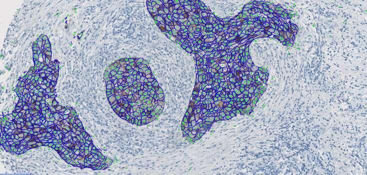
13 FINAL SEGMENTATION
14 MEASURING STAINING INTENSITY Using a special algorithm quantifying brown
15 MEASURING STAINING INTENSITY Measuring average staining intensity (i.e. brown) below the identified membrane label

16 PLANIMETRIC FEATURES Measuring area of membrane, number of nuclei, and average amount of membrane per identified nucleus.
17 Preliminary results of comparison to consensus scores obtained by manual microscopy 7,5 HercepTest (Dako) 7 6,5 Score by Visiopharm image analysis algorithm 6 5,5 5 4,5 n=71 (work-inprogress) 4 3, Consensus score by Manual Microscopy
18 Please feel free to visit the Visiopharm booth for further discussions and a live demo by Jakob Raundahl, PhD
Image analysis in IHC overview, considerations and applications
 Image analysis in IHC overview, considerations and applications Rasmus Røge, MD, Institute of Pathology, Aalborg University Hospital NordiQC workshop September 2016 Aalborg, Denmark Outline Theory Image
Next generation image analysis for immunohistochemistry quantitation
 Next generation image analysis for immunohistochemistry quantitation Ben Vainer Department of Pathology, Rigshospitalet University of Copenhagen Medical Center Men are only so good as their technical developments
HER2 CISH pharmdx TM Kit Interpretation Guide Breast Cancer
 P A T H O L O G Y HER2 CISH pharmdx TM Kit Interpretation Guide Breast Cancer FROM CERTAINTY COMES TRUST For in vitro diagnostic use HER2 CISH pharmdx Kit HER2 CISH pharmdx Kit is intended for dual-color
HER2 FISH pharmdx TM Interpretation Guide - Breast Cancer
 P A T H O L O G Y HER2 FISH pharmdx TM Interpretation Guide - Breast Cancer For In Vitro Diagnostic Use FDA approved as an aid in the assessment of patients for whom Herceptin TM (trastuzumab) treatment
Use of tissue micro array (TMA) in routine clinical analysis
 in routine clinical analysis") 2nd European Workshop on Tissue Imaging and Analysis June 25-26, 2010 Use of tissue micro array (TMA) in routine clinical analysis Tim Svenstrup Poulsen Molecular unit Dept. pathology Herlev Hospital Denmark
2nd European Workshop on Tissue Imaging and Analysis June 25-26, 2010 Use of tissue micro array (TMA) in routine clinical analysis Tim Svenstrup Poulsen Molecular unit Dept. pathology Herlev Hospital Denmark
NordiQC External Quality Assurance in Immunohistochemistry
 NordiQC External Quality Assurance in Immunohistochemistry Mogens Vyberg Professor of Clinical Pathology Director of NordiQC Aalborg University Hospital, Aalborg, Denmark AALBORG (~ 200.000 inhabitants)
Abstract. Background. Objective
 Molecular epidemiology of clinical tissues with multi-parameter IHC Poster 237 J Ruan 1, T Hope 1, J Rheinhardt 2, D Wang 2, R Levenson 1, T Nielsen 3, H Gardner 2, C Hoyt 1 1 CRi, Woburn, Massachusetts,
COMPUTER-AIDED HER-2/neu EVALUATION IN EXTERNAL QUALITY ASSURANCE (EQA) OF BREAST CANCER SCREENING PROGRAMME
 OF BREAST CANCER SCREENING PROGRAMME") COMPUTER-AIDED HER-2/neu EVALUATION IN EXTERNAL QUALITY ASSURANCE (EQA) OF BREAST CANCER SCREENING PROGRAMME Maria Lunardi MD Anatomic Pathology Fracastoro Hospital San Bonifacio, Verona -Italy HER2-neu
COMPUTER-AIDED HER-2/neu EVALUATION IN EXTERNAL QUALITY ASSURANCE (EQA) OF BREAST CANCER SCREENING PROGRAMME Maria Lunardi MD Anatomic Pathology Fracastoro Hospital San Bonifacio, Verona -Italy HER2-neu
Quality assurance and quality control in pathology in breast disease centers
 Quality assurance and quality control in pathology in breast disease centers Judith Sandbank M.D. Pathology Assaf-Harofeh Medical Center ISRAEL jsandbank@asaf.health.gov.il 1 st IBDC, 28 th January, 2011
Quantitative Image Analysis of HER2 Immunohistochemistry for Breast Cancer
 Quantitative Image Analysis of HER2 Immunohistochemistry for Breast Cancer Guideline from the College of American Pathologists Early Online Release Publication: Archives of Pathology & Laboratory Medicine
Dr. dr. Primariadewi R, SpPA(K)
") Curriculum Vitae Dr. dr. Primariadewi R, SpPA(K) Education : Medical Doctor from UKRIDA Doctoral Degree from Faculty of Medicine University of Indonesia Pathologist Specialist and Consultant from Faculty
Curriculum Vitae Dr. dr. Primariadewi R, SpPA(K) Education : Medical Doctor from UKRIDA Doctoral Degree from Faculty of Medicine University of Indonesia Pathologist Specialist and Consultant from Faculty
Product Introduction
 Product Introduction Product Codes: HCL026, HCL027 and HCL028 Contents Introduction to HER2 2 HER2 immunohistochemistry 3 Cell lines as controls 5 HER2 Analyte Control DR IHC 7 HER2 Analyte Control DR
Next-Gen Analytics in Digital Pathology
 Next-Gen Analytics in Digital Pathology Cliff Hoyt, CTO Cambridge Research & Instrumentation April 29, 2010 Seeing life in a new light 1 Digital Pathology Today Acquisition, storage, dissemination, remote
Breast cancer: Antibody selection, protocol optimzation controls and EQA
 Breast cancer: Antibody selection, protocol optimzation controls and EQA Workshop in Diagnostic Immunohistochemistry Oud St. Jan/ Old St. John Brugge (Bruges), Belgium June 13th 15nd 2018 Rasmus Røge,
External Quality Assessment of Breast Marker Analysis. NordiQC data
 External Quality Assessment of Breast Marker Analysis NordiQC data Søren Nielsen Scheme Manager NordiQC Aalborg University Hospital, Denmark Aalborg 12.06 2015 Markers assessed in NordiQC Predictive markers
NordiQC - update
 NordiQC - update 00-0 EQUALIS Uppsala 0 Tomas Seidal NordiQC participants NordiQC participants n:30 S DK N 6 F Ice Bel 54 NL 4 Ger 6 Aust USA 0 It 8 Argent 8.. 96% participation in S,DK & N ~ 60% in Finland
Welcome! HER2 TESTING DIAGNOSTIC ACCURACY 4/11/2016
 HER2 TESTING DIAGNOSTIC ACCURACY Can t We Finally Get It Right? Allen M. Gown, M.D. Medical Director and Chief Pathologist PhenoPath Laboratories Seattle, Washington Clinical Professor of Pathology University
Three Hours Thirty Minutes
 INTERPRETATION HER2 IQFISH pharmdx TM Interpretation Guide Three Hours Thirty Minutes it s about time Breast carcinoma (FFPE) stained with HER2 IQFISH pharmdx Gastric cancer (FFPE) stained with HER2 IQFISH
Supplementary Online Content
 Supplementary Online Content Fumagalli D, Venet D, Ignatiadis M, et al. RNA Sequencing to predict response to neoadjuvant anti-her2 therapy: a secondary analysis of the NeoALTTO randomized clinical trial.
IT S ABOUT TIME. IQFISH pharmdx Interpretation Guide THREEHOURSTHIRTYMINUTES. HER2 IQFISH pharmdxtm. TOP2A IQFISH pharmdxtm
 I N T E R P R E TAT I O N IQFISH pharmdx Interpretation Guide TM HER2 IQFISH pharmdxtm TOP2A IQFISH pharmdxtm Breast carcinoma (FFPE) stained with HER2 IQFISH pharmdx Breast carcinoma (FFPE) stained with
The unknown primary tumour: IHC classification part I, the primary panel - Antibody selection, protocol optimization, controls and EQA
 The unknown primary tumour: IHC classification part I, Mogens Vyberg Professor of Clinical Pathology Director of NordiQC Aalborg University Hospital, Aalborg, Denmark the primary panel - Antibody selection,
Reviewer's report. Version: 1 Date: 24 May Reviewer: Cathy Moelans. Reviewer's report:
 Reviewer's report Title: Validation of HER2 testing with core needle biopsy specimens from primary breast cancers in terms of interobserver reproducibility and concordance with surgically resected specimens
Visual interpretation in pathology
 13 Visual interpretation in pathology Tissue architecture (alteration) evaluation e.g., for grading prostate cancer Immunohistochemistry (IHC) staining scoring e.g., HER2 in breast cancer (companion diagnostic
Instant Quality FISH. The name says it all.
 COMPANION DIAGNOSTICS Instant Quality FISH Instant Quality FISH. The name says it all. IQ: Instant Quality every time. Breast carcinoma stained with : Triple filter showing Blue DAPI colors nuclei, FITC
HER2 ISH (BRISH or FISH)
") Assessment Run H14 2018 HER2 ISH (BRISH or FISH) Material Table 1. Content of the multi-block used for the NordiQC HER2 ISH assessment, run H14 HER2 IHC* IHC score Dual - SISH** FISH*** FISH*** HER2/chr17
Assessment Run H14 2018 HER2 ISH (BRISH or FISH) Material Table 1. Content of the multi-block used for the NordiQC HER2 ISH assessment, run H14 HER2 IHC* IHC score Dual - SISH** FISH*** FISH*** HER2/chr17
Digital Pathology and CAP Guidelines
 Digital Pathology and CAP Guidelines Frequently asked questions The VENTANA family of digital pathology products empowers you with the convenience of a comprehensive image and workflow solution. When used
The unkown primary tumour: IHC Classification, antibody selection, protocol optimization, controls and EQA (part I)
") The unkown primary tumour: IHC Classification, antibody selection, protocol optimization, Mogens Vyberg Professor of Clinical Pathology Director of NordiQC Aalborg University Hospital, Aalborg, Denmark
The unkown primary tumour: IHC Classification, antibody selection, protocol optimization, Mogens Vyberg Professor of Clinical Pathology Director of NordiQC Aalborg University Hospital, Aalborg, Denmark
DENOMINATOR: All melanoma pathology reports for primary malignant cutaneous melanoma
 Quality ID #397: Melanoma Reporting National Quality Strategy Domain: Communication and Care Coordination Meaningful Measure Area: Transfer of Health Information and Interoperability 2019 COLLECTION TYPE:
Quality Assurance and Quality Control in the Pathology Dept.
 Quality Assurance and Quality Control in the Pathology Dept. Judith Sandbank M.D. Pathology Assaf-Harofeh Medical Center ISRAEL jsandbank@asaf.health.gov.il 2 nd IBDC, 9 th February, 2012 Pathology as
Technique and feasibility of a dual staining method for estrogen receptors and AgNORs
 151 Technical note Technique and feasibility of a dual staining method for estrogen receptors and AgNORs Lukas Günther a, and Peter Hufnagl b a Department of Surgery, University of Heidelberg, Heidelberg,
Prosigna BREAST CANCER PROGNOSTIC GENE SIGNATURE ASSAY
 Prosigna BREAST CANCER PROGNOSTIC GENE SIGNATURE ASSAY Methodology The test is based on the reported 50-gene classifier algorithm originally named PAM50 and is performed on the ncounter Dx Analysis System
Prosigna BREAST CANCER PROGNOSTIC GENE SIGNATURE ASSAY
 Prosigna BREAST CANCER PROGNOSTIC GENE SIGNATURE ASSAY GENE EXPRESSION PROFILING WITH PROSIGNA What is Prosigna? Prosigna Breast Cancer Prognostic Gene Signature Assay is an FDA-approved assay which provides
Breast Cancer Interpretation Guide
 Breast Cancer Interpretation Guide UCT D O R P NEW ERBB2/ C E P S ht e ZytoLig lor Prob o C l a u 2D D17S12 ng to the i d r o c c a ting for re-tes idelines 2013 ASCO Gu Breast Cancer Interpretation Guide
PROTOCOL SENTINEL NODE BIOPSY (NON OPERATIVE) BREAST CANCER - PATHOLOGY ASSESSMENT
 BREAST CANCER - PATHOLOGY ASSESSMENT") PROTOCOL SENTINEL NODE BIOPSY (NON OPERATIVE) BREAST CANCER - PATHOLOGY ASSESSMENT Author: Dr Sally Ann Hales On behalf of the Breast and pathology CNGs Written: March 2005 Reviewed by CNG: June 2009 &
PROTOCOL SENTINEL NODE BIOPSY (NON OPERATIVE) BREAST CANCER - PATHOLOGY ASSESSMENT Author: Dr Sally Ann Hales On behalf of the Breast and pathology CNGs Written: March 2005 Reviewed by CNG: June 2009 &
Interpreting Therapeutic Response on Immune Cell Number and Spatial Distribution within the Tumor Microenvironment. Lorcan Sherry, CSO OracleBio
 Interpreting Therapeutic Response on Immune Cell Number and Spatial Distribution within the Tumor Microenvironment Lorcan Sherry, CSO OracleBio Company Overview OracleBio is a specialised CRO providing
Combination of life science entrepreneurs, software development and machine vision experts & recognized scientists.
 Combination of life science entrepreneurs, software development and machine vision experts & recognized scientists. Kaisa Helminen CEO Previously: Sartorius Thermo Scientific Finnzymes Johan Lundin MD,
Quality Assurance in Immunohistochemistry: Experiences from NordiQC
 Nordic immunohistochemical Quality Control 2 Quality Assurance in Immunohistochemistry: Experiences from NordiQC Prof. Mogens Vyberg NordiQC Institute of Pathology Aalborg University Hospital Aalborg,
Layered-IHC (L-IHC): A novel and robust approach to multiplexed immunohistochemistry So many markers and so little tissue
: A novel and robust approach to multiplexed immunohistochemistry So many markers and so little tissue") Page 1 The need for multiplex detection of tissue biomarkers. There is a constant and growing demand for increased biomarker analysis in human tissue specimens. Analysis of tissue biomarkers is key to
Page 1 The need for multiplex detection of tissue biomarkers. There is a constant and growing demand for increased biomarker analysis in human tissue specimens. Analysis of tissue biomarkers is key to
Deciphering the biology that drives response to immunotherapy
 Deciphering the biology that drives response to immunotherapy Phenoptics TM Quantitative Pathology Platform Trent Norris, Field Application Scientist September 15, 2016 HUMAN HEALTH ENVIRONMENTAL HEALTH
Assessment Run B HER-2
 Assessment Run B1 2006 HER-2 The slide to be stained for HER-2 comprised: 1. Cell line JIMT-1 (Amplified)* 2. Cell line MDA-453 (Amplified) 3. Cell line MCF-7 (Not amplified) 4. Cell line BT474 (Amplified)
Assessment Run B HER-2 IHC. HER-2/chr17 ratio**
 Assessment Run B2 20 HER-2 IHC Material The slide to be stained for HER-2 comprised the following 5 tissues: IHC HER-2 Score* (0, +, 2+,3+) FISH HER-2/chr7 ratio**. Breast ductal carcinoma 0..3 2. Breast
Assessment Run B HER2 IHC
 Assessment Run B26 208 HER2 IHC Material The slide to be stained for HER2 comprised the following 5 materials: IHC: HER2 Score* (0, +, 2+, 3+) FISH: HER2 gene/chr 7 ratio**. Breast carcinoma, no. 2+..3
MEDICAL POLICY. Proprietary Information of YourCare Health Plan
 MEDICAL POLICY SUBJECT: HER-2 TESTING IN INVASIVE BREAST OR PAGE: 1 OF: 7 If the member's subscriber contract excludes coverage for a specific service it is not covered under that contract. In such cases,
Assessment of Breast Cancer with Borderline HER2 Status Using MIP Microarray
 Assessment of Breast Cancer with Borderline HER2 Status Using MIP Microarray Hui Chen, Aysegul A Sahin, Xinyan Lu, Lei Huo, Rajesh R Singh, Ronald Abraham, Shumaila Virani, Bal Mukund Mishra, Russell Broaddus,
Guideline. Associated Documents ASCO CAP 2018 GUIDELINES and SUPPLEMENTS -
 Guideline Subject: ASCO CAP 2018 HER2 Testing for Breast Cancer Guidelines - Recommendations for Practice in Australasia Approval Date: December 2018 Review Date: December 2022 Review By: HER2 testing
Tissue microarray TMA
 Tissue microarray TMA Tibor Krenacs 1st Department of Pathology & Experimental Cancer Research Semmelweis University Budapest Austro-Hungarian Pathology Congress October 1-3rd 2009 TMA - tissue microarray
Immunohistochemical classification of the unknown primary tumour (UPT) Part I. Prof. Mogens Vyberg NordiQC Institute of Pathology Aalborg, Denmark
 Part I. Prof. Mogens Vyberg NordiQC Institute of Pathology Aalborg, Denmark") Immunohistochemical classification of the unknown primary tumour (UPT) Part I Prof. Mogens Vyberg NordiQC Institute of Pathology Aalborg, Denmark Tumours of unknown origin: Histology Brain tumour - biopsy
Immunohistochemical classification of the unknown primary tumour (UPT) Part I Prof. Mogens Vyberg NordiQC Institute of Pathology Aalborg, Denmark Tumours of unknown origin: Histology Brain tumour - biopsy
Does digital IHC need digital tissue controls?
 VILNIUS UNIVERSITY Digital immunohistochemistry platform for the staining variation monitoring based on integration of image and statistical analyses with laboratory information system Arvydas Laurinavicius
MEDICAL POLICY. Proprietary Information of Excellus Health Plan, Inc. A nonprofit independent licensee of the BlueCross BlueShield Association
 MEDICAL POLICY SUBJECT: HER-2 TESTING IN INVASIVE BREAST OR PAGE: 1 OF: 7 If a product excludes coverage for a service, it is not covered, and medical policy criteria do not apply. If a commercial product,
Assessment Run B HER2 IHC
 Assessment Run B24 2017 HER2 IHC Material The slide to be stained for HER2 comprised the following 5 materials: IHC: HER2 Score* (0, 1+, 2+, 3+) FISH: HER2 gene/chr 17 ratio** 1. Breast carcinoma, no.
Disruptive innovation in molecular diagnostics. Hilde Windels CEO Biocartis 25 March 2017
 Disruptive innovation in molecular diagnostics Hilde Windels CEO Biocartis 25 March 2017 1 One of the key innovations in healthcare in the last decade PERSONALISED MEDICINE or HIGH PRECISION MEDICINE From
UCL-Advanced Diagnostics. 2015/16 Service Update
 UCL-Advanced Diagnostics 2015/16 Service Update About UCL-Advanced Diagnostics UCL Advanced Diagnostics (UCL-AD) is the immunohistochemistry, in situ and molecular diagnostics service arm of the UCL-Cancer
Removal of sentinel lymph node(s)
") Removal of sentinel lymph node(s) Exceptional healthcare, personally delivered What is a sentinel lymph node? The sentinel lymph nodes (glands) are the first lymph nodes in your armpit to which breast
Removal of sentinel lymph node(s) Exceptional healthcare, personally delivered What is a sentinel lymph node? The sentinel lymph nodes (glands) are the first lymph nodes in your armpit to which breast
Immune Cell Phenotyping in Solid Tumors using Quantitative Pathology
 Immune Cell Phenotyping in Solid Tumors using Quantitative Pathology James R. Mansfield Director of Quantitative Pathology Applications 2009 PerkinElmer What is Quantitative Pathology? Quantitative Pathology
Comparative Analysis of Methods Used in Breast Cancer HER2 and Sentinel Lymph Node Diagnosis
 SHORT THESIS FOR THE DEGREE OF DOCTOR OF PHILOSOPHY (Ph.D.) Comparative Analysis of Methods Used in Breast Cancer HER2 and Sentinel Lymph Node Diagnosis by Monika Francz MD Supervisor: Zoltán Szöllősi
Workflow. Connecting the Pieces For Total Patient Care
 Workflow Connecting the Pieces For Total Patient Care Biocare provides a full line of IHC and molecular pathology products for cancer and infectious disease diagnosis. From a full range of equipment: including
Digital image analysis: a review of reproducibility, stability and basic requirements for optimal results
 APMIS 120: 276 289 Review Article Ó 2011 The Authors APMIS Ó 2011 APMIS DOI 10.1111/j.1600-0463.2011.02854.x Digital image analysis: a review of reproducibility, stability and basic requirements for optimal
CC01 - Colon Cancer Tissue Microarray
 Reveal Biosciences offers Histochemical Staining, Immunohistochemistry (IHC), In Situ Hybridization (ISH), Whole Slide Imaging, and Quantitative Image Analysis on any TMA CC01 - Colon Cancer Tissue Microarray
College of American Pathologists. Pathology Performance Measures included in CMS 2012 PQRS
 College of American Pathologists Pathology Performance Measures included in CMS 2012 PQRS Breast Cancer Resection Pathology Reporting Measure #99 pt category (primary tumor) and pn category (regional lymph
Case 26 Male 37. Right jawline 5mm nodule?keloid. The best diagnosis is:
 Case 26 Male 37. Right jawline 5mm nodule?keloid. The best diagnosis is: A. Desmoplastic Spitz naevus B. Atypical Spitz Tumour C. Spitzoid melanoma D. Deep penetrating naevus E. Spitz naevus Case 26: M
Bioimaging and Functional Genomics
 Bioimaging and Functional Genomics Elisa Ficarra, EPF Lausanne Giovanni De Micheli, EPF Lausanne Sungroh Yoon, Stanford University Luca Benini, University of Bologna Enrico Macii,, Politecnico di Torino
Conflict of Interest. Thank you. I have no relevant conflicts of interest in regards to the content of this presentation.
 What now... Conflict of Interest I have no relevant conflicts of interest in regards to the content of this presentation. Thank you Amy Clayton, MD Mayo Clinic Rochester, MN The Duke Experience 2005 2013
Automatic analysis of virtual slides to help in the determination of well established prognostic parameters in breast carcinomas
 Loews hotel Le Concorde, Quebec Canada August 3-5, 2011 H.I.Q Automatic analysis of virtual slides to help in the determination of well established prognostic parameters in breast carcinomas N Elie 1,
Digital Pathology Diagnosis Assistance System
 August 3, 2011 Digital Pathology Diagnosis Assistance System Computer s s eye and Pathologist s s eye Ogura M, Saito A, Graf HP, Marugame A, Kiyuna T, Yamashita Y, Cosatto E, Malon C & Fukumoto M. Innovative
Clinical utility of cancer biomarkers assessed by virtual microscopy
 Clinical utility of cancer biomarkers assessed by virtual microscopy Lina Seiz Clinical Research Unit Head: Prof. Dr. Manfred Schmitt Department of Obstetrics and Gynecology Klinikum rechts der Isar Established
Classification of the unknown primary tumour: the primary IHC panel
 CIQC/CAP-ACP SEMINAR 2013: DIAGNOSTIC IHC AND MOLECULAR PATHOLOGY Classification of the unknown primary tumour: the primary IHC panel Aalborg University Hospital Denmark Tumours of unknown origin: Histology
Quality control of PD-L1 in NSCLC - Results of two German ring trials Dr. Andreas Scheel University Hospital Cologne
 Quality control of PD-L1 in NSCLC - Results of two German ring trials 2017-06-07 Dr. Andreas Scheel University Hospital Cologne Step-wise approach Predictive value Biological significance Technical aspects
Case Scenario 1 History and Physical 3/15/13 Imaging Pathology
 Case Scenario 1 History and Physical 3/15/13 The patient is an 84 year old white female who presented with an abnormal mammogram. The patient has a five year history of refractory anemia with ringed sideroblasts
Evaluation of Breast Specimens Removed by Needle Localization Technique
 Evaluation of Breast Specimens Removed by Needle Localization Technique Specimen Handling: The breast specimen when received should be measured and grossly inspected for any orientation designated by the
Whitney A. High, MD, JD, MEng
 ADS Dermatopathology Meeting 2014 Selected Adnexal Tumors Whitney A. High, MD, JD, MEng Associate Professor, Dermatology & Pathology Director of Dermatopathology (Dermatology) University of Colorado School
Dako IT S ABOUT TIME. Interpretation Guide. Agilent Pathology Solutions. ALK, ROS1 and RET IQFISH probes (Dako Omnis) MET IQFISH probe (Dako Omnis)
 MET IQFISH probe (Dako Omnis)") INTERPRETATION Dako Agilent Pathology Solutions IQFISH Interpretation Guide ALK, ROS1 and RET IQFISH probes (Dako Omnis) MET IQFISH probe (Dako Omnis) IT S ABOUT TIME For In Vitro Diagnostic Use ALK, ROS1,
INTERPRETATION Dako Agilent Pathology Solutions IQFISH Interpretation Guide ALK, ROS1 and RET IQFISH probes (Dako Omnis) MET IQFISH probe (Dako Omnis) IT S ABOUT TIME For In Vitro Diagnostic Use ALK, ROS1,
Version 2 of these Guidelines were drafted in response to published updated ASCO/CAP HER2 test Guideline Recommendations-
 Introduction: These guidelines represent systematically developed statements to assist in the provision of quality assured HER2 testing in breast and gastric/ gastro-oesophageal carcinoma. They are based
Definiens. Tissue Studio 4.2. Tutorial 8: Cell Simulation and Classification
 Definiens Tissue Studio 4.2 Tutorial 8: Cell Simulation and Classification Tutorial 8: Cell Simulation and Classification Imprint and Version Copyright 2015 Definiens AG. All rights reserved. This document
CANCER. Clinical Validation of Breast Cancer Predictive Markers
 Clinical Validation of Breast Cancer Predictive Markers David Hicks, MD Loralee McMahon, MS, HTL(ASCP) CANCER The human body is composed of billions of highly regulated cells Cancer cells no longer respond
NIH Public Access Author Manuscript Cancer Epidemiol Biomarkers Prev. Author manuscript; available in PMC 2011 January 1.
 NIH Public Access Author Manuscript Published in final edited form as: Cancer Epidemiol Biomarkers Prev. 2010 January ; 19(1): 144 147. doi:10.1158/1055-9965.epi-09-0807. Feasibility Study for Collection
Gaining New Insights Through IF Multiplexed Staining and Analysis. Tyna Hope, Ph.D. P.Eng Biomarker Imaging Research Laboratory October 5, 2017
 Gaining New Insights Through IF Multiplexed Staining and Analysis Tyna Hope, Ph.D. P.Eng Biomarker Imaging Research Laboratory Assessing more from Tissue Sections Gaps with More Common Methods Most common
PRODUCT INFORMATION. New Reagents for Dako CoverStainer. Choose the H&E staining intensity you want.
 PRODUCT INFORMATION Primary Staining Dako CoverStainer Reagents New Reagents for Dako CoverStainer. Choose the H&E staining intensity you want. New flexible staining intensities to fit y Dako H&E Staining
The High-End Solution for Real-Time Patient Tracking
 The High-End Solution for Real-Time Patient Tracking You trust your planning and delivery systems to constantly achieve the best possible results. Now you can trust the Catalyst to provide the same level
Sentinel Node Biopsy in the Treatment of Oral Cancer. Patient Information Leaflet
 Sentinel Node Biopsy in the Treatment of Oral Cancer Patient Information Leaflet Introduction Please read carefully through this information sheet, which describes a new procedure we are providing at this
Breast Unit - University of Heidelberg - Heidelberg, Germany
 - Heidelberg, Germany General Information New breast cancer cases treated per year 600 Breast multidisciplinarity team members 17 Radiologists, surgeons, pathologists, medical oncologists, radiotherapists
DIAGNOSTIC SLIDE SEMINAR: PART 1 RENAL TUMOUR BIOPSY CASES
 DIAGNOSTIC SLIDE SEMINAR: PART 1 RENAL TUMOUR BIOPSY CASES Dr. Andrew J. Evans MD, PhD, FACP, FRCPC Consultant in Genitourinary Pathology University Health Network, Toronto, ON Case 1 43 year-old female,
(a) Significant biological processes (upper panel) and disease biomarkers (lower panel)
 Significant biological processes (upper panel) and disease biomarkers (lower panel)") Supplementary Figure 1. Functional enrichment analyses of secretomic proteins. (a) Significant biological processes (upper panel) and disease biomarkers (lower panel) 2 involved by hrab37-mediated secretory
Supplementary Figure 1. Functional enrichment analyses of secretomic proteins. (a) Significant biological processes (upper panel) and disease biomarkers (lower panel) 2 involved by hrab37-mediated secretory
PD-L1 Analyte Control DR
 Quality in Control PD-L1 Analyte Control DR PD-L1_PI_v2 Product Codes: HCL019, HCL020 and HCL021 Contents PD-L1 Analyte Control DR 2 What is PD-L1? 3 The Role of PD-L1 in Cancer 3 PD-L1 Assessment 4 PD-L1
Assessment performed on Friday, September 18, 2015, at Vancouver General Hospital
 Assessors report for ciqc Run 49: ATRX (June 2015) Assessors: S Yip and J Won (recorder) Assessment performed on Friday, September 18, 2015, at Vancouver General Hospital Background The combined application
Supplementary Online Content
 Supplementary Online Content Rimm DL, Han G, Taube JM, et al. A prospective, multi-institutional, pathologistbased assessment of 4 immunohistochemistry assays for PD-L1 expression in non small cell lung
LN04 - Lymphoma Tissue Microarray
 Reveal Biosciences offers Histochemical Staining, Immunohistochemistry (IHC), In Situ Hybridization (ISH), Whole Slide Imaging, and Quantitative Image Analysis on any TMA LN04 - Lymphoma Tissue Microarray
Anti-hTERT Antibody (SCD-A7)
") Quality in Control Anti-hTERT Antibody (SCD-A7) htert_ab_pi_v1 Product Code: HCL025 Contents htert and Telomerase 2 Negative htert 3 Positive htert 4 Guidance and additional data 5 Case Study: RMH12-001
Quality in Control Anti-hTERT Antibody (SCD-A7) htert_ab_pi_v1 Product Code: HCL025 Contents htert and Telomerase 2 Negative htert 3 Positive htert 4 Guidance and additional data 5 Case Study: RMH12-001
Priti Lal, MD, 1 Paulo A. Salazar, 1 Clifford A. Hudis, MD, 2 Marc Ladanyi, MD, 1 and Beiyun Chen, MD, PhD 1. Abstract
 Anatomic Pathology / DUAL- VS SINGLE-COLOR SCORING IN IMMUNOHISTOCHEMICAL AND FISH HER-2 TESTING HER-2 Testing in Breast Cancer Using Immunohistochemical Analysis and Fluorescence In Situ Hybridization
Mamma Centrum / Zelený Pruh - Prague, Czech Republic
 - Prague, Czech Republic General Information New breast cancer cases treated per year 490 Breast multidisciplinarity team members 29 Radiologists, surgeons, pathologists, medical oncologists, radiotherapists
Determination of HER2 Amplification by In Situ Hybridization. When Should Chromosome 17 Also Be Determined?
 Anatomic Pathology / FISH f o r HER2: Wh e n to Use Ch r o m o s o m e 17 Determination of HER2 Amplification by In Situ Hybridization When Should Chromosome 17 Also Be Determined? John M.S. Bartlett,
Update: Morphologic Considerations in Mesothelioma within the Pleural and Peritoneal Cavities. Douglas J. Hartman, MD June 7, 2018
 Update: Morphologic Considerations in Mesothelioma within the Pleural and Peritoneal Cavities Douglas J. Hartman, MD June 7, 2018 Objectives Review Historical Features associated with prognosis Present
HSL-Advanced Diagnostics 2018 / 19 Test & Service List
 HSL-Advanced Diagnostics 2018 / 19 Test & Service List 2018/19 TEST & SERVICE LIST Haematoxylin & Eosin H&E H&E per slide Routine Immunohistochemistry Immunohistochemical demonstration of an antigen in
A View to the Future: The Development of Targeted Therapy for Melanoma. Michael Davies, M.D., Ph.D.
 A View to the Future: Science to Survivorship Symposium September 26, 2009 The Development of Targeted Therapy for Melanoma Michael Davies, M.D., Ph.D. Assistant Professor, Melanoma Medical Oncology How
Final Project Report Sean Fischer CS229 Introduction
 Introduction The field of pathology is concerned with identifying and understanding the biological causes and effects of disease through the study of morphological, cellular, and molecular features in
Breast cancer diagnostic solutions Deliver diagnostic confidence
 Breast cancer diagnostic solutions Deliver diagnostic confidence 2 Breast cancer diagnostic solutions Roche Tissue Diagnostics is committed to improving outcomes in breast cancer Breast cancer...the most
A712(19)- Test slide, Breast cancer tissues with corresponding normal tissues
- Test slide, Breast cancer tissues with corresponding normal tissues") A712(19)- Test slide, Breast cancer tissues with corresponding normal tissues (formalin fixed) For research use only Specifications: No. of cases: 12 Tissue type: Breast cancer tissues with corresponding
A712(19)- Test slide, Breast cancer tissues with corresponding normal tissues (formalin fixed) For research use only Specifications: No. of cases: 12 Tissue type: Breast cancer tissues with corresponding
Optimal algorithm for HER2 testing
 Optimal algorithm for HER2 testing The revised definition of IHC 2+ (equivocal) is invasive breast cancer with Weak to moderate complete membrane staining observed in >10% of tumor cells. (see Figure 1
Contemporary Classification of Breast Cancer
 Contemporary Classification of Breast Cancer Laura C. Collins, M.D. Vice Chair of Anatomic Pathology Professor of Pathology Beth Israel Deaconess Medical Center and Harvard Medical School Boston, MA Outline
10 years of NordiQC Why are 30% of labs still getting it wrong?
 Mogens Vyberg & Søren Nielsen NordiQC Institute of Pathology Aalborg University Hospital Aalborg, Denmark May 29th 2015 10 years of NordiQC Why are 30% of labs still getting it wrong? Nothing to declare
NCDB Vendor Webinar: NCDB Call for Data January 2018 and Upcoming RQRS Revisions
 NCDB Vendor Webinar: NCDB Call for Data January 2018 and Upcoming RQRS Revisions American American College College of of Surgeons 2013 Content 2014 Content cannot be be reproduced or or repurposed without