59 yo male with past medical history of prostate carcinoma, presented with upper abdominal pain
|
|
|
- Percival Webb
- 5 years ago
- Views:
Transcription


1 December yo male with past medical history of prostate carcinoma, presented with upper abdominal pain Contributed by: Divya Sharma, MD. Fellow, Gastrointestinal Pathology, Department of Pathology and Laboratory Medicine, Indiana University School of Medicine Romil Saxena, MBBS, FRCPath Professor,Indiana University School of Medicine, Department of Pathology and Laboratory Medicine Shaoxiong Chen, MD, PhD. Assistant Professor, Indiana University School of Medicine, Department of Pathology and Laboratory Medicine.

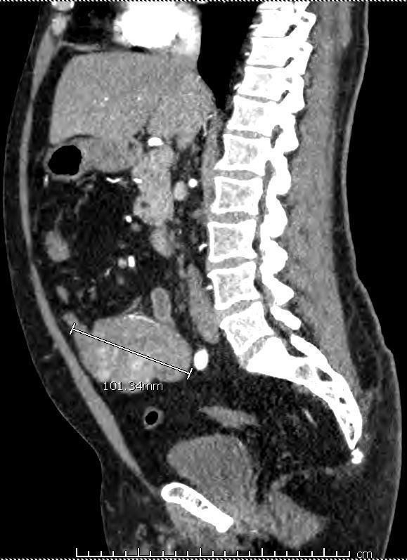
2 Clinical History: 59 yo male with past medical history of prostate carcinoma, presented with upper abdominal pain radiating to the back and melena. CT scan to rule out a dissecting aortic aneurysm revealed an abnormally enhancing, 8x9 cm mesenteric mass that was adherent to the small bowel.the clinical differential diagnosis included desmoid tumor, carcinoid tumor, sclerosing mesenteritis and lymphoma. Radiology: (Figure ) Fig Computer tomography (CT) abdomen and pelvis, coronal section
3 Fig. 1.1

4 1.1- Computer tomography (CT) abdomen and pelvis, axial section Fig Computer tomography (CT) abdomen and pelvis, sagittal section Gross: Small bowel resection specimen revealed a 10.5 x 6x 5.5 cm, friable, partially degenerated, nodular mesenteric mass that extends to the lumen of small bowel. Cut surface was pink tan- red brown solid with areas of cystic degeneration. Micro: The tumor was noted to extend through the muscularis propria and ulcerating the overlying intestinal mucosa (Fig2.0). The tumor cells were arranged in nests, sheets and short fascicles with characteristic radial arrangement around blood vessels (Fig ). Cytologically, they were variably epithelioid, spindly and rhabdoid with abundant clear, granular and palely eosinophilic cytoplasm and round vesicular nuclei with small nucleoli. (Fig )


5 Figure Submucosal mass, ulcerating overlying mucosa. 20x Figure Tumor cells arranged in nests, and sheets showing epithelioid -spindly morphology. 200x


6 Figure 2.2 Figure Tumor cells showing abundant clear to eosinophilic cytoplasm, with rhabdoid like features. 400x


7 Figure 2.4 Figure 2.5 Figure 2.6


8 Immunohistochemical staining:fig Figure Immunohistochemical stain for smooth muscle actin; 400x 2.8 Immunohistochemical stain for Melan A; 400x Figure 2.8
 Perivascular epithelioid cell tumors (PEComas) are an unusual family of")
9 Figure Immunohistochemical stain for HMB-45; 400x Final Diagnosis and Discussion: Perivascular epithelioid cell tumor (PEComa) Perivascular epithelioid cell tumors (PEComas) are an unusual family of neoplasms that show dual smooth muscle and melanocytic differentiation. They arise at any site including soft tissues of extremities, visceral soft tissue (clear cell, sugar tumor, lymphangioleiomyomatosis), kidney (angiomyolipoma), gynecologic tract, and urinary tract, as well as liver pancreas and GI tract(1). Limited data exist on PEComas occurring in the GI tract, but majority of the cases seem to occur in the colon(2-4)in general, PEComas of the visceral sites show female predilection and typically occur in middle aged adults, with a median patient age of 46 years (5). Presenting symptoms of PEComas involving the GI tract may include abdominal pain, GI bleed and obstructive symptoms. Although angiomyolipomas are often associated with tuberous sclerosis, GI PEComas are usually sporadic(3). However, regardless of the presence or absence of the clinical syndrome, most PEComas show loss of TSC1 and TSC2, resulting in upregulation of the mtor pathway, which can be targeted with mtor inhibitors such as sirolimus (6-7). Less frequently, translocations involving the TFE3 gene are present(8). Tumors are usually reasonably well circumscribed but unencapsulated. The cut surface is usually white and solid and may show areas of hemorrhage. Tumor size ranges from 3 cm to 6 cm.
10 The tumor cells of PEComa typically grow in nests, sheets, or short fascicles, and show a characteristic radial arrangement around blood vessels. The cells are variably epithelioid spindled with abundant clear, granular, and pale eosinophilic cytoplasm and round vesicular nuclei with small nucleoli. The cells may also show myoid appearance. Atypia is variable within tumors; malignant forms of PEComa may show striking cytologic atypia. Mitosis is variable with malignant tumors showing higher mitotic rate and necrosis. The vascular network is usually prominent and composed of capillaries and medium-sized thin walled vessels. Given the potentially wide differential diagnosis for these tumors, immunohistochemistry is usually needed to confirm a diagnosis of PEComa. PEComa shows immunohistochemical staining for both myoid and melanocytic differentiation. Smooth muscle markers (alpha-isoform actin, muscle specific, desmin, caldesmon, calponin and smooth-muscle myosin) are variably expressed in terms of intensity and extent. Similarly, markers of melanocytic differentiation (HMB-45, MART-1, tyrosinase and microphthalmia transcription factor [MiTF]) are also variably expressed. HMB-45 is the most frequent ly expressed with expression of S100 being uncommon in PEComa. TFE3 protein is present in a subset of PEComas with TFE3 gene fusions. The expression of TFE3 is mutually exclusive with MiTF expression.(8).kit, keratin, and CD34 expression are typically absent(5). The main differential diagnostic considerations for PEComas of the GI tract include pure smooth muscle tumors (leiomyoma and leiomyosarcoma; HMB45-; Melan A-), epithelioid GIST(KIT+; DOG1+), clear cell sarcoma (S100+; SMA-; Desmin-). The translocation t(12;22)(q13;q12) is also detected in clear cell sarcoma, paraganglioma (synaptophysin+; chromogranin+), metastatic renal cell carcinoma (EMA +; PAX-8 +), and metastatic melanoma (S100 +; SMA-; desmin-). PEComas are tumors of variable and somewhat unpredictable biologic potential. In most cases, they pursue a benign course, but in deep visceral soft tissues, these lesions show a range of clinical behaviors ranging from benign to aggressive, suggested by the presence of marked atypia, high mitotic activity, or necrosis. References: 1. Hornick JL, Fletcher CD. PEComa: what do we know so far? Histopathology 2006;48: Freeman HJ, Webber DL. Perivascular epithelioid cell neoplasm of the colon. World J Gastrointest Oncol 2010;2: Shi HY, Wei LX, Sun L, et al. Clinicopathologic analysis of 4 perivascular epithelioid cell tumors (PEComas) of the gastrointestinal tract. Int J Surg Pathol 2010;18: Ryan P, Nyugen VH, Gholoum S et al. Polypoid PEComa in the rectum of a 15 yearold girl: case report and review of PEComa in the gastrointestinal tract. Am J Surg Pathol 2009;33:
11 5. Folpe AL, Mentzel T, Lehr HA, et al. Perivascular epithelioid cell neoplasms of soft tissue and gynecologic origin: a clinicopathologic study of 26 cases and review of literature. Am J Surg Pathol 2005; 29: Pan CC, Chung MY, Ng KF, et al. Constant allelic alteration on chromosome 16p (TSC2 gene) in perivascular epithelioid cell tumor (PEComa): genetic evidence of relationship of PEComa with angiomyolipoma. J Pathol 2008;214: Wagner AJ, Malinowska-Kolodziej I, Morgan JA, et al. Clinical activity of mtor inhibition with sirolimus in malignant perivascular epithelioid cell tumors: targeting pathogenic activation of mtorc1 in tumors. J Clin Oncol 2010;28: Agrani P, Aulmann S, Illei PB, et al. A distinctive set of PEComas harbors TFE3 gene fusions. Am J Surg Pathol 2010;34:
Financial disclosures
 Mesenchymal Neoplasms with Melanocytic Differentiation By Konstantinos Linos MD, FCAP, FASDP Bone, Soft Tissue and Dermatopathology Assistant Professor of Pathology Dartmouth-Hitchcock Medical Center Geisel
A 25 year old female with a palpable mass in the right lower quadrant of her abdomen
 May 2016 A 25 year old female with a palpable mass in the right lower quadrant of her abdomen Contributed by: Paul Ndekwe, MD, Resident Physician, Indiana University School of Department of Pathology and
Special slide seminar
 Special slide seminar Tomáš Rozkoš The Fingerland Department of Pathology Charles University Medical Faculty and Faculty Hospital in Hradec Králové Czech Republic Case history, 33 years old resistance
A 42-year-old woman with a liver mass
 April 2016 Case of the Month A 42-year-old woman with a liver mass Contributed by: Natalia I. Rush, MD, Resident Physician, Indiana University School of Medicine, Department of Pathology and Laboratory
1/10/2018. Soft Tissue Tumors Showing Melanocytic Differentiation. Overview. Desmoplastic/ Spindle Cell Melanoma
 2016 MFMER slide-1 2016 MFMER slide-2 2016 MFMER slide-3 Soft Tissue Tumors Showing Melanocytic Differentiation Andrew L. Folpe, M.D. Professor of Laboratory Medicine and Pathology Mayo Clinic, Rochester,
Diplomate of the American Board of Pathology in Anatomic and Clinical Pathology
 A 33-year-old male with a left lower leg mass. Contributed by Shaoxiong Chen, MD, PhD Assistant Professor Indiana University School of Medicine/ IU Health Partners Department of Pathology and Laboratory
21/07/2017. Hobnail endothelial cells are not the same as epithelioid endothelial cells
 UPDATE IN CUTANEOUS VASCULAR S DERMATOPATHOLOGY SESSION BELFAST PATHOLOGY JUNE 21/2017 Dr E Calonje St John s Institute of Dermatology, London, United Kingdom THE FAMILY OF VASCULAR S WITH EPITHELIOID
Science & Technologies RETROPERITONEAL TUMOR: DIFFERENTIAL DIAGNOSIS BEYOND THE USUALLY SUSPECTED. Medical University Sofia, Bulgaria
 RETROPERITONEAL TUMOR: DIFFERENTIAL DIAGNOSIS BEYOND THE USUALLY SUSPECTED Vesela Ivanova *, Tihomir Dikov *, Goran Derimachkovski **, Petar Panchev ** * Department of General and Clinical Pathology, Medical
Case 2. Dr. Sathima Natarajan M.D. Kaiser Permanente Medical Center Sunset
 Case 2 Dr. Sathima Natarajan M.D. Kaiser Permanente Medical Center Sunset History 24 year old male presented with a 3 day history of right flank pain, sharp in nature Denies fever, chills, hematuria or
SMOOTH MUSCLE TUMOURS
 SMOOTH MUSCLE TUMOURS NORMAL SMOOTH MUSCLE Cytology Immunohistochemistry Ultrastructure Masson Trichrome Smooth Muscle Ultrastructure Many myofilaments running parallel to the long axis of the smooth
Case 27 Male 42. Painless, static, well-circumscribed, subcutaneous nodule right lower leg,?lipoma. The best diagnosis is:
 Case 27 Male 42. Painless, static, well-circumscribed, subcutaneous nodule right lower leg,?lipoma. The best diagnosis is: A. Angiosarcoma B. Haemangiopericytoma C.Myopericytoma D.Myofibroma E. Angioleiomyoma
ACCME/Disclosures ALK FUSION-POSITIVE MESENCHYMAL TUMORS. Tumor types with ALK rearrangements. Anaplastic Lymphoma Kinase. Jason L.
 Companion Meeting of the International Society of Bone and Soft Tissue Pathology The Evolving Concept of Mesenchymal Tumors ALK FUSION-POSITIVE MESENCHYMAL TUMORS Jason L. Hornick, MD, PhD March 13, 2016
No financial or other disclosures
 Case 2014-5 Esther N. Bit-Ivan, DO Northwestern University Jason Wang, MD Jason Park, MD Korgun Koral, MD Children s Medical Center Charles Timmons, MD Veena Rajaram, MD No financial or other disclosures
أملس عضلي غرن = Leiomyosarcoma. Leiomyosarcoma 1 / 5
 Leiomyosarcoma 1 / 5 EPIDEMIOLOGY Exact incidence is unknown, but older studies suggest that leiomyosarcomas comprise approximately 3 percent of soft-tissue sarcomas. Superficial leiomyosarcoma occurs
Disclosure. Relevant Financial Relationship(s) None. Off Label Usage None MFMER slide-1
 None. Off Label Usage None MFMER slide-1") Disclosure Relevant Financial Relationship(s) None Off Label Usage None 2013 MFMER slide-1 Case Presentation A 43 year old male, with partial nephrectomy for a right kidney mass 2013 MFMER slide-2 2013
Disclosure Relevant Financial Relationship(s) None Off Label Usage None 2013 MFMER slide-1 Case Presentation A 43 year old male, with partial nephrectomy for a right kidney mass 2013 MFMER slide-2 2013
The World Health Organization defines PEComas as mesenchymal
 ORIGINAL ARTICLE Perivascular Epithelioid Cell Neoplasms of Soft Tissue and Gynecologic Origin A Clinicopathologic Study of 26 Cases and Review of the Literature Andrew L. Folpe, MD,* Thomas Mentzel, MD,
Self assessment case. Dr Saleem Taibjee Dorset County Hospital, Dorchester
 Self assessment case Dr Saleem Taibjee saleemtaibjee@gmail.com Dorset County Hospital, Dorchester Clinical details 34-year-old man: Shave excision Skin tag / papilloma left thigh The best diagnosis is:
Case: The patient is a 24 year- old female who was found to have multiple mural nodules within the antrum. Solid and cystic components were noted on
 Case: The patient is a 24 year- old female who was found to have multiple mural nodules within the antrum. Solid and cystic components were noted on imaging. There is no significant past medical history.
International Journal of Health Sciences and Research ISSN:
 International Journal of Health Sciences and Research www.ijhsr.org ISSN: 2249-9571 Case Report Malignant Gastrointestinal Stromal tumor of the Sigmoid Colon with Perforation and Peritonitis - an Unusual
Original Article From angiomyolipoma to malignant epithelioid angiomyolipoma of the kidney, a case report with a history of eight years
 Int J Clin Exp Med 2015;8(11):21252-21256 www.ijcem.com /ISSN:1940-5901/IJCEM0015600 Original Article From angiomyolipoma to malignant epithelioid angiomyolipoma of the kidney, a case report with a history
Perivascular Epithelioid Cell Tumor in the Stomach
 Journal of Pathology and Translational edicine 2017; 51: 428432 CASE STUDY Perivascular Epithelioid Cell Tumor in the Stomach Sun Ah Shin Jiwoon Choi Kyung Chul oon Woo Ho Kim Department of Pathology,
Uterine mesenchymal tumors: Hereditary perspectives
 Uterine mesenchymal tumors: Hereditary perspectives Two hereditary syndromes are known to be related to uterine mesenchymal tumors: Hereditary Leiomyomatosis and Renal Cell Carcinoma syndrome (HLRCC) and
Two cases of perivascular epithelioid cell tumor of the uterus: clinical, radiological and pathological diagnostic challenge
 DOI 10.1186/s40001-017-0248-y European Journal of Medical Research CASE REPORT Open Access Two cases of perivascular epithelioid cell tumor of the uterus: clinical, radiological and pathological diagnostic
Radio-Pathologic Workup of a Retroperitoneal Abdominal Mass
 Radio-Pathologic Workup of a Retroperitoneal Abdominal Mass Joe Carlson Advanced Radiology Clerkship Harvard Medical School Year IV September 12, 2002 84 year old Male Presented to PCP With Abdominal Pain
Desmoplastic Melanoma R/O BCC. Clinical Information. 74 y.o. man with lesion on left side of neck r/o BCC
 R/O BCC Sabine Kohler, M.D. Professor of Pathology and Dermatology Dermatopathology Service Stanford University School of Medicine Clinical Information 74 y.o. man with lesion on left side of neck r/o
Selected Pseudomalignant Soft Tissue Tumors of the Skin and Subcutis
 Selected Pseudomalignant Soft Tissue Tumors of the Skin and Subcutis Andrew L. Folpe, M.D. Professor of Laboratory Medicine and Pathology Mayo Clinic, Rochester, MN folpe.andrew@mayo.edu 2016 MFMER slide-1
Extrapulmonary Lymphangioleiomyoma: Clinicopathological Analysis of 4 Cases
 The Korean Journal of Pathology 2014; 48: 188-192 ORIGINAL ARTICLE Extrapulmonary Lymphangioleiomyoma: Clinicopathological Analysis of 4 Cases Dae Hyun Song In Ho Choi Sang Yun Ha Kang Min Han Jae Jun
3/24/2017 DENDRITIC CELL NEOPLASMS: HISTOLOGY, IMMUNOHISTOCHEMISTRY, AND MOLECULAR GENETICS. Disclosure of Relevant Financial Relationships
 DENDRITIC CELL NEOPLASMS: HISTOLOGY, IMMUNOHISTOCHEMISTRY, AND MOLECULAR GENETICS Jason L. Hornick, M.D., Ph.D. Director of Surgical Pathology and Immunohistochemistry Brigham and Women s Hospital Professor
Cutaneous Mesenchymal Neoplasms with EWSR1 Rearrangement
 Cutaneous Mesenchymal Neoplasms with EWSR1 Rearrangement By Konstantinos Linos MD, FCAP, FASDP Bone, Soft Tissue and Dermatopathology Assistant Professor of Pathology Dartmouth-Hitchcock Medical Center
Radiological Appearance of Renal Leiomyoma: two cases report and review of the literature
 J Radiol Sci 2012; 37: 139-143 Radiological Appearance of Renal Leiomyoma: two cases report and review of the literature Wei-Ni Liao 1 Chi-Kuan Chen 2 Fei-Shih Yang 1,3 Department of Radiology 1, Department
Enterprise Interest Nothing to declare
 Enterprise Interest Nothing to declare Diagnoses one would not like to miss in soft tissue pathology early in your career Marta Sbaraglia, MD Department of Pathology Hospital of Treviso University of Padua
A case of pedunculated intraperitoneal leiomyoma
 Jichi Medical University Journal Chio Shuto Kuniyasu Soda Takayoshi Yoshida Fumio Konishi Abstract We report a very rare case of a pedunculated intraperitoneal leiomyoma in the parietal peritoneum of the
Uncommon of the Uncommon: Malignant Perivascular Epithelioid Cell Tumor of the Lung
 Case Report Thoracic Imaging http://dx.doi.org/10.3348/kjr.2013.14.4.692 pissn 1229-6929 eissn 2005-8330 Korean J Radiol 2013;14(4):692-696 Uncommon of the Uncommon: Malignant Perivascular Epithelioid
Case Presentation. Maha Akkawi, MD, Fatima Obeidat, MD, Tariq Aladily, MD. Department of Pathology Jordan University Hospital Amman, Jordan
 Case Presentation Maha Akkawi, MD, Fatima Obeidat, MD, Tariq Aladily, MD Department of Pathology Jordan University Hospital Amman, Jordan The 25th Annual Congress of the ADIAP The 8/11/2013 1 5th International
Article begins on next page
 Leiomyoma of the Vulva Rutgers University has made this article freely available. Please share how this access benefits you. Your story matters. [https://rucore.libraries.rutgers.edu/rutgers-lib/50624/story/]
Classification (1) Classification (3) Classification (2) Spindle cell lesions. Spindle cell lesions of bladder (Mills et al.
 Classification (3) Classification (2) Spindle cell lesions. Spindle cell lesions of bladder (Mills et al.") Non-epithelial tumours and nonepithelial tumour-like lesions of the bladder Dr Jonathan H Shanks The Christie NHS Foundation Trust, Manchester, UK Classification (1) Myofibroblastic proliferations and
Non-epithelial tumours and nonepithelial tumour-like lesions of the bladder Dr Jonathan H Shanks The Christie NHS Foundation Trust, Manchester, UK Classification (1) Myofibroblastic proliferations and
Case E1. Female aged 63 years Right Nephrectomy Two separate tumours Section of each tumour
 Case E1 Female aged 63 years Right Nephrectomy Two separate tumours Section of each tumour Tumour 1 Upper pole tumour 28mm macro diameter Circumscribed Friable cut surface Tumour 2 Middle pole Part solid
Newer soft tissue entities
 Newer soft tissue entities Examples among fibroblastic tumors Turku, May 6, 2010 Markku Miettinen, M.D. AFIP, Washington, DC Fibroblastic neoplasms Solitary fibrous tumor /Hemangiopericytoma Low-grade
Clear Cell Sarcoma of Right Knee with Bone Marrow Metastasis: A Case Report and review of Literature
 Case Report DOI: 10.21276/APALM.2017.1107 Clear Cell Sarcoma of Right Knee with Bone Marrow Metastasis: A Case Report and review of Literature Divya Shelly*, Shashank Mishra, Divya Gupta and Reena Bharadwaj
Long lasting stable disease with mtor inhibitor treatment in a patient with a perivascular epithelioid cell tumor: A case report and literature review
 ONCOLOGY LETTERS 12: 4739-4743, 2016 Long lasting stable disease with mtor inhibitor treatment in a patient with a perivascular epithelioid cell tumor: A case report and literature review EZEQUIEL FLECHTER
Alveolar Soft Part Sarcoma of the Uterine Cervix: A Case Report and Review of the Literature
 The Korean Journal of Pathology 2014; 48: 361-365 CASE STUDY Alveolar Soft Part Sarcoma of the Uterine Cervix: A Case Report and Review of the Literature Hyun Ju Lee Department of Pathology, Soonchunhyang
Solitary Fibrous Tumor of the Kidney with Massive Retroperitoneal Recurrence. A Case Presentation
 246) Prague Medical Report / Vol. 113 (2012) No. 3, p. 246 250 Solitary Fibrous Tumor of the Kidney with Massive Retroperitoneal Recurrence. A Case Presentation Sfoungaristos S., Papatheodorou M., Kavouras
Case: The patient is a 62 year old woman with a history of renal cell carcinoma that was removed years ago. A 2.4 cm liver mass was found on CT
 Case: The patient is a 62 year old woman with a history of renal cell carcinoma that was removed years ago. A 2.4 cm liver mass was found on CT during follow- up. ALT, AST, Alk Phos and bilirubin were
SOFT TISSUE TUMOR PATHOLOGY: AN UPDATE
 SOFT TISSUE TUMOR PATHOLOGY: AN UPDATE Jason L. Hornick, MD, PhD July 18, 2013 Department of Pathology Brigham and Women s Hospital Harvard Medical School Boston, MA, USA I have no disclosures. New Soft
Gastrointestinal stromal tumor
 Gastrointestinal stromal tumor 영남의대병리학교실 최준혁 Classification of gastrointestinal mesenchymal tumor Gastrointestinal stromal tumor(gist) Smooth muscle tumors : leiomyoma, leiomyosarcoma Neurogenic tumors
Case Report Malignant perivascular epithelioid cell tumor (PEComa) of cervix with TFE3 gene rearrangement: a case report
 of cervix with TFE3 gene rearrangement: a case report") Int J Clin Exp Pathol 2014;7(9):6409-6414 www.ijcep.com /ISSN:1936-2625/IJCEP0001547 Case Report Malignant perivascular epithelioid cell tumor (PEComa) of cervix with TFE3 gene rearrangement: a case report
Int J Clin Exp Pathol 2014;7(9):6409-6414 www.ijcep.com /ISSN:1936-2625/IJCEP0001547 Case Report Malignant perivascular epithelioid cell tumor (PEComa) of cervix with TFE3 gene rearrangement: a case report
GUT-C 11/30/2017. Debasmita Das, M.D. PGY-1 Danbury Hospital
 GUT-C 11/30/2017 Debasmita Das, M.D. PGY-1 Danbury Hospital CLINICAL SUMMARY 8/2017 59 year old female Presented to the ED with 1 month history of general malaise, fever and weight loss PMH: Significant
Financial disclosures
 Cutaneous Mesenchymal Neoplasms with EWSR1 Rearrangement By Konstantinos Linos MD, FCAP, FASDP Bone, Soft Tissue and Dermatopathology Assistant Professor of Pathology Dartmouth-Hitchc Geisel School of
EPITHELIOID ANGIOMYOLIPOMA OF THE KIDNEY MIMICKING RENAL CELL CARCINOMA: A CLINICOPATHOLOGIC ANALYSIS OF CASES AND LITERATURE REVIEW
 EPITHELIOID ANGIOMYOLIPOMA OF THE KIDNEY MIMICKING RENAL CELL CARCINOMA: A CLINICOPATHOLOGIC ANALYSIS OF CASES AND LITERATURE REVIEW Chia-Chun Tsai, 1 Wen-Jeng Wu, 1,3 Ching-Chia Li, 1,3 Chii-Jye Wang,
Images In Gastroenterology
 Images In Gastroenterology Thong-Ngam D, et al. THAI J GASTROENTEROL 2005 Vol. 6 No. 2 May - Aug. 2005 105 Imaging of Gastrointestinal Stromal Tumors Pornpim Fuangtharnthip, M.D. Narumol Hargroove, M.D.
An Overview of Genital Stromal Tumors
 An Overview of Genital Stromal Tumors By Konstantinos Linos MD, FCAP, FASDP Bone, Soft Tissue and Dermatopathology Assistant Professor of Pathology Dartmouth-Hitchcock Medical Center Geisel School of Medicine
Case of the month. Dr Charles Bénière, Institut universitaire de pathologie, Lausanne
 Case of the month Dr Charles Bénière, Institut universitaire de pathologie, Lausanne Clinical history 39 years old male, smoker (19 pack-year) without any prior medical record nor professional exposure.
Cutaneous metastases. Thaddeus Mully. University of California, San Francisco Professor, Departments of Pathology and Dermatology
 Cutaneous metastases Thaddeus Mully University of California, San Francisco Professor, Departments of Pathology and Dermatology DISCLOSURE OF RELATIONSHIPS WITH INDUSTRY Thaddeus Mully Course C005 Essential
Mody. AIS vs. Invasive Adenocarcinoma of the Cervix
 Common Problems in Gynecologic Pathology Michael T. Deavers, M.D. Houston Methodist Hospital, Houston, Texas Common Problems in Gynecologic Pathology Adenocarcinoma in-situ (AIS) of the Cervix vs. Invasive
Respiratory Tract Cytology
 Respiratory Tract Cytology 40 th European Congress of Cytology Liverpool, UK Momin T. Siddiqui M.D. Professor of Pathology and Laboratory Medicine Director of Cytopathology Emory University Hospital, Atlanta,
Endometrial Stromal Tumors
 Endometrial Stromal Tumors WHO Categories: Endometrial Stromal Nodule (ESN) Endometrial Stromal Sarcoma, low grade (LGESS) Endometrial Stromal Sarcoma, high grade (HGESS) Undifferentiated Uterine Sarcoma
What really matters When and Why. Pathology of Uterine Mesenchymal Lesions. Nafisa Wilkinson London
 What really matters When and Why Pathology of Uterine Mesenchymal Lesions Nafisa Wilkinson London Patient centred approach immunohistochemistry Histological diagnosis Next generation sequencing Genetic
Glomus Tumor of the Stomach
 J Radiol Sci 2011; 36: 49-53 Glomus Tumor of the Stomach Yuan-Chun Huang 1 Chen-Te Chou 2 Yu-Cheng Hong 1 Shang-Yun Ho 1 Kwo-Whei Lee 1 Hwa-Koon Wu 1 Department of Medical Imaging 1, Changhua Christian
CyclinD1 Positive High-Grade Endometrial Stromal Sarcoma: A Fascinating Entity!
 Case Report DOI: 10.21276/APALM.1530 CyclinD1 Positive High-Grade Endometrial Stromal Sarcoma: A Fascinating Entity! Divya Shelly*, Imtiaz Ahmed, Sampath K. Srinivasagowda and Reena Bharadwaj Department
Cellular Neurothekeoma
 Cellular Neurothekeoma Scott W Binder, MD Pritzker Professor of Pathology & Dermatology Sr. Vice Chair Director, Pathology Clinical Services Chief, Dermatopathology Geffen/UCLA School of Medicine Clinical
Malignant Peripheral Nerve Sheath Tumor
 C H A P T E R 120 Malignant Peripheral Nerve Sheath Tumor Currently, malignant peripheral nerve sheath tumor (MPNST) is the most commonly used generic name for the neoplasms known in the past as neurosarcoma,
hemangioblastoma of the retroperitoneum
 Int J Clin Exp Pathol 2014;7(4):1777-1781 www.ijcep.com /ISSN:1936-2625/IJCEP1401042 Case Report Yong Huang 1, Xiang-Chun Han 2, Guo-Shi Lv 3 1 Department of Pathology, 251 Hospital of PLA, Zhangjiakou,
Immunohistochemical Evaluation of Necrotic Malignant Melanomas
 Anatomic Pathology / EVALUATION OF NECROTIC MALIGNANT MELANOMAS Immunohistochemical Evaluation of Necrotic Malignant Melanomas Daisuke Nonaka, MD, Jordan Laser, MD, Rachel Tucker, HTL(ASCP), and Jonathan
Intussuception due to gastrointestinal stromal tumor with neural differentiation in a patient with. Von Recklinghausen Neurofibromatosis,
 Turkish Journal of Cancer Vol 31/ No.4 /2001 Intussuception due to gastrointestinal stromal tumor with neural differentiation in a patient with Von Recklinghausen Neurofibromatosis (NF-1): A case report
Kidney Case 1 SURGICAL PATHOLOGY REPORT
 Kidney Case 1 Surgical Pathology Report February 9, 2007 Clinical History: This 45 year old woman was found to have a left renal mass. CT urography with reconstruction revealed a 2 cm medial mass which
The Relevance of Cytologic Atypia in Cutaneous Neural Tumors
 The Relevance of Cytologic Atypia in Cutaneous Neural Tumors Recent Findings - New Developments New Problems Zsolt B. Argenyi, M.D. Professor of Pathology & Dermatology Director of Dermatopathology Department
Diagnosis of a granular cell tumour at the abdominal wall using fine needle aspiration cytology and histology: Case report
 Case Report Diagnosis of a granular cell tumour at the abdominal wall using fine needle aspiration cytology and histology: Case report Journal of International Medical Research 2015, Vol. 43(4) 592 596!
Synonyms. Nephrogenic metaplasia Mesonephric adenoma
 Nephrogenic Adenoma Synonyms Nephrogenic metaplasia Mesonephric adenoma Definition Benign epithelial lesion of urinary tract with tubular, glandular, papillary growth pattern Most frequently in the urinary
Fine Needle Aspiration Cytology of Gastric Glomus Tumor
 The Korean Journal of Pathology 2010; 44: 448-52 DOI: 10.4132/KoreanJPathol.2010.44.4.448 Fine Needle Aspiration Cytology of Gastric Glomus Tumor - A Case Report - Dong Geun Lee Kyu Yun Jang Myoung Ja
Pathology of Sarcoma ELEANOR CHEN, MD, PHD, ASSISTANT PROFESSOR DEPARTMENT OF PATHOLOGY UNIVERSITY OF WASHINGTON
 Pathology of Sarcoma ELEANOR CHEN, MD, PHD, ASSISTANT PROFESSOR DEPARTMENT OF PATHOLOGY UNIVERSITY OF WASHINGTON Presentation outline Background and epidemiology of sarcomas Sarcoma classification Sarcoma
A 9cm mass was excised from the jejunal wall and mesentery of a 33 year old woman.
 A Few Observations on Gastrointestinal Stromal Tumors and Their Differential Diagnosis E. Montgomery A 9cm mass was excised from the jejunal wall and mesentery of a 33 year old woman. 1 2 3 CD117/c-kit
Leiomyosarcoma Of The Intestine With Osseous Differentiation- A Rare Presentation
 International Journal Of Medical Science And Clinical Inventions Volume 2 issue 04 2015 page no. 866-871 ISSN: 2348-991X Available Online At: http://valleyinternational.net/index.php/our-jou/ijmsci Leiomyosarcoma
case report Oman Medical Journal [2016], Vol. 31, No. 1: 60 64
![case report Oman Medical Journal [2016], Vol. 31, No. 1: 60 64](/thumbs/90/102852192.jpg "case report Oman Medical Journal [2016], Vol. 31, No. 1: 60 64") case report Oman Medical Journal [2016], Vol. 31, No. 1: 60 64 Malignant Gastric Glomus Tumor: A Case Report and Literature Review of a Rare Entity Shaesta Zaidi * and Maha Arafah Department of Histopathology,
case report Oman Medical Journal [2016], Vol. 31, No. 1: 60 64 Malignant Gastric Glomus Tumor: A Case Report and Literature Review of a Rare Entity Shaesta Zaidi * and Maha Arafah Department of Histopathology,
Unusual Osteoblastic Secondary Lesion as Predominant Metastatic Disease Spread in Two Cases of Uterine Leiomyosarcoma
 49 Unusual Osteoblastic Secondary Lesion as Predominant Metastatic Disease Spread in Two Cases of Uterine Leiomyosarcoma Loredana Miglietta a Maria Angela Parodi b Luciano Canobbio b Luca Anselmi c a Medical
Affiliazione autori0. Riccardo Ricci Journal Club GIPAD, settore GIST Anatomia Patologica, Università Cattolica, Roma
 GIST Manifesting as a Retroperitoneal Tumor: Clinicopathologic Immunohistochemical, and Molecular Genetic Study of 112 Cases American Journal of Surgical Pathology, 2017, 41:577-585 Miettinen M*; Felisiak-Golabek
Benign and malignant epithelial lesions: Seborrheic keratosis: A common benign pigmented epidermal tumor occur in middle-aged or older persons more
 Benign and malignant epithelial lesions: Seborrheic keratosis: A common benign pigmented epidermal tumor occur in middle-aged or older persons more common on the trunk; but extremities, head and neck are
Keywords solitary fibrous tumor, dedifferentiation, dedifferentiated solitary fibrous tumor, STAT6, GRIA2, cytokeratin, rhabdomyosarcomatous
 758452IJSXXX10.1177/1066896918758452International Journal of Surgical PathologyCreytens et al research-article2018 Pitfalls in Pathology Multifocal Cytokeratin Expression in a Dedifferentiated Solitary
Leiomyosarcoma usually arises in the uterus, gastrointestinal
 Case Report 430 Lower Gastrointestinal Bleeding due to Small Bowel Metastasis from Leiomyosarcoma in the Tibia Kun-Chun Chiang, MD; Chun-Nan Yeh, MD; Hsin-Nung Shih 1, MD; Yi-Yin Jan, MD; Miin-Fu Chen,
Case Report Glomus tumor of uncertain malignant potential of the lung: a case report and review of literature
 Int J Clin Exp Pathol 2015;8(11):15402-15406 www.ijcep.com /ISSN:1936-2625/IJCEP0015761 Case Report Glomus tumor of uncertain malignant potential of the lung: a case report and review of literature Peng-Zhi
Diagnostic problems in uterine smooth muscle tumors
 Diagnostic problems in uterine smooth muscle tumors Marina Kos Ljudevit Jurak Clinical Department of Pathology, Clinical Hospital Center Sestre milosrdnice, Zagreb Institute of Pathology, University of
57th Annual HSCP Spring Symposium 4/16/2016
 An Unusual Malignant Spindle Cell Lesion to Involve the Breast Erinn Downs-Kelly, D.O. Associate Professor of Pathology University of Utah & ARUP Laboratories No disclosures Case 39 y/o female with no
Brief History. Identification : Past History : HTN without regular treatment.
 Brief History Identification : Name : 陳 x - Admission : 94/10/06 Gender : male Age : 75 y/o Chief Complaint : Urinary difficulty for months. Past History : HTN without regular treatment. Brief History
Case Report A Rare Case with a Solitary Fibrous Tumour of the Colon and an Epithelioid Angiomyolipoma of the Kidney
 Volume 2013, Article ID 324538, 7 pages http://dx.doi.org/10.1155/2013/324538 Case Report A Rare Case with a Solitary Fibrous Tumour of the Colon and an Epithelioid Angiomyolipoma of the Kidney Thong Quang
Tumors of kidney and urinary bladder
 Tumors of kidney and urinary bladder Overview of kidney tumors Benign and malignant Of the benign: papillary adenoma -cortical -small (0.5cm) -in 40% of population -clinically insignificant The most common
Various hereditary, acquired and neoplastic conditions can lead to cyst formation in the kidney.
 Dr. Fatima AlAl-Hashimi Hashimi,, MD, FRCPath Salmaniya Medical Complex, Bahrain Various hereditary, acquired and neoplastic conditions can lead to cyst formation in the kidney. The most frequently encountered
IMMUNOPROFILES OF THE MAJOR RENAL NEOPLASMS (%staining)
") Stain Clear Cell Papillary IMMUNOPROFILES OF THE MAJOR RENAL NEOPLASMS (%staining) Chromophobe Collecting Duct Carcinom a Sarcomatoid Xp11 Translocat ion Dr Jon Oxley See also www.jonoxley.com Page 1 MTSCC
Stain Clear Cell Papillary IMMUNOPROFILES OF THE MAJOR RENAL NEOPLASMS (%staining) Chromophobe Collecting Duct Carcinom a Sarcomatoid Xp11 Translocat ion Dr Jon Oxley See also www.jonoxley.com Page 1 MTSCC
RENAL CELL CARCINOMA 2 to 3% of All New Visceral Cancers Peak Incidence is 6th Decade M:F = 2:1 Grossly is a Bright Yellow, Necrotic Mass with a Pseud
 GENITOURINARY PATHOLOGY Kathleen M. O Toole Toole, M.D. RENAL CELL CARCINOMA 2 to 3% of All New Visceral Cancers Peak Incidence is 6th Decade M:F = 2:1 Grossly is a Bright Yellow, Necrotic Mass with a
Dr Sanjiv Manek Oxford. Oxford Pathology Course 2010 for FRCPath Illustration-Cellular Pathology. Oxford Radcliffe NHS Trust
 Dr Sanjiv Manek Oxford Oxford Pathology Course 2010 for FRCPath Illustration-Cellular Pathology. Oxford Radcliffe NHS Trust Ovarian Endometrial Vulvo-vaginal Cervical Illustration-Cellular Pathology. Oxford
Disclosures. Parathyroid Pathology. Objectives. The normal parathyroid 11/10/2012
 Disclosures Parathyroid Pathology I have nothing to disclose Annemieke van Zante MD/PhD Assistant Professor of Clinical Pathology Associate Chief of Cytopathology Objectives 1. Review the pathologic features
Subepithelial Lesions of the Gut: When Should I Worry?
 Subepithelial Lesions of the Gut: When Should I Worry? President, ASGE Chairman, GI & Hepatology Scottsdale, AZ Faigel.douglas@mayo.edu Case 55 yo male with reflux EGD for Barrett s Screening SET, mucosal
CASE REPORT Benign epithelioid peripheral nerve sheath tumour resembling schwannoma
 Malaysian J Pathol 2014; 36(3) : 217 221 CASE REPORT Benign epithelioid peripheral nerve sheath tumour resembling schwannoma Thejasvi KRISHNAMURTHY MD and SR NIVEDITHA MD, DNB Department of Pathology,
Understanding Your GIST Pathology Report
 Gastrointestinal Stromal Tumor Understanding Your GIST Pathology Report Jason L. Hornick, MD PhD Harvard Medical School Brigham and Women's Hospital Alexander J.F. Lazar, MD PhD Sarcoma Research Center
Endometrial Stromal Sarcoma
 May 26, 2011 By Sushila Ladumor, MD [1] Endometrial stromal sarcoma (ESS) is a rare malignant tumor of the endometrium, occurring in the age group of 40-50 years. History The 50-year-old, female patient
Post-test Self-assessment Cases
 Post-test Self-assessment Cases Ibrahim Khalifeh, M.D. Associate Professor Department of Pathology American University of Beirut Medical Center Beirut, Lebanon Case I History A 69 year old gentleman presenting
(2/3 PRCC!) (2/3 PRCC!)
 (2/3 PRCC!)") Approach to the Incidental Solid Renal Mass Stuart G. Silverman, MD, FACR Professor of Radiology Harvard ard Medical School Director, Abdominal Imaging and Intervention Brigham and Women s Hospital Boston,
Approach to the Incidental Solid Renal Mass Stuart G. Silverman, MD, FACR Professor of Radiology Harvard ard Medical School Director, Abdominal Imaging and Intervention Brigham and Women s Hospital Boston,
the urinary system pathology Dr. Fairoz A Eltorgman
 the urinary system pathology Dr. Fairoz A Eltorgman Tumors of the renal pelvis & kidney Benign tumors of the renal pelvis: Hemangioma Leiomyoma Malignant tumors: Transitional cell carcinoma Squamous cell
DIAGNOSTIC SLIDE SEMINAR: PART 1 RENAL TUMOUR BIOPSY CASES
 DIAGNOSTIC SLIDE SEMINAR: PART 1 RENAL TUMOUR BIOPSY CASES Dr. Andrew J. Evans MD, PhD, FACP, FRCPC Consultant in Genitourinary Pathology University Health Network, Toronto, ON Case 1 43 year-old female,
Sclerosing Perivascular Epithelioid Cell Tumor of the Lung: A Case Report with Cytologic Findings
 Journal of Pathology and Translational Medicine 2016; 50: 238-242 CSE STUDY Sclerosing Perivascular Epithelioid Cell Tumor of the Lung: Case Report with Cytologic Findings Ha Yeon Kim Jin Hyuk Choi Hye
Update on Cutaneous Mesenchymal Tumors. Thomas Brenn
 Update on Cutaneous Mesenchymal Tumors Thomas Brenn Cutaneous Mesenchymal Tumours Wide morphological and biological spectrum Myofibroblastic, smooth muscle, neural, vascular, apidocytic, undifferentiated;
Management of perivascular epithelioid cell tumor of the liver: A case report and review of the literature
 148 Management of perivascular epithelioid cell tumor of the liver: A case report and review of the literature DAREN LIU *, DIKE SHI *, YUANLIANG XU and LIPING CAO Department of Surgery, Second Affiliated