Introduction to the Course and the Techniques. Jeffry R. Alger, PhD Ahmanson-Lovelace Brain Mapping Center Department of Neurology
|
|
|
- Grant Neal
- 5 years ago
- Views:
Transcription
1 Introduction to the Course and the Techniques Jeffry R. Alger, PhD Ahmanson-Lovelace Brain Mapping Center Department of Neurology CTSI Neuroimaging April 2014
2 Rationale for the Course
3 Translational Clinical Science Cycle
 Use of imaging to make clinical trials of drugs or devices more scientifically valid (imaging biomarkers) or less")
4 Translational Clinical Science Cycle Discovery of new approaches to image human anatomy and physiology (basic science bench to bedside) Using imaging to discover new information about human disease Routine clinical use of imaging (diagnosis and treatment response assessment) Use of imaging to make clinical trials of drugs or devices more scientifically valid (imaging biomarkers) or less expensive (surrogate endpoints) Clinical trials that test the diagnostic power of imaging
5 Course Components Discovery of new approaches to image human anatomy and physiology Nader Pouratian, MD Danny JJ Wang, PhD Using imaging to discover new information about human disease Katherine Narr, PhD Paul M. Vespa, MD Use of multimodal imaging in clinical trials David S. Liebeskind, MD Nancy L. Sicotte, MD Benjamin M. Ellingson, PhD
6 MRI 101 Laboratory Friday April 11 at 11 AM Meet at the entrance to the Ahmanson-Lovelace Brain Mapping Center (660 CE Young Drive South) Content MRI Safety The look and feel of MRI For patients and investigators
7 Medical Imaging Medical Imaging is based on the interaction between electromagnetic radiation and living tissue The brain is surrounded by bone Ultrasonography does not play a major role in brain imaging Neuroimaging is equally based on interaction between Electromagnetic radiation and bone Electromagnetic radiation and brain
8 Neuroimaging Requirements Brain imaging requires electromagnetic radiation that is safe penetrates bone interacts with brain to produce contrast to anatomy/function/physiology
9 Water Salts Metabolites Neurotransmitters Protein Lipids Membranes Cells Blood vessels Hemoglobin Brain Components

10 Electromagnetic radiation is composed on oscillating electric and magnetic fields (wave frequency is inversely proportional to wavelength)
 Positron Emission tomography")
11 Brain Heating Poor Bone Penetration Molecular Damage EcoG/EEG MRI Near Infared Brain Imaging Optical Brain Imaging Computed tomography (CT) Positron Emission tomography (PET)
12 Bone penetration Bone excludes Infrared Light Visible Light Ultraviolet Light Bone is penetrated by High intensity Xrays Computed Tomography Xray Angiography Radio frequency waves Magnetic Resonance Imaging Microwaves Gamma rays Positron Emission Tomography

13 Microwave Imaging?
14 Imaging with Microwaves? Microwave-based imaging is impractical due to Tissue heating Poor penetration Small charged particles (Na +, K +, Cl - ) in tissue can move in resonance with the applied microwave field Friction between the moving ions and medium produces heat. Beginning to be a factor for MRI because newest MRI frequencies approach the microwave region of the electromagnetic spectrum
15 Electric Field
16 EcoG/EEG (very low frequency electric field imaging)
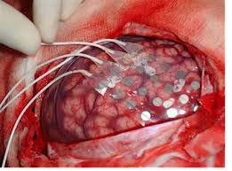
17 Electrocortigraphy
18 Electrocortigraphy (EcoG) Uses electrodes placed on the brain cortex to detect time varying electric fields produced by neural activity A functional brain imaging technique Very low frequency Detection of E-fields is much enhanced if electrodes are placed directly on the brain surface Bone is an electric insulator Electric field strength decays rapidly with distance Safety Requires neurosurgery Applicable only to specific patient populations
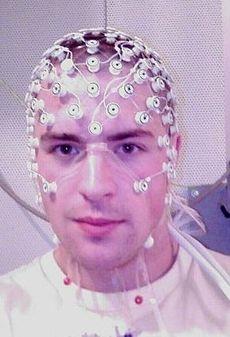
19 Electroencephalagraphic Imaging

20 Magnetic Fields
21 Magnetic Fields Movement of electric charge (electric current) produces a dipole magnetic field Movement of a magnetic field produces electric current Magnetic fields always have two or more poles All magnetism is fundamentally due to electric current
22 Magnetic Resonance Imaging (low frequency magnetic field imaging)
23 Magnetic Resonance Imaging MRI utilizes the magnetic resonance signal produced by the protons ( 1 H) of tissue water. MRI has become the method of choice for nondestructive visualization of brain anatomy Anatomic contrast depends on microscopic biophysical properties of tissue that (mostly) influence how fast water molecules can move Functional MRI techniques trick the proton MR signal to depend on brain function Usually indirect detection of blood flow or oxygenation changes that accompany neural activity

24 B0 B0 H O Magnetic Resonance Image H O H H H B0 B0 One volume element from the imaged tissue section H Proton spin magnets oriented parallel or antiparallel to the applied magnetic field and precessing about it
25 MRI Magnetic Fields B 0 penetrates bony structures allowing the brain to be seen through bone B 0 is typically 1.5 Tesla or greater (30,000 times the strength of the earth s magnetic field) Nuclear magnets rotate around B 0 at a characteristic frequency in the radio frequency region of the electromagnetic spectrum 63 Mhz at 1.5 T Addition of an oscillating magnetic field (B 1 ) interacts with the nuclear magnets causing them to produce a signal Addition of spatial variation in B 0 (magnetic field gradients) supports imaging
26 Magnetic Field Gradient(s) and Imaging A magnetic field gradient is a smooth variation in B 0 from one position to another position Anterior-to-posterior variation Superior-to-inferior variation Left-to-right variation Gradients are purposefully applied as part of the imaging process


27 High High Frequency bandwidth of selective excitation Low B0 in presence of S/I magnetic field gradient Low MR signal frequency MRI of the selected slice

28 sagittal axial coronal
29
30 The Image Signal discussed thus far is depicted as gray level of each element in the image White = greater signal Gray = less signal Black = no signal MR Images usually have no absolute calibration
31 MRI Contrast Signal/image creation in MRI uses a complex set of pulses of the oscillating B 1 magnetic field A pulse sequence Adjustment of pulse sequence timing parameters can be used to create different types of signal contrast to cellular composition T1-weighted imaging: Gray matter, white matter and cerebrospinal fluid give unique signal intensities Often used for brain morphometry studies T2-weighted imaging: Fluid produces prominent signals Often used for evaluating different types of lesions
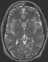
32 T2-weighted Imaging

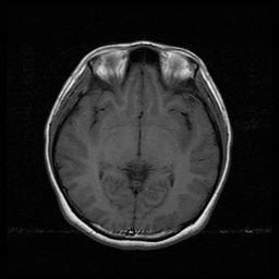
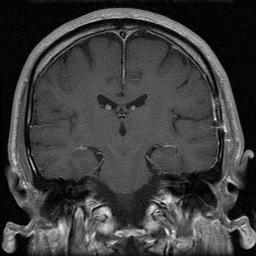
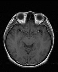
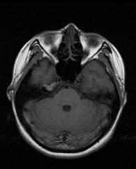
33 T1 weighted imaging (with contrast)
34 MRI Contrast Agents Typical contrast agents are chelates of metals ions (e.g. gadolinium) having unpaired electron spins Water molecules must touch the agent to enhance signal r Typical contrast reagents are designed to not pass the blood brain barrier Signal is enhanced only where blood brain barrier is damaged
35 Computed Tomography

36 Computed Tomography Technique

37 CT Contrast
38 CT contrast Intrinsic CT contrast is within brain is limited Gray matter, white matter and CSF can be distinguished, but not as effectively as with MRI CT neuroimaging often uses intravenous contrast agent (iodinated materials) to create vascular contrast CT angiography (CTA) Visualization of larger blood vessels (usually arteries) CT perfusion (CTP) Dynamic measurement of contrast passage to measure/image microvascular perfusion
39 CT Safety Xrays strip electrons from molecules Ionizing radiation Damages molecules In neuroimaging large xray intensity is used to overcome absorption by calcium in bone Xray exposure is sometimes a limiting factor Dynamic studies such as CTP Multiple studies on the same patient Allergic reaction to contrast agent Renal insufficiency that results in poor clearance of contrast agent
40 Positron Emission Tomography
41 Positron Emission Tomography Based on the existence of a subatomic particle called a positron (a positive electron) Certain unstable nuclear isotopes emit positrons Requires an onsite cyclotron Emitted positrons move a small distance before encountering a negative electron causing decay to two gamma rays

42 PET Technique

43 PET Imaging
44 PET Tracers 18 Fluorodeoxyglucose (FDG) is a tracer of glucose metabolism Injected into vascular system Transported into cells by glucose transporters FDG metabolism reaches a dead end after phosphorylation tracer of glucose metabolism H 2 15 O Injected into the vascular system Penetrates all membranes Washout rate depends on cerebral blood flow 15 O 2 Delivered by inhalation Tracer converted to CO 2 by respiration and washed out by blood flow Analysis of time course can be used to determine the rate of oxygen metabolism if a CBF measurement is also done Many other PET tracers are in development The basis of molecular imaging

45 PET Tracers
46 PET Safety Issues Gamma ray exposure Chemical safety Positron emitters tend to have short half lives Tracer molecules must be synthesized immediately before use Pharmacological effects of the tracer Many PET tracers are analogs of drugs or neurotransmitters
47 Image Processing/Interpretation (be critical)
48 What property is shown by the image? Gray scale and color are used to convey information about tissue properties The image looks cool but what is being shown? Is the image quantitative or only approximate MRI and CT data are often approximate PET images are often quantitative What post-acquisition image manipulations have occurred?
49 Learning Points Neuroimaging techniques can be described at a basic level without using complex physics All techniques use some form of electromagnetic radiation that can pass through bone Contrast depends on interaction between radiation in brain
50 Learning Points EcoG Functional contrast Requires neurosurgery MRI Uses radio waves and magnetic fields Anatomic, vascular and functional contrast Extensive use of contrast agents PET Primarily uses tracers that emit positrons and gamma rays Ionizing radiation Requires cyclotron and chemistry CT Uses xrays Ionizing radiation Anatomic and vascular contrast
MRI and CT of the CNS
 MRI and CT of the CNS Dr.Maha ELBeltagy Assistant Professor of Anatomy Faculty of Medicine The University of Jordan 2018 Computed Tomography CT is used for the detection of intracranial lesions. CT relies
Syllabus References. Resources. Video: MRI Introduction
 MRI Lesson Outline Syllabus References 9.6.4.2.5 Define precessing and relate the frequency of the precessing to the composition of the nuclei and the strength of the applied external magnetic field 9.6.4.2.6
P2 Visual - Perception
 P2 Visual - Perception 2014 SOSE Neuroimaging of high-level visual functions gyula.kovacs@uni-jena.de 11/09/06 Functional magnetic resonance imaging (fmri) The very basics What is fmri? What is MRI? The
HSC Physics. Module 9.6. Medical Physics
 HSC Physics Module 9.6 Medical Physics Contextual Outline 9.6 Medical Physics (28 indicative hours) The use of other advances in technology, developed from our understanding of the electromagnetic spectrum,
Non-Invasive Techniques
 Non-Invasive Techniques Key: Does not hurt the organism Psychology 372 Physiological Psychology Steven E. Meier, Ph.D. Listen to the audio lecture while viewing these slides or view the video presentation
Non-Invasive Techniques
 Many Procedures Non-Invasive Techniques Key: Does not hurt the organism Psychology 372 Physiological Psychology Steven E. Meier, Ph.D. Listen to the audio lecture while viewing these slides or view the
Medical Use of Radioisotopes
 Medical Use of Radioisotopes Therapy Radioisotopes prove to be useful in the application of brachytherapy, the procedure for using temporary irradiation close to the area of disease (i.e. cancer) 10% Medical
Medical imaging X-ray, CT, MRI, scintigraphy, SPECT, PET Györgyi Műzes
 Medical imaging X-ray, CT, MRI, scintigraphy, SPECT, PET Györgyi Műzes Semmelweis University, 2nd Dept. of Medicine Medical imaging: definition technical process of creating visual representations about
Outline. Biological Psychology: Research Methods. Dr. Katherine Mickley Steinmetz
 Biological Psychology: Research Methods Dr. Katherine Mickley Steinmetz Outline Neuroscience Methods Histology Electrophysiological Recordings Lesion Neuroimaging Neuroanatomy Histology: Brain structure
Laura Tormoehlen, M.D. Neurology and EM-Toxicology Indiana University
 Laura Tormoehlen, M.D. Neurology and EM-Toxicology Indiana University Disclosures! No conflicts of interest to disclose Neuroimaging 101! Plain films! Computed tomography " Angiography " Perfusion! Magnetic
Radionuclides in Medical Imaging. Danielle Wilson
 Radionuclides in Medical Imaging Danielle Wilson Outline Definitions History and development Radionuclide applications & techniques in imaging Conclusion Definition #1 : Radionuclide An unstable nucleus
Applicable Neuroradiology
 For the Clinical Neurology Clerkship LSU Medical School New Orleans Amy W Voigt, MD Clerkship Director Introduction The field of Radiology first developed following the discovery of X-Rays by Wilhelm Roentgen
Option D: Medicinal Chemistry
 Option D: Medicinal Chemistry Basics - unstable radioactive nuclei emit radiation in the form of smaller particles alpha, beta, positron, proton, neutron, & gamma are all used in nuclear medicine unstable
Magnetic Resonance Imaging on Soft Tissue. Jiten K. Mistry Calvin Gan
 Magnetic Resonance Imaging on Soft Tissue 1 Jiten K. Mistry Calvin Gan Outline Background of Medical Imaging Introduction to MRI How MRI works MRI of Soft Tissue Benefits & Risks Recent Advances 2 The
PHYSICS OF MRI ACQUISITION. Alternatives to BOLD for fmri
 PHYSICS OF MRI ACQUISITION Quick Review for fmri HST-583, Fall 2002 HST.583: Functional Magnetic Resonance Imaging: Data Acquisition and Analysis Harvard-MIT Division of Health Sciences and Technology
Brain and Cognition. Cognitive Neuroscience. If the brain were simple enough to understand, we would be too stupid to understand it
 Brain and Cognition Cognitive Neuroscience If the brain were simple enough to understand, we would be too stupid to understand it 1 The Chemical Synapse 2 Chemical Neurotransmission At rest, the synapse
Photoacoustic Imaging and Therapy in Biomedicine. Nicholas Tobey and Grace Yook. Optical Engineering. Dr. Kasra Daneshvar
 Photoacoustic Imaging 1 Photoacoustic Imaging and Therapy in Biomedicine Nicholas Tobey and Grace Yook Optical Engineering Dr. Kasra Daneshvar July 16, 2010 Photoacoustic Imaging 2 Abstract When a pulsed
COGNITIVE SCIENCE 17. Peeking Inside The Head. Part 1. Jaime A. Pineda, Ph.D.
 COGNITIVE SCIENCE 17 Peeking Inside The Head Part 1 Jaime A. Pineda, Ph.D. Imaging The Living Brain! Computed Tomography (CT)! Magnetic Resonance Imaging (MRI)! Positron Emission Tomography (PET)! Functional
Nuclear Medicine and PET. D. J. McMahon rev cewood
 Nuclear Medicine and PET D. J. McMahon 150504 rev cewood 2018-02-15 Key Points Nuclear Medicine and PET: Imaging: Understand how Nuc Med & PET differ from Radiography & CT by the source of radiation. Be
Beyond fmri. Joe Kable Summer Workshop on Decision Neuroscience August 21, 2009
 Beyond fmri Joe Kable Summer Workshop on Decision Neuroscience August 21, 2009 What are the strengths of fmri?! Noninvasive, safe! Can be done in humans! Verified correlate of neural activity! Great spatio-temporal
Stroke Imaging Basics. Jeremy Hopkin M.D.
 Stroke Imaging Basics Jeremy Hopkin M.D. Goals Introduce the basic physical properties of imaging used in stroke. Understand why each modality is used in the setting of stroke. Understand some strengths
Basics of nuclear medicine
 Basics of nuclear medicine Prof. dr. Davor Eterović Prof. dr. Vinko Marković Radioisotopes are used both in diagnostics and in therapy Diagnostics gamma emitters are used since gamma rays can penetrate
Molecular Imaging and the Brain
 Molecular imaging technologies are playing an important role in neuroimaging, a branch of medical imaging, by providing a window into the living brain. Where CT and conventional MR imaging provide important
Ways to Study Brain Structures and Functioning. Can physically trace connections. Ablation. Is the most primitive Can be done with any structures
 Ways to Study Brain Structures and Functioning Can physically trace connections Is the most primitive Can be done with any structures Ablation Can remove a piece of the brain and see what happens If the
Non Contrast MRA. Mayil Krishnam. Director, Cardiovascular and Thoracic Imaging University of California, Irvine
 Non Contrast MRA Mayil Krishnam Director, Cardiovascular and Thoracic Imaging University of California, Irvine No disclosures Non contrast MRA-Why? Limitations of CTA Radiation exposure Iodinated contrast
Certification Review. Module 28. Medical Coding. Radiology
 Module 28 is the study of x-rays, using radiant energy and other imaging techniques, such as resonance imaging or ultrasound, to diagnose illnesses and diseases. Vocabulary Barium enema (BE): lower gastrointestinal
The Physics of Medical Imaging
 VEA Bringing Learning to Life Program Support Notes Senior Secondary The Physics of Medical Imaging 27mins Program Support Notes by Ian Walter, Dip.App.Chem.; G.Dip.Ed.Admin.; TTTC Produced by VEA Pty
45 Hr PET Registry Review Course
 45 HR PET/CT REGISTRY REVIEW COURSE Course Control Document Timothy K. Marshel, MBA, R.T. (R), (N)(CT)(MR)(NCT)(PET)(CNMT) The PET/CT Training Institute, Inc. SNMMI-TS 028600-028632 45hr CEH s Voice Credits
Positron Emission Tomography Computed Tomography (PET/CT)
") Positron Emission Tomography Computed Tomography (PET/CT) What is Positron Emission Tomography Computed Tomography (PET/CT) Scanning? What are some common uses of the procedure? How should I prepare for
Positron Emission Tomography Computed Tomography (PET/CT) What is Positron Emission Tomography Computed Tomography (PET/CT) Scanning? What are some common uses of the procedure? How should I prepare for
Introduction to Modern Imaging Physics and Techniques used in Clinical Neurology
 Introduction to Modern Imaging Physics and Techniques used in Clinical Neurology Benjamin M. Ellingson, Ph.D., M.S. Associate Professor of Radiology, Biomedical Physics, Bioengineering, and Psychiatry
Announcements. Final Exam will be a take-home exam. Format similar to the short assignment (no multiple choice, etc.)
") Announcements Final Exam will be a take-home exam Format similar to the short assignment (no multiple choice, etc.) Will be handed out at end of last class period (Thursday June 5 th ) Due by 6 pm June
Announcements Final Exam will be a take-home exam Format similar to the short assignment (no multiple choice, etc.) Will be handed out at end of last class period (Thursday June 5 th ) Due by 6 pm June
Methods for assessing the brain basis of developmental disorders
 Announcements LIGN171: Child Language Acquisition http://ling.ucsd.edu/courses/lign171 Final Exam will be a take-home exam Format similar to the short assignment (no multiple choice, etc.) Will be handed
Brain Tumors. What is a brain tumor?
 Scan for mobile link. Brain Tumors A brain tumor is a collection of abnormal cells that grows in or around the brain. It poses a risk to the healthy brain by either invading or destroying normal brain
Methods of Visualizing the Living Human Brain
 Methods of Visualizing the Living Human Brain! Contrast X-rays! Computerized Tomography (CT)! Magnetic Resonance Imaging (MRI)! Positron Emission Tomography (PET)! Functional MRI! Magnetoencephalography
A&P Key Terms 01 Human Body Anatomy & Physiology
 Cover Page A&P Key Terms 01 Human Body Anatomy & Physiology Author: OpenStax College Published 2015 About Us Powered by QuizOver.com The Leading Online Quiz & Exam Creator Create, Share and Discover Quizzes
Cardiac Imaging. Kimberly Delcour, DO, FACC. Mahi Ashwath, MD, FACC, FASE. Director, Cardiac CT. Director, Cardiac MRI
 Cardiac Imaging Kimberly Delcour, DO, FACC Director, Cardiac CT Mahi Ashwath, MD, FACC, FASE Director, Cardiac MRI Cardiac Imaging Discuss the clinical applications of and indications for: Cardiac CT Nuclear
LESSON 1.3 WORKBOOK. How can we study the behaving brain?
 LESSON 1.3 WORKBOOK How can we study the behaving brain? We are in the middle of a technological revolution when it comes to how closely we can look at the behaving brain. Scientists and doctors now have
3/1/18. Overview of the Talk. Important Aspects of Neuroimaging Technology
 3/1/18 Considerations for the Use of Neuroimaging for Predicting Recovery of Speech and Language in Aphasia Linda I. Shuster, Ph.D., CCC-SLP Overview of the Talk Important aspects of neuroimaging technology
Introduction to Radiology
 Introduction - Lecture 1 436 Teams Introduction to Radiology Objectives Introduce the various Medical Imaging Modalities. Understand the basics of image generation. Relate imaging to gross anatomy. Appreciate
Eavesdropping on the Mind. COGS 17 - Winter 2019 Andrew Shibata
 Eavesdropping on the Mind COGS 17 - Winter 2019 Andrew Shibata Announcements - Midterm I is next Tuesday! - Exam is worth 25% of your grade - Homework 1 is due at the exam (worth 2.5% of grade) - Review
PHYSICS 2: HSC COURSE 2 nd edition (Andriessen et al) CHAPTER 20 Radioactivity as a diagnostic tool (pages 394-5)
 CHAPTER 20 Radioactivity as a diagnostic tool (pages 394-5)") PHYSICS 2: HSC COURSE 2 nd edition (Andriessen et al) CHAPTER 20 Radioactivity as a diagnostic tool (pages 394-5) 1. (a) A radioisotope is an isotope that is unstable and will emit particles from the nucleus
PHYSICS 2: HSC COURSE 2 nd edition (Andriessen et al) CHAPTER 20 Radioactivity as a diagnostic tool (pages 394-5) 1. (a) A radioisotope is an isotope that is unstable and will emit particles from the nucleus
Special Imaging MUSCULOSKELETAL INFECTION. Special Imaging. Special Imaging. 18yr old male pt What is it? Additional Imaging
 MUSCULOSKELETAL INFECTION Additional Imaging May assist in diagnosis and, possibly, treatment Help create the picture May help differentiate from neoplasia 18yr old male pt What is it? Lymphoma Ewings
Chapter Overview. Chapter 1. Anatomy. Physiology
 Chapter Overview Chapter 1 An Introduction to the Human Body Define Anatomy and Physiology Levels of Organization Characteristics of Living Things Homeostasis Anatomical Terminology 1 2 Anatomy Describes
COMENIUS-Project: SM&CLIL Radiation & Medicine
 Medical imaging refers to the techniques and processes used to create images of the human body (or parts thereof) for clinical purposes. Thanks to modern mathematics and computer technology, medical imaging
MR Advance Techniques. Vascular Imaging. Class II
 MR Advance Techniques Vascular Imaging Class II 1 Vascular Imaging There are several methods that can be used to evaluate the cardiovascular systems with the use of MRI. MRI will aloud to evaluate morphology
The Biological Level of Analysis: Studying the Brain
 The Biological Level of Analysis: Studying the Brain In the past the study of the brain was limited to people suffering from head injuries and the effects of accidental damage. It was only possible to
Functional aspects of anatomical imaging techniques
 Functional aspects of anatomical imaging techniques Nilendu Purandare Associate Professor & Consultant Radiologist Tata Memorial Centre Functional/metabolic/molecular imaging (radioisotope scanning) PET
ADVANCES IN RADIATION TECHNOLOGIES IN THE TREATMENT OF CANCER
 ADVANCES IN RADIATION TECHNOLOGIES IN THE TREATMENT OF CANCER Bro. Dr. Collie Miller IARC/WHO Based on trends in the incidence of cancer, the International Agency for Research on Cancer (IARC) and WHO
Nature of Radiation and DNA damage
 Nature of Radiation and DNA damage Index 1. What is radiation? 2. Ionizing Radiation 3. Interaction of Gamma-radiation with Matter 4. Radiobiology 5. Direct and Indirect action of radiation 6. Steps of
OTHER NON-CARDIAC USES OF Tc-99m CARDIAC AGENTS Tc-99m Sestamibi for parathyroid imaging, breast tumor imaging, and imaging of other malignant tumors.
 DEFINITION OF CARDIAC RADIOPHARMACEUTICAL: A radioactive drug which, when administered for purpose of diagnosis of heart disease, typically elicits no physiological response from the patient. Even though
CARDIAC MRI. Cardiovascular Disease. Cardiovascular Disease. Cardiovascular Disease. Overview
 CARDIAC MRI Dr Yang Faridah A. Aziz Department of Biomedical Imaging University of Malaya Medical Centre Cardiovascular Disease Diseases of the circulatory system, also called cardiovascular disease (CVD),
Biomedical Research 2013; 24 (3): ISSN X
: ISSN X") Biomedical Research 2013; 24 (3): 359-364 ISSN 0970-938X http://www.biomedres.info Investigating relative strengths and positions of electrical activity in the left and right hemispheres of the human brain
Biomedical Research 2013; 24 (3): 359-364 ISSN 0970-938X http://www.biomedres.info Investigating relative strengths and positions of electrical activity in the left and right hemispheres of the human brain
RADIOLOGY (MEDICAL IMAGING)
") RADIOLOGY (MEDICAL IMAGING) Radiology is the study of the diagnosis of disease by the use of radiant energy (radiation). In the past this meant the use of X-rays to make an image. Today many other forms
RADIOLOGY (MEDICAL IMAGING) Radiology is the study of the diagnosis of disease by the use of radiant energy (radiation). In the past this meant the use of X-rays to make an image. Today many other forms
Neuroimaging and Assessment Methods
 Psych 2200, Lecture 5 Experimental Design and Brain Imaging Methods Tues Sept 15, 2015 Revised TA office hours (Sam), today 4-5p, and wed 11:30-1:30. I will not have office hours this thurs but you should
*smith&nephew. MRI Safety Information & Parameters for Smith & Nephew Orthopaedics AG. Knee Implants
 Knee Implants MRI Safety Information & Parameters for Smith & Nephew Orthopaedics AG Knee Implants *smith&nephew Supporting healthcare professionals for over 150 years Summary All knee implants of Smith
HST.582J / 6.555J / J Biomedical Signal and Image Processing Spring 2007
 MIT OpenCourseWare http://ocw.mit.edu HST.582J / 6.555J / 16.456J Biomedical Signal and Image Processing Spring 2007 For information about citing these materials or our Terms of Use, visit: http://ocw.mit.edu/terms.
Announcements. Exam 1. VII. Imaging techniques of the brain. Anatomical/Structural Scans. Structural Scans: CT. Structural Scans: CT 2/17/2014
 Exam 1 None at the moment! Announcements Mean 78.0% Median 80% Mode 86% Min 26% Max 98% Std Dev 12.6% VII. Imaging techniques of the brain A. CT: anatomical B. MRI: anatomical C. fmri: functional D. SPECT
Radiologic Imaging Magnetic Resonance Imaging (MRI)
") Radiologic Imaging X-ray has always been the golden rule in diagnosing and treating podiatric patients. Unfortunately, for some patients the diagnosis is not as evident. That is when we need to utilize
Radiologic Imaging X-ray has always been the golden rule in diagnosing and treating podiatric patients. Unfortunately, for some patients the diagnosis is not as evident. That is when we need to utilize
Exam 1. Mean 78.0% Median 80% Mode 86% Min 26% Max 98% Std Dev 12.6%
 Exam 1 Mean 78.0% Median 80% Mode 86% Min 26% Max 98% Std Dev 12.6% None at the moment! Announcements VII. Imaging techniques of the brain A. CT: anatomical B. MRI: anatomical C. fmri: functional D. SPECT
BIOLOGICAL EFFECTS OF
 BIOLOGICAL EFFECTS OF RADIATION Natural Sources of Radiation Natural background radiation comes from three sources: Cosmic Radiation Terrestrial Radiation Internal Radiation 2 Natural Sources of Radiation
STRUCTURAL ORGANIZATION OF THE NERVOUS SYSTEM
 STRUCTURAL ORGANIZATION OF THE NERVOUS SYSTEM STRUCTURAL ORGANIZATION OF THE BRAIN The central nervous system (CNS), consisting of the brain and spinal cord, receives input from sensory neurons and directs
MOLECULAR AND CELLULAR NEUROSCIENCE
 MOLECULAR AND CELLULAR NEUROSCIENCE BMP-218 November 4, 2014 DIVISIONS OF THE NERVOUS SYSTEM The nervous system is composed of two primary divisions: 1. CNS - Central Nervous System (Brain + Spinal Cord)
HEAD AND NECK IMAGING. James Chen (MS IV)
") HEAD AND NECK IMAGING James Chen (MS IV) Anatomy Course Johns Hopkins School of Medicine Sept. 27, 2011 OBJECTIVES Introduce cross sectional imaging of head and neck Computed tomography (CT) Review head
HEAD AND NECK IMAGING James Chen (MS IV) Anatomy Course Johns Hopkins School of Medicine Sept. 27, 2011 OBJECTIVES Introduce cross sectional imaging of head and neck Computed tomography (CT) Review head
Dosimetry, see MAGIC; Polymer gel dosimetry. Fiducial tracking, see CyberKnife radiosurgery
 Subject Index Acoustic neuroma, neurofibromatosis type 2 complications 103, 105 hearing outcomes 103, 105 outcome measures 101 patient selection 105 study design 101 tumor control 101 105 treatment options
CEREBRAL BLOOD FLOW AND METABOLISM
 Supported by: HURO/0901/069/2.3.1 HU-RO-DOCS CEREBRAL BLOOD FLOW AND METABOLISM Part 3 Modern imaging methods SPECT, PET, nmri History of Nuclear Medicine Starts with the invention of the X-ray 1946: radioactive
Radioactivity. Alpha particles (α) :
 :") Radioactivity It is the property of an element that causes it to emit radiation Discovered by Becquerel (1896) Radiation comes from the nucleus of the atom There are three types of radiation : alpha particles
Radioactivity It is the property of an element that causes it to emit radiation Discovered by Becquerel (1896) Radiation comes from the nucleus of the atom There are three types of radiation : alpha particles
FOR CMS (MEDICARE) MEMBERS ONLY NATIONAL COVERAGE DETERMINATION (NCD) FOR MAGNETIC RESONANCE IMAGING:
 MEMBERS ONLY NATIONAL COVERAGE DETERMINATION (NCD) FOR MAGNETIC RESONANCE IMAGING:") National Imaging Associates, Inc. Clinical guidelines SINUS MRI Original Date: November 2007 Page 1 of 5 CPT Codes: 70540, 70542, 70543 Last Review Date: July 2014 NCD 220.2 MRI Last Effective Date: July
National Imaging Associates, Inc. Clinical guidelines SINUS MRI Original Date: November 2007 Page 1 of 5 CPT Codes: 70540, 70542, 70543 Last Review Date: July 2014 NCD 220.2 MRI Last Effective Date: July
Radiation physics and radiation protection. University of Szeged Department of Nuclear Medicine
 Radiation physics and radiation protection University of Szeged Department of Nuclear Medicine Radiation doses to the population 1 Radiation doses to the population 2 Sources of radiation 1 Radiation we
ASL Perfusion Imaging: Concepts and Applications
 ASL Perfusion Imaging: Concepts and Applications David C. Alsop, Ph.D. Beth Israel Deaconess Medical Center and Harvard Medical School, Boston USA INTRODUCTION Arterial Spin Labeling (ASL) perfusion imaging
*smith&nephew. MRI Safety Information & Parameters for Smith & Nephew Orthopaedics AG. Shoulder Implants
 Shoulder Implants MRI Safety Information & Parameters for Smith & Nephew Orthopaedics AG Shoulder Implants *smith&nephew Supporting healthcare professionals for over 150 years Summary All shoulder implants
Exam Practice Guide. Units 1 & 2 Physics: Detailed Study 5 - Investigations: Medical physics Examination Questions
 Exam Practice Guide Units 1 & 2 Physics: Detailed Study 5 - Investigations: Medical physics Examination Questions Key Features: 22 original examination style questions on all examinable topics. Full solutions
Yin-Hui Siow MD, FRCPC Director of Nuclear Medicine Southlake Regional Health Centre
 Yin-Hui Siow MD, FRCPC Director of Nuclear Medicine Southlake Regional Health Centre Today Introduction to CT Introduction to MRI Introduction to nuclear medicine Imaging the dementias The Brain ~ 1.5
Chapter 8. Ionizing and Non-Ionizing Radiation
 Chapter 8 Ionizing and Non-Ionizing Radiation Learning Objectives By the end of the chapter the reader will be able to: Define the terms ionizing radiation and nonionizing radiation State the differences
Introduction. Cardiac Imaging Modalities MRI. Overview. MRI (Continued) MRI (Continued) Arnaud Bistoquet 12/19/03
 MRI (Continued) Arnaud Bistoquet 12/19/03") Introduction Cardiac Imaging Modalities Arnaud Bistoquet 12/19/03 Coronary heart disease: the vessels that supply oxygen-carrying blood to the heart, become narrowed and unable to carry a normal amount
Introduction Cardiac Imaging Modalities Arnaud Bistoquet 12/19/03 Coronary heart disease: the vessels that supply oxygen-carrying blood to the heart, become narrowed and unable to carry a normal amount
Arteriogram An X-ray of an artery after the injection of dye.
 A Abscess A localized collection of pus in any part of the body, usually surrounded by inflamed tissue. Anesthetic An agent that causes loss of sensation with or without the loss of consciousness. Angiography,
Nuclear neurology. Zámbó Katalin Department of Nuclear Medicine
 Nuclear neurology Zámbó Katalin Department of Nuclear Medicine To refresh your memory Brain has a high rate of oxidative metabolism. It has no reserves either of oxygen or of glucose and has a very limited
Chapter 16 Worksheet Code It
 Name: Class: Date: ID: A Chapter 16 Worksheet 3 2 1 Code It True/False Indicate whether the statement is true or false. 1. CT scans generate three-dimensional images. 2. An ultrasound produces images of
Neural Correlates of Human Cognitive Function:
 Neural Correlates of Human Cognitive Function: A Comparison of Electrophysiological and Other Neuroimaging Approaches Leun J. Otten Institute of Cognitive Neuroscience & Department of Psychology University
Hemodynamics and fmri Signals
 Cerebral Blood Flow and Brain Activation UCLA NITP July 2011 Hemodynamics and fmri Signals Richard B. Buxton University of California, San Diego rbuxton@ucsd.edu... The subject to be observed lay on a
Restoring Tumorous MRI Brain Images
 Restoring Tumorous MRI Brain Images Yadavan Varatharajah Summer Ventures 2007 Appalachian State University Abstract Brain tumors are caused by many environmental, lifestyle, and genetic factors. This project
Principles of nuclear metabolic imaging. Prof. Dr. Alex Maes AZ Groeninge Kortrijk and KULeuven Belgium
 Principles of nuclear metabolic imaging Prof. Dr. Alex Maes AZ Groeninge Kortrijk and KULeuven Belgium I. Molecular imaging probes A. Introduction - Chemical disturbances will precede anatomical abnormalities
X-Ray & CT Physics / Clinical CT
 Computed Tomography-Basic Principles and Good Practice X-Ray & CT Physics / Clinical CT INSTRUCTORS: Dane Franklin, MBA, RT (R) (CT) Office hours will be Tuesdays from 5pm to 6pm CLASSROOM: TIME: REQUIRED
Chapter 12: Mass Spectrometry: molecular weight of the sample
 Structure Determination: hapter 12: Mass Spectrometry- molecular weight of the sample; formula hapter 12: Infrared Spectroscopy- indicated which functional groups are present hapter 13: Nuclear Magnetic
RADIOLOGY TEACHING CONFERENCE
 RADIOLOGY TEACHING CONFERENCE John Athas, MD Monica Tadros, MD Columbia University, College of Physicians & Surgeons Department of Otolaryngology- Head & Neck Surgery September 27, 2007 CT SCAN IMAGING
OPTION I TEST REVIEW
 IB PHYSICS 3 Name: Period: Date: DEVIL PHYSICS BADDEST CLASS ON CAMPUS OPTION I TEST REVIEW s2. This question is about defects of hearing. The graph below shows an audiogram for a person who has not been
Bone PET/MRI : Diagnostic yield in bone metastases and malignant primitive bone tumors
 Bone PET/MRI : Diagnostic yield in bone metastases and malignant primitive bone tumors Lars Stegger, Benjamin Noto Department of Nuclear Medicine University Hospital Münster, Germany Content From PET to
Physiological and Physical Basis of Functional Brain Imaging 6. EEG/MEG. Kâmil Uludağ, 20. November 2007
 Physiological and Physical Basis of Functional Brain Imaging 6. EEG/MEG Kâmil Uludağ, 20. November 2007 Course schedule 1. Overview 2. fmri (Spin dynamics, Image formation) 3. fmri (physiology) 4. fmri
Molecular Imaging and Cancer
 Molecular Imaging and Cancer Cancer causes one in every four deaths in the United States, second only to heart disease. According to the U.S. Department of Health and Human Services, more than 512,000
Medical Physics 4 I3 Radiation in Medicine
 Name: Date: 1. This question is about radiation dosimetry. Medical Physics 4 I3 Radiation in Medicine Define exposure. A patient is injected with a gamma ray emitter. The radiation from the source creates
NEUROSCIENCE. W1 Divisions of the nervous system PSYC1002 NOTES
 PSYC1002 NOTES NEUROSCIENCE W1 Divisions of the nervous system Nervous system: - CNS o Brain and spinal cord - Peripheral Nervous System o Sensory nerves o Motor nerves o Autonomic nervous system o Enteric
An investigation of the effect of ionising radiation on nurses and their patients during dialysis
 International Scholars Journals African Journal of Nursing and Midwifery ISSN 2198-4638 Vol. 2 (7), pp. 548-552, September, 2015. Available online at www.internationalscholarsjournals.org International
The neurolinguistic toolbox Jonathan R. Brennan. Introduction to Neurolinguistics, LSA2017 1
 The neurolinguistic toolbox Jonathan R. Brennan Introduction to Neurolinguistics, LSA2017 1 Psycholinguistics / Neurolinguistics Happy Hour!!! Tuesdays 7/11, 7/18, 7/25 5:30-6:30 PM @ the Boone Center
Anatomy and Physiology
 Anatomy and Physiology Anatomy---Study of Structure Gross anatomy Systemic anatomy Regional anatomy Microscopic anatomy Physiology---Study of Function Neurophysiology Muscle physiology Respiratory Renal
Hemodynamics and fmri Signals
 Cerebral Blood Flow and Brain Activation UCLA NITP July 2010 Hemodynamics and fmri Signals Richard B. Buxton University of California, San Diego rbuxton@ucsd.edu... The subject to be observed lay on a
Radiopharmacy. Prof. Dr. Çetin ÖNSEL. CTF Nükleer Tıp Anabilim Dalı
 Prof. Dr. Çetin ÖNSEL CTF Nükleer Tıp Anabilim Dalı What is Nuclear Medicine? Nuclear Medicine is the branch of medicine concerned with the use of radionuclides in the study and the diagnosis of diseases.
Psychologists who map the brain s fissures (grooves on the brain which appear as a deep fold) and inner recesses
 and inner recesses") Also called psychobiologists Psychologists who map the brain s fissures (grooves on the brain which appear as a deep fold) and inner recesses Methods include: 1) Recording 2) Stimulating 3) Lesioning 4)
Also called psychobiologists Psychologists who map the brain s fissures (grooves on the brain which appear as a deep fold) and inner recesses Methods include: 1) Recording 2) Stimulating 3) Lesioning 4)
Introduction. Visual Perception Aditi Majumder, UCI. Perception is taken for granted!
 Introduction Visual Perception Perception is taken for granted! Slide 2 1 Perception is very complex Perceive Locate Identify/Recognize Different objects Their relationship with each other Qualitative
What Radiologists do?
 Multimodality Imaging in Oncology 2018 March 5 th 9th Diagnostic Imaging in Oncology What Radiologists do? Chikako Suzuki, MD, PhD Department of Diagnostic Radiology, KS Solna Department of Molecular Medicine
The physiology of the BOLD signal What do we measure with fmri?
 The physiology of the BOLD signal What do we measure with fmri? Methods and Models in fmri, 10.11.2012 Jakob Heinzle Translational Neuromodeling Unit (TNU) Institute for Biomedical Engineering (IBT) University
MR Angiography in the evaluation of Lower Extremity Arterial Disease
 March 2001 MR Angiography in the evaluation of Lower Extremity Arterial Disease Ted Mau, Harvard Medical School Year III Objectives We will cover: Indications for Magnetic Resonance Angiography (MRA) Basic