Nuclear Medicine and PET. D. J. McMahon rev cewood
|
|
|
- Charles Hardy
- 5 years ago
- Views:
Transcription


1 Nuclear Medicine and PET D. J. McMahon rev cewood
2 Key Points Nuclear Medicine and PET: Imaging: Understand how Nuc Med & PET differ from Radiography & CT by the source of radiation. Be aware of As Low As Reasonably Practicable, ALARP Understand the difference between the scintillation camera and the PET scanner Be aware of the chemical 18 F FDG (fluorodeoxyglucose) & the element Tc (Technetium) Understand what the color spectrum means in a PET visualization: Brighter, hotter colors indicate high glucose uptake; brain, liver, cancer Therapy: Be aware of interventional nuclear medicine therapy and radiosurgery techniques; Radiopharmaceuticals, Brachytherapy, Radiosurgery, Gamma Knife, Linac.
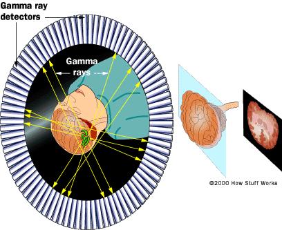
3 Nuclear Medicine Imaging - The application of radioactive substances in the diagnosis of disease. - Nuclear medicine, in a sense, is "radiology done inside out" or "endoradiology" because it records radiation emitting from within the body rather than radiation that is generated by external sources like X-rays. - Nuclear medicine scans differ from radiology as the emphasis is not on imaging anatomy but imaging the function instead. It is called a physiological imaging modality. - In nuclear medicine imaging, radiopharmaceuticals are taken internally, intravenously or orally. Then, external detectors (gamma cameras) capture and form images from the radiation emitted by the radiopharmaceuticals. - All human radiation exposures should be kept As Low As Reasonably Practicable, ALARP (replaces ALARA).

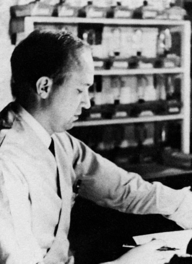

4 John Lawrence Anger scintillation camera Hal Anger
5
 or")
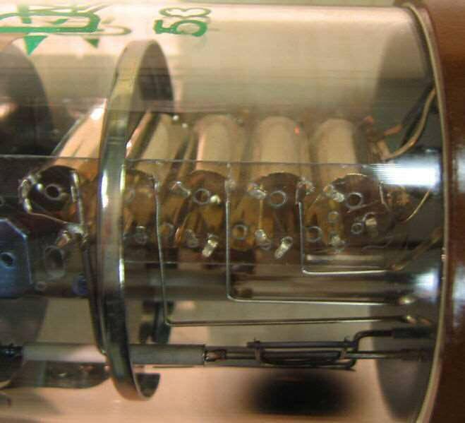
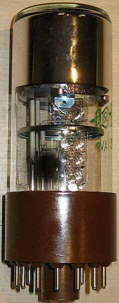
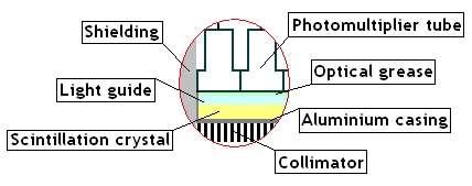
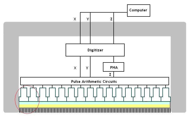
6 Gamma Imaging Camera ( gic ) or γ-camera
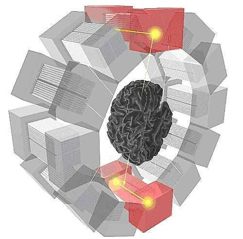
7
8
9

10


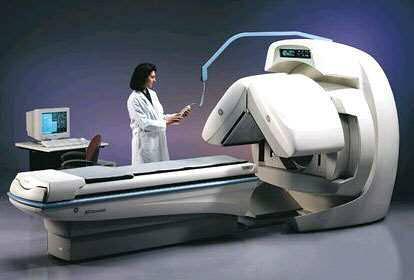
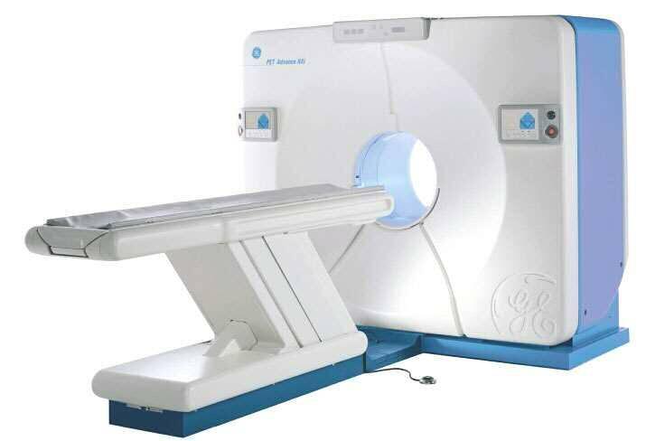
11 GE Maxicamera 400 Gamma Camera PET Scanner
12


13 Patient with infection: Patient with joint disease: Nuclear medicine scans can be superimposed, using software or hybrid cameras, on images from modalities such as CT or MRI to highlight the part of the body in which the radiopharmaceutical is concentrated. Often referred to as image fusion or co-registration.

14 Before-&-after isotope studies of two patients treated with a study medication for Non-Hodgkins Lymphomas:
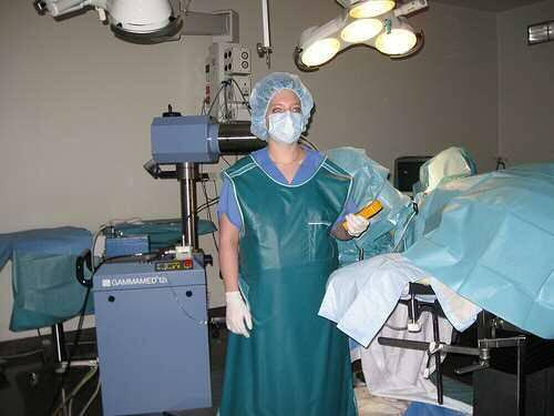
15 PET: Positron Emission Tomography PET is both a medical and research tool. It is used heavily in clinical oncology. A tomographic technique that uses isotopes which emit two gamma rays rather than one, enabling images with a high signal-to-noise ratio and good spatial resolution. PET is used for physiology and function rather than anatomy. PET scans are more expensive than "conventional" imaging using computed tomography (CT) and magnetic resonance imaging (MRI) Oncology scans using 18 F FDG (fluorodeoxyglucose) tracer make up over 90% of all PET scans in current practice This tracer is a glucose analog that is taken up by glucose-using cells. This results in intense radiolabeling of tissues with high glucose uptake, such as the brain, the liver, and most cancers. Brighter, hotter colors indicate high glucose uptake
16

17 47 y/o female, pre- & post chemotherapy for leukemia:

18 MRI and PET scans of brain, normal and with Alzheimer s Disease:
.")
19 (animation) Maximum Intensity Projection (MIP) of a wholebody positron emission tomography (PET) acquisition of a 79 kg (174 lb) weighting female after intravenous injection of 371 MBq of 18 F-FDG (one hour prior measurement). The investigation has been performed as part of a tumor diagnosis prior to applying a radiotherapy (tumor staging step) Besides normal accumulation of the tracer in the heart, bladder, kidneys and brain, liver metastases of a colorectal tumor are clearly visible within the abdominal region of the image. MIPS-anim.gif
20
21 Interventional Nuclear Medicine Therapy... Radiopharmaceuticals typically consist of a radioactive atom (also known as a radionuclide) combined with a cell-targeting molecule that seeks and destroys cancer cells. Some radionuclides have the ability to target specific cells on their own. Some Conditions include: hyperthyroidism and thyroid cancer refractory lymphoma neuroendocrine tumors palliative bone pain treatment Brachytherapy (from Greek word for "short"), or internal radiation therapy for prostate cancer. A doctor or clinician implants radioactive seeds into the prostate gland using an ultrasound for guidance. Anywhere from 40 to 100 seeds are commonly implanted.
22 Interventional Nuclear Medicine Therapy... Radiosurgery Radiosurgery is the destruction of precisely selected areas of tissue using ionizing radiation rather than excision with a blade. Like other forms of radiation therapy (also called radiotherapy), it is usually used to treat cancer. The principle of this instrument is to hit the intra-cranial target with narrow beams of radiation from multiple directions. The beam paths converge in the target volume, delivering a lethal cumulative dose of radiation there, while limiting the dose to the adjacent healthy tissue. In stereotactic radiosurgery (SRS), the word "stereotactic" refers to a threedimensional coordinate system that enables accurate correlation of a virtual target seen in the patient's diagnostic images.
23 Interventional Nuclear Medicine Therapy Gamma Knife The Gamma Knife is used to treat brain tumors. It consists of several radioactive sources placed in a kind of helmet with central channels for irradiation with gamma rays. This produces slit-like radiation lesions for functional neurosurgical procedures to treat pain, movement disorders, or behavioral disorders that do not respond to conventional treatment. Linac A similar approach uses a linear particle accelerator or Linac which emits high energy X-rays, called Cyberknife therapy. Radiosurgery does not remove the tumor but inactivates it biologically. Lack of growth of the lesion is normally considered to be treatment success. In the U.S. the Food and Drug Administration (FDA) regulates radiosurgery devices, whereas the Gamma Knife is regulated by the Nuclear Regulatory Commission.
24

25 PET videos: iphone app for PET visualization: Scans of brain injury for a lawsuit: Major players in Nuc Med > ADAC/Philips > GE > Hitachi/Vision > IS 2 > Siemens > SMV > Sopha > Toshiba > NuQuest Workstations > Segami Workstations
Option D: Medicinal Chemistry
 Option D: Medicinal Chemistry Basics - unstable radioactive nuclei emit radiation in the form of smaller particles alpha, beta, positron, proton, neutron, & gamma are all used in nuclear medicine unstable
Medical Use of Radioisotopes
 Medical Use of Radioisotopes Therapy Radioisotopes prove to be useful in the application of brachytherapy, the procedure for using temporary irradiation close to the area of disease (i.e. cancer) 10% Medical
ADVANCES IN RADIATION TECHNOLOGIES IN THE TREATMENT OF CANCER
 ADVANCES IN RADIATION TECHNOLOGIES IN THE TREATMENT OF CANCER Bro. Dr. Collie Miller IARC/WHO Based on trends in the incidence of cancer, the International Agency for Research on Cancer (IARC) and WHO
Molecular Imaging and Cancer
 Molecular Imaging and Cancer Cancer causes one in every four deaths in the United States, second only to heart disease. According to the U.S. Department of Health and Human Services, more than 512,000
Radionuclides in Medical Imaging. Danielle Wilson
 Radionuclides in Medical Imaging Danielle Wilson Outline Definitions History and development Radionuclide applications & techniques in imaging Conclusion Definition #1 : Radionuclide An unstable nucleus
PHYS 383: Applications of physics in medicine (offered at the University of Waterloo from Jan 2015)
") PHYS 383: Applications of physics in medicine (offered at the University of Waterloo from Jan 2015) Course Description: This course is an introduction to physics in medicine and is intended to introduce
PHYS 383: Applications of physics in medicine (offered at the University of Waterloo from Jan 2015) Course Description: This course is an introduction to physics in medicine and is intended to introduce
MEDICAL MANAGEMENT POLICY
 PAGE: 1 of 8 This medical policy is not a guarantee of benefits or coverage, nor should it be deemed as medical advice. In the event of any conflict concerning benefit coverage, the employer/member summary
General Nuclear Medicine
 General Nuclear Medicine What is General Nuclear Medicine? What are some common uses of the procedure? How should I prepare? What does the equipment look like? How does the procedure work? How is the procedure
Molecular Imaging and Breast Cancer
 Molecular Imaging and Breast Cancer Breast cancer forms in tissues of the breast usually in the ducts, tubes that carry milk to the nipple, and lobules, the glands that make milk. It occurs in both men
An introduction to different types of radiotherapy
 An introduction to different types of radiotherapy Radiotherapy can cure cancer. It is delivered to around half of cancer patients and is a vital part of curative treatment in around 40% of patients 1.
Stereotactic Radiosurgery. Extracranial Stereotactic Radiosurgery. Linear accelerators. Basic technique. Indications of SRS
 Stereotactic Radiosurgery Extracranial Stereotactic Radiosurgery Annette Quinn, MSN, RN Program Manager, University of Pittsburgh Medical Center Using stereotactic techniques, give a lethal dose of ionizing
Medical imaging X-ray, CT, MRI, scintigraphy, SPECT, PET Györgyi Műzes
 Medical imaging X-ray, CT, MRI, scintigraphy, SPECT, PET Györgyi Műzes Semmelweis University, 2nd Dept. of Medicine Medical imaging: definition technical process of creating visual representations about
A. DeWerd. Michael Kissick. Larry. Editors. The Phantoms of Medical. and Health Physics. Devices for Research and Development.
 Larry Editors A. DeWerd Michael Kissick The Phantoms of Medical and Health Physics Devices for Research and Development ^ Springer Contents 1 Introduction to Phantoms of Medical and Health Physics 1 1.1
Understanding Radiation Therapy. For Patients and the Public
 Understanding Radiation Therapy For Patients and the Public Introduction to Radiation Oncology Radiation has been an effective tool for treating cancer for more than 100 years. Radiation oncologists are
Positron Emission Tomography Computed Tomography (PET/CT)
") Positron Emission Tomography Computed Tomography (PET/CT) What is Positron Emission Tomography Computed Tomography (PET/CT) Scanning? What are some common uses of the procedure? How should I prepare for
Positron Emission Tomography Computed Tomography (PET/CT) What is Positron Emission Tomography Computed Tomography (PET/CT) Scanning? What are some common uses of the procedure? How should I prepare for
CEREBRAL BLOOD FLOW AND METABOLISM
 Supported by: HURO/0901/069/2.3.1 HU-RO-DOCS CEREBRAL BLOOD FLOW AND METABOLISM Part 3 Modern imaging methods SPECT, PET, nmri History of Nuclear Medicine Starts with the invention of the X-ray 1946: radioactive
Atoms for Health. Atoms for Health The. Atoms for Health - Division of Nuclear Health - Dept of Nuclear Aplications P Andreo DIR-NAHU 1
 International Atomic Energy Agency Atoms for Health The human side of nuclear applications NUCLEAR APPLICATIONS IN HEALTH A UNIQUE MANDATE OF THE UN SYSTEM The Agency shall seek to accelerate and enlarge
PET-MRI in malignant bone tumours. Lars Stegger Department of Nuclear Medicine University Hospital Münster, Germany
 PET-MRI in malignant bone tumours Lars Stegger Department of Nuclear Medicine University Hospital Münster, Germany Content From PET to PET/MRI General considerations Bone metastases Primary bone tumours
Basics of nuclear medicine
 Basics of nuclear medicine Prof. dr. Davor Eterović Prof. dr. Vinko Marković Radioisotopes are used both in diagnostics and in therapy Diagnostics gamma emitters are used since gamma rays can penetrate
PHYSICS 2: HSC COURSE 2 nd edition (Andriessen et al) CHAPTER 20 Radioactivity as a diagnostic tool (pages 394-5)
 CHAPTER 20 Radioactivity as a diagnostic tool (pages 394-5)") PHYSICS 2: HSC COURSE 2 nd edition (Andriessen et al) CHAPTER 20 Radioactivity as a diagnostic tool (pages 394-5) 1. (a) A radioisotope is an isotope that is unstable and will emit particles from the nucleus
PHYSICS 2: HSC COURSE 2 nd edition (Andriessen et al) CHAPTER 20 Radioactivity as a diagnostic tool (pages 394-5) 1. (a) A radioisotope is an isotope that is unstable and will emit particles from the nucleus
EORTC Member Facility Questionnaire
 Page 1 of 9 EORTC Member Facility Questionnaire I. Administrative Data Name of person submitting this questionnaire Email address Function Phone Institution Address City Post code Country EORTC No Enter
Molecular Imaging and the Brain
 Molecular imaging technologies are playing an important role in neuroimaging, a branch of medical imaging, by providing a window into the living brain. Where CT and conventional MR imaging provide important
Brain Tumors. What is a brain tumor?
 Scan for mobile link. Brain Tumors A brain tumor is a collection of abnormal cells that grows in or around the brain. It poses a risk to the healthy brain by either invading or destroying normal brain
KEYWORDS: nuclear medicine; gamma camera; radiopharmaceutical activities.
 Radiopharmaceutical Activities Administered for Diagnostic Procedures in Nuclear Medicine in the First Six Months of the Gamma Camera Use in the Clinical Center of Montenegro - Podgorica Nevenka Antovic
RADIOTHERAPY: TECHNOLOGIES AND GLOBAL MARKETS
 RADIOTHERAPY: TECHNOLOGIES AND GLOBAL MARKETS HLC176A February 2015 Neha Maliwal Project Analyst ISBN: 1-62296-043-2 BCC Research 49 Walnut Park, Building 2 Wellesley, MA 02481 USA 866-285-7215 (toll-free
COMENIUS-Project: SM&CLIL Radiation & Medicine
 Medical imaging refers to the techniques and processes used to create images of the human body (or parts thereof) for clinical purposes. Thanks to modern mathematics and computer technology, medical imaging
Bone PET/MRI : Diagnostic yield in bone metastases and malignant primitive bone tumors
 Bone PET/MRI : Diagnostic yield in bone metastases and malignant primitive bone tumors Lars Stegger, Benjamin Noto Department of Nuclear Medicine University Hospital Münster, Germany Content From PET to
Breast Cancer. What is breast cancer?
 Scan for mobile link. Breast Cancer Breast cancer is a malignant tumor in or around breast tissue. It usually begins as a lump or calcium deposit that develops from abnormal cell growth. Most breast lumps
Medical Physics 4 I3 Radiation in Medicine
 Name: Date: 1. This question is about radiation dosimetry. Medical Physics 4 I3 Radiation in Medicine Define exposure. A patient is injected with a gamma ray emitter. The radiation from the source creates
Brain Tumor Treatment
 Scan for mobile link. Brain Tumor Treatment Brain Tumors Overview A brain tumor is a group of abnormal cells that grows in or around the brain. Tumors can directly destroy healthy brain cells. They can
Index. Surg Oncol Clin N Am 16 (2007) Note: Page numbers of article titles are in boldface type.
 Note: Page numbers of article titles are in boldface type.") Surg Oncol Clin N Am 16 (2007) 465 469 Index Note: Page numbers of article titles are in boldface type. A Adjuvant therapy, preoperative for gastric cancer, staging and, 339 B Breast cancer, metabolic
Surg Oncol Clin N Am 16 (2007) 465 469 Index Note: Page numbers of article titles are in boldface type. A Adjuvant therapy, preoperative for gastric cancer, staging and, 339 B Breast cancer, metabolic
Monitoring Patients Undergoing Cancer Therapy. By Timothy K. Egan
 F E A T U R E By Timothy K. Egan Before placement in a computed tomography scanner, a patient is fitted with an immobilization device. Immobilization ensures that the same area of the patient is scanned
Nuclear Medicine: Manuals. Nuclear Medicine. Nuclear imaging. Emission imaging: study types. Bone scintigraphy - technique
 Nuclear Medicine - Unsealed radioactive preparations the tracer mixes with the patients body fluids on a molecular level (e.g. after intravenous injection) - 3 main fields: - In vitro : measuring concentrations
45 Hr PET Registry Review Course
 45 HR PET/CT REGISTRY REVIEW COURSE Course Control Document Timothy K. Marshel, MBA, R.T. (R), (N)(CT)(MR)(NCT)(PET)(CNMT) The PET/CT Training Institute, Inc. SNMMI-TS 028600-028632 45hr CEH s Voice Credits
Breast Cancer. What is breast cancer?
 Scan for mobile link. Breast Cancer Breast cancer is a malignant tumor in or around breast tissue. It usually begins as a lump or calcium deposit that develops from abnormal cell growth. Most breast lumps
Principles of nuclear metabolic imaging. Prof. Dr. Alex Maes AZ Groeninge Kortrijk and KULeuven Belgium
 Principles of nuclear metabolic imaging Prof. Dr. Alex Maes AZ Groeninge Kortrijk and KULeuven Belgium I. Molecular imaging probes A. Introduction - Chemical disturbances will precede anatomical abnormalities
Dosimetry, see MAGIC; Polymer gel dosimetry. Fiducial tracking, see CyberKnife radiosurgery
 Subject Index Acoustic neuroma, neurofibromatosis type 2 complications 103, 105 hearing outcomes 103, 105 outcome measures 101 patient selection 105 study design 101 tumor control 101 105 treatment options
Nuclear Medicine in the Diabetic Foot
 26.11.2015, Uniklinik Balgrist Nuclear Medicine in the Diabetic Foot Martin Hüllner Nuklearmedizin und Neuroradiologie, USZ / UZH Outline A. Imaging modalities brief technical overview B. Nuclear medicine
POSITRON EMISSION TOMOGRAPHY (PET)
") Status Active Medical and Behavioral Health Policy Section: Radiology Policy Number: V-27 Effective Date: 08/27/2014 Blue Cross and Blue Shield of Minnesota medical policies do not imply that members should
Status Active Medical and Behavioral Health Policy Section: Radiology Policy Number: V-27 Effective Date: 08/27/2014 Blue Cross and Blue Shield of Minnesota medical policies do not imply that members should
Radiation Therapy for Cancer: Questions and Answers
 Radiation Therapy for Cancer: Questions and Answers Key Points Radiation therapy uses ionizing radiation to kill cancer cells and shrink tumors (see Question 1). About half of all people with cancer are
The Physics of Medical Imaging
 VEA Bringing Learning to Life Program Support Notes Senior Secondary The Physics of Medical Imaging 27mins Program Support Notes by Ian Walter, Dip.App.Chem.; G.Dip.Ed.Admin.; TTTC Produced by VEA Pty
Radiopharmacy. Prof. Dr. Çetin ÖNSEL. CTF Nükleer Tıp Anabilim Dalı
 Prof. Dr. Çetin ÖNSEL CTF Nükleer Tıp Anabilim Dalı What is Nuclear Medicine? Nuclear Medicine is the branch of medicine concerned with the use of radionuclides in the study and the diagnosis of diseases.
A Patient s Guide to SRS
 A Patient s Guide to SRS Stereotactic Radiosurgery 230 Nebraska St. Sioux City, IA 51101 NOTES 230 Nebraska St. Sioux City, IA 51101 Contents page Introduction 1 SRS and how it works 2 The technology involved
Guideline & Reports 医学物理学会教育委員会資料
 Guideline & Reports 医学物理学会教育委員会資料 2017/11 更新 report title year keyword 1 keyword 2 AAPM TG211 Classification and evaluation strategies of auto-segmentation approaches for PET 2017 PET autosegmentation
Certification Review. Module 28. Medical Coding. Radiology
 Module 28 is the study of x-rays, using radiant energy and other imaging techniques, such as resonance imaging or ultrasound, to diagnose illnesses and diseases. Vocabulary Barium enema (BE): lower gastrointestinal
Appendix A: Introduction to Imaging Modalities for Which Data Were Collected in the 2017 Imaging Inventory
 Appendix A: Introduction to Imaging Modalities for Which Data Were Collected in the 207 Imaging Inventory Computed Tomography Computed tomography (CT) employs X-rays as a source of ionizing radiation,
Regions Hospital Delineation of Privileges Radiation Oncology
 Regions Hospital Delineation of Privileges Radiation Oncology Applicant s Last First M. Instructions: Place a check-mark where indicated for each core group you are requesting. Review education and basic
Compact Gamma Camera for Detection of Prostate Cancer
 Compact Gamma Camera for Detection of Prostate Cancer Presented at: Human Interest Panel Federal Laboratory Consortium Annual Conference Nashville, Tennessee Brookhaven National Laboratory and Hybridyne
Austin Radiological Association Nuclear Medicine Procedure PET SODIUM FLUORIDE BONE SCAN (F-18 NaF)
") Austin Radiological Association Nuclear Medicine Procedure PET SODIUM FLUORIDE BONE SCAN (F-18 NaF) Overview Indication Sodium Fluoride F18 injection is a radioactive diagnostic agent for positron emission
Austin Radiological Association Nuclear Medicine Procedure PET SODIUM FLUORIDE BONE SCAN (F-18 NaF) Overview Indication Sodium Fluoride F18 injection is a radioactive diagnostic agent for positron emission
Colorectal Cancer Treatment
 Scan for mobile link. Colorectal Cancer Treatment Colorectal cancer overview Colorectal cancer, also called large bowel cancer, is the term used to describe malignant tumors found in the colon and rectum.
"The Good Side of Radiation: Medical Applications"
 "The Good Side of Radiation: Medical Applications" J. Battista, Ph.D. Medical Physicist London Regional Cancer Program LHSC http://www.macmillan.org.uk/images/cancerinfo Role of Medical Physicists Diagnostic
Pancreatic Cancer. What is pancreatic cancer?
 Scan for mobile link. Pancreatic Cancer Pancreatic cancer is a tumor of the pancreas, an organ that is located behind the stomach in the abdomen. Pancreatic cancer does not always cause symptoms until
Subject: Image-Guided Radiation Therapy
 04-77260-19 Original Effective Date: 02/15/10 Reviewed: 01/25/18 Revised: 01/01/19 Subject: Image-Guided Radiation Therapy THIS MEDICAL COVERAGE GUIDELINE IS NOT AN AUTHORIZATION, CERTIFICATION, EXPLANATION
Austin Radiological Association Ga-68 NETSPOT (Ga-68 dotatate)
") Austin Radiological Association Ga-68 NETSPOT (Ga-68 dotatate) Overview Ga-68 dotatate binds to somatostatin receptors, with highest affinity for subtype 2 receptors (sstr2). It binds to cells that express
Austin Radiological Association Ga-68 NETSPOT (Ga-68 dotatate) Overview Ga-68 dotatate binds to somatostatin receptors, with highest affinity for subtype 2 receptors (sstr2). It binds to cells that express
Proiect IMAGO-MOL: Dezvoltarea imagisticii funcţionale în regiunea Nord-Est. Walther Bild, MD, PhD
 Proiect IMAGO-MOL: Dezvoltarea imagisticii funcţionale în regiunea Nord-Est Walther Bild, MD, PhD Nuclear medicine Originates in the mid-1920s in Freiburg, Germany, when George de Hevesy made experiments
Children's (Pediatric) Nuclear Medicine
 Nuclear Medicine") Scan for mobile link. Children's (Pediatric) Nuclear Medicine Children s (pediatric) nuclear medicine imaging uses small amounts of radioactive materials called radiotracers, a special camera and a computer
Scan for mobile link. Children's (Pediatric) Nuclear Medicine Children s (pediatric) nuclear medicine imaging uses small amounts of radioactive materials called radiotracers, a special camera and a computer
Typical PET Image. Elevated uptake of FDG (related to metabolism) Lung cancer example: But where exactly is it located?
 Lung cancer example: But where exactly is it located?") Typical PET Image Elevated uptake of FDG (related to metabolism) Lung cancer example: But where exactly is it located? PET/CT Oncology Imaging Anatometabolic fusion images are useful in the management
Typical PET Image Elevated uptake of FDG (related to metabolism) Lung cancer example: But where exactly is it located? PET/CT Oncology Imaging Anatometabolic fusion images are useful in the management
Physical Bases : Which Isotopes?
 Physical Bases : Which Isotopes? S. Gnesin Institute of Radiation Physics, Lausanne University Hospital, Lausanne, Switzerland 1/53 Theranostic Bruxelles, 2 Octobrer 2017 Theranostic : use of diagnostic
Radiation Exposure to Staff Using PET/CT Facility
 Egyptian J. Nucl. Med., Vol. 8, No. 2, December 2013 1 Editorial Radiation Exposure to Staff Using PET/CT Facility Taalab, Kh; and Mohsen, Z Department of Nuclear Medicine, International Medical Center;
Positron Emission Tomography - Computed Tomography (PET/CT)
") Scan for mobile link. Positron Emission Tomography - Computed Tomography (PET/CT) Positron emission tomography (PET) uses small amounts of radioactive materials called radiotracers, a special camera and
Scan for mobile link. Positron Emission Tomography - Computed Tomography (PET/CT) Positron emission tomography (PET) uses small amounts of radioactive materials called radiotracers, a special camera and
General Nuclear Medicine
 Scan for mobile link. General Nuclear Medicine Nuclear medicine imaging uses small amounts of radioactive materials called radiotracers that are typically injected into the bloodstream, inhaled or swallowed.
Stereotactic Radiosurgery and Stereotactic Body Radiation Therapy
 Stereotactic Radiosurgery and Stereotactic Body Radiation Therapy Policy Number: Original Effective Date: MM.05.008 05/12/1999 Line(s) of Business: Current Effective Date: HMO; PPO; QUEST Integration 04/01/2015
Medical Policy An independent licensee of the Blue Cross Blue Shield Association
 PET Scanning: Oncologic Applications Page 1 of 88 Medical Policy An independent licensee of the Blue Cross Blue Shield Association Title: Positron Emission Tomography (PET) Scanning: Oncologic Applications
Intensity-Modulated and Image- Guided Radiation Treatment. Outline. Conformal Radiation Treatment
 Intensity-Modulated and Image- Guided Radiation Treatment J. Daniel Bourland, PhD Professor Departments of Radiation Oncology, Physics, and Biomedical Engineering Wake Forest University School of Medicine
I. Equipments for external beam radiotherapy
 I. Equipments for external beam radiotherapy 5 linear accelerators (LINACs): Varian TrueBeam 6, 10 & 18 MV photons, 6-18 MeV electrons, image-guided (IGRT) and intensity modulated radiotherapy (IMRT),
Original Date: April 2016 Page 1 of 7 FOR CMS (MEDICARE) MEMBERS ONLY
 MEMBERS ONLY") National Imaging Associates, Inc. Clinical guidelines STEREOTACTIC RADIATION THERAPY: STEREO RADIOSURGERY (SRS) AND STEREOTACTIC BODY RADIATION THERAPY (SBRT) CPT4 Codes: Please refer to pages 5-6 LCD
National Imaging Associates, Inc. Clinical guidelines STEREOTACTIC RADIATION THERAPY: STEREO RADIOSURGERY (SRS) AND STEREOTACTIC BODY RADIATION THERAPY (SBRT) CPT4 Codes: Please refer to pages 5-6 LCD
PET imaging of cancer metabolism is commonly performed with F18
 PCRI Insights, August 2012, Vol. 15: No. 3 Carbon-11-Acetate PET/CT Imaging in Prostate Cancer Fabio Almeida, M.D. Medical Director, Arizona Molecular Imaging Center - Phoenix PET imaging of cancer metabolism
Lecture 1. Lecture 1: The Different Modalities
 Lecture 1 Lecture 1: The Different Modalities In this Lecture Understanding the difference between the different modalities available Learn when to chose the appropriate modality Trust me, during the next
Arteriogram An X-ray of an artery after the injection of dye.
 A Abscess A localized collection of pus in any part of the body, usually surrounded by inflamed tissue. Anesthetic An agent that causes loss of sensation with or without the loss of consciousness. Angiography,
Cardiac Nuclear Medicine
 Cardiac Nuclear Medicine What is Cardiac Nuclear Medicine? What are some common uses of the procedure? How should I prepare? What does the equipment look like? How does the procedure work? How is the procedure
Linac or Non-Linac Demystifying And Decoding The Physics Of SBRT/SABR
 Linac or Non-Linac Demystifying And Decoding The Physics Of SBRT/SABR PhD, FAAPM, FACR, FASTRO Department of Radiation Oncology Indiana University School of Medicine Indianapolis, IN, USA Indra J. Das,
Stereotactic Radiosurgery and Stereotactic Body Radiation Therapy
 Stereotactic Radiosurgery and Stereotactic Body Radiation Therapy Policy Number: Original Effective Date: MM.05.008 05/12/1999 Line(s) of Business: Current Effective Date: HMO; PPO; QUEST 03/01/2013 Section:
Has radiotherapy the potential being focal?
 Has radiotherapy the potential being focal? György Kovács & Alexander Schlaefer* Interdisciplinary Brachytherapy Unit and *Institute of Robotics and Cognitive Systems, University of Lübeck / 1 100% 90%
RADIOLOGIC AND IMAGING SCIENCE (RIS)
") Kent State University Catalog 2017-2018 1 RADIOLOGIC AND IMAGING SCIENCE (RIS) RIS 34001 INTRODUCTION TO DIAGNOSTIC MEDICAL SONOGRAPHY 1 Credit Provides an introduction to diagnostic medical sonography.
Kent State University Catalog 2017-2018 1 RADIOLOGIC AND IMAGING SCIENCE (RIS) RIS 34001 INTRODUCTION TO DIAGNOSTIC MEDICAL SONOGRAPHY 1 Credit Provides an introduction to diagnostic medical sonography.
What Is Nuclear Medicine?
 What Is Nuclear Medicine? An Introductory Guide For Patients And Their Families What is nuclear medicine? Nuclear medicine is a type of medical imaging that uses small amounts of radioactive material (called
Radiotherapy. Marta Anguiano Millán. Departamento de Física Atómica, Molecular y Nuclear Facultad de Ciencias. Universidad de Granada
 Departamento de Física Atómica, Molecular y Nuclear Facultad de Ciencias. Universidad de Granada Overview Introduction Overview Introduction Brachytherapy Radioisotopes in contact with the tumor Overview
Radioactivity. Alpha particles (α) :
 :") Radioactivity It is the property of an element that causes it to emit radiation Discovered by Becquerel (1896) Radiation comes from the nucleus of the atom There are three types of radiation : alpha particles
Radioactivity It is the property of an element that causes it to emit radiation Discovered by Becquerel (1896) Radiation comes from the nucleus of the atom There are three types of radiation : alpha particles
7. Radioisotopes in Medicine
 7. Radioisotopes in Medicine Radionuclides were first used for therapeutic purposes almost 100 years following the observation by Pierre Curie that radium sources brought into contact with the skin produced
Isotopes in Functional Cancer Imaging
 Seeing and Believing: i Medical Isotopes in Functional Cancer Imaging François Bénard, MD, FRCPC BCCancer Cancer Agency and University of British Columbia Nuclear Medicine 101 A radioactive atom is produced
Nuclear Medicine Head and Neck Region. Bán Zsuzsanna, MD University of Pécs, Department of Nuclear Medicine
 Nuclear Medicine Head and Neck Region Bán Zsuzsanna, MD University of Pécs, Department of Nuclear Medicine Thyroid scintigraphy Parathyroid scintigraphy F18-FDG PET examinations in head and neck cancer
Radiotherapy physics & Equipments
 Radiotherapy physics & Equipments RAD 481 Lecture s Title: An Overview of Radiation Therapy for Health Care Professionals Dr. Mohammed Emam Vision :IMC aspires to be a leader in applied medical sciences,
An Introduction to PET Imaging in Oncology
 January 2002 An Introduction to PET Imaging in Oncology Janet McLaren, Harvard Medical School Year III Basics of PET Principle of Physiologic Imaging: Allows in vivo visualization of structures by their
Radiologic Imaging Magnetic Resonance Imaging (MRI)
") Radiologic Imaging X-ray has always been the golden rule in diagnosing and treating podiatric patients. Unfortunately, for some patients the diagnosis is not as evident. That is when we need to utilize
Radiologic Imaging X-ray has always been the golden rule in diagnosing and treating podiatric patients. Unfortunately, for some patients the diagnosis is not as evident. That is when we need to utilize
Flattening Filter Free beam
 Dose rate effect in external radiotherapy: biology and clinic Marta Scorsetti, M.D. Radiotherapy and Radiosurgery Dep., Istituto Clinico Humanitas, Milan, Italy Brescia October 8th/9th, 2015 Flattening
RADIOLOGY (MEDICAL IMAGING)
") RADIOLOGY (MEDICAL IMAGING) Radiology is the study of the diagnosis of disease by the use of radiant energy (radiation). In the past this meant the use of X-rays to make an image. Today many other forms
RADIOLOGY (MEDICAL IMAGING) Radiology is the study of the diagnosis of disease by the use of radiant energy (radiation). In the past this meant the use of X-rays to make an image. Today many other forms
WN MEDICAL IMAGING. RADIOTHERAPY. MEDICAL PHYSICS. NUCLEAR MEDICINE. RADIOACTIVITY
 WN MEDICAL IMAGING. RADIOTHERAPY. MEDICAL PHYSICS. NUCLEAR MEDICINE. RADIOACTIVITY Definitions : (from various online encyclopaedias/dictionaries) Medical Imaging/Radiology/ Diagnostic imaging: the use
Therapeutic Medical Physics. Stephen J. Amadon Jr., Ph.D., DABR
 Therapeutic Medical Physics Stephen J. Amadon Jr., Ph.D., DABR Outline 1. Why physicists are needed in medicine 2. Branches of medical physics 3. Physics in Radiation Oncology 4. Treatment types and Treatment
Molecular Imaging. Review article. Faruk Dalagija, Amela Mornjaković, Irmina Sefić. Introduction
 Review article Molecular Imaging Faruk Dalagija, Amela Mornjaković, Irmina Sefić Institute of Radiology, Clinical Center University of Sarajevo Sarajevo Corresponding author: Faruk Dalagija Visoka zdravstvena
Radiation physics and radiation protection. University of Szeged Department of Nuclear Medicine
 Radiation physics and radiation protection University of Szeged Department of Nuclear Medicine Radiation doses to the population 1 Radiation doses to the population 2 Sources of radiation 1 Radiation we
PET IMAGING (POSITRON EMISSION TOMOGRAPY) FACT SHEET
 FACT SHEET") Positron Emission Tomography (PET) When calling Anthem (1-800-533-1120) or using the Point of Care authorization system for a Health Service Review, the following clinical information may be needed to
Positron Emission Tomography (PET) When calling Anthem (1-800-533-1120) or using the Point of Care authorization system for a Health Service Review, the following clinical information may be needed to
HEALTHFIRST 2011 RADIOLOGY PROGRAM CODE LIST
 HEALTHFIRST 2011 RADIOLOGY PROGRAM CODE LIST Outpatient Radiology utilization call Carecore at 1-877-773-6964 Modality CPT CODE Description CT SCANS 70450 CT HEAD/BRAIN W/O CONTRAST CT SCANS 70460 CT HEAD/BRAIN
Non-Invasive Techniques
 Non-Invasive Techniques Key: Does not hurt the organism Psychology 372 Physiological Psychology Steven E. Meier, Ph.D. Listen to the audio lecture while viewing these slides or view the video presentation
Non-Invasive Techniques
 Many Procedures Non-Invasive Techniques Key: Does not hurt the organism Psychology 372 Physiological Psychology Steven E. Meier, Ph.D. Listen to the audio lecture while viewing these slides or view the
Itroduction to the Nuclear Medicine: biophysics and basic principles. Zámbó Katalin Department of Nuclear Medicine
 Itroduction to the Nuclear Medicine: biophysics and basic principles Zámbó Katalin Department of Nuclear Medicine NUCLEAR MEDICINE Application of the radioactive isotopes in the diagnostics and in the
PINPOINTING RADIATION THERAPY WITH THE PRECISION OF MR.
 GE Healthcare PINPOINTING RADIATION THERAPY WITH THE PRECISION OF MR. MR Radiation Oncology Suite MAXIMIZE YOUR PRECISION. HELP MINIMIZE PATIENT COMPLICATIONS. Our goal in MR radiation oncology is to
Chapter 16 Worksheet Code It
 Name: Class: Date: ID: A Chapter 16 Worksheet 3 2 1 Code It True/False Indicate whether the statement is true or false. 1. CT scans generate three-dimensional images. 2. An ultrasound produces images of
Diagnostic Radiology. MR Imaging (MRI) Body. Cancer Care Information January 5, 2004
 Body. Cancer Care Information January 5, 2004") All information included in this document was collected from http://www.radiologyinfo.org/content/about/rsna.htm#rsna Diagnostic Radiology MR Imaging (MRI) Body What is Magnetic Resonance Imaging (MRI)
All information included in this document was collected from http://www.radiologyinfo.org/content/about/rsna.htm#rsna Diagnostic Radiology MR Imaging (MRI) Body What is Magnetic Resonance Imaging (MRI)
Steven G. Marsh MS, DABMP, Medical Health Physics RSO - Baystate Health Springfield, MA
 Steven G. Marsh MS, DABMP, Medical Health Physics RSO - Baystate Health Springfield, MA Radioactive Materials License: NRC or Agreement State Broad Scope & Medical Component Limited License Regulations
Head and Neck Cancer Treatment
 Scan for mobile link. Head and Neck Cancer Treatment Head and neck cancer overview The way a particular head and neck cancer behaves depends on the site in which it arises (the primary site). For example,
Lecture 13 Radiation Onclolgy
 Lecture 13 Radiation Onclolgy Radiation Oncology: Tumors attacked with ionizing radiation Photons (gamma rays) High Energy Electrons Protons Other particles Brachytherapy: implants of beta emitters Ionizing