Pediatric Soft Tissue Tumors
|
|
|
- Juliet Fisher
- 5 years ago
- Views:
Transcription


1 Pediatric Soft Tissue Tumors Jerzy Klijanienko MD PhD MIAC Institut Curie Paris, France 1 - -
2 General 2 - -
3 Cancer incidence in children Type of malignancy % Hematology 38.6 CNS 19 Neuroblastoma 9.2 Kidney 8.7 Soft tissue % Bone 4 Retinoblastoma 3.7 Germ cell tumors 3.4 Liver 1.2 Others
4 Anterior: Lymphoma, germ cell tumours Mediastinum: Midlle: Lymphoma, germ cell tumours Posterior: Ganglioneuroblastoma, neuroblastoma Lung: Pleuropulmonary blastoma, Ewing-family tumours Thorax: Ewing-family tumours, synovial sarcoma Limbs: Congenital fibrosarcoma Rabdomyosarcoma Synovial sarcoma Ewing-family tumours Osteosarcoma Vascular tumours From Barocca and Bom Sucesso Retroperitoneum and abdominal cavity: Lymphoma Germ cell tumours Ewing-family tumours Neuroblastoma Rhabdomyosarcoma Desmoplastic small round cell tumour Synovial sarcoma Kidney: Nephroblastoma, rhabdoid tumour, clear cell sarcoma, mesoblastic nephroma Liver: Hepatoblastoma, mesenchymal hamarthoma, adenoma

5 Vascular tumours Germ cell tumours Retinoblastoma Langerhans histiocytosis Ewing-family tumours Non-small cell sarcoma Synovial sarcoma Neuroblastoma, not otherwise specified Pleuropulmonary blastoma Rhabdoid tumour Hepatoblastoma Mesoblastic nephroma Congenital fibrosarcoma Nephroblastoma Clear cell sarcoma Hodgkin lymphoma Epithelioid tumour Rhabdomyosarcoma Carcinoma Embryonal sarcoma Anaplastic large cell lymphoma Burkitt lymphoma Osteosarcoma Lymphoblastic /Diffuse large B-cell lymphoma Hodgkin lymphoma From Barocca and Bom Sucesso 2014
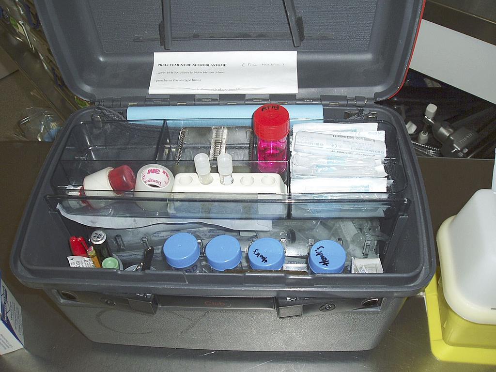
6 Technical aspects Surgical biopsy Core needle biopsy Fine needle biopsy (aspiration) Molecular diagnosis 6 - -

7 Tumor board meeting before biopsy is mandatory oncologists surgeons radiologists anaesthesiologists pathologists
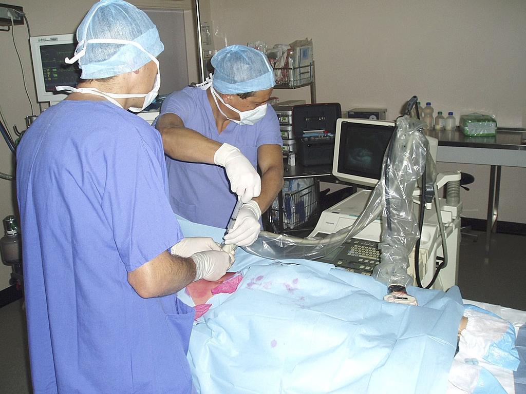
8 Procedure 8 - -
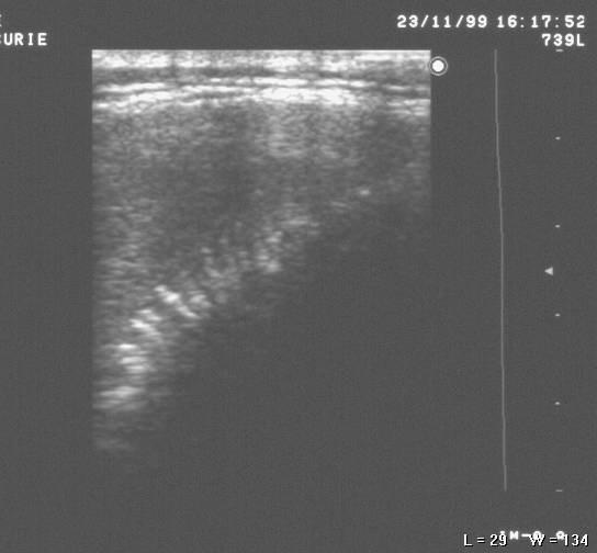
9 Which procedure? Enough material FNA +/- cell-block Percutaneous CNB Surgical biopsy +/- to +++ +/- to to +++ Safe /- to ++ Easy +++ +/- to +++ +/- to + Low cost
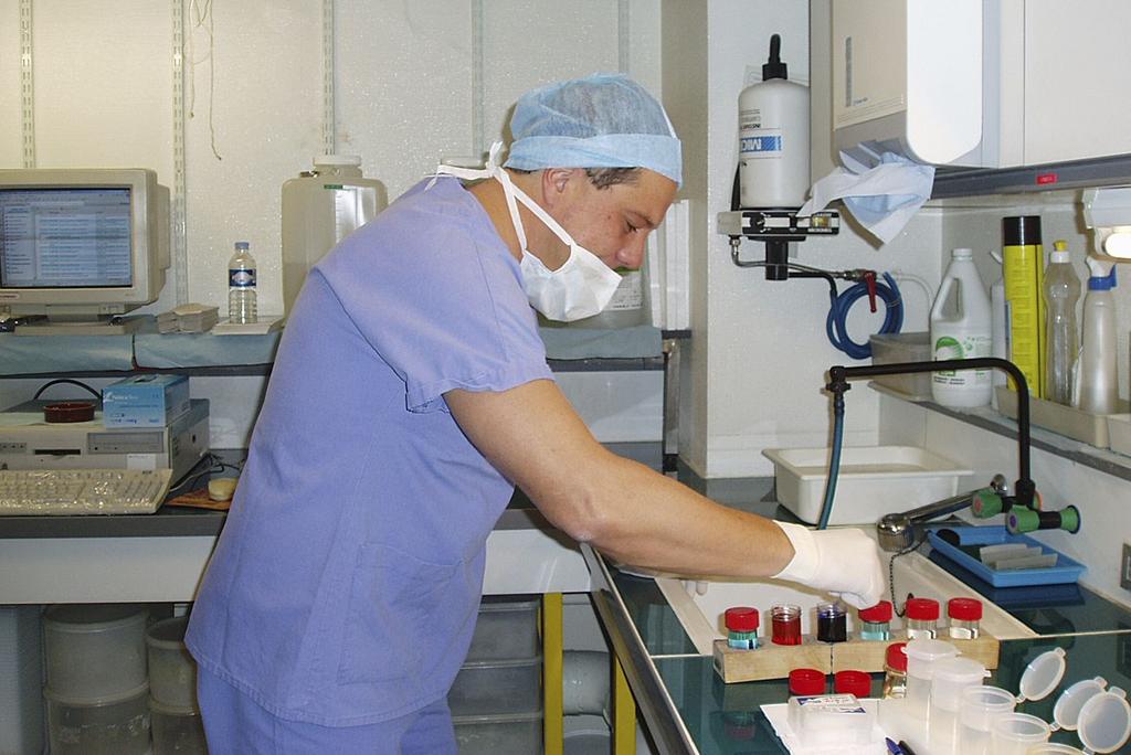
10 Diagnostic samples distribution CNB CB FNA Diagnosis Cryopreservation Pool Karyotyping Molecular Diagnosis Flow Cytometry
11 11 - -
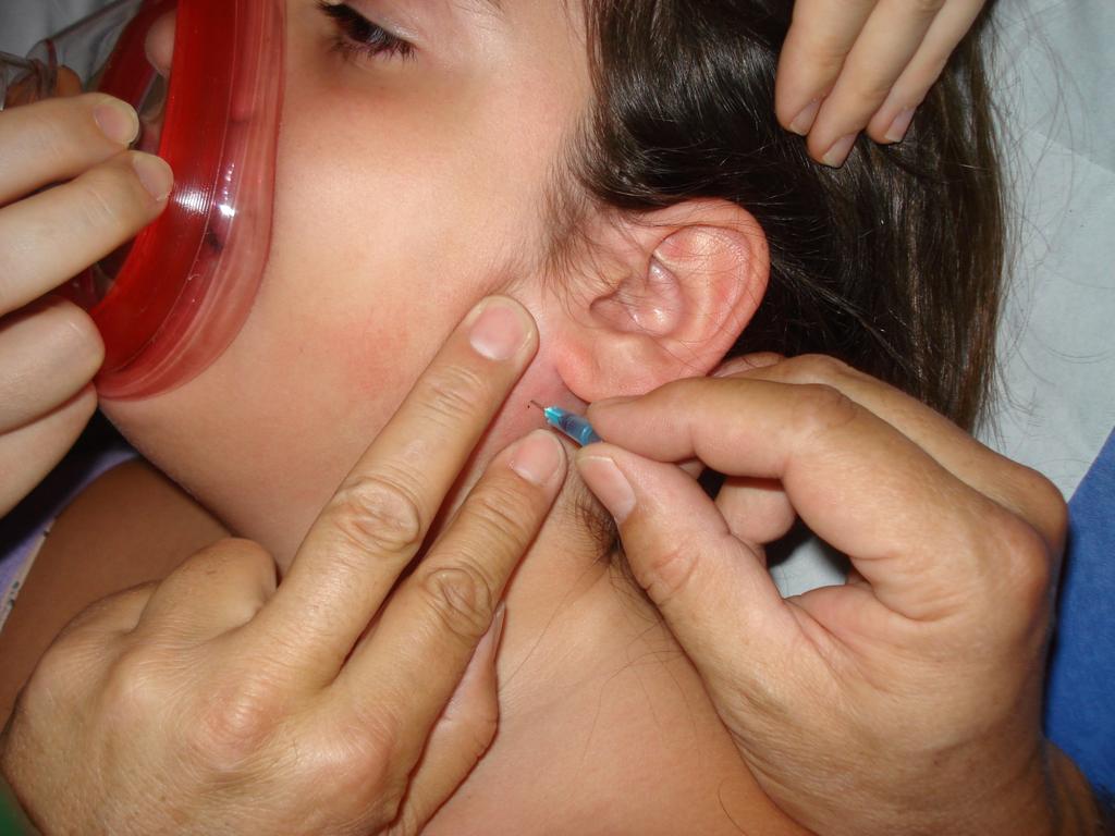
12 Palpable lesions

13 Calinox Emla
14 14 - -

15 Pool Cryopreservation CNB CB FNA
16 Non-palpable lesions Ultrasound-guided
17 17 - -
18 18 - -
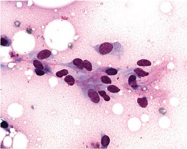
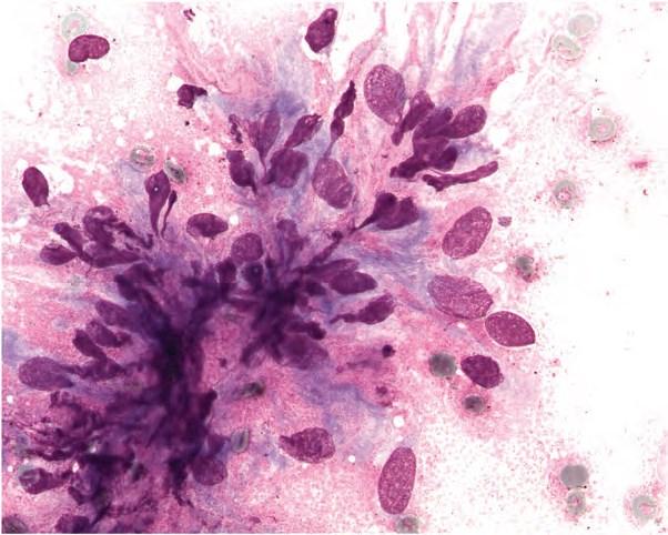
19 19 - -
20 Morphologic and molecular diagnoses
21 21 - -
22 CNB or Cell bloc Diagnosis Immunohistochemistry
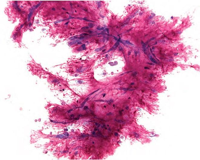
23 23 - -

24 Two main groups of STT


25 Spindle cell tumors (mainly superficial in children)

26 Proposed cytological classification Low-grade spindle cell tumors Tumors with fibrillary stroma Malignant spindle-cell tumors Myxoid tumors Atypical lipomateous tumors Pleomorphic sarcomas Epithelioid tumors Round cell sarcomas
27 Morphological approach in FNA to differentiate STT Low-grade spindle cell tumors Spindle and oval cells without prominent cytonuclear atypia Mitotic figures are scant or absent Connective debris Naked nuclei No necrosis Inflammatory cells Variable cellularity
28 Morphological approach in FNA to differentiate STT (in pediatrics) Low-grade spindle cell tumors Possibility of diagnosis: 1. Fibromatoses or desmoids 2. Nodular fasciitis 3. Dermatofibrosarcoma protuberans 4. Congenital enfantile fibrosarcoma
29 Spindle cell tumors molecular Entity Fibromatoses / desmoids Chromosome abnormality Beta Catenin Gene fusions Nodular fasciitis t(17;22)(p13;q12.3) USP6-MYH9 DFSP t(17;22)(q21.33;q13.1) COLIA1/PDGFB Cong. Enf. Fibrosarcoma t(p12;q15)(p13.2;q25.3) ETV6/NTRK3 + STAT6 antibody (SFT)
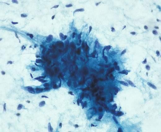
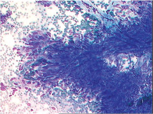
30 Low-grade spindle cell tumors 1. Fibromatoses or desmoids 2. Nodular fasciitis 3. Dermatofibrosarcoma protuberans 4. Fibrosarcoma
31 31 - -
32 32 - -
33 33 - -
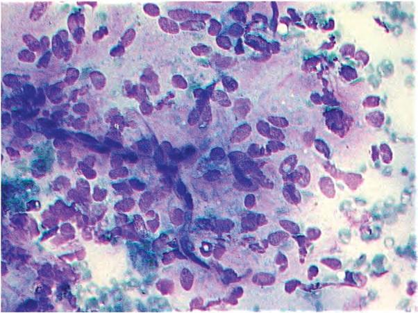
34 34 - -

35 35 - -
36 36 - -
37 Clinical and Fine Needle Aspiration Key Features In favor Difficulties Against - Poorly cellular material comprising spindle-shaped regular cells - Hyalinized tissue fragments - Characteristic clinical and radiological presentations - Some smears may be rich with a mixture of fibroblasts/myofibroblasts - Atypical cells, numerous mitotic figures
38 Low-grade spindle cell tumors 1. Fibromatoses or desmoids 2. Nodular fasciitis 3. Dermatofibrosarcoma protuberans 4. Fibrosarcoma
39 39 - -
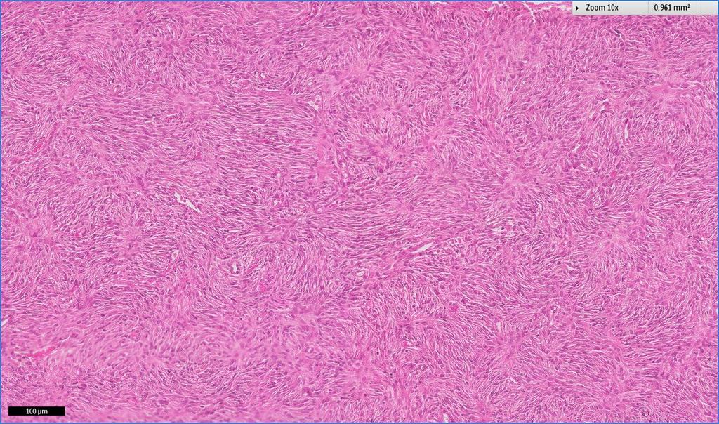
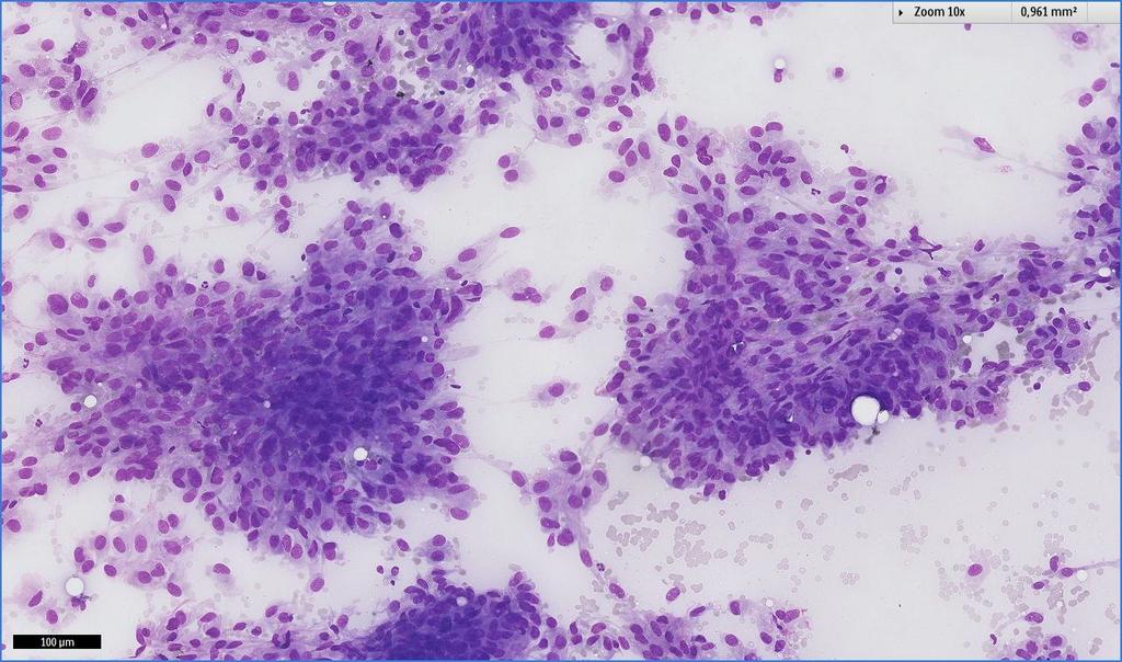
40 40 - -
41 Clinical and Fine Needle Aspiration Key Features In favor Difficulties Against - Hypercellular smears showing mononuclear cells with eccentric, regular nuclei - Multinucleated giant cells - Macrophages or inflammatory background - Mitotic figures or Myxoid background - Necrosis, important cytonuclear atypia, atypical mitoses
42 Low-grade spindle cell tumors 1. Fibromatoses or desmoids 2. Nodular fasciitis 3. Dermatofibrosarcoma protuberans 4. Fibrosarcoma
43 43 - -
44 44 - -
45 45 - -
46 46 - -

47 Storiform
48 48 - -
49 49 - -
50 50 - -
51 51 - -
52 Clinical and Fine Needle Aspiration Key Features In favor Difficulties - Smears rich in spindle cells isolated or clustered - Discrete cytonuclear atypia. - Rare mitotic figures Against -Numerous mitotic figures -Giant multinucleated cells, histiocytic cells - Fibrillary stroma
53 Low-grade spindle cell tumors 1. Fibromatoses or desmoids 2. Nodular fasciitis 3. Dermatofibrosarcoma protuberans 4. Cong Inf Fibrosarcoma
54 54 - -
55 55 - -
56 56 - -
57 57 - -
58 58 - -
59 59 - -
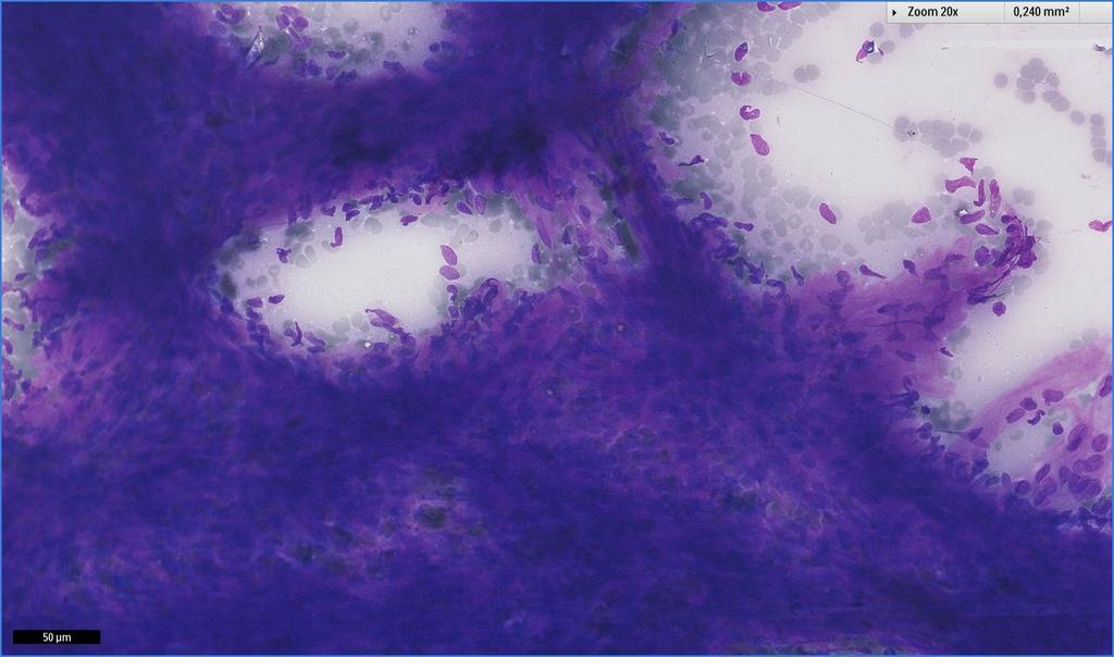
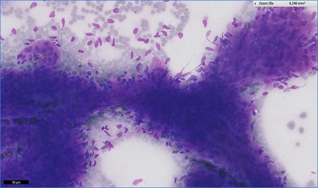
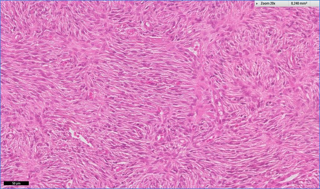
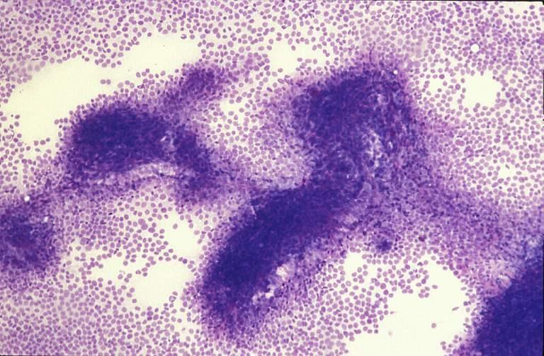
60 Clinical and Fine Needle Aspiration Key Features In favor - Spindle and polymorphous cells - Aggregates of connective tissue - Wavy fascicles - No mitotic activity - Deep soft tissue mass - Exclusion of other spindle-shaped tumor Difficulties - Some smears may be paucicellular - Roundish/epithelioid cells Against - Giant cells - Fibrillary stroma - Epithelial cells - High cellularity - Lipoblasts
61 61 - -
62 62 - -
63 Content children and adolescents in last 20 yrs - malignant 44%, benign 32% and pseudotumors in 24%. - The diagnosis of benign was made in 50% using clinico-radiologic data, - The diagnosis of benign was made in 79% in benign and in 86% in pseudotumors using FNA. - The diagnosis of malignant was made in 39% using clinico-radiologic data, - The diagnosis of malignant was made in 89% using FNA

64 Round cell tumors
65 Round cell tumors Round cell sarcomas Diagnosis by definition Round cell tumors Diagnosis by round cell component
66 Proposed cytological classification 2011 Low-grade spindle cell tumors Tumors with fibrillary stroma Malignant spindle-cell tumors Myxoid tumors Atypical lipomateous tumors Pleomorphic sarcomas Epithelioid tumors Round cell sarcomas
67 Morphological approach to differentiate STT Round cell sarcomas Roundish, clearly malignant cells Moderate cytonuclear atypia Numerous mitotic figures Necrosis may be present Usually extremely high cellularity

68 Morphological approach to differentiate STT Round cell sarcomas Diagnosis by definition 1. Embryonnal and alveolar rhabdomyosarcoma 2. Ewing sarcoma/ppnet 3. Desmoplastic small round cell tumor 4. Extraskeletal mesenchymal chondrosarcoma 5. Rhabdoid tumor
69 Morphological approach to differentiate STT Round cell tumors Diagnosis by round cell component 1. Poorly diff synovial sarcoma 2. Round cell myxoid liposarcoma 3. Neuroblastic tumors 4. Nephroblastoma 5. Langerhans cell histiocytosis 6. Round cell osteosarcoma
70 Morphological approach to differentiate STT Round cell sarcomas How to diagnose? - Clinical and radiological informations are important - Round cell pattern (RCP) * Specific RCP for diagnosis (NB, Ewing,.) ** Not specific RCP for diagnosis - Immuno(histo/cyto)chemistry - Specific molecular alterations in many entities

71 Morphological approach to differentiate STT Round cell sarcomas Strong points of FNA - Round cell pattern (RCP) well seen - Hypercellular material * Cell pooling for molecular techniques ** Cell blocs for IHC - Rapidity - On situ diagnosis for CNB indication etc
72 Morphological approach to differentiate STT Round cell sarcomas What to do on morphology? - Search for rhabdomyoblasts, binucleation, rosettes, and double cell population - Chondroid is usually well detected - Poorly differentiated synovial sarcoma may mimick Ewing sarcoma, search for rosettes and double cell population


73 Morphological approach to differentiate STT Round cell sarcomas What to do on ICH? NB84 INI1/SMARB1 Chromogranin Synaptophisin CD45-RB-LCA Desmin MyoD1 CD99-Mic2 AE1/AE

74 Morphological approach to differentiate STT Round cell sarcomas What to do in molecular diagnosis? -PCR -FISH -Karyotyping -CGH FISH with a EWSR1 dualcolor breakapart probe on a Ewing tumor
75 Molecular specific diagnosis in RCT PNET/Ewing: fusion transcript EWS FLI (85%) Alveolar RMS: fusion transcript PAX FKHR Allelic loss of 1p36 in neuroblastoma Desmoplastic small round cell tumor: EWS/WT1 Synovial sarcoma: SYT/SSX1 or 2 or
76 Round cell sarcomas by definition 1. Embryonnal and alveolar rhabdomyosarcoma 2. Ewing sarcoma/ppnet 3. Desmoplastic small round cell tumor 4. Extraskeletal mesenchymal chondrosarcoma 5. Rhabdoid tumor

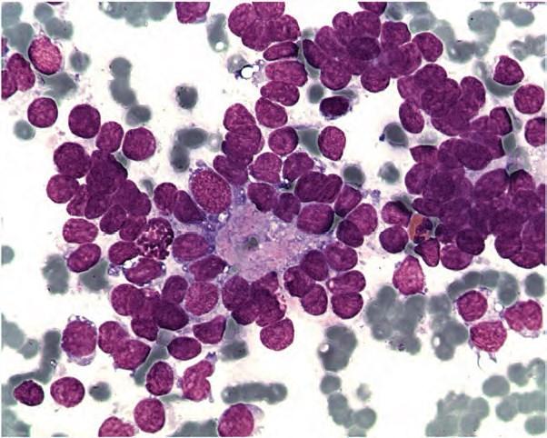
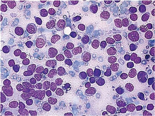
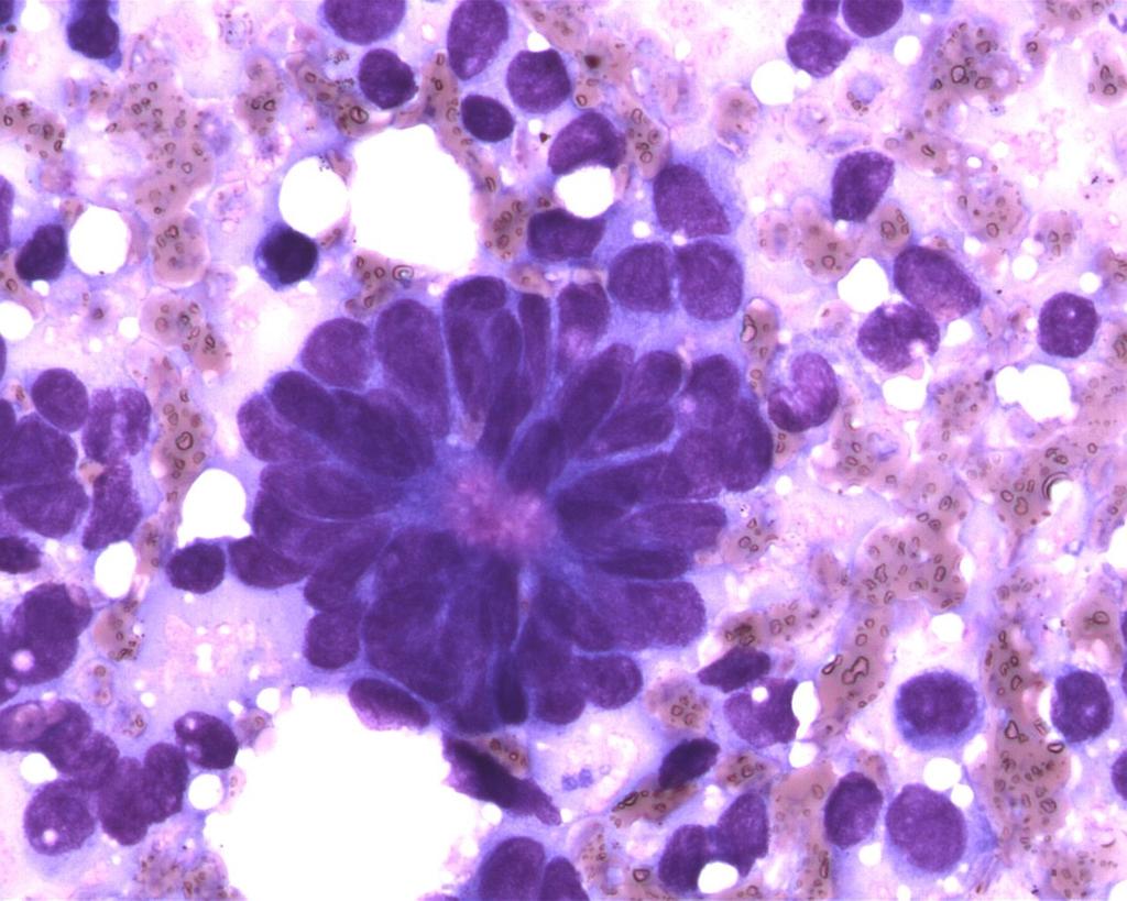
77 Round cell sarcomas by definition 1. Embryonnal and alveolar rhabdomyosarcoma 2. Ewing sarcoma/ppnet 3. Desmoplastic small round cell tumor 4. Extraskeletal mesenchymal chondrosarcoma 5. Rhabdoid tumor

78 RMS

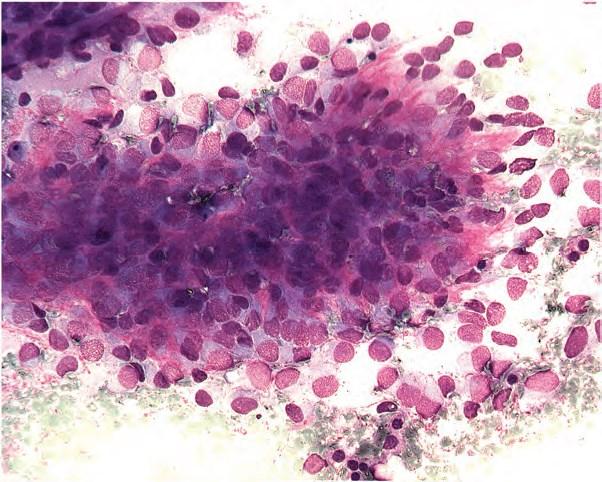
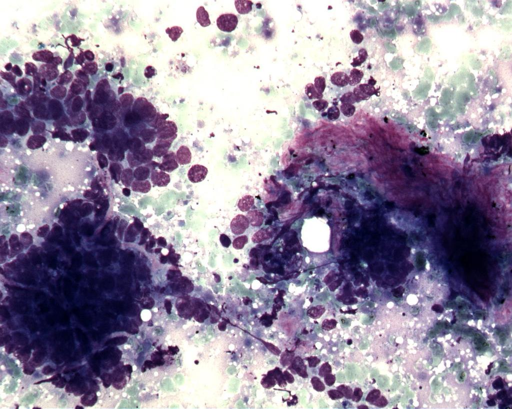
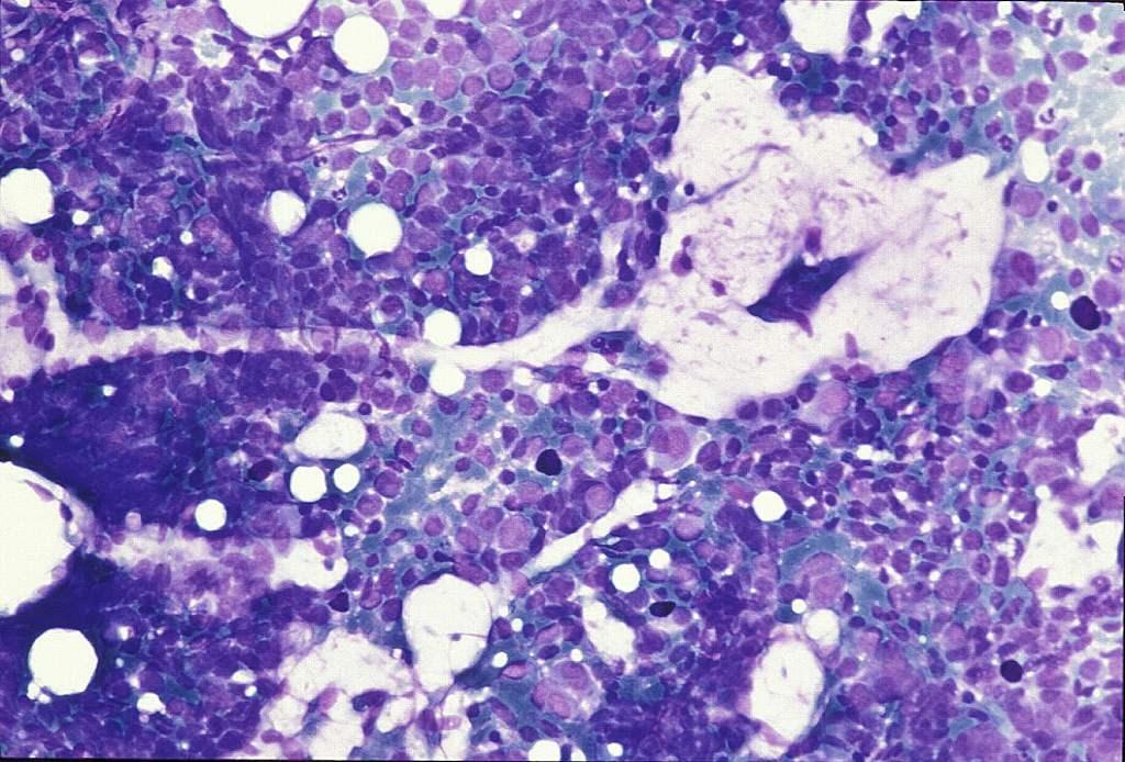
79 RMS
80 Why FNA? To diagnose RMS To provide material for karyotyping or molecular biology techniques Atypical sites

81 RMS

82 82 - RMS -
83 RMS-desmin
84 Cellular components Rhabdomyoblasts Excentric nuclei Binucleated cells Spindle-shaped cells Abundant cytoplasms

85 85 RMS - -

86 RMS-alv

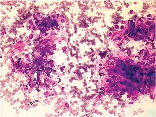
87 RMS-alv
88 88 - -

89 RMS-alv
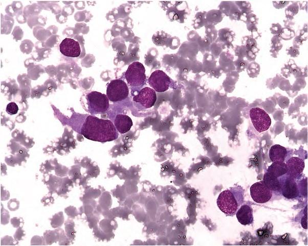
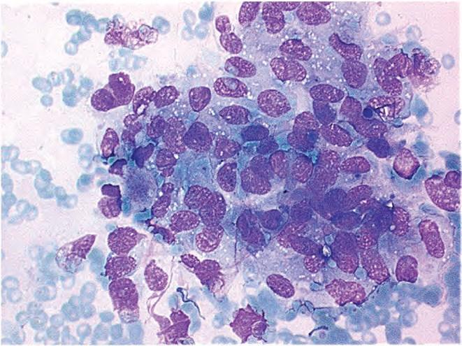
90 90 - -

91 Gluteal
92 92 - -
93 93 - -
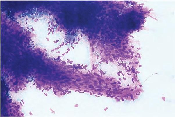
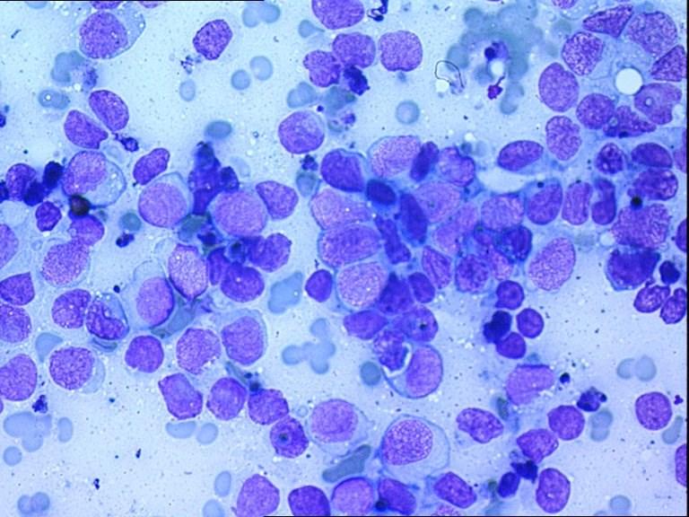
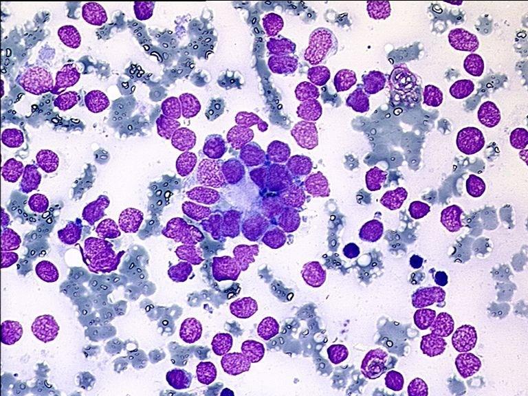
94 Clinical and Fine Needle Aspiration Key Features (RMS) Yes Maybe No -Roundish cells, rhabdomyoblastic cells, alveolar structures, positivity of muscular markers -Specific fusion transcript PAX FKHR (alveolar) - Polymorphous morphology, spindle-shaped cells - Double round cell population - Rosettes - Perinuclear inclusions - Papillary structures - Epithelial cells

95 Round cell sarcomas by definition 1. Embryonnal and alveolar rhabdomyosarcoma 2. Ewing sarcoma/ppnet 3. Desmoplastic small round cell tumor 4. Extraskeletal mesenchymal chondrosarcoma 5. Rhabdoid tumor
96 96 - -

97 PNET
98 Why FNA? To diagnose Ewing/PNET To provide material for karyotype or molecular biology techniques
99 PNET/EWING
100 PNET/EWING
101 Cellular components Round, irregular cells «neuroendocrine» pattern Rosette-like structures
102 PNET/EWING
103
104 PNET/EWING
105 PNET/EWING-cell bloc
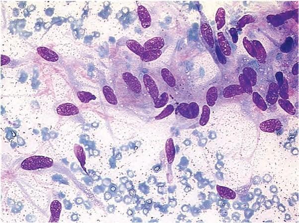
106
107
108
109 Clinical and Fine Needle Aspiration Key Features (ES/PNET) Yes Maybe No -Young adult - Double population of large and small cells - Rosette formation - CD Specific fusion transcript EWS FLI and karyotypic translocation (85%) - Spindle cells, necrosis - Extraskeletal localizations - No specific genomic abnormality or absence of abnormality (15%) - Fibrillary stroma - Osteoid
110 Round cell sarcomas by definition 1. Embryonnal and alveolar rhabdomyosarcoma 2. Ewing sarcoma/ppnet 3. Desmoplastic small round cell tumor 4. Extraskeletal mesenchymal chondrosarcoma 5. Rhabdoid tumor
111
112

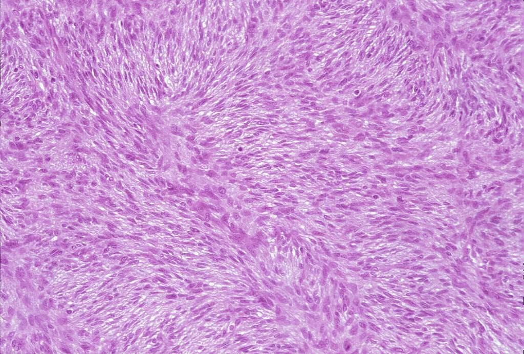
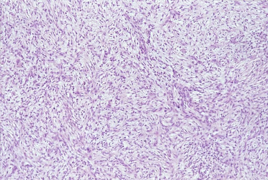
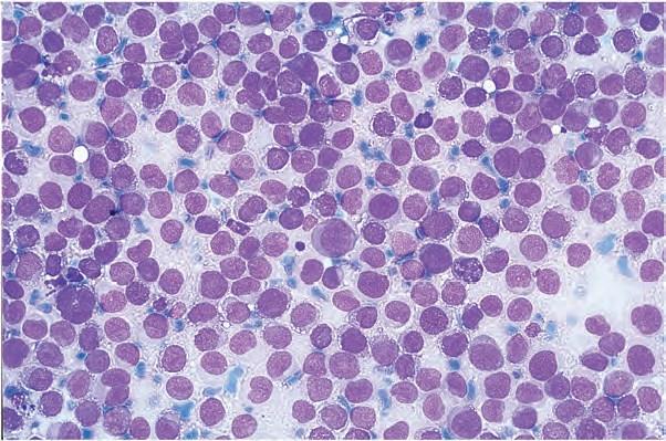
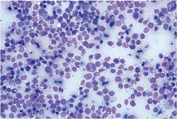
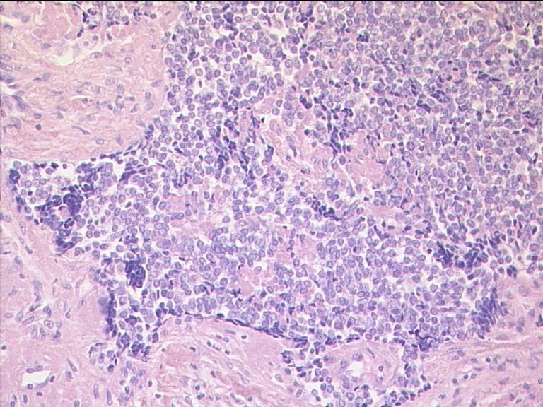
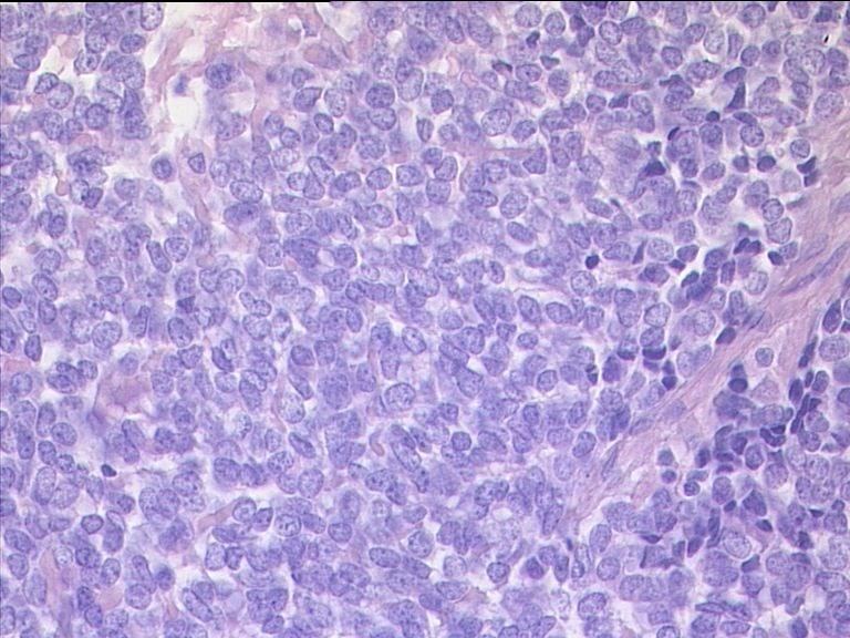
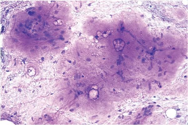
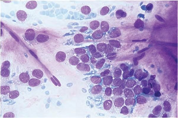
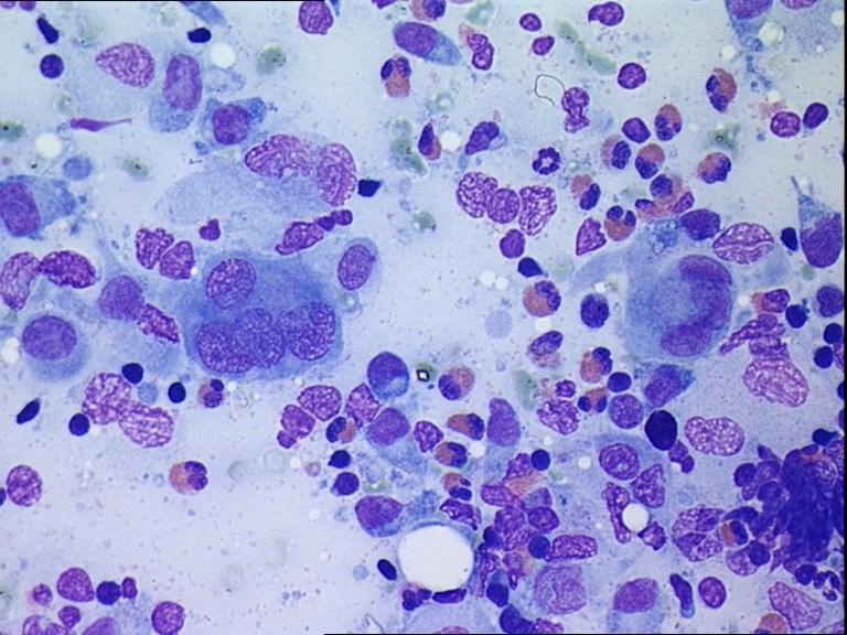
113 Desmin
114 Clinical and Fine Needle Aspiration Key Features (DSRCT) Yes Maybe No - Age - Intraabdominal site - Poorly differentiated round cells with inconspitious cytoplasm - Paranuclear cytoplasmic densities (Inter fil +) - Specific molecular transcript (EWS/WT1) - Sarcomateous polymorphous cells - Extensive necrosis - Rhabdomyoblasts, fibrillary stroma, rosettes
115 Round cell sarcomas by definition 1. Embryonnal and alveolar rhabdomyosarcoma 2. Ewing sarcoma/ppnet 3. Desmoplastic small round cell tumor 4. Extraskeletal mesenchymal chondrosarcoma 5. Rhabdoid tumor
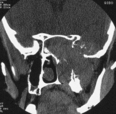
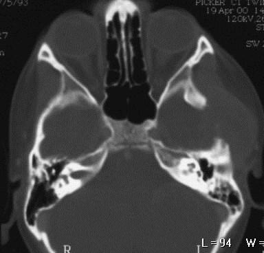
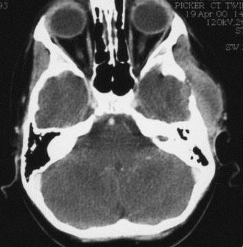
116
117
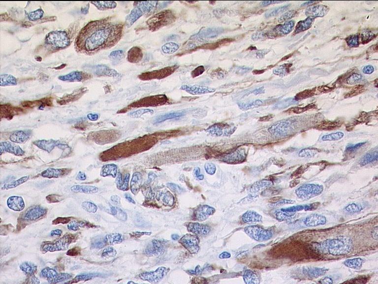
118 Clinical and Fine Needle Aspiration Key Features (EMCS) Yes -Double component of small round cells and malignant chondroid -Specific molecular transcript (EWSR1, TAF15, TCF12/NR4A3) Maybe No - Lack of malignant chondroid component - Rosettes, physaliphorous cells
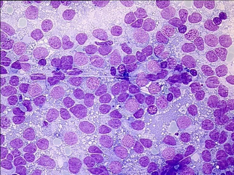
119 Round cell sarcomas by definition 1. Embryonnal and alveolar rhabdomyosarcoma 2. Ewing sarcoma/ppnet 3. Desmoplastic small round cell tumor 4. Extraskeletal mesenchymal chondrosarcoma 5. Rhabdoid tumor
120 Rhabdoid tumor 1978 RMS-like morphology Kidney and extra-kidney localizations Homozygous deletion of 22q % in the first year, 85% in 2 yrs Aggressive behaviour
121 Rhabdoid tumor Monomorphous appearance RMS-like proliferation Dispersed cells Epithelioid cords Cytoplasmic inclusions
122 Why FNA? To diagnose Rhabdoid tumor To provide material for karyotyping or molecular biology techniques

")

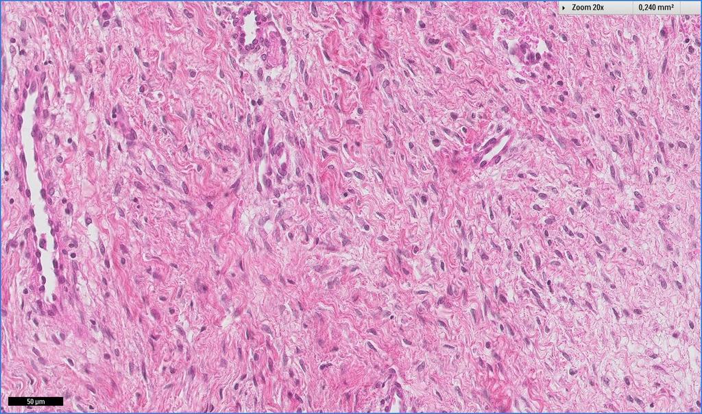
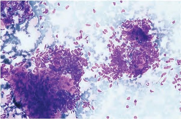
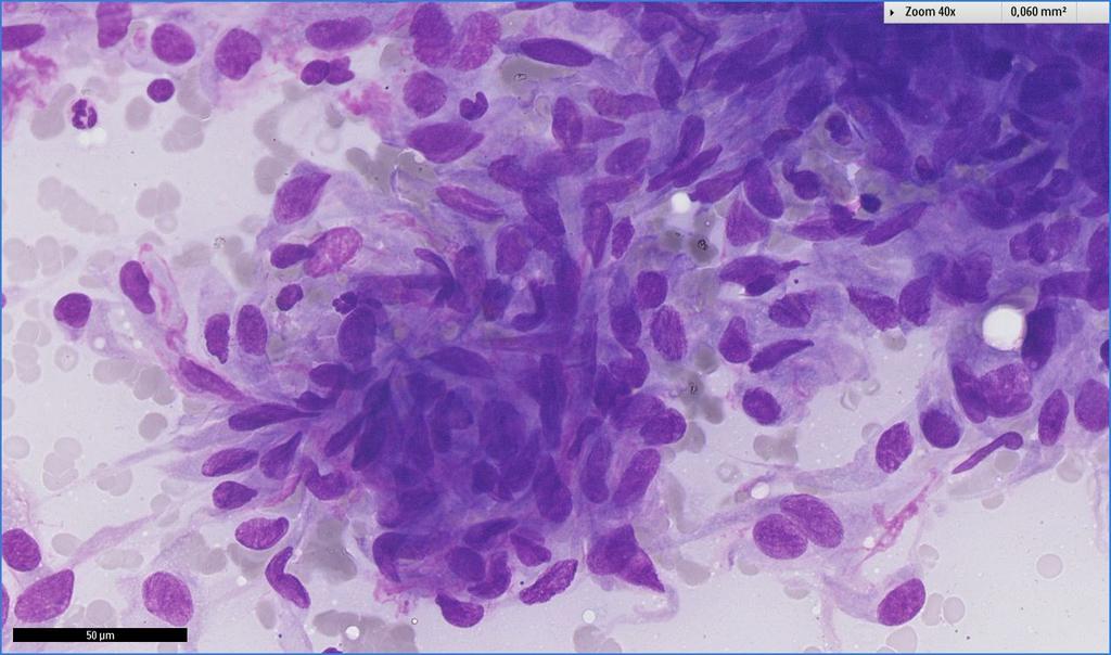
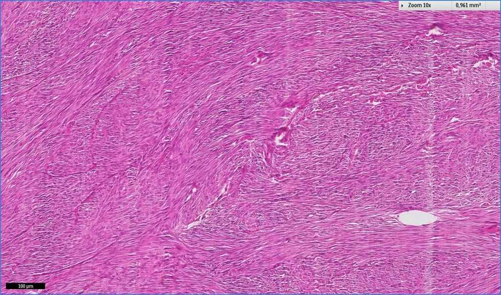
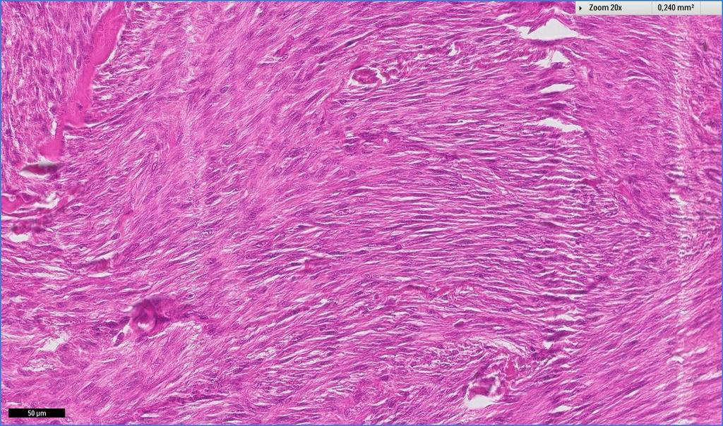
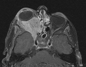
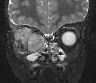
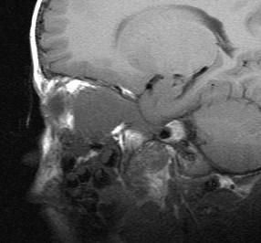
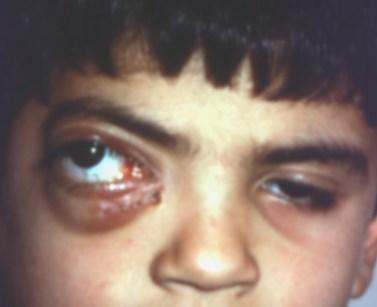
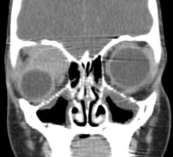
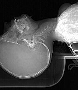
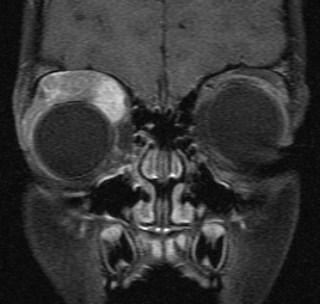
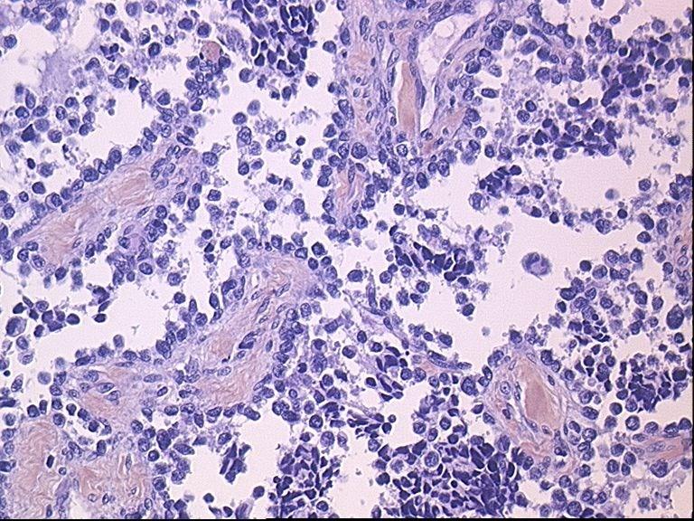
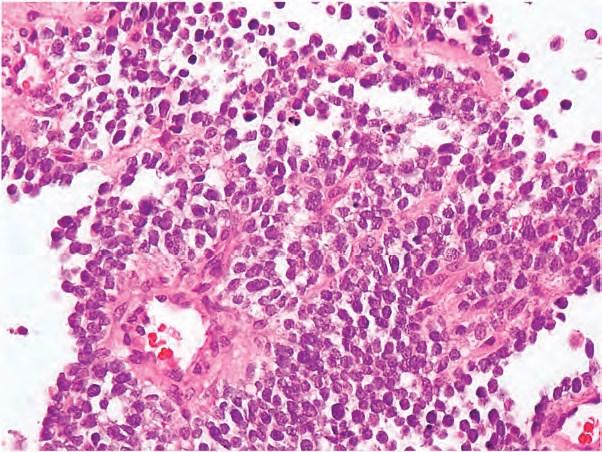
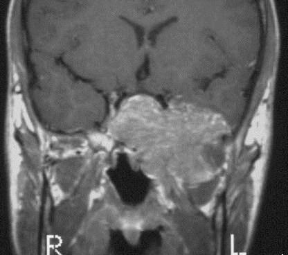
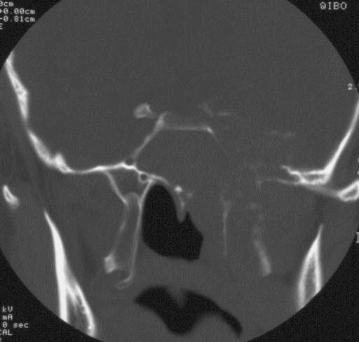
123 A B C Enfant de 14 mois présentant une masse cervicale droite isolée, prise initialement pour une malformation artérioveineuse et surveillée. Progression clinique rapide en 3 mois (figure A) faisant redresser le diagnostic en tumeur rhabdoïde des parties molles. Aspect IRM coronal T2 (figure B) et axial T2 Fat Sat (figure C).
124 RbT

125 EMA
126 Cellular components Rhabdomyoblasts-like Perinuclear bodies Nucleoli

127 RbT

128 RbT

129 RhT
130 Clinical and Fine Needle Aspiration Key Features (RT) Yes Maybe No - Young age, kidney localization - Round-to-oval rhabdomyoblastic cells - Perinuclear inclusions that are keratin (+) - SMARB1/INI1 negativity - Lack of perinuclear densities. - Epithelioid pattern with clusters - True rhabdomyoblasts, alveolar structures
131 Tumors with RC component 1. Poorly differentiated synovial sarcoma 2. Round cell myxoid liposarcoma 3. Neuroblastic tumors 4. Nephroblastoma 5. Langerhans cell histiocytosis 6. Round cell osteosarcoma

132 Tumors with RC component 1. Poorly differentiated synovial sarcoma (spindle?) 2. Round cell myxoid liposarcoma 3. Neuroblastic tumors 4. Nephroblastoma 5. Langerhans cell histiocytosis 6. Round cell osteosarcoma
133
134
135
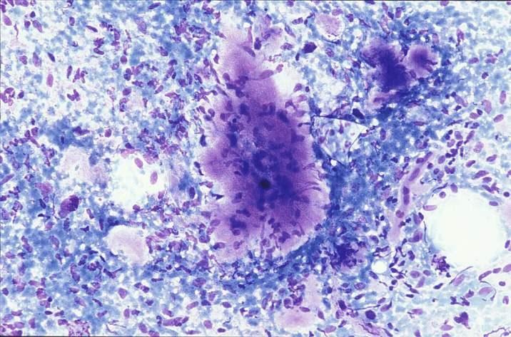
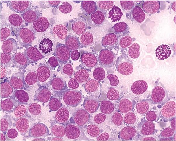
136 Clinical and Fine Needle Aspiration Key Features (SS) Yes Maybe No - Highly cellular smears with oval-to-spindleshaped cytoplasm - Branching tumor tissue fragmentsand vessel stalks -Cohesive epithelial cells in biphasic subtype -SYT/SSX1 or 2 or 4 abnormality - Secretory mucin - Rosette-like structures - Mitotic figures and connective stromal components are usually scarce - True rosettes - Double population of roundish cells

137 Tumors with RC component 1. Poorly differentiated synovial sarcoma 2. Round cell myxoid liposarcoma 3. Neuroblastic tumors 4. Nephroblastoma 5. Langerhans cell histiocytosis 6. Round cell osteosarcoma
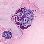
138
139
140 Clinical and Fine Needle Aspiration Key Features (MLipoSa) Yes Maybe No - Abundant myxoid background - Isolated and regular small spindle-shaped and stellated cells - Extremities dermal and subcutaneous localization - FUS, EWSR1/DDIT3 abnormality - Round cells - Giant multinucleated cells - Lack of cytonuclear atypia - High-grade atypical cells and mitotic figures - Deep localization
141 Tumors with RC component 1. Poorly differentiated synovial sarcoma 2. Round cell myxoid liposarcoma 3. Neuroblastic tumors 4. Nephroblastoma 5. Langerhans cell histiocytosis 6. Round cell osteosarcoma
142 Tumors with RC component 1. Poorly differentiated synovial sarcoma 2. Round cell myxoid liposarcoma 3. Neuroblastic tumors 4. Nephroblastoma 5. Langerhans cell histiocytosis 6. Round cell osteosarcoma
143 Tumors with RC component 1. Poorly differentiated synovial sarcoma 2. Round cell myxoid liposarcoma 3. Neuroblastic tumors 4. Nephroblastoma 5. Langerhans cell histiocytosis 6. Round cell osteosarcoma
144 Histiocytosis X
145 Why FNA? To confirm diagnosis Clinics + Radiology + FNA = Diagnosis
146 Cellular components Eosinophilic leukocytes Macrophages Mono-, bi-, multinucleated cells

147 H-X
148

149 Tumors with RC component 1. Poorly differentiated synovial sarcoma 2. Round cell myxoid liposarcoma 3. Neuroblastic tumors 4. Nephroblastoma 5. Langerhans cell histiocytosis 6. Round cell osteosarcoma
150

151 Thank you!!!!
Tumores de células pequeñas, redondas y azules: diagnóstico diferencial cuando el tiempo apremia
 Tumores de células pequeñas, redondas y azules: diagnóstico diferencial cuando el tiempo apremia Sílvia Bagué Servei de Patologia Hospital de Sant Pau Barcelona Soft tissue sarcomas Heterogeneous group
Disclosures. An update on ancillary techniques in the diagnosis of soft tissue tumors. Ancillary techniques. Introduction
 Disclosures An update on ancillary techniques in the diagnosis of soft tissue tumors. I have nothing to disclose. Andrew Horvai, MD, PhD Clinical Professor, Pathology Introduction Ancillary techniques
Klinisch belang van chromosomale translocatie detectie in sarcomen
 Translocations in sarcomas Klinisch belang van chromosomale translocatie detectie in sarcomen Judith V.M.G. Bovée, M.D., Ph.D. Department of Pathology Leiden University Medical Center RNA binding DNA binding
Spindle Cell Lesions Of The Breast. Emad Rakha Professor of Breast Pathology and Consultant Pathologist
 Spindle Cell Lesions Of The Breast Emad Rakha Professor of Breast Pathology and Consultant Pathologist * SCLs comprise a wide spectrum of diseases, ranging from reactive processes to aggressive malignant
5/10. Pathology Soft tissue tumors. Farah Bhani. Mohammed Alorjani
 5/10 Pathology Soft tissue tumors Mohammed Alorjani Farah Bhani Slides are included in this sheet. Objectives: Soft tissue tumors 1. Describe soft tissue tumors. 2. Understand the classification of soft
Molecular pathology in soft tissue tumors. Sylvia Höller Pathologie
 Molecular pathology in soft tissue tumors Sylvia Höller Pathologie When do we perform molecular testing? Morphology and IHC are not clearly fitting with an entity some translocations are entity specific
Surgical Pathology Evening Specialty Conference USCAP 2015
 Surgical Pathology Evening Specialty Conference USCAP 2015 John R. Goldblum, M.D. Chairman, Department of Pathology, Cleveland Clinic Professor of Pathology, Cleveland Clinic Lerner College of Medicine
Case 2. Dr. Sathima Natarajan M.D. Kaiser Permanente Medical Center Sunset
 Case 2 Dr. Sathima Natarajan M.D. Kaiser Permanente Medical Center Sunset History 24 year old male presented with a 3 day history of right flank pain, sharp in nature Denies fever, chills, hematuria or
Immunohistochemistry in Bone and Soft Tissue Tumors. Sahar Rassi Zankoul, MD
 Immunohistochemistry in Bone and Soft Tissue Tumors Sahar Rassi Zankoul, MD Introduction Bone tumors represent a wide variety of tumors of various origins and malignant potentials. These different tumor
Newer soft tissue entities
 Newer soft tissue entities Examples among fibroblastic tumors Turku, May 6, 2010 Markku Miettinen, M.D. AFIP, Washington, DC Fibroblastic neoplasms Solitary fibrous tumor /Hemangiopericytoma Low-grade
Diagnostic Cytology of Cancer Cases
 Diagnostic Cytology of Cancer Cases Somporn Techangamsuwan Companion Animal Cancer Research Unit (CAC-RU) Department of Pathology, Faculty of Veterinary Science, Chulalongkorn University 1 Tumor or Non-tumor
Evening Specialty Conference Bone and Soft Tissue Pathology. Diagnostic pitfalls in bone and soft tissue pathology
 Evening Specialty Conference Bone and Soft Tissue Pathology. Case 1 Elizabeth G Demicco, MD, PhD Mount Sinai Hospital, New York Disclosure of Relevant Financial Relationships USCAP requires that all planners
Tumors of Adipose Tissue Tumors Epidemiology Clinical Features. Morphology. Mature Adipocytes Separated by delicate fibrous septa
 Tumors of Adipose Tissue Lipoma Liposarcoma Most commonly happens in female The most common soft tissue tumor o Originates from matured Adipocytes Most commonly happes at the 4 th and 5 th decade of life
57th Annual HSCP Spring Symposium 4/16/2016
 An Unusual Malignant Spindle Cell Lesion to Involve the Breast Erinn Downs-Kelly, D.O. Associate Professor of Pathology University of Utah & ARUP Laboratories No disclosures Case 39 y/o female with no
From Morphology to Molecular Pathology: A Practical Approach for Cytopathologists Part 1-Cytomorphology. Songlin Zhang, MD, PhD LSUHSC-Shreveport
 From Morphology to Molecular Pathology: A Practical Approach for Cytopathologists Part 1-Cytomorphology Songlin Zhang, MD, PhD LSUHSC-Shreveport I have no Conflict of Interest. FNA on Lymphoproliferative
Diplomate of the American Board of Pathology in Anatomic and Clinical Pathology
 A 33-year-old male with a left lower leg mass. Contributed by Shaoxiong Chen, MD, PhD Assistant Professor Indiana University School of Medicine/ IU Health Partners Department of Pathology and Laboratory
1/10/2018. Soft Tissue Tumors Showing Melanocytic Differentiation. Overview. Desmoplastic/ Spindle Cell Melanoma
 2016 MFMER slide-1 2016 MFMER slide-2 2016 MFMER slide-3 Soft Tissue Tumors Showing Melanocytic Differentiation Andrew L. Folpe, M.D. Professor of Laboratory Medicine and Pathology Mayo Clinic, Rochester,
PITFALLS AND TRAPS IN THE DIAGNOSIS AND STAGING OF RENAL TUMOURS OF CHILDHOOD. Gordan M. Vujanić Cardiff, U.K.
 PITFALLS AND TRAPS IN THE DIAGNOSIS AND STAGING OF RENAL TUMOURS OF CHILDHOOD Gordan M. Vujanić Cardiff, U.K. RENAL TUMOURS OF CHILDHOOD - CLASSIFICATION (2016) Nephroblastic tumours Mesenchymal tumours
3/27/2017. Disclosure of Relevant Financial Relationships
 Ophthalmic Pathology Evening Specialty Conference USCAP 2017 5 th March, 2017 Mukul K. Divatia, MD Assistant Professor Department of Pathology & Genomic Medicine Weill Cornell Medical College Houston Methodist
Anaplastic Large Cell Lymphoma (of T cell lineage)
") Anaplastic Large Cell Lymphoma (of T cell lineage) Definition T-cell lymphoma comprised of large cells with abundant cytoplasm and pleomorphic, often horseshoe-shaped nuclei CD30+ Most express cytotoxic
Anaplastic Large Cell Lymphoma (of T cell lineage) Definition T-cell lymphoma comprised of large cells with abundant cytoplasm and pleomorphic, often horseshoe-shaped nuclei CD30+ Most express cytotoxic
Cutaneous Mesenchymal Neoplasms with EWSR1 Rearrangement
 Cutaneous Mesenchymal Neoplasms with EWSR1 Rearrangement By Konstantinos Linos MD, FCAP, FASDP Bone, Soft Tissue and Dermatopathology Assistant Professor of Pathology Dartmouth-Hitchcock Medical Center
Selected Pseudomalignant Soft Tissue Tumors of the Skin and Subcutis
 Selected Pseudomalignant Soft Tissue Tumors of the Skin and Subcutis Andrew L. Folpe, M.D. Professor of Laboratory Medicine and Pathology Mayo Clinic, Rochester, MN folpe.andrew@mayo.edu 2016 MFMER slide-1
Financial disclosures
 An update on immunohistochemical markers in mesenchymal neoplasms By Konstantinos Linos MD, FCAP, FASDP Assistant Professor of Pathology Geisel School of Medicine at Dartmouth Dartmouth-Hitchcock Medical
ACCME/Disclosures. Everything is spindle - how far can we go with limited FNA material? Everything is spindle how far can we go? Everything is spindle
 ACCME/Disclosures The USCAP requires that anyone in a position to influence or control the content of CME disclose any relevant financial relationship WITH COMMERCIAL INTERESTS which they or their spouse/partner
Keywords solitary fibrous tumor, dedifferentiation, dedifferentiated solitary fibrous tumor, STAT6, GRIA2, cytokeratin, rhabdomyosarcomatous
 758452IJSXXX10.1177/1066896918758452International Journal of Surgical PathologyCreytens et al research-article2018 Pitfalls in Pathology Multifocal Cytokeratin Expression in a Dedifferentiated Solitary
Lung Tumor Cases: Common Problems and Helpful Hints
 Lung Tumor Cases: Common Problems and Helpful Hints Brandon T. Larsen, MD, PhD Senior Associate Consultant Department of Laboratory Medicine and Pathology Mayo Clinic Arizona Arizona Society of Pathologists
Mody. AIS vs. Invasive Adenocarcinoma of the Cervix
 Common Problems in Gynecologic Pathology Michael T. Deavers, M.D. Houston Methodist Hospital, Houston, Texas Common Problems in Gynecologic Pathology Adenocarcinoma in-situ (AIS) of the Cervix vs. Invasive
Methoden / Methods inc. ICCC-3 105
 Methoden / Methods inc. ICCC-3 105 Internationale Klassifikation der Krebserkrankungen bei Kindern (ICCC-3) Zuordnung von ICD-O-3-Codes für Morphologie und Topographie zu diagnostischen Kategorien International
Molecular Genetics of Paediatric Tumours. Gino Somers MBBS, BMedSci, PhD, FRCPA Pathologist-in-Chief Hospital for Sick Children, Toronto, ON, CANADA
 Molecular Genetics of Paediatric Tumours Gino Somers MBBS, BMedSci, PhD, FRCPA Pathologist-in-Chief Hospital for Sick Children, Toronto, ON, CANADA Financial Disclosure NanoString - conference costs for
FNA of Thyroid. Toward a Uniform Terminology With Management Guidelines. NCI NCI Thyroid FNA State of the Science Conference
 FNA of Thyroid NCI NCI Thyroid FNA State of the Science Conference Toward a Uniform Terminology With Management Guidelines Thyroid Thyroid FNA Cytomorphology NCI Thyroid FNA State of the Science Conference
What is New in the 2015 WHO Lung Cancer Classification? Zhaolin Xu, MD, FRCPC, FCAP
 What is New in the 2015 WHO Lung Cancer Classification? Zhaolin Xu, MD, FRCPC, FCAP Professor, Dept of Pathology, Dalhousie University, Canada Pulmonary Pathologist and Cytopathologist, QEII HSC Senior
Pathology of Sarcoma ELEANOR CHEN, MD, PHD, ASSISTANT PROFESSOR DEPARTMENT OF PATHOLOGY UNIVERSITY OF WASHINGTON
 Pathology of Sarcoma ELEANOR CHEN, MD, PHD, ASSISTANT PROFESSOR DEPARTMENT OF PATHOLOGY UNIVERSITY OF WASHINGTON Presentation outline Background and epidemiology of sarcomas Sarcoma classification Sarcoma
The Genetics of Myoepithelial Tumors: salivary glands, soft tissue and bone
 The Genetics of Myoepithelial Tumors: salivary glands, soft tissue and bone Cristina Antonescu, MD Memorial Sloan-Kettering Cancer Center, New York Nothing to declare Disclosure Spectrum of Myoepithelial
Diagnostic Value of Immunohistochemistry in Soft Tissue Tumors
 Original Article DOI: 10.21276/APALM.1637 Diagnostic Value of Immunohistochemistry in Soft Tissue Tumors Sridevi. V*., Susruthan Muralitharan., and Thanka. J Dept of Pathology, SriMuthukumaran Medical
INDEX. in this web service Cambridge University Press
 actin 14 adamantinoma 202, 290 292, 297 adenocarcinoma 136 adipocytes in hibernoma 149, 150 in lipoblastoma 148 in lipoma 141, 142, 145 in liposarcoma 152 in myelolipoma 151 adrenal gland tumors see myelolipoma
The diagnostic challenge of paediatric small round cell tumours
 1 The diagnostic challenge of paediatric small round cell tumours K. Skordilias A.G. Howatson INTRODUCTION On writing about paediatric small round cell tumours (SRCTs) one cannot avoid the obvious question:
Journal of Solid Tumors, April 2012, Vol. 2, No. 2
 ORIGINAL ARTICLE Utility of fluorescence in situ hybridization in subclassifying unclassified high-grade sarcomas: A study of 40 cases using break-apart probes of EWSR1, FOXO1A, SS18 and DDIT3 genes Alfredo
ACCME/Disclosures ALK FUSION-POSITIVE MESENCHYMAL TUMORS. Tumor types with ALK rearrangements. Anaplastic Lymphoma Kinase. Jason L.
 Companion Meeting of the International Society of Bone and Soft Tissue Pathology The Evolving Concept of Mesenchymal Tumors ALK FUSION-POSITIVE MESENCHYMAL TUMORS Jason L. Hornick, MD, PhD March 13, 2016
IMMUNOHISTOCHEMISTRY IN THE DIAGNOSIS OF SOFT TISSUE TUMORS
 IMMUNOHISTOCHEMISTRY IN THE DIAGNOSIS OF SOFT TISSUE TUMORS Nicolas de Saint Aubain Somerhausen Institut Jules Bordet / Hôpital Erasme nicolas.desaintaubain@synet.be ForPath 2005 1 I. Ancillary techniques
Problem 1: Differential of Neuroendocrine Carcinoma 3/23/2017. Disclosure of Relevant Financial Relationships
 Differential of Neuroendocrine Carcinoma Alain C. Borczuk,MD Weill Cornell Medicine Disclosure of Relevant Financial Relationships USCAP requires that all faculty in a position to influence or control
Enterprise Interest Nothing to declare
 Enterprise Interest Nothing to declare Diagnoses one would not like to miss in soft tissue pathology early in your career Marta Sbaraglia, MD Department of Pathology Hospital of Treviso University of Padua
Presentation material is for education purposes only. All rights reserved URMC Radiology Page 1 of 98
 Presentation material is for education purposes only. All rights reserved. 2011 URMC Radiology Page 1 of 98 Radiology / Pathology Conference February 2011 Brooke Koltz, Cytopathology Resident Presentation
Aspen conference on pediatric disease. July through August Bone and Soft Tissue Update. David M. Parham, MD. Rhabdomyoma and rhabdomyosarcoma
 Aspen conference on pediatric disease July through August 2014 Bone and Soft Tissue Update David M. Parham, MD Rhabdomyoma and rhabdomyosarcoma Embryonic rhabdomyogenesis is a highly conserved process
Salivary gland tumor cytologic and histologic correlation: Algorithmic and risk stratification based approaches
 Salivary gland tumor cytologic and histologic correlation: Algorithmic and risk stratification based approaches Christopher C. Griffith, MD, PhD Raja R. Seethala, MD 1. Salivary gland tumor cytology: A
Malignant Peripheral Nerve Sheath Tumor
 C H A P T E R 120 Malignant Peripheral Nerve Sheath Tumor Currently, malignant peripheral nerve sheath tumor (MPNST) is the most commonly used generic name for the neoplasms known in the past as neurosarcoma,
Case year female. Routine Pap smear
 Case 1 57 year female Routine Pap smear Diagnosis? 1. Atypical glandular cells of unknown significance (AGUS) 2. Endocervical AIS 3. Endocervical adenocarcinoma 4. Endometrial adenocarcinoma 5. Adenocarcinoma
Financial disclosures
 Cutaneous Mesenchymal Neoplasms with EWSR1 Rearrangement By Konstantinos Linos MD, FCAP, FASDP Bone, Soft Tissue and Dermatopathology Assistant Professor of Pathology Dartmouth-Hitchc Geisel School of
Part 1. Slides 1-38, Rita Alaggio Soft tissue tumors Trondheim 14. mars 2013
 Part 1 Slides 1-38, Rita Alaggio Soft tissue tumors Trondheim 14. mars 2013 Pediatric Pathology Soft Tissue Tumors AN UPDATE Rita Alaggio Azienda Ospedaliera Università di Padova Soft Tissue Tumors More
Fine Needle Aspiration Biopsy of a Myxoid Leiomyosarcoma with Epithelioid Features and It Metastasized to the Abdominal Wall - A Case Report -
 The Korean Journal of Pathology 2010; 44: 220-4 DOI: 10.4132/KoreanJPathol.2010.44.2.220 Fine Needle Aspiration Biopsy of a Myxoid Leiomyosarcoma with Epithelioid Features and It Metastasized to the Abdominal
Contents Part I Introduction 1 General Description 2 Natural History: Importance of Size, Site, Histopathology
 Contents Part I Introduction 1 General Description... 3 1.1 Introduction... 3 1.2 Incidence and Prevalence... 5 1.3 Predisposing and Genetic Factors... 8 References... 16 2 Natural History: Importance
Special slide seminar
 Special slide seminar Tomáš Rozkoš The Fingerland Department of Pathology Charles University Medical Faculty and Faculty Hospital in Hradec Králové Czech Republic Case history, 33 years old resistance
GUT-C 11/30/2017. Debasmita Das, M.D. PGY-1 Danbury Hospital
 GUT-C 11/30/2017 Debasmita Das, M.D. PGY-1 Danbury Hospital CLINICAL SUMMARY 8/2017 59 year old female Presented to the ED with 1 month history of general malaise, fever and weight loss PMH: Significant
Rhabdomyomas and Rhabdomyosarcomas (RMS) David M. Parham, MD Chief of Anatomic Pathology
 David M. Parham, MD Chief of Anatomic Pathology") Rhabdomyomas and Rhabdomyosarcomas (RMS) David M. Parham, MD Chief of Anatomic Pathology Tumors of skeletal muscle: Rhabdomyomas and rhabdomyosarcomas Embryonal muscle 2 3 4 5 6 7 8 Rhabdomyoma Benign
Rhabdomyomas and Rhabdomyosarcomas (RMS) David M. Parham, MD Chief of Anatomic Pathology Tumors of skeletal muscle: Rhabdomyomas and rhabdomyosarcomas Embryonal muscle 2 3 4 5 6 7 8 Rhabdomyoma Benign
Fluorescence In Situ Hybridization in the Diagnosis of Soft Tissue Neoplasms: A Review. Munir R. Tanas, MD and John R.
 REVIEW ARTICLE Fluorescence In Situ Hybridization in the Diagnosis of Soft Tissue Neoplasms: A Review Munir R. Tanas, MD and John R. Goldblum, MD Abstract: This paper presents an overview of the role of
The Relevance of Cytologic Atypia in Cutaneous Neural Tumors
 The Relevance of Cytologic Atypia in Cutaneous Neural Tumors Recent Findings - New Developments New Problems Zsolt B. Argenyi, M.D. Professor of Pathology & Dermatology Director of Dermatopathology Department
Disclosure of Relevant Financial Relationships
 Ewing and Ewing like sarcomas Using Genetic Signatures in Refining Small Blue Round Cell Tumor Classification Cristina Antonescu, MD Department of Pathology Disclosure of Relevant Financial Relationships
Almost any suspected tumor can be aspirated easily and safely. Some masses are more risky to aspirate including:
 DOES THIS PATIENT HAVE CANCER? USING IN-HOUSE CYTOLOGY TO HELP YOU MAKE THIS DIAGNOSIS. Joyce Obradovich, DVM, Diplomate, ACVIM (Oncology) Animal Cancer & Imaging Center, Canton, Michigan Almost every
Classification (1) Classification (3) Classification (2) Spindle cell lesions. Spindle cell lesions of bladder (Mills et al.
 Classification (3) Classification (2) Spindle cell lesions. Spindle cell lesions of bladder (Mills et al.") Non-epithelial tumours and nonepithelial tumour-like lesions of the bladder Dr Jonathan H Shanks The Christie NHS Foundation Trust, Manchester, UK Classification (1) Myofibroblastic proliferations and
Non-epithelial tumours and nonepithelial tumour-like lesions of the bladder Dr Jonathan H Shanks The Christie NHS Foundation Trust, Manchester, UK Classification (1) Myofibroblastic proliferations and
Case 1 10/2/17. Myxoid Soft Tissue Tumors & Tumor-like Lesions. Myxofibro- or Fibromyxo-?: Myxoid Soft Tissue Tumours We Are All Mixed Up About
 Myxoid Soft Tissue Tumors & Tumor-like Lesions Myxofibro- or Fibromyxo-?: Myxoid Soft Tissue Tumours We Are All Mixed Up About Rajiv M. Patel, M.D. RCPA NZ ASM 2017 (4:15-5:00pm, Saturday, 23-09-17) Heterogenous
EPIDEMIOLOGICAL AND MORPHOLOGICAL CHARACTERISTICS OF CUTANEOUS ROUND CELL TUMORS DIAGNOSED USING ASPIRATIVE CYTOLOGY IN DOGS
 Scientific Works. Series C. Veterinary Medicine. Vol. LXIII (1) ISSN 2065-1295; ISSN 2343-9394 (CD-ROM); ISSN 2067-3663 (Online); ISSN-L 2065-1295 Abstract EPIDEMIOLOGICAL AND MORPHOLOGICAL CHARACTERISTICS
A 25 year old female with a palpable mass in the right lower quadrant of her abdomen
 May 2016 A 25 year old female with a palpable mass in the right lower quadrant of her abdomen Contributed by: Paul Ndekwe, MD, Resident Physician, Indiana University School of Department of Pathology and
Respiratory Tract Cytology
 Respiratory Tract Cytology 40 th European Congress of Cytology Liverpool, UK Momin T. Siddiqui M.D. Professor of Pathology and Laboratory Medicine Director of Cytopathology Emory University Hospital, Atlanta,
Molecular Diagnosis of Soft Tissue Tumors: Avoid Pitfalls
 Molecular Diagnosis of Soft Tissue Tumors: Avoid Pitfalls Cristina Antonescu, MD Department of Pathology Memorial Sloan-Kettering Cancer Center, New York Overview I. When should we rely on the help of
Conceptual Evolution of Soft Tissue Tumors Classification
 Conceptual Evolution of Soft Tissue Tumors Classification Angelo P. Dei Tos M.D. Departments of Pathology & Oncology Treviso, Italy How WHO classification was reshaped Pathologists and Cytogeneticists
IN THE NAME OF GOD Dr. Kheirandish Oral and maxillofacial pathology
 IN THE NAME OF GOD Dr. Kheirandish Oral and maxillofacial pathology ORAL FOCAL MUCINOSIS Uncommon Tumorlike Cutaneous myxoid cyst Overproduction of hyaluronic acid by firoblasts Young adults Female Gingiva
Slide Seminar Spanish Society of Pathology
 Slide Seminar Spanish Society of Pathology John R. Goldblum, M.D. Chairman, Department of Anatomic Pathology Cleveland Clinic Professor of Pathology Cleveland Clinic Lerner College of Medicine 1921 Original
Grading of Bone Tumors
 Grading of Bone Tumors Joon Hyuk Choi, M.D. Department of Pathology College of Medicine, Yeungnam University Introduction to grading system of bone tumor used at Mayo Clinic WHO Histologic Classification
Mayo Medical Laboratories
 Mayo Medical Laboratories Virtual Lectures 2014 MFMER 2016 MFMER slide-1 Virtual Lectures Planning Committee Disclosure Summary As a provider accredited by ACCME, College of Medicine, Mayo Clinic (Mayo
SOFT TISSUE TUMOR PATHOLOGY: AN UPDATE
 SOFT TISSUE TUMOR PATHOLOGY: AN UPDATE Jason L. Hornick, MD, PhD July 18, 2013 Department of Pathology Brigham and Women s Hospital Harvard Medical School Boston, MA, USA I have no disclosures. New Soft
Update On Lipomatous Tumors: Old Standbys and New Concepts
 Update On Lipomatous Tumors: Old Standbys and New Concepts John R. Goldblum, M.D. Chairman, Department of Anatomic Pathology Cleveland Clinic Professor of Pathology Cleveland Clinic Lerner College of Medicine
DETERMINATION OF A LYMPHOID PROCESS
 Chapter 2 Applications of Touch Preparation Cytology to Intraoperative Consultations: Lymph Nodes and Extranodal Tissues for Evaluation of Hematolymphoid Disorders INTRODUCTION As discussed in Chap. 1,
Shared Care & Survival CTYA SSCRG (Childhood Cancer Research Group)
") Shared Care & Survival CTYA SSCRG (Childhood Cancer Research Group) January 2013 The NCIN is a UK-wide initiative, working to drive improvements in standards of cancer care and clinical outcomes by improving
Shared Care & Survival CTYA SSCRG (Childhood Cancer Research Group) January 2013 The NCIN is a UK-wide initiative, working to drive improvements in standards of cancer care and clinical outcomes by improving
Ben Witt, MD University of Utah/ARUP Laboratories Assistant Professor of Anatomic Pathology
 Ben Witt, MD University of Utah/ARUP Laboratories Assistant Professor of Anatomic Pathology Review some of the more common cytodiagnoses of the Head and Neck Establish an approach to some of the diagnostic
Fun with Fat. General Rules. Case
 Fun with Fat General Rules Imaging: location (deep vs. superficial) Superficial lesions are seldom liposarcomas Deep lesions may be benign or malignant Myxoid stroma is common in benign and malignant lesions
Introduction to Musculoskeletal Tumors. James C. Wittig, MD Orthopedic Oncologist Sarcoma Surgeon
 Introduction to Musculoskeletal Tumors James C. Wittig, MD Orthopedic Oncologist Sarcoma Surgeon www.tumorsurgery.org Definitions Primary Bone / Soft tissue tumors Mesenchymally derived tumors (Mesodermal)
Difficult Diagnoses and Controversial Entities in Neoplastic Lung
 Difficult Diagnoses and Controversial Entities in Neoplastic Lung Lynette M. Sholl, M.D. Associate Pathologist, Brigham and Women s Hospital Chief, Pulmonary Pathology Service Associate Professor, Harvard
Case 1. Clinical history
 Case 1 Case 1 Clinical history 17-month-old boy with a kidney tumor found during routine childhood care program. CT scan showed a solid mass. Chemotherapy was given for 4 weeks using actinomycin D and
A case of giant cell tumour of soft parts in a horse Francesco Cian 1, Sarah Whiteoak 2, Jennifer Stewart 1
 A case of giant cell tumour of soft parts in a horse Francesco Cian 1, Sarah Whiteoak 2, Jennifer Stewart 1 1 Animal Health Trust, Newmarket, UK 2 608 Equine and Farm Vets, Rowington, UK Signalment: Horse,
3/24/2017 DENDRITIC CELL NEOPLASMS: HISTOLOGY, IMMUNOHISTOCHEMISTRY, AND MOLECULAR GENETICS. Disclosure of Relevant Financial Relationships
 DENDRITIC CELL NEOPLASMS: HISTOLOGY, IMMUNOHISTOCHEMISTRY, AND MOLECULAR GENETICS Jason L. Hornick, M.D., Ph.D. Director of Surgical Pathology and Immunohistochemistry Brigham and Women s Hospital Professor
ARIZONA SOCIETY OF PATHOLOGISTS 13 TH APRIL 2013 HEAD AND NECK CYTOPATHOLOGY. F ZAHRA ALY, MD, PhD
 ARIZONA SOCIETY OF PATHOLOGISTS 13 TH APRIL 2013 HEAD AND NECK CYTOPATHOLOGY F ZAHRA ALY, MD, PhD The main areas sites amenable for cytopathology include lymph nodes, thyroid, major salivary glands especially
21/07/2017. Hobnail endothelial cells are not the same as epithelioid endothelial cells
 UPDATE IN CUTANEOUS VASCULAR S DERMATOPATHOLOGY SESSION BELFAST PATHOLOGY JUNE 21/2017 Dr E Calonje St John s Institute of Dermatology, London, United Kingdom THE FAMILY OF VASCULAR S WITH EPITHELIOID
USCAP COMPANION MEETING INTERNATIONAL SOCIETY OF BONE AND SOFT TISSUE PATHOLOGY DENVER, March 2 nd 2008
 1 USCAP COMPANION MEETING INTERNATIONAL SOCIETY OF BONE AND SOFT TISSUE PATHOLOGY DENVER, March 2 nd 2008 THE EVOLUTION OF SOFT TISSUE TUMOUR TAXONOMY: WHAT STILL NEEDS TO BE DONE? Christopher D.M. Fletcher,
FNA OF SALIVARY GLANDS: A PRACTICAL APPROACH
 FNA OF SALIVARY GLANDS: A PRACTICAL APPROACH FNA of Salivary Glands: Challenges Wide range of neoplastic and non-neoplastic lesions Cytological overlap between the different benign and malignant tumors
Challenges to diagnose metaplastic carcinoma of the breast through cytologic methods: an eight-case series
 CASE REPORT Open Access Challenges to diagnose metaplastic carcinoma of the breast through cytologic methods: an eight-case series Seema Lale 1, Kiyoe Kure 2, Daniel Lingamfelter 3* Abstract Because metaplastic
Objectives. Salivary Gland FNA: The Milan System. Role of Salivary Gland FNA 04/26/2018
 Salivary Gland FNA: The Milan System Dr. Jennifer Brainard Section Head Cytopathology Cleveland Clinic Objectives Introduce the Milan System for reporting salivary gland cytopathology Define cytologic
Update on Sarcomas of the Head and Neck. Kevin Harrington
 Update on Sarcomas of the Head and Neck Kevin Harrington Overview Classification and incidence of sarcomas Clinical presentation Challenges to treatment Management approaches Prognostic factors Radiation-induced
Shintaro Sugita *, Hiroko Asanuma and Tadashi Hasegawa
 Sugita et al. Diagnostic Pathology (2016) 11:37 DOI 10.1186/s13000-016-0486-2 RESEARCH Open Access Diagnostic use of fluorescence in situ hybridization in expert review in a phase 2 study of trabectedin
Research Article Fine Needle Aspiration Cytology as a Primary Diagnostic Modality in Evaluation of Mesenchymal Lesions
 IBIMA Publishing Advances in Cancer Research & Treatment http://www.ibimapublishing.com/journals/acrt/acrt.html Vol. 2013 (2013), Article ID 745756, 8 pages DOI: 10.5171/2013.745756 Research Article Fine
Pathology Mystery and Surprise
 Pathology Mystery and Surprise Tim Smith, MD Director Anatomic Pathology Medical University of South Carolina Disclosures No conflicts to declare Some problem cases Kidney tumor Scalp tumor Bladder tumor
أملس عضلي غرن = Leiomyosarcoma. Leiomyosarcoma 1 / 5
 Leiomyosarcoma 1 / 5 EPIDEMIOLOGY Exact incidence is unknown, but older studies suggest that leiomyosarcomas comprise approximately 3 percent of soft-tissue sarcomas. Superficial leiomyosarcoma occurs
Normal endometrium: A, proliferative. B, secretory.
 Normal endometrium: A, proliferative. B, secretory. Nội mạc tử cung Nội mạc tử cung Cyclic changes in endometrium.. Approximate relationship of useful microscopic changes. Arias-Stella reaction in endometrial
An unusual superficial small round cell sarcoma
 An unusual superficial small round cell sarcoma - Diagnostic problems - Differential diagnosis Antonio Llombart Bosch Isidro Machado Dep. Pathology Univ. Medical School Valencia, Institute of Oncology
Salivary Gland Cytology
 Salivary Gland Cytology Diagnostic challenges and potential pitfalls Tarik M. Elsheikh, MD Professor and Medical Director Anatomic Pathology Cleveland Clinic FNA Salivary Gland Lesions Indications Distinguish
Biopsy Interpretation of Spindle cell proliferations of the Serosa
 Biopsy Interpretation of Spindle cell proliferations of the Serosa Richard Attanoos, Cardiff. U.K. Disclosure of Relevant Financial Relationships USCAP requires that all planners (Education Committee)
Update on Cutaneous Mesenchymal Tumors. Thomas Brenn
 Update on Cutaneous Mesenchymal Tumors Thomas Brenn Cutaneous Mesenchymal Tumours Wide morphological and biological spectrum Myofibroblastic, smooth muscle, neural, vascular, apidocytic, undifferentiated;
ASCP Resident Review Mini-Course Series Session 2: Renal and Soft Tissue Pathology. Carole Vogler MD, FASCP Ema Dragoescu MD
 178 2011 ASCP Resident Review Mini-Course Series Session 2: Renal and Soft Tissue Pathology Carole Vogler MD, FASCP Ema Dragoescu MD 2011 Annual Meeting Las Vegas, NV AMERICAN SOCIETY FOR CLINICAL PATHOLOGY
59 yo male with past medical history of prostate carcinoma, presented with upper abdominal pain
 December 2016 59 yo male with past medical history of prostate carcinoma, presented with upper abdominal pain Contributed by: Divya Sharma, MD. Fellow, Gastrointestinal Pathology, Department of Pathology
Unknown Case 6. Ann T. Moriarty, MD
 Unknown Case 6 Ann T. Moriarty, MD Unknown Case 6 61 year old male with an enlarged cervical lymph node. He has a history of lung carcinoma, renal cell carcinoma and lymphoma. Case 6 Image 1: Fine needle
Endometrial Stromal Tumors
 Endometrial Stromal Tumors WHO Categories: Endometrial Stromal Nodule (ESN) Endometrial Stromal Sarcoma, low grade (LGESS) Endometrial Stromal Sarcoma, high grade (HGESS) Undifferentiated Uterine Sarcoma