UNIVERSITY OF MILAN THE INHIBITION OF ILK 1 INCREASES THE EFFICACY OF ANTI- ANGIOGENIC THERAPIES
|
|
|
- Lorraine Golden
- 6 years ago
- Views:
Transcription

1 UNIVERSITY OF MILAN Department of Pharmacology, Chemotherapy and Medical Tossicology PhD course in pharmacological science (XXV cicle) THE INHIBITION OF ILK 1 INCREASES THE EFFICACY OF ANTI- ANGIOGENIC THERAPIES Coordinator Prof. Alberto Panerai Tutor Dr. Carlo Sala Doctorial Thesis of Dr. ssa Valentina Cea Matricola R08715 Academic year 2011/2012 2
2 3 To my grandfather because with Selvaggi story taught me the importance of studying. I hope he will be now proud of me!
3 ABSTRACT The prognosis of the most common glial tumors, the glioblastoma (GBM; World Health Organization grade IV), remains poor with a 2-year survival rate in less than 20% of the patients despite significant advances in therapeutic options available. The dependence of tumor growth and metastasis on angiogenesis has provided powerful rationale for anti-angiogenic approaches to cancer therapy (Folkman J. 1971; Carmeliet P. et al. 2000). The main aim of this project was the characterization of tumour response to anti-angiogenic therapy and to find out new markers and better therapies to treat them. In a previous study we have demonstrated the role of Integrin Linked Kinase-1 (ILK-1), a kinase involved in cell cycle progression, inhibition of apoptosis, cell growth and migration, in the resistence of glioma to anti-angiogenic drugs. In fact, we found that the expression level of this protein decreases in the first part of the anti-angiogenic treatment and increases after 20 days of treatment in an in vivo model of glioblastoma. In addition we found that the expression levels of ILK-1 correlate with the worst outcome in patients. We demonstrated a strong reduction of tumour growth after the silencing of ILK-1 in our in vivo model. We want to analyze the effects of a small specific inhibitor of ILK-1: QLT0267 alone or in combination with two anti-angiogenic drugs already used in clinic: sorafenib and sunitinib that inhibit tyrosine kinase receptors. The combination of the pharmacologic inhibition of ILK-1 and angiogenesis lead to a significant decrease of tumour volume and vessels formation. We than investigated the molecular mechanism underling the effect of the combination of the inhibition of angiogenesis and ILK-1 studying what pathway is implicated. We speculate that the inhibition of tyrosine kinase receptors and the ILK-1 activity lead, through AKT pathway, to a downregulation of the pathways involved in angiogenesis. Interestingly, we found a strong decrease in glioma cells of HIF-1α protein after the combined treatments and we think that this can be related to the effects on vessel formation and on tumour growth that we have observed in vivo. Our data taken together indicate that the combinatorial administration of compounds that simultaneously inhibit angiogenesis and tumor cell proliferation by targeting specific signaling pathways might results in a significant increase in the therapeutic efficacy. 4
4 1 INTRODUCTION 1.1 CNS TUMORS Several different types of tumors, benign and malignant, have been identified in the central nervous system (CNS). The prognoses for these tumors are related to several factors, such as the age of the patient and the location and histology of the tumor. In adults, about half of all CNS tumors are malignant, whereas in pediatric patients, more than 75% are malignant. For most benign CNS tumors that require treatment, neurosurgeons can offer curative resections or at least provide significant relief from mass effect. Unfortunately, we still lack effective treatments for most primary and secondary malignant CNS tumors. However, the past decade has witnessed an explosion in the understanding of the early molecular events in malignant primary CNS tumors, and for the first time in history, oncologists are seeing that a plethora of new therapies targeting these molecular events are being tested in clinical trials. There is hope on the horizon for the fight against these deadly tumors (Adamson, Rasheed et al.). A glioma is a type of tumor that starts in the brain or spine. It is called glioma because it arises from glial cells. A number of studies have investigated molecular subclasses in Gliomas. There are numerous grading systems in use, the most common is the World Health Organization (WHO) grading system that distinguishes four different grades of gliomas based on the pathologic evaluation of the tumor. According to this grading system, tumours are graded from I (least advanced disease best prognosis) to IV (most advanced disease worst prognosis). Low-grade gliomas [WHO grade II] are well-differentiated (not anaplastic); these are not benign but still portend a better prognosis for the patient. High-grade [WHO grade III-IV] gliomas are undifferentiated or anaplastic; these are malignant and carry a worse prognosis (anaplastic astrocytoma and glioblastoma)(louis, Ohgaki et al. 2007). Grade I tumors are biologically benign and can be cured if they can be surgically resected; grade II tumors are low-grade malignancies that may follow long clinical courses, but early diffuse infiltration of the surrounding brain renders them incurable by surgery; grade III tumors exhibit increased anaplasia and proliferation over grade II tumours and are more rapidly fatal; grade IV tumours exhibit more advanced features of malignancy, including vascular proliferation and necrosis, and as they are recalcitrant to radio/chemotherapy they are generally lethal within 12 month Glioblastoma Multiforme 5
5 Glioblastoma multiforme (GBM, WHO grade IV) is the most frequent malignant tumour of the central nervous system (CNS) representing up to 50% of all primary brain gliomas, accounts for approximately 12-15% of all brain tumors and is one of the most lethal cancers in adults and children; the prognosis of patients with GBM remains poor (Ohgaki, Dessen et al. 2004). Despite aggressive treatments with surgery, radiation, and chemotherapy, median survival is less than 15 months and overall survival is less than 10% at 5 years. The treatment difficulty is due to the exceptionally infiltrative nature of GBM and its proclivity to integrate into normal brain tissue. To date, the management of patients with GBM continues to harbor significant challenges, and comprehensive genetic screens of tumor tissues and signaling pathways have been explored to develop molecular based targeted therapies. Significantly, expression profiling studies have revealed that molecular classification of gliomas may be of prognostic value (Phillips, Kharbanda et al. 2006) Molecular Characterization of GBM GBM is a highly anaplastic and morphologically highly heterogeneous tumor. As reflected in the old moniker multiforme, GBM presents with significant intratumoral heterogeneity on the cytopathological, transcriptional, and genomic levels. The diagnosis of GBMs has been based on a complete clinicopathological assessment and this has been an extremely valuable approach. The pathognomonic features that characterize GBM at the tissue level are the presence of areas of necrosis with surrounding pseudopalisades and microvascular hyperplasia, which are believed to be instrumental to its accelerated growth. Anaplastic astrocytomas are characterized by increased cellularity, nuclear atypia, and mitotic activity. Glioblastomas also contain areas of microvascular proliferation or necrosis. All of these tumors may contain perinuclear halos and a delicate network of branching blood vessels (chicken-wire pattern)(louis, Ohgaki et al. 2007). Malignant gliomas typically contain both neoplastic and stromal tissues, which contribute to their histologic heterogeneity and variable outcome (Phillips, Kharbanda et al. 2006). In the last years there has been important progress in the understanding of the molecular pathogenesis of malignant gliomas, and especially the importance of cancer stem cells; this is important taking into account that malignant transformation in gliomas results from the sequential accumulation of genetic aberrations and the deregulation of growth-factor signaling pathways (Furnari, Fenton et al. 2007). Glioblastomas can be separated into two main subtypes on the basis of biologic and genetic differences Primary glioblastomas typically occur in patients older than 50 years of age and are characterized by EGFR amplification and mutations, loss of heterozygosity of chromosome 10q, deletion of the phosphatase and tensin homologue on chromosome 10 (PTEN), and p16 deletion. Secondary glioblastomas are characterized by mutations in the p53 tumorsuppressor gene, overexpression of the platelet derived growth factor receptor (PDGFR) (Watanabe, Tachibana et al. 1996), abnormalities in the p16 and retinoblastoma (Rb) pathways, and loss of heterozygosity of chromosome 10q. Secondary glioblastomas have transcriptional patterns and aberrations in the DNA copy number that differ markedly from those of primary glioblastomas. Despite their genetic differences, primary and secondary glioblastomas are morphologically indistinguishable and respond similarly to conventional therapy, but they may respond differently to targeted molecular therapies (Ohgaki and Kleihues 2007). 6
6 1.1.4 Pathways Alterations Analysis of GBM genome and signaling pathways, revealed that genetic loss is scattered across the entire genome, affecting almost all chromosomes. The common genetic alterations include epidermal growth factor receptor (EGFR) amplification, mutations in TP53, P16, DCC and RB, and deletions associated with chromosomes 19q and 22q, chromosome 7 gain and chromosome 10 loss. Cell cycle dysregulation and enhanced cell proliferation The RB pathaway. Gliomas circumvent RB-mediated cell cycle inhibition through any of several genetic alterations. The Rb1 gene, which maps to chromosome 13q14, is mutated in almost 25% of high-grade astrocytomas and the loss of 13q typifies the transition from low- to intermediategrade gliomas (Henson, Schnitker et al. 1994) Moreover, amplification of the CDK4 gene on chromosome 12q13-14 accounts for the functional inactivation of RB in 15% high-grade gliomas, and CDK6 is also amplified but at a lower frequency (Costello, Plass et al. 1997). RB activity is also frequently lost through the inactivation of a critical negative regulator of both CDK4 and CDK6, p16 Ink4a (Serrano, Hannon et al. 1993). The importance of the inactivation of the RB pathway in glioma progression is evidenced by the near-universal and mutually exclusive alteration of RB pathway effectors and inhibitors in both primary and secondary GBM. However, the neutralization of this pathway alone is insufficient to abrogate cell cycle control to the extent needed for cellular transformation, suggesting that other important cell cycle regulation pathways complement its activities in preventing gliomagenesis (Huang, Baldwin et al. 2002). The p53 pathway. The p53 signaling pathway has been suggested to be an essential molecular pathway in regulating glioma oncogenesis and is one of the most frequently mutated tumor suppressors in human gliomas. The p53 tumor suppressor prevents the propagation of cells with unstable genomes, predominantly by halting the cell cycle in the G1 phase or instigating a program of apoptosis or proliferative arrest (Vousden and Lu 2002). Mutant p53 is closely associated with the hallmarks of cancer (Solomon, Madar et al.). p53 mutations may not only have lost tumor-suppressive functions but may have also acquired pro-oncogenic properties, which are known as gain-of-function activities (Sigal and Rotter 2000). Mutant p53 can attenuate the expression of classical wild type p53 responsive target genes, including PTEN, p21, and gadd45 (Vikhanskaya, Lee et al. 2007). p53 was found to activate glycogen synthase kinase- 3β(GSK-3 β) by directly binding to this protein (Watcharasit, Bijur et al. 2002). Mitogenic signaling pathways. Many mitogens and their specific membrane receptors are present in overactive form in gliomas Proliferation of normal cells requires activation of mitogenic signaling pathways through diffusible growth factor binding, cell cell adhesion, and/or contact with extracellular matrix (ECM) components. These signals are transduced intracellularly by 7
7 transmembrane receptors that typically activate the PI3K and MAPK signaling pathways. In contrast, tumor cells acquire genomic alterations that greatly reduce their dependence on exogenous growth stimulation, enabling their inappropriate cell division, survival, and motility through the constitutive activation of these pathways. While gliomas overcome the normal impositions on the control of mitogenic signaling through multiple mechanisms, activation of receptor tyrosine kinases (RTKs) appears to be the predominant mechanism. Among the pathway, it is important to notice (the most important??) the MAPK pathway that is activated by both integrins and RTKs, the PI3K/PTEN/AKT pathway. Recent studies emphasize the complexity of cross-talk between the Ras/MAPK and PI3K pathways (Courtois-Cox, Genther Williams et al. 2006). The complicated interplay among these critical molecules highlights the need for detailed dissection of the pathways that are aberrant in each tumor to accurately guide the choice of combination therapies that can simultaneously target multiple pathways. Epidermal growth factor receptor (EGFR) is a transmembrane receptor TK of the ErbB (also known as HER) family that is abnormally activated in many epithelial tumors. Several mechanisms lead to aberrant receptor activation, including receptor overexpression, gene amplification, activating mutations, overexpression of receptor ligands, and/or loss of their negative regulatory mechanisms (Baselga and Arteaga 2005). Half of the tumors with EGFR amplification express a constitutively autophosphorylated variant of EGFR, known as EGFRvIII, that lacks the extracellular ligand-binding domain (exons 2 through 7) (Pelloski, Ballman et al. 2007). Recently, activating mutations in the extracellular domain of EGFR have been identified (Lee, Vivanco et al. 2006). Immunohistochemical studies have demonstrated that GBM could be stratified according to PI3K pathway activation status and that these activation profiles are associated with EGFRvIII expression and PTEN loss (Choe, Horvath et al. 2003). Consistent with enhanced apoptosis resistance by EGFRvIII, activated EGFR has also been shown to confer radioand chemo-resistance to GBM cells (Chakravarti, Chakladar et al. 2002). Thus, EGFR has been a prime target for therapeutic intervention in GBM with differents approaches and the development of sensitive methodologies to monitor the EGFR pool before and during therapy will constitute an important step in advancing the current use of EGFR kinase inhibitors for cancer. Platelet derived growth factor receptor (PDGFR) In addition to the EGFR signaling axis, PDGFRα and its ligands, PDGF-A and PDGF-B, are expressed in gliomas, particularly in high grade tumors, while strong expression of PDGFRβ occurs in proliferating endothelial cells in GBM (Westermark, Heldin et al. 1995). In contrast to EGFR, amplification or rearrangement of PDGFRα is much less common, and a relatively rare oncogenic deletion mutation of PDGFRα (loss of exons 8 and 9) has been described that, similar to EGFRvIII, is constitutively active and enhances tumorigenicity (Clarke and Dirks 2003). The PDGF signaling is a key regulator of glial development, and both ligand and receptors are frequently expressed in gliomas, creating an autocrine loop that stimulates proliferation of the tumor. Growth factor receptor signaling, through intermediate signal-transduction generators, results in the activation of transcriptional programs for survival, proliferation, invasion, and angiogenesis. Common signal-transduction pathways activated by these growth factors are the Ras mitogenactivated protein (MAP) kinase pathway, which is involved in proliferation and cellcycle 8
.")
8 progression, and the phosphatidylinositol 3-kinase (PI3K) Akt mammalian target of rapamycin (mtor) pathways, which are involved in the inhibition of apoptosis and cellular proliferation (Furnari, Fenton et al. 2007). A explanation for the failure of EGFR and PDGFR inhibitors to elicit significant clinical outcomes is that additional RTKs may cooperate to provide a signaling threshold that prevents the inhibition of mitogenic and survival signals through the inactivation of any single RTK. It has been demonstrated that multiple RTKs in addition to EGFR and PDGFR are activated simultaneously in primary GBM patient samples (Stommel, Kimmelman et al. 2007). The discovery of receptor coactivation or cooperation suggests that tumor RTK profiling may be an important step in the development of a personalized GBM therapeutic regimen. 9
9 Fig 1 Cancer pathways. Genetic alterations characteristic of astrocytic glioma lead to aberrant activation of key pathways involved in mitogenic signaling and cell cycle control. In green are shown proto-oncogenes activated by mutation and growthpromoting genes that are commonly overexpressed; in red are shown tumor suppressor genes that are lost or inactivated; in the blue boxes are drugs that specifically target these pathways. From (Furnari, Fenton et al. 2007). Apoptosis A hallmark feature of malignant glioma cells is an intense resistance to death-inducing stimuli such as radio-therapy and chemotherapy. This biological property has been linked to genetic alterations of key regulatory molecules involved in mitogenic signalling, as well as regulatory and effector molecules residing in cell death networks of both extrinsic (death receptor-mediated) and intrinsic (mitichondria-dependent) apoptosis signaling pathways. The most important death receptor systems include TNFR1 (DR1/CD120a), TRAILR1 (DR4/APO-2), TRAILR2 (DR5/KILLER/TRICK2), and CD95 (DR2/Fas/APO-1), they are cell surface molecules that,upon binding their cognate ligands, recruit adapter molecules for the autoproteolytic processing and activation of caspase (Lavrik, Golks et al. 2005). Several lines of evidence support important roles of these death receptors in glioma pathogenesis (Steinbach and Weller 2004). The expression levels of these death receptors and in particular of their corresponding (antagonistic) decoy receptors may correlate with susceptibility of glioma cells to death ligand-induced apoptosis. Necrosis While highly resistant to therapeutic apoptotic stimuli, GBM tumor cells exhibit the paradoxical propensity for extensive cellular necrosis. Indeed, necrosis is the most prominent form of spontaneous cell death in GBM, presented as foci of micronecrosis surrounded by broad hypercellular zones contiguous with normal tissue or by parenchymal infiltrates; in GBM, the presence of necrosis is an important diagnostic feature, clinical studies indicate that as the degree of necrosis advances, the patient's prognosis worsens. While limited blood supply and anoxia due to a microthrombotic process has been identified as an important cause of necrosis, the molecular basis for this necrotic phenotype, particularly in the context of intense apoptotic therapy resistance, has recently come into focus with the discovery and characterization of the Bcl2-like 12 (Bcl2L12) protein. One of the many possible pathways leading to necrosis formation may involve increased tumor cell secretion of tumor necrosis factor. Procoagulation and antiapoptotic mechanisms resulting from certain pathways could prevent the completion of tumor necrosis factor-induced apoptosis and could promote necrosis as the final mode of cell death (Raza, Lang et al. 2002). While limited blood supply and anoxia due to a microthrombotic process has been identified as an important cause of necrosis, the molecular basis for this necrotic phenotype, particularly in the context of intense apoptotic therapy resistance, has recently come into focus with the discovery and characterization of the Bcl2-like 12 (Bcl2L12) protein. Bcl2L12 has been shown to be a potent inhibitor of post-mitochondrial apoptosis signal transduction that is significantly overexpressed in primary GBMs (Stegh, Kim et al. 2007). The anti-apoptotic actions of Bcl2L12 relate significantly to its capacity to neutralize effector caspase activity downstream from mitochondrial dysfunction. These activities of Bcl2L12 are highly relevant to the necrotic process, in fact suppression of caspase activity downstream from mitochondria redirects the death program from apoptosis to necrosis. 10
10 1.2 ANGIOGENESIS Angiogenesis and tumor cell invasion play a critical role in glioma development and growth, even during the earliest phases (Bello, Giussani et al. 2004). Indeed, the formation of abnormal tumor vasculature and glioma cell invasion along white matter tracts are proposed to be the major causes of the therapeutic resistance of these tumors; thus, glioma remains a fatal disease despite advances in surgical and medical therapy. Glioma tumors are an example of highly vascularized tumors, which induce angiogenesis by upregulating Vascular Endothelial Growth Factor (VEGF) and its downstream pathways. Indeed, several molecular abnormalities have been described in glioma that promote angiogenesis, such as mutations and/or upregulation of PI3K/Akt and the VEGF receptor (VEGFR) in the glioma endothelium (Plate, Breier et al. 1994). Interestingly, each of these signaling pathways involves alterations that can be therapeutically targeted (Duda, Batchelor et al. 2007). Evaluation of drugs that target these pathways requires novel preclinical and clinical experimental trial design to define the optimal drug dose and delivery times to avoid toxicity during the first months of treatment (Bertolini, Shaked et al. 2006; Duda, Cohen et al. 2007) Furthermore, whether these agents can be used in combination with classical cytotoxic chemotherapy, what molecular markers can predict response and whether they can be potentiated by such combinatorial treatments are important issues that remain to be explored Angiogenesis in glioma Angiogenesis, the formation of new blood vessels, is a critical step during tumorigenesis and represents a pathological hallmark of cancer. When a solid tumor, such as a brain tumor, grows larger than a critical size (1 2 mm in diameter), it must recruit new blood vessels to supply the required oxygen and nutrition levels necessary for its survival and proliferation. This process comprises the formation of new blood vessels from pre-existing ones and is a crucial step in the progression of cancer from a small and localized neoplasm to a highly aggressive tumor. Angiogenesis plays a crucial role in glioma development and growth (Bello, Giussani et al. 2004). Gliomas are highly vascularized tumors and neovascularization in and around the tumor are well characterized. Holash et al reported also vascular cooption before the real amgiogenesis in a experimental model of glioma (Holash, Maisonpierre et al. 1999). The level of angiogenesis is correlated with the aggressiveness of gliomas and is often associated with prognosis. Glioblastomas (GBM) are the most lethal cancer and the most vascularized brain cancer, with the highest degree of vascular proliferation and endothelial cell hyperplasia (Brem, Cotran et al. 1972). Patients with high tumor microvascular densities exhibit shorter postoperative survival rates than patients with low microvascular densities (Leon, Folkerth et al. 1996; Birlik, Canda et al. 2006). Perivascular migration, proliferation, and angiogenesis are closely associated and progress concurrently in gliomas. Neovascularization in and around the malignant glioma is well recognized. Glioblastoma, one of the most well-studied tumor types, with regard to angiogenesis, are known to have blood vessels of increased diameter with highly permeability, tickened basement membranes, and highly 11
11 proliferative endothelial cells. Such intense vascularization might be responsible for the peritumoral edem, one of the pathological feature of GBM. Angiogenesis requires three distinct steps: (1) blood vessel breakdown, (2) degradation of the vessel basement membrane and the surrounding extracellular matrix (ECM), and (3) migration of endothelial cells and the formation of new blood vessels (Onishi, Ichikawa et al.). The first step in forming new blood vessels from existing vessels is the dissolution of aspects of native vessels. Glioma cells first accumulate around the existing cerebral blood vessels and lift off the astrocytic foot processes, which leads to the disruption of the normal contact between endothelial cells and the basement membrane (Zagzag, Amirnovin et al. 2000). Degradation of the vessel basement membrane and surrounding ECM, which also facilitates the invasion of endothelial cells, is an integral part of the ongoing angiogenic process. The matrix metalloproteinase (MMP) family enzymes that degrade components of ECM consist of four groups according to their substrates: collagenases, gelatinases, stromelysins, and membrane-associated MMPs. Gelatinases-A (MMP-2) and gelatinases-b (MMP-9) are highly expressed in astrocytomas, and their expression levels, especially those of MMP-9, correlate with the histological grade of tumor. Both MMP-2 and MMP-9 have been detected in blood vessels as well as in tumor cells. MMP-2 and MMP-9 expression is strongly induced by hypoxia, and these two molecules appear to have a synergistic effect on basement membrane degradation (Lakka, Gondi et al. 2005). After regression of existing vessels and breakdown of the basement membrane, endothelial cells proliferate and migrate toward the tumor cells expressing pro-angiogenic compounds. Integrin αvβ3 and αvβ1 are upregulated in endothelial cells during angiogenesis, enhancing endothelial cell adhesion and migration. In addition to migration of endothelial cells, migration of pericytes is an important part of tumor vessel formation. Platelet-derived growth factor (PDGF) secretion by activated endothelial cells recruits pericytes to the site of newly sprouting vessels and aids in establishing a new basement membrane (Brooks, Clark et al. 1994; Ferrara and Kerbel 2005). At least five distinct mechanisms of neovascularization in GBMs have been identified: i) vascular co-option, ii) angiogenesis, iii) vasculogenesis, iv) vascular mimicry, and v) glioblastoma-endothelial cell transdifferentiation. These mechanisms are not independent of one another, but rather are interlinked and are controlled, at least in part, by similar processes (Fig 2). 12
12 A Sprouting angiogenesis B Vasculogenesis C Vessel co-option D Vascular mimicry E Tumor cell EC differentiation Fig 2.. Modes of vessel formation. Vessel formation can occur by sprouting angiogenesis (A), by vasculogenesis:the recruitment of bone-marrow-derived endothelial progenitor cells (EPCs) that differentiated into endothelial cells (B), by co-opting pre-existing vessels (C), by vascular mimicry, in witch tumor vessels are lined by tumor cells (D), tumor cells with cytogenetic abnormalities can originate endothelial cells (E). Modified from (Carmeliet and Jain). Vascular co-option Is the first mechanism by which gliomas achieve their vasculature; this process involves organization of tumor cells into cuffs around normal microvessels. Holash et al were the first to definitively demonstrate vessel co-option. Early tumors were well vascularized through vessel cooption, and it was not until approximately 4 weeks after implantation that, after vascular regression, a robust angiogenic response was seen at the viable tumor periphery. In the interim, the majority of tumor vasculature was co-opted from normal brain vasculature (Figure 2C). Coopted vessels have been shown to express angiopoietin-2 (ANG-2) (Holash, Maisonpierre et al. 1999). At this stage, and in the presence of VEGF, angiogenic vessel sprouting occurs. Possible molecular links between hypoxia and vascular co-option include the up-regulation of ANG-2 by hypoxia through HIF-1 dependent mechanisms and the presence of a HIF-1 binding hypoxia response element location identified in the first intron of the ANG-2 gene (ANGPT2) (Das, Yeger et al. 2005). Angiogenesis Vascular co-option is followed by the development of new vessels from pre-existing ones (Figure 2A), a process known as angiogenesis. This mechanism is integral to both physiological and pathological processes. Glioma-associated sprouting angiogenesis begins with an angiopoietinmediated breakdown of existing vessels. After vascular co-option, persistent up-regulation of ANG-2 and TIE-2 in endothelial and tumor cells promotes disruption of endothelial and perivascular cell junctions, resulting in vessel disruption. A key early event is the proteolysis of the basement membrane and extracellular matrix due to the activity of matrix metalloproteinases (MMPs). In the presence of ANG-2, VEGF promotes migration and proliferation of endothelial cells and stimulates sprouting of new blood vessels. Acquisition of the tip and stalk phenotypes 13
13 among endothelial cells exposed to proangiogenic stimuli involves the delta-like 4 (DLL-4)/Notch pathway. The final stages of angiogenesis involve capillary morphogenesis, mediated largely by integrins α3β1 and αvβ3, as well as by CD44.17 Activated endothelial cells secrete plateletderived growth factor (PDGF), which recruits pericytes to the newly formed vessel,18 aided by the ANG/TIE pathway. Negative feedback by endogenous antiangiogenic factors, as well as accumulation of extracellular matrix, may modulate the process of vascular modeling (Kalluri 2003). The end result of the neoplastic angiogenic process is a characteristically abnormal vascular network, with dilated and tortuous vessels and abnormal branching and arteriovenous shunts, which can also lead to abnormal perfusion. GBMs in particular have immature vasculature, with excessive leakiness, that can contribute to the breakdown of the blood-brain barrier. In addition to physical disruption of existing vessels by lifting and displacement of astrocytic foot processes by glioma cells, induction of leakiness by VEGF and vesiculovacuolar organelles contribute to an abnormal blood-brain barrier in the setting of glioma (Nagy, Dvorak et al. 2007). The permeability of newly formed vascular channels is increased, compared with that of mature capillaries. Normal capillaries of the brain maintain the integrity of the blood-brain barrier, but the blood vessels of experimental and human brain tumors are structurally altered and have increased capillary permeability, in part due to lack of a basal lamina (resulting from persistent angiogenic stimuli leading to incomplete maturation). Several key pathways have been identified in the process of glioma-associated angiogenesis, including erythropoietin and its receptor, DLL4 and its receptor Notch, macrophage migration inhibitory factor (MIF), neuropilin- 2 (NRP2), placental growth factor (PlGF), and basic fibroblast growth factor (bfgf), among others. The most studied and best characterized factor is VEGF. Vasculogenesis Vasculogenesis involves differentiation of circulating bone marrow-derived cells (BMDCs) known as endothelial progenitor cells (EPCs) Figure 2B. VEGF, which has been shown to play a critical role in angiogenesis, also contributes to EPC migration and proliferation. Supporting a role for vasculogenesis in glioma neovascularization is the observation that impaired recruitment of BMDCs interferes with tumor growth (Ruzinova, Schoer et al. 2003). Although vasculogenesis by definition refers to differentiation of EPCs, accumulating evidence suggests that in addition to bone marrow-derived EPCs, bone marrow derived tumor-associated macrophages (TAMs) circulate in the blood and home to sites of pathological neovascularization and differentiate into endothelial cells or macrophages (Venneri, De Palma et al. 2007). Vascular Mimicry A fourth mechanism of glioma vascularization, vascular mimicry, is defined as the ability of tumor cells to form functional vessel-like networks (Figure 2D). Shaifer et al suggested a link between vascular mimicry in GBMs and vascular radioresistance (Shaifer, Huang et al.). Under angiogenic conditions, glioma cells formed threedimensional vascular networks, but characterization of these glioma cells consistently revealed the absence of endothelial-specific markers, suggestive of vascular mimicry. In a study of 101 human glioma samples, Liu et al found a correlation between vascular mimicry and World Health Organization tumor grade. Tumors that contained evidence of vascular mimicry, defined immunohistochemically as CD34 PAS +, were more likely to be higher grade and more aggressive, and these patients had shorter 14
14 overall survival times than those without vascular mimicry. Interestingly enough, tumors exhibiting vascular mimicry had lower microvascular densities than those that did not, indicating that vascular mimicry provides a complementary neovascularization pathway (Liu, Zhang et al.). Glioblastoma-Endothelial cell Transdifferentiation The most recently described mechanism of glioma neovascularization involves transdifferentiation of glioma cells into an endothelial phenotype (Figure 2E). As with vascular mimicry, the hypothesis of endothelial transdifferentiation of tumor cells originated with human cutaneous melanoma models. The rather recent identification of glioma endothelial cell transdifferentiation has yet to be confirmed, and will need to be further investigated for complete validation Role of hypoxia in Angiogenesis The role of hypoxia and HIF1α in vascular co-option has been discussed in the previous paragraph; but Hypoxia has a great role also in angiogenesis, vasculogenesis and in the differentiation of tumoral cells in endothelial cells. Hypoxia has long been known as a major stimulator of angiogenesis in GBMs. Angiogenic factors (many of which are up-regulated by hypoxia). In particular, VEGF, which is up-regulated by hypoxia, stimulates vascularization during embryogenesis and in neoplastic tissues (Semenza). The VEGF family consists of five members: VEGF-A (referred to here simply as VEGF), VEGF- B, VEGF-C, VEGF-D, and placental growth factor (PlGF). VEGF exerts its effects on the vascular endothelium through binding to several high-affinity receptors, including VEGFR-1 (also known as FLT-1) and VEGFR-2 (also known as FLK-1 and KDR). The expression of VEGF and VEGFR correlates with the grade of diffuse astrocytomas, is crucial for glioma growth, and displays a temporal and spatial correlation with the angiogenesis seen in human gliomas. Hypoxia induces HIF-1α expression in GBMs and is the main molecular basis for the activation of VEGF gene transcription, leading to angiogenesis. The expression level of HIF-1α and VEGF in both human and murine gliomas is intense around areas of necrosis in pseudopalisading tumor cells, suggesting that this pattern of HIF-1α and VEGF expression is modulated by tumor oxygenation (Plate, Breier et al. 1992). The molecular switch that is at the basis of vasculogenesis, is largely induced by hypoxic conditions and promotes the recruitment of circulating BMDCs (Zagzag, Esencay et al. 2008). Although no evidence exists for a direct relationship between hypoxia and vascular mimicry in GBMs, Sun et al demonstrated that hypoxia influences vascular mimicry in melanoma models (in which vascular mimicry was first described) (Sun, Zhang et al. 2007). There are suggestion in GBMs of a tumor-derived endothelial cell population. Tumor-derived endothelial cells were not a result of fusion between tumor cells and endothelial cells. Reduced oxygen concentration enhanced this morphological change, in fact tumor-derived endothelial cells colocalized with hypoxic portions of the tumor, suggesting a role for hypoxia and HIF-1 in the transdifferentiation of GBM cells (Soda, Marumoto et al.). 15
15 1.2.3 Role of Glioma stem cells in Angiogenesis Some experimental evidence suggests that GSCs play a critical role in tumor progression, at least in part, through their promotion of angiogenesis. Furthermore, tumors with a high GSC content are highly angiogenic. VEGF and SDF-1α have been shown to be important angiogenic factors released by GSC (Folkins, Shaked et al. 2009). Folkins et al recently described increased microvessel density and tumor perfusion, as well as mobilization and homing of EPCs, in tumors rich in cancer stem cells (CSCs).Thus, the molecular mechanisms controlling vasculogenesis and leading to the mobilization and recruitment of EPCs and bone marrowderived tumor-associated macrophages to the neovascularization of brain tumors have been partially elucidated and appear to be similar, at least in part, to those described for angiogenesis (Folkins, Shaked et al. 2009). Dong et al demonstrated the incorporation of vascular mimicry in the neovascularization process in a xenograft model injected with human GSC-derived tumors. They identified the formation of patterned tubular networks by tumor cells mimicking endothelial-lined vascular networks. They suggest that cell fusion could be one mechanism by which vascular mimicry occurs (Dong, Zhang et al.). The close association between endothelial cells and neural stem cells was demonstrated by Shen et al, who established that factors secreted by endothelial cells stimulate self-renewal of neural stem cells and proposed that endothelial cells are an integral component of the stem cell niche. This vascular niche has also been applied to GSCs and is the protective glioma microenvironment, in which GSCs are able to freely proliferate and remain undifferentiated, completely unaffected by any external influences (Shen, Goderie et al. 2004). Another group have demonstrated that a substantial fraction of CD31+ endothelial cells expressed the same chromosomal aberrations as were present in tumor cells within the specimen, suggesting that at least some endothelial cells originate from the tumor itself (Ricci-Vitiani, Pallini et al.). Cultures of GSPCs in transdifferentiation medium resulted in a characteristic flagstone morphology within 10 days, and culture in Matrigel ultimately produced vessel-like structures, with some cells appearing as endotheliocytes. The transdifferentiation process was associated with an increase in transcription and expression levels of markers of vascular endothelial cells (Dong, Zhao et al.) Vascular Endothelial Growth Factor: the most important pro-angiogenic molecule A large number of pro-angiogenic factors have been identified; perhaps the best characterized is vascular endothelial growth factor (VEGF, also known as vascular permeability factor), which is relatively unique among growth factors in terms of its specificity for the vascular endothelium. This family of structurally related molecules includes VEGF-A, VEGF-B, VEGF-C, VEGF-D, and placental growth factor (PlGF) (Hicklin and Ellis 2005). The major mediator of tumor angiogenesis is VEGF-A, usually referred to as VEGF. VEGF signals mainly through VEGF receptor 2 (VEGFR-2), which is expressed at elevated levels by endothelial cells engaged in 16

16 angiogenesis and by circulating bone marrow derived endothelial progenitor cells. VEGF, the most potent direct-acting angiogenic protein known, is a diffusible endothelial cell-specific mitogen and angiogenic factor that also increases vascular permeability (Connolly, Heuvelman et al. 1989). It elicits a pronounced angiogenic response in a variety of in vivo models. Endothelial cell survival in newly formed vessels is VEGF-dependent. VEGF overproduction has been identified as a major factor underlying pathological angiogenesis in in vivo conditions such as psoriasis, macular degeneration, and tumor proliferation (Alon, Hemo et al. 1995). Hypoxia appears to be an important stimulus for VEGF production in both malignant and normal cells Transcription of VEGF mrna is also induced by a variety of growth factors and cytokines, including PDGF, EGF, tumor necrosis factor alpha, TGF-b1, and interleukin 1-beta. In addition to its role in the paracrine stimulation of angiogenesis, VEGF may also have an autocrine stimulatory effect on tumor cells (Figure 3). Recent evidence suggests that VEGF may not only play a role in inducing angiogenesis but also is important in promoting the survival of new vessels formed in tumors. Fig 3.. Paracrine and autocrine stimulation by angiogenic growth factors. From (McMahon 2000) Three high-affinity cognate endothelial receptors for VEGF have been identified: VEGFR-1/Flt-1, VEGFR-2/Flk-1/KDR, and VEGFR-3/Flt-4. These receptors function as signaling molecules during vascular development VEGFR-1 and VEGFR-2 are cell surface receptor tyrosine kinases (RTKs), which are localized on endothelial cells during embryogenic development. VEGF RTKs are single-pass transmembrane receptors that possess intrinsic cytoplasmic enzymatic activity, catalyzing the transfer of the gamma-phosphate of ATP to tyrosine residues in protein substrates VEGF RTKs, members of a large family of RTKs, are essential components of signal transduction pathways that affect cell proliferation, differentiation, migration, and metabolism. Activation of VEGF RTKs occurs through ligand binding, which facilitates receptor dimerization and autophosphorylation of tyrosine residues in the cytoplasmic portion. The phosphotyrosine residues either enhance receptor catalytic activity or provide docking sites for downstream signaling proteins VEGFR-2 is exclusively expressed in endothelial cells and appears to play a 17
17 pivotal role in endothelial cell differentiation and vasculogenesis. Many studies using molecular techniques have provided evidence for the role of VEGFR-2 in tumor vascularization, growth, and metastasis (Millauer, Wizigmann-Voos et al. 1993). Most types of human cancer cells express VEGF, often at elevated levels; this is a likely consequence of the numerous and diverse genetic and epigenetic ways in which VEGF can be induced (Kerbel and Folkman 2002). Hypoxia, a characteristic of solid tumors, is an important inducer of VEGF. Its effect is mediated through the hypoxia-inducible transcription factors 1α and 2α (Semenza 2003). 18
18 1.3 CURRENT THERAPIES FOR GLIOMAS The current standard of care consists of surgical resection followed by radiotherapy and chemotherapy (Stupp and Roila 2009). However, the effectiveness of surgical resection is often compromised due to the lack of a defined tumour margin and a tumour burden located at a close proximity to vital anatomical structures in the brain. The chemotherapeutic agent are also alchilant agents that damage the DNA directly. The most used chemotherapeutic agent for the treatment of gliomas is Temozolomide; it is the alkylating drug more tolerable and effective, when combined with radiation therapy in patients with an initial glioma is able to significantly prolong the survival rate; patients with glioblastoma treated with radiotherapy combined with tmz compared with patients treated with only radiation therapy have a median survival of 14.6 months compared to 12.1 months with radiotherapy alone. However, this therapy has a low degree of efficiency, in fact there are many patients who after treatment undergo a new tumor progression (Butowski, Sneed et al. 2006) Anti-angiogenic therapy Tumors require nutrients and oxygen in order to grow, and new blood vessels provide these requirements. GBM cells are characterized by their invasive abilities and striking angiogenic potential. The blood vessels formed by tumor cells are structurally and functionally abnormal: the blood vessels are leaky and dilated, the endothelial cells exhibit aberrant morphology, the pericytes are loosely attached or absent and the basement membrane is incomplete (Durupt, Koppers-Lalic et al.). These abnormalities lead to an abnormal tumor microenvironment that is characterized by interstitial hypertension, hypoxia and acidosis. The abnormal vasculature represents a barrier to the delivery and efficacy of anticancer therapeutic agents. These observations suggest that if the structure and function of tumor vessels could be corrected, then the tumor microenvironment might be normalized, ultimately improving the efficacy of cancer treatments. As a key mediator of angiogenesis, VEGF and its receptors are targets for anticancer therapies (Ferrara, Gerber et al. 2003), in addition to conventional therapies. Targeting the cells that support tumor growth, rather than the actual tumor cells, represents a relatively new approach to cancer therapy. This approach is particularly promising because these support cells are genetically stable and therefore less likely to develop mutations that will allow them to develop drug resistance in a rapid manner. A significant challenge for anti-angiogenic therapy is to design combination protocols that can counteract the diverse angiogenic stimuli produced by the tumor and its microenvironment. VEGF signaling inhibitors have been shown to significantly suppress or delay tumor growth in several animal models (Kim, Li et al. 1993) and in clinical trials. The humanized monoclonal anti- VEGF antibody bevacizumab is the first VEGF-targeting drug approved for use in patients with metastatic colorectal cancer (Hurwitz, Fehrenbacher et al. 2004), metastatic breast cancer, lung cancer, renal cell carcinoma and glioblastoma multiforme (Chamberlain). 19
19 VEGF expression is regulated by intrinsic and extrinsic factors. Hypoxia and hypoglycemia are major stimulators of VEGF expression (Shweiki, Itin et al. 1992). Factors that can potentiate VEGF production and stimulate angiogenesis include tumor necrosis factor and transforming growth factor. Several approaches have been used to eliminate the hypoxic cells within tumors (Bernhard). Anti-angiogenic strategies Angiogenesis inhibitors have been divided into two classes: direct and indirect (Relf, LeJeune et al. 1997). Direct angiogenesis inhibitors, such as endostatin, target the microvascular ECs, preventing their response to various proangiogenic stimuli and thereby enhancing the effects of chemotherapy. In contrast, indirect angiogenesis inhibitors interfere with the proangiogenic communication between the tumor cells and the endothelial cell compartments. Anti-angiogenic therapies act predominantly by blocking the binding of VEGF to its receptor and comprise neutralizing antibodies against the ligand or the receptor, soluble receptors, or small molecule inhibitors directed against the tyrosine kinase activity of the VEGF receptors. Due to the potential of tumor escape when specific, indirect anti-angiogenic agents (e.g., anti- VEGF) are delivered individually, appropriate combination protocols employing these agents are required for maximal benefit (Kerbel and Folkman 2002). Abdollahi and co-workers show that the treatment of tumor xenografts with a combination of endostatin and with VEGF blockers results in an enhanced therapeutic effect, which may be attributed to the endostatin-mediated downregulation of many regulators of proangiogenic pathways and suppression of alternative angiogenic mechanisms that might be upregulated by VEGF blockade (Abdollahi, Lipson et al. 2003). Here, we focus on several molecules that interfere with the VEGF/VEGFR signaling pathway, which have been evaluated in clinical trials for solid tumors. In Table 2, we summarize the available treatments and the relative clinical phases and results. Indirect anti-angiogenic drugs As mentioned previously, Bevacizumab (Avastin) is a humanized neutralizing monoclonal antibody that blocks the binding of human VEGF to its receptors. A significant tumor response was observed in response to Bevacizumab treatment: the 6-month progression-free survival was 32% in GBM patients (Vredenburgh, Desjardins et al. 2007). However, glioblastoma appears to adapt rapidly to anti-vegf therapy, resulting in rapid tumor progression without improvement in overall survival (Xu, Chen et al.; Norden, Drappatz et al. 2009). In recent study demonstrated that anti-vegf therapies can significantly reduce the vascular supply, as demonstrated by a decrease in intratumoral blood flow and a strong reduction of large and medium-size blood vessels, however these events were also shown to be accompanied by a strong increase in infiltrating tumor cells in adjacent brain parenchyma (Keunen, Johansson et al.). Finally, a preclinical study (Lucio-Eterovic, Piao et al. 2009) and a clinical trial (Lorgis, Maura et al.) suggest that high doses of bevacizumab could directly enhance the invasiveness of human glioblastoma cell lines and that dosages lower than those currently used might improve patient outcome. 20
20 In the endothelial cells of normal animals, VEGF-A treatment results in the upregulation of both integrins α1β1 and α2β1. The functional blocking of these integrins impairs angiogenesis in vitro and reduces VEGF-A-induced angiogenesis and tumor growth in vivo (Senger, Perruzzi et al. 2002; Sweeney, DiLullo et al. 2003). αvβ3 integrins are highly expressed by proliferating and activated vascular endothelial cells. Therefore, they are a major contributor to the formation of vasculature by supporting the migration and survival of endothelial cells (Brooks, Clark et al. 1994). The blockade of αvβ3 integrins inhibits tumor angiogenesis as well as blood vessel formation in in vivo models (Drake, Cheresh et al. 1995; Friedlander, Brooks et al. 1995). Consequently, αvβ3 might represent a potential target in anti-angiogenic therapy. Antagonizing integrins has generally included the targeting of the receptor binding sites or other nearby sites, although new alternative approaches target downstream signaling proteins. Cilengitide is a cyclic RGD-peptide inhibitor of αvβ3 and αvβ5 integrins. Blocking αvβ3 integrin inhibits blood vessel formation in vivo (Fu, Ponce et al. 2007). In a phase II trial, cilengitide was associated with a median survival of 10 months in recurrent glioma patients (Reardon, Fink et al. 2008). Cilengitide is currently in clinical phase III studies for the treatment of glioblastomas and is in phase II studies for the treatment of several other tumor types, including breast cancer, squamous cell cancer, non-small cell lung cancer and melanoma (Reardon, Neyns et al.; Vermorken, Guigay et al.). Other drugs targeting integrins include the following agents: Abergrin is a humanized antibody against αvβ3 integrins. It blocks integrin binding to vitronectin and fibrinogen, preventing cell adhesion, migration, proliferation and integrin-mediated cell signaling (Cai, Wu et al. 2006). Volociximab is a chimeric human-mouse monoclonal antibody that binds to α5β1 integrins. It induces cell death and prevents capillary tube formation in vitro. In vivo, volociximab exhibits anti-tumor and anti-angiogenic effects (Ricart, Tolcher et al. 2008). Increased matrix metalloproteinase (MMP) levels are associated with glioma invasion and angiogenesis. Marimastat reduces MMP levels in patients with gliomas (Levin, Phuphanich et al. 2006). Phase II clinical trials evaluating the administration of marimastat in combination with temozolomide demonstrated promising results (the progression free survival after six months was 39%), although further investigation is needed for the associated therapy-induced joint pain (Groves, Puduvalli et al. 2002). Sorafenib (Nexavar) is a multi-kinase inhibitor of VEGFR2-3, PDGFR, Raf kinase and c-kit. It is currently approved for the treatment of advanced HCC and renal cell carcinoma. Phase II trials evaluating the efficacy of sorafenib in patients with malignant glioma are currently ongoing (Wong, Prawira et al. 2009). Hypertension is a specific side effects of sorafenib and of most antiangiogenic agents due to the decreased production of nitric oxide and prostacyclines in vascular endothelial cells (Maitland, Kasza et al. 2009). Cediranib (Recentin) is a potent inhibitor of both VEGFR-1 and VEGFR-2. It also exhibits activity against c-kit, PDGFR-beta and FLT4. It is well tolerated, and an inverse correlation was found between cediranib dose- and time-dependent treatment and soluble VEGFR-2 (Fiedler, Mesters et al.). 21
21 Sunitinib (Sutent) is a multi-kinase inhibitor of VEGFR 1-3, RET and PDGFR, approved for treatment of RCC, imatinib-resistant gastrointestinal stromal tumors (GIST) and pancreatic neuroendocrine tumors (pnet) (Raymond, Dahan et al.; Demetri, van Oosterom et al. 2006; Motzer, Hutson et al. 2007). A recent preclinical study (Matsumoto, Batra et al.) shows that after starting sunitinib treatment, there is a period when tumor oxygenation is higher in treated compared to untreated mice. The improved oxygenation suggests that the residual blood vessels had improved function in terms of delivering oxygen and nutrients. A synergistic delay in tumor growth was observed when radiation was applied during the enhanced tumor oxygenation after 4 days of sunitinib administration. Imatinib is a kinase inhibitor of PDGFR, c-kit and bcr-abl. Administration of imatinib at low concentrations can act as a cytostatic agent, whereas at high concentrations, it predominantly behaves as a cytotoxic agent (Ranza, Mazzini et al.). Imatinib monotherapy has failed due to the limited penetration of the drug across the BBB, and for that reason, the inhibition of PDGFR alone is insufficient to prevent the growth of malignant gliomas (Wen, Yung et al. 2006). Anti-angiogenic therapies are integrated into the treatment strategies for many different tumor types. However, not all patients respond to therapy; only a few benefit with progression-free survival. In most tumors, anti-angiogenic treatment is combined with chemotherapy. Furthermore, a major problem of this therapy is the development of resistance. Extensive evidence indicates that anti-angiogenic therapy might actually enhance tumor progression by promoting an invasive phenotype that allows for tumor cells to escape angiogenic inhibition. The identification of predictive biological markers of objective response will be critical for the assessment of the response rates correlated with overall survival and of the development of resistance to anti-angiogenic drugs. These markers will provide important indices to aid in the improvement of therapeutic efficacy or in the development of alternative anti-angiogenic therapies in the event of treatment failure. Drug Target Clinical phase Results Endostatin (Endostar) Interfere with the proangiogenic action of growth factors Phase 2005 III Significant and clinical improvement in response rate, median time to tumor progression, and clinical benefit rate in combination with chemotherapy (Wang, Sun et al. 2005) Bevacizumab (Avastin) Monoclonal anti-vegf antibody Approved 2004 in In May 2009, the FDA approved Avastin as a single agent for the treatment of recurrent GBM based on the demonstration of objective response rates in two single-arm trials: AVF3708g 22
22 and NCI 06-C-0064E Cilengitide Selective inhibitor of αv integrins Orphan drug by European Medicines Agency in 2008 Phase II trial in conjunction with chemotherapy and radiation: EMD in 2010 phase II trial in recurrent glioblastomas. The efficacy of the cilengitide alone is modest, but it is adequately delivered to the tumor (Gilbert, Kuhn et al.). In a phase II study, the addition of cilengitide to standard chemoradiotherapy demonstrated promising activity in GBM (Stupp, Hegi et al.) Etaracizumab (Abegrin) Humanized Monoclonal Antibody Direct Against the Human αvβ3 integrin Phase II/Phase I Well tolerated with no evidence of immunogenicity (Delbaldo, Raymond et al. 2008). Does not improve the effect of decarbazine in a phase II trial of metastatic melanoma (Hersey, Sosman et al.) Volociximab Chimeric monoclonal antibody that binds to and inhibits αvβ1 integrin Phase II Despite insufficient clinical activity in the refractory patient population to continue the study, weekly volociximab was well tolerated. A better understanding of the mechanism of action of volociximab will inform future development efforts (Bell- McGuinn, Matthews et al.) Marimastat Broad-spectrum matrix metalloproteinase inhibitor Phase III Treatment with marimastat in SCLC and GBM patients does not improve survival (Shepherd, Giaccone et al. 2002; Levin, Phuphanich et al. 2006) Sorafenib Small molecular inhibitor of several tyrosine protein kinases (VEGFR and PDGFR) and Raf kinases Approved in 2007 for liver and kidney cancer Phase I and II trials for brain tumors. Sorafenib can be safely administered (Nabors, Supko et al.; Reardon, Vredenburgh et 23
23 Cediranib Potent inhibitor of VEGFR Sunitinib Multi-target receptor tyrosine kinase inhibitor Imatinib Specific inhibitor of receptor tyrosine kinase Phase I, Phase II Approved for renal cell carcinoma and for imatinibresistant gastrointestinal stromal tumor Approved in 2011 for ten different cancer types al.) Modest single agent activity (Alberts, Fitch et al.; Garland, Chansky et al.). Cediranib monotherapy yielded encouraging responses in recurrent glioblastoma in a phase II study (Batchelor, Duda et al.) Single agent sunitinib exhibited insufficient activity in patients with recurrent glioblastoma in a phase II study (Neyns, Sadones et al.) In brain tumors, it did not show clinically meaningful anti-tumor activity in phase II and phase III trials (Dresemann, Weller et al.; Razis, Selviaridis et al. 2009; Reardon, Dresemann et al. 2009) Table 1. Summary of the available treatments and the relative clinical phase and results Molecular mechanisms of resistance to anti-angiogenic therapy in glioma VEGF is ubiquitously expressed in almost all tumors. Tumor cells have been demonstrated to secrete VEGF, which leads to increased angiogenesis (Jin, Li et al.; Kamat, Rajoria et al.). Although anti-angiogenic treatment yields survival benefits for patients with many different types of aggressive tumors, VEGF pathway inhibitors are nonetheless failing to produce enduring clinical responses in most patients (Kindler, Niedzwiecki et al.; Saltz, Lenz et al. 2007). Alternative pathway activation by tumoral cells One way that tumor cells bypass anti-angiogenic therapy is via the activation or upregulation of alternative pro-angiogenic pathways. In preclinical models and in clinical trials, overexpression of fibroblast growth factor 1 and 2, ephrin and angiopoietin was found in tumors that were treated with inhibitors of VEGF signaling (Casanovas, Hicklin et al. 2005). 24
24 Pericytes play an important role in the pathology of aberrant tumor vasculature. The vessels within tumors that survive anti-angiogenic therapy are tightly covered with pericytes, which are recruited by vascular endothelial cells to provide VEGF, the most important survival signal for endothelial cells (Kamba and McDonald 2007). Important features of hypoxic remodeling include the loss of small vessels and extensive proliferation of vascular mural cells (MC) in the surviving vasculature. PDGF-B appears to play a significant role in promoting the integrity of vascular networks during conditions of environmental stress. Recruited MCs are key contributors to the maintenance of tumor neovasculature. PDGF-B signaling via the PDGF receptor-β (PDGFR-β) plays a critical role in MC recruitment (Kourembanas, Hannan et al. 1990). Similar to VEGF, PDGF-B expression in ECs is critically regulated by oxygen tension, and PDGF-B overexpression is associated with abnormal proliferation of MCs (Faller 1999). Members of the ephrin family have been shown to play important roles in regulating the assembly of vascular cells. However, increased PDGF-B expression has only been found in recurrent xenografts. PDGFR-β was also found in the large vessels of the recurrent tumors. Prolonged anti-angiogenic therapy significantly alters the expression of angiogenic factors implicated in vascular MC recruitment, causing extensive morphological changes in vessels, including significant increases in diameter and active proliferation of vascular mural cells (Huang, Soffer et al. 2004). Cancer cells can also adapt to the disruption of vessels by extravasating into normal tissues (Rubenstein, Kim et al. 2000). An alternative mechanism of escaping from VEGF blockade might be attributed to the local contribution of VEGF by the host stroma, which is sufficient to maintain persistent vessels and to sustain tumor growth. When host-derived VEGF is blocked, tumors exhibit extensive necrosis (Glade Bender, Cooney et al. 2004). Breast cancer studies have revealed that mammary stromal fibroblasts might produce factors that influence the growth and malignant progression of a tumor via paracrine effects on the tumor-associated endothelium (Hlatky, Tsionou et al. 1994). The tumor recruits different types of cells Tumor hypoxia caused by the loss of functional vasculature after conventional therapy (e.g., irradiation) results in the upregulation of VEGF to stimulate vascular proliferation and is the stimulus for the influx of BMDCs (bone marrow derived endothelial cells). The two principal ways in which a tumor can expand its vasculature as it grows is either by angiogenesis, which involves the sprouting of endothelial cells from nearby normal vessels, or by vasculogenesis, which occurs by the recruitment of circulating endothelial and other cells into the tumor. Both the pharmacological or genetic inhibition of HIF-1alpha attenuates BMDC recruitment and inhibits tumor recurrence. Such BMDC accumulation is composed largely of CD11b+ monocytes. These cells are highly proangiogenic, suggesting that they are attractive targets for enhancing the response of tumors to irradiation (Ruzinova, Schoer et al. 2003; Bailey, Willenbring et al. 2006). In addition, CD11b+Gr-1+ cells (also defined as myeloid-derived suppressor cells, MDSC) have been found to be frequently increased in tumors and to mediate their resistance to anti-vegf treatments by producing several angiogenic factors including G-CSF and Bv8 (Shojaei, Wu et al. 2007). 25
25 Other cells are important for tumor growth and angiogenesis. For instance, there is an inverse relationship between macrophage density and vascular density (Leek, Lewis et al. 1996). Hypoxia upregulates the production of pro-angiogenic growth factors and cytokines by tumor-associated macrophages (TAM) (Lewis and Pollard 2006). Macrophage infiltration was demonstrated to be a prerequisite step for the angiogenic switch, which correlates with the transition to a malignant tumor phenotype (Lin, Nguyen et al. 2001). TAMs secrete a number of mitogenic cytokines and growth factors, which are involved in a range of paracrine loops that promote tumor cell proliferation and growth. A number of studies have shown that TAM infiltration correlates with increased cell proliferation growth of many tumors (Bingle, Brown et al. 2002). The indirect role of TAMs in angiogenesis is also essential for tumor growth, as they provide oxygen and nutrients. Microglia and macrophages can be recruited either by resident brain microglia or by activated perivascular macrophages. Microglia are recruited to the glioma, where they can produce cytokines to benefit glioma cell proliferation and migration. The cytokines produced include MCP-1 (monocyte chemoattractant protein-1) (Platten, Kretz et al. 2003), G-CSF (Granulocyte colony stimulating factor) (Mueller and Fusenig 1999), and several growth factors such as EGF, VEGF, HGF and SCF (Briers, Desmaretz et al. 1994; Lafuente, Adan et al. 1999). HGF and its receptor are expressed in both microglia and glioma and stimulates angiogenesis, metastasis and proliferation (Zhang, Himi et al. 2000; Kunkel, Muller et al. 2001). Upregulation of TGFβ might be involved in promoting tumor proliferation and invasion, whereas TNFα is mainly produced by microglia because it has been found to be overexpressed in human glioma but not in isolated glioma cell lines (Hao, Parney et al. 2002). Initially, infiltration of microglia has been proposed to defend the brain parenchyma against tumor cells (Galarneau, Villeneuve et al. 2007). However, microglia can interact with the tumor environment and, when activated by the glioma, secrete factors including MMPs that degrade the ECM. Thus, utilizing this strategy, glioma cells can invade and expand into the brain parenchyma (Ghosh and Chaudhuri). Some characteristics of macrophages/microglia are also exhibited by tumor cells. The phagocytic activities observed in human GBM are properties of malignant macrophages/microglia (Persson and Englund 2009). Subpopulations of neoplastic GBM cells exhibit the phagocytic behavior of macrophages/microglia. Notably, GBM tumors contain cells that are positive for both the phagocytic macrophage/microglia marker CD68 and tumor markers such as htert. As microglia are the resident macrophages of the brain, subpopulations of the malignant GBM cells could also arise from microglia/macrophages (Seyfried, Kiebish et al.). Myeloid cells have been observed to fuse with tumor cells, producing daughter cells endowed with the invasive properties of myeloid cells and the unlimited proliferative potential of tumor cells (reviewed in (Rachkovsky, Sodi et al. 1998)). More recently, Pawelek et al. demonstrated that the fusion between non-metastatic cells and macrophages can result in cells with the ability to invade and metastasize (Pawelek and Chakraborty 2008). Macrophage/microglial antigens are expressed on neoplastic cells within GBM (Strojnik, Kavalar et al. 2006). It is possible that macrophages fuse with tumor cells during attempts to engulf the cells but that the resulting fusion produces more aggressive and invasive tumor cells (Shabo, Olsson et al. 2009). 26
26 VEGF blockade might be more effective if combined with therapies that also damage endothelial cells. Because endothelial cells proliferate at a slower rate than tumor cells, after the administration of a low-dose cytotoxic therapy, normal endothelial cells might be able to survive during the recovery period (Browder, Butterfield et al. 2000). Anti-VEGF treatment suppresses the vasculature but not the co-opted vessels (Kioi, Vogel et al.). Electron microscopic analysis of capillary formation has found that the complex vascular structures within tumors are composed essentially of progenitor endothelial cells. Cells with ultrastructural features of endothelial progenitors are recruited to the tumor periphery prior to vessel formation. Endothelial progenitors are migratory endothelial cells with characteristic ultrastructural features and the capacity to circulate, proliferate and differentiate into mature endothelial cells (Frontczak-Baniewicz, Czajkowska et al. 2008). Vascular endothelial cells might also represent a target for cytotoxic therapy, as they might be capable of resuming growth during the recovery period after the cytotoxic treatment. However, Browder et al. hypothesized that endothelial cell recovery occurring during this treatment-free period might support the regrowth of tumor cells. This could increase the risk of the emergence of drug-resistant tumor cells (Browder, Butterfield et al. 2000). Considering that chemotherapeutic agents themselves can elicit anti-angiogenic effects, dosing schedules must be carefully designed to induce maximal apoptosis of the endothelial cells. Some chemotherapeutic agents, in particular, exhibit maximal benefit when administered at a low-dose for long treatment periods (metronomic therapy). The same group has conducted a clinical trial in which children with recurrent or progressive cancers were treated with low-dose chemotherapy in combination with antiangiogenic therapy. Forty percent of patients exhibited prolonged or persistent disease-free status for all of the six months of therapy (Kieran, Turner et al. 2005). Recent studies suggest that tumor cells can also be involved in tumor angiogenesis, as neoplastic lesions have been found to contain tumor-derived endothelial cells (TDECs). These cells originate from the tumor-initiating cells but not from EC progenitor cells. Through the activation of HIF- 1alpha, hypoxia plays an important role in endothelial differentiation. This switch is independent of VEGF or FGF. In this model, the VEGF inhibitor treatment elicited no effects on tumor growth (Soda, Marumoto et al.). Tumors can adapt to treatment with angiogenesis inhibitors by activating alternative angiogenesis-promoting mechanisms to sustain tumor growth (Fernando, Koch et al. 2008). In clinical trials evaluating bevacizumab, sorafenib and sunitinib, a minority of individuals failed to show even transitory clinical benefit (Batchelor, Sorensen et al. 2007). In these cases, the tumors exhibited pre-existing resistance, which was attributed to the activation of one or more of the aforementioned evasive resistance mechanisms, not in response to therapy but to the selective pressure of their microenvironment. Thus, it is important to identify markers of resistance and to identify new approaches for targeting angiogenesis. 27
 a phosphoinositide phospholipid-binding domain that mediates phosphoinositide binding, (c) a C-terminal serine/threonine protein kinase domain. Fig 5.. Schematic of ILK-1 primary structure.")
27 1.4 THE IMPORTANCE OF INTEGRIN LINKED KINASE 1 ILK1 is a 59 kda cytoplasmic protein that contains three distinct domains: (a) an N-terminal ankyrin repeat domain that facilitates protein interactions and (b) a phosphoinositide phospholipid-binding domain that mediates phosphoinositide binding, (c) a C-terminal serine/threonine protein kinase domain. Fig 5.. Schematic of ILK-1 primary structure. The three conserved functional domains of integrin-linked kinase (ILK) are represented. Four amino-terminal ankyrin repeats (blue) mediate protein interactions that serve to localize ILK to focal adhesions and also regulate ILK signalling. The pleckstrin homology domain is indicated in yellow, and boxed residues above the sequence highlight conserved amino-acid residues, some of which are required for phosphoinositide binding. Boxed residues below the catalytic domain (green) indicate two non-conserved amino-acid motifs (PRH and SMAD) that align with the conserved HRD and DFG triplets, respectively, found in typical kinase domains. From ILK1 is a serine/threonine kinase that was first discovered as an integrin-binding protein in a yeast two-hybrid screen. It is able to directly activate several signaling pathways downstream of integrins and to participate in integrin signaling crosstalk with growth factors and hormones. Substrates of the ILK1 include integrin β1 (2), myosin light chain (MLC) (5), protein kinase B /Akt (AKT) and Glycogen synthase kinase 3 (GSK-3) (6). ILK1 is a unique kinase because it also functions as an intracellular adaptor protein, coupling a wide variety of signaling proteins to integrin and growth factor signaling. ILK interacting proteins include Pinch (7), Paxillin (8), Parvins (9), Affixin (10), ILK BP (11), p21 activated kinase 1 (Pak1) (12) and estrogen receptor (ER) (13). Physiological signals including growth factors (6), cytokines (14, 15) estrogen (16), and the Wnt pathway (17) can activate ILK. Direct regulators of ILK include phosphoinositide 3- kinase (PI3K) (6), phosphatase and tensin homolog (PTEN) (18), protein phosphatase 2C (19), ILKAP (20) and secreted protein acidic and rich in cysteine (SPARC) (21). ILK is implicated in the regulation of anchorage-dependent cell growth and survival, cell-cycle progression7,10, Epithelia-Mesenchimal Transition (EMT), invasion and migration11, cell motility and contraction12, vascular development13,14, and tumour angiogenesis15,16. Cell proliferation can be modulated by cooperative interactions between growth factor receptor-induced pathways and integrin signaling. These different roles of ILK result from its combined functions as an adaptor protein and its kinase-associated activity. 28
28 1.4.1 ILK-1 signalling and tumorigenesis ILK-1 signaling axis is implicated in many key signaling pathways that are activated in tumour cells promoting anchorage independence, motility, apoptosis, angiogenesis, EMT and tumor progression (22). Overexpression of ILK-1 in epithelial cells enables anchorage independent growth and survival of tumor cells (22 24) and tumorigenicity in nude mice 25 Soon after the discovery of ILK, it became clear that overexpression of active ILK in epithelial cells resulted in the transformation of the cells into anchorage-independent, highly migratory, invasive cells with mesenchymal properties. Many of these oncogenic properties of human cancer cell lines can be suppressed by inhibiting ILK activity or expression, indicating that ILK could be an important therapeutic target in cancer. Increased ILK expression was shown to stimulate the expression and activity of the matrix metalloproteinase. Accumulating evidence implicates ILK1 as a potential oncogene modulating several signaling pathways for cancer cell survival and tumor progression (22). Evidence also implicates ILK1 in regulating tumor angiogenesis; ILK1 increases vascular endothelial growth factor (VEGF), modulate levels of hypoxia inducible factor (HIF1α) and promote cell migration, blood-vessel formation and tumor growth of VEGF-treated endothelial cells (35, 36). ILK1 promotes epithelial to mesenchymal transformation (EMT) of cancer cells by modulating β -catenin/tcf, Snail and TGFβ pathways (38 40). these evolving findings indicate ILK1 signaling has the potential to activate multiple signaling pathways that contribute to the growth advantage of cancer cells. ILK-mediated EMT is accompanied by increased migration and invasion105. Increased ILK expression was shown to stimulate the expression and activity of the matrix metalloproteinase MMP9, through activation of AP1 transcription factor110 (FIG. 3). Inhibition of ILK activity in highly invasive human glioblastoma cells, and in ILK-overexpressing SCp2 mammary epithelial cells, resulted in substantial inhibition of invasion into matrigel, and pharmacological inhibition of MMP9 activity also inhibited invasion110, demonstrating that ILK can promote invasion through upregulation and activation of MMP9. Inhibiting ILK activity or expression also inhibits cell migration Role in angiogenesis Recent studies have implicated a significant role of ILK in vascular development, vascular morphogenesis and tumour angiogenesis. Endothelial-cell-targeted knockout of ILK results in embryonic lethality with severe defects in placental and embryonic vascularization14. ILK was shown to regulate vascular network formation by directing the assembly of integrin-dependent matrix-forming adhesions13. ILK also has an important role in regulating tumour angiogenesis. ILK-overexpressing cells were found to express high levels of vascular endothelial growth factor (VEGF). ILK seems to be a crucial component of the constitutively activated PI3K pathway in these cells, which controls the expression of VEGF through AKT, mammalian target of rapamycin (mtor) and hypoxia-inducible factor-1α (HIF1α). Collectively, these studies demonstrate a crucial role of ILK in the regulation of vascular morphogenesis. Recent findings in a number of cancers indicate that ILK expression is increased in tumours, and that, in general, higher-grade tumours express higher levels of ILK protein, this has been demonstrated in different kind of cancer: prostate cancer, human colon adenocarcinoma, gastric cancers, ovarian cancers, in 29
29 malignant melanomas. These findings that ILK protein levels are increased in several types of cancer indicate that ILK gene transcription, translation or protein stability could be dysregulated in cancer cells. The constitutively high levels of ILK expression and activity in cancer cells compared with the surrounding normal cells, offers a therapeutic window for downregulating ILK activity in the cancer cells to the level of the normal cells. An interesting recent finding indicates that tumours expressing high levels of constitutively activated ILK are more sensitive to radiation-induced cell death128, suggesting that tumours with increased ILK expression or hyperactive ILK might benefit from radiation treatment. An important point when considering ILK as a therapeutic target is that inhibiting ILK activity slows down tumour progression and is cytostatic rather than cytotoxic. As with other potential therapeutics of this ilk, such as anti-angiogenic compounds, anti-ilk therapeutics will probably be most effective when combined with low-dose cytotoxic agents or other compounds acting on parallel signalling pathways Our interest in ILK In our lab we are interested to understand the escape of the tumour to the anti-angiogenic therapy. We treated the mice injected with human tumoral cells with anti-angiogenic compound: PF4-DLR that has been demonstrated to have a strong anti-angiogenic activity (lavoro di andreas).after 10 days of treatment we saw that the gliomas are fully responsive to PF4-DLR treatment, while they start to become unresponsive at 20 days. With a proteomic approach, we identified different proteins that have e different expression profile after 10 or 20 days of treatment. Among the identified proteins we decided to concentrate our attention on ILK. Overexpression of ILK in tumorigenic cells induces the acquisition of an invasive phenotype, and cell transformation. (fare un riferimento ai lavori che ho citato prima). At 10 days tumor grown is mostly inhibited by PF4- DLR and ILK expression is low, at 20 days glioma are resistant to PF4-DLR treatment and ILK expression is high. Furthermore ILK expression is higher compare to untreated gliomas, thus suggesting that the increased expression of ILK is specifically induced by PF4-DLR treatment. These results indicate that ILK is strongly and statistically associated with poor prognosis in human gliomas and can represent a new molecular target for patient therapy. We therefore decided to inhibit ILK1 using sirna in addition to PF4-DLR administration in order to investigate whether this will further improve therapeutic efficacy in vivo over PF4-DLR alone. We then tested whether the inhibitory activity of PF4-DLR can be increased in vivo by combined treatment with PF4-DLR and ILK1 sirna in the experimental glioma model in mice. A first osmotic mini-pump releasing PF4-DLR (0.5 mg/kg/day) was implanted 12 days after tumor injection, and followed, after additional ten days, by the implantation of a second minipump, intrathecally connected, releasing ILK1 sirna (0.4 mg/day). Animals were sacrificed 10 days later. As control, some mice were left untreated and others were treated with PF4-DLR alone for 20 days. The effect of PF4- DLR and ILK1 sirna on tumor angiogenesis was also evaluated by staining with anti-cd31 antibody. PF4-DLR alone significantly reduced the number of vessels, but the reduction was even greater after the combined treatment. 30

30 We measured ILK1 expression in human glioblastomas (GBM), astrocytomas and oligodendrogliomas by immunoblotting. We also correlated ILK1 expression levels of tumors to the time of recurrence (NR: long-term recurrence, more than one year after initial treatment, R: short-term recurrence, less than four months after initial treatment). ILK1 was significantly more expressed in the subset of patients with early recurrence in all three types of glioma. This suggests that gliomas expressing high ILK1 levels are more aggressive. 31
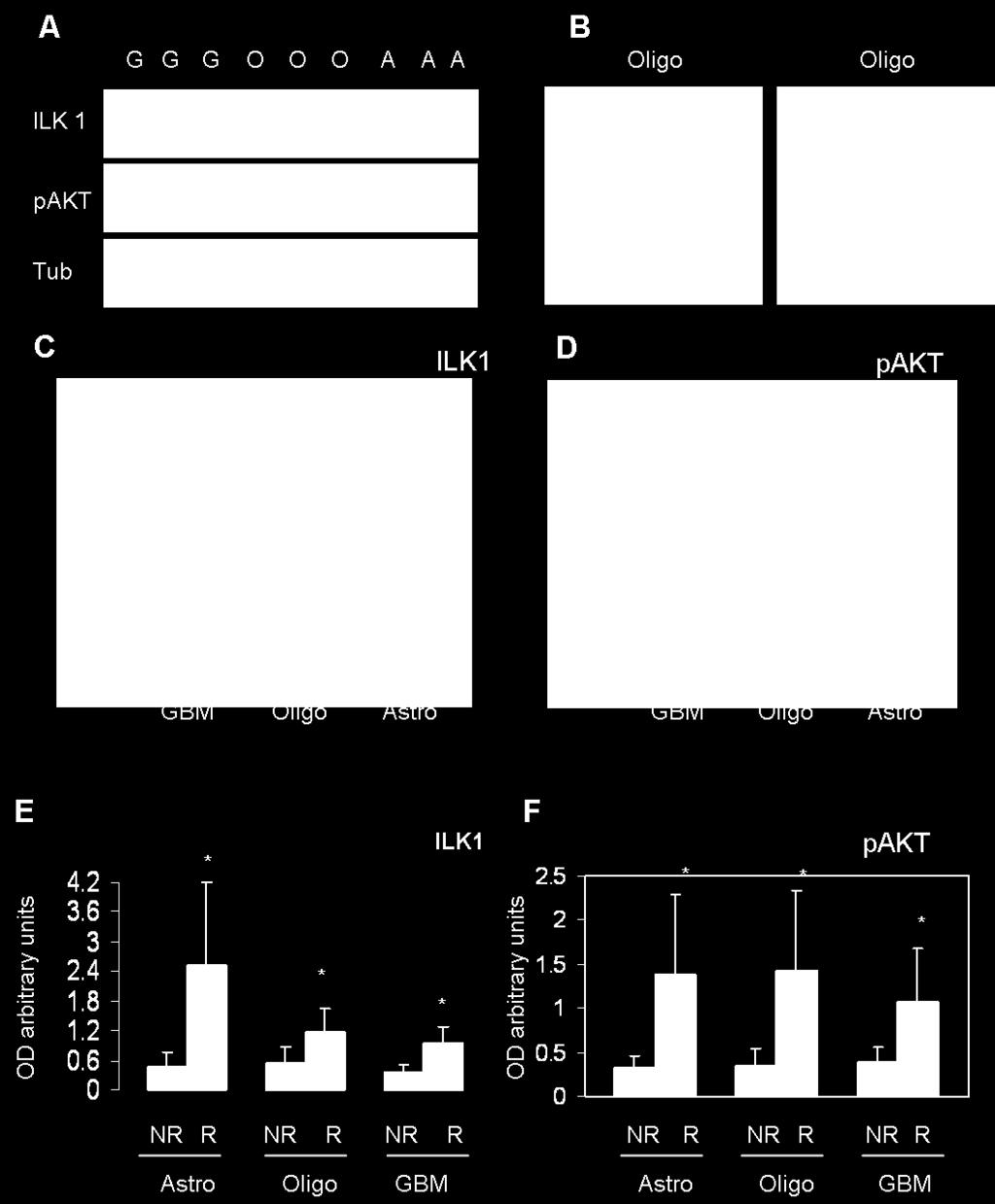
31 32
32 2 AIM OF THE WORK Glioblastoma multiforme (GBM, WHO grade IV) is the most frequent malignant tumour of the central nervous system (CNS) representing up to 50% of all primary brain gliomas, accounts for approximately 12-15% of all brain tumors and is one of the most lethal cancers in adults and children; the prognosis of patients with GBM remains poor (Ohgaki, Dessen et al. 2004). Despite aggressive treatments with surgery, radiation, and chemotherapy, median survival is less than 15 months and overall survival is less than 10% at 5 years. The treatment difficulty is due to the exceptionally infiltrative nature of GBM and its proclivity to integrate into normal brain tissue. To date, the management of patients with GBM continues to harbor significant challenges, and comprehensive genetic screens of tumor tissues and signaling pathways have been explored to develop molecular based targeted therapies. GBM cells are characterized by their invasive abilities and striking angiogenic potential. The blood vessels formed by tumor cells are structurally and functionally abnormal: the blood vessels are leaky and dilated, the endothelial cells exhibit aberrant morphology, the pericytes are loosely attached or absent and the basement membrane is incomplete (Durupt, Koppers-Lalic et al.). These abnormalities lead to an abnormal tumor microenvironment that is characterized by interstitial hypertension, hypoxia and acidosis. The abnormal vasculature represents a barrier to the delivery and efficacy of anticancer therapeutic agents. These observations suggest that if the structure and function of tumor vessels could be corrected, then the tumor microenvironment might be normalized, ultimately improving the efficacy of cancer treatments. As a key mediator of angiogenesis, VEGF and its receptors are targets for anticancer therapies (Ferrara, Gerber et al. 2003), in addition to conventional therapies. Targeting the cells that support tumor growth, rather than the actual tumor cells, represents a relatively new approach to cancer therapy 33
33 3 MATERIALS AND METHODS 3.1 CELL CULTURES AND REAGENTS Cells cultures The human glioma cell line U87-MG (American Type Culture Collection, Manassas, VA) was used in in vitro and in the animal experiments. The cells were cultured in amem (Life Technologies, Inc., Grand Island, NY) supplemented with 2 mm L-glutamine, 10% FBS, and 1000 units/ml gentamycin solution, maintained in T-25 tissue culture flasks in 5% CO2/95% air at 37 ºC in a humidified incubator. For the implantation experiments, U87-MG cells were dispersed with a 0.05% solution of trypsin/edta (Life Technologies), reaction was stopped with FBS. The cells were washed with PBS, and adjusted to a final concentration of 1*10 6 cells/100 µl in PBS. Bovine Corneal Endothelial cells (BCE, LGC Standard) were used in in vitro experiments. The cells were cultured in DMEM low glucose (Life Technologies, Inc., Grand Island, NY) supplemented with 1 mm L-glutamine, 10% FBS, 1000 units/ml gentamycin solution and 1 ng/mg FGF, maintained in T-25 tissue culture flasks in 5% CO2/95% air at 37 ºC in a humidified incubator Reagents Sunitinib Dissolved in DMSO at the concentration of 250 mg/ml, diluted at the concentration of 10 mg/ml in acetate buffer (ph 4.6) and given per os 40mg/Kg/die for the in vivo studies; dissolved in DMSO at the concentration of 10 mm for in vitro studies and given to the cells at three different concentration: 2,5 µm, 5 µm and 10 µm. Molecular structure of Sunitinib 34
34 Sorafenib Dissolved in DMSO at the concentration of 250 mg/ml, diluted at the concentration of 10 mg/ml in acetate buffer (ph 4.6) and given per os 50mg/Kg/die for the in vivo studies; dissolved in DMSO at the concentration of 100 mm for in vitro studies and given to the cells at three different concentration: 2,5 µm, 5 µm and 10 µm. QLT0267 Molecular structure of Sorafenib QLT0267 (QLT Inc, Vancouver BC, Canada) was dissolved in (PEG300 66%, Tween %, Ethanol 95% 25%, citric acid 0.2%) for in vivo studies and given per os 100mg/Kg/die Molecular structure of ILK-1 inhibitors member of pharmacor family 3.2 TREATMENTS Treatments for protein extraction U87 cells were plated into 12 multiwell (Euroclone) in a density of cells/well. The day after were treated with anti-angiogenic drugs (at the concentration of 2,5 µm, 5 µm and 10 µm) alone or in combination with QLT0267 (at the concentration of 100 µm), the control cells received only medium. After 6 hours all the cells were collected in sample buffer, boiled 5 minutes and analyzed by SDS-PAGE treatments for MTS test U87 cells and BCE cells were plated into 96 multiwell (Euroclone) in a density of 5000 cells/well. The day after were treated with anti-angiogenic drugs (at the concentration of 2,5 µm, 5 µm and 10 µm) alone or in combination with QLT0267 (at the concentration of 100 µm), the control cells 35
35 received only medium, and some cells received the vehicle (DMSO). After six hours, 12 hours, 24 hours and 48 hours, we change the medium and we put complete medium plus 20 μl of MTS reagent (CellTiter 96 Promega), we read the plates at 490 nm 3.3 SDS-PAGE, WESTERN BLOT ANALYSIS Proteins were separated in 10% SDS-PAGE and electroblotted onto nitrocellulose membranes in buffer containing M Tris-HCl, M glycine, 20% methanol, ph 8.3 at 240 ma for 120 min. The reactions were performed by incubating with primary antibodies (RT, 2-3h in 4% milk): - rabbit abs anti ILK-1 (1:1000 cell signaling), p-akt (1:1000 cell signaling), HIF-1α (1:500 Santa Cruz Biothecnologies), - mouse abs anti Tubulin (1:1000 Sigma Aldrich), pamakt (1:500 cell signaling). Horseradish peroxidase-conjugated anti-rabbit or anti-mouse secondary antibodies (1:4000 Jackson) were used as secondary antibodies (RT, 1h in 4% milk). Immunoreactive bands were visualized by enhanced chemiluminescence (ECL, GE Healthcare). 36
36 4 RESULTS 4.1 INHIBITION OF ILK-1 We believe that the synergism of angiogenesis inhibitors with ILK1 inhibitors should be tested using small molecular inhibitors blocking ILK1 function and not sirna. Synthetic sirna are extremely expensive and not likely to be used for systemic administration in the clinic In vitro characterization of QLT0267 We have studied in vitro the efficacy of the ILK-1 inhibitor QLT0267. First, we have analyzed the tossicity of QLT0267, for this we have measured the viability of the cells after treatment with a MTS assay. We have treated in four different time point: 2 hours, 4 hours, 6 hours, 12 hours and 24 hours. We found that the tumour cells are almost insensitive to the drug in the first 6 hours, after they start to die (Fig 1A). We have looked at the activity of QLT0267 measuring the activity of ILK-1 (Fig 1B). The immunoblot for pakt shows that the drug is active also at 2h of treatment and remain active until 6h of treatment. We didn t measured the activity at 12 and 24 hours because the U87 cells were dying. The immunoblot for ILK shows that the inhibitor of the kinase didn t change its expression level. 37
; (B) effect of QLT026")
37 Fig 1 effect of the inhibitor of ILK-1 QLT0267 on U87 cells. (A) Viability of U87 cells at 2h, 4h, 6h, 12h and 24h after QLT0267 treatment with MTS assay (** p<0.01); (B) effect of QLT0267 on the activity of ILK-1 in U87 cells at 2h, 4h and 6h of treatment. 4.2 THE COMBINATION OF ANTI-ANGIOGENIC DRUGS AND INHIBITION OF ILK The combination of anti-angiogenic drugs and QLT0267 decreases the viability of tumoral cells and endothelial cells in vitro Since we have demonstrated that the silencing for ILK1 increases the efficacy of anti-angiogenic treatment in vivo, we have treated human tumoral cells U87 and Bovine Corneal Endothelial (BCE) cells with two different anti-angiogenic drugs: sorafenib and sunitinib alone or with the concomitant treatment with the ILK-1 inhibitor QLT0267. We have measured the viability of the cells by MTS assay at 6 hours of treatment (Fig 2). 38
 MTS assay on human tumoral U87 cells after treatment for 6h with sorafenib alone or sunitinib alone (at the concentration of 2,5uM, 5uM and 10uM) and the anti-angiogenic drugs at the same")
38 Fig 2 effect of concomitant treatment with anti-angiogenic drugs and the ILK-1 inhibitor on viability of tumoral cells and endothelial cells. (A) MTS assay on human tumoral U87 cells after treatment for 6h with sorafenib alone or sunitinib alone (at the concentration of 2,5uM, 5uM and 10uM) and the anti-angiogenic drugs at the same concentration plus QLT uM. (B) MTS assay on endothelial BCE cells after treatment for 6h with sorafenib alone or sunitinib alone (at the concentration of 2,5uM, 5uM and 10uM) and the anti-angiogenic drugs at the same concentration plus QLT uM. 39
39 The human tumoral cells are more sensitive to the concomitant treatment with both sorafenib and sunitinib plus QLT0267, in fact the combination of drugs plus the inhibitor decrease the viability to 50% (Fig 2A). On the endothelial cells, the maximum concentration of the anti-angiogenic drugs decrease the viability of the cells of 20% for sorafenib and of 40% for sunitinib. The addition of QLT didn t change the viability of the cells and seems to protect them from the antiangiogenic drugs (Fig 2B) The combination of anti-angiogenic drugs and QLT0267 in vivo reduces tumour growth We then tested the combination of anti-angiogenic drugs and QLT0267 in vivo using a subcutaneous model of glioblastoma (Fig 3). Fig 3 Scheme of in vivo experiment We divided the animals into six groups with seven animal for each group. We injected 1*10 6 human tumoral U87 cells resuspended in PBS into the right flanck of the mice. After 14 days, when the tumours were visible, we started the treatment. All the groups were treated per os every day for 21 days. The control group was treated with vehicle (PEG300 66%, Tween %, Ethanol 95% 25%, citric acid 0.2%, acetate buffer); the other groups were treated with sorafenib alone at the concentration of 50 mg/kg/die, with only sunitinib at the concentration of 40 mg/kg/day and with only QLT0267 at the concentration of 100 mg/kg/day. Finally the remaing two groups were treated with the combination of the two drugs, sorafenib plus QLT0267 and sunitinib plus QLT0267, at the same concentration. After 21 days we sacrificed the animals and we collected the tumours. We then analyzed the volume of the tumours (Fig 4 B). The tumour treated with sorafenib showed a worst outcome respect to the tumours treated with sunitinib; in fact sunitinib administration lead to the reduction of tumour volume of almost 70% respect to the control group. The results given by sorafenib were unexpected because, despite we had an average reduction of 30% of tumour volume respect to the control, we noticed a big variability amoung the group. The QLT alone is able to reduce tumour mass, but the combination of both sunitinib both QLT0267 had the best outcome, in fact we had a 95% of volume reduction in this group. Finally the treatment with sorafenib plus QLT0267 gave a reduction comparable to QLT0267 alone (82%) of control. 40
40 Fig 4 Analysis of tumour volume. Representative picture of three different tumour collected from mice (A). Graph of tumour volume (B) The combination therapy decreases the number of vessel We then looked at the tumor vessel formation. For this purpose, the collected tumors were cut at cryostat and stained by immunofluorescence with an antibody that recognizes the murine CD31, a marker of vessels (Fig 5). Also in this case we found that the treatment with the combination of the two drugs was able to induce the greater reductions in vessel density, 52% reduction in the treatment with sunitinib plus QLT0267 and 46% reduction in the treatment with sorafenib plus QLT0267 (Fig 5B). 41

41 Fig 5 Analysis of vessels density (A) Immunofluorescence of 10uM thick slices cutted at cryostat and stained with antibody against murine CD31 (red) and DAPI (blue), (B) graph representing the quantification of vessel/area. 4.3 EXPRESSION LEVELS OF ILK-1 AND ITS PATHWAY We then investigated the expression levels of ILK-1 in toumoral samples and the activity of the kinase looking at the level of its downstream target: phosphorylated AKT The combination of anti-angiogenic drugs and QLT0267 decreases the expression level of ILK-1 in vivo We have measured the expression level of ILK-1 protein through both immunoblotting of tissue tumour samples (Fig 6A) and immunofluorescence on slices of tumour samples (Fig 6B). In each case we found a strong decrease of ILK-1 expression. The data show that QLT0267 treatment decrease the ILK-1 expression of 20% respect to the control group. The greatest decrease can be 42
 western blot analysis of the expression level of ILK-1 and tubulin in tumour lysates and quantification of normalized intensity C).")
42 found in the combination treatment with Sunitinib plus QLT0267, where we found a reduction of almost 58% respect to the controls (Fig 6C). Fig 6 Expression level of ILK-1 in tumour sample A) western blot analysis of the expression level of ILK-1 and tubulin in tumour lysates and quantification of normalized intensity C). B) immunofluorescence of ILK expression in slices from tumoural sample (in green), in red the staining with the antibody against murine CD31, in blue the DAPI. 43
, we looked at the activity of ILK-1 measuring the phosphorylation level of its major target AKT.")
43 4.3.2 The combination of anti-angiogenic drugs and QLT0267 decreases the activity of of ILK-1 in vivo Since we also found that the QLT0267 is an inhibitor of the activity of ILK-1 but didn t inhibit its expression (see Fig.1), we looked at the activity of ILK-1 measuring the phosphorylation level of its major target AKT. Data showed that the combination therapy sunitinib plus QLT0267 decrease the phosphorilation of AKT of almost 50% of the control group; this data are in agreement with our previous results, in fact we have demonstrated that the silencing of ILK-1 increase the efficacy of anti-angiogenic treatment. 4.4 MOLECULAR MECHANISMS OF ILK-1 ACTIVITY We wanted to better understand the mechanisms underlining the efficacy of combination therapy at a molecular level. Since we knew that the pathway ILK-1 - pakt inhibits the degradation of HIF-1α, an important transcription factor involved in angiogenesis and epithelial mesenchimal transition, and that this results in an increase of the expression level of HIF-1α, we investigated if in our models we had changes in HIF-1α expression The combination of anti-angiogenic drugs and QLT0267 decrease the expression of HIF-1α We treated human U87 glioma cells with sunitinib, sorafenib or QLT0267 at the maximum concentration that we previously used in vitro alone and with the combination of the two drugs: sunitinib plus QLT0267 and sorafenib plus QLT0267. After six hours of treatment, we lysate the cells and we looked at the levels of HIF-1α (Fig 7). The immunoblot shows that there is a strong decrease of HIF-1α after the combination of the two treatment. 44


44 Fig 8 Level of HIF-1α expression. The immunoblot swoes the expression level of HIF-1α after treatment of human glioma U87 cells with anti-angiogenic drugs and QLT0267. We had also checked the level of both ILK-1 and phosphorilated AKT on human glioma cells treated for six hours and, in our model, we see a trend of decrease of pakt in cells treated with QLT0267 alone or in combination with both the antiangiogenic drugs, in contrast we didn t see any differences in the expression levels of ILK-1. These are preliminary data and we need to increase the number of experiments to be sure that this trend is significant. Fig 9 Level of pakt and ILK expression The immunoblot swoes the expression level of ILK-1 and pakt after treatment of human glioma U87 cells with anti-angiogenic drugs and QLT
UNIVERSITY OF MEDICINE AND PHARMACY CRAIOVA PhD SCHOOL. PhD THESIS
 UNIVERSITY OF MEDICINE AND PHARMACY CRAIOVA PhD SCHOOL PhD THESIS THE IMPORTANCE OF TUMOR ANGIOGENESIS IN CEREBRAL TUMOR DIAGNOSIS AND THERAPY ABSTRACT PhD COORDINATOR: Prof. univ. dr. DRICU Anica PhD
The Angiopoietin Axis in Cancer
 Ang2 Ang1 The Angiopoietin Axis in Cancer Tie2 An Overview: The Angiopoietin Axis Plays an Essential Role in the Regulation of Tumor Angiogenesis Growth of a tumor beyond a limiting size is dependent upon
Introduction. Cancer Biology. Tumor-suppressor genes. Proto-oncogenes. DNA stability genes. Mechanisms of carcinogenesis.
 Cancer Biology Chapter 18 Eric J. Hall., Amato Giaccia, Radiobiology for the Radiologist Introduction Tissue homeostasis depends on the regulated cell division and self-elimination (programmed cell death)
Signaling Vascular Morphogenesis and Maintenance
 Signaling Vascular Morphogenesis and Maintenance Douglas Hanahan Science 277: 48-50, in Perspectives (1997) Blood vessels are constructed by two processes: vasculogenesis, whereby a primitive vascular
Mechanisms of Resistance to Antiangiogenic. Martin J. Edelman, MD University of Maryland Greenebaum Cancer Center Dresden, 2012
 Mechanisms of Resistance to Antiangiogenic Agents Martin J. Edelman, MD University of Maryland Greenebaum Cancer Center Dresden, 2012 Angiogenesis: A fundamental attribute of cancer Premise of Anti-angiogenic
PATHOBIOLOGY OF NEOPLASIA
 PATHOBIOLOGY OF NEOPLASIA Department of Pathology Gadjah Mada University School of Medicine dr. Harijadi Blok Biomedis, 6 Maret 2009 [12] 3/17/2009 1 The pathobiology of neoplasia Normal cells Malignant
Heterotypy and Angiogenesis
 Heterotypy and Angiogenesis Tumors are perpetual wounds 1. Normally stroma and epithelia converse at a distance. 2. Juxtaposition of stroma and epithelia is indicative of tissue damage. 4. Activate strategies
The Process of Angiogenesis & Inhibition of Angiogenesis and/or Lymphangiogenesis
 The Process of Angiogenesis & Inhibition of Angiogenesis and/or Lymphangiogenesis Nam Deuk Kim, Ph.D. Pusan National University Contents Part 1. The process of angiogenesis Part 2. The role of angiopoietins
number Done by Corrected by Doctor Maha Shomaf
 number 19 Done by Waseem Abo-Obeida Corrected by Abdullah Zreiqat Doctor Maha Shomaf Carcinogenesis: the molecular basis of cancer. Non-lethal genetic damage lies at the heart of carcinogenesis and leads
Deregulation of signal transduction and cell cycle in Cancer
 Deregulation of signal transduction and cell cycle in Cancer Tuangporn Suthiphongchai, Ph.D. Department of Biochemistry Faculty of Science, Mahidol University Email: tuangporn.sut@mahidol.ac.th Room Pr324
Transformation of Normal HMECs (Human Mammary Epithelial Cells) into Metastatic Breast Cancer Cells: Introduction - The Broad Picture:
 into Metastatic Breast Cancer Cells: Introduction - The Broad Picture:") Transformation of Normal HMECs (Human Mammary Epithelial Cells) into Metastatic Breast Cancer Cells: Introduction - The Broad Picture: Spandana Baruah December, 2016 Cancer is defined as: «A disease caused
Transformation of Normal HMECs (Human Mammary Epithelial Cells) into Metastatic Breast Cancer Cells: Introduction - The Broad Picture: Spandana Baruah December, 2016 Cancer is defined as: «A disease caused
Cancer as a disease of development; Developmental therapies: Anti- Angiogenesis; Stem cells and tissue regeneration.
 Cancer as a disease of development; Developmental therapies: Anti- Angiogenesis; Stem cells and tissue regeneration Mitesh Shrestha What is Cancer? Unrestricted cell growth: tumor cell population 1x10^9
VIII Curso Internacional del PIRRECV. Some molecular mechanisms of cancer
 VIII Curso Internacional del PIRRECV Some molecular mechanisms of cancer Laboratorio de Comunicaciones Celulares, Centro FONDAP Estudios Moleculares de la Celula (CEMC), ICBM, Facultad de Medicina, Universidad
Oncolytic Virotherapy: Targeting Cancer Stem Cells
 Oncolytic Virotherapy: Targeting Cancer Stem Cells Cancer Stem Cells (CSCs) or Cancer Initiating Cells (CICs) A consensus of five defining criteria has been established to affirm the existence of CICs:
Off-Label Treatments. Clinical Trials for Recurrent GBM UCSF Radiation Oncology Course: Management of Recurrent Disease. Outline
 Off-Label Treatments Clinical Trials for Recurrent GBM UCSF Radiation Oncology Course: Management of Recurrent Disease Jennifer Clarke, MD, MPH Assistant Professor Division of Neuro-Oncology Depts of Neurological
1. The metastatic cascade. 3. Pathologic features of metastasis. 4. Therapeutic ramifications. Which malignant cells will metastasize?
 1. The metastatic cascade 3. Pathologic features of metastasis 4. Therapeutic ramifications Sir James Paget (1814-1899) British Surgeon/ Pathologist Paget s disease of Paget s disease of the nipple (intraductal
Neoplasia 18 lecture 8. Dr Heyam Awad MD, FRCPath
 Neoplasia 18 lecture 8 Dr Heyam Awad MD, FRCPath ILOS 1. understand the angiogenic switch in tumors and factors that stimulate and inhibit angiogenesis. 2. list the steps important for tumor metastasis
Contents. Preface XV Acknowledgments XXI List of Abbreviations XXIII About the Companion Website XXIX
 Contents Preface XV Acknowledgments XXI List of Abbreviations XXIII About the Companion Website XXIX 1 General Aspects of Signal Transduction and Cancer Therapy 1 1.1 General Principles of Signal Transduction
Antibody-Drug Conjugates in Glioblastoma Multiforme: Finding Ways Forward
 Transcript Details This is a transcript of a continuing medical education (CME) activity accessible on the ReachMD network. Additional media formats for the activity and full activity details (including
BL-8040: BEST-IN-CLASS CXCR4 ANTAGONIST FOR TREATMENT OF ONCOLOGICAL MALIGNANCIES. Overview and Mechanism of Action Dr.
 BL-8040: BEST-IN-CLASS CXCR4 ANTAGONIST FOR TREATMENT OF ONCOLOGICAL MALIGNANCIES Overview and Mechanism of Action Dr. Leah Klapper, CSO 88 BL-8040: Novel CXCR4 Antagonist For Hematological Cancers Indications:
Tissue repair. (3&4 of 4)
") Tissue repair (3&4 of 4) What will we discuss today: Regeneration in tissue repair Scar formation Cutaneous wound healing Pathologic aspects of repair Regeneration in tissue repair Labile tissues rapid
Tissue repair (3&4 of 4) What will we discuss today: Regeneration in tissue repair Scar formation Cutaneous wound healing Pathologic aspects of repair Regeneration in tissue repair Labile tissues rapid
Osamu Tetsu, MD, PhD Associate Professor Department of Otolaryngology-Head and Neck Surgery School of Medicine, University of California, San
 Osamu Tetsu, MD, PhD Associate Professor Department of Otolaryngology-Head and Neck Surgery School of Medicine, University of California, San Francisco Lung Cancer Classification Pathological Classification
1.The metastatic cascade. 2.Pathologic features of metastasis. 3.Therapeutic ramifications
 Metastasis 1.The metastatic cascade 2.Pathologic features of metastasis 3.Therapeutic ramifications Sir James Paget (1814-1899) British Surgeon/ Pathologist Paget s disease of bone Paget s disease of the
Molecular Oncology, oncology parameters see each test
 Molecular Oncology, oncology parameters see each test DPD deficiency Dihydropyrimidine dehydrogenase deficiency (DPD deficiency) is an autosomal recessive metabolic disorder in which there is absent or
!!! Oncology for Scientists (RPN 530) Metastasis and Angiogenesis Chapter 13 and 14 Oct. 28 th 2014
 Metastasis and Angiogenesis Chapter 13 and 14 Oct. 28 th 2014") Oncology for Scientists (RPN 530) Metastasis and Angiogenesis Chapter 13 and 14 Oct. 28 th 2014 Department of Cancer Genetics Masashi Muramatsu, Ph.D. About Exam First, think and understand the concepts.
Oncology for Scientists (RPN 530) Metastasis and Angiogenesis Chapter 13 and 14 Oct. 28 th 2014 Department of Cancer Genetics Masashi Muramatsu, Ph.D. About Exam First, think and understand the concepts.
THE HALLMARKS OF CANCER
 THE HALLMARKS OF CANCER ONCOGENES - Most of the oncogenes were first identified in retroviruses: EGFR (ErbB), Src, Ras, Myc, PI3K and others (slightly more than 30) - Mutated cellular genes incorporated
RAS Genes. The ras superfamily of genes encodes small GTP binding proteins that are responsible for the regulation of many cellular processes.
 ۱ RAS Genes The ras superfamily of genes encodes small GTP binding proteins that are responsible for the regulation of many cellular processes. Oncogenic ras genes in human cells include H ras, N ras,
Biologics Effects of Targeted Therapeutics
 Report on the isbtc Mini-symposium on Biologics Effects of Targeted Therapeutics Michael B. Atkins, MD Beth Israel Deaconess Medical Center Louis Weiner, M.D. Fox Chase Cancer Center Report on the isbtc
It s s Always Something!
 It s s Always Something! New Approaches in Brain Tumor Treatment Virginia Stark-Vance, M.D. When Something Is a Brain Tumor Brain tumors aren t rare: there are over 100,000/yr Most originate as other cancers
Morphological features and genetic alterations
 Morphological features and genetic alterations Tutor : Audrey Rousseau Caget Lise: Université d Angers Iorio Vittoria: Seconda Università degli studi di Napoli Manaila Roxana: Iuliu Hatieganu University
How genetic & biochemical alterations in brain tumors contribute to epileptogenesis
 How genetic & biochemical alterations in brain tumors contribute to epileptogenesis December 2 nd, 2012 Joon H. Uhm, MD FRCPC Departments of Neurology & Oncology Mayo Clinic, Rochester, MN American Epilepsy
Cancer. The fundamental defect is. unregulated cell division. Properties of Cancerous Cells. Causes of Cancer. Altered growth and proliferation
 Cancer The fundamental defect is unregulated cell division. Properties of Cancerous Cells Altered growth and proliferation Loss of growth factor dependence Loss of contact inhibition Immortalization Alterated
Signaling. Dr. Sujata Persad Katz Group Centre for Pharmacy & Health research
 Signaling Dr. Sujata Persad 3-020 Katz Group Centre for Pharmacy & Health research E-mail:sujata.persad@ualberta.ca 1 Growth Factor Receptors and Other Signaling Pathways What we will cover today: How
Chapter 15: Signal transduction
 Chapter 15: Signal transduction Know the terminology: Enzyme-linked receptor, G-protein linked receptor, nuclear hormone receptor, G-protein, adaptor protein, scaffolding protein, SH2 domain, MAPK, Ras,
Concise Reference. HER2 Testing in Breast Cancer. Mary Falzon, Angelica Fasolo, Michael Gandy, Luca Gianni & Stefania Zambelli
 Concise Reference Testing in Breast Cancer Mary Falzon, Angelica Fasolo, Michael Gandy, Luca Gianni & Stefania Zambelli Extracted from Handbook of -Targeted Agents in Breast Cancer ublished by Springer
CYTOKINE RECEPTORS AND SIGNAL TRANSDUCTION
 CYTOKINE RECEPTORS AND SIGNAL TRANSDUCTION What is Cytokine? Secreted popypeptide (protein) involved in cell-to-cell signaling. Acts in paracrine or autocrine fashion through specific cellular receptors.
Correlation between expression and significance of δ-catenin, CD31, and VEGF of non-small cell lung cancer
 Correlation between expression and significance of δ-catenin, CD31, and VEGF of non-small cell lung cancer X.L. Liu 1, L.D. Liu 2, S.G. Zhang 1, S.D. Dai 3, W.Y. Li 1 and L. Zhang 1 1 Thoracic Surgery,
The PI3K/AKT axis. Dr. Lucio Crinò Medical Oncology Division Azienda Ospedaliera-Perugia. Introduction
 The PI3K/AKT axis Dr. Lucio Crinò Medical Oncology Division Azienda Ospedaliera-Perugia Introduction Phosphoinositide 3-kinase (PI3K) pathway are a family of lipid kinases discovered in 1980s. They have
mirna Dr. S Hosseini-Asl
 mirna Dr. S Hosseini-Asl 1 2 MicroRNAs (mirnas) are small noncoding RNAs which enhance the cleavage or translational repression of specific mrna with recognition site(s) in the 3 - untranslated region
Targeted and immunotherapy in RCC
 Targeted and immunotherapy in RCC Treatment options Surgery (radical VS partial nephrectomy) Thermal ablation therapy Surveillance Immunotherapy Molecular targeted therapy Molecular targeted therapy Targeted
CHAPTER VII CONCLUDING REMARKS AND FUTURE DIRECTION. Androgen deprivation therapy is the most used treatment of de novo or recurrent
 CHAPTER VII CONCLUDING REMARKS AND FUTURE DIRECTION Stathmin in Prostate Cancer Development and Progression Androgen deprivation therapy is the most used treatment of de novo or recurrent metastatic PCa.
Disorders of Cell Growth & Neoplasia. Lecture 4 Molecular basis of cancer
 General Pathology VPM 152 Disorders of Cell Growth & Neoplasia Lecture 4 Molecular basis of cancer Enrique Aburto Apr 2010 Skin tumor in a 10-year-old Rottweiler. Considering the external appearance and
Backgrounder. 1. What are targeted therapies? 2. How do targeted therapies work?
 Backgrounder TARGETED THERAPIES FOR CANCER 1. What are targeted therapies? 2. How do targeted therapies work? 3. What are some of the different types of targeted therapy? 4. What are the potential benefits
Genetics and Cancer Ch 20
 Genetics and Cancer Ch 20 Cancer is genetic Hereditary cancers Predisposition genes Ex. some forms of colon cancer Sporadic cancers ~90% of cancers Descendants of cancerous cells all cancerous (clonal)
Oxygen-independent regulation of HIF-1α levels
 Oxygen-independent regulation of HIF-1α levels Growth factors THIOREDOXIN (TRX) OXIDATIVE STRESS PKC PI3K Akt mtor PTEN HIF1α p70s6k Target gene mrnas SP1 HIF-1α mrna Intratumor hypoxia Genetic alterations
Cell Signaling part 2
 15 Cell Signaling part 2 Functions of Cell Surface Receptors Other cell surface receptors are directly linked to intracellular enzymes. The largest family of these is the receptor protein tyrosine kinases,
Src-INACTIVE / Src-INACTIVE
 Biology 169 -- Exam 1 February 2003 Answer each question, noting carefully the instructions for each. Repeat- Read the instructions for each question before answering!!! Be as specific as possible in each
EGFR: fundamenteel en klinisch
 EGFR: fundamenteel en klinisch Guido Lammering MAASTRO Clinic Maastricht, NL What is EGFR?? The EGFR some facts 1186 amino acids 170 kda Expressed by all cells of epithelial origin Increased activation
Bihong Zhao, M.D, Ph.D Department of Pathology
 Bihong Zhao, M.D, Ph.D Department of Pathology 04-28-2009 Is tumor self or non-self? How are tumor antigens generated? What are they? How does immune system respond? Introduction Tumor Antigens/Categories
Results and Discussion of Receptor Tyrosine Kinase. Activation
 Results and Discussion of Receptor Tyrosine Kinase Activation To demonstrate the contribution which RCytoscape s molecular maps can make to biological understanding via exploratory data analysis, we here
7/6/2015. Cancer Related Deaths: United States. Management of NSCLC TODAY. Emerging mutations as predictive biomarkers in lung cancer: Overview
 Emerging mutations as predictive biomarkers in lung cancer: Overview Kirtee Raparia, MD Assistant Professor of Pathology Cancer Related Deaths: United States Men Lung and bronchus 28% Prostate 10% Colon
Computational Systems Biology: Biology X
 Bud Mishra Room 1002, 715 Broadway, Courant Institute, NYU, New York, USA L#5:(October-18-2010) Cancer and Signals Outline 1 2 Outline 1 2 Cancer is a disease of malfunctioning cells. Cell Lineage: Adult
Molecular Cell Biology - Problem Drill 19: Cell Signaling Pathways and Gene Expression
 Molecular Cell Biology - Problem Drill 19: Cell Signaling Pathways and Gene Expression Question No. 1 of 10 1. Which statement about cell signaling is correct? Question #1 (A) Cell signaling involves receiving
Protocol Abstract and Schema
 Protocol Abstract and Schema A Phase I Trial of p28 (NSC745104), a Non-HDM2 mediated peptide inhibitor of p53 ubiquitination in pediatric patients with recurrent or progressive CNS tumors Description and
Citation: Chen, Wei (2015) Modelling of Tumour-induced Angiogenesis. Doctoral thesis, Northumbria University.
 Modelling of Tumour-induced Angiogenesis. Doctoral thesis, Northumbria University.") Citation: Chen, Wei (2015) Modelling of Tumour-induced Angiogenesis. Doctoral thesis, Northumbria University. This version was downloaded from Northumbria Research Link: http://nrl.northumbria.ac.uk/30235/
Citation: Chen, Wei (2015) Modelling of Tumour-induced Angiogenesis. Doctoral thesis, Northumbria University. This version was downloaded from Northumbria Research Link: http://nrl.northumbria.ac.uk/30235/
Lecture 8 Neoplasia II. Dr. Nabila Hamdi MD, PhD
 Lecture 8 Neoplasia II Dr. Nabila Hamdi MD, PhD ILOs Understand the definition of neoplasia. List the classification of neoplasia. Describe the general characters of benign tumors. Understand the nomenclature
TARGETED THERAPY FOR CHILDHOOD CANCERS
 TARGETED THERAPY FOR CHILDHOOD CANCERS AZIZA SHAD, MD AMEY DISTINGUISHED PROFESSOR OF PEDIATRIC HEMATOLOGY ONCOLOGY, BLOOD AND MARROW TRANSPLANTATION LOMBARDI CANCER CENTER GEORGETOWN UNIVERSITY HOSPITAL
Crosstalk between Adiponectin and IGF-IR in breast cancer. Prof. Young Jin Suh Department of Surgery The Catholic University of Korea
 Crosstalk between Adiponectin and IGF-IR in breast cancer Prof. Young Jin Suh Department of Surgery The Catholic University of Korea Obesity Chronic, multifactorial disorder Hypertrophy and hyperplasia
Receptor mediated Signal Transduction
 Receptor mediated Signal Transduction G-protein-linked receptors adenylyl cyclase camp PKA Organization of receptor protein-tyrosine kinases From G.M. Cooper, The Cell. A molecular approach, 2004, third
Angiogenesis as a therapeutic target
 Angiogenesis as a therapeutic target Lecture Experimentelle Krebsforschung SS 07 Prof. Gerhard Christofori Institute of Biochemistry and Genetics Department of Clinical-Biological Sciences University of
I TESSUTI: Dott.ssa Liliana Belgioia Università degli Studi di Genova
 I TESSUTI: 1. Repair, Radiosensitivity, Recruitment, Repopulation, Reoxygenation 2. Acute and chronic hypoxia 3. Tissue microenvironment and tissue organization Dott.ssa Liliana Belgioia Università degli
PI3K Background. The SignalRx R & D pipeline is shown below followed by a brief description of each program:
 PI3K Background The phosphatidylinositol 3-kinase (PI3K) pathway is a key cell signaling node whose dysregulation commonly results in the transformation of normal cells into cancer cells. The role of PI3K
RINDOPEPIMUT (CDX-110) IN GLIOBLASTOMA
 IN GLIOBLASTOMA") RINDOPEPIMUT (CDX-110) IN GLIOBLASTOMA MULTIFORM GEINO 2014 Dra Estela Pineda Madrid Hospital Clínic Barcelona EGFRvIII in glioblastoma multiform The most common mutation of EGFR in GBM Expressed in 30%
RINDOPEPIMUT (CDX-110) IN GLIOBLASTOMA MULTIFORM GEINO 2014 Dra Estela Pineda Madrid Hospital Clínic Barcelona EGFRvIII in glioblastoma multiform The most common mutation of EGFR in GBM Expressed in 30%
New Approaches in Brain Tumor Treatment. Virginia Stark-Vance, M.D.
 New Approaches in Brain Tumor Treatment Virginia Stark-Vance, M.D. The Primary Brain Tumors es for PicturesMCD for 004.JPG 00 Meningioma 30% Glioblastoma 20% Astrocytoma 10% Nerve sheath 8% Pituitary 6%
WHY TARGETTING SIGNALLING PATHWAYS?
 WHY TARGETTING SIGNALLING PATHWAYS? Cancer cells are particularly sensitive to stress therefore sensitive to inhibition of their hyper activated signaling proteins the re instatement of lost tumor suppressors.
Molecular biology :- Cancer genetics lecture 11
 Molecular biology :- Cancer genetics lecture 11 -We have talked about 2 group of genes that is involved in cellular transformation : proto-oncogenes and tumour suppressor genes, and it isn t enough to
609G: Concepts of Cancer Genetics and Treatments (3 credits)
") Master of Chemical and Life Sciences Program College of Computer, Mathematical, and Natural Sciences 609G: Concepts of Cancer Genetics and Treatments (3 credits) Text books: Principles of Cancer Genetics,
Master of Chemical and Life Sciences Program College of Computer, Mathematical, and Natural Sciences 609G: Concepts of Cancer Genetics and Treatments (3 credits) Text books: Principles of Cancer Genetics,
Dr Rodney Itaki Lecturer Anatomical Pathology Discipline. University of Papua New Guinea School of Medicine & Health Sciences Division of Pathology
 Neoplasia Dr Rodney Itaki Lecturer Anatomical Pathology Discipline University of Papua New Guinea School of Medicine & Health Sciences Division of Pathology General Considerations Overview: Neoplasia uncontrolled,
Disorders of Cell Growth & Neoplasia
 General Pathology VPM 152 Disorders of Cell Growth & Neoplasia Lecture 3 Rate of growth, local invasion, and metastasis. Molecular basis of cancer (normal cell-cycle and cellular proliferation). Enrique
Processing of VEGF-C and -D by the Proprotein Convertases: Importance in Angiogenesis, Lymphangiogenesis, and Tumorigenesis
 Processing of VEGF-C and -D by the Proprotein Convertases: Importance in Angiogenesis, Lymphangiogenesis, and Tumorigenesis ii Colloquium Digital Library of Life Sciences This e-book is an original work
Early Embryonic Development
 Early Embryonic Development Maternal effect gene products set the stage by controlling the expression of the first embryonic genes. 1. Transcription factors 2. Receptors 3. Regulatory proteins Maternal
Development of Carcinoma Pathways
 The Construction of Genetic Pathway to Colorectal Cancer Moriah Wright, MD Clinical Fellow in Colorectal Surgery Creighton University School of Medicine Management of Colon and Diseases February 23, 2019
Tissue renewal and Repair. Nisamanee Charoenchon, PhD Department of Pathobiology, Faculty of Science
 Tissue renewal and Repair Nisamanee Charoenchon, PhD Email: nisamanee.cha@mahidol.ac.th Department of Pathobiology, Faculty of Science Topic Objectives 1. Describe processes of tissue repair, regeneration
Chapter 9. Cellular Signaling
 Chapter 9 Cellular Signaling Cellular Messaging Page 215 Cells can signal to each other and interpret the signals they receive from other cells and the environment Signals are most often chemicals The
Imaging Biomarkers of Response to Radiation and Anti-angiogenic Agents in Brain Tumors
 Imaging Biomarkers of Response to Radiation and Anti-angiogenic Agents in Brain Tumors by Caroline Chung A thesis submitted in conformity with the requirements for the degree of Masters of Science Institute
p53 and Apoptosis: Master Guardian and Executioner Part 2
 p53 and Apoptosis: Master Guardian and Executioner Part 2 p14arf in human cells is a antagonist of Mdm2. The expression of ARF causes a rapid increase in p53 levels, so what would you suggest?.. The enemy
CNS pathology Third year medical students. Dr Heyam Awad 2018 Lecture 12: CNS tumours 2/3
 CNS pathology Third year medical students Dr Heyam Awad 2018 Lecture 12: CNS tumours 2/3 Pilocytic astrocytoma Relatively benign ( WHO grade 1) Occurs in children and young adults Mostly: in the cerebellum
Animal Tissue Culture SQG 3242 Biology of Cultured Cells. Dr. Siti Pauliena Mohd Bohari
 Animal Tissue Culture SQG 3242 Biology of Cultured Cells Dr. Siti Pauliena Mohd Bohari The Culture Environment Changes of Cell s microenvironment needed that favor the spreading, migration, and proliferation
Nintedanib in Oncology Backgrounder
 For media outside the US, UK and Canada only Nintedanib in Oncology Backgrounder 1. What is nintedanib? 2. How does nintedanib work? 3. Data overview 4. Additional clinical data 5. Nintedanib approval
Test Bank for Robbins and Cotran Pathologic Basis of Disease 9th Edition by Kumar
 Link full download:https://getbooksolutions.com/download/test-bank-for-robbinsand-cotran-pathologic-basis-of-disease-9th-edition-by-kumar Test Bank for Robbins and Cotran Pathologic Basis of Disease 9th
Multistep nature of cancer development. Cancer genes
 Multistep nature of cancer development Phenotypic progression loss of control over cell growth/death (neoplasm) invasiveness (carcinoma) distal spread (metastatic tumor) Genetic progression multiple genetic
Human Lung Cancer Pathology and Cellular Biology Mouse Lung Tumor Workshop
 Human Lung Cancer Pathology and Cellular Biology Mouse Lung Tumor Workshop Jan 7 th and 8 th, 2014 Brigitte Gomperts, MD University of California, Los Angeles Lung Structure and Function Airway Epithelial
A particular set of insults induces apoptosis (part 1), which, if inhibited, can switch to autophagy. At least in some cellular settings, autophagy se
, which, if inhibited, can switch to autophagy. At least in some cellular settings, autophagy se") A particular set of insults induces apoptosis (part 1), which, if inhibited, can switch to autophagy. At least in some cellular settings, autophagy serves as a defence mechanism that prevents or retards
A particular set of insults induces apoptosis (part 1), which, if inhibited, can switch to autophagy. At least in some cellular settings, autophagy serves as a defence mechanism that prevents or retards
Imaging of glycolytic metabolism in primary glioblastoma cells with
 63 Chapter 5 Imaging of glycolytic metabolism in primary glioblastoma cells with RIMChip 5.1. Introduction Glioblastoma(GBM) is one of the most common brain tumors 1. It is composed of heterogeneous subpopulations
Tumors of the Nervous System
 Tumors of the Nervous System Peter Canoll MD. PhD. What I want to cover What are the most common types of brain tumors? Who gets them? How do they present? What do they look like? How do they behave? 1
Convergent and Divergent Mechanisms in Aging and Cancer
 Convergent and Divergent Mechanisms in Aging and Cancer Mariana S. De Lorenzo, PhD Department of Cell Biology & Molecular Medicine delorems@umdnj.edu LEARNING OBJECTIVES 1. To identify convergent and divergent
Breast Cancer: the interplay of biology, drugs, radiation. Prof. L. Livi Università degli Studi di Firenze. Brescia, October 3rd 4th, 2013
 Breast Cancer: the interplay of biology, drugs, radiation Prof. L. Livi Università degli Studi di Firenze Brescia, October 3rd 4th, 2013 BACKGROUND (1) The complex interactions between tumor-specific signaling
Generation of post-germinal centre myeloma plasma B cell.
 Generation of post-germinal centre myeloma. DNA DAMAGE CXCR4 Homing to Lytic lesion activation CD38 CD138 CD56 Phenotypic markers Naive Secondary lymphoid organ Multiple myeloma is a malignancy of s caused
Contemporary Management of Glioblastoma
 Contemporary Management of Glioblastoma Incidence Rates of Primary Brain Tumors Central Brain Tumor Registry of the United States, 1992-1997 100 Number of Cases per 100,000 Population 10 1 0.1 x I x I
Cancer Tumor Therapy Drug Vicrostatin Shows. Promising Inhibition of Glioma Growth and. Angiogenesis in Vivo. Rupan Bose
 Bose p. 1 of 23. Cancer Tumor Therapy Drug Vicrostatin Shows Promising Inhibition of Glioma Growth and Angiogenesis in Vivo Rupan Bose B.A. Neuroscience 2011 Candidate Senior Thesis Dana and David Dornsife
INTRODUCTION Ovarian cancer is the leading cause of mortality from gynecologic malignancies in the industrialized countries and is responsible for
 INTRODUCTION Ovarian cancer is the leading cause of mortality from gynecologic malignancies in the industrialized countries and is responsible for more deaths than both cervical and endometrial tumours.
Practice of Medicine-1 Ovarian Cancer Clinical Correlation
 Practice of Medicine-1 Ovarian Cancer Clinical Correlation Amir A. Jazaeri, M.D. Assistant Professor, Division of Gynecologic Oncology American Cancer Society Female Cancers 2000 Statistics Reprinted by
Glioblastoma pathophysiology: or a
 Glioblastoma pathophysiology: A or a? M.J. van den Bent The Brain Tumor Center at Erasmus MC Cancer Center Rotterdam, the Netherlands Pathophysiology: pathophysiology seeks to explain the physiological
T-cell activation T cells migrate to secondary lymphoid tissues where they interact with antigen, antigen-presenting cells, and other lymphocytes:
 Interactions between innate immunity & adaptive immunity What happens to T cells after they leave the thymus? Naïve T cells exit the thymus and enter the bloodstream. If they remain in the bloodstream,
Cancer. Questions about cancer. What is cancer? What causes unregulated cell growth? What regulates cell growth? What causes DNA damage?
 Questions about cancer What is cancer? Cancer Gil McVean, Department of Statistics, Oxford What causes unregulated cell growth? What regulates cell growth? What causes DNA damage? What are the steps in
Cancer and Oncogenes Bioscience in the 21 st Century. Linda Lowe-Krentz
 Cancer and Oncogenes Bioscience in the 21 st Century Linda Lowe-Krentz December 1, 2010 Just a Few Numbers Becoming Cancer Genetic Defects Drugs Our friends and family 25 More mutations as 20 you get older
Corporate Medical Policy
 Corporate Medical Policy Molecular Analysis for Targeted Therapy for Non-Small Cell Lung File Name: Origination: Last CAP Review: Next CAP Review: Last Review: molecular_analysis_for_targeted_therapy_for_non_small_cell_lung_cancer