Pelvic tumor in childhood Classification, imaging approach and radiological findings
|
|
|
- Simon Reynolds
- 6 years ago
- Views:
Transcription

1
2 Pelvic tumor in childhood Classification, imaging approach and radiological findings M. Mearadji International Foundation for Pediatric Imaging Aid Rotterdam, The Netherlands
3 Solid pelvic masses in childhood composed from a group of rare and heterogeneous tumors originating from different pelvic organs or structure. Principally the pelvic mass should be categorized in three groups: I. Rhabdomyosarcoma II. Germ cell tumors III. Other rare and incidental tumors localized in the pelvic region, also to be found anywhere in the body with different origin and histology
4 Following earlier report the pelvic tumors are strongly related to the compartmental location. Anterior midline mostly is the location for rhabdomyosarcoma (RMS). Middle midline is the location for a germ cell tumor as well as a RMS. Posterior midline frequently is the origin of a neurogenic or a germ cell tumor. Lateral pelvic region other soft tissue and bone tumors.
5 Patient material The patient material includes 90 cases of pelvic and testicular tumors in male and female. The anatomic location, the value and limitation of performed different imaging procedures were retrospectively analyzed.
6 I. Rhabdomyosarcoma (RMS) Tumor will arise from primitive cell in any organ. Represents 5% - 10% of malignant solid tumor in childhood Ranking 4 th in frequency after: CNS Neuroblastoma Wilms tumor Bimodial presentation, primary peak 2-5 years of age, secondary years of age. Site classification following intergroup RMS study: Head and Neck (35 %) Genitortinary system (26 %) Extremities (19 %) Other bodyparts (20 %)
7 Patient material of rhabdomyosarcoma There are 18 reviewed cases, 13 boys and 5 girls with the following localisation. Prostate 2 Bladder 4 Vagina 3 Paratesticular 5 Originated from urachus 4
8 Imaging approaches of RMS Conventional as excretory urogram and micturation cystourethrogram as well as angiography in 4 cases (from more than 20 years ago). Later ultrasound routinly used as first diagnostic procedure. Use of CT or MRI or both in all remaining cases.


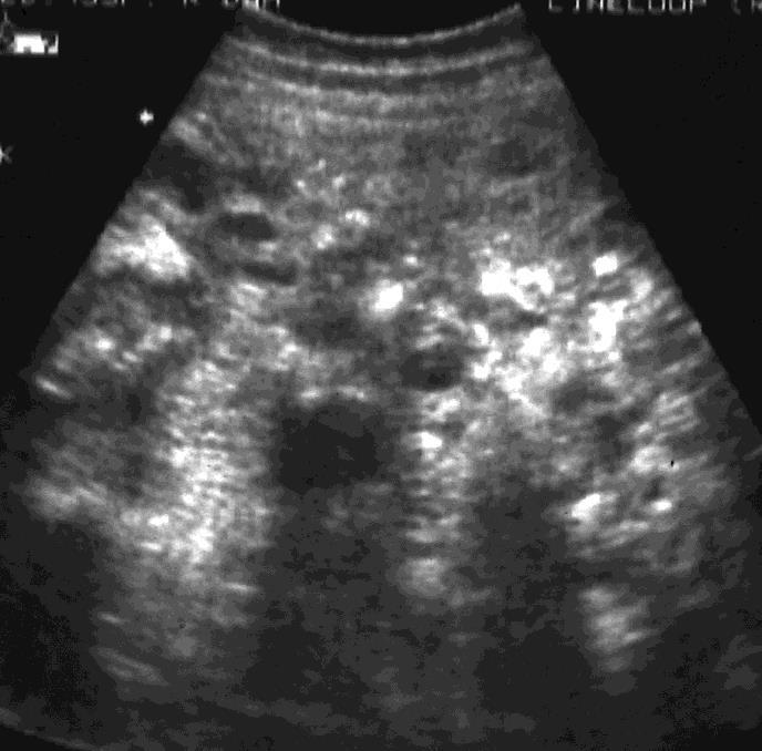
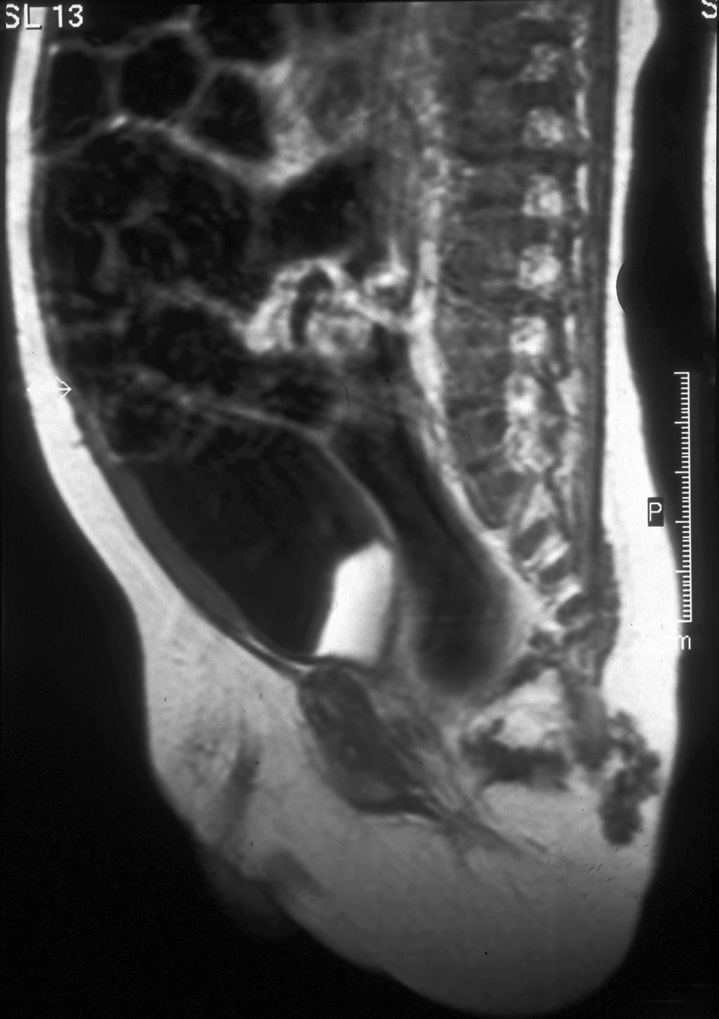
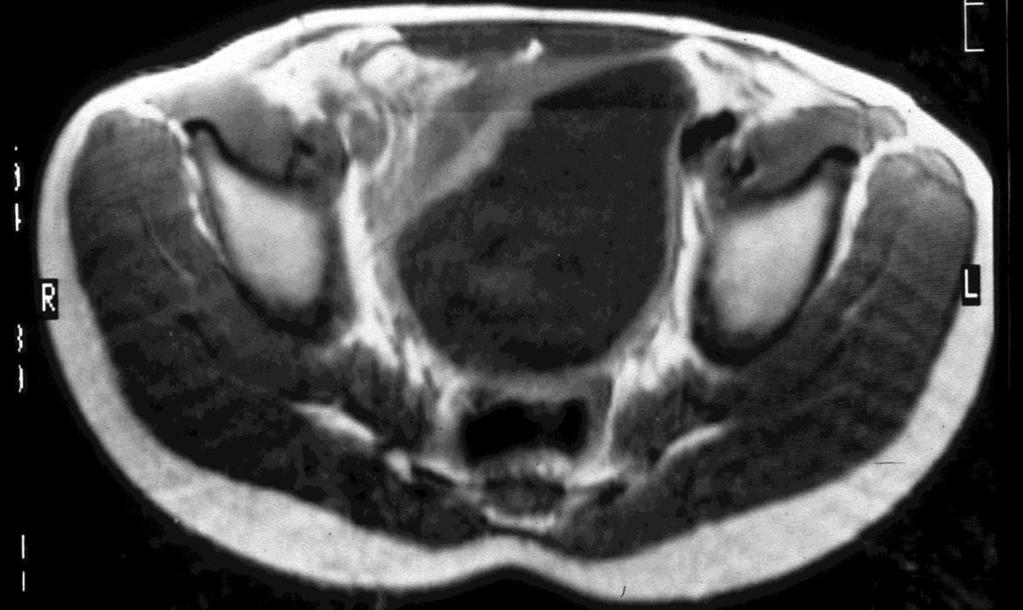
9 RMS of the prostate RMS of the prostate grows rapidly and extend outside the capsule invading the bladder and posterior urethra. This tumor mostly is histologically botroid of type.

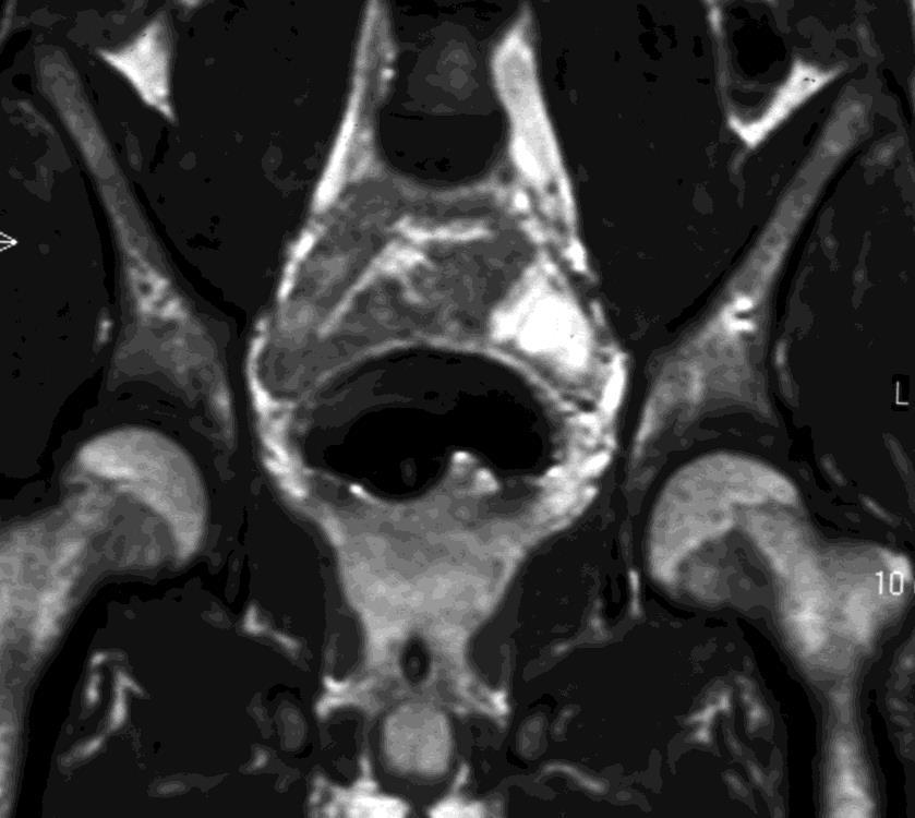
10 15-year-old boy with a prostatic rhabdomyosarcoma invading the bladder


11 RMS of the vagina Most RMS of the vagina arise in the anterior wall of the vagina adjacent to the cervix.


12 Paratesticular RMS The RMS of the testis are histological embryonic of aspect with intermediate prognosis originated of spermatic cord. The patients are less than 5 years of age.


13 RMS of the bladder and urachus RMS typically involves the submucousal region of the trigon, histological is botroid with a good prognosis. RMS in the dome of the bladder is mostly urachal of origin.
14 II. Germ cell tumors in children Germ cell tumors in children are rare entities contributing more than 3% of all pediatric cancer, frequently they encountered in the gonads, but they also are located incidentally in other regions such as in pineal gland, retroperitoneum as well as the sacral area. Histological classification of the germ cell tumors is complex, principally they can be categorized as shown in following table.
15 Germ cell Unipotential Multipotential Germinoma Seminoma Dysgerminoma Gonadoblastoma Teratoma Embryonal carcinoma Benigne And Mature Benigne And Immature Sacrococcygeal Teratoma Yolk Sac tumor Mixed Chorio carcinoma
16 Patient material germ cell tumor (60) Girls 48 Boys 12 Mature and immature ovarian teratoma 15 Mature teratoma of the testis 3 Sacrococcygeaal teratoma 33 Sacrococcygeal teratoma 7 Yolk Sac tumor (testicular) 1 Non seminoma 1
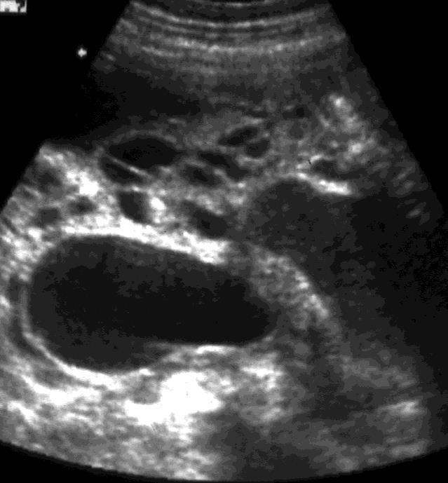
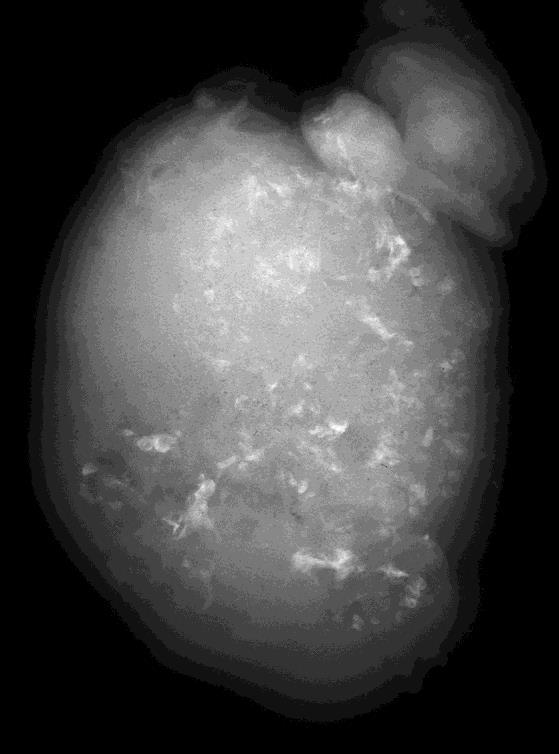
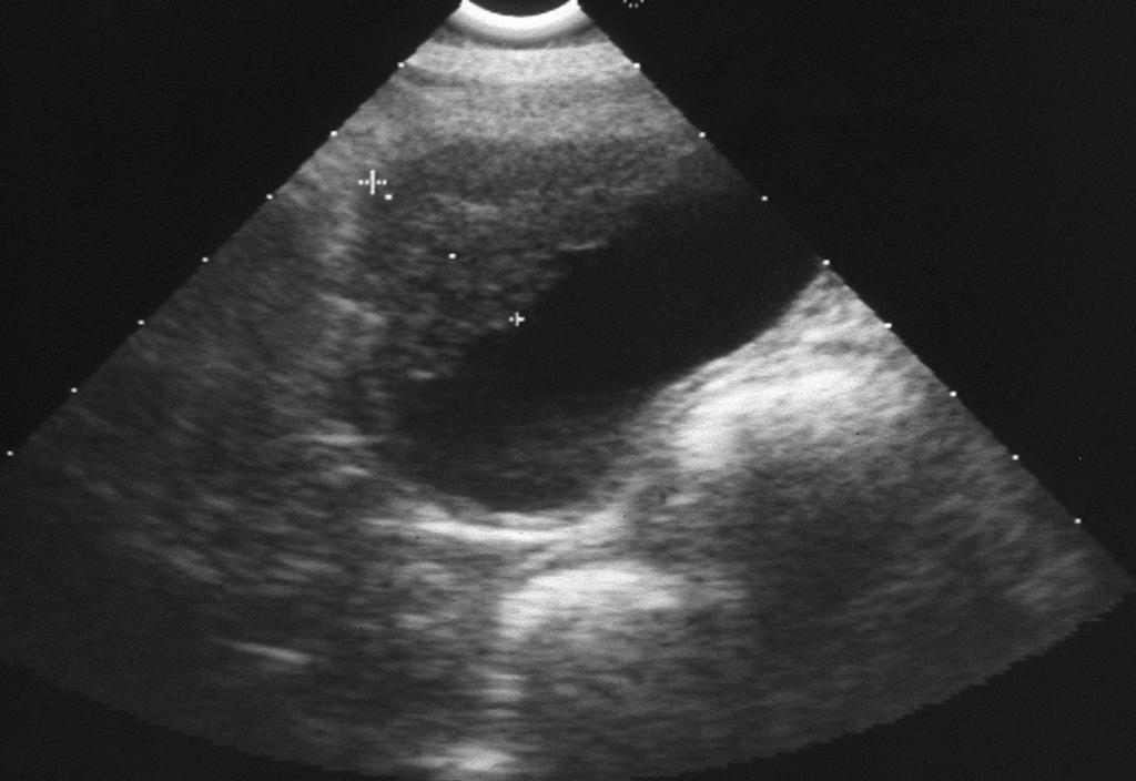
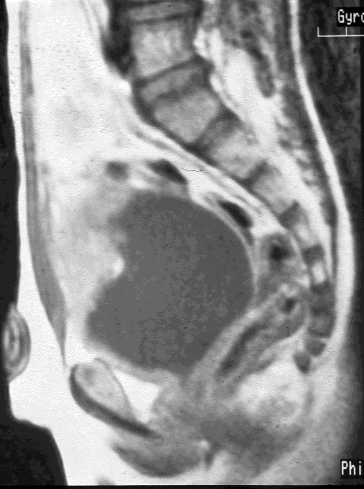
17 Ovarian teratoma Cystic teratoma is by far the most common tumor, accounting for more than 90 % benign ovarian neoplasms. On the basis histological examination teratomas are classified as mature (90 %), immature (containing embryonic neural elements) and malignant. The sonographic appearance of teratomas is variable depending on relative amount of sebum, serous fluid, calcium, hair and fat. The tissue characteristic, location and extension of teratomas could be easily demonstrated by MRI and CT.

18 Ovarian teratoma (histologically mature)

19 Ovarian teratoma (histologically immature with dissemination of glia tissue)
20 Primary testicular germ cell tumor Teratoma is the principal benign germ cell tumor of the testis. Affected boys are usually younger than 4 years of age (85 %). Teratomas on later life tend to be more aggressive and malignant. Yolk Sac carcinoma, teratocarcinoma and choriocarcinomas are all the malignant type of the testicular germ cell tumor.


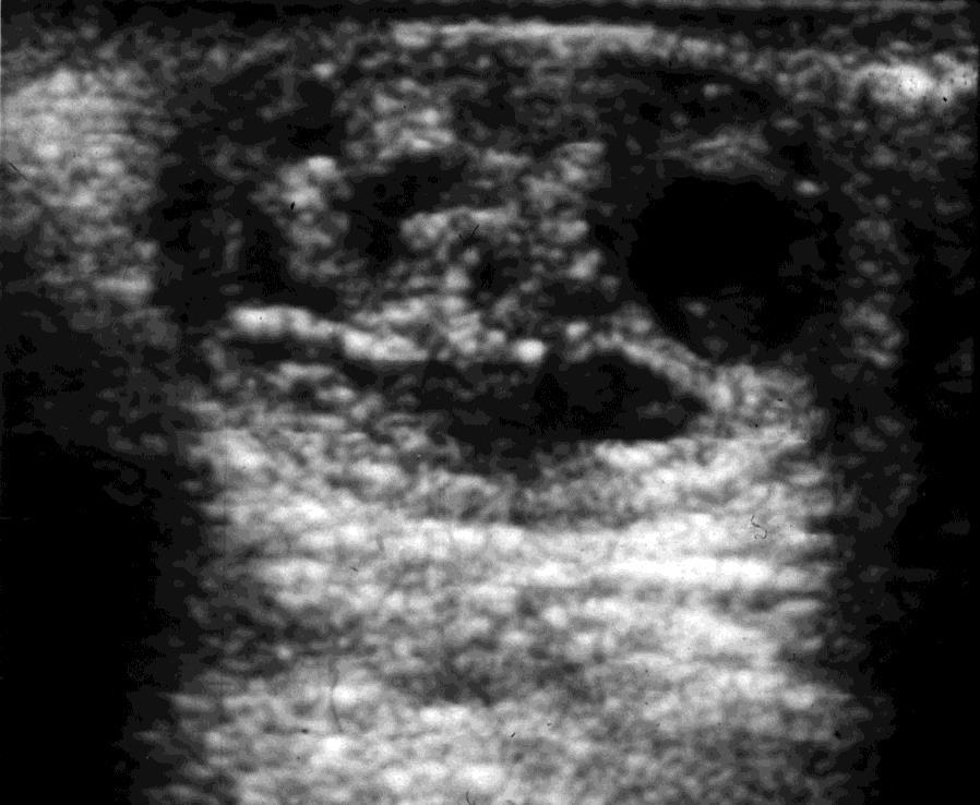
21 Mature teratoma of the right testis.
22 Sacrococcygeal teratoma Sacrococcygeal teratoma represents about 40 % of the germ cell tumors. Females are more affected than males (4:1). The frequency of malignancy depends on age: 7 % (girls) and 10 % (boys) in infants younger than 2 months of age. 47 % (girls) and 66 % (boys) on later life.

23 47 % 34 % 9 % 10 %
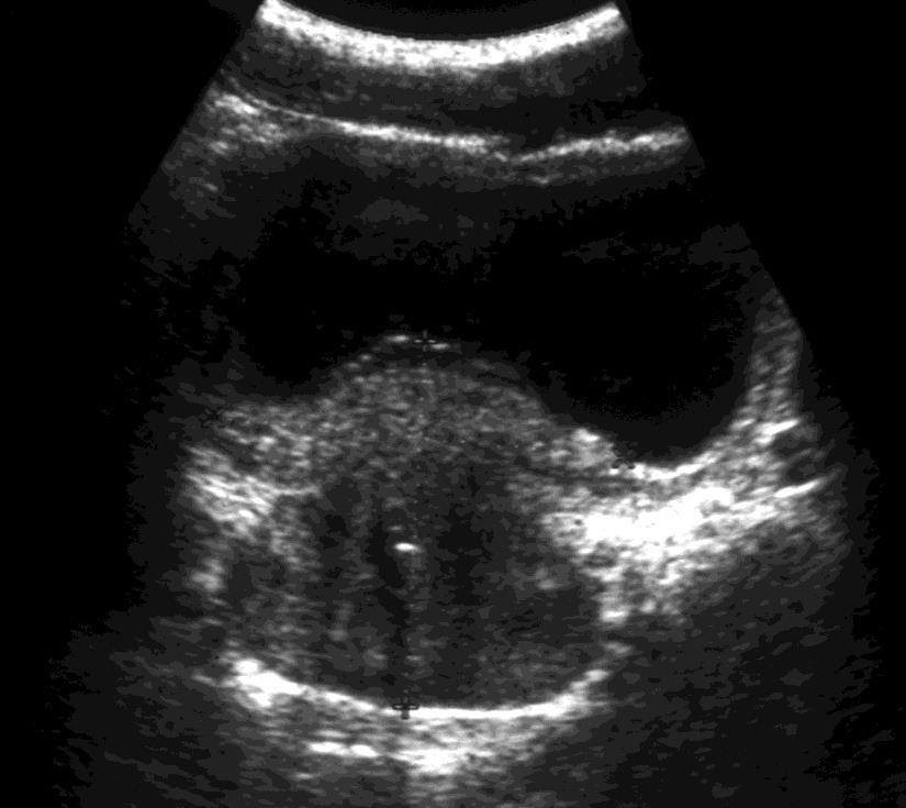
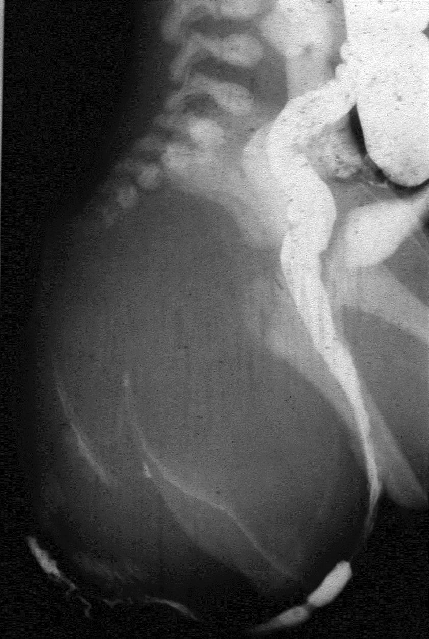
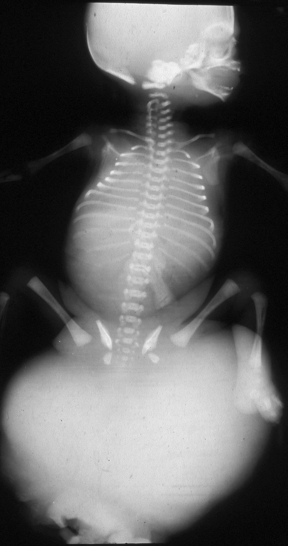
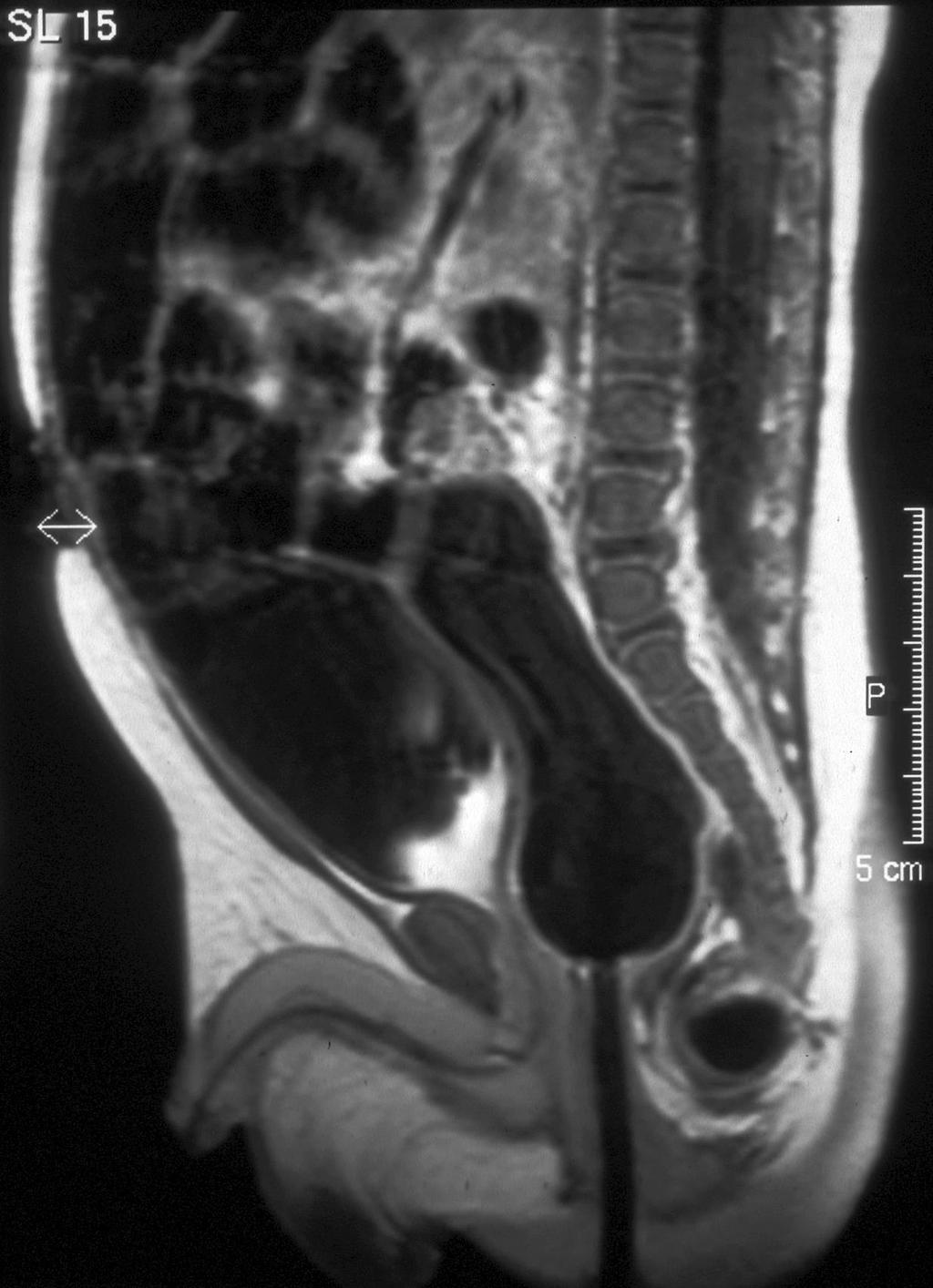
24 Sacrococcygeal teratoma Imaging appearance of the tumor is variable depending on the relative amounts of soft tissue and cystic components. Sonography can provide useful information regarding the internal characteristics of the sacrococcygeal teratomas. MRI is the first modality of choice to determine the total extent of the mass.
25

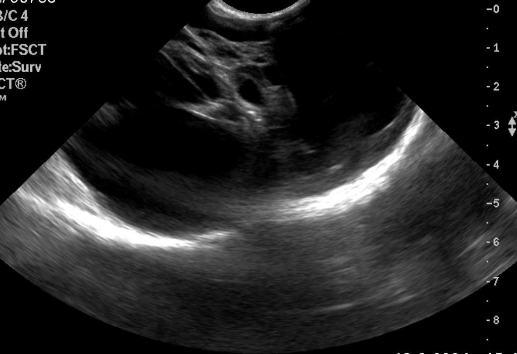
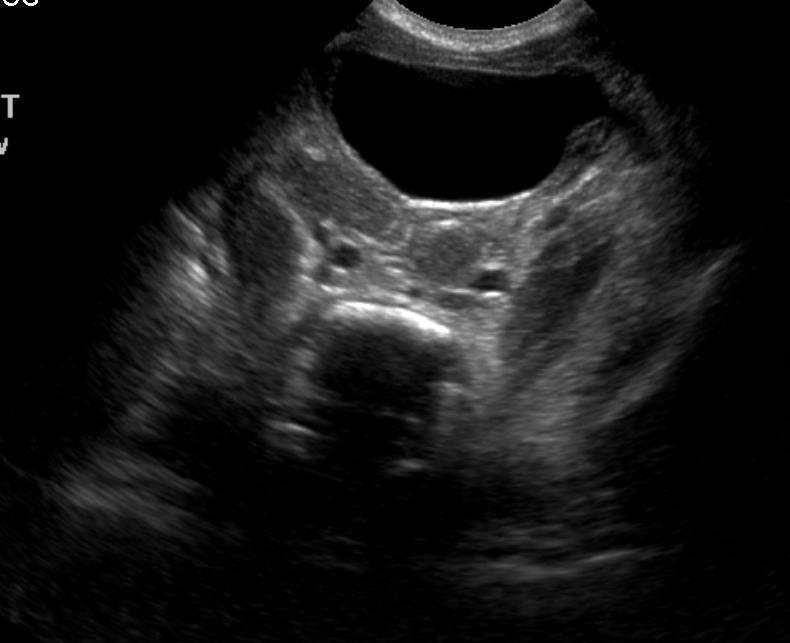
26 Newborn baby with a large sacrococcygeal teratoma type I
27


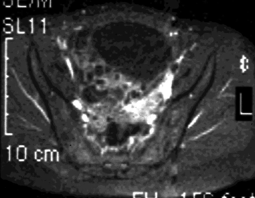
28 Malignant pelvic Yolk Sac tumor
29 III. Miscellaneous Pelvic masses categorized in this group have no relation to RMS or germ cell tumors, neither histologically nor pathogenetically. Some of them however originate from the genital system (4 cases). They could be osteogenic or related to pelvic soft tissue (5 cases). Neurogenic and lymphatic neoplasms are a subgroup under this category of pelvic masses (3 cases).


30 A boy with neurofibromatosis with a neurofibroma of the prostate gland.


31 Pelvic Ewing sarcoma right-sided. Note the huge soft tissue Pelvic osteosarcoma right-sided. Note the massive destruction of the ileum
32 Malignant fibrohistiocytoma

33 Primary malignant pelvic lymphoma
34 Conclusions Dividing the pelvic space into three midline compartments (anterior, middle and posterior) as well as lateral and pelvic floor is useful to define the tumor type. Rhabdomyosarcoma of the lower urogenital system is more frequent in male patients (ratio 2:1) and originates from epididymis, prostate bladder and urachus. Sacrococcygeal teratoma is the most common germ cell tumor in girls with an increasing malignancy of ± 10 % in neonates and up to 66 % in later life.
35 Conclusions Sonography should be considered a sufficient modality in recognition of scrotal mass. Additional contribution of other modalities is not relevant. Sonography is a sufficient imaging modality in neonatal sacrococcygeal teratoma type I and cystic abdominal teratoma on later life if little or no soft tissue is detected. Sonography is only an initial procedure for evaluation of pelvic malignancy, MRI more than CT is required for recognition of tumoral extension and extrapelvic metastasis. CT and MRI are imaging modalities of choice in diagnosis of all masses located around close to the urogenital system ostegenic, or non-osteogenic.
Leukaemia 35% Lymphoma 14%
 Distribution ib ti of Cancers in Children under 15 years Leukaemia 35% Lymphoma 14% Neuroblastoma 9% Other 5% Liver 1% Retinoblastoma 3% Bone and STS 15% CNS 20% Wilms' 8% 30-40% Mortality Germ Cell Tumours
-The cause of testicular neoplasms remains unknown
 - In the 15- to 34-year-old age group, they are the most common tumors of men. - include: I. Germ cell tumors : (95%); all are malignant. II. Sex cord-stromal tumors: from Sertoli or Leydig cells; usually
Adrenal masses in infancy and childhood: A clinical and radiological overview M. Mearadji
 Adrenal masses in infancy and childhood: A clinical and radiological overview M. Mearadji International Foundation for Pediatric Imaging Aid Introduction Neoplastic adrenal masses usually originate from
Note: The cause of testicular neoplasms remains unknown
 - In the 15- to 34-year-old age group, they are the most common tumors of men. - Tumors of the testis are a heterogeneous group of neoplasms that include: I. Germ cell tumors : 95%; all are malignant.
Patient Information. Age: 8 y/o Sex: Female. Date of Admission: Date of Discharge:
 Patient Information Age: 8 y/o Sex: Female Date of Admission: 92-10-08 Date of Discharge: 92-10-18 Chief Complaint Severe admominal pain and vomiting with dysuria since last afternoon Present Illness Lower
Neckmasses in infancy and childhood: Clinical and radiological classification and imaging approaches M. Mearadji
 Neckmasses in infancy and childhood: Clinical and radiological classification and imaging approaches M. Mearadji International Foundation for Pediatric Imaging Aid Introduction Neck masses are a frequent
Germ cell tumours UK SH. Ivo Leuschner. Kiel Pediatric Tumor Registry, Institute of Pathology University Hospital of Schleswig-Holstein Campus Kiel
 Germ cell tumours Ivo Leuschner Kiel Pediatric Tumor Registry, Institute of Pathology University Hospital of Schleswig-Holstein Campus Kiel UK SH Old histogenetic Concept of Germ cell tumours Pluripotent
Retroperitoneal Teratoma Heather Borders, MD
 Retroperitoneal Teratoma Heather Borders, MD 03/04/2012 History Newborn with congenitally diagnosed mass. No other clinical symptoms. Diagnosis Retroperitoneal Teratoma; Immature teratoma, grade 1, with
Pediatric Soft-Tissue Sarcomas. Beth McCarville, MD St. Jude Children s Research Hospital Memphis, Tn
 Pediatric Soft-Tissue Sarcomas Beth McCarville, MD St. Jude Children s Research Hospital Memphis, Tn Overview Histologic classifications Characteristic imaging features Helpful clinical characteristics
Quiz 1. Assign Race 1, Race 2 and Spanish Hispanic Origin to the following scenarios.
 Quiz 1 Assign Race 1, Race 2 and Spanish Hispanic Origin to the following scenarios. 1. 62 year old Brazilian female Race 1 Race 2 Spanish/Hispanic Origin 2. 43 year old Asian male born in Japan Race 1
Methoden / Methods inc. ICCC-3 105
 Methoden / Methods inc. ICCC-3 105 Internationale Klassifikation der Krebserkrankungen bei Kindern (ICCC-3) Zuordnung von ICD-O-3-Codes für Morphologie und Topographie zu diagnostischen Kategorien International
Rare Tumours of Male Genital Organs
 Rare Tumours of Male Genital Organs 1. Epithelial Tumours of Prostate 1.1 General Results Table 1. Epithelial Tumours of Prostate: Incidence, Trends, Survival Flemish Region 2001-2010 Incidence Trend Survival
STAGING AND FOLLOW-UP STRATEGIES
 ATHENS 4-6 October 2018 European Society of Urogenital Radiology STAGING AND FOLLOW-UP STRATEGIES Ahmet Tuncay Turgut, MD Professor of Radiology Hacettepe University, Faculty of Medicine Ankara 2nd ESUR
7 Mousa. Obada Zalat. Mohammad Badi
 7 Mousa Obada Zalat Mohammad Badi Tumors of the ovaries Last lecture we talked about surface epithelial tumors of the ovaries (the most common type). But there are many other types of tumors of germ cell
OVARIES. MLS Basic histological diagnosis MLS HIST 422 Semester 8- batch 7 L13 Dr: Ali Eltayb.
 OVARIES MLS Basic histological diagnosis MLS HIST 422 Semester 8- batch 7 L13 Dr: Ali Eltayb. OBJECTIVES Recognize different disease of ovaries Classify ovarian cyst Describe the pathogenesis, morphology
Ultrasound of malignant testicular lesions. Arne Hørlyck Department of Radiology Aarhus University Hospital, Skejby
 Ultrasound of malignant testicular lesions Arne Hørlyck Department of Radiology Aarhus University Hospital, Skejby Testis Ultrasound is fantastic!! Scrotum Extratesticular mass: Benign Intratesticular
Doppler ultrasound of the abdomen and pelvis, and color Doppler
 - - - - - - - - - - - - - Testicular tumors are rare in children. They account for only 1% of all pediatric solid tumors and 3% of all testicular tumors [1,2]. The annual incidence of testicular tumors
Musculoskeletal Sarcomas
 Musculoskeletal Sarcomas Robert C. Orth, M.D., Ph.D. Edward B. Singleton Department of Pediatric Radiology Texas Children s Hospital Page 0 xxx00.#####.ppt 9/23/2012 9:01:18 AM No disclosures Page 1 xxx00.#####.ppt
3 cell types in the normal ovary
 Ovarian tumors 3 cell types in the normal ovary Surface (coelomic epithelium) the origin of the great majority of ovarian tumors (neoplasms) 90% of malignant ovarian tumors Totipotent germ cells Sex cord-stromal
Pediatric Retroperitoneal Masses Radiologic-Pathologic Correlation
 Acta Radiológica Portuguesa, Vol.XVIII, nº 70, pág. 61-70, Abr.-Jun., 2006 Pediatric Retroperitoneal Masses Radiologic-Pathologic Correlation Marilyn J. Siegel Mallinckrodt Institute of Radiology, Washington
3 cell types in the normal ovary
 Ovarian tumors 3 cell types in the normal ovary Surface (coelomic epithelium) the origin of the great majority of ovarian tumors 90% of malignant ovarian tumors Totipotent germ cells Sex cord-stromal cells
Male Genital Cancers in the US in Frequency of Types
 Germ Cell Tumors of the Testis Pathology, Immunohistochemistry, and the Often Confusing Appearance of Their Metastases Charles Zaloudek, MD Department of Pathology UCSF Male Genital Cancers in the US in
Testis. Protocol applies to all malignant germ cell and malignant sex cord-stromal tumors of the testis, exclusive of paratesticular malignancies.
 Testis Protocol applies to all malignant germ cell and malignant sex cord-stromal tumors of the testis, exclusive of paratesticular malignancies. Protocol revision date: January 2005 Based on AJCC/UICC
Survival in Teenagers and Young. Adults with Cancer in the UK
 Survival in Teenagers and Young Adults with Cancer in the UK Survival in Teenagers and Young Adults (TYA) with Cancer in the UK A comparative report comparing TYA cancer survival with that of children
Case 2. Dr. Sathima Natarajan M.D. Kaiser Permanente Medical Center Sunset
 Case 2 Dr. Sathima Natarajan M.D. Kaiser Permanente Medical Center Sunset History 24 year old male presented with a 3 day history of right flank pain, sharp in nature Denies fever, chills, hematuria or
Prenatal and Neonatal MRI of Sacrococcygeal Teratoma With Surgical Correlation
 Prenatal and Neonatal MRI of Sacrococcygeal Teratoma With Surgical Correlation Ali Mahmood, M.D., and Nadia F. Mahmood, M.D. Citation: Mahmood A, Mahmood NF. Prenatal and Neonatal MRI of Sacrococcygeal
Dr.Dafalla Ahmed Babiker Jazan University
 Dr.Dafalla Ahmed Babiker Jazan University Brain tumors are the second commonest malignancy in children Infratentorial tumors are more common As a general rule they do not metastasize out of the CNS, but
CNS TUMORS. D r. Ali Eltayb ( U. of Omdurman. I ). M. Path (U. of Alexandria)
. M. Path (U. of Alexandria)") CNS TUMORS D r. Ali Eltayb ( U. of Omdurman. I ). M. Path (U. of Alexandria) CNS TUMORS The annual incidence of intracranial tumors of the CNS ISmore than intraspinal tumors May be Primary or Secondary
CNS TUMORS D r. Ali Eltayb ( U. of Omdurman. I ). M. Path (U. of Alexandria) CNS TUMORS The annual incidence of intracranial tumors of the CNS ISmore than intraspinal tumors May be Primary or Secondary
COLOR DOPPLER ULTRASOUND IN EVALUATION OF SCROTAL LESIONS
 COLOR DOPPLER ULTRASOUND IN EVALUATION OF SCROTAL LESIONS Desai Sanjay D Associate Professor, Department of Radiology, RCSM Govt. Medical College, Kolhapur. ABSTRACT: Color Doppler ultrasound is a non-invasive,
Gynaecological Malignancies
 Gynaecological Malignancies Dr Rodney Itaki Lecturer Anatomical Pathology Discipline University of Papua New Guinea Division of Pathology School of Medicine & Health Sciences Overview Genital tract tumors
Testicular Germ Cell Tumors; A Simplistic Approach
 Testicular Germ Cell Tumors; A Simplistic Approach Merce Jorda, MD, PhD, MBA Professor and Vice Chair, Director of Anatomic Pathology Director of Genitourinary Pathology Service Interim Director of Cytopathology
Radiation Oncology Study Guide
 Radiation Oncology Study Guide For the Initial CertificationQualifying (Computer-Based) Examination General and Radiation Oncology This examination is designed to assess your understanding of the entire
Transitional Cell Papilloma 2-3% Inverted Papilloma Rare
 BLADDER TUMORS Benign Transitional Cell Papilloma 2-3% Inverted Papilloma Rare Malignant Transitional (Urothelial) Carcinoma90% Carcinoma In-Situ (By Itself) 5-10% Squamous Cell Carcinoma 3-7% Adenocarcinoma
Painless palpable scrotal mass
 Clinical Case - Test Yourself Urogenital Painless palpable scrotal mass Charis Anastasiadis, Georgia Kyriakopoulou, Charikleia Triantopoulou Radiology Department, Konstantopoulio General Hospital of Nea
Cardiff MRCS OSCE Courses Testicular Cancer
 Testicular Cancer Scenario: A 40-year-old male presents to the surgical out-patient clinic with a 6-8 week history of a painless lump in his left scrotum. He however complains of a dull ache in the scrotum
Diseases of the penis & testis
 Diseases of the penis & testis Done by : Saef B AL-Abbadi Diseases of penis, Condyloma Acuminatum A benign tumor *Tend to recur but only rarely progress into in situ or invasive cancers read this = genital
Chapter 1 MAGNITUDE AND LEADING SITES OF CANCER
 Chapter 1 MAGNITUDE AND LEADING SITES OF CANCER Table 1.1 gives the total number of cancers diagnosed at five different hospital based cancer registries (HBCRs), over the period of two years from 1st January
Intraperitoneal cysts in infancy and childhood An overview and sonographic differentiation
 Intraperitoneal cysts in infancy and childhood An overview and sonographic differentiation M. Mearadji International Foundation for Pediatric Imaging Aid Rotterdam, The Netherlands Intraperitoneal cysts
Testicular tumors; Ultrasonographic and Pathologic correlation
 Testicular tumors; Ultrasonographic and Pathologic correlation Poster No.: C-0106 Congress: ECR 2014 Type: Educational Exhibit Authors: Y. Kim, S. W. Shin, E. T. Kim, M. Y. Kim ; Kuri City/KR, 1 1 2 1
incidence rate x 100,000/year
 Tier R=rare C=common Cancer Entity European crude and age adjusted incidence by cancer, years of diagnosis 2000 and 2007 Analisys based on 83 population-based cancer registries * applying the European
Clinical summary. Male 30 year-old with past history of non-seminomous germ cell tumour. Presents with retroperitoneal lymphadenopathy on CT.
 Clinical summary Male 30 year-old with past history of non-seminomous germ cell tumour. Presents with retroperitoneal lymphadenopathy on CT. For restaging PET/CT. PET/CT findings No significant FDG uptake
Testicular Malignancies /8/15
 Collecting Cancer Data: Testis 2014-2015 NAACCR Webinar Series January 8, 2015 Q&A Please submit all questions concerning webinar content through the Q&A panel. Reminder: If you have participants watching
Male genital tract tumors. SiCA. Division of Urology, Department of Surgery, Faculty of Medicine Siriraj Hospital.
 Male genital tract tumors Division of Urology, Department of Surgery, Faculty of Medicine Siriraj Hospital. adenocarcinoma Prostate Cancer most common male cancer in western countries more detected in
بسم هللا الرحمن الرحيم. Prof soha Talaat
 بسم هللا الرحمن الرحيم Ovarian tumors The leading indication for gynecologic surgery. Preoperative characterization of complex solid and cystic adnexal masses is crucial for informing patients about possible
Pathology of Mediastinal Tumors
 SAMO Meeting Lucerne 2009 Pathology of Mediastinal Tumors Alex Soltermann Most common lesions (adults) Clinical presentation 50% of the patients are asymptomatic, lesion discovered incidentally Symptoms
Tinh hoàn
 Tinh hoàn Tinh hoàn Tinh hoàn Tiền liệt tuyến Tiền liệt tuyến Mào tinh hoàn Mào tinh hoàn Túi tinh Túi tinh Túi tinh Túi tinh So-called cystadenoma of seminal vesicle. Gross appearance of granulomatous
Teratomas and yolk-sac tumours
 Diagnosis CTR 1954-1968 Male Female Leukaemia 252 212 Lymphoma (Hodgkin's and non-hodgkin's) 66 30 Medulloblastoma 56 21 Neuroblastoma 67 35 Connective tissue 108 79 Presacral teratoma 3 11 Table II Childhood
Pathology of the female genital tract
 Pathology of the female genital tract Common illnesses of the female genital tract Before menarche Developmental anomalies Tumors (ovarial teratoma) Amenorrhea Fertile years PCOS, ovarian cysts Endometriosis
A215- Urinary bladder cancer tissues
 A215- Urinary bladder cancer tissues (formalin fixed) For research use only Specifications: No. of cases: 45 Tissue type: Urinary bladder cancer tissues No. of spots: 2 spots from each cancer case (90
Incidental Discovery of Testicular Microlithiasis: What Is the Importance of Ultrasound Surveillance? Two Case Reports
 Published online: October 24, 2013 1662 6575/13/0063 0520$38.00/0 This is an Open Access article licensed under the terms of the Creative Commons Attribution-NonCommercial 3.0 Unported license (CC BY-NC)
 INTRAUTERINE DEVICE = IUD INTRAUTERINE DEVICE = IUD CONGENITAL DISORDERS Pyometra = pyometrea is a uterine infection, it is accumulation of purulent material in the uterine cavity. Ultrasound is usually
Pathology of Ovarian Tumours. Dr. Jyothi Ranganathan MD ( Path) AFMC Pune PDCC (Cytopathology) PGI Chandigarh
 AFMC Pune PDCC (Cytopathology) PGI Chandigarh") Pathology of Ovarian Tumours Dr. Jyothi Ranganathan MD ( Path) AFMC Pune PDCC (Cytopathology) PGI Chandigarh Outline Incidence Risk factors Classification Pathology of tumours Tumour markers Prevention
Pathology of Ovarian Tumours Dr. Jyothi Ranganathan MD ( Path) AFMC Pune PDCC (Cytopathology) PGI Chandigarh Outline Incidence Risk factors Classification Pathology of tumours Tumour markers Prevention
Teratocarcinoma In A Young Boy- An Unusual Presentation
 Human Journals Case Report November 2015 Vol.:2, Issue:1 All rights are reserved by Atia Zaka-ur-Rab et al. Teratocarcinoma In A Young Boy- An Unusual Presentation Keywords: Boy, Testicular Mass, Teratocarcinoma
Testicular fibrous hamartoma: A case report
 Ped Urol Case Rep 2015; 2(5):5-9 DOI: 10.14534/PUCR.2015512749 Ped Urol Case Rep PEDIATRIC UROLOGY CASE REPORTS ISSN: 2148 2969 Journal homepage: http://www.pediatricurologycasereports.com Testicular fibrous
Annual report of Gynecologic Oncology Committee, Japan Society of Obstetrics and Gynecology, 2013
 bs_bs_banner doi:10.1111/jog.12360 J. Obstet. Gynaecol. Res. Vol. 40, No. 2: 338 348, February 2014 Annual report of Gynecologic Oncology Committee, Japan Society of Obstetrics and Gynecology, 2013 Daisuke
CODING TUMOUR MORPHOLOGY. Otto Visser
 CODING TUMOUR MORPHOLOGY Otto Visser INTRODUCTION The morphology describes the tissue of the tumour closest to normal tissue Well differentiated tumours are closest to normal Undifferentiated tumours show
Ovarian Tumors. Andrea Hayes-Jordan MD FACS, FAAP Section Chief, Pediatric Surgery/Surgical Onc. UT MD Anderson Cancer Center
 Ovarian Tumors Andrea Hayes-Jordan MD FACS, FAAP Section Chief, Pediatric Surgery/Surgical Onc. UT MD Anderson Cancer Center Case 13yo female with abdominal pain Ultrasound shows huge ovarian mass Surgeon
Clinical indications for positron emission tomography
 Clinical indications for positron emission tomography Oncology applications Brain and spinal cord Parotid Suspected tumour recurrence when anatomical imaging is difficult or equivocal and management will
Chapter 14 The Reproductive System
 Biology 12 Name: Reproductive System Per: Date: Chapter 14 The Reproductive System Complete using BC Biology 12, page 436-467 14. 1 Male Reproductive System pages 440-443 1. Distinguish between gametes
Brief History. Identification : Past History : HTN without regular treatment.
 Brief History Identification : Name : 陳 x - Admission : 94/10/06 Gender : male Age : 75 y/o Chief Complaint : Urinary difficulty for months. Past History : HTN without regular treatment. Brief History
Institute of Pathology First Faculty of Medicine Charles University. Ovary
 Ovary Barrett esophagus ph in vagina between 3.8 and 4.5 ph of stomach varies from 1-2 (hydrochloric acid) up to 4-5 BE probably results from upward migration of columnar cells from gastroesophageal junction
Lung sequestration and Scimitar syndrome
 Lung sequestration and Scimitar syndrome Imaging approaches M. Mearadji International Foundation for Pediatric Imaging Aid Rotterdam, The Netherlands Pulmonary sequestration Pulmonary sequestration (PS)
Recently, there has been an increasing incidence among women and younger persons
 Bladder Tumors Benign Transitional Cell Papilloma 2-3% Inverted Papilloma Rare Malignant Transitional (Urothelial) Carcinoma 90% Carcinoma In-Situ (By Itself) 5-10% Squamous Cell Carcinoma 3-7% Adenocarcinoma
Prepubertal Testicular Teratomas and Epidermoid Cysts
 ORIGINAL RESEARCH Prepubertal Testicular Teratomas and Epidermoid Cysts Comparison of Clinical and Sonographic Features Min-Yung Chang, MD, Hyun Joo Shin, MD, Hyun Gi Kim, MD, Myung-Joon Kim, MD, PhD,
MRI OF TESTICULAR MALIGNANCIES
 ATHENS 4-6 October 2018 European Society of Urogenital Radiology MRI OF TESTICULAR MALIGNANCIES Effrosyni I. Styliara, Athina C. Tsili, Alexia Psichou, Nikolaos Sofikitis, Maria I. Argyropoulou Department
Pathology Mystery and Surprise
 Pathology Mystery and Surprise Tim Smith, MD Director Anatomic Pathology Medical University of South Carolina Disclosures No conflicts to declare Some problem cases Kidney tumor Scalp tumor Bladder tumor
Mediastinal Germ Cell Tumors
 Mediastinal Germ Cell Tumors Anja C. Roden, M.D. Department of Laboratory Medicine and Pathology, Mayo Clinic, Rochester, MN, USA 2018 MFMER slide-1 Disclosure I have no relevant financial relationships
Case Scenario 1 Discharge Summary Pathology Report Final Diagnosis: Oncology Consult
 Case Scenario 1 Discharge Summary A 31-year-old Brazilian male presented with a 6 month history of right-sided scrotal swelling. Backache was present for 2 months and a history of right epididymitis was
Dr Sneha Shah Tata Memorial Hospital, Mumbai.
 Dr Sneha Shah Tata Memorial Hospital, Mumbai. Topics covered Lymphomas including Burkitts Pediatric solid tumors (non CNS) Musculoskeletal Ewings & osteosarcoma. Neuroblastomas Nasopharyngeal carcinomas
Epidemiology of AYA tumours. Dan Stark, MD Consultant in Medical Oncology Leeds UK
 Epidemiology of AYA tumours Dan Stark, MD Consultant in Medical Oncology Leeds UK Summary Why we should study the epidemiology of AYA tumours What is already known What is not already known and is important
Impact of central surgical review in a study of malignant germ cell tumors
 Impact of central surgical review in a study of malignant germ cell tumors Deborah F. Billmire, Indiana University Frederick J. Rescorla, Indiana University Jonathan H. Ross, Case Western Reserve University
MRI IN THE CHARACTERIZATION OF SEMINOMATOUS AND NONSEMINOMATOUS GERM CELL TUMORS OF THE TESTIS
 MRI IN THE CHARACTERIZATION OF SEMINOMATOUS AND NONSEMINOMATOUS GERM CELL TUMORS OF THE TESTIS Ambesh Deshar *, Gyanendra KC and Zhang Lopsang *Department of Medical Imaging and Nuclear Medicine, First
Evaluation of Neck Mass. Disclosure. Learning Objectives 3/24/2014. Karen T. Pitman MD, FACS Banner MDACC, Gilbert AZ. Nothing to disclose
 Evaluation of Neck Mass Karen T. Pitman MD, FACS Banner MDACC, Gilbert AZ Nothing to disclose Disclosure Learning Objectives 1. Describe a systematic method to evaluate a patient with a neck mass 2. Select
Retroperitoneal Ganglioneuroma Encasing the Celiac and Superior Mesenteric Arteries
 Case Study TheScientificWorldJOURNAL (2004) 4, 974 977 ISSN 1537-744X; DOI 10.1100/tsw.2004.198 Retroperitoneal Ganglioneuroma Encasing the Celiac and Superior Mesenteric Arteries Justin K. Nelms, Eric
Tumour Markers. For these reasons, only a handful of tumour markers are commonly used by most doctors.
 Tumour Markers What are Tumour Markers? Tumour markers are substances that can be found in the body when cancer is present. They are usually found in the blood or urine. They can be products of cancer
Pelvic Pain in the Pediatric Patient Susan D. John, M.D.
 Pelvic Pain in the Pediatric Patient Susan D. John, M.D. RSNA 2012 Patients First Objectives After attending this presentation, participants will be able to: Understand the common congenital and acquired
Exercise. Discharge Summary
 Exercise Discharge Summary A 32-year-old Brazilian male presented with a 6 month history of right-sided scrotal swelling. Backache was present for 2 months and a history of right epididymitis was present
2% of all malignancies Male predominance Patients usually more than 60 years old
 Benign Bladder Tumors Transitional Cell Papilloma 2-3% Inverted Papilloma Rare Malignant Transitional (Urothelial) Carcinoma 90% Carcinoma In-Situ (By Itself) 5-10% Squamous Cell Carcinoma 3-7% Adenocarcinoma
Pathology of Sarcoma ELEANOR CHEN, MD, PHD, ASSISTANT PROFESSOR DEPARTMENT OF PATHOLOGY UNIVERSITY OF WASHINGTON
 Pathology of Sarcoma ELEANOR CHEN, MD, PHD, ASSISTANT PROFESSOR DEPARTMENT OF PATHOLOGY UNIVERSITY OF WASHINGTON Presentation outline Background and epidemiology of sarcomas Sarcoma classification Sarcoma
Case Scenario 1 Discharge Summary Pathology Report Final Diagnosis: Oncology Consult
 Case Scenario 1 Discharge Summary A 31-year-old Brazilian male presented with a 6 month history of right-sided scrotal swelling. Backache was present for 2 months and a history of right epididymitis was
International Journal of Medical and Health Sciences
 International Journal of Medical and Health Sciences Journal Home Page: http://www.ijmhs.net ISSN:2277-4505 Case Report Histomorphological Specrtum of Malignant Germ Cell Tumours: An Overview and Report
Bilateral meconium hydrocele An uncommon case
 Bilateral meconium hydrocele An uncommon case Didem Turcan 1, Evrim Yılmaz 1, Deniz Arık 1, Baran Tokar 2 1 Department of Pathology, 2 Department of Pediatric Surgery, Introduction Meconium hydrocele is
BENIGN & MALIGNANT TESTIS DISEASES. Gary J. Faerber, M.D. Associate Professor, Dept of Urology March 2009 OBJECTIVES
 BENIGN & MALIGNANT TESTIS DISEASES Gary J. Faerber, M.D. Associate Professor, Dept of Urology March 2009 OBJECTIVES 1. Become familiar with the scrotal contents and their anatomical relationship with each
Prof. Dr. med. Beata BODE-LESNIEWSKA Institute of Pathology and Molecular Pathology University Hospital; Zurich
 Prof. Dr. med. Beata BODE-LESNIEWSKA Institute of Pathology and Molecular Pathology University Hospital; Zurich 32 year old man 2 months history of growing left supraclavicular lymph nodes Antibiotic treatment
Other Sites. Table 2 Continued. MPH Rules 11/8/07. NAACCR Webinar Series 1
 MPH s 11/8/07 Other s 1 Table 2 Continued Use this two-page table to select combination histology codes. Compare the terms in the diagnosis to the terms in Columns 1 and 2. If the terms match, code the
Update on RECIST and Staging of Common Pediatric Tumors Ethan A. Smith, MD
 Update on RECIST and Staging of Common Pediatric Tumors Ethan A. Smith, MD Section of Pediatric Radiology C.S. Mott Children s Hospital University of Michigan ethans@med.umich.edu Disclosures No relevant
Medical Science, Volume 13, Number 53, October 29, Adult presentation of retroperitoneal cystic teratoma: a case report
 CASE REPORT PATHOLOGY Medical Science, Volume 13, Number 53, October 29, 2014 ISSN 2321 7359 EISSN 2321 7367 Medical Science The International W Adult presentation of retroperitoneal cystic teratoma: a
PDF created with pdffactory Pro trial version
 Neuroblastoma Tumor derived from neural crest cell that form the sympathetic ganglia&adrenal medulla. Causes *unknown. *familial neuroblastoma has been reported but is rare. * The incidence is 1:100,000
Neoplasia part I. Dr. Mohsen Dashti. Clinical Medicine & Pathology nd Lecture
 Neoplasia part I By Dr. Mohsen Dashti Clinical Medicine & Pathology 316 2 nd Lecture Lecture outline Review of structure & function. Basic definitions. Classification of neoplasms. Morphologic features.
HISTOPATHOLOGICAL STUDY OF TESTICULAR TUMORS IN NORTH-WEST RAJASTHAN (BIKANER REGION)
") HISTOPATHOLOGICAL STUDY OF TESTICULAR TUMORS IN NORTH-WEST RAJASTHAN (BIKANER REGION) *Rankawat Madhu Sudan, Vyas S.P. and Swami Sarika Department of Pathology, Sardar Patel Medical College, Bikaner *Author
HISTOPATHOLOGICAL STUDY OF TESTICULAR TUMORS IN NORTH-WEST RAJASTHAN (BIKANER REGION) *Rankawat Madhu Sudan, Vyas S.P. and Swami Sarika Department of Pathology, Sardar Patel Medical College, Bikaner *Author
Radiology Pathology Conference
 Radiology Pathology Conference Nadia F. Yusaf, M.D. PGY-3 1/29/2010 Presentation material is for education purposes only. All rights reserved. 2010 URMC Radiology Page 1 of 90 Case 1 60 year- old man presents
DISORDERS OF MALE GENITALS
 Wit JM, Ranke MB, Kelnar CJH (eds): ESPE classification of paediatric endocrine diagnosis. 9. Testicular disorders/disorders of male genitals. Horm Res 2007;68(suppl 2):63 66 ESPE Code Diagnosis OMIM ICD10
FINALIZED SEER SINQ QUESTIONS
 0076 Source 1: WHO Class CNS Tumors pgs: 33 MP/H Rules/Histology--Brain and CNS: What is the histology code for a tumor originating in the cerebellum and extending into the fourth ventricle described as
Presacral Neuroblastoma Joseph Junewick, MD FACR
 Presacral Neuroblastoma Joseph Junewick, MD FACR 01/12/2010 History 16 month old male with irritability. Diagnosis Presacral Neuroblastoma Additional Clinical Initial US to evaluate for intussusception
came from a carcinoma and in 12 from a sarcoma. Ninety lesions were intrapulmonary and the as the chest wall and pleura. Details of the primary
 Thorax 1982;37:366-370 Thoracic metastases MARY P SHEPHERD From the Thoracic Surgical Unit, Harefield Hospital, Harefield ABSTRACI One hundred and four patients are reviewed who were found to have thoracic
Shared Care & Survival CTYA SSCRG (Childhood Cancer Research Group)
") Shared Care & Survival CTYA SSCRG (Childhood Cancer Research Group) January 2013 The NCIN is a UK-wide initiative, working to drive improvements in standards of cancer care and clinical outcomes by improving
Shared Care & Survival CTYA SSCRG (Childhood Cancer Research Group) January 2013 The NCIN is a UK-wide initiative, working to drive improvements in standards of cancer care and clinical outcomes by improving
Protocol for the Examination of Lymphadenectomy Specimens From Patients With Malignant Germ Cell and Sex Cord-Stromal Tumors of the Testis
 Protocol for the Examination of Specimens From Patients With Malignant Germ Cell and Sex Cord-Stromal Tumors of the Testis Version: Testis 4.0.1.1 Protocol Posting Date: February 2019 Accreditation Requirements
Analysis of Germ Cell Tumors of Ovary in a Tertiary Care Hospital: A Two Year Retrospective Study
 Original Article DOI: 10.17354/ijss/2016/212 Analysis of Germ Cell Tumors of Ovary in a Tertiary Care Hospital: A Two Year Retrospective Study Muktanjalee Deka 1, Chandan Jyoti Saikia 2, Rukmini Bezbaruah
Clinical and epidemiological characteristics of children with germ cell tumors: A single center experience in a developing country
 The Turkish Journal of Pediatrics 2017; 59: 410-417 DOI: 10.24953/turkjped.2017.04.007 Original Clinical and epidemiological characteristics of children with germ cell tumors: A single center experience